Mycobacterial MazG Safeguards Genetic Stability viaHousecleaning of 5-OH-dCTPLiang-Dong Lyu1,2.*, Bi-Kui Tang1,3., Xiao-Yong Fan4, Hui Ma4, Guo-Ping Zhao1,2,5,6*
1 CAS-Key Laboratory of Synthetic Biology, Institute of Plant Physiology and Ecology, Shanghai Institutes for Biological Sciences, Chinese Academy of Sciences, Shanghai,
China, 2 Department of Microbiology and Li Ka Shing Institute of Health Sciences, The Chinese University of Hong Kong, Prince of Wales Hospital, Shatin, New Territories,
Hong Kong SAR, China, 3 Anhui Key Laboratory of Infection and Immunity, Department of Life Science, Bengbu Medical College, Bengbu, China, 4 Shanghai Public Health
Clinical Center Affiliated with Fudan University, Shanghai, China, 5 Key Laboratory of Medical Molecular Virology affiliated with the Ministry of Education and Health,
Shanghai Medical College, Department of Microbiology, School of Life Sciences, Fudan University, Shanghai, China, 6 Shanghai-MOST Key Laboratory for Health and
Disease Genomics, Chinese National Human Genome Center, Shanghai, China
Abstract
Generation of reactive oxygen species and reactive nitrogen species in phagocytes is an important innate immune responsemechanism to eliminate microbial pathogens. It is known that deoxynucleotides (dNTPs), the precursor nucleotides to DNAsynthesis, are one group of the significant targets for these oxidants and incorporation of oxidized dNTPs into genomic DNAmay cause mutations and even cell death. Here we show that the mycobacterial dNTP pyrophosphohydrolase MazGsafeguards the bacilli genome by degrading 5-OH-dCTP, thereby, preventing it from incorporation into DNA. Deletion of the(d)NTP pyrophosphohydrolase-encoding mazG in mycobacteria leads to a mutator phenotype both under oxidative stressand in the stationary phase of growth, resulting in increased CG to TA mutations. Biochemical analyses demonstrate thatmycobacterial MazG can efficiently hydrolyze 5-OH-dCTP, an oxidized nucleotide that induces CG to TA mutation uponincorporation by polymerase. Moreover, chemical genetic analyses show that direct incorporation of 5-OH-dCTP into mazG-null mutant strain of Mycobacterium smegmatis (Msm) leads to a dose-dependent mutagenesis phenotype, indicating that5-OH-dCTP is a natural substrate of mycobacterial MazG. Furthermore, deletion of mazG in Mycobacterium tuberculosis (Mtb)leads to reduced survival in activated macrophages and in the spleen of infected mice. This study not only characterizes themycobacterial MazG as a novel pyrimidine-specific housecleaning enzyme that prevents CG to TA mutation by degrading 5-OH-dCTP but also reveals a genome-safeguarding mechanism for survival of Mtb in vivo.
Citation: Lyu L-D, Tang B-K, Fan X-Y, Ma H, Zhao G-P (2013) Mycobacterial MazG Safeguards Genetic Stability via Housecleaning of 5-OH-dCTP. PLoS Pathog 9(12):e1003814. doi:10.1371/journal.ppat.1003814
Editor: Helena Ingrid Boshoff, National Institutes of Health, United States of America
Received July 18, 2013; Accepted October 23, 2013; Published December 5, 2013
Copyright: � 2013 Lyu et al. This is an open-access article distributed under the terms of the Creative Commons Attribution License, which permits unrestricteduse, distribution, and reproduction in any medium, provided the original author and source are credited.
Funding: This work was supported by grants from the National Natural Science Foundation of China (No. 30970077, 31121001, 31300126 and 30901276), theResearch Unit Fund of Li Ka Shing Institute of Health Sciences (No. 7103506), the Hong Kong Health and Medical Research Fund (No. 12110622), the ChinaPostdoctoral Science Foundation (No. 20110490754, 2012M510899 and 12R21417000), the SIBS Postdoctoral Research Fund (No. 2011KIP509) and ShanghaiRising-Star Program (No. 12QH1401900). The funders had no role in study design, data collection and analysis, decision to publish, or preparation of themanuscript.
Competing Interests: The authors have declared that no competing interests exist.
* E-mail: [email protected] (LDL); [email protected] (GPZ)
. These authors contributed equally to this work.
Introduction
Oxidative damage to DNA and the DNA precursors, deoxy-
nucleotides (dNTPs) is an inevitable mutagenic challenge occur-
ring in normal aerobic metabolism, generating a large amount of
reactive oxygen species (ROS) as by-products during respiration or
oxidation-reduction reaction [1-3]. Oxidative DNA damage is also
an important innate immune response mechanism implemented
by phagocytes, which produce large amount of ROS and reactive
nitrogen species (RNS) as a bactericidal strategy to eliminate
microbial pathogens [4,5]. Increasing evidence shows that the
nucleotide pool is a significant target for oxidative modification via
ROS and substantial portion of the oxidative damage to genomic
DNA is caused by incorporation of oxidized dNTPs from the
nucleotide pool [3,6,7]. Due to their ambiguous conformation
(anti/syn) compared to that of the canonical dNTPs, incorporation
of oxidized dNTPs into DNA is known to cause mispairing and
mutation, and may be related to carcinogenesis, aging and
neurodegeneration [6,8-10]. Recent studies also established that
incorporation of oxidized dNTPs into DNA is a major causative
mechanism for bacterial cell death induced by bactericidal
antibiotics [11,12]. Therefore, like the DNA repair enzymes,
elimination of the oxidatively damaged dNTPs from the nucleo-
tide pool is an important defense line for cells to maintain genetic
stability.
Cells have evolved a group of non-canonical nucleotide-specific
bio-degradation enzymes, named housecleaning enzyme, to
eliminate the oxidized non-canonical dNTPs from the nucleotide
pool and thus prevent their incorporation into DNA [13,14].
These proteins belong to four structural superfamilies: 1)
dUTPase, 2) ITPase, 3) Nudix (nucleoside diphosphate linked to
an X moiety, or MutT-like) hydrolase, and 4) all-a NTP
pyrophosphohydrolase (MazG NTP-PPase) [14]. The dUTPase
and ITPase are NTP phosphatases that target dUTP, an
intermediate during dTTP synthesis, and ITP/XTP, the deam-
ination products of purine nucleotides, respectively. MutT is the
PLOS Pathogens | www.plospathogens.org 1 December 2013 | Volume 9 | Issue 12 | e1003814
best-studied Nudix hydrolase specific for oxidatively damaged
nucleotides [14,15]. Escherichia coli MutT is the first characterized
Nudix enzyme with 8-oxo-dGTP and 8-oxo-GTP as its natural
substrates. Deletion of mutT in E. coli results in increased AT to CG
mutation in both DNA and mRNA [8,16]. MTH1, the MutT-like
protein in humans, is active against 8-oxo-dGTP, 8-oxo-dATP
and 2-OH-dATP [17]. Depletion of MTH1 in mice leads to a
higher incidence of spontaneous tumorigenesis [18], while in
human cells, MTH1 is involved in maintenance of genome
stability and suppression of degenerative disorders such as
neurodegeneration and carcinogenesis [6,7,19]. However, all the
natural substrates for the MutT-like proteins that have been
characterized in various organisms so far have been the oxidized
purine nucleotides [15].
Oxidized pyrimidine nucleotides likely have a mutagenic effect
similar to that of oxidized purine nucleotides. First, dCTP and
dTTP can be oxidatively modified by ROS to form 5-OH-dCTP
and 5-CHO-dUTP, respectively [20,21]. Second, direct incorpo-
ration of 5-OH-dCTP or 5-CHO-dUTP into E. coli cells may
cause an increase in mutation frequency, and both of these
oxidized nucleotides may be mispaired with adenine rather than
guanine leading to CG to TA mutation [10,22]. Furthermore, 5-
OH-dCTP is known to be incorporated into DNA more efficiently
than 8-oxo-dGTP catalyzed by the exonuclease-free Klenow
fragment [10]. Finally, it was found that the amount of 5-OH-dC
in normal or oxidized cellular DNA is comparable to that of 8-
oxo-dG [1,23]. In addition to their role in mutagenesis, oxidized
pyrimidine nucleotides also show a highly lethal effect on E. coli,
indicating that these non-canonical nucleotides may disturb
normal DNA replication and nucleotide metabolism [22].
Therefore, it is reasonable to conclude that cells have evolved
housecleaning enzymes to eliminate oxidized pyrimidine nucleo-
tides [10,22]. However, although various enzymes responsible for
the removal of oxidized pyrimidine in DNA molecules have been
identified [24–26], the long-awaited housecleaning enzyme
specific for elimination of oxidized pyrimidine nucleotides has
yet to be characterized.
MazG-like proteins are widespread in all three domains of life
and have been biochemically characterized as NTP-PPase while
structurally categorized into the all-a NTP pyrophosphohydrolases
superfamily unrelated to the MutT-like housecleaning enzymes
[14,27,28]. It was found that E. coli MazG can regulate cellular
(p)ppGpp levels and thus, may control programmed cell death
under starvation conditions [29]. However, the mechanism
whereby MazG regulates the cellular (p)ppGpp levels is still
unclear. Structure-based modeling study of MazG from sulfolobus
solfataricus suggested that 2-OH-dATP might be its most likely
substrate and thus proposed, for the first time, a probable role of
housecleaning for this enzyme [27]. Recently, it was reported that
RS21-C6, a MazG-like enzyme in mice, showed a preference for
degrading dCTP and its derivatives, with 5-I-dCTP as the most
preferred substrate in vitro [30]. This attempt to identify a
pyrimidine-specific housecleaning enzyme was echoed by struc-
ture-based analysis, which found that RS21-C6 binds to 5-methyl
dCTP [31]. However, the natural substrate of the MazG family
proteins remained unclear because none of the suggested functions
involving regulation of pyrimidine synthesis [30], prevention of
inappropriate DNA methylation [31], or antimutagenesis by
degrading abnormal dCTP [27,30] have ever been verified in vivo.
Although mycobacterial MazG has been characterized as a
potent NTP pyrophosphohydrolase capable of hydrolyzing all
canonical (d)NTPs in vitro, MazG can also hydrolyze dUTP and 8-
oxo-dGTP, with their affinity to these substrates being similar to
their affinity to the canonical dNTPs (Km,1 mM). Therefore, it is
unlikely that these non-canonical nucleotides are the natural
substrate of the mycobacterial MazG in vivo [28]. In this study, we
demonstrate that 5-OH-dCTP is a natural substrate of mycobac-
terial MazG by means of enzymatic and chemical genetic analyses.
In addition to confirming the antimutator function of MazG, we
show that deletion of mazG in the virulent Mtb strain H37Rv
results in reduced survival in activated macrophage and mice. Our
results reveal that mycobacterial MazG is a novel housecleaning
enzyme involved in a pathway preventing the CG to TA mutation
and ensuring the survival of Mtb in vivo.
Results
Mycobacterial mazG is an antimutatorPreviously, we demonstrated that lack of the MazG NTP-PPase
activity in Msm strain mc2 155 rendered the bacilli more
susceptible to killing by hydrogen peroxide (H2O2) [28]. In order
to test whether the oxidative stress resistant effect of the
mycobacterial MazG is truly attributable to its potential house-
cleaning function in degrading certain oxidatively damaged
dNTP(s), the spontaneous rifampicin-resistance mutation frequen-
cies in wild-type and mazG-null (DmazG::hyg) Msm (bacterial strains
used in this study are list in Table S1) were measured under
different physiological conditions. We showed that the rifampicin-
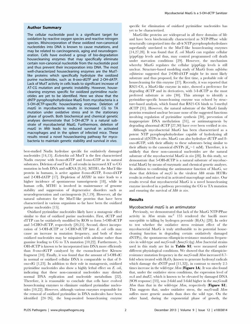
resistance mutation frequency in the mazG-null Msm increased 8.7-
fold when treated with H2O2 (known to generate hydroxyl radicals
which damage the dNTP pool [11,32]), in contrast to merely 2.5
times increase in the wild-type Msm (Figure 1A). It was also found
that, under the oxidative stress conditions, the expression level of
recA and dnaE2, which is known to be elevated by damaged DNA
(SOS response) [33], was 2-fold and 3-fold higher in the mazG-null
Msm than that in the wild-type Msm, respectively (Figure S1).
This suggests that, under oxidative stress, the mazG-null Msm
suffers more genetic assaults than does the wild type. On the
other hand, during the exponential phase of growth, the
Author Summary
The cellular nucleotide pool is a significant target foroxidation by reactive oxygen species and reactive nitrogenspecies. Misincorporation of these oxidized non-canonicalnucleotides into DNA is known to cause mutations, andmay be related to carcinogenesis, aging and neurodegen-eration. Cells have evolved a group of bio-degradationhousecleaning enzymes that may specifically eliminatecertain non-canonical nucleotide from the nucleotide pooland thus prevent their incorporation into DNA. The mostwell-characterized housecleaning enzymes are the MutT-like proteins which specifically hydrolyze the oxidizedpurine nucleotides, such as 8-oxo-dGTP and 2-OH-dATP.Lack of MutT activity in cells leads to significant increase ofAT-CG mutation and genetic instability. However, house-cleaning enzymes specific for oxidized pyrimidine nucle-otides are yet to be identified. Here we show that thedNTP pyrophosphohydrolase MazG from mycobacteria is a5-OH-dCTP-specific housecleaning enzyme. Deletion ofmazG in mycobacteria results in increased CG to TAmutation under oxidative stress and in the stationaryphase of growth. Both biochemical and chemical geneticanalyses demonstrate that 5-OH-dCTP is a natural sub-strate of mycobacterial MazG. Furthermore, deletion ofmazG in Mtb leads to reduced survival in activatedmacrophages and in the spleen of infected mice. Theseresults reveal a novel housecleaning pathway for myco-bacteria to maintain genetic stability and survival in vivo.
Mycobacterial MazG Is a 5-OH-dCTP Sanitizer
PLOS Pathogens | www.plospathogens.org 2 December 2013 | Volume 9 | Issue 12 | e1003814
rifampicin-resistance mutation frequency of mazG-null Msm is
comparable to that of the wild-type Msm (Figure 1A).
We also measured the rifampicin-resistance mutation frequency
in the stationary phase of growth, a stage known to accumulate
metabolic byproducts and mutations [34]. It was found that the
rifampicin-resistance mutation frequency in 5-day-old mazG-null
Msm cultures was 2.5 times greater than that in wild type
(Figure 1A), suggesting a mutator phenotype of mazG-null Msm
during the stationary phase of growth. A similar result was
observed with 8-day-old cultures (data not shown), indicating
prolonged incubation during the stationary phase does not further
increase the mutation frequency in mazG-null Msm. To test
whether mazG plays the same function in Mtb, we constructed a
DmazG::hyg null mutation in the virulent Mtb strain H37Rv by
means of allelic exchange and the deletion of this gene was
confirmed by Southern blot (Figure S2). The mazG-null Mtb
exhibited the same mutator phenotype as that of the mazG-null
Msm (Figure 1B), showing a 2.5-fold increase in rifampicin-
resistance mutation frequency compared to that of the wild type
under oxidative stress or the stationary phase of growth.
In order to test the cytotoxic effects of H2O2 upon the bacteria
studied, we measured the survival rates of the mazG-null strains
versus the wild-type strains of Msm and Mtb under the same H2O2
treatment conditions as that for mutation analysis. The survival
rate of the mazG-null Msm decreased only slightly after 5 hours of
H2O2 treatment compared to that of the wild type, while in the
mazG-null Mtb, no significant effect was observed (Figure 1C).
These data suggest that other than the change of mutation
frequencies, H2O2 treatment in this study did not induce other
major physiological change affecting the survival of the bacilli.
Because the wild-type mazG gene complements all of the defective
phenotypes of the mazG-null mutants (Figure 1), the antimutator
role of mycobacterial MazG is genetically established.
Mycobacterial MazG prevents CG to TA mutationIt has been shown that incorporation of different oxidized
dNTPs into DNA preferentially induces a specific spectrum of
mutation, e.g., 8-oxo-dGTP leads to AT to CG mutation [35,36]
while 5-OH-dCTP induces GC to AT mutation [9,10,22].
Therefore, we compared the mutation spectra between the
mazG-null Msm and its parental strains to infer the probable
substrate of mycobacterial MazG.
We sequenced the cluster I region of the rpoB gene [37] from
randomly isolated rifampicin-resistant colonies. All of the sequenc-
es contained single nonsynonymous nucleotide variations. Of
these, .99% were located within the cluster I region (the
remaining mutations occurred outside of the cluster I region)
and caused mutations in the well characterized rifampicin-
resistance mutation hot spots (Table S2 and S3). Of the
mutations detected, the frequency of CG to TA mutation
exhibited a significant difference between the wild type and the
mazG-null mutant (Table 1). Among the rifampicin-resistant
mutants derived from the exponential-phase cells, the CG to TA
mutation frequency in wild-type Msm increased from 2.261028 in
the untreated samples to 6.861028 in the H2O2 treated cultures
(,3-fold increase), while in the mazG-null Msm, the frequency of
this type of mutations increased significantly from 0.861028 to
14.361028 (,18-fold increase). Meanwhile, the rifampicin-resis-
tant mutants of the wild-type Msm exhibited a CG to TA mutation
frequency in the stationary-phase cells similar to that in the
exponential-phase cells (1.961028 and 2.261028, respectively),
suggesting that the CG to TA mutation rate is likely constant
during replication in the wild-type Msm. However, the CG to TA
mutation in the mazG-null Msm increased 26-fold, from 0.861028
in the exponential-phase cells to 20.561028 in the stationary-
phase cells (Table 1). These results clearly suggest that
mycobacterial MazG is involved in safeguarding genomic DNA
Figure 1. The antimutator role of MazG in Msm (A) and Mtb (B). Both the bacterial culture conditions and the methods for determination ofmutation frequencies were illustrated in Materials and Methods in detail. The frequencies conferring resistance to rifampicin in wild-type (wt), mazG-null (DmazG) and the complemented mutant (compl) strains were determined in exponential phase (OD600,0.5) with or without oxidative stress andin the stationary growth phase. Oxidative stress was induced by treating exponential phase cultures with 10 mM H2O2 for 5 h (Msm) or 24 h (Mtb).Stationary phase was at the 5th-day or 28th-day of culture for Msm or Mtb, respectively. (C) Survival rate of Msm and Mtb strains after exposure toH2O2. The numbers shown are mean 6 S.E. of 3 independent experiments totaling 15 cultures of Msm and 6 of Mtb.doi:10.1371/journal.ppat.1003814.g001
Mycobacterial MazG Is a 5-OH-dCTP Sanitizer
PLOS Pathogens | www.plospathogens.org 3 December 2013 | Volume 9 | Issue 12 | e1003814
by preventing CG to TA mutation under adverse growth
conditions.
5-OH-dCTP is a preferred substrate for mycobacterialMazG
CG to TA transition, the most common base substitution
occurring in aerobic organisms [38,39], can be induced by
incorporation of oxidatively damaged nucleotides into DNA,
especially oxidized pyrimidine nucleotides [9,10,22]. We mea-
sured the MazG NTP-PPase activity towards 5-OH-dCTP, 5-
CHO-dUTP and 2-OH-dATP, all of which are known to induce
CG to TA mutation when incorporated into DNA [9,22,40].
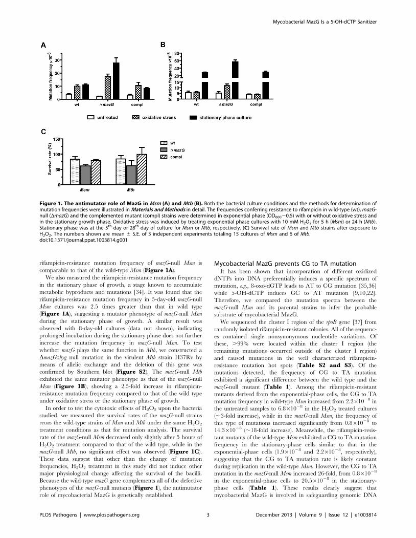
Each substrate was mixed with mycobacterial MazG proteins of
different origin and was incubated at 37uC for 10 minutes. The
hydrolyzed product, pyrophosphate, was quantified by an enzyme
coupled colorimetric method [28]. It was found that all of these
substrates were hydrolyzed into monophosphate derivatives and
pyrophosphate in a time- and enzyme concentration-dependent
manner (Figure 2A). Of the nucleotides examined, 5-OH-dCTP
and 2-OH-dATP were the most preferred substrates for the Mtb
MazG, with Km values of 1.9 and 2.4 mM, respectively, approx-
imately 26 times lower than that of their canonical nucleotides
(Table 2 and Figure 2B). It appears that 5-CHO-dUTP is
unlikely to be the natural substrate of Mtb MazG, shown by its
extremely high Km value (,500 mM, Table 2). The Msm MazG
exhibited similar kinetic constants compared to its Mtb counter-
part, except for 2-OH-dATP, which showed a Km of 311 mM(Table S4), much higher than that of the Mtb MazG (Table 2).
Based on the kinetic constants and the same antimutator role of
MazG in Msm and Mtb, we conclude that 5-OH-dCTP is the most
likely natural substrate of mycobacterial MazG.
5-OH-dCTP is an in vivo substrate of mycobacterial MazGTo further characterize the natural substrate of mycobacterial
MazG under cellular physiological conditions, we compared the in
vivo mutagenic effects of these oxidized nucleotides in wild-type
and mazG-null Msm strains using an established in vivo incorpora-
tion assay [22,36].
Of the nucleotides tested, only 5-OH-dCTP exhibited a
mutagenic effect upon the mazG-null Msm in a dose dependent
manner (Figure 3A–C). When treated with 100 mM 5-OH-
dCTP, the mazG-null mutant showed a ,2 fold increase (P,0.01)
in rifampicin-resistance mutation frequency compared to that of
the wild-type Msm (Figure 3B). The increased mutation
frequency of the mazG-null mutant can be restored to normal by
complementation with a single copy of the wild-type mazG from
Table 1. mazG-null Msm exhibited elevated CG to TA mutation under oxidative stress conditions and in the stationary phase ofgrowth.
Growth phase Strain (n) Mutation frequency 61028 (n)
CGRTA CGRAT CGRGC GCRCG GCRTA ATRGC ATRCG ATRTA
exponential wt (30) 2.2 (17) (0) (0) (0) 0.5 (4) 1.2 (9) (0) (0)
wt+H2O2 (40) 6.8 (27) (0) (0) (0) 1.2 (5) 0.8 (3) 1.2 (5) (0)
DmazG (43) 0.8 (18) 0.05 (1) (0) 0.05 (1) 0.09 (2) 0.7 (14) 0.3 (7) (0)
DmazG+H2O2 (69) 14.3 (57) (0) (0) (0) (0) 2.0 (8) 1.0 (4) (0)
stationary wt (47) 1.9 (8) (0) 3 (13) (0) 5.1 (22) 0.2 (1) (0) 0.7 (3)
DmazG (45) 20.5 (34) 1.9 (3) 4.7 (8) (0) 0.6 (1) (0) (0) (0)
Spontaneous rifampicin-resistant colonies were collected from 3 independent experiments (see Methods). Cluster I region of rpoB were PCR-amplified using pfu DNApolymerase and sequenced bi-directionally. All of the sequenced colonies contain single non-synonymous mutations (see also Table S1 and S2). wt, wild-type Msm;DmazG, mazG-null Msm.doi:10.1371/journal.ppat.1003814.t001
Figure 2. The NTP-PPase activity of mycobacterial MazG against 5-OH-dCTP. (A) Time- and enzyme concentration-dependent hydrolysis of5-OH-dCTP. 5-OH-dCTP (200 mM) was incubated with 1 mg or varied amounts (from 1 mg to 4 mg) of heterogeneously expressed MazG purified tonearly SDS-PAGE homogeneity. The reaction was carried out at 37uC and terminated after 10 min or at the time points as indicated. PPi,pyrophosphate. Shown are mean 6 S.E. of 3 repeats. (B) Michaelis-Menten curves of MazG with 5-OH-dCTP or dCTP as substrate. The hydrolyticproduct PPi is shown as mM/10 min. Data shown are mean 6 S.E. of 3 independent experiments.doi:10.1371/journal.ppat.1003814.g002
Mycobacterial MazG Is a 5-OH-dCTP Sanitizer
PLOS Pathogens | www.plospathogens.org 4 December 2013 | Volume 9 | Issue 12 | e1003814
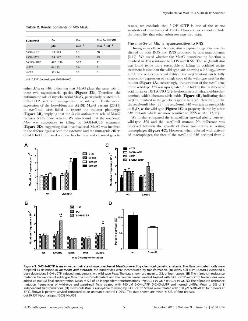
either Msm or Mtb, indicating that MazG plays the same role in
these two mycobacteria species (Figure 3B). Therefore, the
antimutator role of mycobacterial MazG, particularly related to 5-
OH-dCTP induced mutagenesis, is inferred. Furthermore,
expression of the loss-of-function A219E MazG variant [28,41]
in mazG-null Msm failed to restore the mutator phenotype
(Figure 3B), implying that the in vivo antimutator role of MazG
requires NTP-PPase activity. We also found that the mazG-null
Msm was susceptible to killing by 5-OH-dCTP treatment
(Figure 3D), suggesting that mycobacterial MazG was involved
in the defense against both the cytotoxic and the mutagenic effects
of 5-OH-dCTP. Based on these biochemical and chemical genetic
results, we conclude that 5-OH-dCTP is one of the in vivo
substrates of mycobacterial MazG. However, we cannot exclude
the possibility that other substrates may also exist.
The mazG-null Mtb is hypersensitive to RNSDuring intracellular infection, Mtb is exposed to genetic assaults
elicited by both ROS and RNS produced by host macrophages
[5,42]. We tested whether the MazG housecleaning function is
involved in Mtb resistance to ROS and RNS. The mazG-null Mtb
was found to be more susceptible to killing by acidified nitrite
treatment in vitro than the wild-type Mtb, showing a 0.8-log10 lower
CFU. The reduced survival ability of the mazG mutant can be fully
restored by expression of a single copy of the wild-type mazG in the
mutant (Figure 4A). Accordingly, transcription of the mazG gene
in the wild-type Mtb was upregulated 3,5 fold by the treatment of
acid nitrite or DETA/NO (2,2-(hydroxynitrosohydrazino)-bisetha-
namine), which liberates nitric oxide (Figure 4B), indicating that
mazG is involved in the genetic response to RNS. However, unlike
the mazG-null Msm [28], the mazG-null Mtb was just as susceptible
to H2O2 as the wild type (Figure 1C), a property shared by other
Mtb mutants which are more sensitive to RNS in vitro [43,44].
We further compared the intracellular survival ability between
wild-type Mtb and the mazG-null mutant. No difference was
observed between the growth of these two strains in resting
macrophages (Figure 4C). However, when infected with activat-
ed macrophages, the titer of the mazG-null Mtb declined from 2
Figure 3. 5-OH-dCTP is an in vivo substrate of mycobacterial MazG proved by chemical genetic analysis. The Msm competent cells wereprepared as described in Materials and Methods, the nucleotides were incorporated by transformation. (A) mazG-null Msm (DmazG) exhibited adose-dependent 5-OH-dCTP induced mutagenesis. wt, wild-type Msm. The data shown are mean 6 S.E. of four repeats. (B) The rifampicin-resistancemutation frequencies of wild-type Msm, the mazG-null mutant and the complemented mutant treated with 5-OH-dCTP and dCTP. Nucleotides wereadded at 100 mM final concentration. Mean 6 S.E of 12 independent transformations. **p,0.01 vs wt, * p,0.05 vs wt. (C) The rifampicin-resistancemutation frequencies of wild-type and mazG-null Msm treated with 100 mM 2-OH-dATP, 5-CHO-dUTP and normal dNTPs. Mean 6 S.E of 8independent transformations. (D) mazG-null Msm is susceptible to killing by 5-OH-dCTP. Strains were treated with 100 mM 5-OH-dCTP for 5 hours at37uC. Shown is percent survival compared to an untreated control (100%). The data shown are mean 6 S.E. of four repeats.doi:10.1371/journal.ppat.1003814.g003
Table 2. Kinetic constants of Mtb MazG.
Substrate Km kcat kcat /Km (6100)
mM min21 min21 mM21
5-OH-dCTP 1.960.3 1.3 68
2-OH-dATP 2.460.7 1.9 79
5-CHO-dUTP 4976140 56.2 11
d-ATP 64623 5.6 9
d-CTP 51614 3.2 6
doi:10.1371/journal.ppat.1003814.t002
Mycobacterial MazG Is a 5-OH-dCTP Sanitizer
PLOS Pathogens | www.plospathogens.org 5 December 2013 | Volume 9 | Issue 12 | e1003814
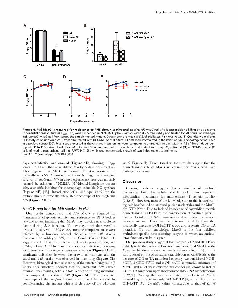
days post-infection and onward (Figure 4D), showing 1 log10
lower CFU than that of wild-type Mtb by 5 days post-infection.
This suggests that MazG is required for Mtb resistance to
intracellular RNS. Consistent with this finding, the attenuated
survival of mazG-null Mtb in activated macrophages was partially
rescued by addition of NMMA (NG-Methyl-L-arginine acetate
salt), a specific inhibitor for macrophage inducible NO synthase
(Figure 4E) [45]. Introduction of a wild-type mazG into the
mutant strain restored the attenuated phenotype of the mazG-null
Mtb (Figure 4D–E).
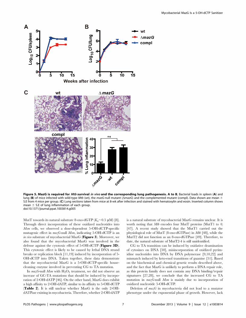
MazG is required for Mtb survival in vivoOur results demonstrate that Mtb MazG is required for
maintenance of genetic stability and resistance to RNS both in
vitro and ex vivo, indicating that MazG may function as a virulence
factor during Mtb infection. To investigate whether mazG is
involved in survival of Mtb in vivo, immune-competent mice were
infected by a low-dose aerosol challenge with Mtb strains.
Compared to wild-type Mtb, the mazG-null Mtb exhibited 1.1-
log10 lower CFU in mice spleens by 4 weeks post-infection, and
0.7-log10 lower CFU by 8 and 12 weeks post-infection, indicating
an attenuation at the stage of persistent infection (Figure 5A). No
significant difference between the growth of wild-type and the
mazG-null Mtb strains was observed in mice lung (Figure 5B).
However, histological stained sections of the infected lung tissue (8
weeks after infection) showed that the mazG-null Mtb caused
minimal pneumonitis, with a 3-fold reduction in lung inflamma-
tion compared to wild-type Mtb (Figure 5C). The attenuated
phenotype of the mazG-null mutant can be fully restored by
complementing the mutant with a single copy of the wild-type
mazG (Figure 5). Taken together, these results suggest that the
housecleaning role of MazG is required for Mtb survival and
pathogenesis in vivo.
Discussion
Growing evidence suggests that elimination of oxidized
nucleotides from the cellular dNTP pool is an important
safeguarding mechanism for maintenance of genetic stability
[2,3,6,7]. However, most of the knowledge about this houseclean-
ing role has focused on oxidized purine nucleotides and the MutT-
like NTP-PPase. Due to lack of knowledge of pyrimidine specific
housecleaning NTP-PPase, the contribution of oxidized pyrimi-
dine nucleotides to DNA mutagenesis and its related mechanism
remains unclear. Here we characterized a NTP-PPase that
specifically degrades 5-OH-dCTP in vivo and prevents CG to TA
mutation. To our knowledge, MazG is the first oxidized
pyrimidine-specific housecleaning enzyme to which an antimu-
tator function can be assigned.
Our previous study suggested that 8-oxo-dGTP and dUTP are
unlikely to be the natural substrates of mycobacterial MazG, as the
Km values for these nucleotides are substantially high [28]. In this
study, based on the observation that deletion of mazG leads to the
increase of CG to TA mutation frequency, we considered 5-OH-
dCTP, 5-CHO-dUTP and 2-OH-dATP as putative substrates of
MazG, and all of these oxidized nucleotides are known to induce
CG to TA mutations upon incorporated into DNA by polymerase
[9,22,40]. Among the substrates tested, mycobacterial MazG
showed high affinity towards 5-OH-dCTP (Km = 1.9 mM) and 2-
OH-dATP (Km = 2.4 mM), values comparable to that of E. coli
Figure 4. Mtb MazG is required for resistance to RNS shown in vitro and ex vivo. (A) mazG-null Mtb is susceptible to killing by acid nitrite.Exponential phase cultures (OD600,0.5) were suspended in 7H9-OADC pH4.5 with or without 2.5 mM NaNO2 and treated for 20 hours. wt, wild-typeMtb; DmazG, mazG-null Mtb; compl, the complemented mutant. Data shown are mean 6 S.E. of triplicates. * p,0.05 vs wt. (B) Quantitative real-timePCR analyses of mazG and dosR from Mtb treated with DETA/NO or acid nitrite. All data were normalized to the levels of sigA. The dosR gene was usedas a positive control [70]. Results are expressed as the changes in expression levels compared to untreated samples. Mean 6 S.E of three independentrepeats. C to E, Survival of wild-type Mtb, the mazG-null mutant and the complemented mutant in resting (C), activated (D) or NMMA treated (E)cells of murine macrophage cell line RAW264.7. Shown is one representative result of two independent experiments.doi:10.1371/journal.ppat.1003814.g004
Mycobacterial MazG Is a 5-OH-dCTP Sanitizer
PLOS Pathogens | www.plospathogens.org 6 December 2013 | Volume 9 | Issue 12 | e1003814
MutT towards its natural substrate 8-oxo-dGTP (Km,0.5 mM) [8].
Through direct incorporation of these oxidized nucleotides into
Msm cells, we observed a dose-dependent 5-OH-dCTP-specific
mutagenic effect in mazG-null Msm, indicating 5-OH-dCTP is an
in vivo substrate of mycobacterial MazG (Figure 3). Moreover, we
also found that the mycobacterial MazG was involved in the
defense against the cytotoxic effect of 5-OH-dCTP (Figure 3D).
This cytotoxic effect is likely to be caused by lethal DNA strand
breaks or replication block [11,19] induced by incorporation of 5-
OH-dCTP into DNA. Taken together, these data demonstrate
that the mycobacterial MazG is a 5-OH-dCTP-specific house-
cleaning enzyme involved in preventing CG to TA mutation.
In mazG-null Msm with H2O2 treatment, we did not observe an
increase of GC-TA mutations that should be induced by incorpo-
ration of 2-OH-dATP [46]. On the other hand, MazG does exhibit
a high affinity to 2-OH-dATP, similar to its affinity to 5-OH-dCTP
(Table 2). It is still unclear whether MazG is the only 2-OH-
dATPase existing in mycobacteria. Therefore, whether 2-OH-dATP
is a natural substrate of mycobacterial MazG remains unclear. It is
worth noting that Mtb encodes four MutT proteins (MutT1 to 4)
[47]. A recent study showed that the MutT1 carried out the
physiological role of MutT (8-oxo-dGTPase) in Mtb [48], while the
MutT2 did not function as an 8-oxo-dGTPase [49]. Therefore, to
date, the natural substrate of MutT2-4 is still unidentified.
CG to TA transition can be induced by oxidative deamination
of cytosines on DNA [50], misincorporation of oxidized pyrim-
idine nucleotides into DNA by DNA polymerase [9,10,22] and
mismatch induced by keto-enol transitions of guanine [51]. Based
on the biochemical and chemical genetic results described above,
and the fact that MazG is unlikely to perform a DNA repair role,
as this protein family does not contain any DNA binding/repair
signatures [27,28], we conclude that the increased CG to TA
mutation in mazG-null Msm is mainly due to incorporation of
oxidized nucleotide 5-OH-dCTP.
Deletion of mazG in mycobacteria did not lead to a mutator
phenotype under the exponential phase of growth. However, lack
Figure 5. MazG is required for Mtb survival in vivo and the corresponding lung pathogenesis. A to B, Bacterial loads in spleen (A) andlung (B) of mice infected with wild-type Mtb (wt), the mazG-null mutant (DmazG) and the complemented mutant (compl). Data shown are mean 6S.E from 4 mice per group. (C) Lung sections taken from mice at 8-wk after infection and stained with hematoxylin and eosin. Inserted column showsmean 6 S.E of lung inflammation of each group.doi:10.1371/journal.ppat.1003814.g005
Mycobacterial MazG Is a 5-OH-dCTP Sanitizer
PLOS Pathogens | www.plospathogens.org 7 December 2013 | Volume 9 | Issue 12 | e1003814
of MazG activity in mycobacteria resulted in higher CG to TA
mutation under both oxidative stress and the stationary growth
phase, compared to that of the parental strains (Table 1). The
likely mechanism underlying this stress-related mutagenesis is that
under stress conditions, mycobacterial cells may accumulate 5-
OH-dCTP and lacking MazG, more 5-OH-dCTP is misincorpo-
rated into DNA. Moreover, down-regulated DNA repair activity
under these stress conditions [34,52] may also contribute to the
stress-related mutagenesis observed in the mazG-null strains.
Nonetheless, although the related molecular mechanism of 5-
OH-dCTP induced stress-related mutagenesis remains to be
determined, as the host environment for Mtb parasitism is always
adverse, 5-OH-dCTP induced mutagenesis may be hypothesized
to play an important role in the microevolution process of the
infected Mtb under stress conditions, i.e. emergence of drug
resistant mutations during bacterial infection. In this connection, it
is worth noting that CG to TA transition is a dominant mutation
in Mtb isolated from either macaques with latent/reactivated
infection or humans [53,54].
It is still unclear why the lack of the 5-OH-dCTP sanitization
function in Mtb results in hypersusceptibility to RNS (Figure 4).
RNS is a group of radicals derived from nitric oxide (NON) which
are produced by macrophage as antimicrobial effector molecules
[5]. An important antimicrobial action of RNS is inhibition of
DNA replication and repair. It was found that NON can inhibit
DNA synthesis by zinc mobilization from DNA-binding metallo-
proteins [55]. RNS can also inhibit ribonucleotide reductase [56],
and thus, limit the availability of precursors for the synthesis and
repair of DNA. Based on the fact that lack of MazG activity leads
to increased incorporation of 5-OH-dCTP into DNA (Table 1), a
possible explanation for the hypersusceptibility of mazG-null Mtb to
RNS is that inhibition of DNA repair activities and lack of DNA
precursors caused by RNS mediated enzyme inactivation result in
higher levels of genetic instability (such as DNA strand breakage)
in mazG-null Mtb than that in the wild-type Mtb.
During infection, Mtb is exposed to an oxidative environment of
host macrophages rich in DNA-damaging ROS and RNS.
Therefore, safeguarding of the genetic information is essential
for mycobacterial survival, especially during the non-replicating
dormancy stage, as slow or non-replicated genomic DNA and
diminished DNA repair activities are likely lead to more genetic
assaults than that during fast growing phase [52,57]. Our results
demonstrated that deletion of mazG leads to attenuated survival of
Mtb in mice spleen during the persistent infection phase,
suggesting that oxidative damage to nucleotides and the subse-
quent genetic assault is one of the bactericidal effects of the
adaptive immune response (corresponding to the bacterial
persistent infection stage). This is consistent with the data
indicating that genes involved in removal of oxidized pyrimidines
are essential for Mtb survival during primates’ infection [58].
Although the difference between the lung and spleen microenvi-
ronments exposed to Mtb is unclear, it is conceivable that the
immune responses and metabolic constraints are different between
the two tissues. Interestingly, tissue specific attenuation have been
demonstrated for several Mtb mutants, including the dosR, fadD26,
mptpB and narG mutants [59–62].
Recent studies have proven that bactericidal antibiotics-induced
ROS production within bacterial cells is a common mechanism for
cell death [12,63–65], predominantly elicited by incorporation of
8-oxo-dGTP into DNA [11]. Therefore, it is not surprising that 5-
OH-dCTP and other oxidized nucleotides have a similar
bactericidal effect, as shown in our results (Figure 3D andFigure 5A). These findings suggest that clinical treatment of
tuberculosis with specific inhibitors of housecleaning enzymes
might facilitate Mtb elimination, especially when combined with
bactericidal antibiotics which are known to induce oxidative stress.
Materials and Methods
Ethics statementSix-to-eight week old female C57BL/6 mice were purchased
from the Shanghai SLAC Laboratory Animal Company. The mice
were housed and cared for in a specific pathogen-free (SPF)
biosafety level 3 facility at Shanghai Public Health Clinical Center.
Mice were provided food and water ad libitum as well as appropriate
monitoring and clinical care. Animal experiments were carried out
in strict accordance with the regulations in the Guidance
Suggestions for the Care and Use of Laboratory Animals issued
by the Ministry of Science and Technology of the People’s Republic
of China. The protocol was approved by the Chinese Science
Academy Committee on Care and Use of Laboratory Animals and
the Laboratory Animal Ethical Board of Shanghai Public Health
Clinical Center (Permit Number: 2012A002).
Bacterial strains and culture conditionsBacterial strains used in this study are list in Table S1. Bacterial
culturing was performed as described [66]. Msm strains were
grown at 37uC in 7H9 broth (BD Difco), or on Luria-Bertani agar
supplemented with 0.5% glycerol (LBG agar). Mtb strains were
grown at 37uC in 7H9 broth supplemented with 10% OADC
(7H9-OADC), or on 7H11 plates supplemented with 10% OADC
(7H11-OADC). When required, the following antibiotics were
used at the specified concentrations: kanamycin (15 mg/ml),
hygromycin B (150 mg/ml for Msm and 50 mg/ml for Mtb) and
rifampicin (250 mg/ml for Msm and 10 mg/ml for Mtb). For
treatment with acid NO, Mtb stains grown to OD600,0.5 were
pelleted and re-suspended in 7H9-OADC pH 4.5 (adjusted by
1 M citrate) with or without 2.5 mM NaNO2 [45]. After 20 h
treatment, bacteria were plated on 7H11-OADC, CFUs were
counted after 3,4 weeks culture at 37uC
Generation of mazG mutants and complemented strainsThe mazG-null mutant was generated by the phage transduction
method [66]. mazG-null Msm and the complemented strains were
generated as described [28]. To construct a transducing phage for
Mtb mazG knockout, the left homologue arm was PCR amplified
using primers KOP1 and KOP2 (primers used in this study are
listed in Table S5). The right homologue arm was PCR amplified
using primers KOP3 and KOP4. The PCR products were ligated
into the AflII/XbaI and HindIII/XhoI sites of pYUB854. The
recombinant transducing phage was used to construct the mazG-
null Mtb as described [28]. The mazG-null mutant was verified by
southern blot and PCR (Figure S2). The probe for Southern blot
was PCR amplified using primers SB1and SB2. A dUTP-biotin
labeled probe (Fermentas) was used for Southern blot analysis of
the PstI/KpnI digested chromosomal DNA on the Hybond-N+
nylon membrane (GE Amersham), according to the standard
method [67]. Primers used for genotyping PCR were P1, P2 and
P3. The complementation plasmid for mazG-null Mtb was
generated by ligating the PCR product amplified using primers
C1 and C2 into the BamHI and HindIII sites of pMV306.
Expression of Mtb mazG was controlled by its own promoter
(1142671–1143646).
Determination of rifampicin-resistance mutationfrequency
Single colonies of various Msm strains from the 7H11 agar plate
were inoculated in 5 ml media and cultured at 37uC for 48 h (2
Mycobacterial MazG Is a 5-OH-dCTP Sanitizer
PLOS Pathogens | www.plospathogens.org 8 December 2013 | Volume 9 | Issue 12 | e1003814
weeks for Mtb strains). For determination of rifampicin-resistance
mutation frequency, the cultures were inoculated with 1% of
primary culture in 20 ml 7H9 media (in a 100-ml flask) without
antibiotics and grown at 37uC with rolling (150 rpm) to exponential
phase (OD600,0.5). Then 10 ml of the cultures were treated with
10 mM H2O2, and another 10 ml cultures were untreated. After
incubation at 37uC, 150 rpm, for 5 h, CFU per ml was determined
by plating; the cell pellet from 3 ml culture was plated on LBG agar
(3 plates of each sample) containing 250 mg/ml rifampicin (Sigma-
Aldrich). The CFU and rifampicin-resistant colonies were counted
after culturing at 37uC for 4 days (28 days for Mtb strains). The
rifampicin-resistance mutant frequency was calculated by dividing
the number of rifampicin-resistant colonies on each plate by the
counts of the total viable cells plated. Rifampicin-resistance
mutation frequencies of Mtb strains were determined by the same
method, except that the oxidative stress was elicited by resuspending
the exponential-phase cell pellet in 7H9 media containing 10 mM
H2O2, followed with incubating at 37uC for 24 h. Mtb strains were
plated on 7H11-OADC with or without 10 mg/ml rifampicin. For
determination of rifampicin-resistance mutation frequency of the
stationary phase cultures, cells were cultured in liquid media for 5
days (for Msm) or 28 days (for Mtb) and plated as described above.
Three independent experiments were performed with totaling 15
cultures of each Msm strains and 6 of Mtb.
Analysis of mutation spectraRifampicin-resistant colonies were collected from three inde-
pendent experiments. The isolated colonies were grown in 1 ml
7H9 at 37uC for 1 week. Cells were pelleted and suspended in
50 ml TE buffer (10 mM Tris–HCl, pH 8.0, and 1 mM EDTA)
and incubated at 95uC for 10 min to extract the genome DNA
[37]. The lysate was centrifuged at 12000 g for 5 min. The
supernatant was used as template to amplify (using pfu DNA
polymerase) the fragment containing the cluster I region of rpoB
using primers Rpo1 and Rpo2. All PCR products were sequenced
by bi-directionally. Mutation spectra of the sequenced region were
analyzed by BioEdit software.
NTP-PPase assayProtein expression and purification was performed as described
[28]. Protein was purified to nearly SDS-PAGE homogeneity.
Protein concentration was determined by the bicinchoninic acid
(BCA) method [68]. The oxidized nucleotides used as substrates
for MazG were purchased from TriLink Biotechnologies Inc. (5-
OH-dCTP) or Hongene Biotechnologies Inc. (2-OH-dATP and 5-
CHO-dUTP). The NTP-PPase activity of MazG was assayed as
described [28]. The NTP-PPase assay was carried out in 20 ml
reaction buffer (20 mM Tris-HCl, pH 7.5, 5 mM MgSO4,
100 mM NaCl) containing 1 mg mycobacterial MazG and
substrate nucleoside triphosphates at 37uC for 10,20 min. The
reaction was stopped by heating at 65uC for 5 min, and 10-20 ml
products were applied for pyrophosphate assay (Molecular Probes)
according to the manufacturer’s instructions. Reactions with heat
inactivated (95uC for 20 min) MazG protein were set up as a
background controls. GraphPad Prism 5.0 (GraphPad Software,
Inc.) was used for enzyme kinetic constants analysis.
Incorporation of nucleotides into Msm competent cellsThe Msm competent cells were prepared from 400-ml cultures
(OD600 = 0.8,1.0) as previously described [69]. Incorporation of
nucleotides into Msm competent cells was performed as described
[22,36]. Briefly, Nucleotide solution (100 mM final concentration)
was added to 150 ml competent cells suspension and the mixture
was placed on ice for 10 min. After heat shock treatment (42uC for
90 sec and then on ice for 30 min), 2 ml 7H9 was added and the
cells were incubated at 37uC with rolling (150 rpm) for 5 h. After
treatment, 2 ml of culture was centrifuged at 4000 g for 5 min and
plated on LBG agar containing 250 mg/ml rifampicin. The
remaining culture was diluted and plated onto LBG agar for
CFU determination. Rifampicin-resistance mutation frequencies
were calculated as described above.
RNA extraction and quantitative real-time PCRWild-type Mtb (OD600,0.5) was treated with acid NO or
2.5 mM DETA/NO for 1 h. Total RNA was extracted with
TRIzol-Reagent (Invitrogen) and further purified with RiboPure-
Bacteria kit (Ambion). Briefly, cell pellet was resuspended in 1 ml
TRIzol reagent, mixed with 400 ml 0.1 mm Zirconia Beads
(BioSpec Products) and lysed in a mini-beadbeater (Biospec) for
three cycles (40 s at maximal speed) with cooling on ice for 1 min
between pulses. RNA was extracted according to the protocol of
TRIzol-Reagent. The extracted RNA was further purified using
the RiboPure-Bacteria kit followed by DNase I treatment to
eliminate DNA contamination. cDNA was synthesized using the
SuperScript III First Strand kit (Invitrogen) with random hexamer
primer. Target gene transcript levels were measured by real-time
PCR using SYBRH Premix Ex Taq GC (TaKaRa) on Mastercy-
cler ep realplex thermal cyclers: 95uC 60 sec, 40 cycles of 95uC5 sec, 62uC 8 sec and 72uC 20 sec, followed by melting curve
analysis. Data were normalized to sigA and expressed as fold
change compared to the untreated samples. PCR primers for sigA,
mazG, dosR, recA and dnaE2 are listed in Table S5.
Macrophage infectionThe murine macrophage cell line RAW264.7 was grown in
DMEM medium (GIBCO) supplemented with 10% fetal calf serum
(FCS) and incubated at 37uC with 5% CO2. For Mtb infection, cells
were plated at a density of 2.06105 cells per well in 24-well plates
without antibiotics and activated with 200 U/ml murine IFN-c(Peprotech) for 16 h [45]. Cells were primed with 1 mg/ml
lipopolysaccharides (LPS, Sigma) for 1 h and then infected at a
multiplicity of infection (MOI) of 2:1 (bacteria:cells). After 4 h
incubation at 37uC with 5% CO2, cells were washed three times
with DMEM to remove extracellular bacteria and cultured with
complete DMEM medium. To inhibit macrophage NO production
during the infection cause, NMMA (Sigma) was added to the culture
medium at a final concentration of 400 mM. At indicated time
points, bacteria were released with PBS solution containing 0.05%
Tween-80 and 0.025% SDS, and plated onto 7H11-OADC plates.
CFUs were counted after 3,4 weeks culture at 37uC.
Mice infectionMice were infected with wild-type Mtb, mazG-null mutant or
complemented strain at an inhaled aerosol dose of 100-200 CFU
per lung by an inhalation exposure system (Glas-Col, Terre Haute,
IN). At indicated time points, mice were sacrificed and lung and
spleen homogenates (four mice per group) were plated onto 7H11-
OADC followed by incubation at 37uC for 4 weeks.
HistopathologyLung sections stained with hematoxylin and eosin were
photographed using a Nikon Optiphot 2 microscope fitted with
a camera which was connected to a computer. The Image Pro Plus
program (Media Cybernetics) was utilized to objectively assess the
level of inflammation present in each image. To quantify the
percent area inflamed, we determined the mean percent inflamed
area from three to five lung sections of each mouse.
Mycobacterial MazG Is a 5-OH-dCTP Sanitizer
PLOS Pathogens | www.plospathogens.org 9 December 2013 | Volume 9 | Issue 12 | e1003814
Statistical analysisStatistical significance was determined with the unpaired two-
tailed Student’s t test at P,0.05 level of significance using
GraphPad Prism 5.0 software.
Supporting Information
Figure S1 mazG-null Msm exhibited higher level of SOSresponse under oxidative stress. Expression level of recA and
dnaE2 from exponential phase bacteria and oxidative stressed
samples (treated with 10 mM H2O2 for 1 h) were measured by
quantitative real-time PCR and normalized to sigA. Shown are fold
change compared to the untreated samples. wt, wild-type Msm;
DmazG, mazG-null Msm. Mean6 S.E. of three independent
repeats.
(TIF)
Figure S2 Characterization of mazG-null Mtb. (A)
Schematic diagrams of wild-type (wt) and the mazG-null (DmazG)
loci. The primers used for PCR are shown as arrows. (B) Southern
blot analysis of wt Mtb and the DmazG mutant. A dUTP-biotin
labeled fragment was used to probe PstI/KpnI-digested chromo-
somal DNA separated by 0.8% agarose gel. Sizes of DNA bands
are as indicated. (C) Analysis of PCR products from wt Mtb and
the DmazG mutant. C1 and C2 are two hygromycin-resistant
colonies.
(TIF)
Table S1 Bacteria strains used in this study.(PDF)
Table S2 Codon mutations determined in exponentialphase Msm-derived rifampicin-resistant mutant. Codon
427,429, 432 and 442 are rifampicin-resistant hot spots of rpoB.
(PDF)
Table S3 Codon mutations determined in stationaryphase (5-day) Msm-derived rifampicin-resistant mu-tant. All listed codons are rifampicin-resistant hot spots of rpoB.
(PDF)
Table S4 Kinetic constants of Msm MazG.(PDF)
Table S5 Primers used in this study.(PDF)
Acknowledgments
We thank Dougtas B. Lowrie from Shanghai Public Health Clinical Center
for his critical reading and helpful comments to this manuscript.
Author Contributions
Conceived and designed the experiments: LDL GPZ. Performed the
experiments: LDL BKT XYF HM. Analyzed the data: LDL BKT XYF
GPZ. Contributed reagents/materials/analysis tools: LDL XYF. Wrote the
paper: LDL BKT XYF GPZ.
References
1. Wagner JR, Hu CC, Ames BN (1992) Endogenous oxidative damage of
deoxycytidine in DNA. Proc Natl Acad Sci U S A 89: 3380–3384.
2. Sekiguchi M, Tsuzuki T (2002) Oxidative nucleotide damage: consequences andprevention. Oncogene 21: 8895–8904.
3. Haghdoost S, Sjolander L, Czene S, Harms-Ringdahl M (2006) The nucleotidepool is a significant target for oxidative stress. Free Radic Biol Med 41: 620–626.
4. Nathan C, Shiloh MU (2000) Reactive oxygen and nitrogen intermediates in the
relationship between mammalian hosts and microbial pathogens. Proc Natl
Acad Sci U S A 97: 8841–8848.
5. Fang FC (2004) Antimicrobial reactive oxygen and nitrogen species: conceptsand controversies. Nat Rev Microbiol 2: 820–832.
6. Rai P (2010) Oxidation in the nucleotide pool, the DNA damage response and
cellular senescence: Defective bricks build a defective house. Mutat Res 703: 71–81.
7. Ventura I, Russo MT, De Luca G, Bignami M (2010) Oxidized purine
nucleotides, genome instability and neurodegeneration. Mutat Res 703: 59–65.
8. Maki H, Sekiguchi M (1992) MutT protein specifically hydrolyses a potent
mutagenic substrate for DNA synthesis. Nature 355: 273–275.
9. Feig DI, Sowers LC, Loeb LA (1994) Reverse chemical mutagenesis:identification of the mutagenic lesions resulting from reactive oxygen species-
mediated damage to DNA. Proc Natl Acad Sci U S A 91: 6609–6613.
10. Purmal AA, Kow YW, Wallace SS (1994) 5-Hydroxypyrimidine deoxynucleo-
side triphosphates are more efficiently incorporated into DNA by exonuclease-free Klenow fragment than 8-oxopurine deoxynucleoside triphosphates. Nucleic
Acids Res 22: 3930–3935.
11. Foti JJ, Devadoss B, Winkler JA, Collins JJ, Walker GC (2012) Oxidation of the
guanine nucleotide pool underlies cell death by bactericidal antibiotics. Science336: 315–319.
12. Gutierrez A, Laureti L, Crussard S, Abida H, Rodriguez-Rojas A, et al. (2013)
beta-lactam antibiotics promote bacterial mutagenesis via an RpoS-mediated
reduction in replication fidelity. Nat Commun 4: 1610.
13. Bessman MJ, Frick DN, O’Handley SF (1996) The MutT proteins or ‘‘Nudix’’hydrolases, a family of versatile, widely distributed, ‘‘housecleaning’’ enzymes.
J Biol Chem 271: 25059–25062.
14. Galperin MY, Moroz OV, Wilson KS, Murzin AG (2006) House cleaning, a
part of good housekeeping. Mol Microbiol 59: 5–19.
15. McLennan AG (2006) The Nudix hydrolase superfamily. Cell Mol Life Sci 63:123–143.
16. Taddei F, Hayakawa H, Bouton M, Cirinesi A, Matic I, et al. (1997)Counteraction by MutT protein of transcriptional errors caused by oxidative
damage. Science 278: 128–130.
17. Fujikawa K, Kamiya H, Yakushiji H, Fujii Y, Nakabeppu Y, et al. (1999) The
oxidized forms of dATP are substrates for the human MutT homologue, thehMTH1 protein. J Biol Chem 274: 18201–18205.
18. Tsuzuki T, Egashira A, Igarashi H, Iwakuma T, Nakatsuru Y, et al. (2001)
Spontaneous tumorigenesis in mice defective in the MTH1 gene encoding 8-
oxo-dGTPase. Proc Natl Acad Sci U S A 98: 11456–11461.
19. Nakabeppu Y, Oka S, Sheng Z, Tsuchimoto D, Sakumi K (2010) Programmed
cell death triggered by nucleotide pool damage and its prevention by MutT
homolog-1 (MTH1) with oxidized purine nucleoside triphosphatase. Mutat Res
703: 51–58.
20. Jaruga P, Dizdaroglu M (1996) Repair of products of oxidative DNA base
damage in human cells. Nucleic Acids Res 24: 1389–1394.
21. Murata-Kamiya N, Kamiya H, Muraoka M, Kaji H, Kasai H (1997)
Comparison of oxidation products from DNA components by gamma-
irradiation and Fenton-type reactions. J Radiat Res 38: 121–131.
22. Fujikawa K, Kamiya H, Kasai H (1998) The mutations induced by oxidatively
damaged nucleotides, 5-formyl-dUTP and 5-hydroxy-dCTP,in Escherichia coli.
Nucleic Acids Res 26: 4582–4587.
23. Fraga CG, Shigenaga MK, Park JW, Degan P, Ames BN (1990) Oxidative
damage to DNA during aging: 8-hydroxy-29-deoxyguanosine in rat organ DNA
and urine. Proc Natl Acad Sci U S A 87: 4533–4537.
24. Hatahet Z, Kow YW, Purmal AA, Cunningham RP, Wallace SS (1994) New
substrates for old enzymes. 5-Hydroxy-29-deoxycytidine and 5-hydroxy-29-
deoxyuridine are substrates for Escherichia coli endonuclease III and
formamidopyrimidine DNA N-glycosylase, while 5-hydroxy-29-deoxyuridine is
a substrate for uracil DNA N-glycosylase. J Biol Chem 269: 18814–18820.
25. Purmal AA, Lampman GW, Bond JP, Hatahet Z, Wallace SS (1998) Enzymatic
processing of uracil glycol, a major oxidative product of DNA cytosine. J Biol
Chem 273: 10026–10035.
26. D’Ham C, Romieu A, Jaquinod M, Gasparutto D, Cadet J (1999) Excision of
5,6-dihydroxy-5,6-dihydrothymine, 5,6-dihydrothymine, and 5-hydroxycyto-
sine from defined sequence oligonucleotides by Escherichia coli endonuclease
III and Fpg proteins: kinetic and mechanistic aspects. Biochemistry 38: 3335–
3344.
27. Moroz OV, Murzin AG, Makarova KS, Koonin EV, Wilson KS, et al. (2005)
Dimeric dUTPases, HisE, and MazG belong to a new superfamily of all-alpha
NTP pyrophosphohydrolases with potential ‘‘house-cleaning’’ functions. J Mol
Biol 347: 243–255.
28. Lu LD, Sun Q, Fan XY, Zhong Y, Yao YF, et al. (2010) Mycobacterial MazG is
a novel NTP pyrophosphohydrolase involved in oxidative stress response. J Biol
Chem 285: 28076–28085.
29. Gross M, Marianovsky I, Glaser G (2006) MazG — a regulator of programmed
cell death in Escherichia coli. Mol Microbiol 59: 590–601.
30. Nonaka M, Tsuchimoto D, Sakumi K, Nakabeppu Y (2009) Mouse RS21-C6 is
a mammalian 29-deoxycytidine 59-triphosphate pyrophosphohydrolase that
prefers 5-iodocytosine. Febs J 276: 1654–1666.
31. Wu B, Liu Y, Zhao Q, Liao S, Zhang J, et al. (2007) Crystal structure of RS21-
C6, involved in nucleoside triphosphate pyrophosphohydrolysis. J Mol Biol 367:
1405–1412.
32. Imlay JA, Chin SM, Linn S (1988) Toxic DNA damage by hydrogen peroxide
through the Fenton reaction in vivo and in vitro. Science 240: 640–642.
Mycobacterial MazG Is a 5-OH-dCTP Sanitizer
PLOS Pathogens | www.plospathogens.org 10 December 2013 | Volume 9 | Issue 12 | e1003814
33. Boshoff HI, Reed MB, Barry CE, 3rd, Mizrahi V (2003) DnaE2 polymerase
contributes to in vivo survival and the emergence of drug resistance in
Mycobacterium tuberculosis. Cell 113: 183–193.
34. Saint-Ruf C, Pesut J, Sopta M, Matic I (2007) Causes and consequences of DNA
repair activity modulation during stationary phase in Escherichia coli. Crit Rev
Biochem Mol Biol 42: 259–270.
35. Michaels ML, Cruz C, Grollman AP, Miller JH (1992) Evidence that MutY and
MutM combine to prevent mutations by an oxidatively damaged form of
guanine in DNA. Proc Natl Acad Sci U S A 89: 7022–7025.
36. Inoue M, Kamiya H, Fujikawa K, Ootsuyama Y, Murata-Kamiya N, et al.
(1998) Induction of chromosomal gene mutations in Escherichia coli by direct
incorporation of oxidatively damaged nucleotides. New evaluation method for
mutagenesis by damaged DNA precursors in vivo. J Biol Chem 273: 11069–
11074.
37. Heep M, Brandstatter B, Rieger U, Lehn N, Richter E, et al. (2001) Frequency
of rpoB mutations inside and outside the cluster I region in rifampin-resistant
clinical Mycobacterium tuberculosis isolates. J Clin Microbiol 39: 107–110.
38. Schaaper RM, Danforth BN, Glickman BW (1986) Mechanisms of spontaneous
mutagenesis: an analysis of the spectrum of spontaneous mutation in the
Escherichia coli lacI gene. J Mol Biol 189: 273–284.
39. Schaaper RM, Dunn RL (1991) Spontaneous mutation in the Escherichia coli
lacI gene. Genetics 129: 317–326.
40. Satou K, Harashima H, Kamiya H (2003) Mutagenic effects of 2-hydroxy-dATP
on replication in a HeLa extract: induction of substitution and deletion
mutations. Nucleic Acids Res 31: 2570–2575.
41. Zheng H, Lu L, Wang B, Pu S, Zhang X, et al. (2008) Genetic basis of virulence
attenuation revealed by comparative genomic analysis of Mycobacterium
tuberculosis strain H37Ra versus H37Rv. PLoS One 3: e2375.
42. Darwin KH, Nathan CF (2005) Role for nucleotide excision repair in virulence
of Mycobacterium tuberculosis. Infect Immun 73: 4581–4587.
43. Darwin KH, Ehrt S, Gutierrez-Ramos JC, Weich N, Nathan CF (2003) The
proteasome of Mycobacterium tuberculosis is required for resistance to nitric
oxide. Science 302: 1963–1966.
44. Venugopal A, Bryk R, Shi S, Rhee K, Rath P, et al. (2011) Virulence of
Mycobacterium tuberculosis depends on lipoamide dehydrogenase, a member of
three multienzyme complexes. Cell Host Microbe 9: 21–31.
45. Chan J, Xing Y, Magliozzo RS, Bloom BR (1992) Killing of virulent
Mycobacterium tuberculosis by reactive nitrogen intermediates produced by
activated murine macrophages. J Exp Med 175: 1111–1122.
46. Kamiya H (2003) Mutagenic potentials of damaged nucleic acids produced by
reactive oxygen/nitrogen species: approaches using synthetic oligonucleotides
and nucleotides: survey and summary. Nucleic Acids Res 31: 517–531.
47. Dos Vultos T, Blazquez J, Rauzier J, Matic I, Gicquel B (2006) Identification of
Nudix hydrolase family members with an antimutator role in Mycobacterium
tuberculosis and Mycobacterium smegmatis. J Bacteriol 188: 3159–3161.
48. Patil AG, Sang PB, Govindan A, Varshney U (2013) Mycobacterium
tuberculosis MutT1 (Rv2985) and ADPRase (Rv1700) proteins constitute a
two-stage mechanism of 8-oxo-dGTP and 8-oxo-GTP detoxification and
adenosine to cytidine mutation avoidance. J Biol Chem 288: 11252–11262.
49. Sang PB, Varshney U (2013) Biochemical properties of MutT2 proteins from
Mycobacterium tuberculosis and M. smegmatis and their contrasting anti-
mutator roles in Escherichia coli. J Bacteriol 195: 1552–1560.
50. Kreutzer DA, Essigmann JM (1998) Oxidized, deaminated cytosines are a
source of C —. T transitions in vivo. Proc Natl Acad Sci U S A 95: 3578–
3582.
51. Bebenek K, Pedersen LC, Kunkel TA (2011) Replication infidelity via a
mismatch with Watson-Crick geometry. Proc Natl Acad Sci U S A 108: 1862–1867.
52. Bjedov I, Tenaillon O, Gerard B, Souza V, Denamur E, et al. (2003) Stress-
induced mutagenesis in bacteria. Science 300: 1404–1409.53. Ioerger TR, Koo S, No EG, Chen X, Larsen MH, et al. (2009) Genome analysis
of multi- and extensively-drug-resistant tuberculosis from KwaZulu-Natal, SouthAfrica. PLoS One 4: e7778.
54. Ford CB, Lin PL, Chase MR, Shah RR, Iartchouk O, et al. (2011) Use of whole
genome sequencing to estimate the mutation rate of Mycobacterium tuberculosisduring latent infection. Nat Genet 43: 482–486.
55. Schapiro JM, Libby SJ, Fang FC (2003) Inhibition of bacterial DNA replicationby zinc mobilization during nitrosative stress. Proc Natl Acad Sci U S A 100:
8496–8501.56. Lepoivre M, Fieschi F, Coves J, Thelander L, Fontecave M (1991) Inactivation
of ribonucleotide reductase by nitric oxide. Biochem Biophys Res Commun 179:
442–448.57. Barry CE, 3rd, Boshoff HI, Dartois V, Dick T, Ehrt S, et al. (2009) The
spectrum of latent tuberculosis: rethinking the biology and interventionstrategies. Nat Rev Microbiol 7: 845–855.
58. Dutta NK, Mehra S, Didier PJ, Roy CJ, Doyle LA, et al. (2010) Genetic
requirements for the survival of tubercle bacilli in primates. J Infect Dis 201:1743–1752.
59. Fritz C, Maass S, Kreft A, Bange FC (2002) Dependence of Mycobacteriumbovis BCG on anaerobic nitrate reductase for persistence is tissue specific. Infect
Immun 70: 286–291.60. Singh R, Rao V, Shakila H, Gupta R, Khera A, et al. (2003) Disruption of
mptpB impairs the ability of Mycobacterium tuberculosis to survive in guinea
pigs. Mol Microbiol 50: 751–762.61. Malhotra V, Sharma D, Ramanathan VD, Shakila H, Saini DK, et al. (2004)
Disruption of response regulator gene, devR, leads to attenuation in virulence ofMycobacterium tuberculosis. FEMS Microbiol Lett 231: 237–245.
62. Dhar N, McKinney JD (2010) Mycobacterium tuberculosis persistence mutants
identified by screening in isoniazid-treated mice. Proc Natl Acad Sci U S A 107:12275–12280.
63. Kohanski MA, Dwyer DJ, Hayete B, Lawrence CA, Collins JJ (2007) A commonmechanism of cellular death induced by bactericidal antibiotics. Cell 130: 797–
810.64. Wang X, Zhao X (2009) Contribution of oxidative damage to antimicrobial
lethality. Antimicrob Agents Chemother 53: 1395–1402.
65. Grant SS, Kaufmann BB, Chand NS, Haseley N, Hung DT (2012) Eradicationof bacterial persisters with antibiotic-generated hydroxyl radicals. Proc Natl
Acad Sci U S A 109: 12147–12152.66. Bardarov S, Bardarov Jr S, Jr., Pavelka Jr MS, Jr., Sambandamurthy V, Larsen
M, et al. (2002) Specialized transduction: an efficient method for generating
marked and unmarked targeted gene disruptions in Mycobacterium tubercu-losis, M. bovis BCG and M. smegmatis. Microbiology 148: 3007–3017.
67. Sambrook J, Fritsch EF, Maniatis T (1989) Molecular Cloning: A LaboratoryManual, 2nd Ed. , Cold Spring Harbor Laboratory, Cold Spring Harbor, NY.
68. Walker JM (1994) The bicinchoninic acid (BCA) assay for protein quantitation.Methods Mol Biol 32: 5–8.
69. Van Kessel JC, Hatfull GF (2008) Mycobacterial recombineering. Methods Mol
Biol 435: 203–215.70. Voskuil MI, Schnappinger D, Visconti KC, Harrell MI, Dolganov GM, et al.
(2003) Inhibition of respiration by nitric oxide induces a Mycobacteriumtuberculosis dormancy program. J Exp Med 198: 705–713.
Mycobacterial MazG Is a 5-OH-dCTP Sanitizer
PLOS Pathogens | www.plospathogens.org 11 December 2013 | Volume 9 | Issue 12 | e1003814


![Expression Insulin/lnterleukin-I ReceptorAntagonist Hybrid ......(15) cDNAprobes which were labeled using the Multiprime cDNA Labeling Kit and [32P]dCTP(AmershamInternational, Little](https://static.cupdf.com/doc/110x72/602a2a83a29db2272a7e5895/expression-insulinlnterleukin-i-receptorantagonist-hybrid-15-cdnaprobes.jpg)