


Multi-scale directional filtering based method for Follicular Lymphoma gradingALİCAN BOZKURT, A. ENIS CETIN
MUSCLE WORKSHOP, ANTALYA
03.10.2013
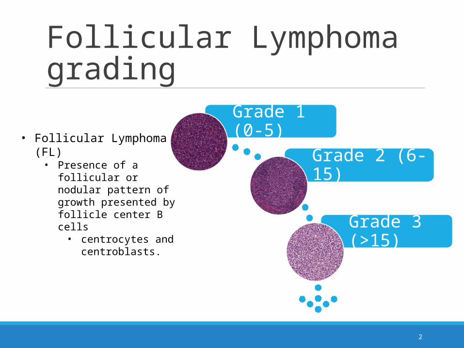
Follicular Lymphoma grading
Grade 1 (0-5)
Grade 2 (6-15)
Grade 3 (>15)
2
• Follicular Lymphoma (FL) • Presence of a follicular or
nodular pattern of growth presented by follicle center B cells
• centrocytes and centroblasts.

Follicular Lymphoma grading
3
Grade 3Grade 2Grade 1
Follicular Lymphoma grading
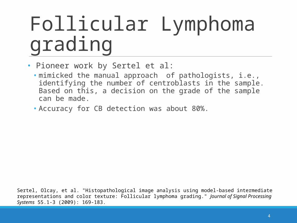
• Pioneer work by Sertel et al: • mimicked the manual approach of pathologists, i.e., identifying the number
of centroblasts in the sample. Based on this, a decision on the grade of the sample can be made.
• Accuracy for CB detection was about 80%.
4
Sertel, Olcay, et al. "Histopathological image analysis using model-based intermediate representations and color texture: Follicular lymphoma grading." Journal of Signal Processing Systems 55.1-3 (2009): 169-183.
Follicular Lymphoma grading
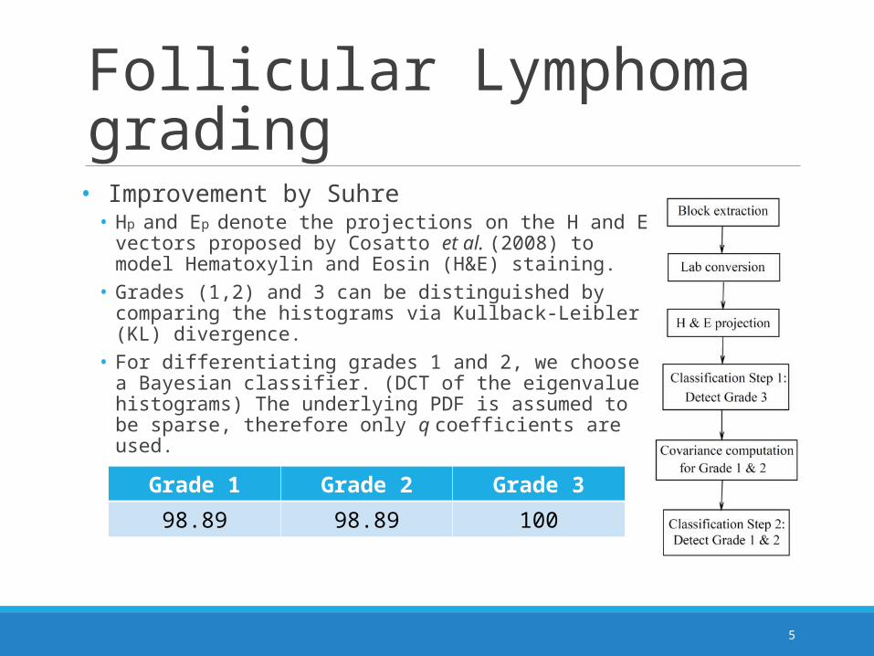
• Improvement by Suhre• Hp and Ep denote the projections on the H and E vectors proposed
by Cosatto et al. (2008) to model Hematoxylin and Eosin (H&E) staining.
• Grades (1,2) and 3 can be distinguished by comparing the histograms via Kullback-Leibler (KL) divergence.
• For differentiating grades 1 and 2, we choose a Bayesian classifier. (DCT of the eigenvalue histograms) The underlying PDF is assumed to be sparse, therefore only q coefficients are used.
5
Grade 1 Grade 2 Grade 3
98.89 98.89 100

Follicular Lymphoma grading
• Our Work• Approaches the problem as texture recognition program• Based on a novel multi-scale feature extraction method• LDA• SVM
6

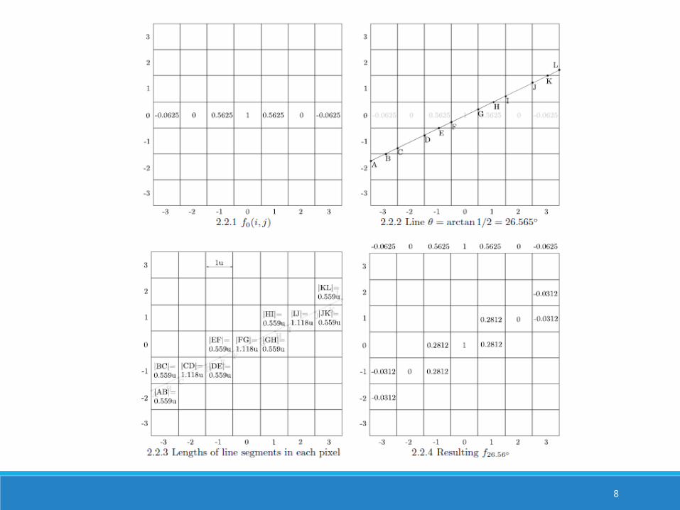
Directional filtering•Main idea: rotating a 1D filter along desired orientation
•Easy for θ=k x 45°, k=0,1,2,…
•Not easy for θ≠k x 45°• Bilinear/cubic interpolation• Our method: coefficients proportional to length of line segments enclosed
by pixels• Also used in CT
7
Herman, Gabor T. "Image reconstruction from projections." Image Reconstruction from Projections: Implementation and Applications 1 (1979).
8

Directional filtering
9

Directional Filtering
10
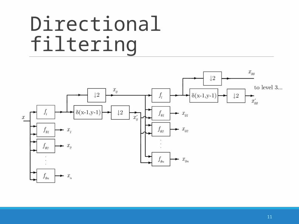
Directional filtering
11

12
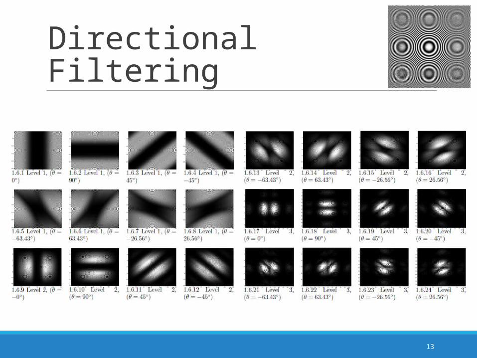
Directional Filtering
13

Feature extractionStep 0
• Input Image
14

Feature extractionStep 0
• Input Image
Step 1
• Convert Image to gray level
15

Feature extraction
: 0,082091 0,084891 0,060045 0,080689 0,085836 0,060873
: 0,14791 0,15201 0,11201 0,14617 0,15402 0,11424
: 0,22597 0,24064 0,11976 0,23731 0,24072 0,12753
: 0,36203 0,35692 0,17401 0,37765 0,34842 0,19024
: 0,49943 0,54883 0,35954 0,55623 0,56736 0,30949
: 0,6949 0,65361 0,46078 0,72141 0,68851 0,39779
Φ = [μ1 σ1 μ2 σ2 μ3 σ3]
μ1σ1
μ2σ2
σ3μ3
(1x36 feature vector)
Step 0• Input Image
Step 1
• Convert Image to gray level
Step 2
• Extract Features
50 100 150 200 250 300 350 400 450 500
50
100
150
200
250
300
50 100 150 200 250 300 350 400 450 500
50
100
150
200
250
300
50 100 150 200 250 300 350 400 450 500
50
100
150
200
250
300
50 100 150 200 250 300 350 400 450 500
50
100
150
200
250
300
50 100 150 200 250
20
40
60
80
100
120
140
160
50 100 150 200 250
20
40
60
80
100
120
140
160
50 100 150 200 250
20
40
60
80
100
120
140
160
50 100 150 200 250
20
40
60
80
100
120
140
160
50 100 150 200 250
20
40
60
80
100
120
140
160
50 100 150 200 250
20
40
60
80
100
120
140
160
16

Classification
[ θ1 ][ θ2 ]
.
.
.[ θN ]
10-fold CV
SVMTrain
Paramete
r search for C
and γ
SVMClassifyTraining
Test
Model
features
Mean Accuracy
PCA
LDA
17

Dataset Same dataset used by Suhre
90 images per grade
18
Grade 3Grade 2Grade 1

Background
19

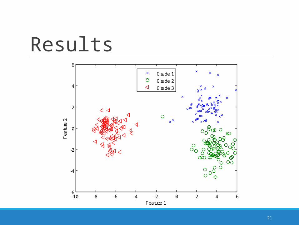
Results
Follicular Lymphoma•Max: 100.00 (Dir. Fil.)•SoA: 99.26
20
[20] A. Suhre, Novel Methods for Microscopic Image Processing, Analysis, Classification and Compression. PhD thesis, Bilkent University, 2013.
Results
21
-10 -8 -6 -4 -2 0 2 4 6-6
-4
-2
0
2
4
6
Feature 1
Fea
ture
2
Grade 1
Grade 2
Grade 3
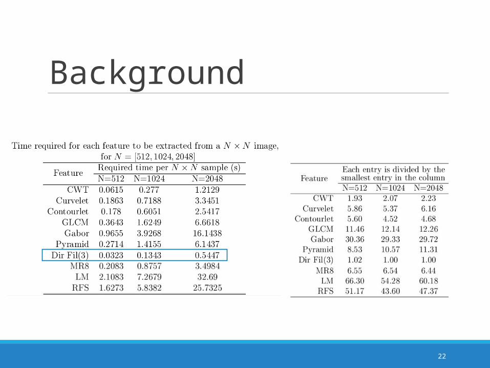
Background
22

Results
23
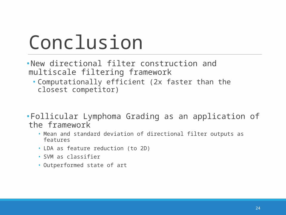
Conclusion•New directional filter construction and multiscale filtering framework
• Computationally efficient (2x faster than the closest competitor)
•Follicular Lymphoma Grading as an application of the framework• Mean and standard deviation of directional filter outputs as features• LDA as feature reduction (to 2D)• SVM as classifier• Outperformed state of art
24

Thank You!
25


