MRI History and HardwareBasic Safety IssuesIntroduction to fMRI
John VanMeter, Ph.D.Center for Functional and Molecular
Imaging
Terms Used for MRI
NMR (Nuclear Magnetic Resonance)
MR (Magnetic Resonance)
MRI (Magnetic Resonance Imaging)

Pauli, Stern and Gerlach - 1920’s Pauli postulated that atomic nuclei (e.g. H, C,
etc) have two properties: spin and magnetic moment
Further, the rate of spin occurs at a given frequency depending on the nuclei
Stern & Gerlach demonstrate this in pure gases Shot beam of gas through a static magnetic Produced multiple smaller beamlets
Rabi - 1937 Rabi showed that nuclei absorb energy if the
frequency matched the “resonant frequency” of the nuclei
Showed resonance frequency is dependent on static magnetic field strength
Measured resonance frequency of the lithium nucleus

Edward Purcell - 1945 Detected resonance frequency in bulk matter Used current passing through paraffin wax in a strong
magnetic field Changed strength of magnetic field over time At first did not see any change in current but
hypothesized it would take some time for relaxation of the spins to occur
Repeated experiment after leaving wax in magnetic field overnight and had success
Basis of Nuclear Magnetic Resonance Spectroscopy and MRI

Felix Bloch - 1945 Similar experiment to Purcell’s except using water in
a brass box inside a magnetic field Used a transmitter coil to send electromagnetic
energy into the box and receiver coil to measure changes in energy absorbed by the water
Was also able to measure magnetic resonance effect This basic setup is the basis of NMR spectrometers
used in biochemistry With some additional refinements it is also the basis
modern MRI scanners
Raymond Damadian - 1971
Discovery: Rat Tumor has a relaxation time longer than normal tissue
Differences in relaxation time provides one form of tissue contrast - T1

Paul Lauterbur - 1973
• Used GRADIENTS to distinguish spatially localized signals PHASE ENCODING
• Also, used GRADIENTS to manipulate the frequency of the spins to localize signals. He referred to this as Zeumatography FREQUENCY ENCODING
Both techniques needed to encode spatial location of signals

First MR Image - 1973 Lauterbur created image
by applying gradients at different angles to produce 1D projections
Combining projections forms image (back-projection reconstruction technique)
Inefficient as time needed for each angle equivalent to a single acquisition

Sir Peter Mansfield - 1974
Devised selective excitation of a slice again using gradients
Slice Select
Identifies where in a 3D object to collect signal from
Richard Ernst - 1975
Used 2D-FT Two-Dimensional Fourier
Transformation
Needed to reconstruct images, which are encoded with frequency and phase
Faster alternative to back-projection technique

Sir Peter Mansfield - 1976 Developed very efficient way to collect
data using technique called echo planar imaging (EPI)
Transmits 1 RF pulse per slice Rapidly switches gradients and records EPI used today in fMRI!
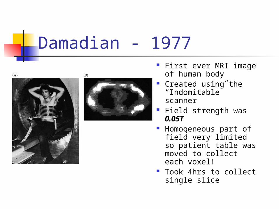
Damadian - 1977 First ever MRI image of
human body Created using the
“Indomitable” scanner Field strength was
0.05T Homogeneous part of
field very limited so patient table was moved to collect each voxel!
Took 4hrs to collect single slice

FDA Clears First MRI Scanner - 1985
Minicomputers such as the PDP-11 and VAX become widely available
GE develops first “high-field” (1.5T) commercial MRI scanner (1982)
Medicare starts paying for MRI scans (1985)
VAX 11/750 (1982)

1990’s
FUNCTIONAL IMAGING

5 Nobel Laureates for MRI
Bloch, Purcell (1952)
Lauterbur, Mansfield (2003)
Rabi (1944)
Nobel Controversy - 2003 Damadian took out full page ads in NY Times and
Washington Post protesting award to Lauterbur and Mansfield
“This Year’s Nobel Prize in Medicine. The Shameful Wrong That Must Be Righted”
“The Nobel Prize Committee for Physiology or Medicine chose to award the prize, not to the medical doctor/research scientist who made the breakthrough discovery on which all MRI technology is based, but to two scientists who later made technological improvements based on his discovery”
"I know that had I never been born, there would be no MRI today"
MRI Hardware

Basic MRI Hardware Magnet
Large magnetic field that is homogeneous over a large area Aligns protons in the body
Radiofrequency (RF) coils Transmit and Receive RF energy into and from the body
Gradients Induce linear change in magnetic field Spatial encoding
Computer System and Console Patient Handling System

Types of Magnets Permanent Iron Core
Low Field “Open” Resistive Electromagnet
Up to 0.2T Superconducting Magnet
Cools wire coil with cryogens 0.5T to 35T

Electromagnets Field proportional to
number of loops relative to cross-section area of each loop
Increases in current also increases field strength
Field highest and most homogenous at center of coil

Properties of Superconducting Magnets Very high field strengths generated
Cool magnet’s wire coil using cryogens (liquid helium and possibly nitrogen) to near absolute zero
Reduces resistance to zero for certain metals Provides stable and homogeneous magnetic field
over a relatively large area Once ramped up no electricity used (relatively cheap) MAGNET ALWAYS ON! New dangers specific to these types of magnets

RF (Radiofrequency) Coils Used to transmit and receive RF energy Needed to create images

Coil Designs Closer coil is to object being imaged the
better signal Variety of coils designed for specific body
parts
Surface Coil Volume Coil(aka Birdcage Coil)

Coil Design Affects Images

Gradient Coils Induce small linear changes in magnetic field
along one or more dimensions Produces two types of spatial encoding
referred to as Frequency and Phase Encoding
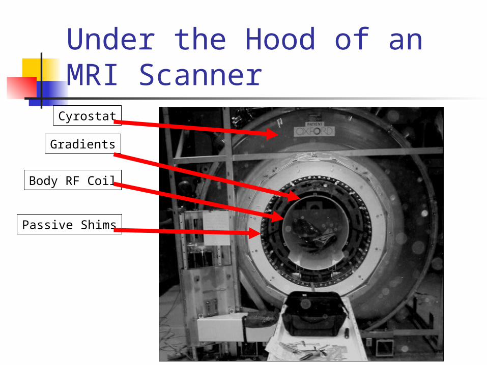
Under the Hood of an MRI Scanner
Gradients
Body RF Coil
Passive Shims
Cyrostat
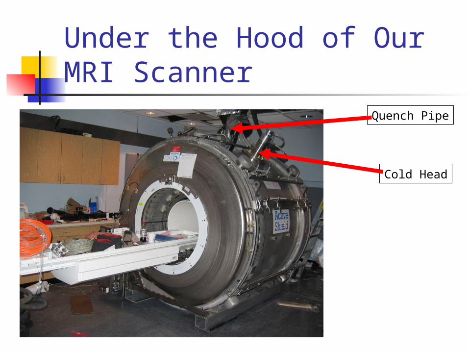
Under the Hood of Our MRI Scanner
Quench Pipe
Cold Head

Computer System and Console
Image reconstruction and post processing is computationally intensive
Standard workstation sufficient for basic clinical MRI system
Multi-processor systems with gigabytes of memory needed for functional MRI and DTI (Diffusion Tensor Imaging) scanning
Console computer coordinates everything
Patient Handling System Methods to get patient in and out of the
scanner Alignment of the body part to be
scanned with isocenter of the scanner Labeling of scans with appropriate
identifiers and anatomic labels

MRI Safety
MRI Safety
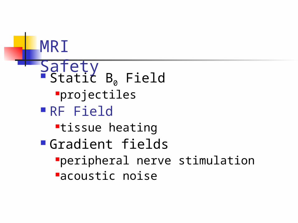
Static B0 FieldProjectilesImplants/other materials in the body
RF Fieldtissue heating
Gradient fieldsperipheral nerve stimulationacoustic noise

Forces on Ferrous Objects
Crash cart meets a 1.5T magnet
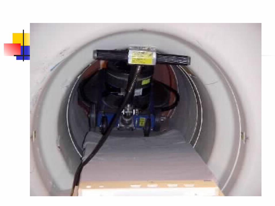


Welding tank
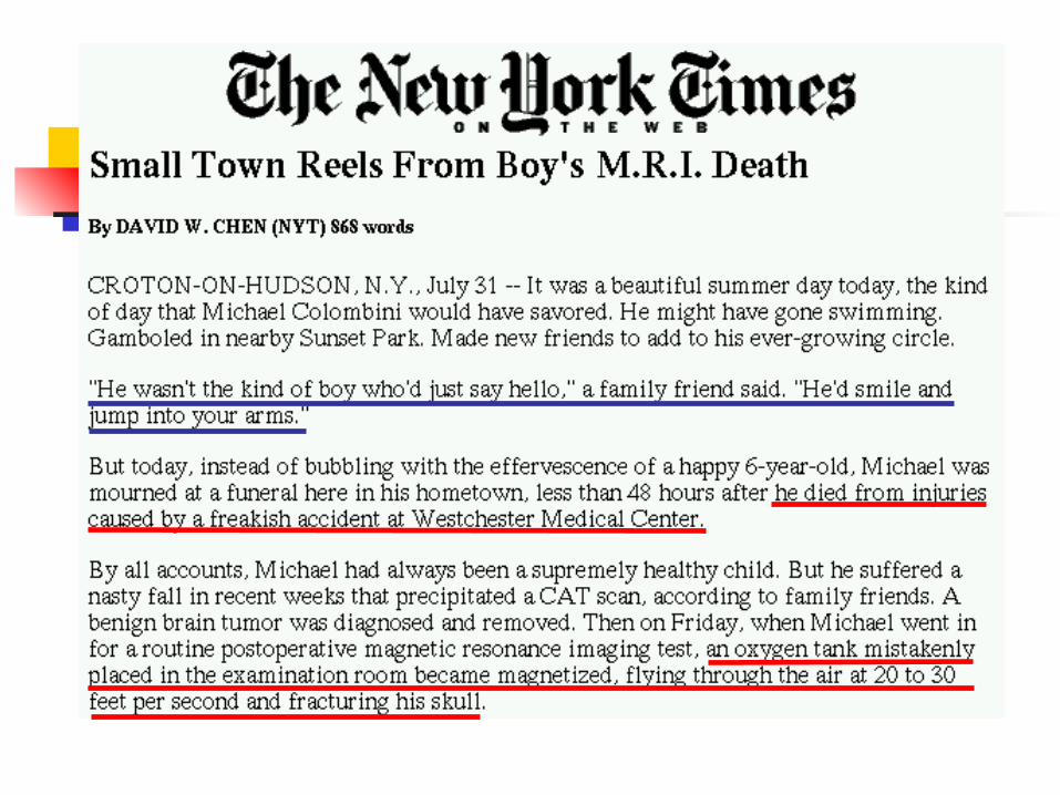

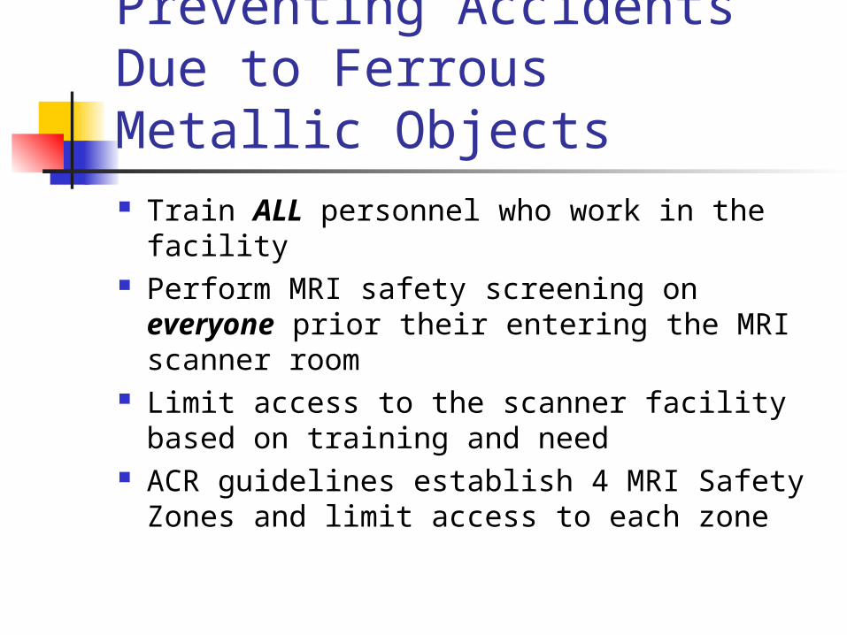
Preventing Accidents Due to Ferrous Metallic Objects Train ALL personnel who work in the facility Perform MRI safety screening on everyone
prior their entering the MRI scanner room Limit access to the scanner facility based on
training and need ACR guidelines establish 4 MRI Safety Zones
and limit access to each zone
MRI Safety Static B0 Field
projectiles RF Field
tissue heating Gradient fields
peripheral nerve stimulationacoustic noise

RF Exposure Standards
The FDA limits RF exposure to less than a 1 degree C rise in core body temperature
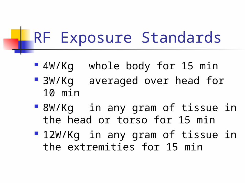
RF Exposure Standards
4W/Kg whole body for 15 min 3W/Kg averaged over head for 10
min 8W/Kg in any gram of tissue in
the head or torso for 15 min 12W/Kg in any gram of tissue in
the extremities for 15 min

MRI Safety Static B0 Field
projectiles RF Field
tissue heating Gradient fields
peripheral nerve stimulationacoustic noise

Stimulation Caused by the SwitchingGradient Fields Nerve stimulation Acoustic trauma Burn from looped cables
be careful when using anything with electrical wires or cables in the scanner
Changing B field Creates voltage,current and heat
V ~ (Area) x (dB/dt)

Introduction to Functional MRI

Difference BetweenMRI & fMRI
From: Daniel Bulte
Centre for Functional MRI of the Brain
University of Oxford
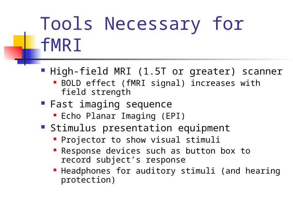
Tools Necessary for fMRI High-field MRI (1.5T or greater) scanner
BOLD effect (fMRI signal) increases with field strength
Fast imaging sequence Echo Planar Imaging (EPI)
Stimulus presentation equipment Projector to show visual stimuli Response devices such as button box to record
subject’s response Headphones for auditory stimuli (and hearing
protection)


Functional Brain Mapping with MRI
Basic concept - changes in neuronal activity produces a measurable change in MR signal
Collect 100-500 MRI scans continuously (1 every 2-3s each typically cover 30-50 slices)
Experimenter induces changes in activity at known points in time by having subject perform some cognitive or motoric task
Analyses statistically tests for MR signal changes that corresponding to experimental task

Fixation
time
Basic fMRI Experiment
Thumb movement

Data Analysis
430435440445450455460465470475480
0 20 40 60 80 100
Time
AU
Identify voxels with signal changes matched to the timing of experiment Tapping Tapping
Tapping
Rest Rest Rest

Unimanual Thumb Flexion
L R
Right Thumb Left Thumb

fMRI Compared to Other Functional Techniques

Examples of fMRI

Activity in a Vegetative State

Super Bowl Ads Marco Iacoboni at UCLA used fMRI to
examine the brain’s response to different super bowl ads
Ranked ads based on brain responses Found differences in the ads that
stimulated the brain most and those people reported as liking the most
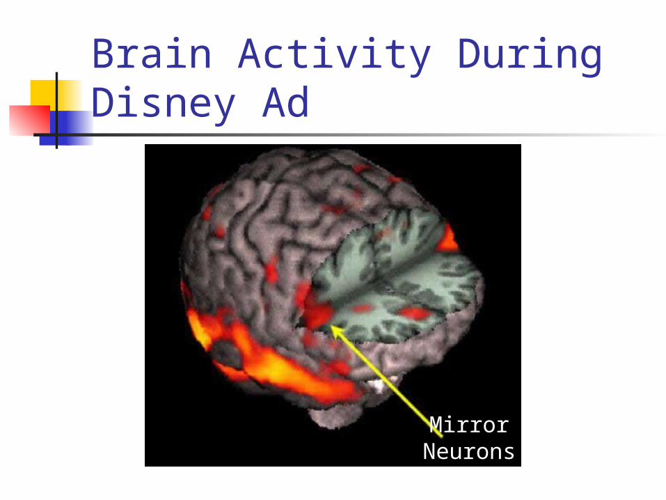
Brain Activity During Disney Ad
Mirror Neurons
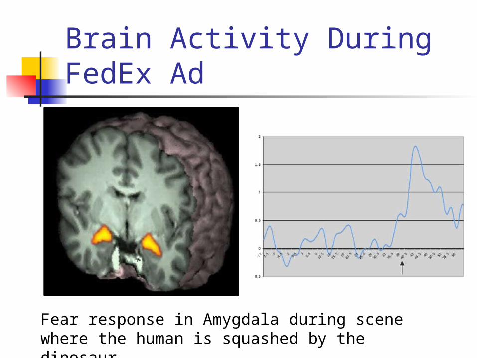
Brain Activity During FedEx Ad
Fear response in Amygdala during scene where the human is squashed by the dinosaur

Caution Needed Interpretation of the
signal changes depends on a lot of factors
Communication of results with public needs to be approached with care
McCabe & Castel (2008, Cognition) brain imaging increased perceived credibility of research compared to bar graphs