www.elsevier.com/locate/meegid
Available online at www.sciencedirect.com
Infection, Genetics and Evolution 8 (2008) 433–438
Molecular characterization of Thai Ehrlichia canis and
Anaplasma platys strains detected in dogs§
Danai Pinyoowong a, Sathaporn Jittapalapong b, Fanan Suksawat c,Roger W. Stich d, Arinthip Thamchaipenet a,*
a Department of Genetics, Faculty of Science, Kasetsart University, Thailandb Department of Parasitology, Faculty of Veterinary Medicine, Kasetsart University, Thailandc Department of Medicine, Faculty of Veterinary Medicine, Khon Kaen University, Thailand
d Department of Veterinary Pathobiology, University of Missouri, Columbia, MO, USA
Received 31 March 2007; accepted 12 June 2007
Available online 19 June 2007
Abstract
Canine monocytic ehrlichiosis caused by Ehrlichia canis is of veterinary importance worldwide. In Thailand, there has been little information
available on E. canis and its phylogeny. The objective of this study was to characterize and establish molecular structure and phylogeny of Thai
Ehrlichia and Anaplasma strains. Genus-specific primers for Ehrlichia and Anaplasma were used to amplify the 16S rRNA gene from naturally
infected canine blood samples, and these amplicon sequences were compared with other sequences from GenBank. Both homology and secondary
structure analysis of 16S rRNA sequences indicated that they were novel E. canis and A. platys strains. Phylogenetic analysis revealed that the Thai
E. canis strain was closely related and formed a single cluster with E. canis from different countries. A. platys found in this study showed close
relationship with earlier report of A. platys from Thailand. To our knowledge this report represents the first molecular characterization of the nearly
complete 16S rRNA gene from E. canis in dogs from Thailand.
# 2007 Elsevier B.V. All rights reserved.
Keywords: Ehrlichia canis; Anaplasma platys; 16S rDNA; Phylogenetic tree; Dogs; Bangkok; Thailand
1. Introduction
Canine monocytic ehrlichiosis (CME) and canine cyclic
thrombocytopenia (CCT) are caused by Ehrlichia canis and
Anaplasma platys, respectively. These bacteria are classified in
the rickettsial family Anaplasmataceae, which includes obligate
intracellular prokaryotic parasites that reside within a para-
sitophorous vacuole (Dumler et al., 2001). In canine hosts, E.
canis is infective for monocytes while A. platys infect platelets
(Greene and Harvey, 1990). Rhipicephalus sanguineus ticks
are considered the primary vector of both pathogens (Groves
et al., 1975; Greene and Harvey, 1990). Globally distributed,
well-characterized pathogens such as E. canis offer unique
opportunities to study coevolution and interaction between
§ This study was presented in part as an abstract at the MEEGID VIII
Congress, Bangkok, Thailand, 30 November to 2 December 2006.
* Corresponding author. Tel.: +66 2 562 5444x4208; fax: +66 2 579 5528.
E-mail address: [email protected] (A. Thamchaipenet).
1567-1348/$ – see front matter # 2007 Elsevier B.V. All rights reserved.
doi:10.1016/j.meegid.2007.06.002
tick-borne pathogens and their vertebrate and invertebrate hosts
(Stich et al., in press).
Although CME and CCTare considered enzootic throughout
Thailand, these conclusions are based on diagnoses that rely on
clinical signs, haematological abnormalities and microscopic
examination of peripheral blood. Thus, these diagnoses are
often ambiguous and may fail to identify the pathogen species
involved. Molecular diagnostic methods allow direct detection
of these etiologic agents and sequence analysis facilitates their
comparison to geographically diverse strains. To our knowl-
edge, detailed genetic and phylogenetic information about E.
canis and A. platys in Thailand are limited to a single report of
an A. platys 16S rRNA gene (16S rDNA) sequence (Suksawat
et al., 2001).
In this study, we investigated diagnosed cases of canine
ehrlichiosis from a private laboratory in Bangkok, to confirm
the presence of E. canis and A. platys and to compare Thai
strains to those from other regions. PCR and 16S rDNA
sequence analysis were used to characterize the molecular
D. Pinyoowong et al. / Infection, Genetics and Evolution 8 (2008) 433–438434
features of these infections, which allowed identification of a
novel strain of A. platys that differed from that of the previous
report of Suksawat et al. (2001). Moreover, E. canis from this
country was genetically verified for the first time.
2. Materials and methods
2.1. DNA extraction from canine blood
Blood samples were collected with EDTA from dogs
diagnosed with clinical ehrlichiosis and submitted to a private
laboratory in Bangkok. The blood was chilled until arrival at the
laboratory and then stored at �20 8C. Total DNA was isolated
from 100 ml of thawed blood using the phenol/chloroform
extraction method of Sambrook and Russell (2001).
2.2. Primers designed for 16S rDNA amplification
Primers for amplification of Ehrlichia and Anaplasma
16S rDNA were designed from nucleotide sequences deposited
in GenBank database (DQ342324, AF414873, AF414870,
AF414869, AB211163, U23503, CR767821, U96436,
AB196302 and AF318946). All the sequences were aligned
for the maximum homology by ClustalW Version 8.1 (Thompson
et al., 1994). Conserved regions were selected and specific
oligonucleotide primers named ATT062F (50CCTGGCTCA–
GAACGAACGCT30) and ATT062R (50GATCCAGCCGCA-
GGTTCACC30) were derived.
2.3. 16S rDNA amplification and sequencing
DNA isolated from dog blood was used as a template to
amplify the majority of the 16S rRNA gene from Ehrlichia and
Anaplasma spp. by PCR. Amplifications were generated using
3–6 ml of genomic DNA with primers ATT062F and ATT062R
for each 20 ml PCR reaction mixture in a Peltier thermal cycler
(MJ Research, Watertown, MA, USA), by 30 cycles of 30 s at
94.0 8C, 30 s at 64.0 8C, and 1 min at 72.0 8C, preceded by
4 min at 94.0 8C and followed by 4 min at 72.0 8C. PCR
products were examined by 0.8% agarose gel electrophoresis
and ethidium bromide staining. Amplicons were purified with
QIAquick PCR purification kits (QIAGEN) according to the
manufacturer’s protocol. Direct sequencing was carried on
the PCR product using the same PCR primers, ATT062F
and ATT062R. In order to cover both strands of the PCR
fragment completely, another set of primers, ATT066F
(50CCCTGGTAGTCCACGCTG30) and ATT067R (50CAGC-
GTGGACTACCAGGG30), were designed for annealing in the
middle of the 16S rDNA amplicons and used for sequencing
towards 30 and 50 ends, respectively.
2.4. Phylogenetic analysis
Multiple sequence alignments of amplicons and 16S rDNA
sequences from GenBank were performed using the ClustalW
Version 1.8 (Thompson et al., 1994). Phylogenetic trees
were inferred using neighbor-joining (NJ) analysis by MEGA
software Version 3.1 (Kumar et al., 2004). The distance matrix of
nucleotide divergences was calculated according to Kimura’s
two-parameter model furnished by MEGA. A bootstrap re-
sampling technique of 1000 replications was performed to
statistically support the reliabilities of the nodes on the trees. The
differences between nucleotide positions were confirmed by
DnaSP Version 4.10 (Rozas et al., 2003). Neorickettsia sennetsu
was used as an outgroup.
2.5. Nucleotide sequences accession numbers
The 16S rDNA sequences from this study were deposited at
GenBank database under accession numbers EF139458 for E.
canis-Bangkok and EF139459 for A. platys-Bangkok. Other
Anaplasmataceae 16S rDNA sequences from Genbank
(and their accession numbers) included 10 E. canis strains
reported from China (AF162860), Japan (AF536827), Peru
(DQ915970), South Africa (U54805), Spain (AY394465),
Venezuela (AF373613, AF373612) and the USA (M73226,
M73221), eight A. platys strains from China (AF156784),
France (AF303467), Japan (AY077619, AF536828), Spain
(AY530806), Thailand (AF286699), Venezuela (AF287153)
and the USA (M82801), E. chaffeensis (U23503), E. ewingii
(U96436), E. muris (NC_007354, AB196302), E. ruminantium
(CR767821), A. bovis (AB211163), A. centrale (AF414869), A.
marginale (AF414873), A. ovis (AF414870), A. phagocyto-
philum (DQ342324) and N. sennetsu (M73219).
3. Results
3.1. Amplification and sequencing of 16S rDNA
The identity of agents associated with diagnosed CME and
CCT are often assumed based on clinical diagnosis and
microscopic examination of peripheral blood. Blood samples
from dogs diagnosed with clinical ehrlichiosis were screened to
compare these agents to previously reported strains. Template
was prepared and assayed by PCR with primers ATT062F and
ATT062R, which were designed for specific amplification of
Ehrlichia and Anaplasma 16S rDNA. Approximately 1.5 kb
amplicons corresponding to the expected size of targeted 16S
rRNA gene fragments were obtained (data not shown).
Amplicons from two individual canine blood samples were
purified and directly sequenced with the same primers. The
nearly complete 16S rRNA gene sequences of 1478 (51% AT)
and 1481 (52% AT) bp were identical to consensus 16S rDNA
sequences for E. canis and A. platys, respectively. These
sequences were named E. canis-Bangkok and A. platys-
Bangkok and deposited as new 16S rDNA sequences in
GenBank.
3.2. Molecular characterization of E. canis from Thai dogs
E. canis and A. platys are distributed globally, and it is
generally assumed that all strains primarily utilize dogs and
rhipicephaline ticks as vertebrate and invertebrate hosts.
However, closely related anaplasmal pathogens, including
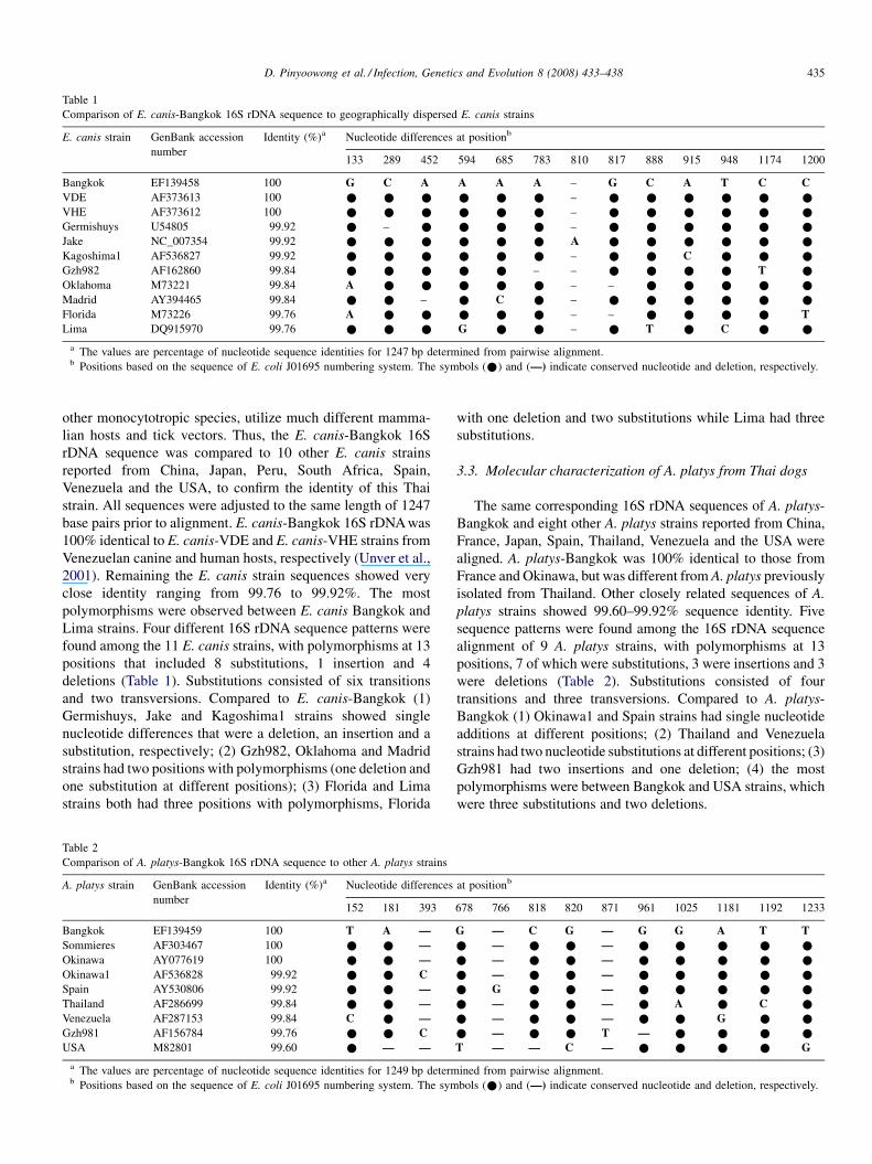
Table 1
Comparison of E. canis-Bangkok 16S rDNA sequence to geographically dispersed E. canis strains
E. canis strain GenBank accession
number
Identity (%)a Nucleotide differences at positionb
133 289 452 594 685 783 810 817 888 915 948 1174 1200
Bangkok EF139458 100 G C A A A A – G C A T C C
VDE AF373613 100 * * * * * * – * * * * * *
VHE AF373612 100 * * * * * * – * * * * * *
Germishuys U54805 99.92 * – * * * * – * * * * * *
Jake NC_007354 99.92 * * * * * * A * * * * * *
Kagoshima1 AF536827 99.92 * * * * * * – * * C * * *
Gzh982 AF162860 99.84 * * * * * – – * * * * T *
Oklahoma M73221 99.84 A * * * * * – – * * * * *
Madrid AY394465 99.84 * * – * C * – * * * * * *
Florida M73226 99.76 A * * * * * – – * * * * T
Lima DQ915970 99.76 * * * G * * – * T * C * *
a The values are percentage of nucleotide sequence identities for 1247 bp determined from pairwise alignment.b Positions based on the sequence of E. coli J01695 numbering system. The symbols (*) and (—) indicate conserved nucleotide and deletion, respectively.
D. Pinyoowong et al. / Infection, Genetics and Evolution 8 (2008) 433–438 435
other monocytotropic species, utilize much different mamma-
lian hosts and tick vectors. Thus, the E. canis-Bangkok 16S
rDNA sequence was compared to 10 other E. canis strains
reported from China, Japan, Peru, South Africa, Spain,
Venezuela and the USA, to confirm the identity of this Thai
strain. All sequences were adjusted to the same length of 1247
base pairs prior to alignment. E. canis-Bangkok 16S rDNA was
100% identical to E. canis-VDE and E. canis-VHE strains from
Venezuelan canine and human hosts, respectively (Unver et al.,
2001). Remaining the E. canis strain sequences showed very
close identity ranging from 99.76 to 99.92%. The most
polymorphisms were observed between E. canis Bangkok and
Lima strains. Four different 16S rDNA sequence patterns were
found among the 11 E. canis strains, with polymorphisms at 13
positions that included 8 substitutions, 1 insertion and 4
deletions (Table 1). Substitutions consisted of six transitions
and two transversions. Compared to E. canis-Bangkok (1)
Germishuys, Jake and Kagoshima1 strains showed single
nucleotide differences that were a deletion, an insertion and a
substitution, respectively; (2) Gzh982, Oklahoma and Madrid
strains had two positions with polymorphisms (one deletion and
one substitution at different positions); (3) Florida and Lima
strains both had three positions with polymorphisms, Florida
Table 2
Comparison of A. platys-Bangkok 16S rDNA sequence to other A. platys strains
A. platys strain GenBank accession
number
Identity (%)a Nucleotide differences
152 181 393
Bangkok EF139459 100 T A —
Sommieres AF303467 100 * * —
Okinawa AY077619 100 * * —
Okinawa1 AF536828 99.92 * * C
Spain AY530806 99.92 * * —
Thailand AF286699 99.84 * * —
Venezuela AF287153 99.84 C * —
Gzh981 AF156784 99.76 * * C
USA M82801 99.60 * — —
a The values are percentage of nucleotide sequence identities for 1249 bp determb Positions based on the sequence of E. coli J01695 numbering system. The sym
with one deletion and two substitutions while Lima had three
substitutions.
3.3. Molecular characterization of A. platys from Thai dogs
The same corresponding 16S rDNA sequences of A. platys-
Bangkok and eight other A. platys strains reported from China,
France, Japan, Spain, Thailand, Venezuela and the USA were
aligned. A. platys-Bangkok was 100% identical to those from
France and Okinawa, but was different from A. platys previously
isolated from Thailand. Other closely related sequences of A.
platys strains showed 99.60–99.92% sequence identity. Five
sequence patterns were found among the 16S rDNA sequence
alignment of 9 A. platys strains, with polymorphisms at 13
positions, 7 of which were substitutions, 3 were insertions and 3
were deletions (Table 2). Substitutions consisted of four
transitions and three transversions. Compared to A. platys-
Bangkok (1) Okinawa1 and Spain strains had single nucleotide
additions at different positions; (2) Thailand and Venezuela
strains had two nucleotide substitutions at different positions; (3)
Gzh981 had two insertions and one deletion; (4) the most
polymorphisms were between Bangkok and USA strains, which
were three substitutions and two deletions.
at positionb
678 766 818 820 871 961 1025 1181 1192 1233
G — C G — G G A T T
* — * * — * * * * *
* — * * — * * * * *
* — * * — * * * * *
* G * * — * * * * *
* — * * — * A * C *
* — * * — * * G * *
* — * * T — * * * *
T — — C — * * * * G
ined from pairwise alignment.
bols (*) and (—) indicate conserved nucleotide and deletion, respectively.
D. Pinyoowong et al. / Infection, Genetics and Evolution 8 (2008) 433–438436
3.4. 16S rRNA secondary structures
16S rRNA is not subject to the same selective influences on
function as mRNA (i.e., 16S rRNA function relies on structure
rather than codon usage), thus the effects of nucleotide changes
on predicted secondary structures could be more informative
than primary sequence variation alone. The positions of 16S
rDNA sequences defined in Tables 1 and 2 were correlated with
the E. coli J01695 numbering system (Konings and Gutell, 1995).
Comparison of E. canis-Bangkok and A. platys-Bangkok 16S
rRNA predicted secondary structures to that of the E. coli J01695
indicated that both had conserved tetra loops that generally
constrained the rRNA architecture (Woese et al., 1990). Nucleo-
tides at positions 289, 452, 594, 888, 915, 948 and 1200 of E.
canis-Bangkok were common among bacteria while positions
133, 685, 783, 810, 817 and 1174 were different from other
eubacteria. At position 948, we observed T (U) in most samples
except E. canis-Lima that carried C, the latter of which is similar
to those of alphaproteobacteria (Woese, 1987). In A. platys-
Bangkok, nucleotide differences at positions 181, 678, 871 and
1025 were within the common structure of eubacteria. At posi-
tion 393, eubacteria generally carried A (Woese, 1987), but most
of A. platys had a single deletion except for A. platys-Okinawa1
and A. platys-Gzh981 that carried a C. At position 1233, T was
observed in most samples except for A. platys USA, which had a
G that is similar to those of alphaproteobacteria (Woese, 1987).
3.5. Phylogenetic analysis of Ehrlichia and Anaplasma
Ehrlichia and Anaplasma 16S rDNA sequences were used to
generate a phylogenetic tree using the neighbor-joining method
by MEGA software (Version 3.1). In addition to E. canis and A.
platys strains, closely related species included the tick-borne
anaplasmal parasites E. chaffeensis, E. ewingii, E. muris, E.
ruminantium, A. bovis, A. centrale, A. marginale, A. ovis and A.
phagocytophilum. A biologically divergent member of the
Anaplasmataceae, N. sennetsu, was used as the outgroup. The
resultant phylogenetic tree revealed that E. canis-Bangkok and
A. platys-Bangkok were grouped tightly within the other E.
canis and A. platys strains, respectively (Fig. 1). This analysis
revealed that (1) Ehrlichia and Anaplasma were divided into
clearly defined clades; (2) E. canis strains from different
geographic regions were always grouped in a clade independent
from E. chaffeensis, E. ewingii, E. muris and E. ruminantium;
(3) E. ewingii showed the closet relationship to E. canis while
E. ruminantium was the most distant; (4) A. platys strains from
different countries constantly grouped in a clade independent
from A. bovis, A. centrale, A. marginale, A. ovis and A.
phagocytophilum; (5) A. phagocytophilum had the closet
relationship to A. platys while A. centrale, A. marginale and A.
ovis were the most distant; (6) A. marginale clustered in a
branch linked to A. centrale and A. ovis.
4. Discussion
Understanding the epidemiology of these diseases and
infectious cycles of anaplasmal organisms is expected to
facilitate understanding of similar diseases, including zoonoses
that may share similar etiologic agents or vectors. These results
support previous studies that indicated E. canis and A. platys
are uniform worldwide, suggesting that biological observations
with different strains and from different regions could be
laterally applicable. However, it is important to note that these
observations are based on the highly conserved 16S rRNA gene
and that further work with more divergent sequences is needed
to confirm whether these strains are indeed uniform. Moreover,
it is important to remember that potential heterogeneity of
vertebrate and invertebrate hosts from different locations is
likely to influence the biology of these organisms. Thus, global
analyses of evolutionary patterns among E. canis and A. platys
will likely require parallel analyses of such patterns among
their reservoirs and vectors. For example, R. sanguineus, which
is considered an important vector of both pathogens described
in this report, is considered highly host-specific in some regions
but reportedly feeds on diverse medium to large mammalian
hosts (including humans) in other areas. Susceptibility of these
tick strains to various pathogens, including E. canis and A.
platys, could explain reports suggesting E. canis infections of
ruminants and humans (Stich et al., in press; Yu et al., 2007).
Alternatively, similarities among geographically diverse E.
canis strains could be reflective of relatively recent global
spread of this pathogen with its vertebrate and invertebrate
hosts (Yu et al., 2007).
This report describes molecular analysis of nearly complete
16S rDNA sequences of E. canis and A. platys from Bangkok,
Thailand. The sequence alignments and phylogenetic tree
suggested low diversity within E. canis and A. platys strains
based on the close similarity amongst their 16S rDNA
sequences from all geographic regions tested, and these
conclusions are consistent with other reports (de la Fuente et al.,
2006; Aguirre et al., 2006; Yu et al., 2007). Genetic
polymorphisms from comparison of 16S rRNA sequences
based on the E. coli J01695 numbering system (Konings and
Gutell, 1995) also indicated that E. canis-Bangkok and A.
platys-Bangkok were structurally conserved in 16S rRNA
architecture. Therefore, we are confident that all polymorph-
isms observed in these experiments are consistent with these
two species. However, although nucleotide differences at many
positions indicated that E. canis-Bangkok and A. platys-
Bangkok shared some structure with other bacteria, other
nucleotides were different from most eubacteria.
Close similarity among 16S rDNA of A. platys from
different locations worldwide supported the hypothesis that A.
platys strains are not geographically segregated (Huang et al.,
2005). Interestingly, the sequence of A. platys-Bangkok
reported from this study had two nucleotide substitutions
compared to a previously characterized A. platys strain from
Thailand (Suksawat et al., 2001) at positions 1025 and 1192
(Table 2) that appeared through G/A and T/C transitions,
respectively, suggesting that there might be at least two A.
platys strains enzootic to Thailand.
The phylogenetic tree separated two major clusters of
Ehrlichia spp. and Anaplasma spp. As expected, E. canis-
Bangkok was within the Ehrlichia clade and A. platys-Bangkok
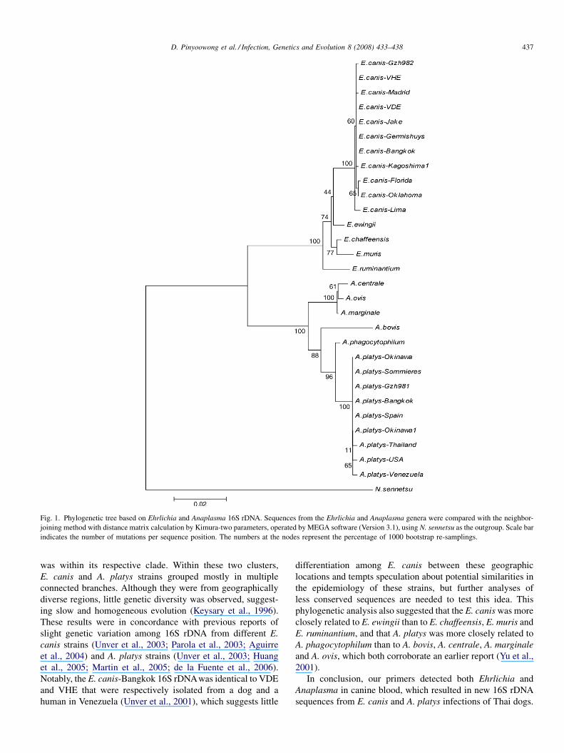
Fig. 1. Phylogenetic tree based on Ehrlichia and Anaplasma 16S rDNA. Sequences from the Ehrlichia and Anaplasma genera were compared with the neighbor-
joining method with distance matrix calculation by Kimura-two parameters, operated by MEGA software (Version 3.1), using N. sennetsu as the outgroup. Scale bar
indicates the number of mutations per sequence position. The numbers at the nodes represent the percentage of 1000 bootstrap re-samplings.
D. Pinyoowong et al. / Infection, Genetics and Evolution 8 (2008) 433–438 437
was within its respective clade. Within these two clusters,
E. canis and A. platys strains grouped mostly in multiple
connected branches. Although they were from geographically
diverse regions, little genetic diversity was observed, suggest-
ing slow and homogeneous evolution (Keysary et al., 1996).
These results were in concordance with previous reports of
slight genetic variation among 16S rDNA from different E.
canis strains (Unver et al., 2003; Parola et al., 2003; Aguirre
et al., 2004) and A. platys strains (Unver et al., 2003; Huang
et al., 2005; Martin et al., 2005; de la Fuente et al., 2006).
Notably, the E. canis-Bangkok 16S rDNAwas identical to VDE
and VHE that were respectively isolated from a dog and a
human in Venezuela (Unver et al., 2001), which suggests little
differentiation among E. canis between these geographic
locations and tempts speculation about potential similarities in
the epidemiology of these strains, but further analyses of
less conserved sequences are needed to test this idea. This
phylogenetic analysis also suggested that the E. canis was more
closely related to E. ewingii than to E. chaffeensis, E. muris and
E. ruminantium, and that A. platys was more closely related to
A. phagocytophilum than to A. bovis, A. centrale, A. marginale
and A. ovis, which both corroborate an earlier report (Yu et al.,
2001).
In conclusion, our primers detected both Ehrlichia and
Anaplasma in canine blood, which resulted in new 16S rDNA
sequences from E. canis and A. platys infections of Thai dogs.
D. Pinyoowong et al. / Infection, Genetics and Evolution 8 (2008) 433–438438
Although the 16S rDNA sequences were highly conserved
among geographically diverse strains of these organisms,
additional analyses of genes more subject to selective pressure
from host environments (e.g., outer membrane protein gene
families) could help elucidate the diversity and evolution of
strains from different geographic areas. Current efforts include
examination of additional canine blood samples to determine
the presence and genetic diversity of Ehrlichia and Anaplasma
spp. found in Thailand.
Acknowledgements
We give special thanks to Veterinary Diagnostic Laboratory
(VDL) center, Bangkok for dog blood samples. This research
was supported by the Graduate School, Kasetsart University.
References
Aguirre, E., Sainz, A., Dunner, S., Amusategui, I., Lopez, L., Rodriguez-
Franco, F., Luaces, I., Cortes, O., Tesouro, M.A., 2004. First isolation and
molecular characterization of Ehrlichia canis in Spain. Vet. Parasitol. 125,
365–372.
Aguirre, E., Tesouro, M.A., Ruiz, L., Amusategui, I., Sainz, A., 2006. Genetic
characterization of Anaplasma (Ehrlichia) platys in dogs in Spain. J. Vet.
Med. B Infect. Dis. Vet. Public Health 53, 197–200.
de la Fuente, J., Torina, A., Naranjo, V., Nicosia, S., Alongi, A., Mantia, F.L.,
Kocan, K.M., 2006. Molecular characterization of Anaplasma platys strains
from dogs in Sicily. Italy Vet. Res. 2, 24–29.
Dumler, J.S., Barbet, A.F., Bekker, C.P., Dasch, G.A., Palmer, G.H., Ray, S.C.,
Rikihisa, Y., Rurangirwa, F.R., 2001. Reorganization of genera in the
families Rickettsiaceae and Anaplasmataceae in the order Rickettsiales:
unification of some species of Ehrlichia with Anaplasma, Cowdria with
Ehrlichia and Ehrlichia with Neorickettsia, descriptions of six new species
combinations and designation of Ehrlichia equi and ‘HGE agent’ as
subjective synonyms of Ehrlichia phagocytophila. Int. J. Syst. Evol.
Microbiol. 51, 2145–2165.
Greene, C.E., Harvey, J.W., 1990. Canine ehrlichiosis. In: Greene, C.E. (Ed.),
Clinical Microbiology and Infectious Diseases of the Dog and Cat. W.B.
Saunders, Philadelphia, pp. 137–148.
Groves, M.G., Dennis, G.L., Amyx, H.L., Huxsoll, D.L., 1975. Transmission of
Ehrlichia canis to dogs by tick (Rhipicephalus sanguineus). Am. J. Vet. Res.
36, 937–940.
Huang, H., Unver, A., Perez, M.J., Orellana, N.G., Rikihisa, Y., 2005. Pre-
valence and molecular analysis of Anaplasma platys in dogs in Lara,
Venezuela. Braz. J. Microbiol. 36, 211–216.
Keysary, A., Waner, T., Rosner, M., Warner, C.K., Dawson, J.E., Zass, R.,
Biggie, K.L., Harrus, S., 1996. The first isolation, in vitro propagation, and
genetic characterization of Ehrlichia canis in Israel. Vet. Parasitol. 62, 331–
340.
Konings, D.A.M., Gutell, R.R., 1995. A comparison of thermodynamic foldings
with comparatively derived structures of 16S and 16S-like rRNAs. RNA 1,
559–574.
Kumar, S., Tamura, K., Nei, M., 2004. MEGA3: integrated software for
molecular evolutionary genetics analysis and sequence alignment. Brief.
Bioinform. 5, 150–163.
Martin, A.R., Brown, G.K., Dunstan, R.H., Roberts, T.K., 2005. Anaplasma
platys: an improved PCR for its detection in dogs. Exp. Parasitol. 109, 176–
180.
Parola, P., Cornet, J.P., Sanogo, Y.O., Miller, R.S., Thien, H.V., Gonzalez, J.P.,
Raoult, D., Telford III, S.R., Wongsrichanali, C., 2003. Detection of
Ehrlichia spp., Anaplasma spp., Rickettsia spp., and other eubacteria in
ticks from the Thai-Myanmar border and Vietnam. J. Clin. Microbiol. 41,
1600–1608.
Rozas, J., Sanchez-Delbarrio, J.C., Messeguer, X., Rozas, R., 2003. DnaSP,
DNA polymorphism analysis by the coalescent and other methods. Bioin-
formatics 19, 2496–2497.
Sambrook, J., Russell, D.W., 2001. In: Molecular Cloning: A Laboratory
Manual. third ed. Cold Spring Harbor Laboratory Press, New York.
Stich, R.W., Schaefer, J.J., Bremer, W.G., Needham, G.R., Jittapalapong, S.,
in press. Host surveys, ixodid tick biology and transmission scenarios as
related to the tick-borne pathogen, Ehrlichia canis. Vet. Parasitol., in
press.
Suksawat, J., Pitulie, C., Arraga-Alvarado, C., Madrigal, K., Hancock, S.I.,
Breitschwerdt, E.B., 2001. Coinfection with three Ehrlichia species in dogs
from Thailand and Venezuela with emphasis on consideration of the 16S
ribosomal DNA secondary structure. J. Clin. Microbiol. 39, 90–93.
Thompson, J.D., Higgins, D.G., Gibson, T.J., 1994. CLUSTALW: improving
the sensitivity of progressive multiple sequence alignment through sequence
weighting, position-specific gap penalties and weight matrix choice.
Nucleic Acids Res. 22, 4673–4680.
Unver, A., Perez, M., Orellana, N., Huang, H., Rikihisa, Y., 2001. Molecular and
antigenic comparison of Ehrlichia canis isolates from dogs, ticks, and a
human in Venezuela. J. Clin. Microbiol. 39, 2788–2793.
Unver, A., Rikihisa, Y., Kawahara, M., Yamamoto, S., 2003. Analysis of 16S
rRNA gene sequence of Ehrlichia canis, Anaplasma platys, and Wol-
bachia species from canine blood in Japan. Ann. N.Y. Acad. Sci. 990,
692–698.
Woese, C.R., 1987. Bacterial evolution. Microbiol. Rev. 51, 221–271.
Woese, C.R., Winker, S., Gutell, R.R., 1990. Architecture of ribosomal RNA:
constraints on the sequence of ‘‘tetra-loops’’. Proc. Natl. Acad. Sci. U.S.A.
87, 8467–8471.
Yu, X.J., Zhang, X.F., McBride, J.W., Zhang, Y., Walker, D.H., 2001. Phylo-
genetic relationships of Anaplasma marginale and ‘Ehrlichia platys’ to
other Ehrlichia species determined by GroEL amino acid sequences. Int. J.
Syst. Evol. Microbiol. 51, 1143–1146.
Yu, X.J., McBride, J.W., Walker, D.H., 2007. Restriction and expansion of
Ehrlichia strain diversity. Vet. Parasitol. 143, 337–346.