University of ZurichZurich Open Repository and Archive
Winterthurerstr. 190
CH-8057 Zurich
http://www.zora.uzh.ch
Year: 2009
Survival and failure rates of orthodontic temporary anchoragedevices: a systematic review
Schätzle, M; Männchen, R; Zwahlen, M; Lang, N P
Schätzle, M; Männchen, R; Zwahlen, M; Lang, N P (2009). Survival and failure rates of orthodontic temporaryanchorage devices: a systematic review. Clinical Oral Implants Research, 20(12):1351-1359.Postprint available at:http://www.zora.uzh.ch
Posted at the Zurich Open Repository and Archive, University of Zurich.http://www.zora.uzh.ch
Originally published at:Clinical Oral Implants Research 2009, 20(12):1351-1359.
Survival and failure rates of orthodontic temporary anchoragedevices: a systematic review
Abstract
Aim: The purpose of this study was to systematically review the literature on the survival rates of palatalimplants, Onplants((R)), miniplates and mini screws. Material and methods: An electronic MEDLINEsearch supplemented by manual searching was conducted to identify randomized clinical trials,prospective and retrospective cohort studies on palatal implants, Onplants((R)), miniplates andminiscrews with a mean follow-up time of at least 12 weeks and of at least 10 units per modality havingbeen examined clinically at a follow-up visit. Assessment of studies and data abstraction was performedindependently by two reviewers. Reported failures of used devices were analyzed using random-effectsPoisson regression models to obtain summary estimates and 95% confidence intervals (CI) of failureand survival proportions. Results: The search up to January 2009 provided 390 titles and 71 abstractswith full-text analysis of 34 articles, yielding 27 studies that met the inclusion criteria. In meta-analysis,the failure rate for Onplants((R)) was 17.2% (95% CI: 5.9-35.8%), 10.5% for palatal implants (95% CI:6.1-18.1%), 16.4% for miniscrews (95% CI: 13.4-20.1%) and 7.3% for miniplates (95% CI: 5.4-9.9%).Miniplates and palatal implants, representing torque-resisting temporary anchorage devices (TADs),when grouped together, showed a 1.92-fold (95% CI: 1.06-2.78) lower clinical failure rate thanminiscrews. Conclusion: Based on the available evidence in the literature, palatal implants andminiplates showed comparable survival rates of >/=90% over a period of at least 12 weeks, and yieldedsuperior survival than miniscrews. Palatal implants and miniplates for temporary anchorage providereliable absolute orthodontic anchorage. If the intended orthodontic treatment would require multipleminiscrew placement to provide adequate anchorage, the reliability of such systems is questionable. Forpatients who are undergoing extensive orthodontic treatment, force vectors may need to be varied or theroots of the teeth to be moved may need to slide past the anchors. In this context, palatal implants orminiplates should be the TADs of choice. To cite this article: Schätzle M, Männchen R, Zwahlen M,Lang NP. Survival and failure rates of orthodontic temporary anchorage devices: a systematic review.
1
Survival and failure rates of orthodontic temporary anchorage devices
A systematic review
Marc Schätzle1,2, Roland Männchen1, Marcel Zwahlen3, & Niklaus P. Lang2
1 Clinic for Orthodontics and Pediatric Dentistry
Center for Dental and Oral Medicine and Cranio-Maxillofacial Surgery University of Zurich, Switzerland
2 Faculty of Dentistry The Prince Philip Dental Hospital The University of Hong Kong Hong Kong SAR PR China
3 Research Support Unit, Institute of Social and Preventive Medicine,
Bern University, Bern, Switzerland and CTU Bern, University Hospital Bern, Bern, Switzerland
Address for correspondence: Dr. med. dent. & Odont Dr. Marc Schätzle Orthodontics Faculty of Dentistry The Prince Philip Dental Hospital 2nd Floor, Block A 34 Hospital Road The University of Hong Kong Sai Ying Pun, Hong Kong SAR PR China Phone: Business +41 - 44 634 32 14 Fax: +41 - 44 634 43 04 E-mail: [email protected] Keywords: skeletal anchorage, survival, failure, human, systematic review Running head: Survival and failure rates of orthodontic temporary anchorage devices
2
Abstract: Aim: The purpose of this study was to systematically review the literature on survival rates of palatal implants, Onplants®, miniplates and mini screws. Material and Methods: An electronic MEDLINE search supplemented by manual searching was conducted to identify randomized clinical trials, prospective and retrospective cohort studies on palatal implants, Onplants®, miniplates and miniscrews with a mean follow-up time of at least 12 weeks and of at least 10 units per modality having been examined clinically at a follow-up visit. Assessment of studies and data abstraction was performed independently by two reviewers. Reported failures of used devices were analyzed using random-effects Poisson regression models to obtain summary estimates and 95% confidence intervals of failure and survival proportions. Results: The search up to January 2009 provided 390 titles and 71 abstracts with full-text analysis of 34 articles, resulting in 27 studies that met the inclusion criteria. In meta-analysis, the failure rate for Onplants® was 17.2% (95% confidence interval: 5.9% - 35.8%), for palatal implants 10.5% (95% CI: 6.1% - 18.1%), for miniscrews 16.4% (95% CI: 13.4% - 20.1%) and 7.3% for miniplates (95% CI: 5.4% - 9.9%). Miniplates and palatal implants, representing torque resisting temporary anchorage devices, when grouped together, showed a 1.92 fold (95% CI: 1.06 – 2.78) lower clinical failure rate than miniscrews. Conclusions: Based on the available evidence in the literature, palatal implants and miniplates showed comparable survival rates of ≥90% over a period of at least 12 weeks, and yielded superior survival than miniscrews. Palatal implants and miniplates for temporary anchorage provide reliable absolute orthodontic anchorage. If the intended orthodontic treatment would require multiple miniscrew placement to provide adequate anchorage, the reliability of such systems is questionable. For patients who are undergoing extensive orthodontic treatment, force vectors may need to be varied or roots of the teeth to be moved may need to slide past the anchors. In this context, palatal implants or miniplates should be the TADs of choice.
3
Introduction
Anchorage is one of the limiting factors in orthodontics, and its control is essential for
successful treatment outcomes. The term “orthodontic anchorage” denotes the nature
and degree of resistance to displacement offered by an anatomic unit. According to the
intended treatment goals, desired tooth movements should, therefore, be maximized, and
undesirable effects minimized. Traditionally, orthodontic therapy used teeth, extraoral
and/or intermaxillary appliances for anchorage. Since patient’s cooperation is not always
optimal (Nanda & Kierl 1992), temporary anchorage devices (TAD) (Daskalogiannakis
2000) have been introduced. TADs are anchored in bone and removed after completion
of the intended orthodontic tooth movement. They are designed to overcome the
limitations of conventional orthodontic anchorage devices (COADs). The anchorage by
means of TADs permits independence in relation to patient compliance (Creekmore &
Eklund 1983) either by supporting the teeth of the reactive unit or by obviating the need
for a reactive at large.
Usually, orthodontic patients present a complete dentition or with extraction sites to be
closed. No edentulous alveolar bone ridges are generally available for the insertion of
TADs. As a consequence, these must be placed in topographical regions distant to the
main area of action.
New additional insertion sites have been offered by the introduction of length-reduced
mid-palatal orthodontic anchorage devices such as titanium flat screws (Triaca et al.
1992), resorbable orthodontic implant anchors (Glatzmaier et al. 1996), T-shaped
orthodontic implants (Wehrbein et al. 1996) (Orthosystem®, Institut Straumann,
Waldenburg, Switzerland) and the Graz implant-supported pendulum (Byloff et al. 2000).
Diameter reduced temporary orthodontic anchorage devices such as miniscrews (<2mm)
in various lengths (Kanomi 1997, Costa et al. 1998) and titanium pins (Bouquet et al.
1996) are inserted into the alveolar bone and L-shaped miniplates with the long arm
exposed into the oral cavity (Umemori et al. 1999) and bollard anchors (De Clerck et al.
2002) are fixed by bone screws in supra-apical regions. Another device, the Onplant®
(Nobel Biocare, Zurich, Switzerland) (Block & Hofmann 1995), placed subperiostally, was
supposed to adhere to bone.
4
Having used these TADs for more than a decade, numerous case reports and scientific
papers have been published documenting the clinical feasibility of the TADs mentioned.
In contrast to prosthetic oral implants, the literature exploring the survival and failures
rates of orthodontic TADs has not been evaluated systematically.
Therefore, the aim of the present systematic review was to determine the survival and
failure rates of palatal implants, mini screws, miniplates and onplants. The focused
question to be answered was: “What are the survival and failure rates of the orthodontic
TADs after a functional period of at least 12 weeks”.
5
Material and Method
A MEDLINE (PubMed and Ovid) search from 1966 up to and including January 2009 was
conducted for English language articles limited to human studies published based on the
following searching terms: 'mini screw‘, ‘miniscrew’, ‘micro screw’, ‘microscrew’, 'micro
implant’, ‘microimplant’, ’mini implant', ‘miniimplant’, ‘palatal implant', ‘miniplate’ and
‘onplant’.
Manual searches of the bibliographies of all full-text articles and related reviews, selected
from the electronic search were additionally performed. Furthermore, the following
journals were searched manually for the years 2004 to January 2009: Clinical Oral
Implants Research, European Journal of Orthodontics, American Journal of Orthodontics
and Dentofacial Orthopedics, Angle Orthodontist, Journal of Clinical Orthodontics, Journal
of Orofacial Orthopopedics, Journal of Adult Orthodontics and Orthognathic Surgery and
International Journal of Oral and Maxillofacial Implants.
From these searches, it was obvious that there were no randomized controlled clinical
trials (RCTs) available comparing all the different types of TADs. However, there were 2
RCTs comparing TADs (Onplants® and palatal implants) to compliance dependent
COADs (Sandler et al. 2008, Feldmann & Bondemark 2008) and one RCT comparing two
different miniscrew types (Wiechmann et al. 2007).
Inclusion criteria
In the absence of RCTs comparing all different types of TADs to each other, this
systematic review was based on the few (3) available RCTs with limited impact and all
prospective or retrospective cohort studies. The additional inclusion criteria for study
selection were:
• Mean TAD loading time of at least 12 weeks
• Publications reported in English
• Included patients had been examined clinically at the follow-up visit, i.e. publications
based on patient records only, on questionnaires or interviews were excluded
• Reported details on the screw types used
6
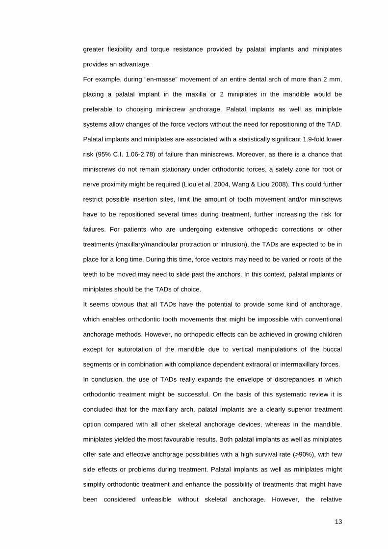
Selection of studies
Figure 1 describes the search strategy employed to identify relevant studies selected for
this review. Titles and abstracts of the MEDLINE searches were initially screened by two
independent reviewers (R.M. and M.S.) for possible inclusion. From a yield of 390 titles
71 were selected for abstract screening (Figure 1). The agreement between the reviewers
using Kappa-statistics was 96.2%. The full text of all studies of possible relevance (34)
was then obtained for independent assessment by the two reviewers. Any disagreement
was resolved by discussion.
Data were extracted independently by the same two reviewers using a data extraction
form.
Excluded studies
Of the 34 full text articles retrieved, 7 were excluded from the final analysis. The main
reasons for exclusion were a mean observation period of less than 12 weeks, loading
time was not clearly indicated, less than 10 units per modality in the study and multiple
publication of the same cohort in different scientific journals at different time points.
Data extraction
Information on the proportions of biological and technical complications was retrieved of
the 27 studies included. Biological complications included disturbances in the function of
the skeletal anchorage device leading to any early removal of the anchorage device prior
to the end of the intended orthodontic treatment or observation period. Healing or
incorporation failures were also included in this category. Technical complications were
not reported in any of the studies, and could therefore not be assessed separately.
From the 27 included studies, the number and percentage of failures was extracted.
Disagreement regarding data extraction was resolved by consensus.
Statistical analysis
Failure rates were calculated by dividing the number of events (failures) after at least 12
weeks of orthodontic loading in the numerator by the total number of each TAD type in
the denominator. For further analysis, the total number of events was considered to be

7
Poisson distributed for a given number of TADs, and Poisson regression with a
logarithmic link-function and total number of TADs per study as an offset variable was
used. To assess heterogeneity of the study-specific event rates, the Spearman
goodness-of-fit statistics and associated P-value were calculated. If the goodness-of-fit P-
value was below 0.05, indicating heterogeneity, random-effects Poisson regression (with
γ-distributed random effects) was used to obtain a summary estimate of the event rates.
Summary failure rate estimates and 95 percent confidence intervals (95% CI) are
reported.
To provide anchorage on either side of the maxilla, only one palatal implant or Onplant®
was needed, whereas at least two fixtures have to be installed if miniplates or miniscrews
are used.
To evaluate the possible failure of at least one out of two fixtures, it was assumed that
failures of these objects may occur independently. The probability to remain free of failure
was therefore calculated by multiplying the probability that each object remains free of
failure: (1-riskobject1)*(1-risk object2). Therefore, the probability to encounter at least one
failure becomes 1-(1-riskobject1)*(1-risk object2).
The 95% CI limits for survival proportions were calculated by using the 95% confidence
limits of the event rates. All analyses were performed using Stata®, version 10.1 (Stata
Corporation, College Station, TX, USA).
8
Results
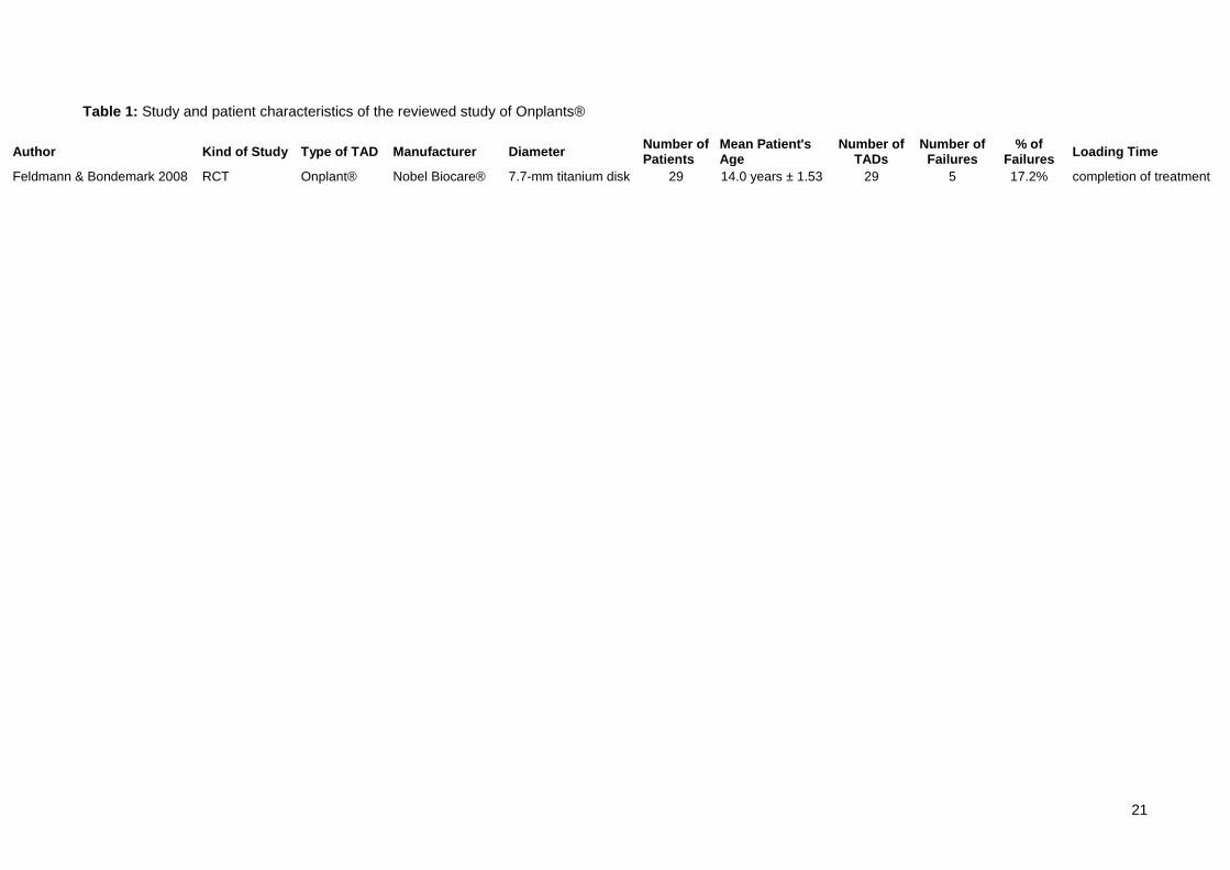
Onplants®
There was only one article fulfilling the inclusion criteria concerning Onplants® (Feldmann
& Bondemark, 2008). In this RCT, 5 out of 29 onplants or 17.2% (95% confidence interval
(CI): 5.9% - 35.8%) failed (Table 1).
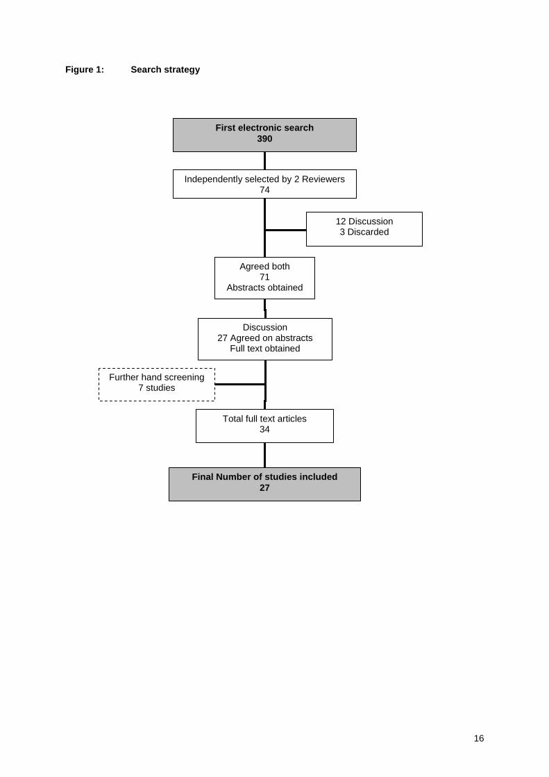
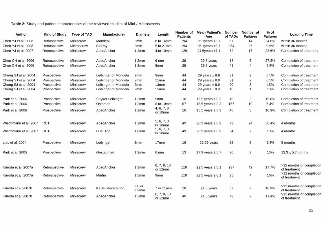
Microiscrews/Microimplants and Miniscrews/Miniimplants
17 studies provided data on the survival of 31 different types of miniscrews (Table 2). A
total of 2374 miniscrews inserted in 1196 patients with a total of 363 or 15.3% failures
could be analyzed (Table 2). Seven studies reported results of prospective cohort studies,
whereas the remaining 10 assessed their results retrospectively. Data of only one RCT
could be extracted comparing two different screw types (Wiechmann et al. 2007).
However, due to the lack of precise data reporting in all these studies no conclusive
statement of survival and/or failure rate of a specific screw type (length and diameter)
regarding their favourable indication, insertion location, insertion technique and type of
loading could be made.
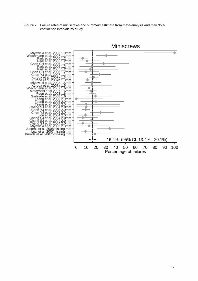
Some reports provided detailed data on diameter and length of the inserted miniscews,
while others pooled the results of a specific miniscrew diameter with various lengths
(Table 2). The mean follow-up time ranged between 120 days to more than 1 year or
completion of the intended orthodontic treatment.
By meta-analysis, the failure rate (Figure 2) was estimated at 16.4% (95% CI 13.4%-
20.1%). By analyzing the influence of screw length and diameter, only the data of screws
with detailed characteristics were considered. 3 groups of diameter were created which
basically separate these 3 “clouds” of diameter and length types (Figure 3). The
miniscrews with a diameter of 2mm or more showed a significantly 1.8-fold lower risk
(95% C.I. 1.1-3.0) to fail than miniscrews of a diameter of 1.2mm or less.
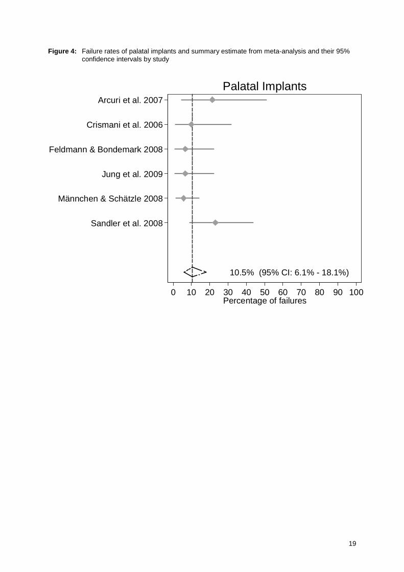
Palatal implants
One retrospective and five prospective cohort studies provided data fulfilling the inclusion
criteria on survival and failure rate of palatal implants (Table 3). Two out of these were
RCTs comparing palatal implants to conventional compliance-dependent orthodontic
9
anchorage (CDOA) (Sandler et al. 2008) only or to CDOA and Onplants® (Feldmann &
Bondemark 2008). However, only one report evaluated the clinical outcome of a larger
number of palatal implants (Männchen & Schätzle 2008). Data of a total of 190 palatal
implants with a follow-up time of at least 12 weeks up to more than 22 months or
completion of the intended orthodontic treatment could be assessed. Nineteen or 10%
out of 190 palatal implants did not provide sufficient anchorage and were lost early or
before the time point of evaluation. In meta-analysis, the failure rate for the whole group
of studies was estimated at 10.5% (95%C.I: 6.1%-18.1%) (Figure 4).
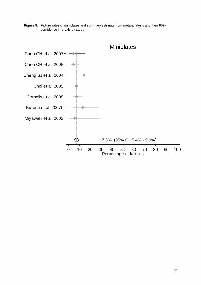
Miniplates
Seven studies out of the 27 included reports provided data on the survival and failure
rates of miniplates (Table 4). Two were prospective cohort studies, the remaining five
evaluated the material presented retrospectively. A total of 586 miniplates in 406 patients
could be followed for at least 120 days up to 1.5 years or completion of the intended
orthodontic treatment, respectively. 43 or 7.3% out of these did not remain stable and had
to be removed early. In meta-analysis, the failure rate (Figure 5) was estimated at 7.3%
(95% CI: 5.4%-9.9%).
By comparing miniplates, palatal implants and miniscrews to each other, none of them
showed statistically significantly higher survival rates than the other due to the wide
scattering within the groups. However, when miniplates and palatal implants representing
torque resisting temporary anchorage devices, were grouped together, they showed a
statistically significant 1.9-fold (95% CI: 1.1 – 2.8, p=0.005) lower clinical failure rate than
did miniscrews.
To achieve the same clinical anchorage on both sides of the arch as with a palatal
implant (10.5% failure rate, 95%C.I: 6.1%-18.1%), 2 minicrews or miniplates have to be
inserted. The probability to have at least one failure, when 2 of these TADs are installed
in the maxilla, was 14.1% (95% C.I. 10.5% - 18.8%) for miniplates and 29.4% (95% C.I.
24.3% - 36%) for miniscrews, respectively.
10
Discussion
The purpose of this systematic review was to evaluate the survival and failure rates of
skeletal temporary anchorage devices (TADs) such as Onplants®, miniplates, palatal
implants and mini- or microscrews after a loading time of at least 12 weeks. No RCTs
were available comparing all types of these TADs. RCTs comparing these four treatment
modalities may be difficult to conduct both from a logistic as well as ethical point of view
since such anchorage is usually chosen on specific patient indications. In the absence of
these kinds of RCTs, a lower level of evidence, i.e. RCT’s comparing some TADs to
conventional orthodontic anchorage devices (COAD) and prospective and retrospective
cohort studies, were included in this systematic review. TAD survival and failure rates are
only meaningful if anchorage is provided at least for the major part of orthodontic therapy.
Hence, a minimal period of 12 weeks of functional anchorage was chosen in the
evaluation.
Prior to the use of TADs, COADs offered the only possibility for sufficient anchorage to
control undesired tooth movements. The main disadvantage of many of these devices
was the fact that treatment outcomes depended to a high degree on patient compliance
(Nanda & Kierl 1992). Hence, the comparison of survival and failure rates of the different
types of TADs is of great prognostic value in future orthodontic treatment planning. But it
has to be remembered that TADs are usually inappropriate in growing patients in whom
influencing the skeletal growth is additionally indicated.
There were only two randomized clinical trials (Feldmann & Bondemark 2008; Sandler et
al. 2008) comparing the efficacy of COADs to TADs (palatal implants or Onplants®)
within the same patient cohort. One of these studies reported significantly higher
proportions of failed palatal implants than the other (Sandler et al. 2008). Most of the
failed palatal implants had been placed during the initial phase of the investigation,
representing results of a learning curve of the surgeons involved with this “relatively new”
technique. Similar problems were encountered in one retrospective study (Arcuri et al.
2007).
In contrast to conventional oral implants, the orthodontic anchorage implants of the time
such as palatal implant yielded an emergence profile with a 90-degree shoulder. This
bore the danger of “over-winding” the implant during installation with a subsequent loss of
11
the primary stability. It is obvious that such designed features made the installation of
palatal implants technique sensitive. If the two studies mentioned are eliminated from the
analysis, palatal implants showed a failure rate of only 6.7%. This is slightly below that
reported for miniplates (7.3% C.I. 95%: 5.4%-9.9%).
In recent years, a new palatal implant (with a modified, slightly concave, tulip-shaped
conical emergence profile) was developed with the purpose of reducing the risk of over-
winding the implant during installation (Orthoimplant®, Straumann AG, Basel,
Switzerland). To date, only one prospective cohort study is available on this new
generation of palatal implants (Jung et al. 2008) reporting very favourable survival rates
(93.3%) (Table 3). Furthermore, a recently published experimental human study on
palatal implants with this novel design (Schätzle et al. 2009) yielded a high primary
stability and a 100% survival for the whole observation period. Considering all studies on
palatal implants, the meta-analysis presented a mean failure rate of 10.5% (95%C.I:
6.1%-18.1%) rendering this treatment a reliable option with sufficient predictability for
routine clinical use (Figure 3).
Compared to COAD (headgear, transpalatal arch), palatal implants provided equal
(compliant patients, Sandler et al. 2008) or statistically significantly better clinical
anchorage reinforcement (Feldmann & Bondemark 2008). There were more technical
problems and a significantly higher failure rate with the Onplant® system and hence, the
palatal implant may be considered the anchorage system of choice for TAD (Feldmann &
Bondemark 2008). Palatal implants were better tolerated than Onplant® devices as well
as extraction of premolars in terms of patient centered outcomes (pain intensity,
discomfort, and analgesic consumption) (Feldmann et al. 2007).
After an observation period of at least 12 weeks, miniplates showed a slightly higher
success rate of 92.7% than palatal implants (89.5%). It has to be realized, however, that
this difference was mainly caused by early surgical failures in two studies mentioned
above (Arcuri et al. 2007, Sandler et al. 2008). A direct comparison of the efficacy of
miniplates to that of palatal implants with respect to survival has not been performed.
Considering the fact that 2 miniplates have to be installed instead of one palatal implant
to achieve the same anchorage in the maxilla, the presumptive risk for failure for the
miniplates has to be assumed at 14.1% (95% C.I. 10.5% - 18.8%) for the miniplates.
12
Even though the majority of the included in this review deal with miniscrews, there was no
study describing clinical or diagnostic criteria in relation to screw length or screw
diameter. Only one RCT (Wiechmann et al. 2007) directly compared two different screw
diameters (1.1 and 1.6mm) of various lengths to each other. Small screw diameter was
identified as a risk factor for failure. These findings are in accordance with the results
from this present systematic review. An approximately 2-fold increased failure rate was
identified for miniscrews of a diameter of ≤1.2mm compared to miniscrews of a diameter
of 2mm or more. Moreover, two other single retrospective studies (Chen et al. 2007,
Miyawaki et al. 2003) came to the same conclusion. But in contrast to another
retrospective study (Chen et al. 2006) this RCT (Wiechmann et al. 2007) failed to identify
screw length as a possible risk factor for failure. Too many different screw lengths and
insertion sites had been included in the study, resulting in a wide scattering of the data.
However, it seems to be important that the tipping moment at the bone edge be
considered (Büchter et al. 2005). These findings are in accordance with data from two
experimental implant studies that are dealing with different force levels (Melsen & Lang
2001, Hsieh et al. 2008). Therefore, controlled clinical trials with clear selection criteria for
screw length and diameter including the applied tipping moments are encouraged.
The dynamics of TAD loss (loss over time) is an important factor for decision making in
orthodontic treatment planning. The Kaplan-Meier analysis of Wiechmann and co-workers
(2007) showed that the major miniscrew failures occurred within 100 to 150 days after the
start of orthodontic loading. At this point, a change of the treatment plan may be difficult
or impossible. With respect to palatal implants, reports indicate that implant loss occurred
predominantly in the unloaded healing period (Arcuri et al. 2007, Männchen & Schätzle
2008, Sandler et al. 2008). This in turn means that once a palatal implant is
osseointegrated, no implant loss is to be expected.
It is clear that the placement and removal of a miniplate or palatal implant is a more
complex procedure than that associated with the installation of a miniscrew. The surgical
intervention for both devices is generally well tolerated by the patients (Kuroda et al.
2007, Cornelis et al. 2008) and pain intensity after surgical installation of a palatal implant
is less than that after premolar extraction (Feldmann et al. 2007). It seems that the
13
greater flexibility and torque resistance provided by palatal implants and miniplates
provides an advantage.
For example, during “en-masse” movement of an entire dental arch of more than 2 mm,
placing a palatal implant in the maxilla or 2 miniplates in the mandible would be
preferable to choosing miniscrew anchorage. Palatal implants as well as miniplate
systems allow changes of the force vectors without the need for repositioning of the TAD.
Palatal implants and miniplates are associated with a statistically significant 1.9-fold lower
risk (95% C.I. 1.06-2.78) of failure than miniscrews. Moreover, as there is a chance that
miniscrews do not remain stationary under orthodontic forces, a safety zone for root or
nerve proximity might be required (Liou et al. 2004, Wang & Liou 2008). This could further
restrict possible insertion sites, limit the amount of tooth movement and/or miniscrews
have to be repositioned several times during treatment, further increasing the risk for
failures. For patients who are undergoing extensive orthopedic corrections or other
treatments (maxillary/mandibular protraction or intrusion), the TADs are expected to be in
place for a long time. During this time, force vectors may need to be varied or roots of the
teeth to be moved may need to slide past the anchors. In this context, palatal implants or
miniplates should be the TADs of choice.
It seems obvious that all TADs have the potential to provide some kind of anchorage,
which enables orthodontic tooth movements that might be impossible with conventional
anchorage methods. However, no orthopedic effects can be achieved in growing children
except for autorotation of the mandible due to vertical manipulations of the buccal
segments or in combination with compliance dependent extraoral or intermaxillary forces.
In conclusion, the use of TADs really expands the envelope of discrepancies in which
orthodontic treatment might be successful. On the basis of this systematic review it is
concluded that for the maxillary arch, palatal implants are a clearly superior treatment
option compared with all other skeletal anchorage devices, whereas in the mandible,
miniplates yielded the most favourable results. Both palatal implants as well as miniplates
offer safe and effective anchorage possibilities with a high survival rate (>90%), with few
side effects or problems during treatment. Palatal implants as well as miniplates might
simplify orthodontic treatment and enhance the possibility of treatments that might have
been considered unfeasible without skeletal anchorage. However, the relative

14
effectiveness, efficiency, and indication list of all different temporary anchorage devices
used for various clinical problems need to further be evaluated in prospective controlled
studies.

15
Acknowledgement:
This study has been supported by the Clinical Research Foundation (CRF) for the Promotion of Oral
Health, Brienz, Switzerland.
The authors acknowledge the continuous support of Prof. Odont. Riitta Suuronen, REGA Insitute for
Regenerative Medicine, University of Tampere, Finland and Prof. Dr. Timo Peltomäki, University of
Zurich, School of Dental and Oral Medicine. Likewise, the stimulating encouragement of Prof. Dr.
Urban Hägg, The University of Hong Kong, is highly appreciated.
16
Figure 1: Search strategy
First electronic search 390
Agreed both 71
Abstracts obtained
Discussion 27 Agreed on abstracts
Full text obtained
Total full text articles 34
Final Number of studies included 27
Further hand screening 7 studies
Independently selected by 2 Reviewers 74
12 Discussion 3 Discarded
17
Figure 2: Failure rates of miniscrews and summary estimate from meta-analysis and their 95% confidence intervals by study
16.4% (95% CI: 13.4% - 20.1%)Kuroda et al. 2007bmissing mm
Luzi et al. 2007missing mmJustens et al. 2008missing mm
Miyawaki et al. 2003 2.3mmCheng SJ et al. 2004 2.0mmCheng SJ et al. 2004 2.0mmCheng SJ et al. 2004 2.0mm
Liou et al. 2004 2.0mmChen YJ et al. 2008 2.0mmChen YJ et al. 2008 2.0mm
Cheng SJ et al. 2004 2.0mmTseng et al. 2006 2.0mmTseng et al. 2006 2.0mmTseng et al. 2006 2.0mm
Garfinkle et al. 2008 1.6mmMoon et al. 2008 1.6mm
Motoyoshi et al 2007 1.6mmWiechmann et al. 2007 1.6mm
Kuroda et al. 2007a 1.5mmMiyawaki et al. 2003 1.5mmKuroda et al. 2007b 1.3mmKuroda et al. 2007a 1.3mmChen YJ et al. 2007 1.2mmChen CH et al. 2006 1.2mm
Park et al. 2005 1.2mmPark et al. 2006 1.2mm
Chen CH et al. 2006 1.2mmPark et al. 2006 1.2mmPark et al. 2006 1.2mm
Wiechmann et al. 2007 1.1mmMiyawaki et al. 2003 1.0mm
0 10 20 30 40 50 60 70 80 90 100Percentage of failures
Miniscrews
18
510
1520
Leng
th in
mm
1 1.5 2 2.5Diameter in mm
Only Miniplates
Figure 3: Distribution of screw length by screw diameter (CI:95%)
19
10.5% (95% CI: 6.1% - 18.1%)
Sandler et al. 2008
Männchen & Schätzle 2008
Jung et al. 2009
Feldmann & Bondemark 2008
Crismani et al. 2006
Arcuri et al. 2007
0 10 20 30 40 50 60 70 80 90 100Percentage of failures
Palatal Implants
Figure 4: Failure rates of palatal implants and summary estimate from meta-analysis and their 95% confidence intervals by study
20
7.3% (95% CI: 5.4% - 9.9%)
Miyawaki et al. 2003
Kuroda et al. 2007b
Cornelis et al. 2008
Choi et al. 2005
Cheng SJ et al. 2004
Chen CH et al. 2008
Chen CH et al. 2007
0 10 20 30 40 50 60 70 80 90 100Percentage of failures
Miniplates
Figure 5: Failure rates of miniplates and summary estimate from meta-analysis and their 95% confidence intervals by study
21
Table 1: Study and patient characteristics of the reviewed study of Onplants®
Author Kind of Study Type of TAD Manufacturer Diameter Number of Patients
Mean Patient's Age
Number of TADs
Number of Failures
% of Failures Loading Time
Feldmann & Bondemark 2008 RCT Onplant® Nobel Biocare® 7.7-mm titanium disk 29 14.0 years ± 1.53 29 5 17.2% completion of treatment
22
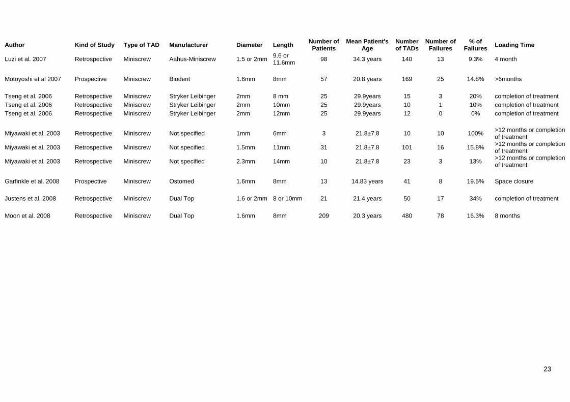
Table 2: Study and patient characteristics of the reviewed studies of Mini-/ Microscrews
Author Kind of Study Type of TAD Manufacturer Diameter Length Number of Patients
Mean Patient's Age
Number of TADs
Number of Failures
% of Failures Loading Time
Chen YJ et al. 2008 Retrospective Miniscrew Mondeal 2mm 8 to 14mm 194 25.1years ±8.7 57 14 24.6% within 36 months Chen YJ et al. 2008 Retrospective Microscrew BioRay 2mm 5 to 21mm 194 25.1years ±8.7 264 25 9.6% within 36 months Chen YJ et al. 2007 Retrospective Miniscrew AbsoAnchor 1.2mm 4 to 10mm 129 24.5years ±7.1 72 17 23.6% Completion of treatment Chen CH et al. 2006 Retrospective Miniscrew AbsoAnchor 1.2mm 6 mm 29 29.8 years 18 5 27.8% Completion of treatment Chen CH et al. 2006 Retrospective Miniscrew AbsoAnchor 1.2mm 8mm 29 29.8 years 41 4 9.8% Completion of treatment Cheng SJ et al. 2004 Prospective Miniscrew Leibinger or Mondela 2mm 9mm 44 29 years ± 8.9 31 2 6.5% Completion of treatment Cheng SJ et al. 2004 Prospective Miniscrew Leibinger or Mondela 2mm 11mm 44 29 years ± 8.9 31 2 6.5% Completion of treatment Cheng SJ et al. 2004 Prospective Miniscrew Leibinger or Mondela 2mm 13mm 44 29 years ± 8.9 20 3 15% Completion of treatment Cheng SJ et al. 2004 Prospective Miniscrew Leibinger or Mondela 2mm 15mm 44 29 years ± 8.9 10 1 10% Completion of treatment Park et al. 2006 Prospective Miniscrew Stryker Leibinger 1.2mm 5mm 10 15.5 years ± 8.3 19 3 15.8% Completion of treatment Park et al. 2006 Prospective Miniscrew Ostomed 1.2mm 6 to 10mm 67 15.5 years ± 8.3 157 10 6.4% Completion of treatment
Park et al. 2006 Prospective Miniscrew AbsoAnchor 1.2mm 4, 6, 7, 8 or 10mm 16 15.5 years ± 8.3 46 5 10.9% Completion of treatment
Wiechmann et al. 2007 RCT Miniscrew AbsoAnchor 1.1mm 5, 6, 7, 8 0r 10mm 49 26.9 years ± 8.9 79 24 30.4% 4 months
Wiechmann et al. 2007 RCT Miniscrew Dual Top 1.6mm 5, 6, 7, 8 0r 10mm 49 26.9 years ± 8.9 54 7 13% 4 months
Liou et al. 2004 Prospective Miniscrew Leibinger 2mm 17mm 16 22-29 years 32 3 9.4% 9 months Park et al. 2005 Prospective Miniscrew Oesteomed 1.2mm 6 mm 13 17.9 years ± 5.7 30 3 10% 12.3 ± 5.7months
Kuroda et al. 2007a Retrospective Miniscrew AbsoAnchor 1.3mm 6, 7, 8, 10 or 12mm 110 22.5 years ± 8.1 237 42 17.7% >12 months or completion
of treatment
Kuroda et al. 2007a Retrospective Miniscrew Martin 1.5mm 9mm 110 22.5 years ± 8.1 25 4 16% >12 months or completion of treatment
Kuroda et al 2007b Retrospective Miniscrew KeSei Medical Ind. 2.0 or 2.3mm 7 or 11mm 18 21.8 years 37 7 18.9% >12 months or completion
of treatment
Kuroda et al 2007b Retrospective Miniscrew AbsoAnchor 1.3mm 6, 7, 8, 10 or 12mm 40 21.8 years 79 9 11.4% >12 months or completion
of treatment
23
Author Kind of Study Type of TAD Manufacturer Diameter Length Number of Patients
Mean Patient's Age
Number of TADs
Number of Failures
% of Failures Loading Time
Luzi et al. 2007 Retrospective Miniscrew Aahus-Miniscrew 1.5 or 2mm 9.6 or 11.6mm 98 34.3 years 140 13 9.3% 4 month
Motoyoshi et al 2007 Prospective Miniscrew Biodent 1.6mm 8mm 57 20.8 years 169 25 14.8% >6months Tseng et al. 2006 Retrospective Miniscrew Stryker Leibinger 2mm 8 mm 25 29.9years 15 3 20% completion of treatment Tseng et al. 2006 Retrospective Miniscrew Stryker Leibinger 2mm 10mm 25 29.9years 10 1 10% completion of treatment Tseng et al. 2006 Retrospective Miniscrew Stryker Leibinger 2mm 12mm 25 29.9years 12 0 0% completion of treatment
Miyawaki et al. 2003 Retrospective Miniscrew Not specified 1mm 6mm 3 21.8±7.8 10 10 100% >12 months or completion of treatment
Miyawaki et al. 2003 Retrospective Miniscrew Not specified 1.5mm 11mm 31 21.8±7.8 101 16 15.8% >12 months or completion of treatment
Miyawaki et al. 2003 Retrospective Miniscrew Not specified 2.3mm 14mm 10 21.8±7.8 23 3 13% >12 months or completion of treatment
Garfinkle et al. 2008 Prospective Miniscrew Ostomed 1.6mm 8mm 13 14.83 years 41 8 19.5% Space closure Justens et al. 2008 Retrospective Miniscrew Dual Top 1.6 or 2mm 8 or 10mm 21 21.4 years 50 17 34% completion of treatment Moon et al. 2008 Retrospective Miniscrew Dual Top 1.6mm 8mm 209 20.3 years 480 78 16.3% 8 months
24
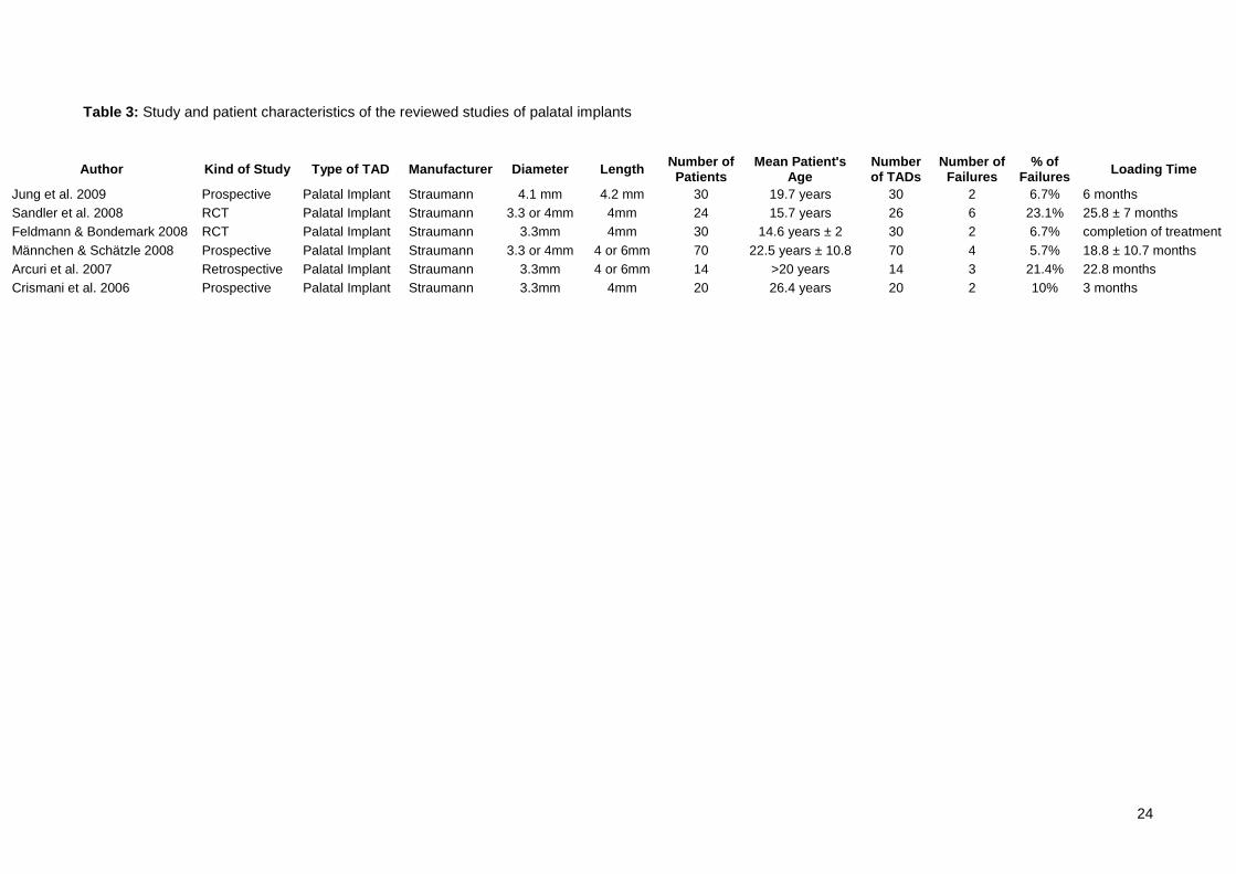
Table 3: Study and patient characteristics of the reviewed studies of palatal implants Author Kind of Study Type of TAD Manufacturer Diameter Length Number of
Patients Mean Patient's
Age Number of TADs
Number of Failures
% of Failures Loading Time
Jung et al. 2009 Prospective Palatal Implant Straumann 4.1 mm 4.2 mm 30 19.7 years 30 2 6.7% 6 months Sandler et al. 2008 RCT Palatal Implant Straumann 3.3 or 4mm 4mm 24 15.7 years 26 6 23.1% 25.8 ± 7 months Feldmann & Bondemark 2008 RCT Palatal Implant Straumann 3.3mm 4mm 30 14.6 years ± 2 30 2 6.7% completion of treatment Männchen & Schätzle 2008 Prospective Palatal Implant Straumann 3.3 or 4mm 4 or 6mm 70 22.5 years ± 10.8 70 4 5.7% 18.8 ± 10.7 months Arcuri et al. 2007 Retrospective Palatal Implant Straumann 3.3mm 4 or 6mm 14 >20 years 14 3 21.4% 22.8 months Crismani et al. 2006 Prospective Palatal Implant Straumann 3.3mm 4mm 20 26.4 years 20 2 10% 3 months
25
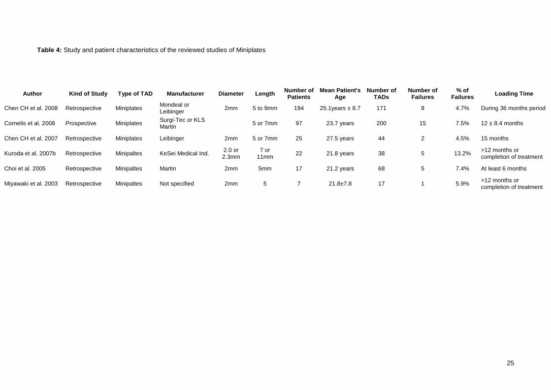
Table 4: Study and patient characteristics of the reviewed studies of Miniplates
Author Kind of Study Type of TAD Manufacturer Diameter Length Number of Patients
Mean Patient's Age
Number of TADs
Number of Failures
% of Failures Loading Time
Chen CH et al. 2008 Retrospective Miniplates Mondeal or Leibinger 2mm 5 to 9mm 194 25.1years ± 8.7 171 8 4.7% During 36 months period
Cornelis et al. 2008 Prospective Miniplates Surgi-Tec or KLS Martin 5 or 7mm 97 23.7 years 200 15 7.5% 12 ± 8.4 months
Chen CH et al. 2007 Retrospective Miniplates Leibinger 2mm 5 or 7mm 25 27.5 years 44 2 4.5% 15 months
Kuroda et al. 2007b Retrospective Minipaltes KeSei Medical Ind. 2.0 or 2.3mm
7 or 11mm 22 21.8 years 38 5 13.2% >12 months or
completion of treatment
Choi et al. 2005 Retrospective Minipaltes Martin 2mm 5mm 17 21.2 years 68 5 7.4% At least 6 months
Miyawaki et al. 2003 Retrospective Minipaltes Not specified 2mm 5 7 21.8±7.8 17 1 5.9% >12 months or completion of treatment
26
References 1. Arcuri, C., Muzzi, F., Santini, F., Barlattani, A. & Giancotti, A.(21007) Five years of experience
using palatal mini-implants for orthodontic anchorage. Journal of Oral Maxillofacial Surgery 65,2492-2497.
2. Block, M. S. & Hoffman, D. R. (1995) A new device for absolute anchorage for orthodontics.
American Journal of Orthodontics and Dentofacial Orthopedics 3, 251-258. 3. Bousquet, F., Bousquet, P., Mauran, G. & Parguel, P. (1996) Use of an impacted post for
anchorage. Journal of Clinical Orthodontics 30, 261-265.
4. Büchter, A., Wiechmann, D., Koerdt, S., Wiesmann, H. P., Piffko, J. & Meyer, U. (2005) Load-related implant reaction of mini-implants used for orthodontic anchorage. Clinical Oral Implants Research 16: 473-479.
5. Byloff, F. K., Karcher, H., Clar, E. & Stoff, F. (2000) An implant to eliminate anchorage loss during
molar distalization: a case report involving the Graz implant-supported pendulum. International Journal of Adult Orthodontics and Orthognathic Surgery 15, 129-137.
6. Chen, C. H., Chang, C. S., Hsieh, C. H., Tseng, Y. C., Shen, Y. S., Huang, I. Y., Yang, C. F.&
Chen, C. M. (2006) The use of microimplants in orthodontic anchorage. Journal of Oral Maxillofacial Surgery 64, 1209-1213.
7. Chen, Y. J., Chang, H. H., Lin, H. Y., Lai, E. H., Hung, H. C. & Yao, C. C. (2008) Stability of
miniplates and miniscrews used for orthodontic anchorage: experience with 492 temporary anchorage devices. Clinical Oral Implants Research 19, 1188-1196.
8. Chen, Y. J., Chang, H. H., Huang, C. Y., Hung, H. C., Lai, E. H. & Yao, C. C. (2007) A
retrospective analysis of the failure rate of three different orthodontic skeletal anchorage systems. Clinical Oral Implants Research 18,768-775.
9. Chen CH, Hsieh CH, Tseng YC, Huang IY, Shen YS, Chen CM. (2007) The use of miniplate
osteosynthesis for skeletal anchorage. Plastic Reconstructive Surgery 120, 232-235. 10. Cheng, S. J., Tseng, I. Y., Lee, J. J. & Kok, S. H. (2004) A prospective study of the risk factors
associated with failure of mini-implants used for orthodontic anchorage. International Journal of Oral and Maxillofacial Implants 19, 100-106.
11. Choi, B. H., Zhu, S. J. & Kim, Y. H. (2005) A clinical evaluation of titanium miniplates as anchors
for orthodontic treatment. American Journal of Orthodontics and Dentofacial Orthopedics 128, 382-384.
12. Cornelis, M. A., Scheffler, N. R., Nyssen-Behets, C., De Clerck, H. J. & Tulloch J. F. (2008)
Patients' and orthodontists' perceptions of miniplates used for temporary skeletal anchorage: a prospective study. American Journal of Orthodontics and Dentofacial Orthopedics 133, 18-24.
13. Costa, A., Raffaini, M. & Melsen, B. (1998) Miniscrews as orthodontic anchorage: a preliminary
report. International Journal of Adult Orthodontics and Orthognathic Surgery 13, 201-209. 14. Crismani, A. G., Bernhart, T., Schwarz, K., Čelar, A. G., Bantleon, H.-P.& Watzek, G. (2006)
Ninety percent success in palatal implants loaded 1 week after placement: a clinical evaluation by resonance frequency analysis. Clinical Oral Implants Research 17, 445–450.
15. Creekmore, T. D. & Eklund, M. K. (1983) The possibility of skeletal anchorage, Journal of Clinical
Orthodontics 17, 266–269. 16. Daskalogiannakis, J. (2000) Glossary of Orthodontic Terms, Quintessence Publishing Co, Leipzig. 17. De Clerck, H., Geerinckx, V. & Siciliano, S. (2002) The Zygoma Anchorage System. Journal of
Clinical Orthodontics 36, 455-459
27
18. Feldmann, I. & Bondemark, L. (2008) Anchorage capacity of osseointegrated and conventional anchorage systems: a randomized controlled trial. American Journal of Orthodontics and Dentofacial Orthopedics 133, 339.e19-28.
19. Feldmann, I., List, T., Feldmann, H. & Bondemark, L. (2007) Pain intensity and discomfort
following surgical placement of orthodontic anchoring units and premolar extraction: a randomized controlled trial. Angle Orthodontist 77, 578-85.
20. Garfinkle, J. S., Cunningham, L. L. Jr, Beeman, C. S., Kluemper, G. T., Hicks, E. P. & Kim, M. O.
(2008) Evaluation of orthodontic mini-implant anchorage in premolar extraction therapy in adolescents. American Journal of Orthodontics and Dentofacial Orthopedics 133, 642-653.
21. Glatzmaier, J., Wehrbein, H. & Diedrich, P. (1995) Die Entwicklung eines resorbierbaren
Implantatsystems zur orthodontischen Verankerung. Fortschritte der Kieferorthopädie 56, 175–181.
22. Herman, R. J., Currier, G. F. & Miyake A. (2006) Mini-implant anchorage for maxillary canine
retraction: a pilot study. American Journal of Orthodontics and Dentofacial Orthopedics 130, 228-235.
23. Hsieh, Y. D., Su, C. M., Yang, Y. H., Fu, E., Chen, H. L. & Kung, S. (2008) Evaluation on the
movement of endosseous titanium implants under continuous orthodontic forces: an experimental study in the dog. Clinical Oral Implants Research 19, 618-623.
24. Jung, B. A., Kunkel, M., Göllner, P., Liechti, T. & Wehrbein, H. (2009) Success rate of second-
generation palatal implants. Angle Orthodontist 79, 85-90. 25. Justens E. & De Bruyn, H. (2008) Clinical outcome of mini-screws used as orthodontic anchorage.
Clinical Implant Dentistry and Related Research 10, 174-80. 26. Kanomi, R. (1997) Mini-implant for orthodontic anchorage. Journal of Clinical Orthodontics 31,
763-767. 27. Kirkwood, B. R. & Sterne, J. A. C. (2003a) Poisson regression. In: Essential Medical Statistics,
249–269. Oxford: Blackwell Science Ltd. 28. Kirkwood, B. R. & Sterne, J. A. C. (2003b) Survival analysis: displaying and comparing survival
patterns. In: Essential Medical Statistics, 272–284. Oxford: Blackwell Science Ltd. 29. Kuroda, S., Sugawara, Y., Deguchi, T., Kyung, H. M. & Takano-Yamamoto, T. (2007) Clinical use
of miniscrew implants as orthodontic anchorage: success rates and postoperative discomfort. American Journal of Orthodontics and Dentofacial Orthopedics 131, 9-15.
30. Liou, E. J., Pai, B. C. & Lin, J. C. (2004) Do miniscrews remain stationary under orthodontic
forces? American Journal of Orthodontics and Dentofacial Orthopedics 126, 42-47. 31. Luzi, C., Verna, C. & Melsen, B. (2007) A prospective clinical investigation of the failure rate of
immediately loaded mini-implants used for orthodontic anchorage. Progress in Orthodontics 8, 192-201.
32. Männchen, R. & Schätzle, M. (2008) Success Rate of Palatal Orthodontic Implants - A prospective
longitudinal study. Clinical Oral Implants Research 19, 665-669. 33. Melsen, B. & Lang, N. P. (2001) Biological reactions of alveolar bone to orthodontic loading of oral
implants. Clinical Oral Implants Research 12: 144–152. 34. Miyawaki, S., Koyama, I., Inoue, M., Mishima, K., Sugahara, T.& Takano-Yamamoto, T. (2003)
Factors associated with the stability of titanium screws placed in the posterior region for orthodontic anchorage. American Journal of Orthodontics and Dentofacial Orthopedics 124, 373-378.
28
35. Moon, C. H., Lee, D. G., Lee, H. S., Im, J. S. & Baek, S. H.(2008) Factors associated with the success rate of orthodontic miniscrews placed in the upper and lower posterior buccal region. Angle Orthodobnist 78,101-106.
36. Motoyoshi, M., Hirabayashi, M., Uemura, M., Shimizu, N. (2006) Recommended placement torque
when tightening an orthodontic mini-implant. Clinical Oral Implants Research 17, 109-114. 37. Nanda, R. S. & Kierl M. J. (1992) Prediction of cooperation in orthodontic treatment. American
Journal of Orthodontics and Dentofacial Orthopedics 102, 15-21. 38. Park, H. S., Lee, S. K. & Kwon, O. W. (2005) Group distal movement of teeth using microscrew
implant anchorage. Angle Orthodontist 75, 602-609. 39. Park, H. S., Jeong, S. H. & Kwon, O. W. (2006) Factors affecting the clinical success of screw
implants used as orthodontic anchorage. American Journal of Orthodontics and Dentofacial Orthopedics 130, 18-25.
40. Sandler, J., Benson, P. E., Doyle, P., Majumder, A., O'Dwyer, J., Speight, P., Thiruvenkatachari,
B. & Tinsley, D. (2008) Palatal implants are a good alternative to headgear: a randomized trial. American Journal of Orthodontics and Dentofacial Orthopedics 133, 51-57.
41. Schätzle, M., Männchen, R. Balbach, U., Hämmerle, C. H. F., Toutenburg, H. & Jung, R. E. (2009)
Stability change of chemically modified SLA titanium palatal implants in humans. A randomized clinical trial. Clinical Oral Implants Research 10.1111/j.1600-0501.2008.01694.x.
42. Triaca, A., Antonini, M. & Wintermantel, E. (1992). Ein neues Titan-Flachschrauben-Implantat zur
orthodontischen Verankerung am anterioren Gaumen. Informationen aus Orthodontie und Kieferorthopädie 24, 251-257.
43. Tseng, Y. C., Hsieh, C. H., Chen, C. H., Shen, Y. S., Huang, I. Y. & Chen, C. M. (2006) The
application of mini-implants for orthodontic anchorage. International Journal Oral and Maxillofacial Surgery 35, 704-707.
44. Umemori, M., Sugawara, J., Mitani, H., Nagasaka, H. & Kawamura, H. (1999) Skeletal anchorage
system for open-bite correction. American Journal Orthodontics and Dentofacial Orthopedics 115, 166-174.
45. Wang, Y. C. & Liou, E. J. (2008) Comparison of the loading behavior of self-drilling and predrilled
miniscrews throughout orthodontic loading. American Journal Orthodontics and Dentofacial Orthopedics 133, 38-43.
46. Wehrbein, H., Glatzmaier, J., Mundwiller, U. & Diedrich, P. (1996). The Orthosystem-a new
implant system for orthodontic anchorage in the palate. Journal of Orofacial Orthopopedics 57, 142-153.
47. Wiechmann, D., Meyer, U. & Büchter, A. (2007) Success rate of mini- and micro-implants used for
orthodontic anchorage: a prospective clinical study. Clinical Oral Implants Research 18, 263-267. 48. Xun C, Zeng X, Wang X. (2007) Microscrew anchorage in skeletal anterior open-bite treatment.
Angle Orthodontist 77, 47-56.