Technical Note
From theand DepartGraduate Sc
The authoand publicaavailable for
Received FAddress co
thopaedic Sucho, Hirosak
� 2020 bElsevier. Thicreativecomm
2212-6287https://doi
Medial Patellofemoral Ligament ReconstructionUsing FiberTape and Knotless SwiveLock Anchors
Yasuyuki Ishibashi, M.D., Yuka Kimura, M.D., Eiji Sasaki, M.D., Shizuka Sasaki, M.D.,Yuji Yamamoto, M.D., and Eiichi Tsuda, M.D.
Abstract: Medial patellofemoral ligament (MPFL) reconstruction usually involves hamstring autografts and is associatedwith donor-site morbidity. Excellent short-term results with MPFL reconstruction using synthetic materials have beenreported. Although synthetics do not sacrifice autologous tissues, their material properties are significantly different fromthose of biological tissues. Therefore, accurate surgical procedures are required to achieve excellent results. The purpose ofthis report was to describe our surgical method for MPFL reconstruction using FiberTape and knotless SwiveLock anchors.
he medial patellofemoral ligament (MPFL) is the
Tprimary ligamentous stabilizer that prevents lateralpatella dislocations. Therefore, MPFL reconstructionsare a logical treatment approach for lateral patellarinstability,1 and the clinical results are excellent with alow re-dislocation rate.2 Although autografts, such ashamstring tendons, are most commonly used for MPFLreconstruction, they have the disadvantage of donor-site morbidity. Synthetic materials do not sacrificeautologous tissues3 and provide excellent mid-termresults.4 However, synthetics have not become popu-lar as an alternative graft option.Recently, the use of polyester suture tape (FiberTape;Arthrex, North Naples, FL) with knotless anchors(SwiveLock; Arthrex) for MPFL reconstruction hasbeen reported.5,6 FiberTape with SwiveLock anchorshas stronger ultimate load than a hamstring tendonwith soft anchors7; however, it stiffness is significantlydifferent from that of autologous tissue.7 This is both an
Department of Orthopaedic Surgery (Y.I., Y.K., E.S., S.S., Y.Y.)ment of Rehabilitation Medicine (E.T.), Hirosaki Universityhool of Medicine, Hirosaki, Japan.rs report that they have no conflicts of interest in the authorshiption of this article. Full ICMJE author disclosure forms arethis article online, as supplementary material.ebruary 29, 2020; accepted April 26, 2020.rrespondence to Yasuyuki Ishibashi, M.D., Department of Or-rgery, Hirosaki University Graduate School of Medicine, 5 Zaifu-i, Aomori, 036-8562, Japan. E-mail: [email protected] the Arthroscopy Association of North America. Published bys is an open access article under the CC BY-NC-ND license (http://ons.org/licenses/by-nc-nd/4.0/)./20325.org/10.1016/j.eats.2020.04.020
Arthroscopy Techniques, Vol 9, No 8
advantage and a disadvantage in MPFL reconstruction.One of the possible complications of synthetics is anoverconstrained patellofemoral (PF) joint, which mayresult in prolonged postoperative rehabilitation, ante-rior knee pain, and degenerative change of the PF joint.The purpose of this Technical Note was to describeMPFL reconstruction using FiberTape and SwiveLockanchors in detail.
Technique
Surgical IndicationPatients with lateral patellar instability but without
severe bony anomalies are good candidates for thisprocedure. Patients with high-grade trochlear dysplasia(Dejour type D), severe PF osteochondral damage, andskeletal immaturity are contraindicated. Bony anoma-lies, such as patella alta and increased tibialtubercleetrochlear groove distance (>25 mm), maynecessitate concomitant bony procedures in addition tothe MPFL reconstruction.
Examination Under Anesthesia and LateralRetinacular ReleaseExamination under anesthesia is routinely performed
to assess patellar tracking and lateral retinacular tight-ness. Then, diagnostic arthroscopy is performed toassess patellar tracking and intra-articular lesions. Ifnecessary, any concomitant lesions are treated. If lateralretinacular tightness is present, lateral retinacularrelease (LRR) is performed. We prefer to perform LRRusing Metzenbaum scissors from the anterolateralportal. After LRR, patellar tracking and improvement oflateral tilt are confirmed.
(August), 2020: pp e1197-e1202 e1197
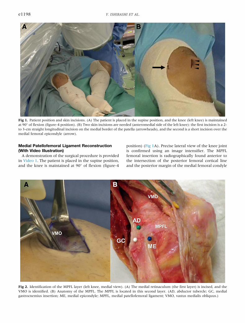
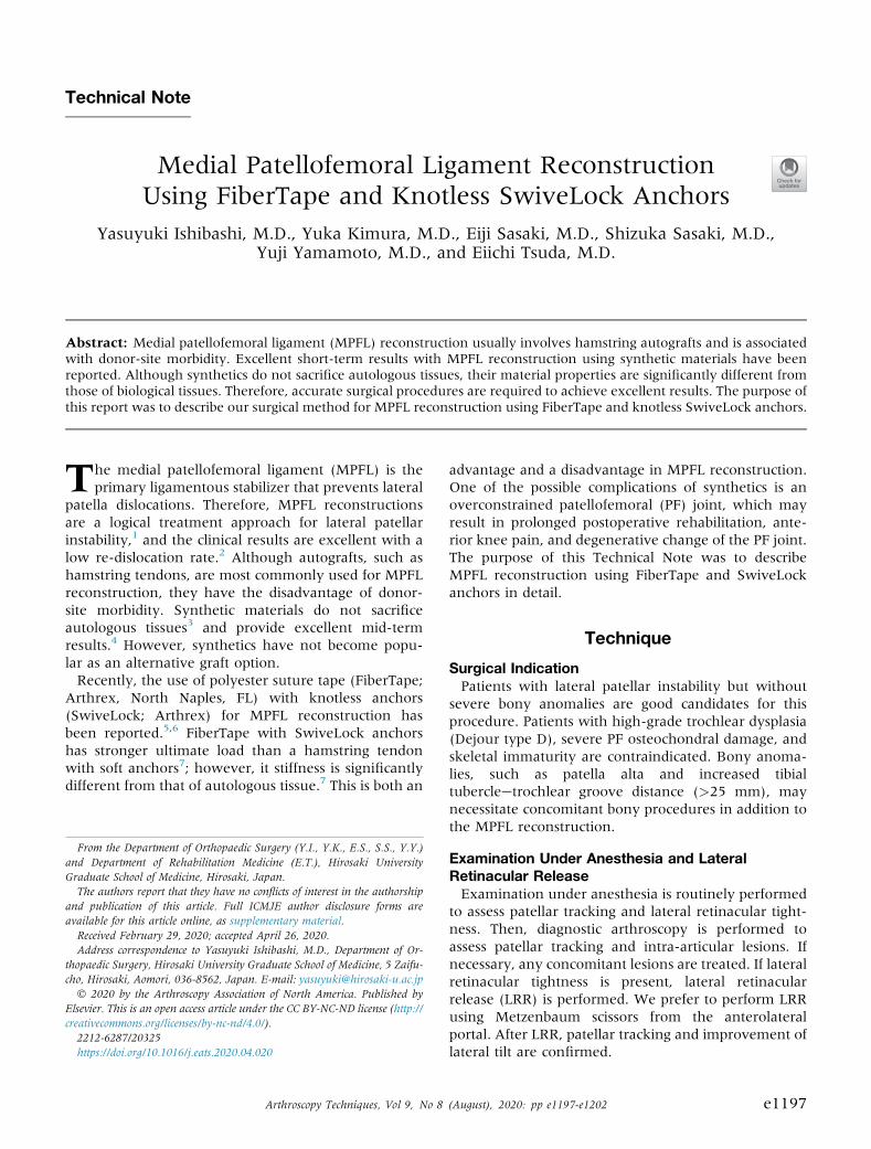
Fig 1. Patient position and skin incisions. (A) The patient is placed in the supine position, and the knee (left knee) is maintainedat 90� of flexion (figure-4 position). (B) Two skin incisions are needed (anteromedial side of the left knee): the first incision is a 2-to 3-cm straight longitudinal incision on the medial border of the patella (arrowheads), and the second is a short incision over themedial femoral epicondyle (arrow).
e1198 Y. ISHIBASHI ET AL.
Medial Patellofemoral Ligament Reconstruction(With Video Illustration)A demonstration of the surgical procedure is provided
in Video 1. The patient is placed in the supine position,and the knee is maintained at 90� of flexion (figure-4
Fig 2. Identification of the MPFL layer (left knee, medial view). (VMO is identified. (B) Anatomy of the MPFL. The MPFL is locagastrocnemius insertion; ME, medial epicondyle; MPFL, medial p
position) (Fig 1A). Precise lateral view of the knee jointis confirmed using an image intensifier. The MPFLfemoral insertion is radiographically found anterior tothe intersection of the posterior femoral cortical lineand the posterior margin of the medial femoral condyle
A) The medial retinaculum (the first layer) is incised, and theted in this second layer. (AD, abductor tubercle; GC, medialatellofemoral ligament; VMO, vastus medialis obliquus.)
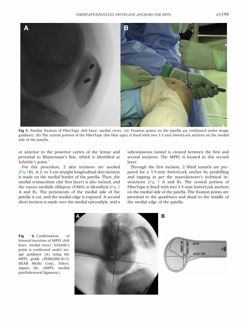
Fig 3. Patellar fixation of FiberTape (left knee, medial view). (A) Fixation points on the patella are confirmed under imageguidance. (B) The central portion of the FiberTape (the blue tape) is fixed with two 3.5-mm SwiveLock anchors on the medialside of the patella.
FIBERTAPE/KNOTLESS SWIVELOCK ANCHORS FOR MPFL e1199
or anterior to the posterior cortex of the femur andproximal to Blumensaat’s line, which is identified asSchöttle’s point.1
For this procedure, 2 skin incisions are needed(Fig 1B). A 2- to 3-cm straight longitudinal skin incisionis made on the medial border of the patella. Then, themedial retinaculum (the first layer) is also incised, andthe vastus medialis obliquus (VMO) is identified (Fig 2A and B). The periosteum of the medial side of thepatella is cut, and the medial edge is exposed. A secondshort incision is made over the medial epicondyle, and a
Fig 4. Confirmation offemoral insertion of MPFL (leftknee, medial view). Schöttle’spoint is confirmed under im-age guidance (A) using theMPFL guide (JBM0200-01/2;BEAR Medic Corp., Tokyo,Japan) (B). (MPFL, medialpatellofemoral ligament.)
subcutaneous tunnel is created between the first andsecond incisions. The MPFL is located in this secondlayer.Through the first incision, 2 blind tunnels are pre-
pared for a 3.5-mm SwiveLock anchor by predrillingand tapping as per the manufacturer’s technical in-structions (Fig 3 A and B). The central portion ofFiberTape is fixed with two 3.5-mm SwiveLock anchorson the medial side of the patella. The fixation points areproximal to the quadrisect and distal to the middle ofthe medial edge of the patella.
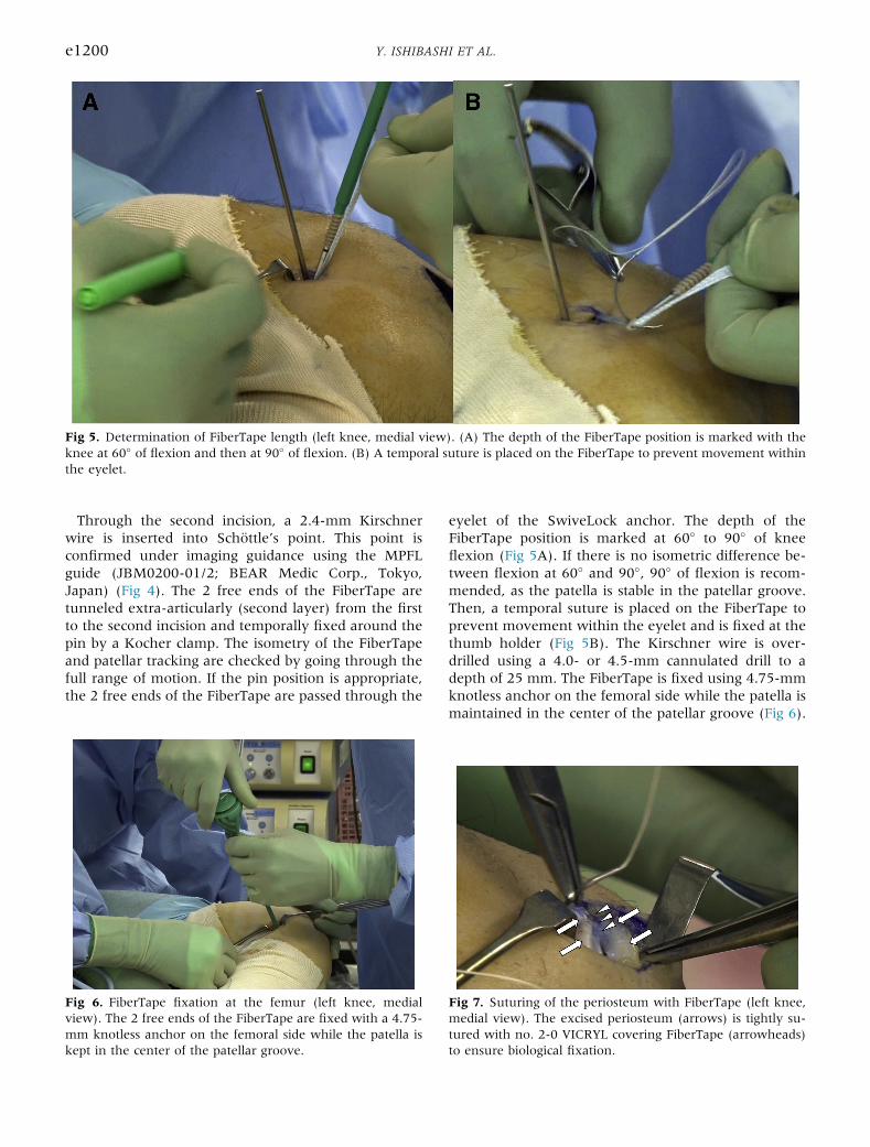
Fig 5. Determination of FiberTape length (left knee, medial view). (A) The depth of the FiberTape position is marked with theknee at 60� of flexion and then at 90� of flexion. (B) A temporal suture is placed on the FiberTape to prevent movement withinthe eyelet.
e1200 Y. ISHIBASHI ET AL.
Through the second incision, a 2.4-mm Kirschnerwire is inserted into Schöttle’s point. This point isconfirmed under imaging guidance using the MPFLguide (JBM0200-01/2; BEAR Medic Corp., Tokyo,Japan) (Fig 4). The 2 free ends of the FiberTape aretunneled extra-articularly (second layer) from the firstto the second incision and temporally fixed around thepin by a Kocher clamp. The isometry of the FiberTapeand patellar tracking are checked by going through thefull range of motion. If the pin position is appropriate,the 2 free ends of the FiberTape are passed through the
Fig 6. FiberTape fixation at the femur (left knee, medialview). The 2 free ends of the FiberTape are fixed with a 4.75-mm knotless anchor on the femoral side while the patella iskept in the center of the patellar groove.
eyelet of the SwiveLock anchor. The depth of theFiberTape position is marked at 60� to 90� of kneeflexion (Fig 5A). If there is no isometric difference be-tween flexion at 60� and 90�, 90� of flexion is recom-mended, as the patella is stable in the patellar groove.Then, a temporal suture is placed on the FiberTape toprevent movement within the eyelet and is fixed at thethumb holder (Fig 5B). The Kirschner wire is over-drilled using a 4.0- or 4.5-mm cannulated drill to adepth of 25 mm. The FiberTape is fixed using 4.75-mmknotless anchor on the femoral side while the patella ismaintained in the center of the patellar groove (Fig 6).
Fig 7. Suturing of the periosteum with FiberTape (left knee,medial view). The excised periosteum (arrows) is tightly su-tured with no. 2-0 VICRYL covering FiberTape (arrowheads)to ensure biological fixation.
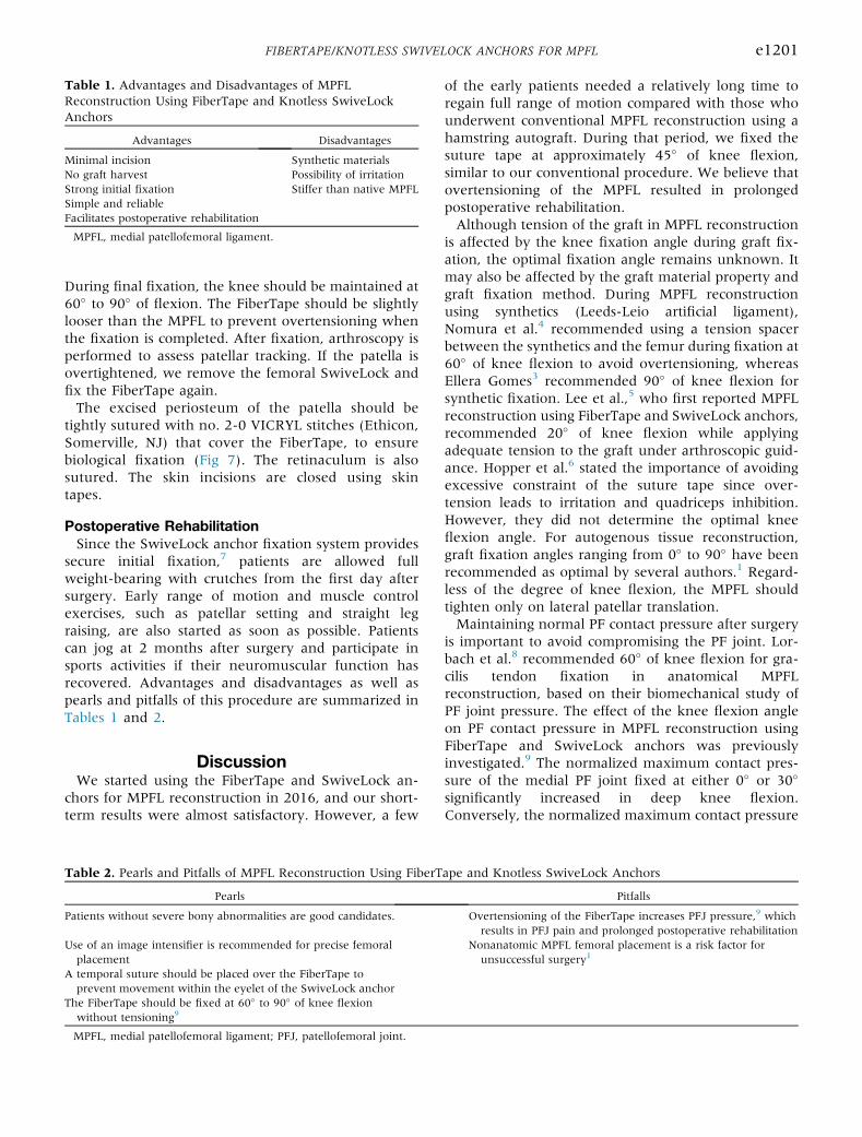
Table 1. Advantages and Disadvantages of MPFLReconstruction Using FiberTape and Knotless SwiveLockAnchors
Advantages Disadvantages
Minimal incision Synthetic materialsNo graft harvest Possibility of irritationStrong initial fixation Stiffer than native MPFLSimple and reliableFacilitates postoperative rehabilitation
MPFL, medial patellofemoral ligament.
FIBERTAPE/KNOTLESS SWIVELOCK ANCHORS FOR MPFL e1201
During final fixation, the knee should be maintained at60� to 90� of flexion. The FiberTape should be slightlylooser than the MPFL to prevent overtensioning whenthe fixation is completed. After fixation, arthroscopy isperformed to assess patellar tracking. If the patella isovertightened, we remove the femoral SwiveLock andfix the FiberTape again.The excised periosteum of the patella should be
tightly sutured with no. 2-0 VICRYL stitches (Ethicon,Somerville, NJ) that cover the FiberTape, to ensurebiological fixation (Fig 7). The retinaculum is alsosutured. The skin incisions are closed using skintapes.
Postoperative RehabilitationSince the SwiveLock anchor fixation system provides
secure initial fixation,7 patients are allowed fullweight-bearing with crutches from the first day aftersurgery. Early range of motion and muscle controlexercises, such as patellar setting and straight legraising, are also started as soon as possible. Patientscan jog at 2 months after surgery and participate insports activities if their neuromuscular function hasrecovered. Advantages and disadvantages as well aspearls and pitfalls of this procedure are summarized inTables 1 and 2.
DiscussionWe started using the FiberTape and SwiveLock an-
chors for MPFL reconstruction in 2016, and our short-term results were almost satisfactory. However, a few
Table 2. Pearls and Pitfalls of MPFL Reconstruction Using FiberT
Pearls
Patients without severe bony abnormalities are good candidates.
Use of an image intensifier is recommended for precise femoralplacement
A temporal suture should be placed over the FiberTape toprevent movement within the eyelet of the SwiveLock anchor
The FiberTape should be fixed at 60� to 90� of knee flexionwithout tensioning9
MPFL, medial patellofemoral ligament; PFJ, patellofemoral joint.
of the early patients needed a relatively long time toregain full range of motion compared with those whounderwent conventional MPFL reconstruction using ahamstring autograft. During that period, we fixed thesuture tape at approximately 45� of knee flexion,similar to our conventional procedure. We believe thatovertensioning of the MPFL resulted in prolongedpostoperative rehabilitation.Although tension of the graft in MPFL reconstruction
is affected by the knee fixation angle during graft fix-ation, the optimal fixation angle remains unknown. Itmay also be affected by the graft material property andgraft fixation method. During MPFL reconstructionusing synthetics (Leeds-Leio artificial ligament),Nomura et al.4 recommended using a tension spacerbetween the synthetics and the femur during fixation at60� of knee flexion to avoid overtensioning, whereasEllera Gomes3 recommended 90� of knee flexion forsynthetic fixation. Lee et al.,5 who first reported MPFLreconstruction using FiberTape and SwiveLock anchors,recommended 20� of knee flexion while applyingadequate tension to the graft under arthroscopic guid-ance. Hopper et al.6 stated the importance of avoidingexcessive constraint of the suture tape since over-tension leads to irritation and quadriceps inhibition.However, they did not determine the optimal kneeflexion angle. For autogenous tissue reconstruction,graft fixation angles ranging from 0� to 90� have beenrecommended as optimal by several authors.1 Regard-less of the degree of knee flexion, the MPFL shouldtighten only on lateral patellar translation.Maintaining normal PF contact pressure after surgery
is important to avoid compromising the PF joint. Lor-bach et al.8 recommended 60� of knee flexion for gra-cilis tendon fixation in anatomical MPFLreconstruction, based on their biomechanical study ofPF joint pressure. The effect of the knee flexion angleon PF contact pressure in MPFL reconstruction usingFiberTape and SwiveLock anchors was previouslyinvestigated.9 The normalized maximum contact pres-sure of the medial PF joint fixed at either 0� or 30�
significantly increased in deep knee flexion.Conversely, the normalized maximum contact pressure
ape and Knotless SwiveLock Anchors
Pitfalls
Overtensioning of the FiberTape increases PFJ pressure,9 whichresults in PFJ pain and prolonged postoperative rehabilitation
Nonanatomic MPFL femoral placement is a risk factor forunsuccessful surgery1
Fig 8. Postoperative computed tomography image of the leftknee (medial view). The femoral fixation point is locatedbetween the AD and the ME (arrowhead). The patellar fixa-tion points are proximal to the quadricet and distal to themiddle of the medial edge of the patella (arrows). (AD,abductor tubercle; ME, medial epicondyle.)
e1202 Y. ISHIBASHI ET AL.
fixed at 60� and 90� remained at the same level as theintact knees at all knee flexion angles. FiberTape fixedby SwiveLock anchors was stiffer than native MPFL.7
Therefore, the suture tape should be fixed at >60� ofknee flexion to avoid excessive PF joint contactpressure.The femoral positioning of the graft and graft tension
are critical steps for the overall outcome of MPFLreconstruction.1 A small displacement of the femoralfixation point dramatically changes the force andpressure to the medial PF cartilage. We have beenperforming postoperative 3-dimensional computed to-mography, and there has been no malpositioning in ourseries (Fig 8). Since SwiveLock fixation does not needan oblique femoral tunnel like ENDOBUTTON fixation,a guidewire can be perpendicularly inserted into thefemoral tunnel under image control. This leads to ac-curate graft placement, which is an advantage of thistechnique. Patellar fracture is one of the complicationsto avoid. Two small blind tunnels for 3.5-mm Swive-Lock anchors may not decrease the strength of thepatella and prevent patella fracture, and this is anotheradvantage of this technique.
There are some limitations to this procedure. Sincesynthetics cannot be replaced by autologous tissue, thelong-term effects on the knee joint are unknown. Ac-cording to a mid-term follow-up study reported byNomura et al.,4 MPFL reconstruction using synthetics issafe and achieves favorable results. Since the FiberTapefixed by SwiveLock anchors was stiffer than the nativeMPFL, minor malpositioning is a risk factor for technicalfailure. Further clinical studies are necessary to deter-mine the long-term results of this procedure.
References1. Sanchis-Alfonso V. Guidelines for medial patellofemoral
ligament reconstruction in chronic lateral patellar insta-bility. J Am Acad Orthop Surg 2014;22:175-182.
2. Lippacher S, Dreyhaupt J, Williams SR, Reichel H,Nelitz M. Reconstruction of the medial patellofemoral lig-ament: Clinical outcomes and return to sports. Am J SportsMed 2014;42:1661-1668.
3. Ellera Gomes JL. Medial patellofemoral ligament recon-struction for recurrent dislocation of the patella: A pre-liminary report. Arthroscopy 1992;8:335-340.
4. Nomura E, Horiuchi Y, Kihara M. A mid-term follow-up ofmedial patellofemoral ligament reconstruction using anartificial ligament for recurrent patellar dislocation. Knee2000;7:211-215.
5. Lee PYF, Golding D, Rozewicz S, et al. Modern syntheticmaterial is a safe and effective alternative for medialpatellofemoral ligament reconstruction. Knee Surg SportsTraumatol Arthrosc 2018;26:2716-2721.
6. Hopper GP, Heusdens CHW, Dossche L, Mackay GM.Medial patellofemoral ligament repair with suture tapeaugmentation. Arthrosc Tech 2019;8:e1-e5.
7. Tsushima T, Tsukada H, Saaki S, et al. Biomechanicalanalysis of medial patellofemoral ligament reconstruction:FiberTape� with knotless anchors versus a semitendinosustendon autograft with soft anchors. J Orthop Sci 2019;24:663-667.
8. Lorbach O, Haupert A, Efe T, et al. Biomechanical evalu-ation of MPFL reconstructions: Differences in dynamiccontact pressure between gracilis and fascia lata graft. KneeSurg Sports Traumatol Arthrosc 2017;25:2502-2510.
9. Sakamoto Y, Sasaki S, Kimura Y, Yamamoto Y, Tsuda E,Ishibashi Y. Patellofemoral contact pressure for medialpatellofemoral ligament reconstruction using suture tapevaries with the knee flexion angle: A biomechanical eval-uation. Arthroscopy 2020;36:1390-1395.