C L I N I C S I NP L A S T I C
S U R G E R Y
Clin Plastic Surg 33 (2006) 117–127
117
Low-Level Laser-AssistedLiposuction: the Neira 4 L TechniqueRodrigo Neira, MD*, Luiz Toledo, MD,Jose Arroyave, BSc, MSc, TEM, SEM, Efrain Solarte, Dr, Rer, Nat, MSc,Carolina Isaza, MD, MSc, Oscar Gutierrez, MD, MSc,William Criollo, BSc, MSc, Hugo Ramirez, VMD, MSc,Maria I. Gutierrez, MD, MSc, PhD, Clara L. Ortiz-Neira, MD
& Mechanisms of action of low-level lasers & Discussion
Effects of low-level lasers on the adiposecell membrane and mitochondriaEffect of laser light on the bodyBiophysical effects of laser light onadipose cells
& Low-level laser-assisted liposuctionThe procedureTreatment of complications
Department of Plastic Surgery, Maxillofacial andCali, Colombia* Corresponding author. 103 Watermill Road, Greer, SCE-mail address: [email protected] (R. Neira).
0094-1298/06/$ – see front matter © 2005 Elsevier Inc. All rightsplasticsurgery.theclinics.com
& Summary& Acknowledgments& References
Liposuction, a procedure that carves out bodyfat by suction and grafts fat in other places to rec-reate a harmonic, anatomic, and physiologic cor-poral shape, is an art. Although this procedure isthe most commonly performed cosmetic surgery(384,686 procedures were performed in the UnitedStates in 2003) [1], it is not considered altogetherbenign [2]. In 2000, the mortality rate for pa-tients having liposuction was similar to that forpersons dying in motor vehicle accidents in theUnited States—about 20 in every 100,000 proce-dures done by physicians certified by the AmericanBoard of Plastic Surgery [3].Liposuction causes severe trauma to the adipose
tissue and crushes nerves, vessels, and connectivetissue, generating a strong inflammatory response[4]. The patient undergoing liposuction is at risk formultiple problems, such as thrombosis, endothelialedema, fat embolism, immunocompromise, infec-
tion, local necrosis, transitory and definitive vasculardamage, and lengthy healing processes. Hetter [4]described the surgical trauma in a classic lipoplasty ascrush injuries and referred to these wounds as ‘‘theinternal burn,’’ stressing the loss of serum albuminas well as red cell mass and the release of intracel-lular products. These events initiate a giant inflam-matory cascade that causes a marked fourth-spacephenomenon during the first 3 days after surgery.In an effort to mitigate the trauma of classic lipo-
suction, surgeons have used external laser-assistedliposuction to reduce inflammation and controlpain after surgery. This article describes low-levellaser-assisted liposuction (LLLL), known as theNeira 4 L technique, a new procedure that our sur-gical team has developed to moderate the inflam-matory response, reduce trauma to tissues, andpromote subcutaneous wound healing after lipo-plasty-assisted liposuction.
Hand Surgery, Pontificia Universidad Javeriana,
29650.
reserved. doi:10.1016/j.cps.2005.09.005
118 Neira et al
Mechanisms of action of low-level lasers
Effects of low-level lasers on the adipose cellmembrane and mitochondria
Low-level laser irradiation modulates the attach-ment of cells in vitro by means of cytochrome Coxidase and activates the mitochondrial signalingpathway, which increases the effect of the laser onthe cell membrane and the mitochondria [5] andthe number of contacts of the endoplasmic reticu-lum with the mitochondria and plasma membrane[6]. Irradiation with a low-level laser increases ATPlevels in cells cultivated in vitro, increasing thesurvival of the cell by increasing cell energy thatcould be necessary to repair and heal tissues aftersurgical trauma [7]. The increase in cellular ATPlevels produced by monochromatic red light de-pends on the growth phase of the culture. ATPlevels are insignificant in the lag phase of culturedcells, increase in the log phase, and reach a maxi-mum (about 190%) in cells at the late logarithmicand early plateau phases [8,9]. Iaffaldano and co-workers [10] found increased ATP levels, electricpotential across inner membranes, and action po-tential in the mitochondrial matrix, as well as smallchanges in the matrix configuration. Although in-creasing intracellular cAMP concentrations in vas-cular smooth muscle increases blood flow andpromotes edema, increasing cAMP concentrationsin the endotheliummay suppress edema by enhanc-ing the permeability barrier [11].Radiation of cells with a low-level laser causes
morphologic changes in the mitochondrial lym-phocyte [12]. Because hemoglobin does not absorbin the red and near-infrared spectral region, lightcan penetrate deep into the living tissue.At least three signaling pathways relate cell at-
tachment, the respiratory chain, and the Na+, K+-ATPase, and N+/H+ exchanger activities [13]. Cu(A)and Cu(B) chromophores of cytochrome C oxidasemay be involved as photoacceptors, and varioussignaling pathways between cytochrome C oxidaseand cell attachment regulation are at work.
Effect of laser light on the body
Used in the treatment of a broad range of condi-tions, including acute and chronic pain and injuredtissues, low-level laser therapy (LLLT) has improvedwound healing, reduced edema, and relieved painof various origins.
InflammationLow-level lasers have been used for many years toreduce inflammation. LLLT reversibly suppressesthe action potentials elicited by bradykinin andblocks the conduction of nociceptive signals inprimary afferent nerves [14]. At 4 J/cm2, the anti-
inflammatory effect is superior to that with 8 J/cm2
[15]. LLLT is used to stabilize vascular endotheliato prevent tissue hypoperfusion through endothe-lial edema [16].
PainLLLT for pain involves suppression of late dis-charges in caudal neurons evoked by excitatoryinputs from C-fiber afferents but not of early dis-charges evoked by impulses from A-fiber deltaafferents [17]. These results suggest that low-levellaser energy blocks the depolarization of C-fiberafferents and suppresses impulse conduction ofunmyelinated A-fiber delta afferents in the periph-eral sensory nerve, which causes pain. Kasai andcoworkers [18] showed that LLLT acts as a revers-ible direct suppressor of neuronal activity.In a study of the effect of low-power laser irradia-
tion on pain using substance P in rat spinal dorsalroot ganglion, Ohno [19] found a statistically sig-nificant difference in substance P–like immuno-reactivity between the control group and stimulatedgroup. These results suggest that laser irradiationsuppresses excitation of the unmyelinated C-fibersin the afferent sensory pathway.LLLT on local points in rats with arthritis relieved
arthralgia, reduced swelling in the ankles, and pro-duced instant analgesia [20].
Peripheral nervesIrradiation with a low-level laser can prevent post-traumatic degeneration of peripheral nerves andpostpone degeneration of the central nervous sys-tem [21]. Low-level laser energy inactivates thepotential of the cell membrane, preventing thetransmission of pain through afferent nerves aftersurgery, and acts on the demyelinated nerve tospeed recovery. In one study [22], transcutaneouslow-power irradiation with low-level lasers signifi-cantly increased the rate of regeneration of rat facialnerve after a crush injury when compared with thatof the control group (P < .01).In their classic study, Rochkind and coworkers
[23] found that LLLT improved the recovery ofinjured peripheral nerves and the central nervoussystem, as well as the healing of cutaneous woundsand burns. LLLT applied to crushed injured nervessignificantly increased the compound action poten-tial in unirradiated tissue and irradiated tissue onthe left side. Laser irradiation done on only theright side in bilaterally inflicted cutaneous woundsenhanced recovery on both sides (P < .01). In thenonirradiated control group, all rats sustained ad-vanced necrosis of the feet and bilateral gangrene.These systemic effects have implications for theclinical application of LLLT and for basic researchinto the possible mechanisms involved.
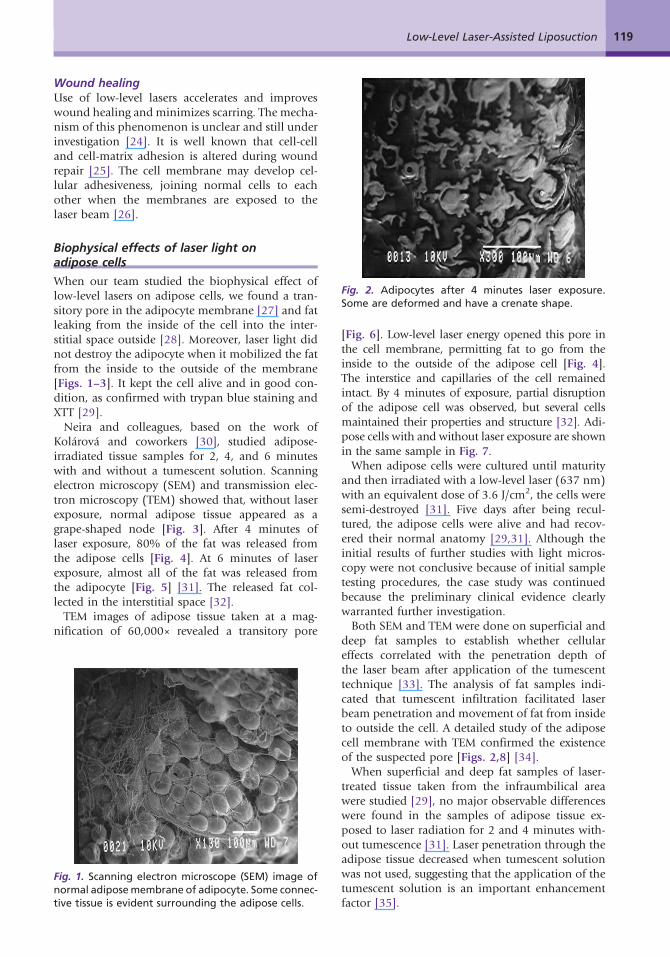
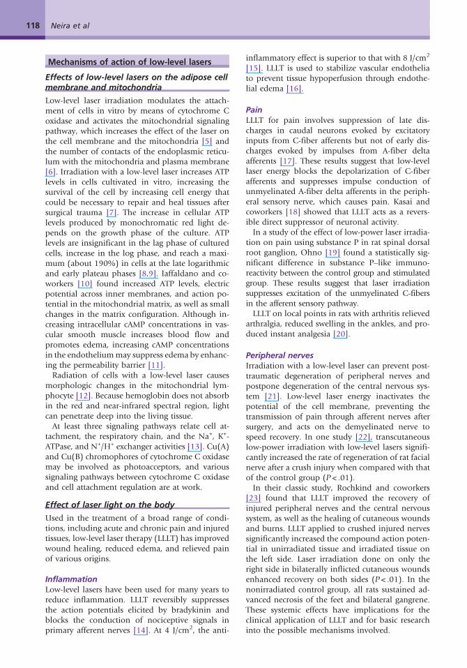
Fig. 2. Adipocytes after 4 minutes laser exposure.Some are deformed and have a crenate shape.
119Low-Level Laser-Assisted Liposuction
Wound healingUse of low-level lasers accelerates and improveswound healing and minimizes scarring. The mecha-nism of this phenomenon is unclear and still underinvestigation [24]. It is well known that cell-celland cell-matrix adhesion is altered during woundrepair [25]. The cell membrane may develop cel-lular adhesiveness, joining normal cells to eachother when the membranes are exposed to thelaser beam [26].
Biophysical effects of laser light onadipose cells
When our team studied the biophysical effect oflow-level lasers on adipose cells, we found a tran-sitory pore in the adipocyte membrane [27] and fatleaking from the inside of the cell into the inter-stitial space outside [28]. Moreover, laser light didnot destroy the adipocyte when it mobilized the fatfrom the inside to the outside of the membrane[Figs. 1–3]. It kept the cell alive and in good con-dition, as confirmed with trypan blue staining andXTT [29].Neira and colleagues, based on the work of
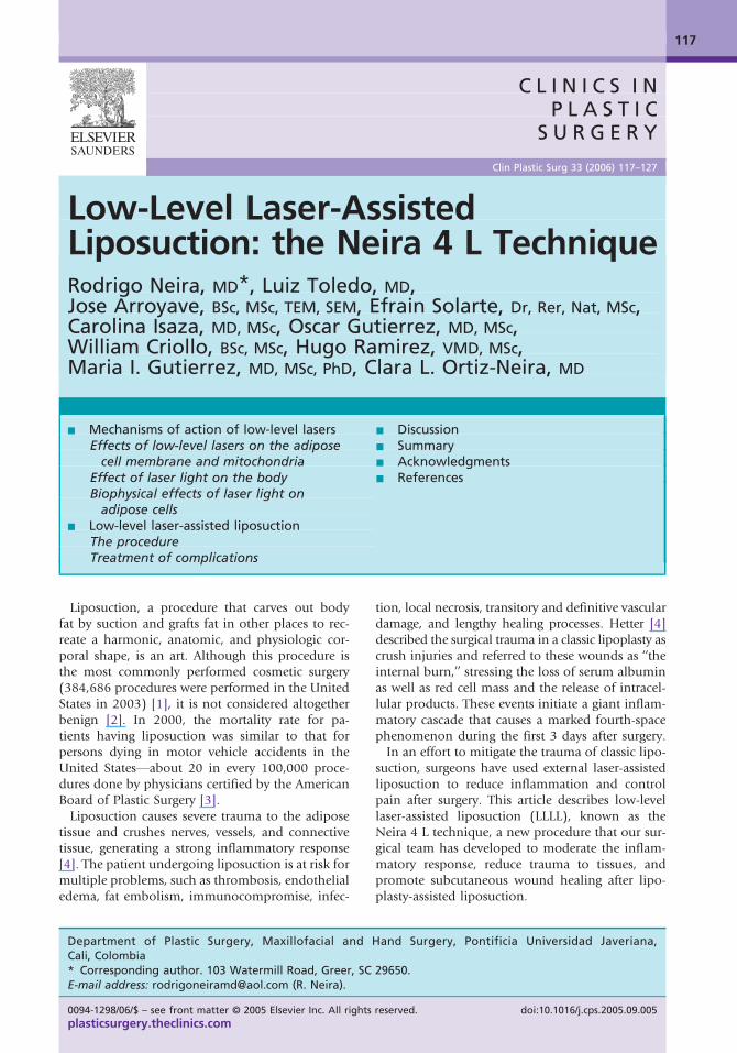
Kolárová and coworkers [30], studied adipose-irradiated tissue samples for 2, 4, and 6 minuteswith and without a tumescent solution. Scanningelectron microscopy (SEM) and transmission elec-tron microscopy (TEM) showed that, without laserexposure, normal adipose tissue appeared as agrape-shaped node [Fig. 3]. After 4 minutes oflaser exposure, 80% of the fat was released fromthe adipose cells [Fig. 4]. At 6 minutes of laserexposure, almost all of the fat was released fromthe adipocyte [Fig. 5] [31]. The released fat col-lected in the interstitial space [32].TEM images of adipose tissue taken at a mag-
nification of 60,000× revealed a transitory pore
Fig. 1. Scanning electron microscope (SEM) image ofnormal adiposemembrane of adipocyte. Some connec-tive tissue is evident surrounding the adipose cells.
[Fig. 6]. Low-level laser energy opened this pore inthe cell membrane, permitting fat to go from theinside to the outside of the adipose cell [Fig. 4].The interstice and capillaries of the cell remainedintact. By 4 minutes of exposure, partial disruptionof the adipose cell was observed, but several cellsmaintained their properties and structure [32]. Adi-pose cells with and without laser exposure are shownin the same sample in Fig. 7.When adipose cells were cultured until maturity
and then irradiated with a low-level laser (637 nm)with an equivalent dose of 3.6 J/cm2, the cells weresemi-destroyed [31]. Five days after being recul-tured, the adipose cells were alive and had recov-ered their normal anatomy [29,31]. Although theinitial results of further studies with light micros-copy were not conclusive because of initial sampletesting procedures, the case study was continuedbecause the preliminary clinical evidence clearlywarranted further investigation.Both SEM and TEM were done on superficial and
deep fat samples to establish whether cellulareffects correlated with the penetration depth ofthe laser beam after application of the tumescenttechnique [33]. The analysis of fat samples indi-cated that tumescent infiltration facilitated laserbeam penetration and movement of fat from insideto outside the cell. A detailed study of the adiposecell membrane with TEM confirmed the existenceof the suspected pore [Figs. 2,8] [34].When superficial and deep fat samples of laser-
treated tissue taken from the infraumbilical areawere studied [29], no major observable differenceswere found in the samples of adipose tissue ex-posed to laser radiation for 2 and 4 minutes with-out tumescence [31]. Laser penetration through theadipose tissue decreased when tumescent solutionwas not used, suggesting that the application of thetumescent solution is an important enhancementfactor [35].
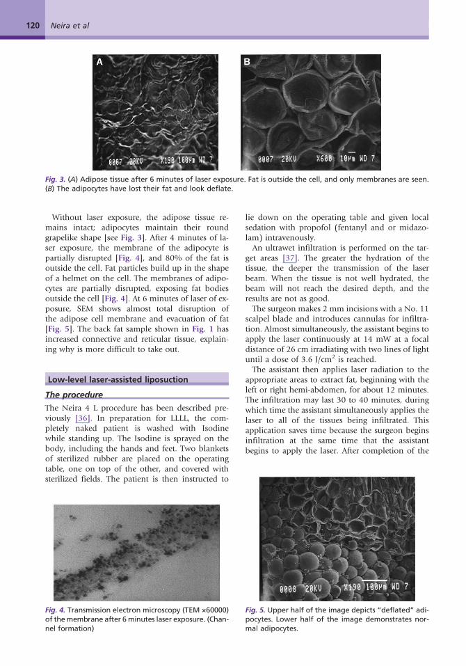
Fig. 3. (A) Adipose tissue after 6 minutes of laser exposure. Fat is outside the cell, and only membranes are seen.(B) The adipocytes have lost their fat and look deflate.
120 Neira et al
Without laser exposure, the adipose tissue re-mains intact; adipocytes maintain their roundgrapelike shape [see Fig. 3]. After 4 minutes of la-ser exposure, the membrane of the adipocyte ispartially disrupted [Fig. 4], and 80% of the fat isoutside the cell. Fat particles build up in the shapeof a helmet on the cell. The membranes of adipo-cytes are partially disrupted, exposing fat bodiesoutside the cell [Fig. 4]. At 6 minutes of laser of ex-posure, SEM shows almost total disruption ofthe adipose cell membrane and evacuation of fat[Fig. 5]. The back fat sample shown in Fig. 1 hasincreased connective and reticular tissue, explain-ing why is more difficult to take out.
Low-level laser-assisted liposuction
The procedure
The Neira 4 L procedure has been described pre-viously [36]. In preparation for LLLL, the com-pletely naked patient is washed with Isodinewhile standing up. The Isodine is sprayed on thebody, including the hands and feet. Two blanketsof sterilized rubber are placed on the operatingtable, one on top of the other, and covered withsterilized fields. The patient is then instructed to
Fig. 4. Transmission electron microscopy (TEM ×60000)of themembrane after 6 minutes laser exposure. (Chan-nel formation)
lie down on the operating table and given localsedation with propofol (fentanyl and or midazo-lam) intravenously.An ultrawet infiltration is performed on the tar-
get areas [37]. The greater the hydration of thetissue, the deeper the transmission of the laserbeam. When the tissue is not well hydrated, thebeam will not reach the desired depth, and theresults are not as good.The surgeon makes 2 mm incisions with a No. 11
scalpel blade and introduces cannulas for infiltra-tion. Almost simultaneously, the assistant begins toapply the laser continuously at 14 mW at a focaldistance of 26 cm irradiating with two lines of lightuntil a dose of 3.6 J/cm2 is reached.The assistant then applies laser radiation to the
appropriate areas to extract fat, beginning with theleft or right hemi-abdomen, for about 12 minutes.The infiltration may last 30 to 40 minutes, duringwhich time the assistant simultaneously applies thelaser to all of the tissues being infiltrated. Thisapplication saves time because the surgeon beginsinfiltration at the same time that the assistantbegins to apply the laser. After completion of the
Fig. 5. Upper half of the image depicts “deflated” adi-pocytes. Lower half of the image demonstrates nor-mal adipocytes.
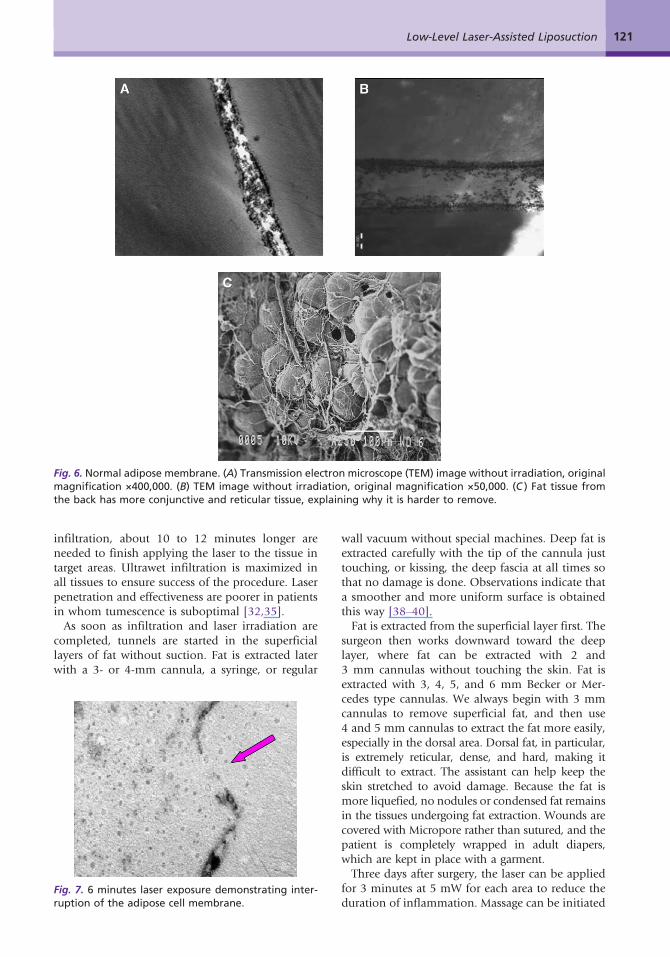
Fig. 6. Normal adipose membrane. (A) Transmission electron microscope (TEM) image without irradiation, originalmagnification ×400,000. (B) TEM image without irradiation, original magnification ×50,000. (C ) Fat tissue fromthe back has more conjunctive and reticular tissue, explaining why it is harder to remove.
121Low-Level Laser-Assisted Liposuction
infiltration, about 10 to 12 minutes longer areneeded to finish applying the laser to the tissue intarget areas. Ultrawet infiltration is maximized inall tissues to ensure success of the procedure. Laserpenetration and effectiveness are poorer in patientsin whom tumescence is suboptimal [32,35].As soon as infiltration and laser irradiation are
completed, tunnels are started in the superficiallayers of fat without suction. Fat is extracted laterwith a 3- or 4-mm cannula, a syringe, or regular
Fig. 7. 6 minutes laser exposure demonstrating inter-ruption of the adipose cell membrane.
wall vacuum without special machines. Deep fat isextracted carefully with the tip of the cannula justtouching, or kissing, the deep fascia at all times sothat no damage is done. Observations indicate thata smoother and more uniform surface is obtainedthis way [38–40].Fat is extracted from the superficial layer first. The
surgeon then works downward toward the deeplayer, where fat can be extracted with 2 and3 mm cannulas without touching the skin. Fat isextracted with 3, 4, 5, and 6 mm Becker or Mer-cedes type cannulas. We always begin with 3 mmcannulas to remove superficial fat, and then use4 and 5 mm cannulas to extract the fat more easily,especially in the dorsal area. Dorsal fat, in particular,is extremely reticular, dense, and hard, making itdifficult to extract. The assistant can help keep theskin stretched to avoid damage. Because the fat ismore liquefied, no nodules or condensed fat remainsin the tissues undergoing fat extraction. Wounds arecovered with Micropore rather than sutured, and thepatient is completely wrapped in adult diapers,which are kept in place with a garment.Three days after surgery, the laser can be applied
for 3 minutes at 5 mW for each area to reduce theduration of inflammation. Massage can be initiated
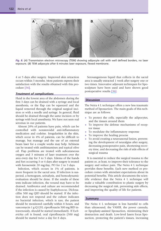
Fig. 8. (A) Transmission electron microscopy (TEM) showing adipocyte cell with well defined borders, no laserexposure. (B) TEM adipocyte after 6 minutes laser exposure, flexed membrane.
122 Neira et al
4 or 5 days after surgery. Improved skin retractionoccurs within 3 months. Most patients express theirsatisfaction with the results obtained with this pro-cedure [36].
Treatment of complications
Fluid in the lowest area of the abdomen during thefirst 3 days can be drained with a syringe and localanesthesia, or the flap can be squeezed and theliquid removed through the original surgical inci-sion or with a needle and syringe. In general, fluidshould be drained through the same incision or bysyringe with local anesthesia. We have not seen realseromas in our patients.Almost 20% of patients have pain, which can be
controlled with nonsteroidal anti-inflammatorymedication and codeine. Irregularities in the skin,which occur in 4% of patients, can be difficult tomanage, but massage and the use of an externalbeam laser for a couple weeks may help. Itchinesscan be treated with antihistamines and topical oliveoil. Flap problems are treated with subcutaneousoxygen and 3 minutes of laser treatment over thearea every day for 3 to 5 days. Edema of the handsand feet occurring 3 or 4 days after surgery is treatedwith furosemide 20 mg/day, PO for 2 to 3 days.Infection, which occurs in 1% of patients, is
more frequent in the sacral area. If infection is sus-pected, a hemogram, urinalysis, and hemodynamicevaluation should be done. If the results of thesetests indicate infection, the wound may have to bedrained. Antibiotics and culture are recommendedif the infection is caused by Staphylococcus. Dicloxa-cillin 500 mg QID should be started. If the infec-tion does not respond and the culture indicatesno bacterial infection, which is rare, the patientshould be monitored carefully within 8 hours, andvancomycin 1 g Q12H, ciprofloxacin 500mg BID in-travenously, should be started immediately. If Esch-erichia coli is found, oral ciprofloxacin (500 mg)should be started twice a day for 8 days.
Serosanguinous liquid that collects in the sacralarea is usually extracted 1 week after surgery one ortwo times. Innovative adjuvant techniques for lipo-sculpture have been used and have shown goodpostoperative results [36].
Discussion
The Neira 4 L technique offers a new less traumaticmethod of liposuction. The main goals of this tech-nique are as follows:
• To protect the cells, especially the adipocytes,and the tissues around them
• To improve the defense mechanisms of recep-tor tissue
• To modulate the inflammatory response
• To improve the healing process
• To avoid creating a neuroaxonal lesion, prevent-ing the development of neuralgias after surgery,decreasing postoperative pain, shortening recov-ery time, and decreasing the risk of side effects ofsurgical trauma
It is essential to reduce the surgical trauma to thepatient or, at least, to improve their tolerance to thesurgery and reduce pain. The Neira 4 L techniqueprovides these benefits. Each new method or pro-cedure comes with attendant expectations about itspotential benefits. This article documents the scien-tific evidence that the Neira 4 L technique willmake a valuable contribution to plastic surgery bydecreasing the surgical risk, preventing side effects,and improving the quality of life for patients.
Summary
The Neira 4 L technique is less harmful to cellsthan ultrasound, the VASER, the power cannula,or lipoplasty-assisted liposuction, minimizing theirdestruction and death. Low-level lasers focus lipo-suction, protecting the patient’s tissues, increasing
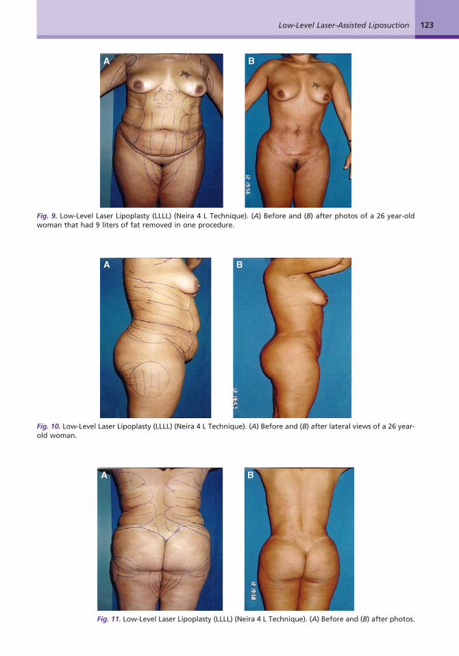
Fig. 9. Low-Level Laser Lipoplasty (LLLL) (Neira 4 L Technique). (A) Before and (B) after photos of a 26 year-oldwoman that had 9 liters of fat removed in one procedure.
Fig. 10. Low-Level Laser Lipoplasty (LLLL) (Neira 4 L Technique). (A) Before and (B) after lateral views of a 26 year-old woman.
Fig. 11. Low-Level Laser Lipoplasty (LLLL) (Neira 4 L Technique). (A) Before and (B) after photos.
123Low-Level Laser-Assisted Liposuction
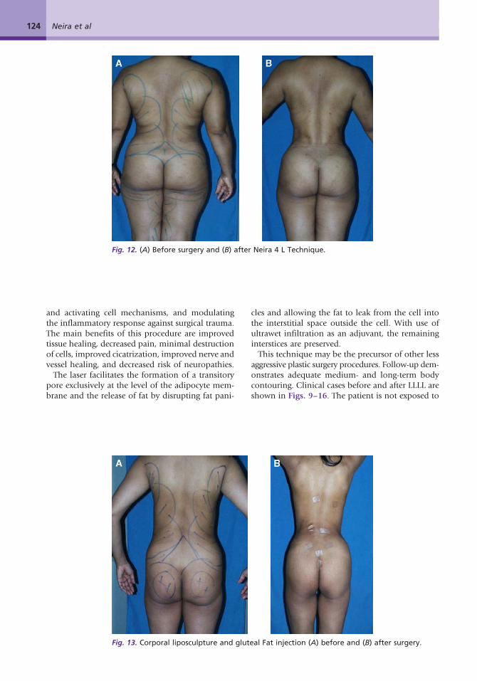
Fig. 12. (A) Before surgery and (B) after Neira 4 L Technique.
124 Neira et al
and activating cell mechanisms, and modulatingthe inflammatory response against surgical trauma.The main benefits of this procedure are improvedtissue healing, decreased pain, minimal destructionof cells, improved cicatrization, improved nerve andvessel healing, and decreased risk of neuropathies.The laser facilitates the formation of a transitory
pore exclusively at the level of the adipocyte mem-brane and the release of fat by disrupting fat pani-
Fig. 13. Corporal liposculpture and glut
cles and allowing the fat to leak from the cell intothe interstitial space outside the cell. With use ofultrawet infiltration as an adjuvant, the remaininginterstices are preserved.This technique may be the precursor of other less
aggressive plastic surgery procedures. Follow-up dem-onstrates adequate medium- and long-term bodycontouring. Clinical cases before and after LLLL areshown in Figs. 9–16. The patient is not exposed to
eal Fat injection (A) before and (B) after surgery.
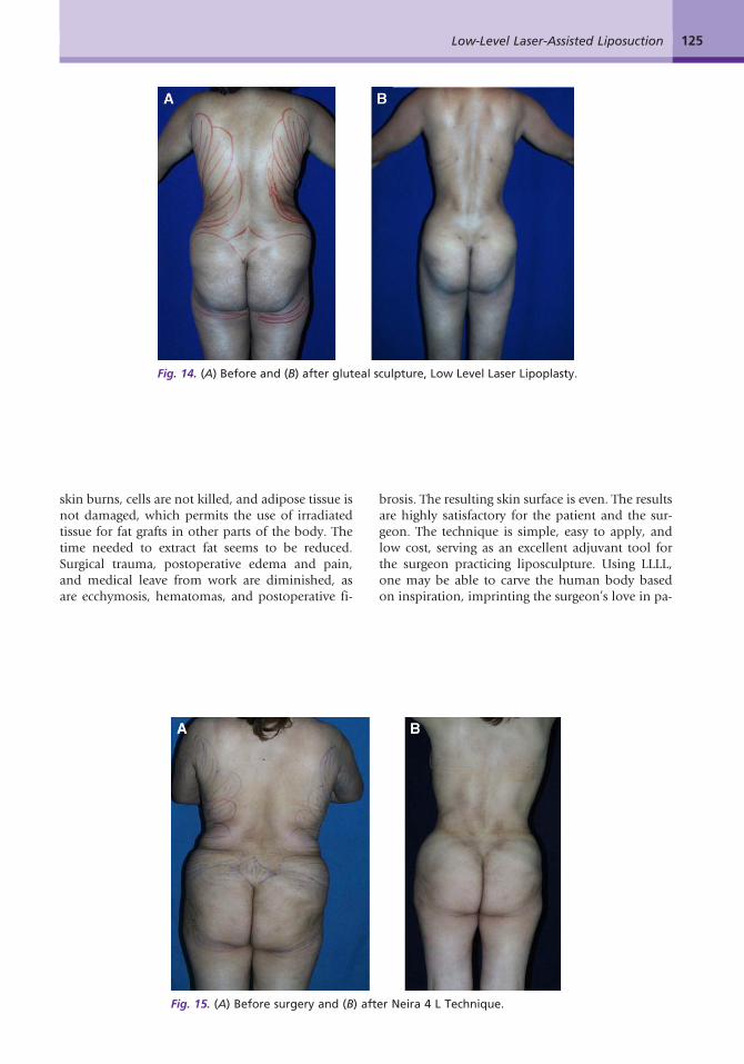
Fig. 14. (A) Before and (B) after gluteal sculpture, Low Level Laser Lipoplasty.
125Low-Level Laser-Assisted Liposuction
skin burns, cells are not killed, and adipose tissue isnot damaged, which permits the use of irradiatedtissue for fat grafts in other parts of the body. Thetime needed to extract fat seems to be reduced.Surgical trauma, postoperative edema and pain,and medical leave from work are diminished, asare ecchymosis, hematomas, and postoperative fi-
Fig. 15. (A) Before surgery and (B) aft
brosis. The resulting skin surface is even. The resultsare highly satisfactory for the patient and the sur-geon. The technique is simple, easy to apply, andlow cost, serving as an excellent adjuvant tool forthe surgeon practicing liposculpture. Using LLLL,one may be able to carve the human body basedon inspiration, imprinting the surgeon’s love in pa-
er Neira 4 L Technique.
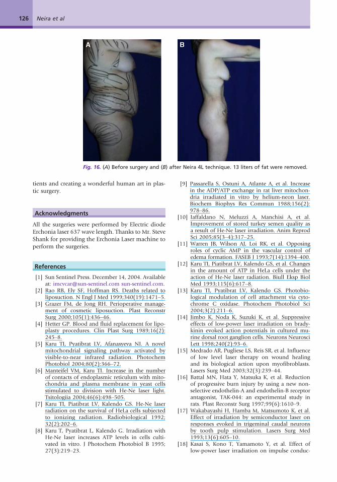
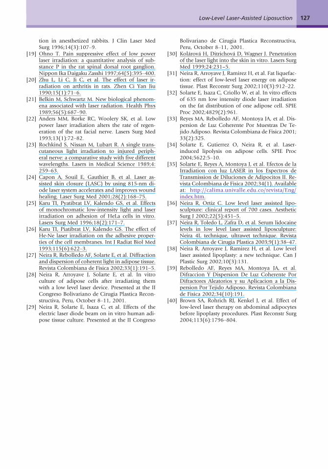
Fig. 16. (A) Before surgery and (B) after Neira 4L technique. 13 liters of fat were removed.
126 Neira et al
tients and creating a wonderful human art in plas-tic surgery.
Acknowledgments
All the surgeries were performed by Electric diodeErchonia laser 637 wave length. Thanks to Mr. SteveShank for providing the Erchonia Laser machine toperform the surgeries.
References
[1] Sun Sentinel Press. December 14, 2004. Availableat: [email protected] sun-sentinel.com.
[2] Rao RB, Ely SF, Hoffman RS. Deaths related toliposuction. N Engl J Med 1999;340(19):1471–5.
[3] Grazer FM, de Jong RH. Perioperative manage-ment of cosmetic liposuction. Plast ReconstrSurg 2000;105(1):436–46.
[4] Hetter GP. Blood and fluid replacement for lipo-plasty procedures. Clin Plast Surg 1989;16(2):245–8.
[5] Karu TI, Pyatibrat LV, Afanasyeva NI. A novelmitochondrial signaling pathway activated byvisible-to-near infrared radiation. PhotochemPhotobiol 2004;80(2):366–72.
[6] Manteifel VM, Karu TI. Increase in the numberof contacts of endoplasmic reticulum with mito-chondria and plasma membrane in yeast cellsstimulated to division with He-Ne laser light.Tsitologiia 2004;46(6):498–505.
[7] Karu TI, Piatibrat LV, Kalendo GS. He-Ne laserradiation on the survival of HeLa cells subjectedto ionizing radiation. Radiobiological 1992;32(2):202–6.
[8] Karu T, Pyatibrat L, Kalendo G. Irradiation withHe-Ne laser increases ATP levels in cells culti-vated in vitro. J Photochem Photobiol B 1995;27(3):219–23.
[9] Passarella S, Ostuni A, Atlante A, et al. Increasein the ADP/ATP exchange in rat liver mitochon-dria irradiated in vitro by helium-neon laser.Biochem Biophys Res Commun 1988;156(2):978–86.
[10] Iaffaldano N, Meluzzi A, Manchisi A, et al.Improvement of stored turkey semen quality asa result of He-Ne laser irradiation. Anim ReprodSci 2005;85(3–4):317–25.
[11] Warren JB, Wilson AJ, Loi RK, et al. Opposingroles of cyclic AMP in the vascular control ofedema formation. FASEB J 1993;7(14):1394–400.
[12] Karu TI, Piatibrat LV, Kalendo GS, et al. Changesin the amount of ATP in HeLa cells under theaction of He-Ne laser radiation. Biull Eksp BiolMed 1993;115(6):617–8.
[13] Karu TI, Pyatibrat LV, Kalendo GS. Photobio-logical modulation of cell attachment via cyto-chrome C oxidase. Photochem Photobiol Sci2004;3(2):211–6.
[14] Jimbo K, Noda K, Suzuki K, et al. Suppressiveeffects of low-power laser irradiation on brady-kinin evoked action potentials in cultured mu-rine dorsal root ganglion cells. Neurons NeurosciLett 1998;240(2):93–6.
[15] Medrado AR, Pugliese LS, Reis SR, et al. Influenceof low level laser therapy on wound healingand its biological action upon myofibroblasts.Lasers Surg Med 2003;32(3):239–44.
[16] Battal MN, Hata Y, Matsuka K, et al. Reductionof progressive burn injury by using a new non-selective endothelin-A and endothelin-B receptorantagonist, TAK-044: an experimental study inrats. Plast Reconstr Surg 1997;99(6):1610–9.
[17] Wakabayashi H, Hamba M, Matsumoto K, et al.Effect of irradiation by semiconductor laser onresponses evoked in trigeminal caudal neuronsby tooth pulp stimulation. Lasers Surg Med1993;13(6):605–10.
[18] Kasai S, Kono T, Yamamoto Y, et al. Effect oflow-power laser irradiation on impulse conduc-
127Low-Level Laser-Assisted Liposuction
tion in anesthetized rabbits. J Clin Laser MedSurg 1996;14(3):107–9.
[19] Ohno T. Pain suppressive effect of low powerlaser irradiation: a quantitative analysis of sub-stance P in the rat spinal dorsal root ganglion.Nippon Ika Daigaku Zasshi 1997;64(5):395–400.
[20] Zhu L, Li C, Ji C, et al. The effect of laser ir-radiation on arthritis in rats. Zhen Ci Yan Jiu1990;15(1):71–6.
[21] Belkin M, Schwartz M. New biological phenom-ena associated with laser radiation. Health Phys1989;56(5):687–90.
[22] Anders MM, Borke RC, Woolery SK, et al. Lowpower laser irradiation alters the rate of regen-eration of the rat facial nerve. Lasers Surg Med1993;13(1):72–82.
[23] Rochkind S, Nissan M, Lubart R. A single trans-cutaneous light irradiation to injured periph-eral nerve: a comparative study with five differentwavelengths. Lasers in Medical Science 1989;4:259–63.
[24] Capon A, Souil E, Gauthier B, et al. Laser as-sisted skin closure (LASC) by using 815-nm di-ode laser system accelerates and improves woundhealing. Laser Surg Med 2001;28(2):168–75.
[25] Karu TI, Pyatibrat LV, Kalendo GS, et al. Effectsof monochromatic low-intensity light and laserirradiation on adhesion of HeLa cells in vitro.Lasers Surg Med 1996;18(2):171–7.
[26] Karu TI, Piatibrat LV, Kalendo GS. The effect ofHe-Ne laser irradiation on the adhesive proper-ties of the cell membranes. Int J Radiat Biol Med1993;115(6):622–3.
[27] Neira R, Rebolledo AF, Solarte E, et al. Diffractionand dispersion of coherent light in adipose tissue.Revista Colombiana de Fisica 2002;33(1):191–5.
[28] Neira R, Arroyave J, Solarte E, et al. In vitroculture of adipose cells after irradiating themwith a low level laser device. Presented at the IICongreso Bolivariano de Cirugia Plastica Recon-structiva, Peru, October 8–11, 2001.
[29] Neira R, Solarte E, Isaza C, et al. Effects of theelectric laser diode beam on in vitro human adi-pose tissue culture. Presented at the II Congreso
Bolivariano de Cirugia Plastica Reconstructiva,Peru, October 8–11, 2001.
[30] Kolárová H, Ditrichová D, Wagner J. Penetrationof the laser light into the skin in vitro. Lasers SurgMed 1999;24:231–5.
[31] Neira R, Arroyave J, Ramirez H, et al. Fat liquefac-tion: effect of low-level laser energy on adiposetissue. Plast Reconstr Surg 2002;110(3):912–22.
[32] Solarte E, Isaza C, Criollo W, et al. In vitro effectsof 635 nm low intensity diode laser irradiationon the fat distribution of one adipose cell. SPIEProc 2002;4829(2):961.
[33] Reyes MA, Rebolledo AF, Montoya JA, et al. Dis-persion de Luz Coherente Por Muestras De Te-jido Adiposo. Revista Colombiana de Fisica 2001;33(2):325.
[34] Solarte E, Gutierrez O, Neira R, et al. Laser-induced lipolysis on adipose cells. SPIE Proc2004;5622:5–10.
[35] Solarte E, Reyes A, Montoya J, et al. Efectos de laIrradiation con luz LASER in los Espectros deTransmission de Diluciones de Adipocitos II. Re-vista Colombiana de Fisica 2002;34(1). Availableat: http://calima.univalle.edu.co/revista/Eng/index.htm.
[36] Neira R, Ortiz C. Low level laser assisted lipo-sculpture: clinical report of 700 cases. AestheticSurg J 2002;22(5):451–5.
[37] Neira R, Toledo L, Zafra D, et al. Serum lidocainelevels in low level laser assisted liposculpture:Neira 4L technique, ultrawet technique. RevistaColombiana de Cirugia Plastica 2003;9(1):38–47.
[38] Neira R, Arroyave J, Ramirez H, et al. Low levellaser assisted lipoplasty: a new technique. Can JPlastic Surg 2002;10(3):131.
[39] Rebolledo AF, Reyes MA, Montoya JA, et al.Difraccion Y Dispersion De Luz Coherente PorDifractores Aleatorios y su Aplicacion a la Dis-persion Por Tejido Adiposo. Revista Colombianade Fisica 2002;34(10):191.
[40] Brown SA, Rohrich RJ, Kenkel J, et al. Effect oflow-level laser therapy on abdominal adipocytesbefore lipoplasty procedures. Plast Reconstr Surg2004;113(6):1796–804.