Local Over-Expression of VEGF-DDNDC in the UterineArteries of Pregnant Sheep Results in Long-TermChanges in Uterine Artery Contractility and AngiogenesisVedanta Mehta1,3*, Khalil N. Abi-Nader1, Panicos Shangaris1, S. W. Steven Shaw1, Elisa Filippi1,
Elizabeth Benjamin1, Michael Boyd2, Donald M. Peebles1, John Martin3, Ian Zachary3, Anna L. David1
1 Institute for Women’s Health, UCL, London, United Kingdom, 2 BSU, Royal Veterinary College, Camden, London, United Kingdom, 3 Centre for Cardiovascular Biology
and Medicine, Division of Medicine, Rayne Building, UCL, London, United Kingdom
Abstract
Background: The normal development of the uteroplacental circulation in pregnancy depends on angiogenic andvasodilatory factors such as vascular endothelial growth factor (VEGF). Reduced uterine artery blood flow (UABF) is acommon cause of fetal growth restriction; abnormalities in angiogenic factors are implicated. Previously we showed thatadenovirus (Ad)-mediated VEGF-A165 expression in the pregnant sheep uterine artery (UtA) increased nitric oxide synthase(NOS) expression, altered vascular reactivity and increased UABF. VEGF-D is a VEGF family member that promotesangiogenesis and vasodilatation but, in contrast to VEGF-A, does not increase vascular permeability. Here we examined theeffect of Ad.VEGF-DDNDC vector encoding a fully processed form of VEGF-D, on the uteroplacental circulation.
Methods: UtA transit-time flow probes and carotid artery catheters were implanted in mid-gestation pregnant sheep (n = 5)to measure baseline UABF and maternal haemodynamics respectively. 7–14 days later, after injection of Ad.VEGF-DDNDC
vector (561011 particles) into one UtA and an Ad vector encoding b-galactosidase (Ad.LacZ) contralaterally, UABF wasmeasured daily until scheduled post-mortem examination at term. UtAs were assessed for vascular reactivity, NOSexpression and endothelial cell proliferation; NOS expression was studied in ex vivo transduced UtA endothelial cells(UAECs).
Results: At 4 weeks post-injection, Ad.VEGF-DDNDC treated UtAs showed significantly lesser vasoconstriction (Emax144.0 v/s184.2, p = 0.002). There was a tendency to higher UABF in Ad.VEGF-DDNDC compared to Ad.LacZ transduced UtAs (50.58% v/s 26.94%, p = 0.152). There was no significant effect on maternal haemodynamics. An increased number of proliferatingendothelial cells and adventitial blood vessels were observed in immunohistochemistry. Ad.VEGF-DDNDC expression incultured UAECs upregulated eNOS and iNOS expression.
Conclusions: Local over-expression of VEGF-DDNDC in the UtAs of pregnant mid-gestation sheep reduced vasoconstriction,promoted endothelial cell proliferation and showed a trend towards increased UABF. Studies in cultured UAECs indicatethat VEGF-DDNDC may act in part through upregulation of eNOS and iNOS.
Citation: Mehta V, Abi-Nader KN, Shangaris P, Shaw SWS, Filippi E, et al. (2014) Local Over-Expression of VEGF-DDNDC in the Uterine Arteries of Pregnant SheepResults in Long-Term Changes in Uterine Artery Contractility and Angiogenesis. PLoS ONE 9(6): e100021. doi:10.1371/journal.pone.0100021
Editor: Masuko Ushio-Fukai, University of Illinois at Chicago, United States of America
Received July 4, 2013; Accepted May 22, 2014; Published June 30, 2014
Copyright: � 2014 Mehta et al. This is an open-access article distributed under the terms of the Creative Commons Attribution License, which permitsunrestricted use, distribution, and reproduction in any medium, provided the original author and source are credited.
Funding: This study was partly funded by University College London Hospital (UCLH) Charities. VM is supported by a Dorothy Hodgkins Postgraduate Awardfrom the UK Medical Research Council and UCLH Charities. Work in IZ9s group is funded by the BHF. This work was undertaken at UCLH/UCL who received aproportion of the funding from the Department of Health9s NIHR Biomedical Research Center9s funding scheme. The funders had no role in study design, datacollection and analysis, decision to publish, or preparation of the manuscript.
Competing Interests: The authors have declared that no competing interests exist.
* Email: [email protected]
Introduction
The normal development of the placenta is key to ensuring an
uncomplicated pregnancy with adequate fetal growth. During
early pregnancy increased maternal cardiac output and tropho-
blast driven modification of the uterine spiral arteries result in a
dramatic increase in uterine perfusion [1] and a fall in utero-
placental resistance, allowing provision of sufficient oxygen and
nutrients for exchange across the placenta. Failure of this normal
physiological process leads to fetal growth restriction (FGR) and
pre-eclampsia (PET), two of the most challenging obstetric
complications. Despite several pre-clinical and clinical trials of
novel drugs and interventions, no effective therapies have been
developed.
The fall in utero-placental resistance in normal pregnancy is
mediated by interstitial extravillous trophoblast secretion of
angiogenic and vasodilatory factors such as vascular endothelial
growth factor (VEGF-A) to promote local blood flow to the uterus
[2,3]. VEGF induces vasodilatation and increases blood flow in
diverse vascular beds [4,5], effects mediated partly through its
stimulation of endothelial production of NO [6] and prostacyclin
[7]. In FGR and PET, there is decreased depth and density of
PLOS ONE | www.plosone.org 1 June 2014 | Volume 9 | Issue 6 | e100021

trophoblast invasion of the spiral arteries [8,9] and myometrial
small arteries show increased vasoconstriction and decreased
endothelium-dependent vasodilatation [10,11]. The invading
cytotrophoblasts secrete VEGF to regulate their acquisition of an
endothelial-like phenotype which allows them to replace the
maternal cells that line the uterine vessels. These cells also depend
on VEGF for their maintenance and growth [2]. In established
FGR, serum levels of VEGF-A165 are significantly diminished
[12]. In PET, placental-derived sFlt-1, a soluble receptor of VEGF
is upregulated, resulting in lowered circulating concentrations of
free VEGF and endothelial dysfunction [13]. Therapeutic
strategies targeting angiogenesis and vasodilatation in the utero-
placental circulation may therefore be of use in treating FGR and
PET.
Previously, we have demonstrated that local over-expression of
VEGF-A165 in the uterine arteries of pregnant sheep from mid-
gestation mediated by adenovirus vector (Ad) transduction, results
in a significant increase in uterine artery blood flow (UABF) for up
to one month after delivery and a reduction in uterine artery
contractility at term [14,15]. These effects are associated with a
short term increase in endothelial NO synthase (eNOS) in the
uterine arteries (UtAs) and long term UtA adventitial neovascu-
larization [15]. We also reported an upregulation of VEGFR-2 in
the UtAs transduced with Ad.VEGF-A165, suggesting this may be
the primary receptor mediating the biological effects observed.
This was achieved without affecting maternal or fetal haemody-
namic parameters.
VEGF-DDNDC [16] is a fully processed form of the VEGF family
member, VEGF-D, generated by proteolytic processing of the N-
and C-termini of full length VEGF-D, which has been shown to
have a significant angiogenic and vasodilatory effect, whereas the
full-length form is primarily lymphangiogenic. VEGF-DDNDC
elicits a more restricted range of biological responses compared
with VEGF-A165 [16,17] including less vascular permeability. In
this study we investigated the effect of Ad-mediated over-
expression of VEGF-DDNDC in the UtAs and uterine artery
endothelial cells (UAECs) of normal pregnant sheep, on eNOS
levels, angiogenesis, vascular reactivity, UABF and maternal
haemodynamics to determine its suitability as a therapeutic agent
for FGR.
Methods
Ethics StatementAll work was conducted in accordance with the UK Animals
(Scientific Procedures) Act (1986), project licence 70/6546 and
approved by the Royal Veterinary College ethics committee and
the UCL Biological Services Unit ethics committee.
AnimalsAll experiments were carried out in time-mated normal
pregnant sheep (Romney breed), not affected by vascular placental
insufficiency. Mid-gestation pregnant sheep (n = 6, 82–109 days of
gestation, term = 145 days) were studied 4–7 days after vector
administration, (‘‘short term’’) for organ bath experiments to
examine vascular reactivity, eNOS activity, neovascularization
and to assess acute toxicity (if any). A separate ‘‘long term’’ group
of mid-gestation pregnant ewes carrying singleton (n = 4) or twin
pregnancies (n = 1) (82–98 days of gestation) were studied until the
end of gestation (Table 1) for assessment of eNOS activity,
neovascularization, UABF measurements, and maternal haemo-
dynamics. In addition, two sheep were injected with only the
vehicle (PBS) and sacrificed at the short-term time point and long-
term time-point each (after injection), to provide control tissue for
histological and haematologic analysis. Normal mid-gestation
pregnant sheep (n = 6, 90–100 days) were used to provide uterine
artery endothelial cells (UAECs) for experiments.
Animal surgery and vector injectionAfter fasting overnight, pregnant ewes at 90.6066.19 days of
gestation underwent general anaesthesia induced with thiopental
sodium 20 mg/kg IV (Thiovet, Novartis Animal Health UK Ltd,
Table 1. Analysis performed on experimental sheep and long-term changes in UABF from baseline to 28 days after vectorinjection.
Animal Fetal number
Post mortemexamination(d after vectorinjection) Side of vector injection % Change in UABF at 28 days
Ad.VEGF-DDNDC Ad.LacZ Ad.VEGF-DDNDC Ad.LacZ
1 Singleton 4 Gravid Non-gravid NA NA
2 Singleton 6 Gravid Non-gravid NA NA
3 Singleton 7 Non-gravid Gravid NA NA
4 Singleton 7 Gravid Non-gravid NA NA
5 Singleton 5 Non-gravid Gravid NA NA
6 Singleton 4 Non-gravid Gravid NA NA
7 Twin 43 Gravid Gravid 23.60 21.23
8 Singleton 41 Non-gravid Gravid 66.14 47.86
9 Singleton 30 Gravid Non-gravid 24.77 4.48
10 Singleton 34 Non-gravid Gravid 32.65 19.85
11 Singleton 45 Non-gravid Gravid 105.76 41.32
UABF: Uterine artery blood flow; VEGF: Vascular Endothelial Growth Factor; NA: Not available; d: days.Animals 1–6 were used for short-term experiments.Animals 7–11 were used for long-term experiments, involving chronic implantation of telemetric flow probes around the uterine arteries.doi:10.1371/journal.pone.0100021.t001
VEGF-D Expression and Uterine Artery Blood Flow
PLOS ONE | www.plosone.org 2 June 2014 | Volume 9 | Issue 6 | e100021
Hertfordshire, UK) and maintained with 2–2.5% isoflurane in
oxygen (Isoflurane-Vet, Merial Animal Health Ltd, Essex, UK)
after intubation. Umbilical artery Doppler measurements, pulsa-
tility index (PI) and resistance index (RI) were acquired [14].
Gestational ages were confirmed by ultrasound using fetal
measurements [18]. For chronic maternal haemodynamic mon-
itoring (n = 4), a blood pressure sensitive PA-D70 catheter (Data
Sciences International, Tilburg, Netherlands) was inserted into the
carotid artery lumen, as described [15]. A laparotomy was then
performed, the UtAs were identified bilaterally and mobilised
immediately proximal to the first bifurcation. For long term
experiments, a transit time flow probe (6 mm 6PS, Transonic
Systems Inc., NY, USA), which can measure blood flow with an
absolute accuracy of 610% was placed around the main UtA on
each side. The cabling from each probe was then exteriorized onto
the ewe’s right flank, and the skin buttons were secured to the skin
as described [19]. The abdomen was closed, the ewe received
standard analgesia and antibiotic prophylaxis and the animal was
then recovered.
For long term experiments, vector was delivered to the UtAs 7–
14 days after flow probe placement (at 100.667.63 days of
gestation). For short-term experiments, sheep only had the vector
injection surgery at 97.5612.72 days of gestation without any
prior probe placement surgery. To deliver the Ad vectors into the
UtAs, the sheep underwent a second general anaesthetic and
laparotomy. The UtAs were identified bilaterally and mobilised
immediately proximal to the first bifurcation, or proximal to the
position of the flow probes. A butterfly needle (21 Gauge) was
inserted into the UtA and the viral vector (561011 viral particles in
10 ml phosphate buffered saline) was injected over a 1 minute
period, during which time the UtA was digitally occluded
proximal to the site of injection and for a further 4 minutes after
removal of the needle [14]. The operators were blinded to which
horn of the uterus received Ad.VEGF-DDNDC at the time of vector
injection. Ad.LacZ vector (561011 viral particles in 10 ml
phosphate buffered saline) was injected into the contralateral
UtA. The ewe received standard analgesia and antibiotic
prophylaxis, and the abdominal incision was closed [19].
Animal monitoring. Measurements of UABF, maternal
blood pressure and heart rate were recorded continuously in the
instrumented ewes, over the 3 days preceding and 7 days
succeeding vector injection to capture acute effects, and for
1 hour daily thereafter, at the same hour of the day, to capture
chronic effects.
UABF was sampled at a rate of 128 Hz and data were
transmitted telemetrically via the skin buttons when they were
connected to the PhysioGear I transmitter system and to the
PhysioView Data Acquisition Software (Transonic Systems Inc.).
The data acquired from the flow probes were analyzed using
Acknowledge software 3.9.1 (Biopac Q10 Systems Inc., CA, USA).
The baseline UABF was calculated as the average of three daily
mean UABF measurements taken for one hour each day before
vector injection. UABF percentage change from baseline was
calculated at specified time points, 7, 14, 21 and 28 days after
vector injection, using the average of three consecutive daily mean
UABF measurements on the day of and one day either side of the
time point. A two-way General Linear Model (GLiM) was used to
compare the UABF percentage change in Ad.VEGF-DDNDC and
Ad.LacZ-injected UtAs at each time point and also the gradients
of UABF percentage change over the length of gestation. The two
factors accounted for in the GLiM analysis were whether the UtA
supplied a gravid or non-gravid horn and whether Ad.VEGF-
DDNDC or Ad.LacZ vector was injected.
Maternal BP and HR were recorded telemetrically. Uploaded
traces were analyzed using Dataquest ART 4.1 software (Data
Sciences International). A two-tailed paired t-test was used to
compare changes in BP before and after the administration of the
vector.
Tissue sampling. Terminal anaesthesia was performed
either one week after vector administration (in short-term animals)
or at the end of gestation (136 to 142 days) to allow sampling of the
UtAs under optimal conditions. The umbilical artery PI and RI
were measured using ultrasound Doppler and compared with pre-
injection values. The UtAs and their next three divisions down to
the level of the uterine wall (vessel diameter 1 mm) as well as other
maternal and fetal tissues were sampled as described previously
[14]. Tissue samples to be used in histological and immunohis-
tochemical studies were fixed in 4% paraformaldehyde for 24 h
and then transferred into 70% alcohol to be subsequently blocked
in paraffin. All other samples were snap frozen in liquid nitrogen
and stored at 280uC.
Organ bath studies for UtA reactivity. The cleared second
and third UtA branches were divided into 3 mm long segments
and examined on two 8-chambered organ bath setups in the
absence and presence of inhibitors as described, namely L-NAME
(300 mM), an NO synthase inhibitor; NS-398 (10 mM), a
cyclooxygenase inhibitor and Apamin (1 mM) a blocker of SK
channels that inhibits the actions of endothelium-derived hyper-
polarizing factor [15].
Histological and blood examination. Paraffinized tissue
sections stained with hematoxylin and eosin were observed
microscopically for histological examination. Maternal and fetal
blood samples obtained before vector injection and at post mortem
(from both the short-term and long-term cohorts) were tested for
routine haematology, biochemistry and liver function tests at the
Clinical Diagnostics Laboratory, RVC Hawkeshead.
Assessment of endothelial cell proliferation and
neovascularization. Paraffinised sections of the main UtA
were double stained with anti-BrdU and anti-vWF antibodies to
assess endothelial cell proliferation and adventitial neovascular-
ization, respectively. Tissue sections were dewaxed and endoge-
nous peroxidase activity was blocked with 0.6% Hydrogen
peroxide for 15 minutes. Antigen retrieval was performed using
0.1% trypsin (215240, BD Biosciences, UK) digestion at 37uC for
10 minutes. The sections were then blocked with 5% non-immune
donkey serum (D9663, Sigma Aldrich, Gillingham, Dorset, UK) at
room temperature for 30 minutes. Polyclonal rabbit anti-human
vWF (1:400, A0082, Dako, Glostrup, Denmark) was used as the
primary antibody and incubated overnight at 4uC, followed by a
biotinylated donkey anti-rabbit secondary antibody (1:100, 711-
065-152, Jackson ImmunoResearch, West Grove, PA, USA) for
1 hour at room temperature. Following one wash with PBS
supplemented with 0.1% bovine serum albumin and two washes
with PBS, the sections were incubated with ABC solution
(PK4000, Vector Laboratories, Peterborough, UK) for one hour.
The deposited ABC complex was detected via covalent conjuga-
tion of biotinylated tyramide (1:1000, Dupont, UK), reacted in the
presence of 0.01% Hydrogen peroxide in PBS for 10 minutes at
room temperature [20]. This treatment with biotinylated tyramide
allowed us to transform an initially noncovalent form of biotin
labeling into a covalent one, to allow the label to withstand the
post-treatment with 2N Hydrochloric acid (HCl) for 45 minutes at
room temperature needed to expose the BrdU antigen. The HCl-
treated sections were briefly washed in PBS, exposed for
30 minutes to 0.1M boric acid/sodium borate buffer (pH 8.5),
washed in PBS again, pre-incubated with 0.5% Triton X-100 and
5% donkey serum in PBS and then incubated with the primary
VEGF-D Expression and Uterine Artery Blood Flow
PLOS ONE | www.plosone.org 3 June 2014 | Volume 9 | Issue 6 | e100021

mouse antibody against BrdU (1:100 Dako, Glostrup, Germany
M0744) overnight at 4uC. This step was followed by a one hour
incubation with a secondary Alexafluor-488 conjugated goat anti-
mouse IgG (1:200, 11001, Invitrogen, Paisley, UK) and then
enhanced with a tertiary Alexafluor-488 conjugated donkey anti-
goat antibody (1:200, 11055, Invitrogen, Paisley, UK), in
combination with Texas Red Streptavidin (016-070-084, Jackson
Immunoresearch) for one hour at room temperature. The sections
were covered with mounting medium with DAPI (H-1200, Vector
Laboratories, UK) and stored in the dark at 4uC before use.
Negative controls were obtained by not exposing the tissue section
to either of the primary antibodies.
Confocal Microscopy. For visualizing the immuno-fluores-
cence double labeling, digital micrographs of the Alexafluor-488
for the BrdU staining and Texas red fluorescence for vWF were
taken representing an area of 1 mm61 mm (1024 pixels61024
pixels; grayscale 0–255) with a Leica TCS 4D confocal laser
microscope using a 206 objective (Milton Keynes, UK). The
fluorescence was excited using low ArKr laser power (0.25 V) at
wavelengths of 488 nm for Alexafluor-488, 568 nm for Texas Red
and 358 nm (ultraviolet) for DAPI, and detected using the BP-
FITC filter for Alexafluor-488, the LP590 filter for Texas Red and
the LP360 filter for DAPI, respectively. Nine consecutive,
equidistant levels were recorded and condensed to a single bitmap
using the MaxIntens algorithm. Proliferating endothelial cells and
adventitial blood vessels (with a distinct lumen) were identified and
counted by two independent observers who were blinded to the
treatment. All analysis was performed in duplicate.
Measurement of VEGF-D protein expression. The quan-
tity of human VEGF-D protein in snap frozen samples of UtA,
uterine wall and whole placentome from two pregnant sheep in
the short-term study and long-term study each was measured by
enzyme-linked immunosorbent assay (R&D Systems, Minneapolis,
MN, USA) as described previously [14]. Human VEGF-D levels
Table 2. Antibodies used to confirm identity of endothelial cells isolated from pregnant sheep uterine arteries.
Primary Antibody Secondary Antibody
polyclonal rabbit anti-human vWF (1:400, A0082, Dako, Glostrup, Germany) Alexafluor-488 donkey anti-rabbit IgG (1:1000, A21206, Invitrogen, Paisley, UK)
rabbit monoclonal anti- b-catenin (1:2000, C2206, Sigma Aldrich, Gillingham,Dorset, UK)
Alexafluor-488 donkey anti-rabbit IgG (1:1000, A21206, Invitrogen, Paisley, UK)
mouse monoclonal anti-VE cadherin (1:500, sc9989, Santa Cruz Biotechnology,Heidelberg, Germany)
Alexafluor-488 goat anti-mouse IgG(1:1000, A11001, Invitrogen, Paisley,UK)
doi:10.1371/journal.pone.0100021.t002
Figure 1. Vascular reactivity of uterine arteries 4–7 days after vector administration. (A) Logarithmic dose-response curve to L-phenylephrine (PE) depicting that the contractile tension generated in the UtAs of pregnant sheep (n = 6) is significantly lower in Ad.VEGF-DDNDC
transduced vessels relative to Ad.LacZ transduced vessels 4–7 days post-vector injection. The contractility of the vessel is expressed as a percentageof the response to KCl. ** p,0.005. (B) Logarithmic dose-response curve to Bradykinin (BK) depicting that the relaxation response generated in theUtAs of pregnant sheep (n = 5) is significantly greater in the Ad.VEGF-DDNDC transduced arteries compared to Ad.LacZ treated vessels 4–7 days post-vector injection. The relaxation is expressed as a percentage of inhibition of PE-induced contractions. * denotes p = 0.05. Error bars denote SEM.doi:10.1371/journal.pone.0100021.g001
VEGF-D Expression and Uterine Artery Blood Flow
PLOS ONE | www.plosone.org 4 June 2014 | Volume 9 | Issue 6 | e100021
were also measured in pre- and post-injection maternal serum
samples and fetal serum samples collected at post-mortem
examination.
Measurements of phosphorylated and total eNOS, Akt
and Erk levels. Protein extracts from the snap-frozen UtA
tissues from both the short-term and long-term studies were used
to estimate levels of phosphorylated(p)-eNOS(Ser1177, 1:1000,
9570, Cell Signaling Technology, Danvers, MA, USA), total (T)-
eNOS (1:3000, 610296, BD Transduction Laboratories), p-Akt
(Ser473, 1:1000, 9271, Cell Signaling Technology), T-Akt (1:1000,
4691, Cell Signaling Technology), p-Erk (Thr202/Tyr204, 1:1000,
9101, Cell Signaling Technology) and T-Erk (1:1000, 9102, Cell
Signaling Technology) by western blotting, as previously described
[15].
Uterine artery endothelial cell (UAEC) isolation. UtAs
from normal mid-gestation pregnant sheep (approximately 90–100
days, n = 6) were dissected free of surrounding connective tissue
and cleared from their origin at the internal iliac artery up to the
level of the 2nd division, under terminal anaesthesia, as described
above. The ewe was then put down with an overdose of
intravenous pentobarbitone and the uterine arteries were ligated
at both ends using 1-0 silk ties and removed as a single piece
(which included the main, first and second branches).
The harvested UtAs were placed in a 10 cm petri-dish in a
sterile laminar flow hood and cleared further of surrounding
connective tissue and blood clots.
At the proximal end, a 23 gauge butterfly needle was introduced
and secured tightly with a haemostat. The vessel was flushed with
M-199 (50 ml, 41150-020, Invitrogen, Paisley, UK) to remove all
blood clots. The distal end of the vessel was then tied with a silk tie
and the vessel was inflated with Endothelial Cell Basal Medium
(EBM, CC3121, Lonza, Slough, UK) containing 5 mg/ml
collagenase (11088815001, Roche Diagnostics, Mannheim, Ger-
many) and 0.5% bovine serum albumin (BSA) (A4503, Sigma
Aldrich, UK) to dissociate endothelial cells from the vessel wall.
The inflated vessel was incubated at 37uC for 15 minutes. The
distal tie was then cut and the contents of the vessel were flushed
into a falcon tube using Endothelial Cell Growth Medium (EGM,
CC4133, Lonza). The endothelial cell fraction was concentrated
by centrifugation and washed two times with EGM to remove all
debris. The freshly isolated cells were considered to be at passage 0
and plated in 4 wells of a 6-well plate (140675, Nunc, Roskilde,
Denmark) in EGM containing 10% Fetal bovine serum, 1%
penicillin-streptomycin (15140-122, Invitrogen, Paisley, UK). All
cell surfaces on which endothelial cells were cultured were treated
with gelatin (G1393, Sigma Aldrich) to enhance adhesion to the
surface. Cells were grown for approximately 6 days and passaged
(passage 1) to T-25 flasks (136196, Nunc). Cells were grown to
70% confluence in T-25 flasks and then passaged (passage 2) to T-
75 flasks (178905, Nunc). Cells were again grown to approximately
70% confluence and passaged once more (passage 3) to T-175
flasks (178883, Nunc). Once ready for passage, the cells were
passaged (passage 4) to 6-well plates for adenovirus infection
experiments. To verify their endothelial identity, primary UAECs
were incubated with Ac-LDL tagged with Alexafluor-488 (L-
23380, Invitrogen, UK). Ac-LDL was added directly to cells
growing in culture in 1 ml EBM (serum-free) to yield a final
concentration of 10 mg/ml and left to incubate for four hours at
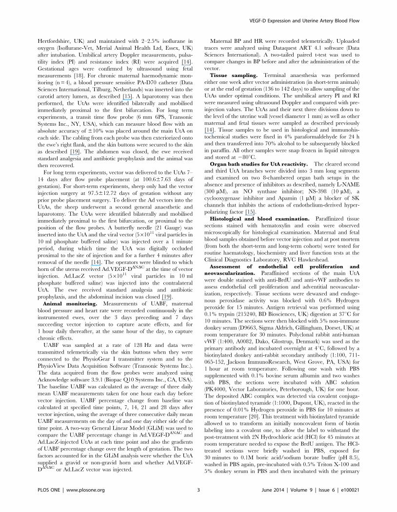
Figure 2. The endothelium-dependent relaxation to bradykinin in the presence of different inhibitors of the relaxation pathway inpregnant sheep uterine arteries, 4–7 days after Ad.VEGF-DDNDC or Ad.LacZ transduction. The contribution of NO, PGI2 and EDHF on therelaxation response to BK were investigated in vessels pre-contracted with PE. Cumulative relaxation curves of BK (10211M to 1026M) wereconstructed under the following conditions: (1) control (no inhibitors); (2) in the presence of L-NAME (300 mM); (3) in the presence of L-NAME and NS-398 (COX-2 inhibitor, 10 mM); (4) in the presence of L-NAME, NS-398 and apamin (1 mM). Relaxation was expressed as a percentage of inhibition of PE-induced contraction. The mean relaxation response of vessels from singleton pregnant sheep was calculated (n = 5). Statistical significance wasassumed at p,0.05. The BK relaxant effect was reduced by L-NAME (p,0.05, n = 5), but not significantly modified by the further addition of NS-398.The remaining endothelium-dependent relaxation (Emax), that was resistant to NS-398 and NO synthase inhibition, was significantly reduced bypretreatment with apamin in both Ad.VEGF-DDNDC and Ad.LacZ treated arteries (P,0.05, n = 5). The residual relaxation that was resistant to thecumulative addition of all three inhibitors was significantly greater in the Ad.VEGF-DDNDC transduced segments compared to Ad.LacZ transducedsegments.doi:10.1371/journal.pone.0100021.g002
VEGF-D Expression and Uterine Artery Blood Flow
PLOS ONE | www.plosone.org 5 June 2014 | Volume 9 | Issue 6 | e100021

Figure 3. Vascular reactivity of uterine arteries 30–45 days after vector administration. (A) Logarithmic dose-response curve to L-phenylephrine (PE) depicting that the contractile tension generated in the UtAs of term pregnant sheep (n = 5) is significantly lower in Ad.VEGF-DDNDC transduced vessels relative to Ad.LacZ transduced vessels 30–45 days post-injection. The contractility of the vessel is expressed as a percentageof the response to KCl. * p,0.005. (B) Logarithmic dose-response curve to Bradykinin (BK) depicting that the relaxation response generated in theUtAs of term pregnant sheep (n = 5) is not significantly different between the Ad.VEGF-DDNDC and Ad.LacZ treated vessels 30–45 days post-injection.The relaxation is expressed as a percentage of inhibition of PE-induced contractions. Error bars denote SEM.doi:10.1371/journal.pone.0100021.g003
Figure 4. Changes in UABF after adenovirus vector injection. Graph showing the percentage increase in UABF from baseline (adjusted to 0)and gradients of percentage increase in UABF in Ad.VEGF-DDNDC and Ad.LacZ transduced UtAs from 5 pregnant sheep. Vector injection = Day 0; Errorbars denote SEM.doi:10.1371/journal.pone.0100021.g004
VEGF-D Expression and Uterine Artery Blood Flow
PLOS ONE | www.plosone.org 6 June 2014 | Volume 9 | Issue 6 | e100021
37uC. The medium was then aspirated and fresh PBS was added.
Cells were then observed under a fluorescent microscope and
photographed on a confocal microscope.
Immunofluorescent staining of UAECs. UAECs (3.56105
cells/well) were seeded on a gelatinized cover-slip in a 6-well plate
and grown to 100% confluence overnight. The next morning, the
medium was aspirated and 4% formaldehyde was added gently
along the edge of each well to fix the cells. The plate was shaken
gently for 15 minutes, after which the formaldehyde was discarded
and the cells were washed twice with PBS. 0.1% Triton X-100
(diluted in PBS) was added to each well to permeabilize the cell
membrane. The solution was aspirated after 10 minutes and the
cells were washed twice with PBS. Primary antibodies were
prepared in PBS containing 0.1% Tween-20 and 1% BSA. The
antibodies used are outlined in Table 2. After the addition of the
primary antibody, the plate was left overnight at 4uC on the
shaking platform. Next morning, the cells were washed three times
in PBS. The appropriate secondary antibodies (Table 2) were
prepared in the same solution and added to the cells for one hour
at room temperature. The wells were again washed three times
with PBS. The coverslip was then gently lifted up and inverted
over a drop of 49,6-diamidino-2-phenylindole (DAPI) solution on a
glass slide (with the cell adherent surface of the coverslip facing
down). After five minutes, the slides were observed under a
fluorescent microscope and subsequently photographed on a
confocal microscope. Negative controls were obtained by omission
of the primary antibody.
Infection of UAECs with Adenovirus vectors. Cultured
UAECs were seeded in a six well plate (3.56105 cells/well), and
infected the following day with Ad.VEGF-DDNDC or Ad.LacZ at
multiplicities of infection (MOI) of 0, 1, 10, 100, 1000 and 10000.
At the same time, serum concentration in the culture medium was
changed to 0.5%. Protein was extracted after 48 hours of infection
for analysis by western blotting for p-eNOS, T-eNOS, p-Akt, T-
Akt, p-Erk and T-Erk as described above.
Results
There were no cases of maternal or fetal mortality and
morbidity. UABF and maternal haemodynamics were measured
successfully in all the ewes with chronically implanted UtA flow
probes and carotid artery blood-pressure sensitive catheters. Gross
examination at the time of post mortem and microscopic
histological examination of ewes and fetuses did not reveal any
pathology. The UtAs did not show any evidence of edema,
leucocyte infiltration or inflammation. There were no detectable
changes in haematological and biochemical profiles or liver
enzyme function when compared with baseline analysis in the
mother at 1 week (n = 3) or 5 weeks (n = 3) after vector injection, or
in the fetal sheep after vector injection when compared with
controls (which had only been injected with the vehicle).
Fetal WeightsFetal weights from singleton pregnancies undergoing long-term
UtA blood flow monitoring (n = 4) were measured at post-mortem
examination and compared to a historical singleton fetal control
group from the same sheep breed (n = 9). The mean gestational
age of the two groups was not statistically different (139.362.5
days v/s 137.863.9 days, p = 0.97, unpaired t-test). The mean
fetal weight in the experimental group was not significantly
different than that in the control group (48636492 grams v/s
469861004 grams, p = 0.45, unpaired t-test).
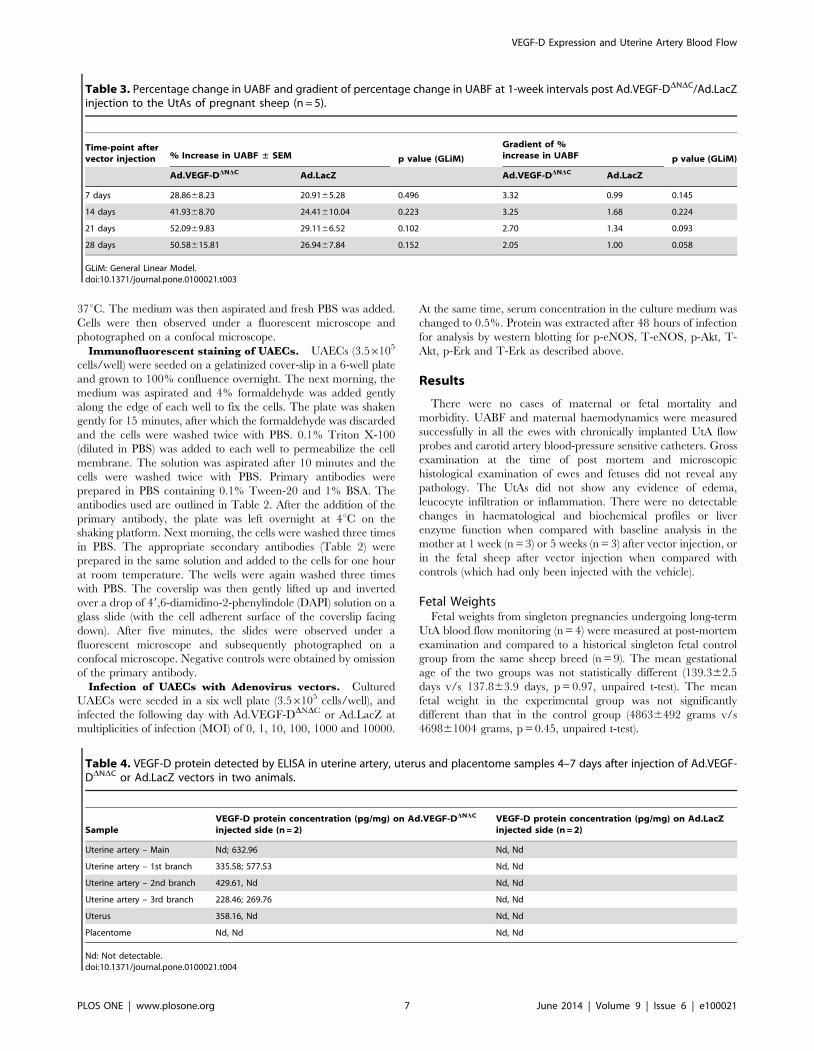
Table 3. Percentage change in UABF and gradient of percentage change in UABF at 1-week intervals post Ad.VEGF-DDNDC/Ad.LacZinjection to the UtAs of pregnant sheep (n = 5).
Time-point aftervector injection % Increase in UABF ± SEM p value (GLiM)
Gradient of %increase in UABF p value (GLiM)
Ad.VEGF-DDNDC Ad.LacZ Ad.VEGF-DDNDC Ad.LacZ
7 days 28.8668.23 20.9165.28 0.496 3.32 0.99 0.145
14 days 41.9368.70 24.41610.04 0.223 3.25 1.68 0.224
21 days 52.0969.83 29.1166.52 0.102 2.70 1.34 0.093
28 days 50.58615.81 26.9467.84 0.152 2.05 1.00 0.058
GLiM: General Linear Model.doi:10.1371/journal.pone.0100021.t003
Table 4. VEGF-D protein detected by ELISA in uterine artery, uterus and placentome samples 4–7 days after injection of Ad.VEGF-DDNDC or Ad.LacZ vectors in two animals.
SampleVEGF-D protein concentration (pg/mg) on Ad.VEGF-DDNDC
injected side (n = 2)VEGF-D protein concentration (pg/mg) on Ad.LacZinjected side (n = 2)
Uterine artery – Main Nd; 632.96 Nd, Nd
Uterine artery – 1st branch 335.58; 577.53 Nd, Nd
Uterine artery – 2nd branch 429.61, Nd Nd, Nd
Uterine artery – 3rd branch 228.46; 269.76 Nd, Nd
Uterus 358.16, Nd Nd, Nd
Placentome Nd, Nd Nd, Nd
Nd: Not detectable.doi:10.1371/journal.pone.0100021.t004
VEGF-D Expression and Uterine Artery Blood Flow
PLOS ONE | www.plosone.org 7 June 2014 | Volume 9 | Issue 6 | e100021

Figure 5. Effects of Ad.VEGF-DDNDC uterine artery transduction on phosphorylated (p) and Total (T) eNOS, Akt and Erk expression.(A) A representative blot shows upregulation of p-eNOS (Ser1177), T-eNOS, p-Akt and p-Erk in Ad.VEGF-DDNDC transduced UtAs compared to Ad.LacZtransduced UtAs 5 days after vector administration, but not 30 days after vector administration. Results are representative of n = 3 independentexperiments each for the short-term and long-term time points. GAPDH was used as a loading control. (B) Densitometric analysis was performed onthe western blots using Image J software, after normalizing against the density of GAPDH, T-Akt or T-Erk, as appropriate. Results are representative ofn = 3 independent experiments. * indicates p,0.05 (t-test).doi:10.1371/journal.pone.0100021.g005
Figure 6. Proliferation and Neovascularization in Ad.VEGF-DDNDC–transduced uterine arteries. Clusters of proliferating endothelial cellsin the short and long term sheep injected with Ad.VEGF-DDNDC (A–D and I-L respectively) and with Ad.LacZ (E–H and M–P respectively). PicturesA,E,I,M, show the staining of the nuclei with DAPI. The arrows in pictures B,F,J show nuclei which are positive to BrdU. The column containing C,G,K,Opictures shows positive staining to vWF. The merged pictures D,H,L,P, show the positive association of the BrdU stained nuclei with vWF whichconfirms that these nuclei belong to proliferating endothelial cells. Scale bar = 50 mm.doi:10.1371/journal.pone.0100021.g006
VEGF-D Expression and Uterine Artery Blood Flow
PLOS ONE | www.plosone.org 8 June 2014 | Volume 9 | Issue 6 | e100021
Fetal liver weights from the experimental group (n = 4) were
compared to a historical singleton fetal control group from the
same sheep breed (n = 10). The mean gestational age of the two
groups was not statistically different (139.362.5 days v/s
138.966.5 days, p = 0.68, unpaired t-test). Mean fetal liver weight
was higher in the experimental group (123.60624.67 grams v/s
106.10621.18 grams), although this increase was not significant
(p = 0.20).
Table 5. Mean number of proliferating endothelial cells and adventitial blood vessels in the uterine arteries of pregnant sheeptransduced with Ad.VEGF-DDNDC or Ad.LacZ and from uninjected sheep.
Treatment administeredDuration ofexperiment
Mean no. of proliferatingendothelial cells (±SEM)
Mean no. of adventitialblood vessels (±SEM)
Ad.VEGF-DDNDC (n = 4) 4–7 days 22.8366.03* 55.1066.82
Ad.LacZ (n = 4) 9.1662.68 50.4165.51
No treatment (n = 2) 7.7561.89 47.6865.40
Ad.VEGF-DDNDC (n = 4) 30–45 days 23.4766.16 77.9166.76*
Ad.LacZ (n = 4) 15.564.37 58.0665.78
No treatment (n = 2) 7.863.74 54.3367.26
* indicates significantly greater (p,0.05) compared to Ad.LacZ (control) by two-way ANOVA.doi:10.1371/journal.pone.0100021.t005
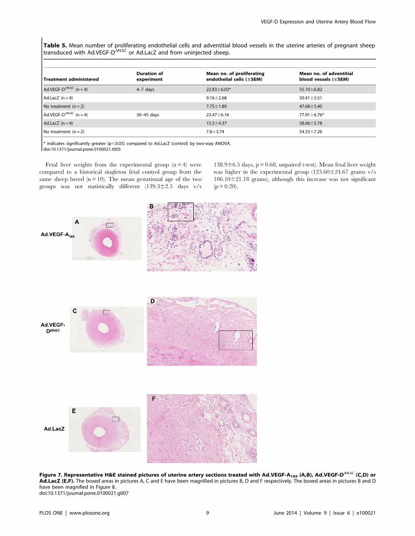
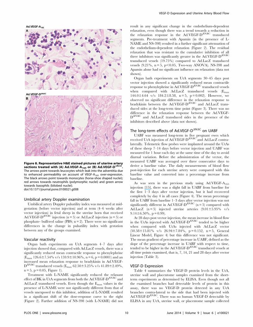
Figure 7. Representative H&E stained pictures of uterine artery sections treated with Ad.VEGF-A165 (A,B), Ad.VEGF-DDNDC (C,D) orAd.LacZ (E.F). The boxed areas in pictures A, C and E have been magnified in pictures B, D and F respectively. The boxed areas in pictures B and Dhave been magnified in Figure 8.doi:10.1371/journal.pone.0100021.g007
VEGF-D Expression and Uterine Artery Blood Flow
PLOS ONE | www.plosone.org 9 June 2014 | Volume 9 | Issue 6 | e100021
Umbilical artery Doppler examinationUmbilical artery Doppler pulsatility index was measured at mid-
gestation (before vector injection) and at term (4–6 weeks after
vector injection) in fetal sheep in the uterine horn that received
Ad.VEGF-DDNDC injection (n = 5) or Ad.LacZ injection (n = 5) or
phosphate- buffered saline (PBS; n = 2). There were no significant
differences in the change in pulsatility index with gestation
between any of the groups examined.
Vascular reactivityOrgan bath experiments on UtA segments 4–7 days after
injection showed that, compared with Ad.LacZ vessels, there was a
significantly reduced mean contractile response to phenylephrine
(Emax 126.667.54% v/s 159.9610.96%, n = 6, p = 0.0001) and an
increased mean relaxation response to bradykinin in Ad.VEGF-
DDNDC transduced vessels (Emax 62.5063.25% v/s 41.8962.49%,
n = 5, p = 0.05, Figure 1).
Treatment with L-NAME significantly reduced the relaxant
effect of BK in UtA segments from both the Ad.VEGF-DDNDC and
Ad.LacZ transduced vessels. Even though the Emax values in the
presence of L-NAME were not significantly different from that of
vessels unexposed to this inhibitor, addition of L-NAME resulted
in a significant shift of the dose-response curve to the right
(Figure 2). Further addition of NS-398 (with L-NAME) did not
result in any significant change in the endothelium-dependent
relaxation, even though there was a trend towards a reduction in
the relaxation response in the Ad.VEGF-DDNDC transduced
segments. Pre-treatment with Apamin (in the presence of L-
NAME and NS-398) resulted in a further significant attenuation of
the endothelium-dependent relaxation (Figure 2). The residual
relaxation that was resistant to the cumulative inhibition of all
three inhibitors was significantly greater in the Ad.VEGF-DDNDC
transduced vessels (19.75%) compared to Ad.LacZ transduced
vessels (9.21%, n = 5, p,0.05, Two-way ANOVA). NS-398 and
Apamin alone had no significant influence on relaxation (data not
shown).
Organ bath experiments on UtA segments 30–45 days post
vector injection showed a significantly reduced mean contractile
response to phenylephrine in Ad.VEGF-DDNDC transduced vessels
when compared with Ad.LacZ transduced vessels (Emax
144.064.64 v/s 184.268.58, n = 5, p = 0.002). However, we
observed no significant difference in the relaxation response to
bradykinin between the Ad.VEGF-DDNDC and Ad.LacZ trans-
duced sides at the long-term time point (Figure 3). There was no
difference in the relaxation response between the Ad.VEGF-
DDNDC and Ad.LacZ transduced sides in the presence of the
inhibitors described above (data not shown).
The long-term effects of Ad.VEGF-DDNDC on UABFUABF was measured long-term in five pregnant ewes which
received UtA injection of Ad.VEGF-DDNDC and Ad.LacZ contra-
laterally. Telemetric flow probes were implanted around the UtAs
of these sheep 7–14 days before vector injection and UABF was
measured for 1 hour each day at the same time of the day to avoid
diurnal variation. Before the administration of the vector, the
measured UABF was averaged over three consecutive days to
derive a baseline value. The daily measurements of blood flow
post-injection for each uterine artery were compared with this
baseline value and converted into a percentage increase from
baseline.
As was seen in the previous study using Ad.VEGF-A165
injection [15], there was a slight fall in UABF from baseline for
the first 1–3 days after vector injection, but it had recovered
completely by day 4 in all cases (Figure 4). The mean percentage
fall in UABF from baseline 1–3 days after vector injection was not
significantly different in Ad.VEGF-DDNDC (n = 5) compared with
Ad.LacZ (n = 5) injected uterine arteries (9.0165.95% v/s
9.1466.50%, p = 0.99).
At 28 days post vector injection, the mean increase in blood flow
in the UtAs injected with Ad.VEGF-DDNDC tended to be higher
when compared with UtAs injected with Ad.LacZ vector
(50.58615.81% v/s 26.9467.84%, p = 0.152, n = 5, General
Linear Model, Figure 4) but this difference was not significant.
The mean gradient of percentage increase in UABF, defined as the
slope of the percentage increase in UABF with respect to time,
tended to be higher in the Ad.VEGF-DDNDC transduced vessels at
all time points examined, that is, 7, 14, 21 and 28 days after vector
injection (Table 3).
VEGF-D ExpressionTable 4 summarizes the VEGF-D protein levels in the UtA,
uterine wall and placentome samples examined from the short-
term experiments as determined by ELISA. Even though not all
the examined branches had detectable levels of protein in this
assay, there was no VEGF-D protein detected in any UtA
branches contra-lateral to the side that had been injected with
Ad.VEGF-DDNDC. There was no human VEGF-D detectable by
ELISA in any UtA, uterine wall, or placentome sample collected
Figure 8. Representative H&E stained pictures of uterine arterysections treated with (A) Ad.VEGF-A165 or (B) Ad.VEGF-DDNDC.The arrows point towards leucocytes which leak into the adventitia dueto enhanced permeability on account of VEGF-A165 over-expression.The black arrows point towards monocytes (horse-shoe shaped nuclei);red arrows towards neutrophils (polymorphic nuclei) and green arrowtowards basophils (bilobed nuclei).doi:10.1371/journal.pone.0100021.g008
VEGF-D Expression and Uterine Artery Blood Flow
PLOS ONE | www.plosone.org 10 June 2014 | Volume 9 | Issue 6 | e100021
from long-term transduced ewes and sham controls. VEGF-D was
also not detected in maternal or fetal blood/serum samples
obtained at vector injection or at post-mortem examination in
short-term and long-term experiments. These findings are similar
to our previous findings for Ad.VEGF-A165 delivery in the UtAs
[14,15].
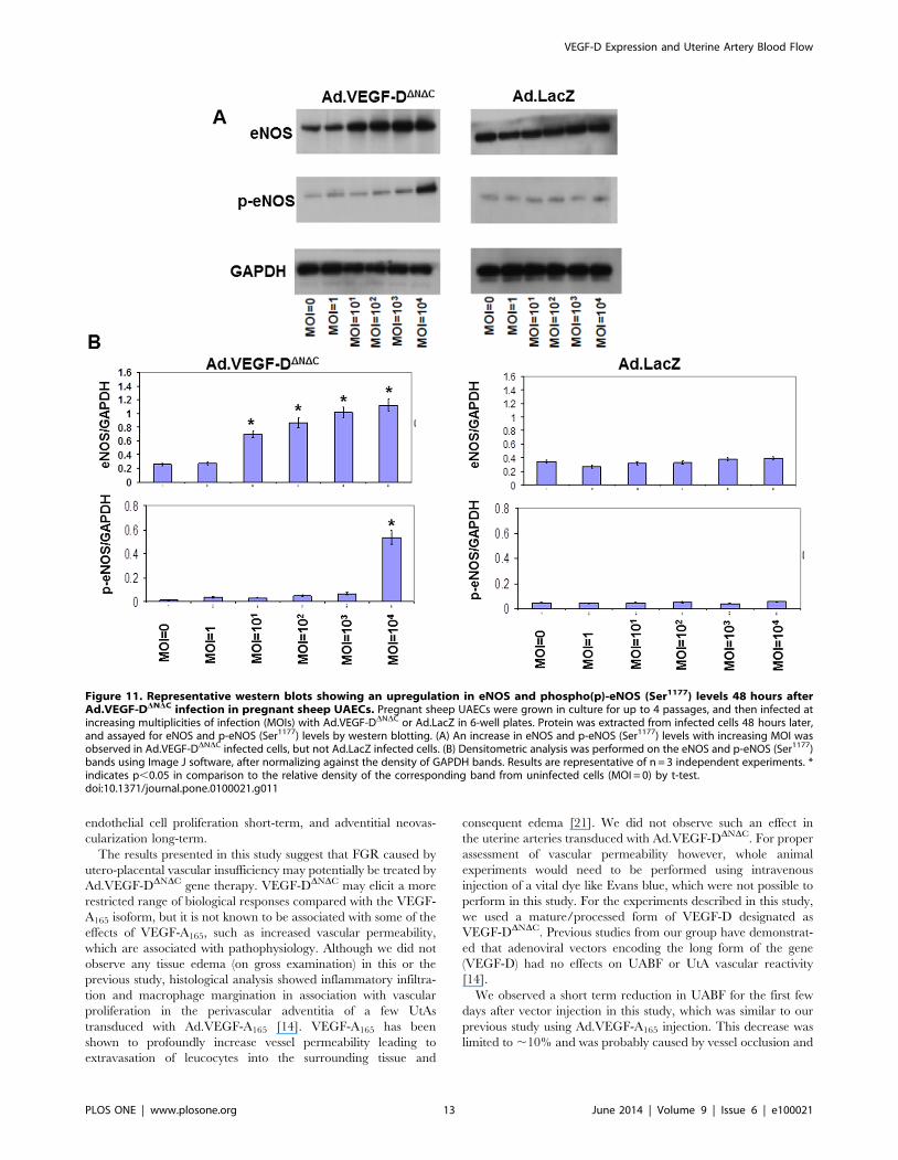
eNOS, Akt and Erk levelsProtein extracts of UtA samples from short-term studies (4–7
days after vector injection) and long-term studies (30–45 days after
vector injection) were analysed for changes in phosphorylated and
total levels of eNOS, Akt and Erk by western blotting. We
observed significantly increased levels of p-eNOS (Ser1177), T-
eNOS, p-Akt and p-Erk in Ad.VEGF-DDNDC transduced UtAs
short-term. However, this difference was not sustained long-term
(Figure 5).
Neovascularization and Endothelial cell proliferationFour to seven days after transduction we observed a significant
increase in the number of proliferating endothelial cells in the
main branch of Ad.VEGF-DDNDC transduced UtAs compared to
Ad.LacZ transduced UtAs or untransduced UtAs from control
sheep at the same gestational age (p = 0.013, n = 4, Two-way
ANOVA, Figure 6). ANOVA showed that the vector type had a
significant effect on the number of proliferating endothelial cells
but whether the UtA was supplying the gravid or non-gravid
uterine horn did not (p = 0.563). There was no significant
difference in the number of adventitial blood vessels (p = 0.301,
n = 4, Two-way ANOVA) between the Ad.VEGF-DDNDC trans-
duced UtAs and Ad.LacZ transduced UtAs. The mean number of
proliferating endothelial cells and adventitial blood vessels in the
Ad.VEGF-DDNDC/Ad.LacZ transduced UtAs and untransduced
UtAs is summarized in Table 5.
After long-term transduction we observed a tendency to higher
numbers of proliferating endothelial cells in the Ad.VEGF-DDNDC
transduced UtAs compared to Ad.LacZ transduced and uninfect-
ed UtAs, though this increase was not significant. (p = 0.159, n = 4,
Two-way ANOVA, Table 5). The number of adventitial blood
vessels was significantly greater in the Ad.VEGF-DDNDC trans-
duced UtAs compared to Ad.LacZ transduced and uninjected
UtAs (p = 0.043, n = 4, Two-way ANOVA). ANOVA showed that
whether the uterine artery was supplying a gravid or non-gravid
uterine horn had no significant effect on the number of adventitial
blood vessels (p = 0.436).
Figure 9. Representative graph showing the (A) diurnal short-term and (B) hourly long-term variation in maternal blood pressurebefore and after the administration of the vector. There were no significant changes in maternal haemodynamics after Ad.VEGF-DDNDC
administration when compared to baseline. Error bars represent SEM.doi:10.1371/journal.pone.0100021.g009
VEGF-D Expression and Uterine Artery Blood Flow
PLOS ONE | www.plosone.org 11 June 2014 | Volume 9 | Issue 6 | e100021
Vascular permeability and inflammationH&E stained sections of the uterine arteries treated with either
Ad.VEGF-A165, Ad.VEGF-DDNDC or Ad.LacZ were examined
microscopically to look for the presence of inflammatory cells, if
any. The adventitia of Ad.VEGF-A165 treated vessels appeared
more diffuse than that of Ad.VEGF-DDNDC or Ad.LacZ treated
vessels, suggestive of edema, and also had a greater number of
nucleated cells (Figures 7 and 8). Higher magnification images
showed that inflammatory cells, particularly neutrophil poly-
morphs, monocytes and basophils could be identified in the
adventitial layer of Ad.VEGF-A165 treated arteries but not
Ad.VEGF-DDNDC treated arteries (Figure 8).
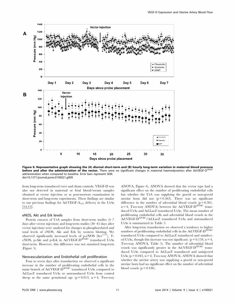
Maternal haemodynamicsMaternal blood pressure (BP) was monitored in 5 ewes. There
were no short term changes in blood pressure in the first 2 days
after vector injection (Figure 9), when VEGF-DDNDC expression
would be expected to be at a maximum level. By 7 days after
vector injection, the maternal mean arterial pressure had increased
marginally from 83.3962.65 mmHg at baseline to
85.6068.15 mmHg. This change is similar to our observations
in the sham-injected control ewes (85.57 mmHg to 88.13 mmHg).
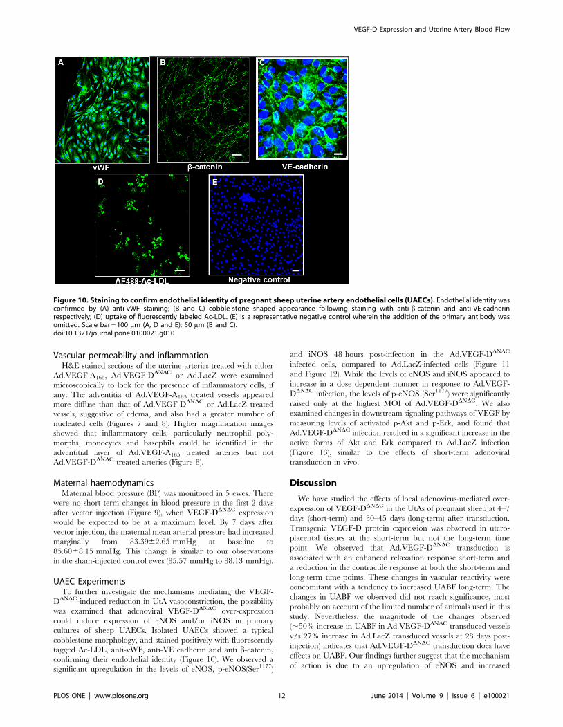
UAEC ExperimentsTo further investigate the mechanisms mediating the VEGF-
DDNDC-induced reduction in UtA vasoconstriction, the possibility
was examined that adenoviral VEGF-DDNDC over-expression
could induce expression of eNOS and/or iNOS in primary
cultures of sheep UAECs. Isolated UAECs showed a typical
cobblestone morphology, and stained positively with fluorescently
tagged Ac-LDL, anti-vWF, anti-VE cadherin and anti b-catenin,
confirming their endothelial identity (Figure 10). We observed a
significant upregulation in the levels of eNOS, p-eNOS(Ser1177)
and iNOS 48 hours post-infection in the Ad.VEGF-DDNDC
infected cells, compared to Ad.LacZ-infected cells (Figure 11
and Figure 12). While the levels of eNOS and iNOS appeared to
increase in a dose dependent manner in response to Ad.VEGF-
DDNDC infection, the levels of p-eNOS (Ser1177) were significantly
raised only at the highest MOI of Ad.VEGF-DDNDC. We also
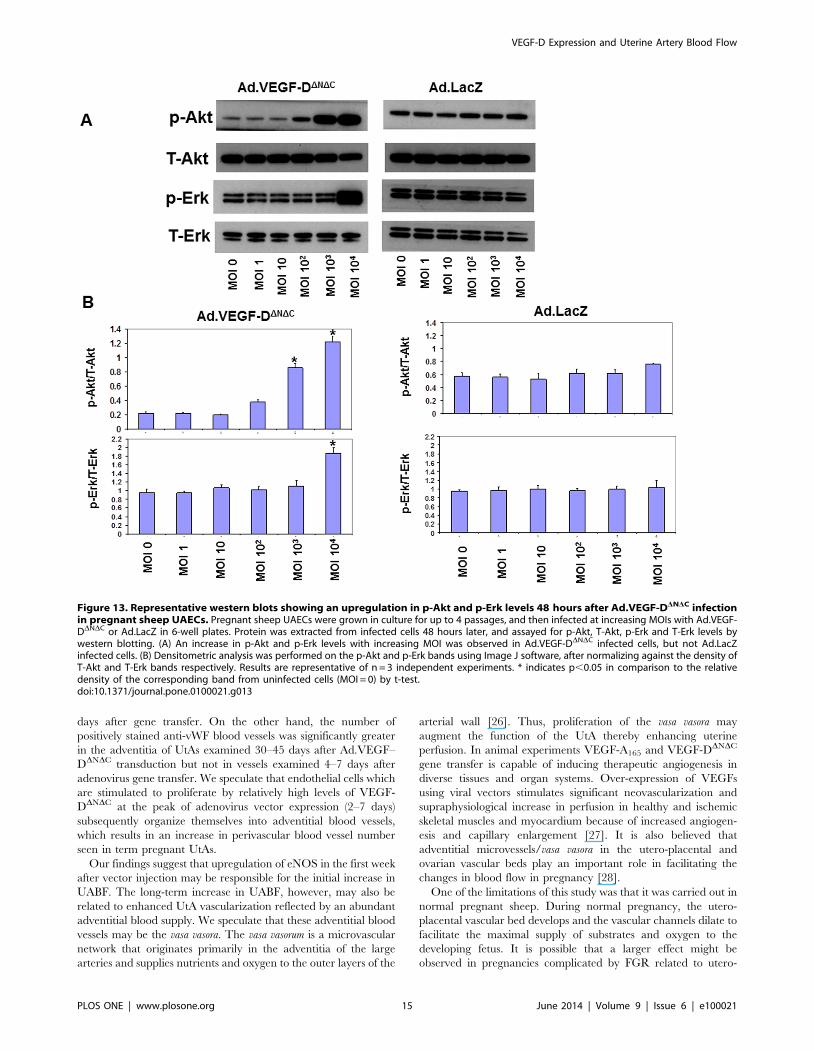
examined changes in downstream signaling pathways of VEGF by
measuring levels of activated p-Akt and p-Erk, and found that
Ad.VEGF-DDNDC infection resulted in a significant increase in the
active forms of Akt and Erk compared to Ad.LacZ infection
(Figure 13), similar to the effects of short-term adenoviral
transduction in vivo.
Discussion
We have studied the effects of local adenovirus-mediated over-
expression of VEGF-DDNDC in the UtAs of pregnant sheep at 4–7
days (short-term) and 30–45 days (long-term) after transduction.
Transgenic VEGF-D protein expression was observed in utero-
placental tissues at the short-term but not the long-term time
point. We observed that Ad.VEGF-DDNDC transduction is
associated with an enhanced relaxation response short-term and
a reduction in the contractile response at both the short-term and
long-term time points. These changes in vascular reactivity were
concomitant with a tendency to increased UABF long-term. The
changes in UABF we observed did not reach significance, most
probably on account of the limited number of animals used in this
study. Nevertheless, the magnitude of the changes observed
(,50% increase in UABF in Ad.VEGF-DDNDC transduced vessels
v/s 27% increase in Ad.LacZ transduced vessels at 28 days post-
injection) indicates that Ad.VEGF-DDNDC transduction does have
effects on UABF. Our findings further suggest that the mechanism
of action is due to an upregulation of eNOS and increased
Figure 10. Staining to confirm endothelial identity of pregnant sheep uterine artery endothelial cells (UAECs). Endothelial identity wasconfirmed by (A) anti-vWF staining; (B and C) cobble-stone shaped appearance following staining with anti-b-catenin and anti-VE-cadherinrespectively; (D) uptake of fluorescently labeled Ac-LDL. (E) is a representative negative control wherein the addition of the primary antibody wasomitted. Scale bar = 100 mm (A, D and E); 50 mm (B and C).doi:10.1371/journal.pone.0100021.g010
VEGF-D Expression and Uterine Artery Blood Flow
PLOS ONE | www.plosone.org 12 June 2014 | Volume 9 | Issue 6 | e100021
endothelial cell proliferation short-term, and adventitial neovas-
cularization long-term.
The results presented in this study suggest that FGR caused by
utero-placental vascular insufficiency may potentially be treated by
Ad.VEGF-DDNDC gene therapy. VEGF-DDNDC may elicit a more
restricted range of biological responses compared with the VEGF-
A165 isoform, but it is not known to be associated with some of the
effects of VEGF-A165, such as increased vascular permeability,
which are associated with pathophysiology. Although we did not
observe any tissue edema (on gross examination) in this or the
previous study, histological analysis showed inflammatory infiltra-
tion and macrophage margination in association with vascular
proliferation in the perivascular adventitia of a few UtAs
transduced with Ad.VEGF-A165 [14]. VEGF-A165 has been
shown to profoundly increase vessel permeability leading to
extravasation of leucocytes into the surrounding tissue and
consequent edema [21]. We did not observe such an effect in
the uterine arteries transduced with Ad.VEGF-DDNDC. For proper
assessment of vascular permeability however, whole animal
experiments would need to be performed using intravenous
injection of a vital dye like Evans blue, which were not possible to
perform in this study. For the experiments described in this study,
we used a mature/processed form of VEGF-D designated as
VEGF-DDNDC. Previous studies from our group have demonstrat-
ed that adenoviral vectors encoding the long form of the gene
(VEGF-D) had no effects on UABF or UtA vascular reactivity
[14].
We observed a short term reduction in UABF for the first few
days after vector injection in this study, which was similar to our
previous study using Ad.VEGF-A165 injection. This decrease was
limited to ,10% and was probably caused by vessel occlusion and
Figure 11. Representative western blots showing an upregulation in eNOS and phospho(p)-eNOS (Ser1177) levels 48 hours afterAd.VEGF-DDNDC infection in pregnant sheep UAECs. Pregnant sheep UAECs were grown in culture for up to 4 passages, and then infected atincreasing multiplicities of infection (MOIs) with Ad.VEGF-DDNDC or Ad.LacZ in 6-well plates. Protein was extracted from infected cells 48 hours later,and assayed for eNOS and p-eNOS (Ser1177) levels by western blotting. (A) An increase in eNOS and p-eNOS (Ser1177) levels with increasing MOI wasobserved in Ad.VEGF-DDNDC infected cells, but not Ad.LacZ infected cells. (B) Densitometric analysis was performed on the eNOS and p-eNOS (Ser1177)bands using Image J software, after normalizing against the density of GAPDH bands. Results are representative of n = 3 independent experiments. *indicates p,0.05 in comparison to the relative density of the corresponding band from uninfected cells (MOI = 0) by t-test.doi:10.1371/journal.pone.0100021.g011
VEGF-D Expression and Uterine Artery Blood Flow
PLOS ONE | www.plosone.org 13 June 2014 | Volume 9 | Issue 6 | e100021
consequent trauma during injection. UABF recovered in all
treated sheep by day 4 after injection.
Ad.VEGF-DDNDC transduction resulted in a significant reduc-
tion in the UtA contractile response at both the short-term and
long-term time points, but an enhancement of the relaxation
response only at the short-term time point. This is most likely to be
because at term, the utero-placental blood vessels are maximally
dilated, and VEGF over-expression, which is known to bring
about vasodilatation, may be unable to further enhance the
relaxation of UtAs. It was further noted that in the short-term
treated vessels, the amount of residual relaxation after cumulative
inhibition with L-NAME, NS-398 and Apamin was significantly
greater in the Ad.VEGF-DDNDC treated vessels compared to
Ad.LacZ treated vessels. This may reflect either another as yet
unidentified VEGF-mediated relaxation mechanism, or, alterna-
tively, augmentation of NO/EDHF-dependent signaling resulting
from the over-expression of VEGF-DDNDC. We plan to do further
experiments with endothelium-independent vasodilators (like
Sodium nitroprusside) to test whether the differences in relaxation
observed were indeed mediated by the endothelium.
Ad.VEGF-DDNDC transduction resulted in an upregulation of
eNOS, p-eNOS (Ser1177), iNOS, p-Akt and p-Erk in pregnant
sheep UAECs at the 48 hour time point. Ser1177 is the same site
phosphorylated in response to shear stress [22] and phosphory-
lation of this site renders eNOS active at resting Ca2+ concentra-
tions. All studies on UAECs were carried out at passage four.
Ovine UAECs retain their primary in vivo characteristics up to
passage four, meaning that expression of key proteins and mRNA
are retained. Levels of eNOS protein and mRNA are found to be
higher in UAECs from pregnant ewes compared to cells from non-
pregnant ewes at the fourth passage [23,24]. Similar to the
findings in UAECs, we observed that Ad.VEGF-DDNDC trans-
duction upregulated eNOS, p-eNOS (Ser1177), p-Akt and p-Erk in
pregnant UtAs for up to at least 7 days after gene transfer, but this
upregulation was no longer evident at the long-term time point.
This may be because continued significant VEGF expression is
needed for long-term eNOS upregulation or, alternatively, eNOS
level at term and in normal pregnancy is already at its peak and
cannot be upregulated further by VEGF over-expression. These
results suggest that VEGF may be activating eNOS via
Phosphoinositide 3-kinase (PI3K) dependent Akt-catalysed phos-
phorylation. PI3Ks are a family of enzymes which play an
important role in cellular physiology, particularly cell growth,
proliferation, differentiation, motility, survival and intracellular
trafficking. They mediate these functions in response to the
binding of different growth factors to cell surface receptors [25].
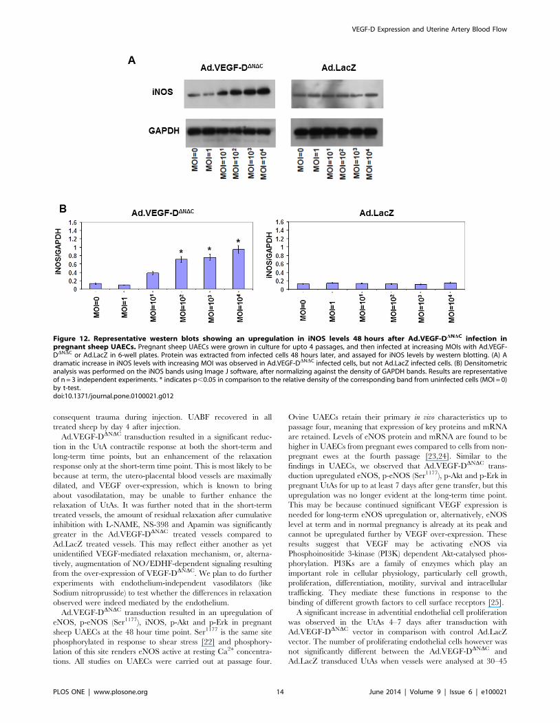
A significant increase in adventitial endothelial cell proliferation
was observed in the UtAs 4–7 days after transduction with
Ad.VEGF-DDNDC vector in comparison with control Ad.LacZ
vector. The number of proliferating endothelial cells however was
not significantly different between the Ad.VEGF-DDNDC and
Ad.LacZ transduced UtAs when vessels were analysed at 30–45
Figure 12. Representative western blots showing an upregulation in iNOS levels 48 hours after Ad.VEGF-DDNDC infection inpregnant sheep UAECs. Pregnant sheep UAECs were grown in culture for upto 4 passages, and then infected at increasing MOIs with Ad.VEGF-DDNDC or Ad.LacZ in 6-well plates. Protein was extracted from infected cells 48 hours later, and assayed for iNOS levels by western blotting. (A) Adramatic increase in iNOS levels with increasing MOI was observed in Ad.VEGF-DDNDC infected cells, but not Ad.LacZ infected cells. (B) Densitometricanalysis was performed on the iNOS bands using Image J software, after normalizing against the density of GAPDH bands. Results are representativeof n = 3 independent experiments. * indicates p,0.05 in comparison to the relative density of the corresponding band from uninfected cells (MOI = 0)by t-test.doi:10.1371/journal.pone.0100021.g012
VEGF-D Expression and Uterine Artery Blood Flow
PLOS ONE | www.plosone.org 14 June 2014 | Volume 9 | Issue 6 | e100021
days after gene transfer. On the other hand, the number of
positively stained anti-vWF blood vessels was significantly greater
in the adventitia of UtAs examined 30–45 days after Ad.VEGF–
DDNDC transduction but not in vessels examined 4–7 days after
adenovirus gene transfer. We speculate that endothelial cells which
are stimulated to proliferate by relatively high levels of VEGF-
DDNDC at the peak of adenovirus vector expression (2–7 days)
subsequently organize themselves into adventitial blood vessels,
which results in an increase in perivascular blood vessel number
seen in term pregnant UtAs.
Our findings suggest that upregulation of eNOS in the first week
after vector injection may be responsible for the initial increase in
UABF. The long-term increase in UABF, however, may also be
related to enhanced UtA vascularization reflected by an abundant
adventitial blood supply. We speculate that these adventitial blood
vessels may be the vasa vasora. The vasa vasorum is a microvascular
network that originates primarily in the adventitia of the large
arteries and supplies nutrients and oxygen to the outer layers of the
arterial wall [26]. Thus, proliferation of the vasa vasora may
augment the function of the UtA thereby enhancing uterine
perfusion. In animal experiments VEGF-A165 and VEGF-DDNDC
gene transfer is capable of inducing therapeutic angiogenesis in
diverse tissues and organ systems. Over-expression of VEGFs
using viral vectors stimulates significant neovascularization and
supraphysiological increase in perfusion in healthy and ischemic
skeletal muscles and myocardium because of increased angiogen-
esis and capillary enlargement [27]. It is also believed that
adventitial microvessels/vasa vasora in the utero-placental and
ovarian vascular beds play an important role in facilitating the
changes in blood flow in pregnancy [28].
One of the limitations of this study was that it was carried out in
normal pregnant sheep. During normal pregnancy, the utero-
placental vascular bed develops and the vascular channels dilate to
facilitate the maximal supply of substrates and oxygen to the
developing fetus. It is possible that a larger effect might be
observed in pregnancies complicated by FGR related to utero-
Figure 13. Representative western blots showing an upregulation in p-Akt and p-Erk levels 48 hours after Ad.VEGF-DDNDC infectionin pregnant sheep UAECs. Pregnant sheep UAECs were grown in culture for up to 4 passages, and then infected at increasing MOIs with Ad.VEGF-DDNDC or Ad.LacZ in 6-well plates. Protein was extracted from infected cells 48 hours later, and assayed for p-Akt, T-Akt, p-Erk and T-Erk levels bywestern blotting. (A) An increase in p-Akt and p-Erk levels with increasing MOI was observed in Ad.VEGF-DDNDC infected cells, but not Ad.LacZinfected cells. (B) Densitometric analysis was performed on the p-Akt and p-Erk bands using Image J software, after normalizing against the density ofT-Akt and T-Erk bands respectively. Results are representative of n = 3 independent experiments. * indicates p,0.05 in comparison to the relativedensity of the corresponding band from uninfected cells (MOI = 0) by t-test.doi:10.1371/journal.pone.0100021.g013
VEGF-D Expression and Uterine Artery Blood Flow
PLOS ONE | www.plosone.org 15 June 2014 | Volume 9 | Issue 6 | e100021

placental insufficiency, where there is evidence of reduced
trophoblast invasion of the spiral arteries, increased resistance to
blood flow and reduced perfusion.
An intravenous infusion of VEGF has been shown to result in
transient tachycardia, hypotension and a decrease in cardiac
output in conscious instrumented rats [29]. In this study however,
we did not observe any changes in maternal haemodynamics,
other than a small fall in BP at the end of gestation, which is
normally observed in sheep [30]. At the same time, no long-term
expression of VEGF-DDNDC could be detected in maternal and
fetal tissues by ELISA, which provides re-assurance against long-
term toxic effects. In our previous studies, we were only able to
detect transgenic VEGF-A165 expression by RT-PCR in the
uterine arteries that had actually been injected with Ad.VEGF-
A165, but not in any other maternal or fetal tissue [14,15]. These
findings support our conclusion that UtA injection of Ad vectors
results in only local transduction and transgenic protein expres-
sion, without eliciting systemic effects which could be deleterious
in pregnancy.
This study was designed with clinical relevance in mind.
Application of the vector injection technique described in this
paper to human patients is particularly challenging. FGR fetuses
are frequently hypoxic, and reductions in UABF during UtA
occlusion may exacerbate the situation resulting in fetal/neonatal
complications. A minimally invasive technique such as transfem-
oral UtA catheterization with temporary balloon occlusion of the
vessel lumen as is used to treat massive obstetric haemorrhage [31]
may decrease UtA trauma and post-injection vasoconstriction.
Further experiments to determine optimal vector dose and the
mode of delivery are required. Another issue is the timing of gene
delivery. Fetuses with advanced growth restriction and cardiovas-
cular compensation through brain sparing have a significant
degree of hypoxemia but are not acidemic until abnormal
precordial venous dopplers are observed that signal decompensa-
tion [32]. This should be considered when deciding on the best
timing for vector application, and especially that the longitudinal
progression of early severe FGR is well defined [33]. In addition,
those fetuses with early FGR and very high umbilical artery
resistance at presentation (.4SD above the mean for gestation)
could be considered at a very high risk for morbidity and mortality
[34]. These fetuses would represent the target population for a
phase I trial of Ad.VEGF-A165/Ad.VEGF-DDNDC gene therapy
and application of the vector in this case would be preferable at a
stage where significant hypoxemia has not yet developed (i.e.
before brain sparing occurs) to prevent acute deterioration during
UtA occlusion. Application at this stage would also give time for
the increased expression of transgenic VEGF protein to have an
effect on UABF and vascular reactivity. If applied too late in the
progression of FGR, the delay in any beneficial changes might not
prevent irreversible damage to a fetus [35].
Conclusions
The studies described here demonstrate that local adenovirus-
mediated VEGF-DDNDC over-expression in the pregnant sheep
UtAs at mid-gestation leads to short and long term changes in UtA
vascular reactivity, and a tendency to increased UABF. The
mechanism of action is likely due to an upregulation of eNOS and
increased endothelial cell proliferation short-term, and adventitial
neovascularization long-term. The magnitude of the changes
observed in this study in terms of UABF, vascular responses on an
organ bath, eNOS upregulation and adventitial neovascularization
are similar to those seen after Ad.VEGF-A165 injection, without
the inflammatory changes that were sometimes observed in the
UtA adventitia. VEGF-DDNDC gene therapy has the potential to
reverse the impaired uteroplacental perfusion found in the
majority of cases of severe early onset FGR. Vector administration
appears to be safe, leading to no detrimental changes in maternal
haemodynamics or pathology. Studies in growth-restricted small
and large animals, optimization of the delivery technique, timing
of delivery and further safety evaluation will be required before
clinical application could be contemplated.
Author Contributions
Conceived and designed the experiments: VM KAN DMP JM IZ ALD.
Performed the experiments: VM KAN PS SWSS EF EB MB DMP ALD.
Analyzed the data: VM KAN PS EB DMP JM IZ ALD. Contributed
reagents/materials/analysis tools: VM EB MB JM IZ ALD. Wrote the
paper: VM KAN PS DMP JM IZ ALDD.
References
1. Lang U, Baker RS, Braems G, Zygmunt M, Kunzel W, et al. (2003) Uterine
blood flow–a determinant of fetal growth. Eur J Obstet Gynecol Reprod Biol
110 Suppl 1: S55–61.
2. Zhou Y, McMaster M, Woo K, Janatpour M, Perry J, et al. (2002) Vascular
endothelial growth factor ligands and receptors that regulate human cytotro-
phoblast survival are dysregulated in severe preeclampsia and hemolysis,
elevated liver enzymes, and low platelets syndrome. Am J Pathol 160: 1405–
1423.
3. Hemberger M, Nozaki T, Masutani M, Cross JC (2003) Differential expression
of angiogenic and vasodilatory factors by invasive trophoblast giant cells
depending on depth of invasion. Dev Dyn 227: 185–191.
4. Ku DD, Zaleski JK, Liu S, Brock TA (1993) Vascular endothelial growth factor
induces EDRF-dependent relaxation in coronary arteries. Am J Physiol 265:
H586–592.
5. Takeshita S, Isshiki T, Ochiai M, Eto K, Mori H, et al. (1998) Endothelium-
dependent relaxation of collateral microvessels after intramuscular gene transfer
of vascular endothelial growth factor in a rat model of hindlimb ischemia.
Circulation 98: 1261–1263.
6. Laitinen M, Zachary I, Breier G, Pakkanen T, Hakkinen T, et al. (1997) VEGF
gene transfer reduces intimal thickening via increased production of nitric oxide
in carotid arteries. Hum Gene Ther 8: 1737–1744.
7. Wheeler-Jones C, Abu-Ghazaleh R, Cospedal R, Houliston RA, Martin J, et al.
(1997) Vascular endothelial growth factor stimulates prostacyclin production and
activation of cytosolic phospholipase A2 in endothelial cells via p42/p44
mitogen-activated protein kinase. FEBS Lett 420: 28–32.
8. Naicker T, Khedun SM, Moodley J, Pijnenborg R (2003) Quantitative analysis
of trophoblast invasion in preeclampsia. Acta Obstet Gynecol Scand 82: 722–
729.
9. Reister F, Frank HG, Kingdom JC, Heyl W, Kaufmann P, et al. (2001)
Macrophage-induced apoptosis limits endovascular trophoblast invasion in the
uterine wall of preeclamptic women. Lab Invest 81: 1143–1152.
10. Ong SS, Baker PN, Mayhew TM, Dunn WR (2005) Remodeling of myometrial
radial arteries in preeclampsia. Am J Obstet Gynecol 192: 572–579.
11. Wareing M, Myers JE, O’Hara M, Baker PN (2005) Sildenafil citrate (Viagra)
enhances vasodilatation in fetal growth restriction. J Clin Endocrinol Metab 90:
2550–2555.
12. Savvidou MD, Yu CK, Harland LC, Hingorani AD, Nicolaides KH (2006)
Maternal serum concentration of soluble fms-like tyrosine kinase 1 and vascular
endothelial growth factor in women with abnormal uterine artery Doppler and
in those with fetal growth restriction. Am J Obstet Gynecol 195: 1668–1673.
13. Sibai B, Dekker G, Kupferminc M (2005) Pre-eclampsia. Lancet 365: 785–799.
14. David AL, Torondel B, Zachary I, Wigley V, Abi-Nader K, et al. (2008) Local
delivery of VEGF adenovirus to the uterine artery increases vasorelaxation and
uterine blood flow in the pregnant sheep. Gene Ther 15: 1344–1350.
15. Mehta V, Abi-Nader KN, Peebles DM, Benjamin E, Wigley V, et al. (2012)
Long-term increase in uterine blood flow is achieved by local overexpression of
VEGF-A(165) in the uterine arteries of pregnant sheep. Gene Ther 19: 925–935.
16. Rissanen TT, Markkanen JE, Gruchala M, Heikura T, Puranen A, et al. (2003)
VEGF-D is the strongest angiogenic and lymphangiogenic effector among
VEGFs delivered into skeletal muscle via adenoviruses. Circ Res 92: 1098–1106.
17. Jia H, Bagherzadeh A, Bicknell R, Duchen MR, Liu D, et al. (2004) Vascular
endothelial growth factor (VEGF)-D and VEGF-A differentially regulate KDR-
mediated signaling and biological function in vascular endothelial cells. J Biol
Chem 279: 36148–36157.
VEGF-D Expression and Uterine Artery Blood Flow
PLOS ONE | www.plosone.org 16 June 2014 | Volume 9 | Issue 6 | e100021
18. Barbera A, Jones OW, 3rd, Zerbe GO, Hobbins JC, Battaglia FC, et al. (1995)
Ultrasonographic assessment of fetal growth: comparison between human andovine fetus. Am J Obstet Gynecol 173: 1765–1769.
19. Abi-Nader KN, Mehta V, Wigley V, Filippi E, Tezcan B, et al. (2010) Doppler
ultrasonography for the noninvasive measurement of uterine artery volumeblood flow through gestation in the pregnant sheep. Reprod Sci 17: 13–19.
20. Werner A, Kloss CU, Walter J, Kreutzberg GW, Raivich G (1998) Intercellularadhesion molecule-1 (ICAM-1) in the mouse facial motor nucleus after axonal
injury and during regeneration. J Neurocytol 27: 219–232.
21. Dvorak HF (2002) Vascular permeability factor/vascular endothelial growthfactor: a critical cytokine in tumor angiogenesis and a potential target for
diagnosis and therapy. J Clin Oncol 20: 4368–4380.22. Dimmeler S, Fleming I, Fisslthaler B, Hermann C, Busse R, et al. (1999)
Activation of nitric oxide synthase in endothelial cells by Akt-dependentphosphorylation. Nature 399: 601–605.
23. Bird IM, Sullivan JA, Di T, Cale JM, Zhang L, et al. (2000) Pregnancy-
dependent changes in cell signaling underlie changes in differential control ofvasodilator production in uterine artery endothelial cells. Endocrinology 141:
1107–1117.24. Gifford SM, Cale JM, Tsoi S, Magness RR, Bird IM (2003) Pregnancy-specific
changes in uterine artery endothelial cell signaling in vivo are both programmed
and retained in primary culture. Endocrinology 144: 3639–3650.25. Vanhaesebroeck B, Guillermet-Guibert J, Graupera M, Bilanges B (2010) The
emerging mechanisms of isoform-specific PI3K signalling. Nat Rev Mol CellBiol 11: 329–341.
26. Heistad DD, Marcus ML, Larsen GE, Armstrong ML (1981) Role of vasavasorum in nourishment of the aortic wall. Am J Physiol 240: H781–787.
27. Rissanen TT, Yla-Herttuala S (2007) Current status of cardiovascular gene
therapy. Mol Ther 15: 1233–1247.
28. Zezula-Szpyra A, Gawronska B, Skipor J (1997) Vasa vasorum of blood and
lymph vessels in the broad ligament of the sheep uterus analyzed by scanning
electron microscopy. Rocz Akad Med Bialymst 42 Suppl 2: 134–146.
29. Yang R, Thomas GR, Bunting S, Ko A, Ferrara N, et al. (1996) Effects of
vascular endothelial growth factor on hemodynamics and cardiac performance.
J Cardiovasc Pharmacol 27: 838–844.
30. Kitanaka T, Gilbert RD, Longo LD (1989) Maternal responses to long-term
hypoxemia in sheep. Am J Physiol 256: R1340–1347.
31. Delotte J, Novellas S, Koh C, Bongain A, Chevallier P (2009) Obstetrical
prognosis and pregnancy outcome following pelvic arterial embolisation for post-
partum hemorrhage. Eur J Obstet Gynecol Reprod Biol 145: 129–132.
32. Baschat AA, Gembruch U, Reiss I, Gortner L, Weiner CP, et al. (2000)
Relationship between arterial and venous Doppler and perinatal outcome in
fetal growth restriction. Ultrasound Obstet Gynecol 16: 407–413.
33. Baschat AA, Gembruch U, Harman CR (2001) The sequence of changes in
Doppler and biophysical parameters as severe fetal growth restriction worsens.
Ultrasound Obstet Gynecol 18: 571–577.
34. Turan OM, Turan S, Gungor S, Berg C, Moyano D, et al. (2008) Progression of
Doppler abnormalities in intrauterine growth restriction. Ultrasound Obstet
Gynecol 32: 160–167.
35. Arduini D, Rizzo G, Romanini C (1992) Changes of pulsatility index from fetal
vessels preceding the onset of late decelerations in growth-retarded fetuses.
Obstet Gynecol 79: 605–610.
VEGF-D Expression and Uterine Artery Blood Flow
PLOS ONE | www.plosone.org 17 June 2014 | Volume 9 | Issue 6 | e100021