9
Benha M. J.
Vol. 30 No 3 Sept. 2013
LAPAROSCOPIC ASSISTED VAGINALLAPAROSCOPIC ASSISTED VAGINALHYSTERECTOMY (LAVH) VERSUS HANDHYSTERECTOMY (LAVH) VERSUS HAND
ASSISTED LAPAROSCOPIC HYSTERECTOMYASSISTED LAPAROSCOPIC HYSTERECTOMY(HALH) IN GYNECOLOGICAL TUMOURS(HALH) IN GYNECOLOGICAL TUMOURS
Sheiref Kotb MD*, Nazem Shams MD*, Ashraf Khater MD**Sheiref Kotb MD*, Nazem Shams MD*, Ashraf Khater MD**and Mohamed El-Metwally M.Sc***and Mohamed El-Metwally M.Sc***
*Professor of General Surgery and Surgical Oncology, Oncology Center,
Faculty of Medicine, Mansoura University
**Assistant Professor of General Surgery and Surgical Oncology, Oncology Center,
Faculty of Medicine, Mansoura University
***Assistant lecturer of surgical oncology, Oncology Center, Mansoura University
AbstractObjectives and Background: Objectives and Background: Hysterectomy is one of the most com-
monly performed major gynecological procedures required for the treat-ment of a number of gynecological disorders. The use of laparoscopictechniques now permits combination of benefits of both abdominal andvaginal routes of hysterectomy. Hand assisted laparoscopic surgery wasfirst described in the early 1990s as a surgical method designed to facil-itate the performance of challenging laparoscopic procedures whilemaintaining the advantages of a minimally invasive approach.
Our present study aims to: Our present study aims to: (1) Evaluate laparoscopic assisted vagi-nal hysterectomy as regard operative time, blood loss, flatulence relieftime, postoperative pain, analgesic requirement, early and late operativecomplications. (2) Compare short and long term clinical results of lapar-oscopic assisted vaginal hysterectomy and hand assisted laparoscopichysterectomy. (3) Evaluate the value of hand piece in laparoscopic hys-terectomy.
Materials and Methods:Materials and Methods: This randomized prospective study washeld at Oncology Center, Mansoura University (OCMU) included sixtyone sequential patients scheduled for hysterectomy at Oncology Center,Mansoura University (OCMU) were divided randomizally (patient by pa-tient) into three groups; group 1(control) included 20 patients who un-
10
Sheiref Kotb, et al....
derwent open hysterectomy, group 2 included 21 patients underwentlaparoscopic assisted vaginal hysterectomy (LAVH) and group 3 includ-ed 20 patients who underwent hand assisted laparoscopic hysterectomy(HALH) From August 2010 to March 2013. Patients were excluded fromthis study if they had contraindications to either vaginal hysterectomy,such as several priorabdominal surgeries, vaginalstenosis or severe en-dometriosis, or to laparoscopy,including underlying medical conditionsthat could be worsened by pneumoperitoneum or the Trendelenburg po-sition. Body mass index (BMI) was not a limiting factor forpatient inclu-sion in the study.
Results:Results: The clinical characteristics of the 61 patients were similaras regard follow up duration, age, parity and uterine size.The indica-tions for hysterectomy among the study groups were nearly similar. Nostatistically significant difference between the two laparoscopic groupsin the operative time. Operative time decreased progressively for bothlaparoscopic groups but more in the HALH group. Operative blood losswas higher in the LAVH group. Two cases in the LAVH group were con-verted to laparotomy to control bleeding and to repair a urinary bladdertear. The HALH group showed less analgesic consumption, earlier am-bulation, shorter hospital stay and earlier regain of daily and coital ac-tivities. On the contrary, much more direct costs.
Conclusion:Conclusion: According to our study; much more scope should beconcentrated in the future towards HALH as our results had shown thatthe HALH group had less analgesic consumption, earlier ambulation,shorter hospital stay and earlier regain of daily and coital activities. Onthe contrary, much more direct costs; which requires much effort to bedirected towards this fruitful technique and more training programmesto our surgeons to increase their experience enriching hand skills inthat emerging technique.
Key words:Key words: Hand-assisted laparoscopy surgery (HALS), Hysterecto-my, Laparoscopic assisted vaginal hysterectomy (LAVH).
11
Benha M. J.
Vol. 30 No 3 Sept. 2013
IntroductionHysterectomy is a procedure in
which the uterus is removed sur-
gically for the treatment of a num-
ber of gynecological disorders and
is one of the most commonly per-
formed major gynecological proce-
dures.(1)
Approximately 494,000 hyster-
ectomies are performed annually
in the United States, making this
procedure one of the most com-
monly performed in women of re-
productive age.(2)
The optimum approach to
hysterectomy would retain the ad-
vantage of abdominal route which
include clear visualization and
ease of manipulation of the ade-
nexial structures, and to combine
these features with principle ad-
vantage of vaginal hysterectomy
namely avoidance of a large ab-
dominal incision. The use of lapar-
oscopic techniques now permits
combination of these benefits.
But, Laparoscopic hysterectomy
has been associated with a higher
risk of urinary tract injury com-
pared with abdominal and vaginal
procedures, and the risks of these
minimally invasive approaches
must be balanced with the bene-
fits.(3)
Currently, there are several
methods of laparoscopic hysterec-
tomy including laparoscopically
assisted vaginal hysterectomy
(LAVH), hand assisted laparoscop-
ic hysterectomy (HALH), total la-
paroscopic hysterectomy (TLH),
and more recently, robotic hyster-
ectomy. Three main types of hys-
terectomy are now used: abdomi-
nal, vaginal, and laparoscopic.
Laparoscopic assisted vaginal hys-
terectomy (LAVH) has already
gained widespread acceptance
since it was first reported by Reich
et al in 1989.(4)
Laparoscopic assisted vaginal
hysterectomy has become a popu-
lar alternative to abdominal hys-
terectomy in cases difficult to
manage via vaginal route alone.(5)
LAVH is now regarded as a safe
and feasible technique for manag-
ing uterine diseases, because it
offers minimal postoperative dis-
comfort, less blood loss, shorter
hospital stay, rapid convales-
cence, and an early return to the
activities of daily living.(6)
12
Sheiref Kotb, et al....
Hand assisted laparoscopic
surgery was first described in the
early 1990s to facilitate the perfor-
mance of challenging laparoscopic
procedures while maintaining the
advantages of a minimally inva-
sive approach.(7)
In this technique, the surgeon’s
non-dominant hand is introduced
into the abdominal cavity by
means of a hand-port device while
maintaining pneumoperitoneum.
The dominant hand is then used
to manipulate instruments in con-
cert with a surgical assistant.
Hand-assisted laparoscopy com-
bines the benefits of laparoscopy
with advantages of a conventional
laparotomy, allowing for improved
exposure, manual exploration,
blunt dissection, and immediate
control of hemostasis.(8)
Materials And MethodsMaterials And MethodsThis randomized controlled
prospective study was held at On-
cology Center, Mansoura Universi-
ty (OCMU) in which 41 patients
with uterine tumours in addition
to 20 patients control; matching
with age were classified into three
groups: Group 1: control group,
open hysterectomy was done for
them(20). Group 2: laparoscopic
assisted vaginal hysterectomy
(LAVH) was done for them; (21 pa-
tients). Group 3: hand assisted la-
paroscopic hysterectomy (HALH)
was done for them; (20 patients).
Patients were excluded from
this study if they had contraindic-
ationsto either vaginal hysterecto-
my, such as several priorabdomi-
nal surgeries, vaginalstenosis or
severe endometriosis, or to lapa-
roscopy,including underlying med-
ical conditions that could be wors-
ened by pneumoperitoneum or the
Trendelenburg position. Body
mass index (BMI) was not a limit-
ing factor forpatient inclusion in
the study.
Full history,general,abdominal
and vaginal examinationswere
conducted for every patient. Com-
plete blood count,liver and renal
functions and electrocardiography
were ordered too. An informed
consent for every patient was ob-
tained. All patients underwent the
same standard preparation prior
tosurgery, including antibiotic
prophylaxis and administration of
low molecular weightheparin
(LMWH).
13
Benha M. J.
Vol. 30 No 3 Sept. 2013
Group 2 (LAVH) A peritoneal
access is performed with a 10-mm
sheath placed supraumbilically
using closed (Veress needle) or
open (Hasson trocar) technique.
CO2 is insufflated with a high-flow
(>3 L/min) insufflator at pressures
<15 mm Hg. The laparoscope is
inserted and upper abdominal
contents are visualized. The pa-
tient is placed in 20o to 30o Tren-
delenburg position for visualiza-
tion of the pelvic structures.
Additional sheaths are placed un-
der laparoscopic guidance with
transabdominal illumination and
avoidance of the major vessels.
Two 5-mm sheaths are placed ap-
proximately 3 to 4 cm medial to
and slightly above the level of the
anterior superior iliac spines. The
inferior epigastric vessels should
be avoided when these sheaths
are being placed. Additional 10-
mm sheath is placed in the su-
prapubic location.
The bowel is manipulated out
of the pelvis with atraumatic for-
ceps to visualize the anatomical
landmarks.The course of every
pelvic urerter is visualized
through the medial leaf of the
broad ligament, and its position is
verified during each portion of the
procedure.
The uterus is placed on lateral
traction (with the help of uterine
manipulator), and the round liga-
ment and infundibulopelvic on
each side is elevated and divided
with the endoscopic scissors using
monopolar electrocautery. The
vaginal phase consists of posterior
colpotomy, followed by clamping,
cutting,and suture-ligating the re-
maining paracervicaltissues. The
uterine vessels are sought and
controlled. After completing the
vaginal phase of LAVH, the uterus
is removed vaginally and the
stump is closed vaginally.
Group 3 (HALH) The procedure
is like group 2, but the intra-
abdominal hand does most of the
retracting action and also tactile
sensation of the ureters.After
freeying the whole uterus the
hand device is removed and the
vagina is opened and the speci-
men is retrieved through the ab-
domen .the vaginal stump is
closed with continuous vicryl su-
tures. Closure of LAP DISC®
wound in two layers first the rec-
tus sheath by vicryl 1-0 then skin
14
Sheiref Kotb, et al....
anda pneumoperitoneum is re-
created to confirmhomeostasis
and re-check for peristalsis of
theureters.
Both umbilical and supra-
pubic ports are closed in two
layers first the rectus sheath by
vicryl 1-0 then skin, while the oth-
er ports are closed only by skin
stitches.
ResultsResultsDuring the period between Au-
gust, 2010 and March, 2013 (a to-
tal of 31 months), 61 patients
were enrolled in the study, of
these; 21 patients were treated
with laparoscopic assisted vaginal
hysterectomy (LAVH) and 20 pa-
tients were treated with hand as-
sisted laparoscopic hysterectomy
(HALH). Together with 20 patients
were treated with open hysterecto-
my(control group).
In our study the clinical char-
acteristics of the 61 patients were
similar as regard follow up dura-
tion, age, parity and uterine size,
the indications for hysterectomy
among the study groups were
nearly similar Uterine fibroids and
endometrial carcinoma had the
highest percentage comprising
82% of indications in all groups.
The operative time: (from the
insertion of the Veress needle in
LAVH group or making the ab-
dominal incision for the hand
piece in HALH group to skin clo-
sure at the last trocar incision
site). The mean operative time of
HALH was shorter than that of
LAVH, (123.50 min and 131.67
min respectively) but this varia-
tion was not statistically signifi-
cant.
A very important observation
was that for both procedures,
there was a decline of the opera-
tive time with progress of the
study (160 min to 105 min in first
group and 190 min to 95 min in
second group). We compared the
operative time between the first
half of cases and the second half
in both groups. The second group
(HALH) showed a statistically sig-
nificant more decrease in the op-
erative time.
Estimated blood loss, the need
for blood transfusion, and hae-
moglopin reduction were higher in
LAVH group, but the difference is
15
Benha M. J.
Vol. 30 No 3 Sept. 2013
not statistically significant.
Two cases (9.5%) of LAVH
group needed laparotomy to con-
trol bleeding in one case and blad-
der injury was detected intra oper-
atively during bladder dissection
in one case of LAVH group.No dif-
ficulty was met in delivering the
uterus in any case in both groups.
We did not do any morcellation for
the specimens. No bowel or ure-
teric injuries occurred.No conver-
sion was need in the HALH group.
Hospital stay in the HALH
group was shorter (3.45 days)
than the LAVH group (4.57). This
difference was statistically signifi-
cant (p=0.007). Five cases (12.2%)
had fever: four in the LAVH due to
urinary tract infection (three cas-
es) and wound infection (one case,
this case was that one who had
laparotomy to control bleeding)
and one case in the HALH group
due to wound infection.
No statistically significant dif-
ference was found between both
groups as regard resumption of
ordinary daily activities. But the
mean duration of resumption of
coital activities (if there were) was
significantly lower in the HALH
group (47.67 days) versus the
LAVH group (58.00 days).
We found no statistically signif-
icant relation between uterine size
and operative time and estimated
blood loss. On the other hand,
both time to begin ambulation and
to regain daily activities are
strongly related to operative time
(p=0.001, p=0.006 respectively).
DiscussionDiscussionIn most studies about laparos-
copic hysterectomy, dysfunctional
uterine bleeding is a major indica-
tion. This is different from our
study which is restricted to cases
with tumors.In our study, the in-
dications for hysterectomy among
the study groups were nearly simi-
lar with uterine fibroids and endo-
metrial carcinoma had highest
percentage comprising 78% of in-
dications in both groups with no
statistically significant difference.
Our series of laparoscopically
assisted vaginal hysterectomy
with mean operative time of 131.5
minutes is comparable to other
studie:178.0 min(9), 253.8 min(10),
270min(11), 120min(12), 144 min
16
Sheiref Kotb, et al....
(13), 12.5min(14), and102 min(15).
Estimated blood loss, the need
for blood transfusion, and hae-
moglopin reduction were higher in
LAVH group, but the difference is
not statistically significant. Mean
estimated blood loss in the LAVH
group was 532.2 ml which is high-
er than other studies: 105.13 ml(9), 433.6ml(10), 500ml(11), 200
ml(12), 457ml(13) and 314ml(15).
In our study, there were no re-
lations between the uterine size
and the operative time or the rate
of complications. But our study
cannot efficiently address this is-
sue because from the start the pa-
tient group is selected with
avoidance of relatively large uterei.
At our institution, we are not
familiar with morcellation because
most of our patients have
malignant or potentially malignant
conditions.
Shiota et al. compared the sur-
gical results (blood loss, operative
time, rates of conversion to lapa-
rotomy, and intraand postopera-
tive complications) among 9
groups classified by uterine
weight. Statistically significant dif-
ferences in surgical outcomes
were found between the group
with a uterine Weight ≥ 800g and
the other groups.So when the ute-
rine weight was ≥ 800g, TAH was
more appropriate because signifi-
cant blood loss and/or complica-
tions would be expected during
LAVH. A removed uterus weighing
800g is reportedly equivalent to a
preoperative uterine size of ap-
proximately 12cm. Therefore,
LAVH may be safely indicated for
patients with a uterine size ≤12cm
(approximately equivalentto the
uterine size at 16-weeks' gesta-
tion).(16)
Strategic considerations are in
the majority of cases (69%) the
reason for converting laparoscopic
hysterectomy to the conventional
abdominal approach. Visibility
and/or mobility problems are the
main reason for this type of con-
version, while uncontrollable
bleeding is the main adverse event
leading to a reactive conversion.
As reported in other studies, BMI
and uterus weight are confirmed
to be independent risk factors for
conversion.(13)
Hospital stay in the HALH
17
Benha M. J.
Vol. 30 No 3 Sept. 2013
group was shorter (3.45 days)
than the LAVH group (4.57 days).
This difference was statistically
significant (p=0.007). Duration of
hospital stay in our study is com-
parable to other studies 5 days(12), 4.5 days(17) and 3.79 days(14). Asian, especially, Korean
studies reported longer durations
of hospital stay: 7 days(10) and 10
days(11).
We also found no statistically
significant difference between both
groups as regard resumption of
ordinary daily activities (mean
time is 24 days). But the mean
duration of resumption of coital
activities (if there were) was signif-
icantly lower in the HALH group
(47.67 days) versus the LAVH
group (58.00 days). Yi et al., in a
meta-analysis,found this period to
vary between 21 to 30 days (mean
is 25 days).(18)
For all malignant cases in the
study, there were no residual or
recurrent tumors. The relatively
small number and the short inter-
val of follow up make this study
inappropriate to discuss the effect
of various laparoscopic approach-
es on the oncologic aspects.
Key Messages:Key Messages:
The hand assisted laparoscopic
technique was successfully devel-
oped and manual access to the la-
paroscopic field facilitated comple-
tion of anotherwise minimally
invasive procedure.
We demonstrated that hand as-
sisted laparoscopic hysterectom-
yis technicallyfeasible, and in se-
lected cases may provide an
alternative to conventional tech-
niques of hysterectomy.
Fewer complications were at-
tributable directly to the HALH
technique.
We feel that modifications in
the technique that reduce surgical
time would be beneficialand that
careful case selection and prepar-
ation is important for a successful
outcome.
We anticipate that the HALS
technique could be readily modi-
fied for development of other mini-
mally invasive procedures of thees-
tablished laparoscopic procedures.
In our study the direct cost of
hand assisted laparoscopic hyster-

18
Sheiref Kotb, et al....
ectomy was much more than la-
paroscopic hysterectomy, because
the LAP DISC® alone costs about
850 bounds. So we recommend its
usage in patients with large uterie
as the indirect costs of conventional
laparotomy may exceed the direct
costs of hand assisted surgery.
ReferencesReferences1.1. Gil-Moreno A., Puig O.,Gil-Moreno A., Puig O.,
Perez-Benavente M. A., et al.Perez-Benavente M. A., et al.
(2005):(2005): Total laparoscopic radical
hysterectomy (type II–III) with pel-
vic lymphadenectomy in early in-
vasive cervical cancer.J Minim In-
vasive Gynecol .;12(2):113–20.
2.2. Centers for Disease Con-Centers for Disease Con-
trol and Prevention (2010):trol and Prevention (2010): Na-
tional Center for Health Statistics.
Number of all-listed procedures
for discharges from short stay
hospitals: United States, 2010.
Available atet:ttp://www.cdc.gov/
nchs/data/nhds/10 Detaileddiag-
nosesprocedures/2010det10_ all-
listedprocedures.pdf. Accessed
Jan. 1, 2013.
3.3. Nieboer T. E., JohnsonNieboer T. E., Johnson
N., Lethaby A., et al. (2009):N., Lethaby A., et al. (2009):
Surgical approach to hysterecto-
my for benign gynaecological dis-
ease. Cochrane Database Syst
Rev.; 3: CD003677.
4.4. Wu J. M., Wechter M. E.,Wu J. M., Wechter M. E.,
Geller E. J., Nguyen T. V. andGeller E. J., Nguyen T. V. and
Visco A. G. (2007): Visco A. G. (2007): Hysterectomy
rates in the United States. Obstet
Gynecol; 110:1091-5.
5.5. Ben Hur H. and Phipps J.Ben Hur H. and Phipps J.
H. (2000):H. (2000): Laparoscopic hysterec-
tomy. J Am Assoc GynecolLa-
paosc.; 7: 140-6.
6. Reich H. and Roberts L.6. Reich H. and Roberts L.
(2003): (2003): Laparoscopic hysterecto-
my in current gynecological prac-
tice .In Gynaecological Practice,
Volume 3, Issue 1, Pages 32-40.
7.7. Munver R., Del Pizzo J. J.Munver R., Del Pizzo J. J.
and Sosa R. E. (2003):and Sosa R. E. (2003): The evolu-
tion and current applications of
hand-assisted laparoscopy. Con-
temp Urol;15: 30– 58.
8.8. Meijer D., Bannenber J.Meijer D., Bannenber J.
J. G. and Jakimowicz J. J.J. G. and Jakimowicz J. J.
(2000): (2000): Hand-assisted laparoscop-
ic surgery. Surg Endosc;14:891–5.
9.9. Ikram M., Saeed M. andIkram M., Saeed M. and
Shazia J. (2012):Shazia J. (2012): Hysterectomy
comparison of laparoscopic assist-
19
Benha M. J.
Vol. 30 No 3 Sept. 2013
ed vaginal versus total abdominal
hysterectomy.. Professional Med J
Mar-Apr.;19(2): 214-221.
10.10. Park J. Y., Kim D. Y.,Park J. Y., Kim D. Y.,
Kim J. H., et al. (2012):Kim J. H., et al. (2012): Laparos-
copic versus open radical hyster-
ectomy for elderly patients with
early-stage cervical cancer. Am J
ObstetGynecol .; 207:195.1-8.
11.11. Hong J. H., Choi J. S.,Hong J. H., Choi J. S.,
Lee J. H., et al. (2012):Lee J. H., et al. (2012): Can la-
paroscopic radical hysterectomy
be a standard surgical modality in
stage IA2–IIA cervical cancer? Gy-
necologic Oncology; 127: 102-6.
12.12. Ding D. C., Chu T. Y.Ding D. C., Chu T. Y.
and Chan Y. H. (2012): and Chan Y. H. (2012): Trend
changes in the proportion of mini-
mal invasive hysterectomies over a
five-year period: A single-center
experience. In Tzu Chi Medical
Journal (24)136-138.
13.13. Twijnstra A. R., Blikken-Twijnstra A. R., Blikken-
daal M. D., vanZwet E. W., et al.daal M. D., vanZwet E. W., et al.
(2012):(2012): Predictors of successful
surgical outcome in laparoscopic
hysterectomy. ObstetGynecol; 119:
700-8.
14.14. Shin J. W., Lee H. H.,Shin J. W., Lee H. H.,
Lee S. P. and Park C. Y. (2011):Lee S. P. and Park C. Y. (2011):
Total Laparoscopic Hysterectomy
and Laparoscopy-Assisted Vaginal
Hysterectomy.JSLS; 15: 218-21.
15.15. Song X., Waters H. C.,Song X., Waters H. C.,
Pan K., et al. (2011):Pan K., et al. (2011): Laparoscop-
ic Supracervical Hysterectomy ver-
sus Laparoscopic-Assisted Vaginal
Hysterectomy. JSLS (15):460–470.
16.16. Shiota M., Kotani Y.,Shiota M., Kotani Y.,
Umemoto M., Tobiume T., et al.Umemoto M., Tobiume T., et al.
(2011):(2011): Indication for Laparoscop-
ically Assisted Vaginal Hysterecto-
my. JSLS; 15: 343-5.
17.17. Soliman H. O., ElsebaieSoliman H. O., Elsebaie
H. I., Gad Z. S., et al. (2011):H. I., Gad Z. S., et al. (2011): La-
paroscopic hysterectomy in the
treatment of endometrial cancer:
NCI experience. Original Research
Article Journal of the Egyptian
National Cancer Institute; Volume
23, Issue 3,101-4.
18.18. Yi Y. X., Zhang W., ZhouYi Y. X., Zhang W., Zhou
Q., et al. (2011): Q., et al. (2011): Laparoscopic-
assisted vaginal hystrectomy vs
abdominal hystrectomy for benign
disease: a meta-analysis of ran-
domized controlled trials. Europe-
an Journal of Obstetrics & Gyne-
cology and Reproductive Biology,
Volume 159, Issue 1, Pages 1-18.

20
Sheiref Kotb, et al....
LAPAROSCOPIC ASSISTED VAGINALLAPAROSCOPIC ASSISTED VAGINALHYSTERECTOMY (LAVH) VERSUSHYSTERECTOMY (LAVH) VERSUSHAND ASSISTED LAPAROSCOPICHAND ASSISTED LAPAROSCOPIC
HYSTERECTOMY (HALH) INHYSTERECTOMY (HALH) INGYNECOLOGICAL TUMOURSGYNECOLOGICAL TUMOURS
Sheiref Kotb MD, Nazem Shams MD, Ashraf Khater MDSheiref Kotb MD, Nazem Shams MD, Ashraf Khater MDand Mohamed El-Metwally and Mohamed El-Metwally M.ScM.Sc
BENHAMEDICALJOURNAL
REPRINT
Published byPublished by
Benha Faculty of MedicineBenha Faculty of Medicine Volume 30 Number 3Sept. 2013
21
Benha M. J.
Vol. 30 No 3 Sept. 2013
IntroductionBurnout is a professional psy-
chological stress-induced syn-
drome defined by the three dimen-
sions: emotional exhaustion,
depersonalisation and low person-
al accomplishment.1–3
Its prevalence is high amongst
physicians. Whippen and Canellos
randomly surveyed 1000 oncolo-
gists and showed that 56% of
them reported being burnt out.4
In a recent meta-analysis of 10
PREVALENCE OF OCCUPATIONAL BURNOUTPREVALENCE OF OCCUPATIONAL BURNOUTAMONG MANSOURA UNIVERSITY HOSPITALS’AMONG MANSOURA UNIVERSITY HOSPITALS’
RESIDENTS AND ASSISTANT LECTURERSRESIDENTS AND ASSISTANT LECTURERS
Ahmed A. Albadry M.Sc, Ahmed N. Sleem MD, Nadia A.Ahmed A. Albadry M.Sc, Ahmed N. Sleem MD, Nadia A.Montasser MD and EL-Sayed A. El-Naggar MDMontasser MD and EL-Sayed A. El-Naggar MDDepartments of Community Medicine & Physchity, Faculty of Medicine,
Mansoura University
AbstractBackground:Background: Burnout syndrome occurs frequently amongst health-
care workers. It has a detrimental effect on the patient–physician rela-tionship. Little is known about the prevalence and causes of burnoutamongst Mansoura university hospitals’ residents and assistant lectur-ers.
Methods:Methods: An anonymous questionnaire was distributed to a repre-sentative sample of Mansoura university hospitals’ residents and assist-ant lecturers (n = 182). It included demographical data, burnout level(Maslach Burnout Inventory). Validated scales were used when availa-ble.
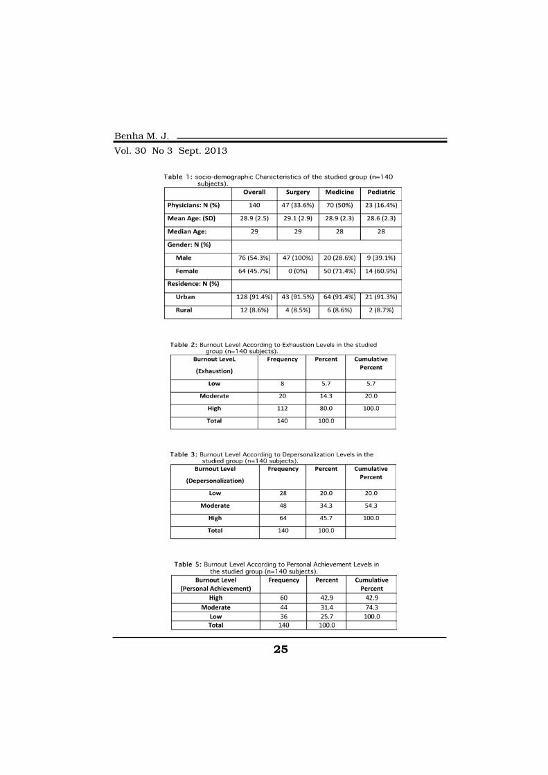
Results:Results: The response rate was 77% (140/182). Emotional exhaus-tion (EE) and Depersonalisation (DP), the major components of burnout,were reported, respectively, by 80 % (n = 112) and 45.7 % (n = 64) of theresidents and assistant lecturers.
Conclusion:Conclusion: The burnout level is high amongst Mansoura universityhospitals assistant lecturers and residents. Interventions are neededand could include support groups, more intense coaching by senior
22
Ahmed A. Albadry, et al....
observational studies in oncology,
the overall prevalence of high emo-
tional exhaustion, depersonaliza-
tion and low personal accomplish-
ment were, respectively, 36% (95%
confidence interval (CI): 31 41),
34% (95% CI: 30–39) and 25%
(95% CI: 16–34).5
Burnout has a detrimental ef-
fect on the physician’s quality of
life and is associated with an in-
creased risk of suicidal ideation.6
It has also been linked to poorer
quality of care, increased medical
errors and lawsuits, decreased
empathy6,7, job withdrawal and
absenteeism.8
Some medical specialties are at
higher risk of burnout. Although a
study comparing burnout
amongst residents in various med-
ical specialties in the United
States reported no significant dif-
ferences between specialties9, two
Finnish studies.10,11
Reported more burnout
amongst doctors who more often
treat chronically ill, incurable or
dying patients. Oncology was one
of these specialties. The factors
associated with stress and burn-
out in Oncology are insufficient
personal or vacation time, a sense
of failure, unrealistic expectations
of patients, cognitive or ethical
dissonance, repeated losses and
grieving or problems concerning
managed care.12
Burnout is highly prevalent
amongst medical residents. Re-
ported levels of burnout attained
76% amongst the residents in an
internal Medicine programme7
and 49.6% amongst US medical
students.6 However, the preva-
lence and causes of burnout
amongst oncology residents have
never been properly studied.
The aims of this study were to
quantify the frequency of burnout
amongst oncology residents and
assistant lecturers, to determine
demographical and psychological
factors associated with burnout.
MethodsA descriptive cross sectional
study carried out targeting resi-
dent physicians and assistant lec-
tures at Mansoura university hos-
pitals to estimate the prevalence
of burnout syndrome among resi-
dents in the hospital.
23
Benha M. J.
Vol. 30 No 3 Sept. 2013
1. Sampling and Sample Size:1. Sampling and Sample Size:
A stratified proportional sam-
pling technique was used to with-
draw the estimated sample 182
residents and assistant lecturers
from different departments in dif-
ferent Mansoura university hospi-
tals in the first phase of the study.
The number of the responders was
140 (77%) residents and assistant
lecturers.
2. Measurement Instrument:2. Measurement Instrument:
The survey included eight dem-
ographic questions before the
Maslach Burnout Inventory-
Human Services Survey MBI-HSS.
The survey was designed by the
researcher to collect specific dem-
ographic information from the Res-
idents and Assistant Lecturers.
The items on the demographic
portion of the survey consisted of
personal and professional infor-
mation such as age, gender, clini-
cal department, number of Experi-
ence years, number of working
hours per week, monthly income
and Residence. The demographic
portion of the survey contained a
small number of questions (eight).
This survey intentionally collected
only minimal personal information
about the participants to avoid
identifying the participant and to
maintain strict confidentiality.
3. Procedure:3. Procedure:
The Maslach Burnout Invento-
ry-Human Services Survey pencil,
self-report measure designed to
assess the three components of
the burnout syndrome: emotional
exhaustion, depersonalization,
and reduced personal accomplish-
ment (Maslach & Jackson, 1986).
There are 22 items which are di-
vided into three subscales and the
items are written in the form of
statements about personal feel-
ings or attitudes such as “I feel
burned out from my work,” “I
don’t really care what et al.,
1996). Respondents answer using
a seven-point Likert-type scale,
with the two extremes of “never
feel the effects” (0) and “feel the ef-
fects every day” (6). Scores are
generated for each subscale by
adding the numeric responses for
the items corresponding with each
scale. Subscale scores range from
a low of 0 to a high of 54 on the
EE subscale, from 0 to 30 on the
DP subscale, and from 0 to 48 on
the PA subscale (Maslach & Jack-
son, 1986). A high degree of burn-
24
Ahmed A. Albadry, et al....
out is reflected by high scores on
the Emotional Exhaustion and De-
personalization subscales and a
low score on the Personal Accom-
plishment subscale. The scores for
each subscale are considered sep-
arately and are not combined into
a single, unitary score, and subse-
quently the three scores are com-
puted for each respondent (Mas-
lach et al., 1996).
The specific categorization of
low, Moderate, or high burnout for
medical occupations is shown in
Table 1 below and by definition a
high degree of burnout is reflected
by high scores on the Emotional
Exhaustion and Depersonalization
subscales and a low score on the
Personal Accomplishment scale
(Maslach et al., 1996).
4. Data Collection:4. Data Collection:
The research for this study was
conducted at Mansoura University
Hospitals. To garner support for
this study the researcher first en-
gaged in a series of requests and
approvals.
The survey was distributed to the
Residents and Assistant Lecturers
in Mansoura University Hospitals
for 182 Participants. The researcher
provided the needed information
about the survey to all participants
so that they would have a clear
understanding of the study and
would understand their voluntary
participation in this study. Each
participant completed the survey
in paper and pen format. The sur-
vey was expected to take approxi-
mately 10 minutes to complete.
The participants were informed
that the information provided in
the instrument would be kept con-
fidential.
Results
25
Benha M. J.
Vol. 30 No 3 Sept. 2013
26
Ahmed A. Albadry, et al....
DiscussionThis study demonstrated that
the prevalence of burnout are high
amongst Mansoura University
Hospitals residents and Assistant
Lecturers. An excessive workload,
psychosomatic disorders or anxio-
lytics intake were independently
associated with increasing emo-
tional exhaustion scores.
Although this study provides a
number of important contribu-
tions, it is important to point out
its limitations. First, as the design
of the study was cross-sectional, it
does not allow causal interpreta-
tions between job characteristics
and health-related variables. Re-
spondents with poor psychological
well-being, i.e. high burnout, may
have reported a negative work en-
vironment. Nevertheless, longitu-
dinal studies have shown that per-
ceived work characteristics were
predictive of psychological distress
and not the reverse.22,23
Second, the present study, like
most burnout and stress studies,
is based on self-reported meas-
ures.24
Which could influence the sta-
tistical analysis. Indeed, the inde-
pendent and dependent variables
are based upon a single source of
information, the participants.25
This can result in an overesti-
mation of the main effects by in-
flating the association between
perceived work environment fac-
tors and strain indicators. Howev-
er, as pointed out by Spector,26
there is a high consistency be-
tween objective and subjective rat-
ings of variables such as those
used in our study.
Despite the limitations dis-
cussed above, this study has
many strengths. Firstly, it is the
first multi-institutional study on
the subject. Secondly, the re-
sponse rate is high, somewhat
higher than those usually found
in studies on medical students.27
thirdly; the study included resi-
dents and assistant lecturers in
medical, surgical and pediatric
fields, and thus evaluated the
three clinical specialties of health-
care service.
The prevalence of burnout in
this study is consistent with the
levels reported in other studies
27
Benha M. J.
Vol. 30 No 3 Sept. 2013
amongst healthcare workers5 or
amongst residents and medical
students6,8 or with levels expect-
ed due to the internal validity of
EE or DP scores.2 However, there
is a lot of heterogeneity between
burnout levels reported by these
studies. Indeed, virtually all the
studies used the MBI to quantify
burnout levels, but the cut-offs
applied were usually those pub-
lished in the initial study,2 which
are probably not relevant for all of
these populations. Cut-offs should
be adapted to take into account
social and cultural differences.1
Factors associated with burn-
out have previously been studied
in oncology12,28 and reported in a
meta-analysis.5
The most important ones are
related to workload, insufficient
personal or vacation time, feeling
of being fallible as a doctor, exces-
sive number of deaths, emotions
and particularly emotional disso-
nance and problems related to the
working environment (excessive
paperwork team communication-
Difficulties...).
As residents are the future on-
cology physicians, their needs and
aspirations should be taken into
account, especially these days
when cancer incidence is on the
increase29,30 and the crude num-
ber of oncologists may become too
low in some countries. Indeed,
some countries are facing a de-
mography crisis in oncology and
should take notice of the fact that,
as shown in our study, burnout is
strongly associated with a desire
to change specialty, or even to
quit Medicine.
In other studies, burnout was
associated with job absenteeism,
intention to leave the organization
and job turnover.8
Fatigue and depression have
been associated with increased
perceived medical errors.31,32 In-
terestingly, burnout was not al-
ways associated with higher rates
of medical errors.32,33
Burnout is detrimental to the
patient–physician relationship in
general Medicine.34 As such, they
should be taken into account by
university hospitals to improve
both student wellness and patient
care. To limit the incidence of
28
Ahmed A. Albadry, et al....
stress and burnout, Shanafelt et
al have proposed a multistep pro-
cess including the identification of
professional goals, the choice of
the most fitting type of practice
and the management of the
stressors specific to that practice
type. This process allows to
determine how to balance compet-
ing personal and professional
goals.35
Intervention studies are
needed, e.g. support groups, more
intense coaching by senior
physicians, training programs on
‘breaking bad news’ and teaching
of stress management skills to
study how to prevent or reverse
burnout. Such courses are
feasible and have proven their
ability to reduce the levels of
burnout in the short and long
term, to improve physicians’
well-being and attitudes
associated with patient-centered
care.37
Recovery from burnout is possi-
ble and was associated with a re-
duction of suicidal ideation in a
population of U.S. medical stu-
dents, confirming that burnout is
a reversible phenomenon.6
In brief, this study shows that
the prevalence of burnout is high
amongst Mansoura University
Hospitals Assistant Lecturers and
residents and is associated with a
poorer perceived health status
and the will to quit Medicine or
change specialty.
Medical schools and University
hospitals should consider this a
cause for concern and develop
screening strategies and interven-
tion programs to improve resi-
dents’ wellness. The results of our
study need to be confirmed and
confronted other major western
countries. Nevertheless, a close
and confident relationship be-
tween teachers and junior doctors
needs to be maintained and en-
hanced during this intense period
of training. Comparisons with sen-
ior oncologists or residents of oth-
er specialties are ongoing to evalu-
ate factors related to burnout that
could be specific to oncology or to
residents.
References1. Maslach C., Schaufeli W.B.1. Maslach C., Schaufeli W.B.
and Leiter M.P. (2001): and Leiter M.P. (2001): Job
burnout. Annu Rev Psychol;
52:397-422.
29
Benha M. J.
Vol. 30 No 3 Sept. 2013
2.2. Maslach C. and JacksonMaslach C. and Jackson
S.E. (1986):S.E. (1986): The measurement of
experienced burnout. J Occup Be-
hav; 2:99-113.
3.3. Maslach C., Jackson S.E.Maslach C., Jackson S.E.
and Leiter M.P. (1996):and Leiter M.P. (1996): Maslach
Burnout Inventory Manual. Palo
Alto, CA.
4.4. Whippen D.A. and Canel-Whippen D.A. and Canel-
los G.P. (1991):los G.P. (1991): Burnout syn-
drome in the practice of oncology:
results of a random survey of
1000 oncologists. J Clin Oncol;
9:1916-20.
5.5. Trufelli D.C., Bensi C.G.,Trufelli D.C., Bensi C.G.,
Garcia J.B., et al. (2008):Garcia J.B., et al. (2008): Burn-
out in cancer professionals: a sys-
tematic review and meta-analysis.
Eur J Cancer Care (Engl); 17:524-
531.
6.6. Dyrbye L.N., ThomasDyrbye L.N., Thomas
M.R., Massie F.S., et al. (2008):M.R., Massie F.S., et al. (2008):
Burnout and suicidal ideation
among US medical students. Ann
Intern Med; 149:334-41.
7.7. Shanafelt T.D., BradleyShanafelt T.D., Bradley
K.A., Wipf J.E. and Back A.L.K.A., Wipf J.E. and Back A.L.
(2002):(2002): Burnout and selfreported
patient care in an internal medi-
cine residency program. Ann In-
tern Med; 136:358-67.
8.8. Prins J.T., Hoekstra-Prins J.T., Hoekstra-
Weebers J.E., Van de Wiel H.B.,Weebers J.E., Van de Wiel H.B.,
et al. (2007):et al. (2007): Burnout among
Dutch medical residents. Int J Be-
hav Med; 14:119-25.
9.9. Martini S., Arfken C.L.,Martini S., Arfken C.L.,
Churchill A. and Balon R. (2004):Churchill A. and Balon R. (2004):
Burnout comparison among resi-
dents in different medical special-
ties. Acad Psychiatry; 28:240-2.
10.10. Korkeila J.A., Toyry S.,Korkeila J.A., Toyry S.,
Kumpulainen K., et al. (2003):Kumpulainen K., et al. (2003):
Burnout and selfperceived health
among Finnish psychiatrists and
child psychiatrists: a national sur-
vey. Scand J Public Health; 31:85-
91.
11.11. Olkinuora M., Asp S.,Olkinuora M., Asp S.,
Juntunen J., et al. (1990):Juntunen J., et al. (1990):
Stress symptoms,burnout and
suicidal thoughts in Finnish phy-
sicians. Soc Psychiatry Psychiatr
Epidemiol; 25:81-6.
12.12. Lyckholm L. (2001):Lyckholm L. (2001):
Dealing with stress, burnout, and
grief in the practice of oncology.
Lancet Oncol; 2:750-5.
30
Ahmed A. Albadry, et al....
13.13. Dewas S., Pointreau Y.,Dewas S., Pointreau Y.,
Rivera S., et al. (2009):Rivera S., et al. (2009): Demogra-
phy of radiation oncology resi-
dents in France in 2008: current
situation and perspectives for the
next three years. Cancer Radio-
ther; 13:153-60.
14.14. Loriot Y., Albiges-SauvinLoriot Y., Albiges-Sauvin
L., Dionysopoulos D., et al.L., Dionysopoulos D., et al.
(2007):(2007): French association of resi-
dents in oncology (AERIO) survey.
Ann Oncol; 21:161-5.
15.15. Grunfeld E., WhelanGrunfeld E., Whelan
T.J., Zitzelsberger L., et al.T.J., Zitzelsberger L., et al.
(2000):(2000): Cancer care workers in
Ontario: prevalence of burnout,
job stress and job satisfaction.
CMAJ; 163:166-9.
16.16. Lee R. and Ashforth B.E.Lee R. and Ashforth B.E.
(1996):(1996): A meta-analytic examina-
tion of the correlates of the three
dimensions of job burnout. J Appl
Psychol; 81:123-33.
17.17. Taris T.W., Le Blanc P.,Taris T.W., Le Blanc P.,
Schaufeli W.B. and SchreursSchaufeli W.B. and Schreurs
P.J.G. (2005):P.J.G. (2005): Are there relation-
ships between the dimensions of
the Maslach Burnout Inventory? A
review and two longitudinal tests.
Work Stress; 19:256-62.
18.18. Roelofs J., Verbraak M.,Roelofs J., Verbraak M.,
Keijsers G.P.J., De BruinKeijsers G.P.J., De Bruin
M.B.N., Schmidt A.J.M. (2005):M.B.N., Schmidt A.J.M. (2005):
Psychometric properties of a
Dutch version of the Maslach
Burnout Inventory-General Survey
(MBI-DV) in individuals with and
without clinical burnout. Stress
Health; 21:17-25.
19. Dion G., Tessier R.19. Dion G., Tessier R.
(1994): (1994): Validation de la traduc-
tion de l’Inventaire d’e´cuisement
professionnel de Maslach et Jack-
son. Can J Behav Sci; 26:210-27.
20. Rascle N., Truchot D.,20. Rascle N., Truchot D.,
Borteyrou X. (2008):Borteyrou X. (2008): Construc-
tion et validation d’une e´chelle de
stress pour des personnels travail-
lant en oncologie. 15e`me congre`s
de l’Association Internationale de
psychologie du travail de langue
francaise, Quebec, Canada.
21.21. Dyrbye L.N., West C.P.,Dyrbye L.N., West C.P.,
Shanafelt T.D. (2009):Shanafelt T.D. (2009): Defining
burnout as a dichotomous vari-
able. J Gen Intern Med; 24:440
[author reply 441].
22.22. Borritz M., Bu¨ltmannBorritz M., Bu¨ltmann
U., Rugulies R., et al. (2006):U., Rugulies R., et al. (2006):
Psychosocial work characteristics
31
Benha M. J.
Vol. 30 No 3 Sept. 2013
as predictors for burnout: findings
from 3-year follow up of the PUMA
study. J Occup Environ Med;
47:1015-25.
23.23. Taylor C., Graham J.,Taylor C., Graham J.,
Potts H.W., Richards M.A. andPotts H.W., Richards M.A. and
Ramirez A.J. (2005):Ramirez A.J. (2005): Changes in
mental health of UK hospital con-
sultants since the mid-1990s.
Lancet; 366:742-4.
24.24. Truchot D. (2004):Truchot D. (2004):
Epuisement professionnel et burn-
out. Concepts, mode`les, interven-
tions. Paris: Dunod.
25.25. Spector P.E. and JexSpector P.E. and Jex
S.M. (1991):S.M. (1991): Relations of job
characteristics from multiple data
sources with employee affect, ab-
sence, turnover intentions and
health. J Appl Psychol; 76:46-53.
26.26. Spector P.E. (1987):Spector P.E. (1987):
Method variance as an artifact in
self-reported affect and percep-
tions at work: myth or significant
problem? J Appl Psychol; 72:438-
43.
27.27. Dyrbye L.N., ThomasDyrbye L.N., Thomas
M.R., Huntington J.L., et al.M.R., Huntington J.L., et al.
(2006):(2006): Personal life events and
medical student burnout: a multi-
center study. Acad Med; 81:374-
84.
28.28. Mount B.M. (1986):Mount B.M. (1986): Deal-
ing with our losses. J Clin Oncol;
4:1127-34.
29.29. Guerin S., Doyon F. andGuerin S., Doyon F. and
Hill C. (2009):Hill C. (2009): The frequency of
cancer in France in 2006,
mortality trends since 1950,
incidence trends since 1980 and
analysis of the discrepancies be-
tween these trends. Bull Cancer;
96:51-7.
30.30. Jemal A., Siegel R.,Jemal A., Siegel R.,
Ward E., et al. (2009):Ward E., et al. (2009): Cancer
statistics, 2009. CA Cancer J Clin;
59:225-49.
31.31. Landrigan C.P., Roths-Landrigan C.P., Roths-
child J.M., Cronin J.W., et al.child J.M., Cronin J.W., et al.
(2004):(2004): Effect of reducing interns’
work hours on serious medical er-
rors in intensive care units. N
Engl J Med; 351:1838-48.
32.32. West C.P., Tan A.D., Ha-West C.P., Tan A.D., Ha-
bermann T.M., Sloan J.A. andbermann T.M., Sloan J.A. and
Shanafelt T.D. (2009):Shanafelt T.D. (2009): Associa-
tion of resident fatigue and dis-
tress with perceived medical er-
32
Ahmed A. Albadry, et al....
rors. JAMA; 302:1294-300.
33.33. Fahrenkopf A.M., Sect-Fahrenkopf A.M., Sect-
ish T.C., Barger L.K., et al.ish T.C., Barger L.K., et al.
(2008):(2008): Rates of medication errors
among depressed and burnt out
residents: prospective cohort
study. BMJ; 336:488-91.
34.34. Truchot D., Bante´gnieTruchot D., Bante´gnie
D. and Roncari N. (2009):D. and Roncari N. (2009):
Burnout, patient compliance,
psychological withdrawal among
GPs: an exploratory study. Oslo,
Norway: European Congress of
Psychology.
35.35. Shanafelt T., Chung H.,Shanafelt T., Chung H.,
White H. and Lyckholm L.J.White H. and Lyckholm L.J.
(2006):(2006): Shaping your career to
maximize personal satisfaction in
the practice of oncology. J Clin
Oncol; 24:4020-6.
36.36. Bragard I., Razavi D.,Bragard I., Razavi D.,
Marchal S., et al. (2006):Marchal S., et al. (2006): Teach-
ing communication and stress
management skills to junior phy-
sicians dealing with cancer pa-
tients: a Belgian interuniversity
curriculum. Support Care Cancer;
14:454-61.
37.37. Krasner M.S., EpsteinKrasner M.S., Epstein
R.M., Beckman H., et al. (2009):R.M., Beckman H., et al. (2009):
Association of an educational pro-
gram in mindful communication
with burnout, empathy, and atti-
tudes among primary care physi-
cians. JAMA; 302:1284-93.

33
Benha M. J.
Vol. 30 No 3 Sept. 2013
PREVALENCE OF OCCUPATIONALPREVALENCE OF OCCUPATIONALBURNOUT AMONG MANSOURABURNOUT AMONG MANSOURA
UNIVERSITY HOSPITALS’ RESIDENTSUNIVERSITY HOSPITALS’ RESIDENTSAND ASSISTANT LECTURERSAND ASSISTANT LECTURERS
Ahmed A. Albadry M.Sc, Ahmed N. Sleem MD, Nadia A.Ahmed A. Albadry M.Sc, Ahmed N. Sleem MD, Nadia A.Montasser MD and EL-Sayed A. El-Naggar MDMontasser MD and EL-Sayed A. El-Naggar MD
BENHAMEDICALJOURNAL
REPRINT
Published byPublished by
Benha Faculty of MedicineBenha Faculty of Medicine Volume 30 Number 3Sept. 2013

33
Benha M. J.
Vol. 30 No 3 Sept. 2013
GLYPICAN-3 EXPRESSION INGLYPICAN-3 EXPRESSION INHEPATOCELLULAR CARCINOMA IN RELATIONHEPATOCELLULAR CARCINOMA IN RELATION
TO THE GRADE OF DIFFERENTIATIONTO THE GRADE OF DIFFERENTIATION
Eman Tawfik Enan MD, Amira Kamal El-Hawary MD,Eman Tawfik Enan MD, Amira Kamal El-Hawary MD,Dina Abd El-Aziz El-Tantawy MD, Nagwa Mokhtar Helal MDDina Abd El-Aziz El-Tantawy MD, Nagwa Mokhtar Helal MD
and Maha Mohamed Abo-Hashem MDand Maha Mohamed Abo-Hashem MDPathology Departement, Faculty of Medicine, Mansoura University, Egypt
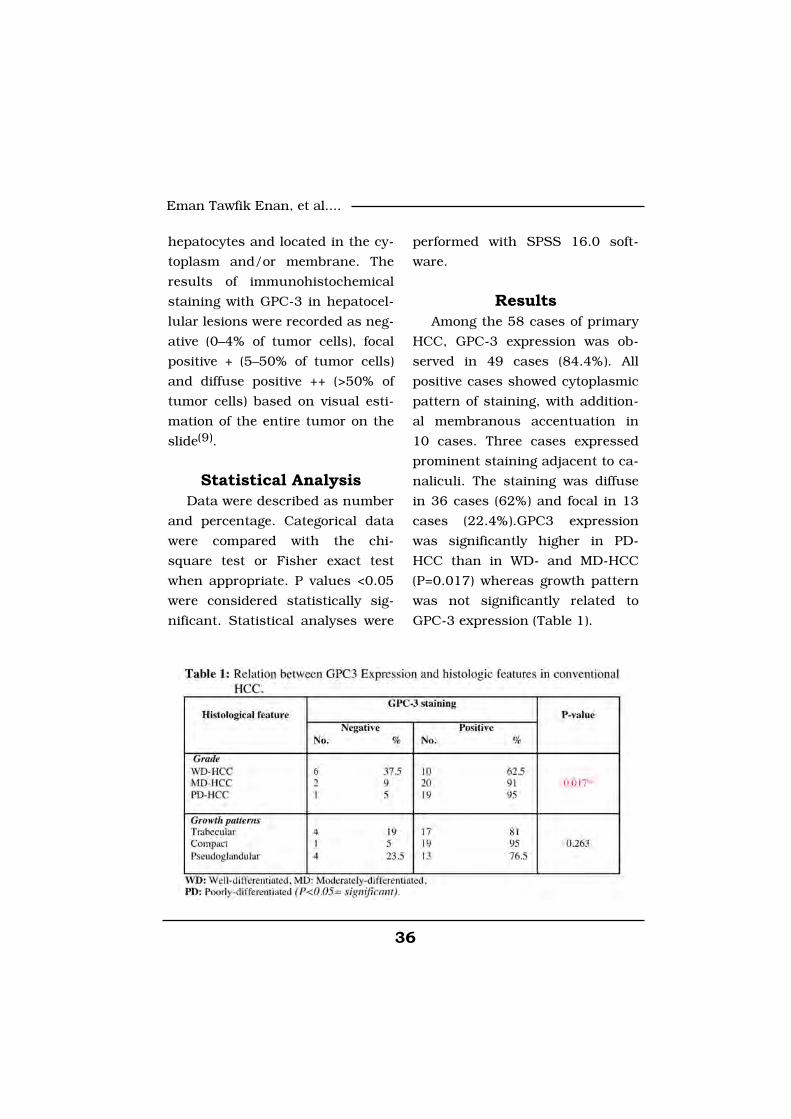
AbstractBackground: Background: Glypican-3 (GPC-3) is an oncofetal protein normally ex-
pressed in fetal liver and placenta but is not found in normal adult liv-er. GPC-3 expression has been reported in 75–100% of hepatocellularcarcinoma (HCC). It has also been suggested that poorly differentiatedhepatocellular carcinomas are more likely to express GPC3. The aim ofthis study was to assess the diagnostic value of GPC-3 immunostainingin HCCs and to analyze its expression profile in relation to the grade ofdifferentiation.
Material and methods: Material and methods: This study was performed on 58 cases of for-malin-fixed, paraffin-embedded cases of HCC obtained from the files ofpathology laboratory of GastroEnterology Center, Mansoura Universityfrom 2009 to 2012. The H&E slides were reviewed to confirm the diag-nosis and assess the grade of differentiation. The following cases werestudied: well differentiated- HCC (WD-HCC) (n=16), moderately differen-tiated HCC (MD-HCC) (n=22), and poorly differentiated HCC (PD-HCC)(n=20)
All cases were immunostained with GPC-3.Results: Results: Among the 58 cases of primary HCC, GPC-3 expression was
observed in 84.4% of cases. The staining was diffuse in 62% of casesand focal in 22.4 % of cases. GPC3 expression was significantly higherin PD-HCC than in WD- and MD-HCC (P value, 0.017).
Conclusion:Conclusion: Our data demonstrate that GPC-3 has high sensitivityto HCC and is more expressed in poorly differentiated tumors. As such,we recommend that this marker should be included in any antibody
34
Eman Tawfik Enan, et al....
IntroductionHepatocellular carcinoma
(HCC) is the most common malig-
nant primary tumor of the liver(1).
HCC affects about a million people
every year worldwide(2). In Egypt
the incidence of HCC has doubled
in the past 10 years, thus becom-
ing the second most incident and
lethal cancer in men(3).
Accurate diagnosis is critically
important to appropriate clinical
management of the patients and
assessment of the prognosis. The
histologic diagnosis of HCC is rel-
atively straightforward when the
tumor recapitulates the cytoarchi-
tectural appearance of the normal
liver. However, HCC exhibiting a
pseudoglandular or poorly differ-
entiated morphology may be diffi-
cult to distinguish from cholangio-
carcinoma or metastatic
adenocarcinoma involving the liver(4).
The presently employed immu-
nohistochemical panels have
greatly facilitated the diagnosis of
HCC. However, there are several
situations where these markers
are of limited use. Hep Par 1 is a
highly sensitive and specific mark-
er of benign and malignant hepa-
tocytes (80%–90% sensitivity,
nearly 100% specificity). However,
expression is decreased in poorly
differentiated HCC and in the scle-
rosing variant of HCC, which can
show immunoreactivity in only
50% of cases. Similarly, polyclonal
CEA have low sensitivity (~50%)
for the diagnosis of poorly differ-
entiated hepatocellular carcinoma(4,5). Hence, Hep Par 1 and poly-
clonal CEA may be less helpful in
the setting of a poorly differentiat-
ed hepatic neoplasm in distin-
guishing hepatocellular carcinoma
and metastatic adenocarcinoma.
Glypican-3 (GPC-3) is an oncof-
etal protein and is a member of
the membrane-bound heparin sul-
fate proteoglycans. This protein is
normally expressed in fetal liver
and placenta but is not found in
normal adult liver. It plays a role
in cell growth, differentiation, and
migration(6). GPC-3 is highly ex-
pressed, both at the mRNA and
panel used to distinguish HCC from cholangiocarcinoma and metastaticcarcinoma to increase diagnostic accuracy.
35
Benha M. J.
Vol. 30 No 3 Sept. 2013
protein level, in HCCs. It has also
been suggested that poorly differ-
entiated hepatocellular carcino-
mas are more likely to express
GPC-3(7). The aim of this study is
to assess the diagnostic value of
GPC3 immunostaining in hepato-
cellular carcinomas and to analyze
its expression profile in relation to
the grade of differentiation.
Materials and MethodsCasesCases
This study was performed on
58 cases of formalin-fixed, paraf-
fin-embedded cases of HCC ob-
tained from the files of pathology
laboratory of GastroEnterology
Center, Mansoura University from
2009 to 2012. Twelve specimens
were trucut needle biopsy, and 46
were from partial hepatectomy
specimens. The available paraffin
blocks were sectioned at 4-5 mi-
crons and stained with haematox-
ylin and eosin. The slides were re-
viewed to confirm the diagnosis
according to the guidelines of the
WHO(8). The studied tumors in-
cluded: Well differentiated- HCC
(WD-HCC) (n=16), moderately dif-
ferentiated HCC (MD-HCC) (n=22),
and poorly differentiated HCC
(PD-HCC) (n=20).
ImmunohistochemistryImmunohistochemistry
Monoclonal antibody to GPC-3
(Clone 1G12, 1:300, Biocare, Con-
cord, CA), was used for staining
all cases of the study. Previously
validated slides were used as posi-
tive control samples. Slides using
phosphate-buffered saline instead
of primary monoclonal antibody
were regarded as negative control
samples. The sections were depa-
raffinized and rehydrated in grad-
ed alcohol. Endogenous peroxide
was blocked by 3% hydrogen per-
oxide treatment, and the antigen
was retrieved using sodium citrate
treatment in a microwave oven at
98°C for 10 minutes. Following
peroxidase block and incubation
with primary antibody for 30 min
at room temperature, the sections
were incubated with horse-radish
peroxidase-labeled secondary anti-
body (UltraVision ONE HRP Poly-
mer; Catalog no.TL-015-HDJ;
Thermo, Fermont, CA) for 30 min-
utes at room temperature, DAB
Plus substrate chromagen for 15
min, and counterstained with
hematoxylin.
Staining was considered posi-
tive when immunoreactivity was
present in at least 5% of lesional
36
Eman Tawfik Enan, et al....
hepatocytes and located in the cy-
toplasm and/or membrane. The
results of immunohistochemical
staining with GPC-3 in hepatocel-
lular lesions were recorded as neg-
ative (0–4% of tumor cells), focal
positive + (5–50% of tumor cells)
and diffuse positive ++ (>50% of
tumor cells) based on visual esti-
mation of the entire tumor on the
slide(9).
Statistical AnalysisData were described as number
and percentage. Categorical data
were compared with the chi-
square test or Fisher exact test
when appropriate. P values <0.05
were considered statistically sig-
nificant. Statistical analyses were
performed with SPSS 16.0 soft-
ware.
ResultsAmong the 58 cases of primary
HCC, GPC-3 expression was ob-
served in 49 cases (84.4%). All
positive cases showed cytoplasmic
pattern of staining, with addition-
al membranous accentuation in
10 cases. Three cases expressed
prominent staining adjacent to ca-
naliculi. The staining was diffuse
in 36 cases (62%) and focal in 13
cases (22.4%).GPC3 expression
was significantly higher in PD-
HCC than in WD- and MD-HCC
(P=0.017) whereas growth pattern
was not significantly related to
GPC-3 expression (Table 1).
37
Benha M. J.
Vol. 30 No 3 Sept. 2013
DiscussionThe differential diagnosis of
HCC varies, depending on the de-
gree of tumor differentiation.
Work-up often requires immuno-
histochemical stains, which
should be judiciously selected
based on the H&E morphological
pattern and the differential diag-
nosis.
Several studies have demon-
strated the efficacy of GPC-3 as a
diagnostic tool in HCC. The re-
ported sensitivity ranges from 75-
100%, with figures of 75-5% in
larger series(10,11,12,13). Our re-
sults are similar to the literature,
with 84.4% of hepatocellular car-
cinoma being positive for GPC-3.
The staining was diffuse in 62% of
cases and focal in about 22%; in-
dicating that the focal staining de-
tectable in a fraction of HCCs
might cause false negative results
in tiny liver biopsies, a finding
that was previously reported by
Shafizadeh et al(7).
Poorly differentiated hepatocel-
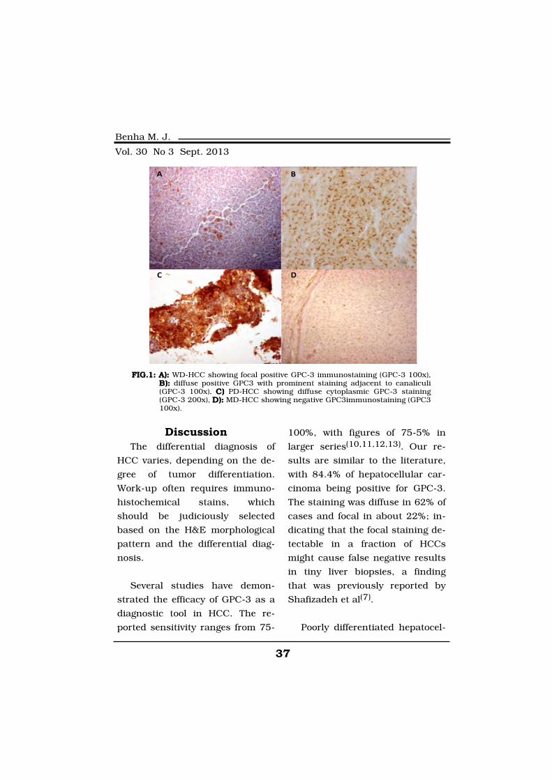
FIG.1: A): FIG.1: A): WD-HCC showing focal positive GPC-3 immunostaining (GPC-3 100x),B):B): diffuse positive GPC3 with prominent staining adjacent to canaliculi(GPC-3 100x), C)C) PD-HCC showing diffuse cytoplasmic GPC-3 staining(GPC-3 200x), D):D): MD-HCC showing negative GPC3immunostaining (GPC3100x).
38
Eman Tawfik Enan, et al....
lular carcinomas pose a diagnostic
problem as they can mimic meta-
static neoplasms. As mentioned
previously, many earlier studies
have demonstrated that the com-
monly used hepatocellular mark-
ers like Hep- Par 1 and pCEA have
low sensitivity for poorly differen-
tiated hepatocellular carcinoma(4,14,15).
The results of the current study
showed that the extent of GPC-3
immunoreactivity was significantly
related to the tumor grading, be-
cause less differentiated HCCs
had a greater immunoreactivity.
This finding is consistent with
that reported by Di Tommaso et al(16) and Shirakawa et al(11). In
other studies, the opposite was
described(12), or there was no as-
sociation with grade(7,10&13).
This discrepancy may be related
to variation in number of studied
cases, different cutoff point of
GPC-3 positivity (5% vs. 10%), or
to the type of sample (whole sec-
tions vs. Tissue microarray).
In conclusion, our data confirm
previous observations regarding
the high rate of expression of
GPC-3 in HCC, particularly poorly
differentiated tumors. As such, we
recommend that this marker
should be included in any anti-
body panel used to distinguish
HCC from cholangiocarcinoma
and metastatic carcinoma to in-
crease diagnostic accuracy.
References1- Crawford J. and Liu C.1- Crawford J. and Liu C.
(2010): (2010): Liver and biliary tract. In:
Kumar V., et al. (Eds.), Robbins
and Cotran Pathologic Basis of
Disease, 8th ed., Elsevier, Phila-
delphia, pp.878-882.
2- Motola K.D., Zamora V.D.,2- Motola K.D., Zamora V.D.,
Uribe M., et al. (2006): Uribe M., et al. (2006): Hepato-
cellular carcinoma. An overeview.
Ann Hepatol, 5: 16–24.
3- Lehman E.M. and Wilson3- Lehman E.M. and Wilson
M.L. (2009):M.L. (2009): Epidemiology of hep-
atitis viruses among hepatocellu-
lar carcinoma cases and healthy
people in Egypt: a systematic re-
view and metaanalysis. Int J Can-
cer: 124 (3), 690.
4- Lau S.K., Prakash S., Gell-4- Lau S.K., Prakash S., Gell-
er S.A., et al. (2002): er S.A., et al. (2002): Compara-
tive immunohistochemical profile
of hepatocellular carcinoma, cho-
langiocarcinoma, and metastatic
39
Benha M. J.
Vol. 30 No 3 Sept. 2013
adenocarcinoma. Hum Pathol ,
33: 1175-1181.
5- Fan Z., Van De Rijn M.,5- Fan Z., Van De Rijn M.,
Montgomery K., et al. (2003):Montgomery K., et al. (2003):
Hep Par 1 antibody stain for the
differential diagnosis of hepatocel-
lular carcinoma: 676 tumors test-
ed using tissue microarrays and
conventional tissue sections. Mod
Pathol, 16: 137-144.
6- Allende D.S. and Yerian L.6- Allende D.S. and Yerian L.
(2009): (2009): Immunohistochemical
markers in the diagnosis of hepat-
ocellular carcinoma. Pathology
Case Reviews, 14: 40-46.
7- Shafizadeh N., Ferrell L.D.7- Shafizadeh N., Ferrell L.D.
and Kakar S. (2008):and Kakar S. (2008): Utility and
limitations of glypican-3 expres-
sion for the diagnosis of hepato-
cellular carcinoma at both ends of
the differentiation spectrum. Mod
Pathol, 21:1011-18.
8- Hirohashi S., Blum H.E.8- Hirohashi S., Blum H.E.
and Ishak K.J. (2000): and Ishak K.J. (2000): Hepato-
cellular carcinoma. In: WHO: pa-
thology & genetics, tumour of the
digestive system. IARC Press, p.
159-72.
9- Di Tommaso L., Destro A.,9- Di Tommaso L., Destro A.,
Seok J.Y., et al. (2009): Seok J.Y., et al. (2009): The ap-
plication of markers (HSP70 GPC3
and GS) in liver biopsies is useful
for detection of hepatocellular car-
cinoma. J Hepatol, 50:746-754.
10- Kandil D., Leiman G., Al-10- Kandil D., Leiman G., Al-
legretta M., et al. (2007):legretta M., et al. (2007): Glypi-
can-3 immunocytochemistry in
liver fine-needle aspirates: a novel
stain to assist in the differentia-
tion of benign and malignant liver
lesions. Cancer, 111:316-322.
11- Shirakawa H., Kuronuma11- Shirakawa H., Kuronuma
T., Nishimura Y., et al. (2009):T., Nishimura Y., et al. (2009):
Glypican-3 is a useful diagnostic
marker form a component of he-
patocellular carcinoma in human
liver cancer. Int J Oncol, 34:649-
56.
12- Zhang L., Liu H., Sun L.,12- Zhang L., Liu H., Sun L.,
et al. (2012): et al. (2012): Glypican-3 as a po-
tential differential diagnosis mark-
er for hepatocellular carcinoma: A
tissue microarray-based study.
Acta Histochemica, 114: 547- 552.
13- Yamauchi N., Watanabe13- Yamauchi N., Watanabe
A., Hishinuma M., et al. (2005):A., Hishinuma M., et al. (2005):
The glypican 3 oncofetal protein is
a promising diagnostic marker for
hepatocellular carcinoma. Mod
40
Eman Tawfik Enan, et al....
Pathol, 18:1591-1598.
14- Minervini M.I., Demetris14- Minervini M.I., Demetris
A.J., Lee R.G., et al. (1997): A.J., Lee R.G., et al. (1997): Util-
ization of hepatocyte-specific anti-
body in the immunocytochemical
evaluation of liver tumors. Mod
Pathol, 10:686-692.
15- Saad R.S., Luckasevic15- Saad R.S., Luckasevic
T.M., Noga C.M., et al. (2004):T.M., Noga C.M., et al. (2004):
Diagnostic value of HepPar1,
pCEA, CD10, and CD34 expres-
sion in separating hepatocellular
carcinoma from metastatic carci-
noma in fine-needle aspiration cy-
tology. Diagn Cytopathol, 30:1-6.
16- Di Tommaso L., Franchi16- Di Tommaso L., Franchi
G., Park Y.N., et al. (2007):G., Park Y.N., et al. (2007): Diag-
nostic value of HSP70, glypican 3,
and glutamine synthetase in he-
patocellular nodules in cirrhosis.
Hepatology, 45:725-734.
41
Benha M. J.
Vol. 30 No 3 Sept. 2013
GLYPICAN-3 EXPRESSION INGLYPICAN-3 EXPRESSION INHEPATOCELLULAR CARCINOMA INHEPATOCELLULAR CARCINOMA INRELATION TO THE GRADE OF DIF-RELATION TO THE GRADE OF DIF-
FERENTIATIONFERENTIATION
Eman Tawfik Enan MD, Amira Kamal El-Hawary MD,Eman Tawfik Enan MD, Amira Kamal El-Hawary MD,Dina Abd El-Aziz El-Tantawy MD,Dina Abd El-Aziz El-Tantawy MD,
Nagwa Mokhtar Helal MDNagwa Mokhtar Helal MDand Maha Mohamed Abo-Hashem MDand Maha Mohamed Abo-Hashem MD
BENHAMEDICALJOURNAL
REPRINT
Published byPublished by
Benha Faculty of MedicineBenha Faculty of Medicine Volume 30 Number 3Sept. 2013
41
Benha M. J.
Vol. 30 No 3 Sept. 2013
EFFECTS OF TAMOXIFEN ON LIPID PROFILESEFFECTS OF TAMOXIFEN ON LIPID PROFILESIN POST-MENOPAUSAL BREASTIN POST-MENOPAUSAL BREAST
CANCER PATIENTSCANCER PATIENTS
Fatma M. F. Akl MD* and Ibrahim A. Abdel Aal MD**Fatma M. F. Akl MD* and Ibrahim A. Abdel Aal MD**Clinical Oncology & Nuclear Medicine Department*, Clinical Pathology Department**,
Faculty of Medicine, Mansoura University
AbstractBackground & Objective: Background & Objective: The risk of cardiovascular mortality in-
creases dramatically in women after menopause because of lipid-metabolism alterations that are attributed to estrogen deprivation. Ta-moxifen is the usual endocrine (anti-estrogen) therapy for hormone re-ceptor-positive breast cancer in pre-menopausal women, and is also astandard in post-menopausal women although aromatase inhibitors arefrequently used in that setting. The long-term use of anti-estrogenagents showed a potential to improve lipid profiles in post-menopausalbreast cancer patients. The present study has been undertaken to as-sess the effect of tamoxifen therapy on plasma lipid profile in postmeno-pausal breast cancer patients.
Patients & Methods:Patients & Methods: The study population consisted of 36 postmen-opausal, primary operable breast cancer patients treated with surgeryin the form of a total mastectomy or a breast-conserving resection withaxillary dissection. The patients were adjusted for adjuvant chemothera-py and or radiation therapy and allocated to tamoxifen 20 mg daily .Serum lipid profiles evaluated were total cholesterol (TC), low-densitylipoprotein cholesterol (LDL-C), high-density lipoprotein cholesterol(HDL-C) and triglycerides (TG). Time points for blood collection were be-fore the start of administration of tamoxifen, 3 and 6 months after thestart of its administration.
Results:Results: The mean level of plasma total cholesterol was significantlydecreased (P<0.001) after 3 and 6 months of tamoxifen treatment com-pared with mean baseline levels. Also , significant decreases were ob-served in mean LDL-C (P<0.001). At 3 and 6 months’ evaluation, atrend toward increase of plasma triglycerides and HDL cholesterol levels
42
Fatma M. F. Akl and Ibrahim A. Abdel Aal
IntroductionBreast cancer is the most fre-
quently diagnosed invasive cancer
in women, with more than 1.3 mil-
lion women worldwide are diag-
nosed with breast cancer each
year, making it the second most
common form of cancer behind
lung cancer[1]. The increased
number of breast cancer diagnos-
es along with improvements in ini-
tial treatments, have led to an in-
crease in the number of breast
cancer survivors[2].
In postmenopausal women with
endocrine-responsive early breast
cancer, adjuvant hormonal thera-
py is the established standard of
care. In postmenopausal women,
the two most commonly used
strategies of endocrine treatment
are either the interference with
estrogen signaling by binding to
the estrogen receptor protein with
a selective estrogen-receptor
modulator(SERM), such as
tamoxifen, or the inhibition of
endogenous estrogen production
by using an aromatase inhibitor
(AI)[3].
Tamoxifen is the usual endo-
crine (anti-estrogen) therapy for
hormone receptor-positive breast
cancer in pre-menopausal women,
and is also a standard in post-
menopausal women although
aromatase inhibitors are also fre-
quently used in that setting[4].
The long-term use of anti-
estrogen agents showed a poten-
tial to improve lipid profiles of
post-menopausal breast cancer
patients ,which attracted atten-
tion in both research and clinical
settings[5].
The risk of cardiovascular mor-
tality increases dramatically in
women after menopause because
of lipid-metabolism alterations
that are attributed to estrogen
deprivation[6]. Levi etal. suggested
that the greatest cause of death in
women with early-stage breast
cancer is heart disease[7].
were observed but, it didn't reach statistical significance.Conclusion:Conclusion: In conclusion, favorable changes of lipid profiles were
detected in postmenopausal patients with breast cancer treated withtamoxifen.
Keywords:Keywords: Tamoxifen, lipid profiles, post-menopause, breast cancer.
43
Benha M. J.
Vol. 30 No 3 Sept. 2013
The role of low-density lipopro-
tein cholesterol (LDL) in the path-
ogenesis of atherosclerosis and
subsequently in coronary heart
disease is well known. Evidence
suggests that increased levels of
LDL are highly correlated with in-
creased risk of heart disease, even
while total cholesterol remains
within normal range[8]. At the
same time, high-density lipopro-
tein (HDL) cholesterol is known to
have a protective effect against
coronary heart disease[9]. The role
of triglycerides is less clear, but
increased levels have been asso-
ciated with risk of cardiovascular
diseases in both women and
men[6].
Some trials demonstrated that
tamoxifen shares the beneficial ef-
fects of estrogens on cardiovascu-
lar risk factors by decreasing total
cholesterol (TC) and low-density
lipoprotein cholesterol (LDL-C)
both in pre-menopausal and post-
menopausal women[10]. It produc-
es no significant change in trigly-
cerides (TGs), high density
lipoprotein cholesterol (HDL-C)
and very low-density lipoprotein
cholesterol (VLDL-C) levels in pre-
menopausal and postmenopausal
women[11].
Cholesterol also is the precur-
sor to steroid hormone synthesis
and endogenous sex steroid hor-
mones which are directly related
to breast cancer risk[12]. On the
other hand, an inverse association
was observed between high-
density lipoprotein cholesterol
(HDL-C) level and breast cancer[13].
The present study has been un-
dertaken to assess the effect of ta-
moxifen therapy on plasma lipid
profile in a sample of egyptian
postmenopausal breast cancer pa-
tients.
Materials and MethodsThis prospective, study was
conducted from June 2010 to De-
cember 2012 at the Clinical On-
cology and Nuclear Medicine De-
partment and the Clinical
Pathology Department, Mansoura
University Hospital.
A total of 36 postmenopausal
patients with primary operable
breast cancer between 48 and 70
years of age were included in this
study.

44
Fatma M. F. Akl and Ibrahim A. Abdel Aal
Patients included in the trial
satisfied the following entry
criteria: (1) all demonstrated a
postmenopausal status (defined
as either no menses for more than
1 year or shorter duration of
amenorrhea with follicle-
stimulating hormone [FSH] levels
in the postmenopausal range); (2)
all had undergone breast surgery
(either lumpectomy or
mastectomy) and were considered
to be potentially curable; (3) none
had received either radiation or
chemotherapy before breast sur-
gery; (4)estrogen receptor ER posi-
tivity was confirmed by histopa-
thology; (5) none had either
diabetes mellitus, renal or hepatic
disease; (6) none had received
drugs known to affect the lipid
and lipoprotein levels; and (7) all
were instructed to follow their
usual diet and maintain weight
during the study period.
All patients underwent surgery
in the form of a total mastectomy
or a breast-conserving resection
with axillary dissection. The pa-
tients were adjusted for adjuvant
chemotherapy and or radiation
therapy and allocated to tamoxifen
20 mg daily .
Serum samples were collected
after overnight fasting (12-14
hours). Time points for blood col-
lection were before the start of ad-
ministration of tamoxifen, 3 and 6
months after the start of its ad-
ministration. Serum lipid profiles
evaluated were total cholesterol
(TC), low-density lipoprotein cho-
lesterol (LDL-C), high-density lipo-
protein cholesterol (HDL-C) and
triglycerides (TG). Lipid profile as-
say was measured by standard
enzymatic methods (Human
GmbH Germany). LDL-C values
were calculated using the equa-
tion of Friedewald[14]. Diagnostic
criteria for lipid abnormalities are
as follows: hyper-LDL-
cholesterolemia, ≥140 mg/dl;
hypo-HDL-cholesterolemia, <40
mg/dl; and hypertriglyceridemia,
≥150 mg/dl.
Statistical MethodsBasic descriptive statistics, in-
cluding means, standard devia-
tions (SD), ranges, and percentag-
es, were used to characterize the
study participants. Changes in lip-
id profiles were analyzed using
Student's paired t-test based on
differences between mean values
before administration of tamoxifen
45
Benha M. J.
Vol. 30 No 3 Sept. 2013
(baseline) and mean values of 3
and 6 months after administration
(significance level ≤ 0.05). Statisti-
cal analysis was done using Sta-
tistical Program for Social Scienc-
es (SPSS 17.0).
ResultsPatient characteristics, includ-
ing age, weight, stage, prior adju-
vant chemotherapy or radiothera-
py, and ECOG performance status
are presented in Table 1.
A total of 36 patients under-
went lipid profile evaluation at
baseline, but decreased to 32 and
31 patients at 3 and 6 months
evaluation, respectively.
Table 2 shows the mean lipo-
protein levels at baseline, 3
months, and 6 months in patients
receiving tamoxifen.
The mean level of plasma total
cholesterol was significantly de-
creased (P<0.001) after 3 and 6
months of tamoxifen treatment
compared with mean baseline lev-
els. Also, similar decreases were
observed in mean LDL-C
(P<0.001).
At 3 and 6 months’ evaluation,
a trend toward increase levels of
HDL cholesterol was observed, but
these values were statistically non
significant in comparison to pre
tamoxifen treatment (P =0.32), (P
=0.06) respectively.
Similarly, a trend toward in-
crease levels of plasma triglyce-
rides were detected at 3 and 6
months’ evaluation but , it didn’t
reach statistical significance(P
=0.14), (P =0.09) respectively.
Tamoxifen significantly de-
creased T-C by 8.06 mg/dl at 3
months and by 21.45 mg/dl at 6
months (p< 0.001) and also de-
creased LDL-C by 10.89 mg/dl at
3 months and by 22.45 mg/dl at 6
months (p< 0.001) (Table 2).
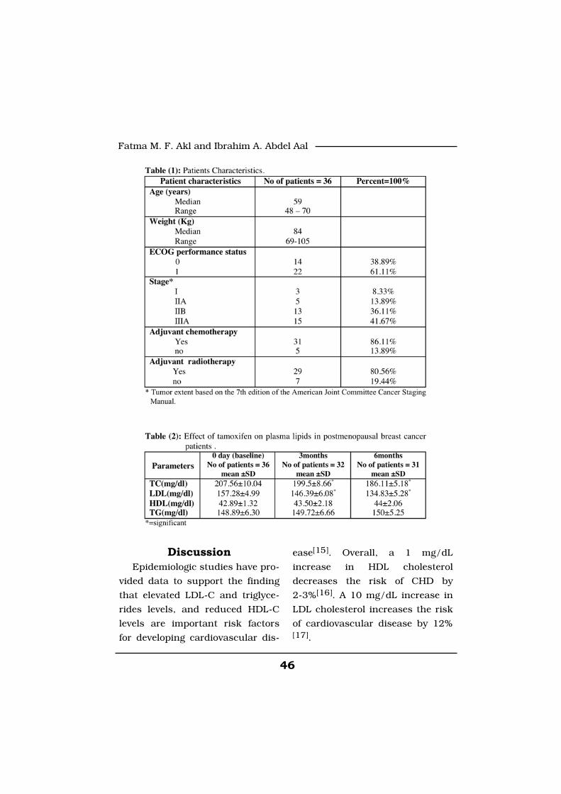
46
Fatma M. F. Akl and Ibrahim A. Abdel Aal
DiscussionEpidemiologic studies have pro-
vided data to support the finding
that elevated LDL-C and triglyce-
rides levels, and reduced HDL-C
levels are important risk factors
for developing cardiovascular dis-
ease[15]. Overall, a 1 mg/dL
increase in HDL cholesterol
decreases the risk of CHD by
2-3%[16]. A 10 mg/dL increase in
LDL cholesterol increases the risk
of cardiovascular disease by 12%[17].
47
Benha M. J.
Vol. 30 No 3 Sept. 2013
Hypercholesterolemia and hy-
pertriglyceridemia are more com-
mon in post-menopausal women
compared with pre-menopausal
women, and the primary cause is
thought to be decreased blood es-
trogen concentrations[18].
It is generally accepted that ad-
juvant hormonal treatment with
tamoxifen has a beneficial effect
on serum lipids of postmenopau-
sal breast cancer patients, by de-
creasing plasma total cholesterol
and low density lipoprotein (LDL)
cholesterol levels[19]. Tamoxifen
also may increase plasma triglyce-
ride (TG) levels [20].
In the present study, we evalu-
ated the effects of tamoxifen on
plasma lipid profile in postmeno-
pausal patients with breast can-
cer. Tamoxifen was found to cause
significant reduction in TC and
LDL-C, while non significant in-
creased levels of plasma triglyce-
rides and HDL cholesterol were
observed. These results are in ac-
cordance with those of Sawada et
al., who demonstrated that tamox-
ifen significantly decreased T-C by
13.4% (p=0.001) and LDL-C by
23.5% (p<0.001)[21].
Our results agree also with
those previously reported by Gup-
ta et al where postmenopausal pa-
tients after tamoxifen therapy
showed significant reduction in to-
tal cholesterol levels by 10.42 mg/
dl at 3 months (P<0.001) and by
16.42 mg/dl (P<0.001) at 6
months. LDL-c also significantly
decreased by 12.5 mg/ dl at 3
months (P<0.001) and by 21.26
mg/dl at 6 months (P<0.001), with
the peak decrease in the LDL-c at
6 months. Triglycerides levels in-
creased by 1.00 mg/dl at 3
months and by 2.6 mg/dl at 6
months but these values were sta-
tistically non significant in com-
parison to pre drug treatment.
Similar increase in HDL-c levels
from baseline value was observed
after 3 and 6 months respectively,
but it was statistically non signifi-
cant[19].
Final findings from another
study confirm the protective effect
of tamoxifen on the lipid profile
and indicate an overall trend for
increasing cholesterol levels after
cessation of tamoxifen[22].
In a study comparing the effect
of toremefine and tamoxifen on
48
Fatma M. F. Akl and Ibrahim A. Abdel Aal
lipid profile of postmenopausal
breast cancer patients ,Tominaga
et al showed the beneficial effect
of tamoxifen to lower TC and
LDL-C, where the tamoxifen group
(n=120) showed significantly de-
creased total cholesterol (P<0.001)
and low-density lipoprotein cho-
lesterol levels (P<0.001); no signifi-
cant changes occurred in high-
density lipoprotein cholesterol
(P=0.297) or triglyceride levels
(P=0.120)[23].
In a study by Patil et al,
postmenopausal breast cancer
patients, after tamoxifen therapy
expressed a highly-significant
reduction in TC and LDL-C levels,
there was no significant
increase in TGs and HDL-C levels
in comparison to baseline
values[24].
Tamoxifen has been associated
with a cardioprotective effect as
confirmed by data from a meta-
analysis of 32 studies in more
than 50.000 patients, and was
also associated with a significant
reduction in myocardial infarc-
tion-linked deaths and a trend to-
ward decreased incidence of myo-
cardial infarction[25].
In general, our present results
are consistent with other previous
reports.
While tamoxifen remains an
important endocrine therapy for
breast cancer, third generation AIs
[anastrozole, letrozole, and exe-
mestane] are increasingly replac-
ing tamoxifen as the preferred
treatment agent for endocrine-
responsive early breast cancer in
postmenopausal women[26].
Hozumi et al studied the effect
of tamoxifen, exemestane and
anastazole on lipid profile of post-
menopausal breast cancer pa-
tients ,he found that TC and LDL-
C rapidly decreased in patients
treated with tamoxifen at 3
months. Compared with anastro-
zole and exemestane patients, TC
and LDL-C were significantly low-
er at all assessment time points in
tamoxifen patients (P<0.05). HDL-
C did not change significantly in
tamoxifen patients and slightly de-
creased in exemestane patients.
Compared with anastrozole pa-
tients, HDL-C was significantly
lower in exemestane patients at 3
months and 1 year (P = 0.0179
and 0.0013, respectively)[27]. Ta-

49
Benha M. J.
Vol. 30 No 3 Sept. 2013
moxifen also has experienced lipid-
lowering and cardioprotective effect
which are not evident with AI[28].
By taking these documented
positive effects of tamoxifen on lip-
ids and cardiovascular outcomes
into account ,we can 't ignore an
effective and cheap hormonal
treatment like tamoxifen specially
in our developing country.
ConclusionThe results of the present study
confirmed the beneficial effects of
tamoxifen on lipid profiles of post-
menopausal women with breast
cancer. The favorable effects of ta-
moxifen might offer benefits for
patients with dyslipidemias or
with histories of atherosclerosis
and ischemic heart disease .
References1. Grayson M. (2012):1. Grayson M. (2012): Breast
cancer. Nature, 485, S49 .
2. American Cancer Society2. American Cancer Society
(2005): (2005): Cancer Facts and Figures,
Atlanta. 2005: http://
www.cancer.org/research/
cancerfactsfigures.
3. Burstein H.J., Prestrud3. Burstein H.J., Prestrud
A.A., Seidenfeld J., et al. (2010):A.A., Seidenfeld J., et al. (2010):
American Society of Clinical
Oncology Clinical Practice
Guideline: update on adjuvant en-
docrine therapy for women with
hormone receptor-positive breast
cancer. J Clin Oncol.; 28
(23):3784-3796.
4. Letrozole Therapy Alone or4. Letrozole Therapy Alone or
in Sequence with Tamoxifen inin Sequence with Tamoxifen in
Women with Breast CancerWomen with Breast Cancer
(2009):(2009): The BIG 1-98 Collabora-
tive Group, N Engl J Med,
361:766 Aug. 20.
5. Riggs B.L. and Hartmann5. Riggs B.L. and Hartmann
L.C. (2003):L.C. (2003): Selective estrogen-
receptor modulators mechanisms
of action and application to clini-
cal practice. N Engl J Med; 348:
618-29.
6. Korhonen T., Savolainen6. Korhonen T., Savolainen
M.J., Ikäheimo M., LinnaluotoM.J., Ikäheimo M., Linnaluoto
M.K., Kervinen K. andM.K., Kervinen K. and
Kesäniemi V.A. (1996):Kesäniemi V.A. (1996): Associa-
tion of lipoprotein cholesterol and
triglycerides with the severity of
coronary artery disease in men
and women. Atherosclerosis,
127:213-220.
7. Levi F., Randimbison L., Te7. Levi F., Randimbison L., Te
50
Fatma M. F. Akl and Ibrahim A. Abdel Aal
V.C. and La Vecchia C. (2002):V.C. and La Vecchia C. (2002):
Long-term mortality of women
with a diagnosis of breast cancer.
Oncology, 63:266-269.
8. Kannel W.B. (1987):8. Kannel W.B. (1987): Meta-
bolic risk factors for coronary
heart disease in women: perspec-
tive from the Framingham Study.
Am Heart J, 114:413-419.
9. Bass K.M., Newschaffer9. Bass K.M., Newschaffer
C.J., KIag M.J. and Bush T.L.C.J., KIag M.J. and Bush T.L.
(1993): (1993): Plasma lipoprotein levels
as predictors of cardiovascular
death in women. Arch Intern Med,
153:2209-2216.
10. Miyauchi K., Sano M., Ki-10. Miyauchi K., Sano M., Ki-
mura M., et al. (2003):mura M., et al. (2003): Effect of
toremifene and tamoxifen on ser-
um lipids in postmenopausal pa-
tients with breast cancer. Proc Am
Soc Clin Oncol; 22:41.
11. Sharma D., Sharma U.,11. Sharma D., Sharma U.,
Bhatnagar V.B. and Singh V.S.Bhatnagar V.B. and Singh V.S.
(2001): (2001): A comparative effect of ta-
moxifen on serum lipid and lipo-
protein profile in pre menopausal
and postmenopausal women with
breast carcinoma and associated
risk of vascular complications. Ind
J Med Sci; 55:37-40.
12. Endogenous Hormones12. Endogenous Hormones
and Breast Cancer Collaborativeand Breast Cancer Collaborative
Group (2002): Group (2002): Endogenous sex
hormones and breast cancer in
postmenopausal women: reanaly-
sis of nine prospective studies.
Journal of the National Cancer In-
stitute. 2002,v.94, p. 606- 616.
13. Furberg A.S., Veierod13. Furberg A.S., Veierod
M.B., Wilsgaard T., Bernstein L.M.B., Wilsgaard T., Bernstein L.
and Thune I. (2004):and Thune I. (2004): Serum
high-density lipoprotein choleste-
rol, metabolic profile, and breast
cancer risk. Journal of the Nation-
al Cancer Institute, v.96, p.1152-
1160.
14. Friedewald W.T., Levy14. Friedewald W.T., Levy
R.I. and Fredrickson D.S.R.I. and Fredrickson D.S.
(1972):(1972): Estimation of the concen-
tration of low-density lipoprotein
cholesterol in plasma, without use
of the preparative ultracentrifuga-
tion. Clin Chem; 18:499-502.
15. Iso H., Naito Y., Sato S.,15. Iso H., Naito Y., Sato S.,
et al. (2001):et al. (2001): Serum triglycerides
and risk of coronary heart disease
among Japanese men and women.
Am J Epidemiol; 153:490-9.
16. Gordon D.J., Probstfield16. Gordon D.J., Probstfield
J.L., Garrison R.J., et al.J.L., Garrison R.J., et al.
51
Benha M. J.
Vol. 30 No 3 Sept. 2013
(1989): (1989): High-density lipoprotein
cholesterol and cardiovascular
disease: four prospective
American studies. Circulation;
79:8-15.
17. Howard B.V., Robbins17. Howard B.V., Robbins
D.C., Sievers M.L., et al. (2000):D.C., Sievers M.L., et al. (2000):
LDL cholesterol as a strong pre-
dictor of coronary heart disease in
diabetic individuals with insulin
resistance and low LDL: the
Strong Heart Study. Arterioscler
Thromb Vasc Biol; 20:830-5.
18. Wenger N.K. (1997): 18. Wenger N.K. (1997): Coro-
nary heart disease: an older wom-
an's major health risk. BMJ;
315:1085-90.
19. Gupta S., Tandon V.R.,19. Gupta S., Tandon V.R.,
Kapoor B., Gupta A., GuptaKapoor B., Gupta A., Gupta
G.D. and Khajuria V. (2006):G.D. and Khajuria V. (2006): Ef-
fects of tamoxifen therapy on plas-
ma lipid profile in patients of
breast cancer. Journal of the As-
sociation of Physicians of India,
v.54, p.183–186.
20. Kusama M., Kaise H., Na-20. Kusama M., Kaise H., Na-
kayama S., Ota D., Misaka T.kayama S., Ota D., Misaka T.
and Aoki T. (2004):and Aoki T. (2004): Crossover
trial for lipid abnormality in post-
menopausal breast cancer pa-
tients during selective estrogen re-
ceptor modulators (SERMs) ad-
ministrations. Breast Cancer Re-
search and Treatment, v. 88,
p.9–16.
21. Sawada S.H., Sato K.,21. Sawada S.H., Sato K.,
Kusuhara M., Ayaori M., Yone-Kusuhara M., Ayaori M., Yone-
mura A., Tamaki K., Hiraidemura A., Tamaki K., Hiraide
H., Mochizuki H. and OhsuzuH., Mochizuki H. and Ohsuzu
F. (2005): F. (2005): Effect of anastrozole
and tamoxifen on lipid metabolism
in Japanese postmenopausal
women with early breast cancer.
Acta Oncologica, 44: 134-141.
22. Markopoulos C.H., Dafni22. Markopoulos C.H., Dafni
U., Misitzis J., Zobolas V., Tzo-U., Misitzis J., Zobolas V., Tzo-
racoleftherakis E., Koukourasracoleftherakis E., Koukouras
D., Xepapadakis G., Papadia-D., Xepapadakis G., Papadia-
mantis J., Venizelos B., Antono-mantis J., Venizelos B., Antono-
poulou Z. and Gogas H. (2009):poulou Z. and Gogas H. (2009):
Extended adjuvant hormonal ther-
apy with exemestane has no detri-
mental effect on the lipid profile of
postmenopausal breast cancer pa-
tients: final results of the ATENA
lipid substudy. Breast Cancer Re-
search, 11:R35.
23. Tominaga T., Kimijima I.,23. Tominaga T., Kimijima I.,
Kimura M., Takatsuka Y., Ta-Kimura M., Takatsuka Y., Ta-
kashima S.H., Nomura Y., Ka-kashima S.H., Nomura Y., Ka-
sumi F., Yamaguchi A., Masu-sumi F., Yamaguchi A., Masu-

52
Fatma M. F. Akl and Ibrahim A. Abdel Aal
da N., Noguchi S.H. and Eshi-da N., Noguchi S.H. and Eshi-
ma N. (2010):ma N. (2010): Effects of Toremi-
fene and Tamoxifen on Lipid Pro-
files in Post-menopausal Patients
with Early Breast Cancer: Interim
Results from a Japanese Phase III
Trial Jpn. J. Clin. Oncol. 40 (7):
627-633.
24. Patil V.W., Singhai R. and24. Patil V.W., Singhai R. and
Patil A.V. (2011):Patil A.V. (2011): Evaluation of
Tamoxifen Therapy in Pre-
Menopausal and Post-Menopausal
Patients of Breast Cancer: a Com-
parative Study .Journal of Re-
search in Medical Education &
Ethics Vol. 1, No. 1, March, 29-
34.
25. Early Breast Cancer Trial-25. Early Breast Cancer Trial-
ists’ Collaborative Groupists’ Collaborative Group
(EBCTCG). (2005): (EBCTCG). (2005): Effects of
chemotherapy and hormonal ther-
apy for early breast cancer on re-
currence and 15-year survival: an
overview of the randomised trials.
Lancet.; 365(9472):1687-1717.
26. Burstein H.J., Prestrud26. Burstein H.J., Prestrud
A.A., Seidenfeld J., et al. (2010):A.A., Seidenfeld J., et al. (2010):
American Society of Clinical
Oncology Clinical Practice
Guideline: update on adjuvant en-
docrine therapy for women with
hormone receptor-positive breast
cancer. J Clin Oncol.;28(23):3784-
3796.
27. Hozumi Y., Suemasu K.,27. Hozumi Y., Suemasu K.,
Takei H., Aihara T., TakeharaTakei H., Aihara T., Takehara
M., Saito T., Ohsumi S., MasudaM., Saito T., Ohsumi S., Masuda
N. and Ohashi Y. (2011):N. and Ohashi Y. (2011): The ef-
fect of exemestane, anastrozole,
and tamoxifen on lipid profiles in
Japanese postmenopausal early
breast cancer patients: final re-
sults of National Surgical Adju-
vant Study BC 04, the TEAM Ja-
pan sub-study. Annals of
Oncology: 1777–1782.
28. Matti S. Aapro (2010): 28. Matti S. Aapro (2010): The
Safety Profile of Aromatase Inhibi-
tors Used in Adjuvant Treatment
of Breast Cancer. EJCMO; 2:(4).
53
Benha M. J.
Vol. 30 No 3 Sept. 2013
EFFECTS OF TAMOXIFEN ON LIPIDEFFECTS OF TAMOXIFEN ON LIPIDPROFILES IN POST-MENOPAUSALPROFILES IN POST-MENOPAUSAL
BREAST CANCER PATIENTSBREAST CANCER PATIENTS
Fatma M. F. Akl MD and Ibrahim A. Abdel Aal MDFatma M. F. Akl MD and Ibrahim A. Abdel Aal MD
BENHAMEDICALJOURNAL
REPRINT
Published byPublished by
Benha Faculty of MedicineBenha Faculty of Medicine Volume 30 Number 3Sept. 2013
53
Benha M. J.
Vol. 30 No 3 Sept. 2013
EFFECTS OF INSULIN AND VITAMIN E ON THEEFFECTS OF INSULIN AND VITAMIN E ON THEEXPRESSION OF GLIAL FIBRILLARY ACIDICEXPRESSION OF GLIAL FIBRILLARY ACIDICPROTEIN AND OXIDATIVE STRESS IN THEPROTEIN AND OXIDATIVE STRESS IN THE
CEREBELLUM OF DIABETIC RATSCEREBELLUM OF DIABETIC RATS
Adel A. Bondok Ph.D, Adel A. Elhawary Ph.D,Adel A. Bondok Ph.D, Adel A. Elhawary Ph.D,Mohamed I. Abdo Ph.D, Rania N. Kamal Ph.DMohamed I. Abdo Ph.D, Rania N. Kamal Ph.D
and *Hany M. Sonpol M.Scand *Hany M. Sonpol M.ScDepartment of Anatomy and Embryology, Faculty of Medicine, Mansoura University
AbstractBackground: Background: Uncontrolled diabetes is associated with increased risk
of the central nervous system complications. Poorly uncontrolled dia-betes leads to cellular changes in the cerebellum particularly for astro-cytes and Bergmann cells, the alterations in activity of these cells couldcontribute to diabetes-related disturbances and affect neurons.
Material and methods: Material and methods: 60 adult male rats were divided into 5groups (12 rat each): -ve control, Streptozotocin (STZ) induced diabetic,diabetic treated with insulin, diabetic treated with vitamin E and diabet-ic treated with both insulin and vitamin E groups. Animals were sacri-ficed after 8 weeks of induction. Cerebellum was removed and pro-cessed for measurement of oxidative stress markers and stained withcresyl violet and GFAP immunohistochemical stain.
Results:Results: There was a significant decrease in the GFAP expression inthe cerebellum of the diabetic rats with minimal dystrophic changes inthe cerebellar neurons associated with significant elevation of the mal-ondialdehyde (MDA) and significant decrease in the superoxide dismu-tase (SOD). Treatment with insulin significantly improved GFAP expres-sion and decreased the alterations in MDA and SOD and the dystrophicneuronal changes. The treatment with vitamin E Improved the GFAP ex-pression and decreased the changes in MDA and SOD and the dys-trophic neuronal changes but less than insulin. The effects of the com-bined treatment with both insulin and vitamin E were better thantreatment with any of them.
54
Adel A. Bondok, et al....
IntroductionDiabetes mellitus (DM) is one of
the most common endocrine dis-
orders affecting almost 6% of the
world's population[1]. Uncon-
trolled (DM) led to CNS complica-
tions concerning with neurotrans-
mitter metabolism, cerebral blood
flow, the blood–brain barrier
(BBB) and microvascular function.
These changes most likely under-
lie the increased risk of stroke,
seizures and dementia, learning
and memory alterations and in-
creased neuronal apoptosis in the
cerebellum of diabetic rats[2]. The
cerebellum is important for a
number of motor and cognitive
functions, including motor learn-
ing, time perception and precise
movement[3,4,5].
Astrocytes play critical roles in
a number of CNS activities includ-
ing production of growth factors[6],
maintenance of the extracellular
environment[7], regulation of syn-
aptic activity and synaptogenesis[8],
formation of the BBB[9,10], neuro-
nal transmission and metabolism[11], protection from reactive oxy-
gen species[12], regulation of the
cerebral microcirculation[13]. As-
trocytes are particularly important in
glutamate uptake and metabolism[7]. The alterations of astrocytes
number are possibly due to oxida-
tive stress and free radical forma-
tion. Hyperglycemia causes a re-
duction in levels of protective
endogenous antioxidants and in-
creases generation of free radicals[14].
Glial fibrillary acidic protein
(GFAP)—is an intermediate cytos-
keletal filament protein specific for
astrocytes. The alteration in the
expression of GFAP is a key indi-
cator of astrocyte activity[15,16]. It
was proved that the number of
GFAP +ve astrocytes increased in
Conclusion:Conclusion: Insulin and vitamin E had protective roles on astrocytesin diabetic status by normalizing the hyperglycaemic state and decreas-ing the oxidative stress.
Key words:Key words: Astrocytes; glial fibrillary acidic protein; diabetes; cere-bellum; oxidative stress
* Corresponding author:* Corresponding author: Hany M. Sonpol, Department of Anatomyand Embryology, Faculty of Medicine, Mansoura University.
55
Benha M. J.
Vol. 30 No 3 Sept. 2013
the hippocampus of STZ-diabetic
mice and rats[17,18].
This study aimed to investigate
the effect of diabetes mellitus on
the expression of GFAP, Lipid per-
oxidation and the neuronal insults
in the cerebellum of STZ induced
diabetic rats after 8 weeks of in-
duction. It also aimed to elucidate
the protective effect of insulin and
vitamin E.
Material and MethodsAnimals Model:Animals Model:
Sixty adult male albino rats
(Sprague Dawly), weighting 175-
185 gm aging 10-12 weeks were
purchased from Mansoura experi-
mental research center (MERC),
Egypt. They were housed in stain-
less steel mesh cages under con-
trol condition of temperature
(23ºC±3), and relative humidity
throughout acclimatization and
experimental periods, with ad lib-
itum access to food and water and
fixed 12:12-hours light/dark cy-
cle. All rats were maintained un-
der specific pathogen-free condi-
tions in the animal house. All the
experiments were carried out ac-
cording to the rules and regula-
tions lay down by the committee
on animals’ experimentation of
Mansoura University.
Induction of diabetes:Induction of diabetes:
To induce experimental dia-
betes, streptozotocin (Sigma
Chemical Co.) was dissolved in
0.9% NaCl (normal saline) (pH:
7.4) and injected intrapetinoneally
(IP) in a dose of 55 mg/kg, within
15 min of preparation[19]. At 24h
after the STZ injection, the blood
glucose level was determined by
Accu-Chek blood glucose meter
(Roche Diagnostic, Germany) and
animals with blood glucose level
measured >350 mg/dl were con-
sidered as diabetic[20].
Experimental design:Experimental design:
After one week of acclimatiza-
tion, the rats were randomly divid-
ed into 5 groups (one control and 4
experimental groups), 12 rats each:
1.1. Group I (-ve controlGroup I (-ve control
group):group): received a single intra-
peritoneal injection of one ml 0.9%
NaCl (normal saline) (pH: 7.4).
2.2. Group 2 (diabetic group):Group 2 (diabetic group):
STZ induced diabetic animals.
3.3. Group 3 (insulin group):Group 3 (insulin group):
diabetic animals received subcuta-
56
Adel A. Bondok, et al....
neous injection of 4U/Kg/day of
Mixtard 30/70 insulin (Novo Nor-
disk Co.) in 2 divided doses at 8
am and 6 pm throughout the du-
ration of the study.
4.4. Group 4 (vitamin EGroup 4 (vitamin E
group):group): diabetic animals received
vitamin E in a dose of 125 mg/kg/
day[14]. Vitamin E was dissolved
in olive oil and administrated by
oral gavage[21].
5.5. Group 5 (vitamin E andGroup 5 (vitamin E and
Insulin):Insulin): diabetic animals received
SC injection of 4U/Kg/day of Mix-
tard 30/70 insulin in 2 divided
doses at 8 am and 6pm and vita-
min E by oral gavage in a dose of
125 mg/kg/day.
Monitoring Blood GlucoseMonitoring Blood Glucose
Level: Level:
A drop of blood from the tail
vein was obtained once per week
and the glucose level was checked
by using ACCU-CHEK glucose test
strip and evaluated with the
ACCU-CHEK glucose meter, (Roche
Diagnostic Co., Germany)[22].
Specimens collection: Specimens collection:
At the assigned time (8 weeks
after induction of diabetes), the
rats of each group were weighted,
anaesthetized with ether inhala-
tion, decapitated and cerebellar
hemispheres were carefully dis-
sected out of the skull. The cere-
bellar samples were divided into
two subgroups; 6 specimens each.
In the first subgroup, mid sag-
ittal sections from the cerebellum
were fixed in 10% neutral buffered
formalin for 2-3 days and used to
prepare paraffin sections for cresyl
violet and GFAP immunohisto-
chemical stains, the rats in this
subgroup subjected to intracardi-
ac fixation after anesthesia and
before dissection of the cerebel-
lum.
The other subgroup was used
to obtain fresh cerebellar speci-
mens for colorimetric assessment
of oxidative stress markers MDA [23]
and SOD[24].
Cresyl violet staining for NisslCresyl violet staining for Nissl
bodies:bodies:
The cerebellar samples were
stained for Nissl bodies [25] to
evaluate the viability of the neu-
rons. The tissues were deparaffi-
nized by keeping the slides in a
57
Benha M. J.
Vol. 30 No 3 Sept. 2013
60º C oven for 30 min then im-
mersed in xylene and rehydrated
through a grading serial of alcohol
concentrations. Tissues were cov-
ered with filtered 1% cresyl fast vi-
olet diluted in distilled water (Flu-
ka Chemika, Switzerland), kept for
15 min., rinsed in distilled water
and passed through the alcohol
series for dehydration. Finally, tis-
sues were rinsed in xylene and
mounted in DPX.
Immunostaining for GFAPImmunostaining for GFAP
protein:protein:
Mounted tissue sections were
deparaffinized and hydrated to
distilled water followed by expo-
sure of to 0.9% hydrogen peroxide
for 20 min at room temperature to
abolish endogenous peroxidase-
like activity. Sections were placed
in 10 mm sodium citrate pH 6.0,
and were heated to boiling for 10
min. Sections were then incubated
for 20 min in 5% normal serum of
the species in which the secon-
dary antibody was made and 2.5%
bovine serum albumin (BSA) in
PBS. GFAP antibody (Abcam co.)
was applied in a dilution 1/1000,
and the tissue sections were incu-
bated overnight at room tempera-
ture. Sections were treated with bio-
tinylated secondary antibodies, then
with horseradish peroxidase enzyme
(HRP), and chromogen 3,3'- Diamin-
obenzidine (DAB). Lastly, sections
were stained with hematoxylin[26].
Quantitation of GFAP positive
immunostained astrocytes: The
tissue sections from the cerebel-
lum were examined under the
light microscope (Zeiss) at magnif-
cations. X100 For morphometric
analysis, the percentage area ratio
of the positively stained astrocytes
were done using program NIH Im-
age J program (National Institutes
of Health, Bethesda, MD, USA).[18].
Statistical AnalysisData were analyzed by one-way
Analysis of Variance (ANOVA) us-
ing the statistical program Graph
Pad Prism version 6 for Windows
(Graph Pad Software, San Diego,
CA). Differences between treat-
ment groups within a given time
period were assessed by the Tu-
key–Kramer Multiple comparison
test (Graph Pad Software, San
Diego, CA). Data were reported as
means ±SEM.
Results1. Effects of Diabetes on Body1. Effects of Diabetes on Body
58
Adel A. Bondok, et al....
Weight (table 1):-Weight (table 1):-
The initial body weight was
181±2.25 gm at the start of the ex-
periment. The body weight of the
control group was 345±2.07 gm
after 8 weeks from the start of the
experiment. Diabetes resulted in a
significant decrease in body
weight (P<0.05) after 8 weeks in
the diabetic groups, as compared
with the control group. The body
weight in the STZ induced diabetic
groups was 249±2.1gm at 8 weeks.
Treatment of the diabetic ani-
mals with insulin only prevented
the marked decrease in the body
weight with a significant difference
compared to the diabetic group,
but still significantly different
from the control groups (P<0.05),
while In treatment of the diabetic
animals with vitamin E only, there
was a significant decrease in the
body weight compared to the con-
trol group with no significant dif-
ference compared to the diabetic
groups at 8 weeks (P<0.05). The
combined treatment with insulin
and vitamin E improved the de-
crease in the body weight more
than insulin or vitamin E treat-
ment alone. There was no signifi-
cant difference between the –ve
control group and group treated
with both insulin and vitamin E at
8 weeks.
2. Blood Glucose Level (table2. Blood Glucose Level (table
1): 1):
During the eight weeks of the
experiment, Streptozotocin admin-
istration caused a significant in-
crease of the blood glucose level
(596.3±9.681 mg/dl) compared to
the –ve control group (P<0.05).
The blood glucose level of the –ve
control group was (125±8.7 mg/
dl). Treatment with insulin signifi-
cantly prevented the increase in
the blood glucose level
(231.5±11.5). Vitamin E treatment
alone did not protect against the
elevation in the blood glucose lev-
el. The mean blood glucose level of
this group was 536±23 mg/dl.
Treatment with both insulin and
vitamin E prevented the marked
elevation in the blood glucose lev-
el. The mean blood glucose level of
this group was 188±5.2 mg/dl.
These levels were significantly low-
er than that of the diabetic group
and not significantly different from
that of the control group.
3. Measurement of oxidative3. Measurement of oxidative
stress markers SOD and MDAstress markers SOD and MDA
59
Benha M. J.
Vol. 30 No 3 Sept. 2013
(tables 2):(tables 2):
The levels of MDA and SOD in
the -ve control group of the cere-
bellum were 0.154 nmol/gm tis-
sue and 28.7 U/gm tissue at 8
weeks respectively. Diabetes re-
sulted in a significant increase in
tissue MDA and significant de-
crease in SOD in cerebellum at 8
weeks versus the –ve control
groups at the same time. Treat-
ment with insulin or vitamin E
significantly improved the changes
of both MDA and SOD after 8
weeks versus the diabetic groups.
The combined treatment with in-
sulin and vitamin E significantly
improved the changes in the levels
of MDA and SOD after 8 weeks
versus the diabetic groups in a
way better than insulin or vitamin
E alone.
4. Cresyl violet staining for4. Cresyl violet staining for
Nissl bodies in neurons:Nissl bodies in neurons:
In the cresyl violet staining of
the -ve control cerebellum, the
cerebellar cortex was formed of
superficial molecular layer and
deeper granular layer with the
purkinje cells layer clearly visible
in between the two layers. Nissl
granules appeared within the cell
body cytoplasm of the Purkinje
cells surrounding the large promi-
nent centrally located nuclei indi-
cating viability of the neurons (Fig.
1 A&B). Some degenerating and
dystrohic changes had been no-
ticed in a few numbers of neurons
in the STZ induced diabetic
groups after 8 weeks including
neuronal hypoplasia (shrunken
hyperchromatic dark stained neu-
rons). Few neurons were hyper-
trophic with abnormal distribution
of Nissl granules (Fig. 1 C&D).
These dystrophic changes were
less visible in vitamin E group af-
ter 8 weeks (Fig. 2 F) and minimal
in insulin group (Fig. 2 E). In the
combined vitamin E and insulin
group, almost all the cells ap-
peared normal and viable (Fig. 2
G).
5. Immunohistochemical stain-5. Immunohistochemical stain-
ing for GFAP protein: ing for GFAP protein:
Immunohistochemistry for the
expression of the GFAP in the cer-
ebellum of the –ve control rats
showed GFAP +ve immunostained
Bergmann cells radiating into the
molecular layer of the cerebellum
and immunostained astrocytes in
the granular layer, purkinje cell
layer, around the blood vessels
and forming the glia limitans
60
Adel A. Bondok, et al....
membrane (Fig. 3 A). In STZ in-
duced diabetic group there was
significant decrease in GFAP +ve
immunostained Bergmann cells
and astrocytes in both molecular
and granular layer respectively
compared versus the –ve control
(Fig. 3 B). Treatment of diabetic
rats with insulin resulted in signif-
icant increase in GFAP +ve immu-
nostained Bergmann cells and as-
trocytes compared versus the
diabetic group (Fig. 3 C). While
treatment of diabetic rats with vi-
tamin E only didn’t change signifi-
cantly the amount of GFAP ex-
pression by Bergmann cells and
astrocytes versus the diabetic
group (Fig. 3 D). Treatment of dia-
betic rats with insulin and vitamin
E resulted in significant increase
in +ve GFAP immunostained Berg-
mann cells and astrocytes versus
the diabetic group (Fig no. 4).
The area ratio % of the GFAP
expression was measured in dif-
ferent regions of cerebellum (su-
perior vermis and inferior vermis)
in both molecular layer and gran-
ular layer (table 3). There was sig-
nificant difference in this ratio of
the diabetic group and vitamin E
group compared to the –ve control
group. The ratio % of the expressed
GFAP protein in the insulin group
and the combined insulin and vi-
tamin E group was significantly
different compared to the diabetic
group while there is no significant
difference compared to the -ve
control group (Graph no. 1).
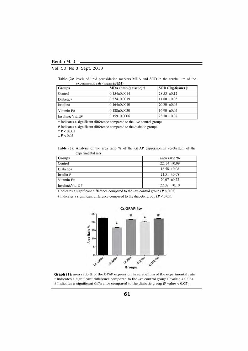
61
Benha M. J.
Vol. 30 No 3 Sept. 2013
Graph (1):Graph (1): area ratio % of the GFAP expression in cerebellum of the experimental rats* Indicates a significant difference compared to the –ve control group (P value < 0.05).# Indicates a significant difference compared to the diabetic group (P value < 0.05).
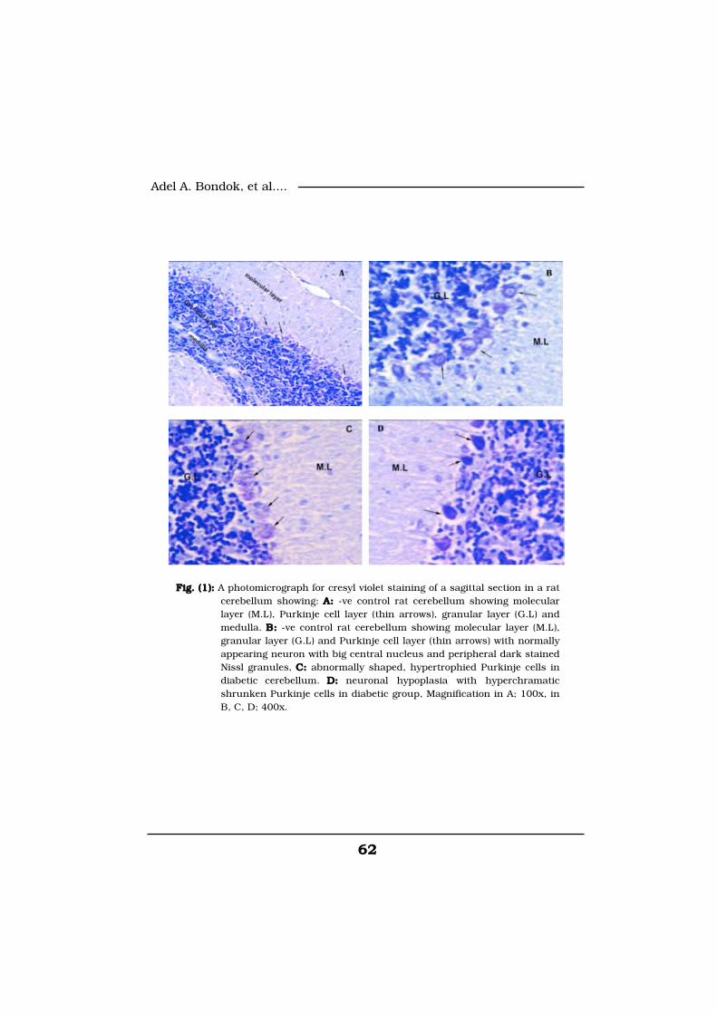
62
Adel A. Bondok, et al....
Fig. (1):Fig. (1): A photomicrograph for cresyl violet staining of a sagittal section in a ratcerebellum showing: A:A: -ve control rat cerebellum showing molecularlayer (M.L), Purkinje cell layer (thin arrows), granular layer (G.L) andmedulla. B:B: -ve control rat cerebellum showing molecular layer (M.L),granular layer (G.L) and Purkinje cell layer (thin arrows) with normallyappearing neuron with big central nucleus and peripheral dark stainedNissl granules, C: C: abnormally shaped, hypertrophied Purkinje cells indiabetic cerebellum. D: : neuronal hypoplasia with hyperchramaticshrunken Purkinje cells in diabetic group, Magnification in A; 100x, inB, C, D; 400x.

63
Benha M. J.
Vol. 30 No 3 Sept. 2013
Fig. (2):Fig. (2): A photomicrograph for cresyl violet staining of a sagittal section in a ratcerebellum showing molecular layer (M.L), Purkinje cell layer (thin ar-rows) and granular layer (G.L) E:E: insulin treated diabetic rat showingminimal changes with multiple pattern purkinje cells; dark stainedcells (thin arrows) and normally appearing purkinje (arrow heads). F:F:vitamin E treated group, showing dark stained shrunken neuron (thinarrows) and normally appearing neurons with rounded with central nu-cleus and peripheral Nissl granules (arrows heads). G: G: combined insu-lin and vitamin E treatment showing normally appearing Purkinje cellswith normal pattern of Nissl granules and large central nucleus. Magni-fication 400x.
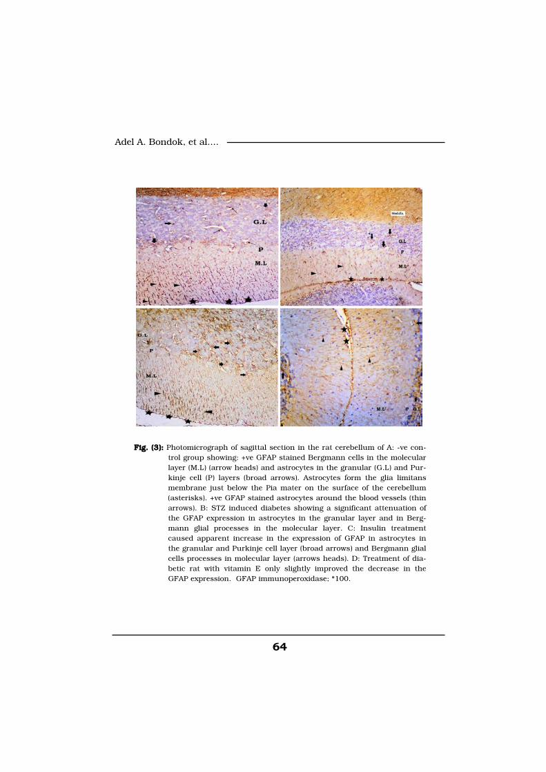
64
Adel A. Bondok, et al....
Fig. (3):Fig. (3): Photomicrograph of sagittal section in the rat cerebellum of A: -ve con-trol group showing: +ve GFAP stained Bergmann cells in the molecularlayer (M.L) (arrow heads) and astrocytes in the granular (G.L) and Pur-kinje cell (P) layers (broad arrows). Astrocytes form the glia limitansmembrane just below the Pia mater on the surface of the cerebellum(asterisks). +ve GFAP stained astrocytes around the blood vessels (thinarrows). B: STZ induced diabetes showing a significant attenuation ofthe GFAP expression in astrocytes in the granular layer and in Berg-mann glial processes in the molecular layer. C: Insulin treatmentcaused apparent increase in the expression of GFAP in astrocytes inthe granular and Purkinje cell layer (broad arrows) and Bergmann glialcells processes in molecular layer (arrows heads). D: Treatment of dia-betic rat with vitamin E only slightly improved the decrease in theGFAP expression. GFAP immunoperoxidase; *100.
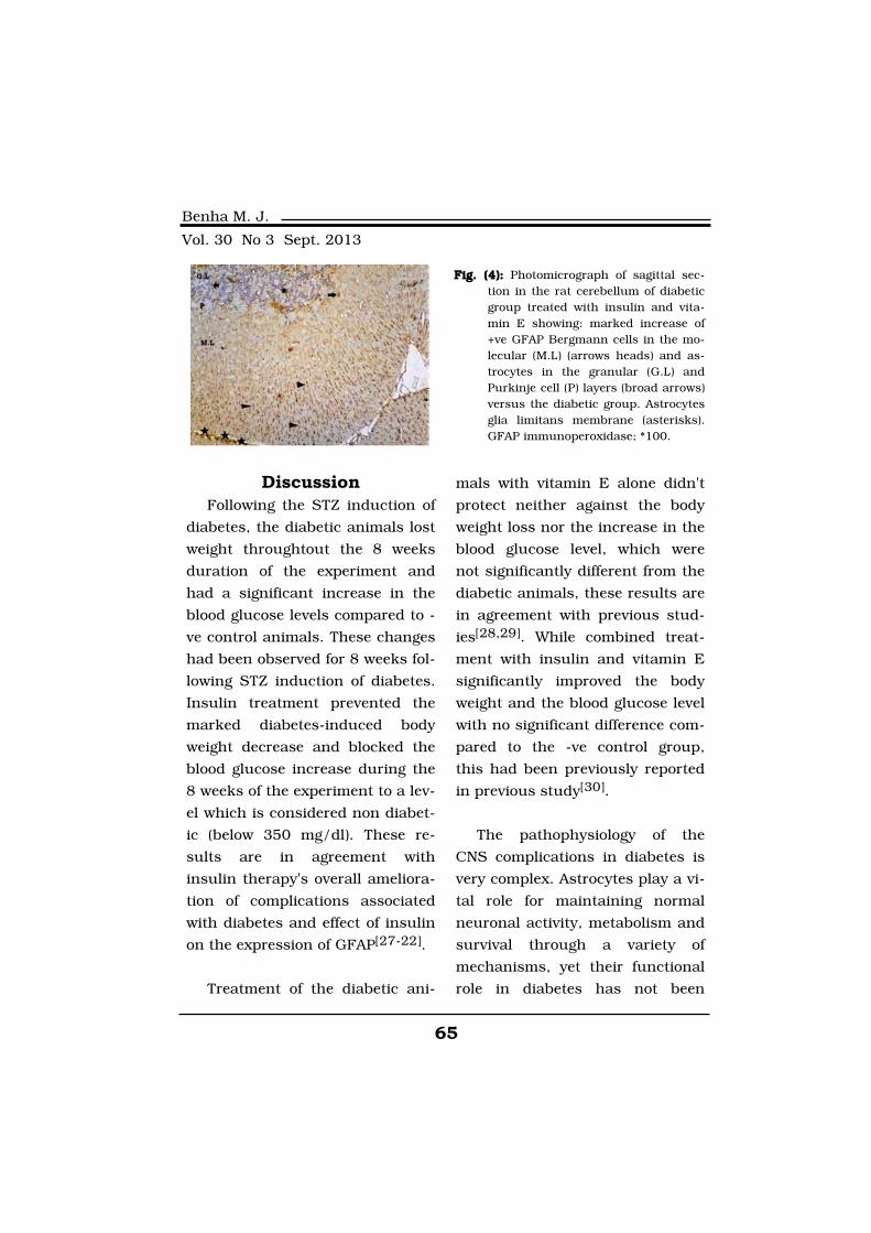
65
Benha M. J.
Vol. 30 No 3 Sept. 2013
DiscussionFollowing the STZ induction of
diabetes, the diabetic animals lost
weight throughtout the 8 weeks
duration of the experiment and
had a significant increase in the
blood glucose levels compared to -
ve control animals. These changes
had been observed for 8 weeks fol-
lowing STZ induction of diabetes.
Insulin treatment prevented the
marked diabetes-induced body
weight decrease and blocked the
blood glucose increase during the
8 weeks of the experiment to a lev-
el which is considered non diabet-
ic (below 350 mg/dl). These re-
sults are in agreement with
insulin therapy's overall ameliora-
tion of complications associated
with diabetes and effect of insulin
on the expression of GFAP[27-22].
Treatment of the diabetic ani-
mals with vitamin E alone didn't
protect neither against the body
weight loss nor the increase in the
blood glucose level, which were
not significantly different from the
diabetic animals, these results are
in agreement with previous stud-
ies[28,29]. While combined treat-
ment with insulin and vitamin E
significantly improved the body
weight and the blood glucose level
with no significant difference com-
pared to the -ve control group,
this had been previously reported
in previous study[30].
The pathophysiology of the
CNS complications in diabetes is
very complex. Astrocytes play a vi-
tal role for maintaining normal
neuronal activity, metabolism and
survival through a variety of
mechanisms, yet their functional
role in diabetes has not been
Fig. (4):Fig. (4): Photomicrograph of sagittal sec-tion in the rat cerebellum of diabeticgroup treated with insulin and vita-min E showing: marked increase of+ve GFAP Bergmann cells in the mo-lecular (M.L) (arrows heads) and as-trocytes in the granular (G.L) andPurkinje cell (P) layers (broad arrows)versus the diabetic group. Astrocytesglia limitans membrane (asterisks).GFAP immunoperoxidase; *100.
66
Adel A. Bondok, et al....
clearly defined[31].
In this study, neuronal degen-
eration could be seen in the cere-
bellum of STZ induced diabetic
animals, these changes were in
the form of neuronal hypoplasia
(hyperchromatic shrunken neu-
rons) or abnormal pattern of dis-
tribution of Nissl granule with
large hypertrophied vacuolated
neurons. These results are in
agreement with[32] who reported
that neurons tend to degenerate
and show a relative inability to
grow and proliferate in diabetes.
Changes in the dentritic process
of neurons in frontal lobe and hip-
pocampus have been reported in
diabetes[33]. Studies showed that
neurons suffer oxidative damage
and undergo apoptosis in dia-
betes[34]. These changes are mini-
mal with insulin treatment but
more visible in groups treated
with vitamin E only. This neuronal
degeneration was prevented by the
combined treatment with insulin
and Vitamin E.[35] proposed that
vitamin E is especially potent as
neuroprotective agent.
The change in astrocyte num-
ber and morphology could be the
consequence of unviable extracel-
lular conditions such as hyperos-
molarity, low nutrient availability,
or increased oxidative stress. The
lack of insulin could be involved
because insulin influences astro-
cyte morphology, differentiation,
and GFAP expression[36]. Glial
cells are widely exceeding in num-
ber the neurons in the adult brain
representing about 90% of human
brain cells [37]. Astrocytes are the
most numerous cells in the CNS[38], which occupy up to 50% of
the total brain volume[39]. One of
the subfamily of the astrocytes is
the Bergmann cells of the cerebel-
lum[40].
There are many discrepancies
concerning changes in GFAP lev-
els in diabetic animals in the pre-
vious studies due different re-
sponse of astrocytes and its
subtype Bergmann glial cells to di-
abetes after variables periods[41,17,14,18]. A second possibility
that may account for the differ-
ence is that not all astrocytes are
identical[42] and astrocytes in dif-
ferent brain regions respond dif-
ferently to diabetes[22].
In the present study STZ dia-
67
Benha M. J.
Vol. 30 No 3 Sept. 2013
betes led to a significant decrease
in the expression of GFAP protein
by astrocytes in the granular layer
of cerebellum and the processes of
Bergmann glial cells in the molec-
ular layer of the cerebellum. This
decrease in GFAP expression
could be due to changes in the
amount of this structural protein
per cell and/or decrease in the
number of GFAP-positive cells.
These observations are consistent
with the results of [18,22,2]. On
contrary Baydas et al.,[14] stated
that diabetes induced a glial reac-
tivity by increasing the expression
of GFAP protein in many parts of
the brain. On the other hand Lu-
chuga-Sancho[2] reported that
Bergmann glial cells were not af-
fected by diabetes, however, in the
current study Bermann Glial pro-
cesses had been apparently affect-
ed as GFAP expression was signifi-
cant decrease after 8 weeks
diabetes. Penky and Nelson[43] re-
ported that there the increase in
the GFAP immunoreactivity and
proliferation of astrocytes is a gen-
eral response to neuronal injury.
Luchuga-Sancho[2] had stated
that there is initial increase in
GFAP expression after one week of
diabetes induction which pointing
to active astrogliosis associated
with induced diabetic cerebellar
injury, followed by significant de-
crease in the expression of GFAP
probably as a result of both de-
creased cell proliferation and in-
creased cell death.
The decrease in the GFAP ex-
pression in the cerebellum was
improved by the treatment with ei-
ther insulin or vitamin E but still
significantly lower than that of the
-ve control group. The combined
treatment with insulin and vita-
min E prevented this decrease and
almost restore the GFAP expres-
sion up to the -ve control level at 8
weeks. This is in agreement with
previous study of[22]. The re-
sponse of the cerebellar astrocytes
and its subfamily Bergmann cell
may vary according to the severity
of the diabetes[31].
In many neurodegenerative dis-
eases, oxidative stress both ini-
tiates and drives the progression
of the pathogenic process[44] and
many of the diabetic complica-
tions such as diabetic neuropathy
are believed to be a result of ex-
cessive accumulation of reactive
oxygen species and of a decreased
68
Adel A. Bondok, et al....
antioxidant defense system[45].
Excessive production of free radi-
cals is believed to be involved in
many diabetic complications, in-
cluding diabetic neuropathy in di-
abetes mellitus[46]. In the current
study STZ induced diabetes
caused oxidative stress as it sig-
nificantly elevated the MDA mark-
er and decreased the SOD marker
of the lipid peroxidation in the cer-
ebellum after 8 weeks of diabetes
induction. These finding are in
agreement with previous results of[47,48,49,14]. It is known that as-
trocytes have more antioxidant ca-
pacity than do neurones[50,51,52]. The glial cells protect
neurones against oxidative stress
and promote neuronal survival as
they express a variety of neuro-
trophic factors and cytokines[14].
The current study demonstrat-
ed that STZ induced diabetes
leads to a decrease in GFAP levels
in astrocytes and its subtype
Bergmann glial cells of the cere-
bellum at 8 weeks of diabetes to-
gether with increased oxidative
stress. These might contribute to
the underlying patho-physiology of
diabetes-induced CNS disorders
and neuronal changes that had
noticed in Purkinje cell layer of
the cerebellum at 8 weeks. These
changes were prevented by treat-
ment with both insulin and vita-
min E.
Astrocytes have an expanding
list of functions in supporting the
neurons and maintain the neuro-
nal environment, the role of astro-
cytes in diabetes-induced CNS
disorders clearly warrants further
investigations and studies to eval-
uate the effects of diabetes on the
functionality of the astrocyte, glu-
tamate transporters, insulin re-
ceptors in astrocytes and neurons,
the effect of diabetes and insulin
treatment for longer periods on
neurons and the molecular chang-
es to assess the effects of insulin
therapy on diabetes-related astro-
cyte alterations.
References1. Wild S., Roglic G., Green1. Wild S., Roglic G., Green
A., Sicree R. and King H. (2004):A., Sicree R. and King H. (2004):
Global prevalence of diabetes.
Diabetes Care, 2004. 27:
1047-1053.
2. Lechuga-Sancho A.M., Ar-2. Lechuga-Sancho A.M., Ar-
roba A.I., Frago L.M., et al.roba A.I., Frago L.M., et al.
(2006):(2006): Reduction in the number

69
Benha M. J.
Vol. 30 No 3 Sept. 2013
of astrocytes and their projections
in associated withincreased syn-
aptic protein density in the hypo-
thalamus of poorly controlled dia-
betic rats. Endocrinology 147,
5314-5324.
3. Thach W.T. (1998):3. Thach W.T. (1998): A role
for the cerebellum in learning
movement coordination; Neurobi-
ol. Learn. Mem. 70 177-188.
4. Salman M.S. (2002):4. Salman M.S. (2002): The
cerebellum: It's about time! But
timing is not everything-new insights
into the role of the cerebellum in
timing motor and cognitive tasks; J.
Child Neurol. 17 1-9.
5. Matsumura M., Sadato N.,5. Matsumura M., Sadato N.,
Kochiyama T., et al. (2004):Kochiyama T., et al. (2004): Role
of the cerebellum in implicit motor
skill learning: A PET study; Brain
Res. Bull. 63 471-483.
6. Travis J. (1994):6. Travis J. (1994): Glia: the
brain's other cells. Science
266:970-972.
7. Laming P.R., Kimelberg H.,7. Laming P.R., Kimelberg H.,
Robinson S., et al. (2000):Robinson S., et al. (2000): Neu-
ronal-glial interactions and beha-
viour. Neurosci Biobehav Rev
24:295-340.
8. Ullian E.M., Sapperstein8. Ullian E.M., Sapperstein
S.K., Christopherson K.S. andS.K., Christopherson K.S. and
Barres B.A. (2001):Barres B.A. (2001): Control of
synapse number by glia. Science
291:657-661.
9. Abbott N.J. (2002):9. Abbott N.J. (2002): Astro-
cyte-endothelial interactions and
the blood brain barrier permeabili-
ty. J Anat 200:629-638.
10. Bondok A.A. (2010):10. Bondok A.A. (2010): Basic
Clinical Neuroanatomy. 2nd ed.
Middle of Delta Publisher: Man-
soura, Egypt. pp 91.
11. Newman E.A. (2003):11. Newman E.A. (2003): New
roles for astrocytes: regulation of
synaptic transmission. Trends
Neurosci 26:536-542.
12. Chen Y. and Swanson12. Chen Y. and Swanson
R.A. (2003):R.A. (2003): Astrocytes and brain
injury. J Cereb Blood Flow Metab
23:137-149.
13. Zonta M., Angulo M.C.,13. Zonta M., Angulo M.C.,
Gobbo S., et al. (2003):Gobbo S., et al. (2003): Neuron
to astrocyte signaling is central to
the dynamic control of brain mi-
crocirculation. Nat Neurosci 6:43-50.
14. Baydas G., Nedzvetskii14. Baydas G., Nedzvetskii
V.S., Tuzcu M., Yasar A. andV.S., Tuzcu M., Yasar A. and
70
Adel A. Bondok, et al....
Kirichenko S.V. (2003):Kirichenko S.V. (2003): Increase
of glial fibrillary acidic protein and
S-100B in hippocampus and cor-
tex of diabetic rats: effects of vita-
min E. Eur J Pharmacol 462:67-
71.
15. Bignami A. and Dahl D.15. Bignami A. and Dahl D.
(1977):(1977): Specificity of the glial fi-
brillary acidic protein for astroglia.
J Histochem Cytochem 25:466-
469.
16. Norton W.T., Aquino D.A.16. Norton W.T., Aquino D.A.
Hozumi I., Chiu F.C. and Bros-Hozumi I., Chiu F.C. and Bros-
nan C.F. (1992):nan C.F. (1992): Quantitative as-
pects of reactive gliosis. Neuro-
chem Res 17:877-885.
17. Saravia F.E., Revsin Y.,17. Saravia F.E., Revsin Y.,
Deniselle M.C.G., et al. (2002):Deniselle M.C.G., et al. (2002):
Increased astrocyte reactivity in
the hippocampus of murine mod-
els of type 1 diabetes: the nono-
bese diabetic (NOD) and strepto-
zotocin-treated mice. Brain Res
957:345-353.
18. Coleman E., Judd R., Hoe18. Coleman E., Judd R., Hoe
L., Dennos J. and Posner P.L., Dennos J. and Posner P.
(2004):(2004): Effects of diabetes mellit-
us on astrocyte GFAP and gluta-
mate transporters in the CNS.
Glia 48:166-178.
19. Renno W.M., Saleh F.,19. Renno W.M., Saleh F.,
Klepacek I. and Al-Awadi F.Klepacek I. and Al-Awadi F.
(2006):(2006): Talin immunogold density
increases in sciatic nerve of dia-
betic rats after nerve growth factor
treatment. Medicina (Kaunas)
42:147-163.
20. Akbarzadeh A., Norouzian20. Akbarzadeh A., Norouzian
D., Mehrabi M.R., et al. (2007):D., Mehrabi M.R., et al. (2007):
Induction of diabetes by STZ in
rats. Ind J.Clin l Bioch 2007, 22,
60- 64.
21. Alzoubi K.H., Khabour21. Alzoubi K.H., Khabour
O.F., Abu Rashid B., Damaj I.M.O.F., Abu Rashid B., Damaj I.M.
and Salah H.A. (2012):and Salah H.A. (2012): The neu-
roprotective effect of vitamin E on
chronic sleep deprivation-induced
memory impairment: The role of
oxidative stress, Behavioural Brain
Research 226 (2012) 205- 210.
22. Coleman E., Dennos J.,22. Coleman E., Dennos J.,
Braden T.D., Judd R. and Pos-Braden T.D., Judd R. and Pos-
ner P. (2010):ner P. (2010): Insulin treatment
prevents diabetes-induced altera-
tions in astrocyte glutamate up-
take and GFAP content in rats at
4 and 8 weeks of diabetes dura-
tion. Brain Res 1306 (2010) 131-
141.
23. Ohkawa H., Ohishi N. and23. Ohkawa H., Ohishi N. and
71
Benha M. J.
Vol. 30 No 3 Sept. 2013
Yagi K. (1979):Yagi K. (1979): Assay for lipid
peroxidation in animal tissues by
thiobarbituric acid reaction. An-
nals of Biochemistry. 1979;
95:351-358.
24. Nishikimi M., N. Appaji24. Nishikimi M., N. Appaji
and K. Yagi (1972):and K. Yagi (1972): The occur-
rence of superoxide anion in the
reaction of reduced phenazine me-
thosulfate and molecular oxygen.
Biochem. Biophys. Res. Com-
mun., 46: 849-854.
25. Bancroft J.D. and Gamble25. Bancroft J.D. and Gamble
M. (2002):M. (2002): Theory and practice of
histological techniques. 5th ed.
Churchill Livingstone: London,
UK.
26. Dugar A., Patanow C.,26. Dugar A., Patanow C.,
O'Callaghan J.P. and LakoskiO'Callaghan J.P. and Lakoski
J.M. (1998):J.M. (1998): Immunohistochemi-
cal localization and quantification
of glial fibrillary acidic protein and
synaptosomal-associated protein
(mol. wt 25000) in the ageing hip-
pocampus following administra-
tion of 5, 7-dihydroxytryptamine.
Neuroscience 85:123-133.
27. Nathan D.M., Zinman B.,27. Nathan D.M., Zinman B.,
Cleary P.A., et al. (2009):Cleary P.A., et al. (2009): Mod-
ern-day clinical course of type1 di-
abetes mellitus after 30 years' du-
ration: the diabetes control and
complications trial/epidemiology
of diabetes interventions and com-
plications and Pittsburgh epidemi-
ology of diabetes complications ex-
perience (1983-2005). Arch.
Intern. Med. 169, 1307-1316.
28. Kedziora-Kornatowska K.,28. Kedziora-Kornatowska K.,
Szram S., Kornatowski T., Sza-Szram S., Kornatowski T., Sza-
dujkis-Szadurski L., Kedziora J.dujkis-Szadurski L., Kedziora J.
and Bartosz G. (2003):and Bartosz G. (2003): Effect of
Vitamin E and Vitamin C Supple-
mentation on Antioxidative State
and Renal Glomerular Basement
Membrane Thickness in Diabetic
Kidney, Nephron Exp Nephrol
2003;95: 134-143.
29. Al Shamsi M., Amin A.29. Al Shamsi M., Amin A.
and Adeghate E. (2004):and Adeghate E. (2004): Benefi-
cial effect of vitamin E on the
metabolic parameters of diabetic
rats, Molecular and Cellular Bio-
chemistry June 2004, Vol. 261,
35-42 .
30. Yoshida M., Kimura H.,30. Yoshida M., Kimura H.,
Kyuki K. and Ito M. (2004):Kyuki K. and Ito M. (2004): Com-
bined effect of vitamin E and insu-
lin on cataracts of diabetic rats fed
a high cholesterol diet.Biol Pharm
Bull. 2004 Mar; 27(3):338-44.

72
Adel A. Bondok, et al....
31. Sofroniew M.V. and Vint-31. Sofroniew M.V. and Vint-
ers H.V. (2010):ers H.V. (2010): Astrocytes: biolo-
gy and pathology. Acta Neuropath-
ol. 2010 Jan; 119(1):7-35.
32. Compston A. (1995):32. Compston A. (1995):
Brain repair. J Intern Med
237:127-134.
33. Martínez-Tellez R.,33. Martínez-Tellez R.,
Gómez-Villalobos Mde J. andGómez-Villalobos Mde J. and
Flores G. (2005):Flores G. (2005): Alteration in
dendritic morphology of cortical
neurons in rats with diabetes mel-
litus induced by streptozotocin.
Brain Res. 2005 Jun 28; 1048(1-
2):108-15.
34. Vincent A.M., McLean34. Vincent A.M., McLean
L.L., Backus C. and FeldmanL.L., Backus C. and Feldman
E.L. (2005):E.L. (2005): Short-term hypergly-
cemia produces oxidative damage
and apoptosis in neurons. FASEB
J. 19, 638-640.
35. Sen C.K., Khanna S. and35. Sen C.K., Khanna S. and
Roy S. (2004):Roy S. (2004): Tocotrienol: the
natural vitamin E to defend the
nervous system? Ann. N. Y. Acad.
Sci. 1031, 127-142.
36. Aizenman Y. and de Vellis36. Aizenman Y. and de Vellis
J. (1987):J. (1987): Synergistic action of
thyroid hormone, insulin and hy-
drocortisone on astrocyte differen-
tiation. Brain Res 414:301-308.
37. Baumann N. and Pham-37. Baumann N. and Pham-
Dinh D. (2002):Dinh D. (2002): Astrocytes,
Encyclopedia of the Human Brain
Vol. 1. 2002, Elsevier Science
(USA).
38. Gordon G.R., Howarth C.38. Gordon G.R., Howarth C.
and MacVicar B.A. (2011):and MacVicar B.A. (2011): Bidi-
rectional control of arteriole diam-
eter by astrocytes. Exp Physiol.
2011 Apr; 96(4):393-9.
39. Yi C.X. and Tschöp M.H.,39. Yi C.X. and Tschöp M.H.,
(2012):(2012): Brain-gut-adipose-tissue
communication pathways at a
glance. Dis Model Mech. 2012
Sep; 5(5):583-7.
40. Palay S.L., Chan-Palay V.,40. Palay S.L., Chan-Palay V.,
(1974):(1974): A guide to the synaptic
analysis of the neuropil. Cold
Spring Harb Symp Quant Biol.
1976; 40:1-16.
41. Barber A.J., Antonetti41. Barber A.J., Antonetti
D.A. and Gardner T.W. (2000):D.A. and Gardner T.W. (2000):
Altered expression of retinal occlu-
din and glial fibrillary acidic pro-
tein in experimental diabetes. In-
vest Ophthalmol Vis Sci 41:3561-
3568.
73
Benha M. J.
Vol. 30 No 3 Sept. 2013
42. Hewett J.A. (2009):42. Hewett J.A. (2009): Deter-
minants of regional and local di-
versity within the astroglial line-
age of the normal centralnervous
system. J Neurochem. 2009 July
17;Epub.
43. Pekny M. and Nilsson M.43. Pekny M. and Nilsson M.
(2005):(2005): Astrocyte activation and
reactive gliosis. Glia 50, 427-434.
44. Coyle J.T. and Puttfarck-44. Coyle J.T. and Puttfarck-
en P. (1993):en P. (1993): Oxidative stress,
glutamate, and neurodegenerative
disorders. Science 262, 689-695.
45. Kesavulu M.M., Giri R.,45. Kesavulu M.M., Giri R.,
Rao B.K. and Apparao C. (2000):Rao B.K. and Apparao C. (2000):
Lipid peroxidation and antioxidant
enzyme levels in type 2 diabetics
with micro vascular complica-
tions. Diabetes Metab. 26, 387-
392.
46. Sima A.A. and Sugimoto46. Sima A.A. and Sugimoto
K., (1999):K., (1999): Experimental diabetic
neuropathy: an update. Diabetolo-
gia 42, 773-788.
47.47. Ercel E., Baydas G., Ak-Ercel E., Baydas G., Ak-
yol A., Eksioglu E., Canpolatyol A., Eksioglu E., Canpolat
and L. (1999):and L. (1999): The effect of vita-
min E on the sciatic nerve lipid
peroxidation in Streptozotocin in-
duced diabetes mellitus. Biomed.
Res. 10, 95-101.
48.48. Celik S., Baydas G. andCelik S., Baydas G. and
Yilmaz O. (2002):Yilmaz O. (2002): Influence of vi-
tamin E on the levels of fatty acids
and MDA in some tissues of dia-
betic rats. Cell Biochem. Funct.
20, 67-71.
49.49. Baydas G., Canatan H.Baydas G., Canatan H.
and Turkoglu A. (2002):and Turkoglu A. (2002): Compar-
ative analyses of the protective ef-
fects of melatonin and vitamin E
on streptozocin-induced diabetes
mellitus. J. Pineal Res. 32, 225-
229.
50.50. Savolainen H. (1978):Savolainen H. (1978):
Superoxide dismutase and glu-
tathione peroxidase activities in
rat brain. Res. Commun. Chem.
Pathol. Pharmacol. 21, 173- 176.
51.51. Raps S.P., Lai J.C.,Raps S.P., Lai J.C.,
Hertz L. and Cooper A.J. (1989):Hertz L. and Cooper A.J. (1989):
Glutathione is present in high
concentrations in cultured astro-
cytes but not in cultured neu-
rones. Brain Res. 593, 398-401.
52.52. Makar T.K., NedergaardMakar T.K., Nedergaard
M., Preuss A., Gelbard A.S., Pe-M., Preuss A., Gelbard A.S., Pe-
rumal A.S. and Cooper A.J.rumal A.S. and Cooper A.J.

74
Adel A. Bondok, et al....
(1994):(1994): Vitamin E, ascorbate, glu-
tathione disulphide and enzymes
of glutathione metabolism in cul-
tures of chick astrocytes and neu-
rons: evidence that astrocytes play
an important role in antioxidative
processes in the brain. J. Neuro-
chem. 62, 45- 53.
75
Benha M. J.
Vol. 30 No 3 Sept. 2013
EFFECTS OF INSULIN AND VITAMINEFFECTS OF INSULIN AND VITAMINE ON THE EXPRESSION OF GLIALE ON THE EXPRESSION OF GLIALFIBRILLARY ACIDIC PROTEIN ANDFIBRILLARY ACIDIC PROTEIN AND
OXIDATIVE STRESS IN THEOXIDATIVE STRESS IN THECEREBELLUM OF DIABETIC RATSCEREBELLUM OF DIABETIC RATS
Adel A. Bondok Ph.D, Adel A. Elhawary Ph.D,Adel A. Bondok Ph.D, Adel A. Elhawary Ph.D,Mohamed I. Abdo Ph.D, Rania N. Kamal Ph.DMohamed I. Abdo Ph.D, Rania N. Kamal Ph.D
and *Hany M. Sonpol M.Scand *Hany M. Sonpol M.Sc
BENHAMEDICALJOURNAL
REPRINT
Published byPublished by
Benha Faculty of MedicineBenha Faculty of Medicine Volume 30 Number 3Sept. 2013
75
Benha M. J.
Vol. 30 No 3 Sept. 2013
WNT/WNT/β-CATENIN SIGNALING PATHWAY IN-CATENIN SIGNALING PATHWAY INBREAST CARCINOGENESIS IN RATSBREAST CARCINOGENESIS IN RATS
Amina Ahmad Baiomy Ph.D, Hoda Ahmad Nada Ph.D,Amina Ahmad Baiomy Ph.D, Hoda Ahmad Nada Ph.D,Lamiaa Arafa Ph.D, Maha Amin Ph.D* Lamiaa Arafa Ph.D, Maha Amin Ph.D*
and Lamiaa El-Abbasy M.Sc. and Lamiaa El-Abbasy M.Sc. Departments of Medical Biochemistry and Pathology*,
Faculty of Medicine, Mansoura University, Egypt.
AbstractBackground: Background: Breast cancer is a common worldwide malignancy
among women. De-regulation of Wnt/β-catenin signalling is increasing-ly being implicated in both experimental and human carcinogenesis;however, its role in breast cancer is unclear.
Aim of work:Aim of work: The goal of this study was to elucidate the role of Wnt/β-catenin signaling in breast carcinogenesis, and to test for a potentialrelationship between Wnt/β-catenin pathway activation and expressionof Glioma-associated oncogene homolog 1 (GLi1), a marker of Hedgehogpathway activation.
Material & Method:Material & Method: We used a methyl-nitrosourea (MNU)-inducedrat breast carcinogenesis model that mimics many essential elements ofhuman breast cancer to investigate the expression pattern of two of themain key players in Wnt signaling (β-catenin and Axin2) by semiquanti-tative reverse transcription polymerase chain reaction (RT-PCR) analy-sis. Also, Gli1 mRNA was analyzed in relation to both genes in order toreveal any possible crosstalk.
Results:Results: The expressions of target genes were significantly upregu-lated in the diseased groups than in control group. There was no signifi-cant correlation between the expression of β-catenin, Axin2, and Gli1.
Conclusion:Conclusion: These results indicate that overexpression of β-catenin,Axin2, and Gli1 may be associated with the malignant transformation ofmammary cells, suggesting a new target for breast chemoprevention.
Key words:Key words: Breast cancer; Wnt signaling; β-catenin; Axin2; Gli1;rats.
76
Amina Ahmad Baiomy, et al....
IntroductionBreast cancer is one of the
most common cancers around the
world with approximately 1.6 mil-
lion new cases and 425 thousand
deaths from the disease in 2010(1). It is a highly heterogeneous
disease represented by tumors
that have a diverse natural histo-
ry, complex histology and a vari-
able response to therapy. Al-
though the molecular events that
trigger breast cancer progression,
including its initiation, promotion
and progression to a fully malig-
nant state, are not fully under-
stood(2). The role of Wnt/ß-
catenin signaling in human breast
cancer has been subject to much
debate(3,4). Although, the first
mammalian Wnt gene, Wnt1, was
originally identified as a locus ac-
tivated by retroviral insertion of
mouse mammary tumor virus
(MMTV), and transgenic Wnt1
overexpression was subsequently
shown to drive mammary tumor
formation in mice(5,6). The defini-
tive evidence linking Wnt signaling
to human breast cancer has been
slow to emerge(7). Moreover, his-
torical failure to identify substan-
tial frequencies of Wnt ligand
overexpression in human breast
tumors hindered appreciation of
the relevance of Wnt signaling to
the human disease. In contrast,
there is abundent evidence that
hyperactive Wnt signaling contrib-
utes to the genesis of a wide range
of other human cancers. Striking
frequencies of aberrant nucleocy-
toplasmic β-catenin accumulation
have now been recorded in multi-
ple human neoplastic conditions,
most notably in colorectal cancers(8). However, mutation of pathway
components, including the APC,
Axin, and CTNNB1 genes (encod-
ing β-catenin), leading to β-catenin
stabilization, and hence activation
of the Wnt/β-catenin pathway, is
now recognized as a common
event in human tumorigenesis(9,10). Because such mutations
are comparatively rare in human
breast carcinomas, excepting fi-
bromatoses and metaplastic tu-
mors(11).
Aberrant reactivation of Hh sig-
naling has also been reported in
breast cancer(12,13). Hh signaling
is also thought to contribute to in-
vasiveness(14). Glioma-associated
oncogene homolog 1 (GLi1) is
thought to be a marker of Hh
pathway activation(15). Although
77
Benha M. J.
Vol. 30 No 3 Sept. 2013
the Hh and Wnt pathways can be
activated concurrently during
breast tumorigenesis, the func-
tional significance of signaling
crosstalk in tumor initiation and
progression has not been estab-
lished(16).
Our goal in the present study
was to further investigate the fre-
quency of Wnt/β-catenin signaling
pathway activation in breast neo-
plasia using means of semiquanti-
tative reverse transcription poly-
merase chain reaction (RT-PCR) of
both mRNA of β-catenin and
Axin2, a specific target gene. An
additional goal was to assess the
relationship between canonical
Wnt pathway activation and Gli
overexpression based on several
lines of evidence indicating that
Wnt and Hedgehog (Hh) signaling
pathways can interact(16-18).
Materials and Methods* Animals* Animals
A total of 55 female Sprague-
Dawley rats, aged 14 days and
weighing (35±5g) were obtained
from and housed in Medical Ex-
perimental Research Centre
(MERC). The animals were housed
4-5 per cage and maintained at
23±2ºC and 12 h light:dark cycle.
They were fed a purified diet (AIN-
76A) and had access to water ad
libitum.
* Experimental Design* Experimental Design
After a 7 days acclimatization
period, the animals were randomly
divided into two groups (experi-
mental and control group). For in-
duction of breast cancer in the ex-
perimental group (n=43), MNU
(Sigma-Aldrich, St. Louis, USA),
(50 mg/kg body weight) was in-
jected i.p. on the day 21 and 65 of
age. The MNU was always dis-
solved immediately before use in
0.9% NaCl adjusted to pH 4 with
acetic acid. The solubility of MNU
in water at room temperature was
1.4 % (w/v)(19). The animals were
weighed once a week. Ten rats of
the experimental group died dur-
ing the experiments. Sex and age
matched control rats (n=12) were
maintained free access to tap wa-
ter and basal diet without any
treatment until scarification. The
experiment was terminated on the
130 th day of the animals age.
* Tissue sampling* Tissue sampling
The abdominal-inguinal mam-
mary glands (AIMG) on both sides,
78
Amina Ahmad Baiomy, et al....
left and right were evaluated for
the presence of grossly detectable
mammary tumours and the dis-
sected animals with tumours were
photographed to provide identifi-
cation record on the location and
gross morphology of lesions (Fig-
ure.1.b). The mammary tissues
were excised from the subcutane-
ous tissue with scissors along
with the fat pads (Figure.1.a), then
cut into 2 pieces, the proximal re-
gion including a lymph node (ab-
dominal gland) and the distal re-
gion (injuinal gland). For
histopathological study, specimen
of the breast (tumour and tumour
adjacent) tissues were fixed in
10% neutral buffered formalin,
embedded in paraffin wax, cut at
5ml thickness and stained with
hematoxylin and eosin (H&E) after
processing. The rest of the breast
tissues were snap frozen in liquid
nitrogen for subsequent molecular
analysis. Mammary tumors were
classified based on the criteria de-
scribed by Russo(20) into hyper-
plasia, carcinoma insitu (CIS) and
adenocarcinoma which shows ob-
vious invasion. Also, The normal
non tumored mammary gland
from rats bearing tumor were col-
lected and analyzed.
* Molecular Biology Assays in* Molecular Biology Assays in
the Breast: the Breast:
RNA Isolation and Semiquan-RNA Isolation and Semiquan-
titative Reverse Transcriptiontitative Reverse Transcription
Polymerase Chain Reaction (RT-Polymerase Chain Reaction (RT-
PCR) Analysis:PCR) Analysis:
Total RNA was extracted from
breast tissue specimen using both
Trifast Reagent (Peqlab, Germany)
followed by GeneJET RNA Purifi-
cation Kit (Fermentas Lifesciences,
U.S.A.). Purity and concentration
of extracted RNA was assessed by
measuring the optical density at
wave lengths of 260, and 280 nm.
Absorbance ratio at 260/280 was
used to determine the purity of
the RNA samples. DNA contami-
nation was assessed using formal-
dehyde agarose gel electrophore-
sis. The RNA is then was
converted into cDNA using Maxi-
ma First Strand cDNA Synthesis
Kit (Thermo Scientific, U.S.A). Two
micrograms of total RNA were re-
verse transcribed into first strand
cDNA in a volume of 20 µl at 10
min at 25°C followed by 15 min at
50°C and heated at 85ºC for 5 min
to terminate the reverse transcrip-
tion reaction. β-catenin, Axin2,
Gli1 and GAPD (housekeeping
gene) genes were amplified from 2
µl cDNA mixtures in a final vol-
79
Benha M. J.
Vol. 30 No 3 Sept. 2013
ume of 20 µl PCR reaction mixture
by adding 18 µl of DreamTaq™
Green PCR Master Mix (2X) (Fer-
mentas Lifesciences, U.S.A). Reac-
tion mixtures were first denatured
at 94ºC for 5 min, and amplifica-
tion was performed for 35 cycles,
at 95ºC for 45 s, annealing (57ºC
for β-catenin, Gli1 and GAPD,
while 62ºC for Axin2) for 1 min,
and at 72ºC for 2 min, followed by
an extension for 10 min at 72ºC.
The PCR primer pairs used were
enlisted in Table 1.
After amplification, the PCR
products were electrophoresed on
agarose gel. The marker used was
50 bp DNA Ladder (GeneRuler™,
Fermentas, Canada) ranging from
1000-50 bp with two reference
bands at 500 and 250 bp. The re-
sulted photos were analyzed with
scion image® release Alpha
4.0.3.2 software for windows®
(Scion image, Scion corporation,
USA) which performs bands detec-
tion and conversion to peaks. Are-
as under each peak were calculat-
ed in square pixels and used in
quantification. Semiquantitative
gene expression was determined
by calculating the ratio between
the square pixel value of the target
genes in relation to the house-
keeping gene. Each target gene ex-
pression was evaluated by com-
parison of the results at each
experimental stage with those of
the normal control.
ResultsExpression of Expression of β-catenin-catenin
mRNA, Axin2 mRNA and Gli1mRNA, Axin2 mRNA and Gli1
mRNA:mRNA:
Comparative analysis of
β-catenin expression (post hoc
test) showed a significant increase
in β-catenin expression in the
different histopathological stages
of MNU-induced breast cancer
(hyperplasia, CIS & adenocarcino-
ma) when compared with the
control group (P<0.01). There was
no statistically significant
difference between these 3 groups.
Also, there was a highly
significant increase in β-catenin
expression in normal mammary
tissue from tumor- bearing rats
when compared with control
(P<0.001).
Comparative analysis of Axin2
expression (post hoc test) showed
a significant increase in Axin2 ex-
pression in the three pathological
groups when compared with the
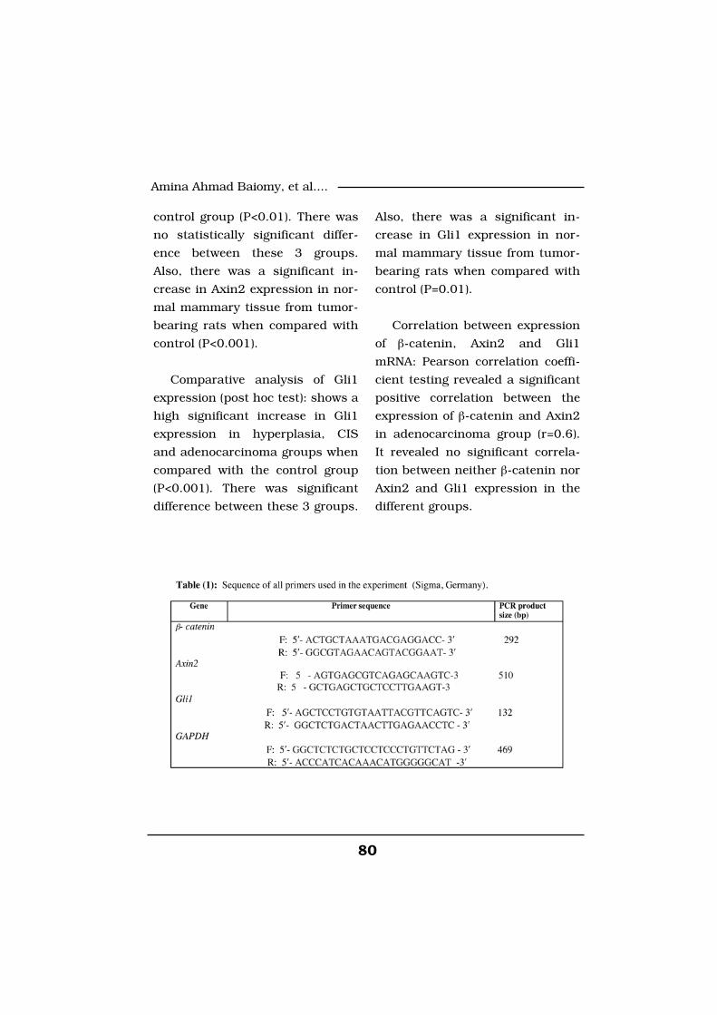
80
Amina Ahmad Baiomy, et al....
control group (P<0.01). There was
no statistically significant differ-
ence between these 3 groups.
Also, there was a significant in-
crease in Axin2 expression in nor-
mal mammary tissue from tumor-
bearing rats when compared with
control (P<0.001).
Comparative analysis of Gli1
expression (post hoc test): shows a
high significant increase in Gli1
expression in hyperplasia, CIS
and adenocarcinoma groups when
compared with the control group
(P<0.001). There was significant
difference between these 3 groups.
Also, there was a significant in-
crease in Gli1 expression in nor-
mal mammary tissue from tumor-
bearing rats when compared with
control (P=0.01).
Correlation between expression
of β-catenin, Axin2 and Gli1
mRNA: Pearson correlation coeffi-
cient testing revealed a significant
positive correlation between the
expression of β-catenin and Axin2
in adenocarcinoma group (r=0.6).
It revealed no significant correla-
tion between neither β-catenin nor
Axin2 and Gli1 expression in the
different groups.
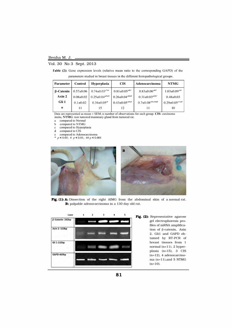
81
Benha M. J.
Vol. 30 No 3 Sept. 2013
Fig. (1):Fig. (1): A:A: Dissection of the right AIMG from the abdominal skin of a normal rat.B:: palpable adenocarcinoma in a 130 day old rat.
A B
Fig. (2):Fig. (2): Representative agarosegel electrophoresis pro-files of mRNA amplifica-tion of β-catenin, Axin2, Gli1 and GAPD ob-tained by RT-PCR ofbreast tissues from 1normal (n=11), 2 hyper-plasia (n=15), 3 CIS(n=12), 4 adenocarcino-ma (n=11),and 5 NTMG(n=10).
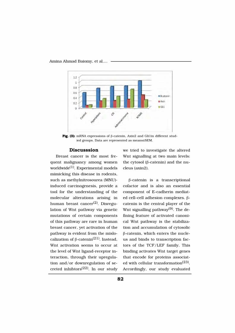
82
Amina Ahmad Baiomy, et al....
DiscusssionBreast cancer is the most fre-
quent malignancy among women
worldwide(1). Experimental models
mimicking this disease in rodents,
such as methylnitrosourea (MNU)-
induced carcinogenesis, provide a
tool for the understanding of the
molecular alterations arising in
human breast cancer(2). Disregu-
lation of Wnt pathway via genetic
mutations of certain components
of this pathway are rare in human
breast cancer, yet activation of the
pathway is evident from the mislo-
calization of β-catenin(21). Instead,
Wnt activation seems to occur at
the level of Wnt ligand-receptor in-
teraction, through their upregula-
tion and/or downregulation of se-
creted inhibtors(22). In our study
we tried to investigate the altered
Wnt signalling at two main levels:
the cytosol (β-catenin) and the nu-
cleus (axin2).
β-catenin is a transcriptional
cofactor and is also an essential
component of E-cadherin mediat-
ed cell–cell adhesion complexes. β-
catenin is the central player of the
Wnt signalling pathway(9). The de-
fining feature of activated canoni-
cal Wnt pathway is the stabiliza-
tion and accumulation of cytosolic
β-catenin, which enters the nucle-
us and binds to transcription fac-
tors of the TCF/LEF family. This
binding activates Wnt target genes
that encode for proteins associat-
ed with cellular transformation(23).
Accordingly, our study evaluated
Fig. (3): Fig. (3): mRNA expressions of β-catenin, Axin2 and Gli1in different stud-ied groups. Data are represented as means±SEM.
83
Benha M. J.
Vol. 30 No 3 Sept. 2013
the mRNA expression of β-catenin
in the different histopathological
stages of MNU-induced breast
cancer (hyperplasia, CIS & adeno-
carcinoma), showing significant
increase when compared to the
control normal rat tissue. This
overexpression of β-catenin mRNA
indicates mostly transcriptional
activation occurring during breast
cancer initiation and progression.
Little is known about regulation of
β-catenin gene expression. Howev-
er, TCF/LEF and AP1 transcrip-
tion factors were found in the pro-
moter region, suggesting possible
direct feedback mechanism for
augmenting β-catenin expression
by β-catenin /TCF/LEF pathway
and one of its downstream targets,
c-Jun(24).
Also, the upregulation of β-
catenin in this study is suggesting
that transcriptional deregulation
of the β-catenin gene itself might
be important during tumor devel-
opment. This suggestion is sup-
ported by other studies. One
study demonstrated the β-catenin
oncogenic effect, as transgenic
mutant β-catenin expressed in
mouse mammary glands leads to
carcinoma(25). Also, Ryo et al.,(26)
and Johnnson et al.,(27) confirmed
elevated β-catenin levels by West-
ern blotting of tumor lysates.
Meanwhile, several studies ob-
served elevated levels of nuclear
and/or cytoplasmic β-catenin de-
tected by immunohistochemical
staining in the majority breast tu-
mor tissue samples, and even that
high β-catenin activity was signifi-
cantly correlated with poor prog-
nosis of breast cancer(28-30).
Axin2 is a direct and specific
transcriptional target of the Wnt/
β-catenin signaling pathway. It is
well recognized that the expres-
sion level of Axin2 is the signature
of the activation of Wnt/β- catenin
signaling(31). Our study revealed
upregulation of Axin2 mRNA ex-
pression in the different histo-
pathological stages of breast can-
cer when compared with the
control group. The overexpression
of Axin2 mRNA could be explained
by activation of Axin2 gene tran-
scription. The effectors of the
Wnt/βcatenin pathway (TCF/LEF)
and E2F1 have shown to be in-
volved in regulation of Axin2 gene
activity(32,33). Beside this, chroma-
tin immunoprecipitation/microarray
analysis of many genes coding for
84
Amina Ahmad Baiomy, et al....
Wnt inhibitors, including Axin2
showed that they were occupied
by EZH2, a subunit of the poly-
comb repressor complex 2 (PRC2).
EZH2 is a methyl transferase that
deposits the repressive H3K27me3
marks on chromatin(34).
The increased expression of
Axin2 in this study is in agree-
ment with other studies who con-
firmed that Axin2 was markedly
upregulated upon mRNA analysis
of high-grade breast tumours(35,36). The oncogenic effect of
Axin2 was a point of question
since Axin2 is known as a nega-
tive regulator of canonical Wnt
signaling in normal cells. Howev-
er, rather than functioning as a
tumor suppressor, Axin2 was
found acting as a potent promot-
er of carcinoma behavior by up-
regulating the activity of the tran-
scriptional repressor, Snail1, in-
ducing a functional epithelial-
mesenchymal transition (EMT)
program and driving metastatic
activity(37). Also, Axin2 induced
chromosomal instability in CRC
has been reported(38).
Nevertheless, it’s worthwhile to
note that our findings involving
the increased Axin2 mRNA expres-
sion in the normal tissue from the
tumored rats are supported by the
recent study of khalil et al.,(39),
that demonstrated a finite fre-
quency of positive nucleocytoplas-
mic staining in normal human
breast tissues, indicating an ac-
tive wnt/ β-catenin signaling path-
way in these tissues. Accordingly ,
We hypothesized that Wnt/β-
catenin pathway activation ob-
served in benign breast tissue
could result from precancerous
changes in morphologically nor-
mal breast, and further reasoned
that breast tissue adjacent to in-
vasive tumors would be more like-
ly to contain such protumorigenic
molecular alterations.
Gli1 has been reported exten-
sively as a universal marker for
Hh-pathway activation. Elevated
Gli1 expression and its role in a
number of cancers has been wide-
ly reported, including a recent
study that the conditional expres-
sion of Gli1 in mouse mammary
glands results in mammary tu-
mours(40). This is consistent with
our findings as the Gli mRNA ex-
pression showed significant in-
crease during the different histo-
85
Benha M. J.
Vol. 30 No 3 Sept. 2013
pathological stages of MNU-
induced breast cancer (hyperpla-
sia, CIS & adenocarcinoma), when
compared to the control normal
rat tissue. The elevated expression
of Gli1 mRNA could be due to
transcriptional activation via both
canonical Hh-pathway-dependent(41,42) and non-canonical Hh-
independent activation of Gli1 in-
cluding TGF-β(43), PI3K/AKT(44),
Ras/Mek(45), and a Gli1-CDK2-
dependent mechanism(46). Our re-
sults revealed statistically signifi-
cant elevation of Gli1 expression
in the adenocarcinoma compared
to the other histopathological
stages of cancer, suggesting the
involvement of Gli1 in cancer pro-
gression. This is consistent with
other studies which reported that
deregulated Hh supports more ag-
gressive phenotypes of human
breast cancers, such as progres-
sion, metastasis, and therapeutic
resistance(47).
A cross talk between the Hh
and Wnt signaling in cancer was
reported in gastric, colon, endo-
metrial and skin cancer(16). De-
pending on the type of tissue Hh
and Wnt signalling pathways can
have cross talk as a positive feed-
back or negative feedback activity.
For example, Hh signalling has in-
hibitory effects on Wnt signalling
in stomach adenocarcinoma(17).
Meanwhile, it has a stimulatory ef-
fect in endometrial cancer(18). The
present study showed that Wnt/β-
catenin and Hh pathways are acti-
vated concurrently during breast
carcinogenesis, although in the
present study, Pearson correlation
coefficient testing revealed no sig-
nificant correlation between ex-
pression of β-catenin, Axin2 and
Gli1 mRNA in the histopathologi-
cal groups. This finding could be
explained by the wide variability
in the rate of increase of the dif-
ferent assayed genes as well as
heterogeneity of expression of
genes between rat samples of the
same histopathological group. The
heterogeneity of gene expression
and interactions of the Wnt and
the Hh pathway activation is pos-
sibly one of the reasons for drug
resistance in breast cancer.
In summary, the Wnt signalling
pathway may be a logical focus for
novel chemopreventive and thera-
peutic strategies because of a like-
ly role as one of the ‘‘driver path-
ways’’ in breast tumourigenesis.
86
Amina Ahmad Baiomy, et al....
References1. Forouzanfar M.H., Fore-1. Forouzanfar M.H., Fore-
man K.J., Delossantos A.M., Lo-man K.J., Delossantos A.M., Lo-
zano R., Murray C.L. and Na-zano R., Murray C.L. and Na-
ghavi M. (2011):ghavi M. (2011): Breast and
cervical cancer in 187 countries
between 1980 and 2010: a sys-
tematic analysis. The Lancet; 378
(9801): 1461-1484.
2. Chan M.M., Lu X., Mer-2. Chan M.M., Lu X., Mer-
chan F.M., Iglehart J.D. andchan F.M., Iglehart J.D. and
Miron P.L. (2005):Miron P.L. (2005): Gene expres-
sion profiling of NMU-induced rat
mammary tumors: cross species
comparison with human breast
cancer. Carcinogenesis; 26 (.8):
134-1353.
3.3. Howe L.R. and cBrownHowe L.R. and cBrown
A.M.C. (2004):A.M.C. (2004): Wnt signaling and
breast cancer. Cancer Biol Ther;
3: 36-41.
4. Smalley M.J. and Dale T.C.4. Smalley M.J. and Dale T.C.
(2001):(2001): Wnt signaling and mam-
mary tumorigenesis. J Mammary
Gland Biol Neoplasia; 6: 37-52.
5. Nusse R., van Ooyen A.,5. Nusse R., van Ooyen A.,
Cox D., Fung Y.K. and VarmusCox D., Fung Y.K. and Varmus
H. (1984):H. (1984): Mode of proviral activa-
tion of a putative mammary onco-
gene (int-1) on mouse chromo-
some 15. Nature; 307: 131-136.
6. Brown A.M.C. (2001): 6. Brown A.M.C. (2001): Wnt
signaling in breast cancer: have
we come full circle?. Breast Can-
cer Res; 3: 351-355.
7.7. Fodde R. and Brabletz T.Fodde R. and Brabletz T.
(2007):(2007): Wnt/b-catenin signaling
in cancer stemness and malignant
behavior. Current Opinion in Cell
Biology; 19:150-158.
8. Giles R.H., van Es J.H. and8. Giles R.H., van Es J.H. and
Clevers H. (2003):Clevers H. (2003): Caught up in a
Wnt storm: Wnt signaling in can-
cer. Biochim Biophys Acta; 1653:
1-24.
9. Polakis P. (2000):9. Polakis P. (2000): Wnt sig-
naling and cancer. Genes Dev; 14:
1837-1851.
10. Candidus S., Bischoff P.,10. Candidus S., Bischoff P.,
Becker K.F. and Hofler H.Becker K.F. and Hofler H.
(1996): (1996): No evidence for mutations
in the alpha- and beta-catenin
genes in human gastric and
breast carcinomas. Cancer Res;
56: 49-52.
11. Abraham S.C., Reynolds11. Abraham S.C., Reynolds
C., Lee J.H., Montgomery E.A.,C., Lee J.H., Montgomery E.A.,
Baisden B.L., Krasinka A.M. andBaisden B.L., Krasinka A.M. and

87
Benha M. J.
Vol. 30 No 3 Sept. 2013
Wu T.T. (2002):Wu T.T. (2002): Fibromatosis of
the breast and mutations involv-
ing the APC/beta-catenin path-
way. Hum Pathol; 33: 39-46.
12. Cui W., Wang L.H., Wen12. Cui W., Wang L.H., Wen
Y.Y., Song M., Li B.L., ChenY.Y., Song M., Li B.L., Chen
X.L., Xu M., An S.X., Zhao J.,X.L., Xu M., An S.X., Zhao J.,
Lu Y.Y., Mi X.Y. and Wang E.H.Lu Y.Y., Mi X.Y. and Wang E.H.
(2010):(2010): Expression and regulation
mechanisms of Sonic Hedgehog in
breast cancer. Cancer Sci ;
101:927-933.
13.13. Souzaki M., Kubo M.,Souzaki M., Kubo M.,
Kai M., Kameda C., Tanaka H.,Kai M., Kameda C., Tanaka H.,
Taquchi T., Tanaka M., OnishiTaquchi T., Tanaka M., Onishi
H. and Katano M. (2011):H. and Katano M. (2011): Hedge-
hog signaling pathway mediates
the progression of non-invasive
breast cancer to invasive breast
cancer. Cancer Sci; 102:373-381.
14. Nagai S., Nakamura M.,14. Nagai S., Nakamura M.,
Yanai K., Wada J., Akiyoshi T.,Yanai K., Wada J., Akiyoshi T.,
Nakashima H., Ohuchida K.,Nakashima H., Ohuchida K.,
Sato N., Tanaka M. and KatanoSato N., Tanaka M. and Katano
M. (2008): M. (2008): Gli1 contributes to the
invasiveness of pancreatic cancer
through matrix metalloproteinase-
9 activation. Cancer Sci; 99:1377-
1384.
15. Lewis M.T. (2001):15. Lewis M.T. (2001): Hedge-
hog signaling in mouse mammary
gland development and neoplasia.
J Mammary Gland Biol Neoplasia;
6:53-66.
16. Sarkar F.H., Li Y., Wang16. Sarkar F.H., Li Y., Wang
Z. and Kong D. (2010): Z. and Kong D. (2010): Cellular
signaling perturbation by natural
products. Cellular Signalling;
21:1541-1547.
17. Yanai K., Nakamura M.,17. Yanai K., Nakamura M.,
Akiyoshi T., Nagai S., Wada J.,Akiyoshi T., Nagai S., Wada J.,
Koga K., Noshiro H., Naggai E.,Koga K., Noshiro H., Naggai E.,
Tsuneyoshi M., Tanaka M. andTsuneyoshi M., Tanaka M. and
Katano M. (2008):Katano M. (2008): Crosstalk of
hedgehog and Wnt pathways in
gastric cancer. Cancer Lett;
263:145-156.
18.18. Liao X., Siu M.Y., AuLiao X., Siu M.Y., Au
C.W., Chan Q.K., Chan H.Y.,C.W., Chan Q.K., Chan H.Y.,
Wong E.S., Ip P.P., Ngan H.Y.Wong E.S., Ip P.P., Ngan H.Y.
and Cheung A.Y. (2009):and Cheung A.Y. (2009): Aber-
rant activation of hedgehog signal-
ing pathway contributes to endo-
metrial carcinogenesis through β-
catenin. Modern Pathology; 22:
839–847.
19. Thompson H.J., M.C. gin-19. Thompson H.J., M.C. gin-
ley J.N., Rothhammer K. andley J.N., Rothhammer K. and
Singh M. (1995):Singh M. (1995): Rapid induction
of mammary intraductal prolifera-
88
Amina Ahmad Baiomy, et al....
tion, ductal carcinoma in situ and
carcinomas by the injection of sex-
ually immature female rats with
1-methyl-1-nitrosourea. Carcino-
genesis;16:2407-2411.
20. Russo J., Tay L.K. and20. Russo J., Tay L.K. and
Russo I.H. (1992):Russo I.H. (1992): Diferentiation
of the mammary gland and sus-
ceptibility to carcinogenesis. Breast
Cancer Res Treatment, 2:5-73.
21.21. Klarmann G.J., DeckerKlarmann G.J., Decker
A. and Farrar W.L. (2008):A. and Farrar W.L. (2008): Epige-
netic gene silencing in the Wnt
pathway in breast cancer. Epige-
netics; 3(2): 59-63.
22.22. Schlange T., Matsuda Y.,Schlange T., Matsuda Y.,
Lienhard S., Huber A. andLienhard S., Huber A. and
Hynes N.E. (2007):Hynes N.E. (2007): Autocrine
Wnt signaling contributes to
breast cell proliferation via the ca-
nonical WNT pathway and EGFR
transactivation. Breast Cancer Re-
search; 9: R63.
23.23. Giles R.H., van Es J.H.Giles R.H., van Es J.H.
and Clevers H. (2003):and Clevers H. (2003): Caught
up in a Wnt storm: Wnt signaling
in cancer. Biochim Biophys Acta;
1653(1): 1-24.
24. Li Q., Dashwood W.M.,24. Li Q., Dashwood W.M.,
Zhong X., Al-Fageeh M. andZhong X., Al-Fageeh M. and
Dashwood R.H. (2004):Dashwood R.H. (2004): Cloning
of the rat h-catenin gene (Ctnnb1)
promoter and its functional analy-
sis compared with the Catnb and
CTNNB1 promoters . Genomics;
83: 231-242.
25.25. Imbert A., Eelkema R.,Imbert A., Eelkema R.,
Jordan S., Feiner H. and CowinJordan S., Feiner H. and Cowin
P. (2001):P. (2001): DN89 β-catenin induc-
es precocious development, diffe-
rentiation, and neoplasia in mam-
mary gland. J Cell Biol; 153:555-
68.
26.26. Ryo A., Nakamura M.,Ryo A., Nakamura M.,
Wulf G., Liou Y.C. and Lu K.P.Wulf G., Liou Y.C. and Lu K.P.
(2001):(2001): Pin1 regulate turnover
and subcellular localization of
beta-catenin by inhibiting its in-
teraction with APC. Nat Cell Biol;
3: 793-801.
27.27. Johnson M., Borg A.,Johnson M., Borg A.,
Nilbert M. and Andersson T.Nilbert M. and Andersson T.
(2000): (2000): Involvement of adenoma-
tous polyposis coli (APC)/ beta-
catenin signaling in human breast
cancer. Eur J Cancer; 36:242-
248.
28. Dolled-Filhart M.,28. Dolled-Filhart M.,
McCabe A., Giltnane J., CreggerMcCabe A., Giltnane J., Cregger
89
Benha M. J.
Vol. 30 No 3 Sept. 2013
M., Camp R.L. and Rimm D.L.M., Camp R.L. and Rimm D.L.
(2006):(2006): Quantitative in situ analy-
sis of beta-catenin expression in
breast cancer shows decreased ex-
pression is associated with poor
outcome. Cancer Res; 66: 5487-
5494.
29. Nakopoulou L., Mylona29. Nakopoulou L., Mylona
E., Papadaki I., Kavantzas N.,E., Papadaki I., Kavantzas N.,
Giannopoulou I., Markaki S.Giannopoulou I., Markaki S.
and Keramopoulos A. (2006):and Keramopoulos A. (2006):
Study of phospho-beta catenin
subcellular distribution in inva-
sive breast carcinomas in relation
to their phenotype and the clinical
outcome. Mod Pathol; 19: 556-
563.
30. Prasad C.P., Gupta S.D.,30. Prasad C.P., Gupta S.D.,
Rath G. and Ralhan R. (2007):Rath G. and Ralhan R. (2007):
Wnt Signaling Pathway in Invasive
Ductal Carcinoma of the Breast:
Relationship between β-Catenin,
Disheveled and Cyclin D1 Expres-
sion. Oncology;73:112117.
31. Jho E.H., Zhang T., Dom-31. Jho E.H., Zhang T., Dom-
on C., Joo C.K., Freund J.N. andon C., Joo C.K., Freund J.N. and
Costantini F. (2002):Costantini F. (2002): Wnt/beta-
catenin/Tcf signaling induces the
transcription of Axin2, a negative
regulator of the signaling pathway,
Mol. Cell. Biol; 22 : 1172-1183.
32.2. Lustig B., Jerchow B.,Lustig B., Jerchow B.,
Sachs M., Weiler S., Pietsch T.,Sachs M., Weiler S., Pietsch T.,
Karsten U., van de Wetering M.,Karsten U., van de Wetering M.,
Clevers H., Schag P.M., Birch-Clevers H., Schag P.M., Birch-
meier W. and Behrens J. (2002):meier W. and Behrens J. (2002):
Negative feedback loop of Wnt sig-
naling through upregulation of
conductin/axin2 in colorectal and
liver tumors. Mol. Cell. Biol; 22 :
1184-1193.
33.33. Hughes T.A. and BradyHughes T.A. and Brady
H.J.M. (2005):H.J.M. (2005): E2F1 up-regulates
axin2 both by direct activation of
transcription and by stabilisation
of axin2 mRNA. Biochem. Bio-
phys. Res. Commun; 329: 1267-
1274.
34.34. Cheng A.S., Lau S.S.,Cheng A.S., Lau S.S.,
Chen Y., Kondo Y., Li M.S., FengChen Y., Kondo Y., Li M.S., Feng
H., Ching A.K., Cheung K.F.,H., Ching A.K., Cheung K.F.,
Wong H.K., Tong J.H., et al.Wong H.K., Tong J.H., et al.
(2011):(2011): EZH2- mediated concor-
dant repression of Wnt antago-
nists promotes β-catenin-
dependent hepatocarcinogenesis.
Cancerc Res; 71: 4028-4039.
35.35. Gabrovska P.N., SmithGabrovska P.N., Smith
R.A., Tiang T., Weinstein S.R.,R.A., Tiang T., Weinstein S.R.,
Haupt L.M. and Griffiths L.R.Haupt L.M. and Griffiths L.R.
(2012):(2012): Development of an eight
gene expression profile implicating
90
Amina Ahmad Baiomy, et al....
human breast tumours of all
grade. Mol Biol Rep; 39(4):3879-
3892.
36.36. Ayyanan A., Civenni G.,Ayyanan A., Civenni G.,
Ciarloni L., Morel C., Mueller N.,Ciarloni L., Morel C., Mueller N.,
Lefort K., Mandinova A., RaffoulLefort K., Mandinova A., Raffoul
W., Fiche M., Dotto G.P. andW., Fiche M., Dotto G.P. and
Brisken C. (2006):Brisken C. (2006): Increased Wnt
signaling triggers oncogenic con-
version of human breast epithelial
cells by a Notch-dependent mech-
anism. Proc Natl Acad Sci USA,
103: 3799-3804.
37.37. Wua Z.Q., Brabletzb T.,Wua Z.Q., Brabletzb T.,
Fearona E., Willisa A.L., HuaFearona E., Willisa A.L., Hua
C.Y., Lia X.Y. and Weissa S.J.C.Y., Lia X.Y. and Weissa S.J.
(2012): (2012): Canonical Wnt suppres-
sor, Axin2, promotes colon carci-
noma oncogenic activity.
PNAS;109(28): 11312-11317.
38. Hadjihannas M.V.,38. Hadjihannas M.V.,
Brückner M. and Behrens J.Brückner M. and Behrens J.
(2010):(2010): Conductin/axin2 and Wnt
signalling regulates centrosome
cohesion. EMBO Rep; 11:317-324.
39. Khalil S., Tan G.A., Giri39. Khalil S., Tan G.A., Giri
D.D., Zhou X.K. and Howe L.R.D.D., Zhou X.K. and Howe L.R.
(2012):(2012): Activation Status of Wnt/
β-Catenin Signaling in Normal and
Neoplastic Breast Tissues: Rela-
tionship to HER2/neu Expression
in Human and Mouse. PLoS ONE;
7(3): e33421.
40. Fiaschi M., Rozell B.,40. Fiaschi M., Rozell B.,
Bergstrom A. and Toftgard R.Bergstrom A. and Toftgard R.
(2000): (2000): Development of mammary
tumors by conditional expression
of GLI1. Cancer Res; 69:4810-
4817.
41. Rubin L. and de Sauvage41. Rubin L. and de Sauvage
F. (2006):F. (2006): Targeting the Hedgehog
pathway in cancer. Nat Rev Drug
Discov; 5:1026-1033.
42.42. Yauch R.L., Gould S.E.,Yauch R.L., Gould S.E.,
Scales S.J., Tang T., Tian H.,Scales S.J., Tang T., Tian H.,
Ahn C.P., Marshall D., Fu L.,Ahn C.P., Marshall D., Fu L.,
Januario T., Kallop D., Nannini-Januario T., Kallop D., Nannini-
Pepe M., Kotkow K., MarstersPepe M., Kotkow K., Marsters
J.C., Rubin L.L. and de SauvageJ.C., Rubin L.L. and de Sauvage
F.J. (2008):F.J. (2008): A paracrine require-
ment for hedgehog signalling in
cancer. Nature ; 455:406-410.
43.43. Dennler S., Andre´ J.,Dennler S., Andre´ J.,
Alexaki I., Li A., Magnaldo T.,Alexaki I., Li A., Magnaldo T.,
Ten Dijke P., Wang X., Verrec-Ten Dijke P., Wang X., Verrec-
chia F. and Mauviel A. (2007):chia F. and Mauviel A. (2007):
Induction of Sonic hedgehog medi-
ators by transforming growth fac-
tor-{beta}: Smad3-dependent acti-
vation of Gli2 and Gli1 expression
91
Benha M. J.
Vol. 30 No 3 Sept. 2013
in vitro and in vivo. Cancer Res;
67: 6981-6986.
44. Riobo N.A., Lu K., Ai X.,44. Riobo N.A., Lu K., Ai X.,
Haines G.M., Emerson C.P. andHaines G.M., Emerson C.P. and
Jr (2006):Jr (2006): Phosphoinositide 3-
kinase and Akt are essential for
sonic hedgehog signaling. Proc
Natl Acad Sci U S A; 103:4505-
4510.
45.45. Stecca B., Mas C., Clem-Stecca B., Mas C., Clem-
ent V., Zbinden M., Correa R.,ent V., Zbinden M., Correa R.,
Piguet V., Beermann F. and RuizPiguet V., Beermann F. and Ruiz
I.A.A. (2007):I.A.A. (2007): Melanomas require
Hedgehog-GLI signaling regulated
by interactions between GLI1 and
the RAS-MEK/AKT pathways.
Proc Natl Acad Sci USA; 104:
5895-5900.
46.46. Rizvi S., DeMars C.,Rizvi S., DeMars C.,
Comba A., Gainullin V., Rizvi Z.,Comba A., Gainullin V., Rizvi Z.,
Almada L., Wang K., LomberkAlmada L., Wang K., Lomberk
G., Ferna´ndez-Zapico M. andG., Ferna´ndez-Zapico M. and
Buttar N. (2010):Buttar N. (2010): Combinatorial
chemoprevention reveals a novel
Smoothened-independent role of
GLI1 in esophageal carcinogene-
sis. Cancer Res; 70: 6787.
47. Carpenter R.L. and Lo47. Carpenter R.L. and Lo
H.W. (2012):H.W. (2012): Hedgehog Pathway
and GLI1 Isoforms in Human
Cancer. Discov Med; 13(69):
105-113.

92
Amina Ahmad Baiomy, et al....
WNT/WNT/β-CATENIN SIGNALING-CATENIN SIGNALINGPATHWAY IN BREASTPATHWAY IN BREAST
CARCINOGENESIS IN RATSCARCINOGENESIS IN RATS
Amina Ahmad Baiomy Ph.D, Hoda Ahmad Nada Ph.D,Amina Ahmad Baiomy Ph.D, Hoda Ahmad Nada Ph.D,Lamiaa Arafa Ph.D, Maha Amin Ph.DLamiaa Arafa Ph.D, Maha Amin Ph.D
and Lamiaa El-Abbasy M.Sc. and Lamiaa El-Abbasy M.Sc.
BENHAMEDICALJOURNAL
REPRINT
Published byPublished by
Benha Faculty of MedicineBenha Faculty of Medicine Volume 30 Number 3Sept. 2013
93
Benha M. J.
Vol. 30 No 3 Sept. 2013
BELOW 2 YEARS CHILDREN CAREGIVERS’BELOW 2 YEARS CHILDREN CAREGIVERS’KNOWLEDGE, ATTITUDE AND BELIEFSKNOWLEDGE, ATTITUDE AND BELIEFS
TOWARDS IMMUNIZATIONTOWARDS IMMUNIZATION
Nadia Abd El-Hamed Montasser MD*,Nadia Abd El-Hamed Montasser MD*,Randah Mohamad Helal MD*, Noha El-Adawi MD*,Randah Mohamad Helal MD*, Noha El-Adawi MD*,
Eman Mostafa**, Fatma Abd El-Rahman**,Eman Mostafa**, Fatma Abd El-Rahman**,Maged Saad** and Soha Hamza**Maged Saad** and Soha Hamza**
*Department of Public Health and Community Medicine, Faculty of Medicine,
Mansoura University, Mansoura, Egypt
** Family Medicine, Ministry of Health and Population
AbstractBackground:Background: Uptake of vaccination services is dependent not only
on provision of these services but also on other factors including knowl-edge and attitude of mothers.
Aim:Aim: To determine the attitude of children below 2 years' caregiverstowards immunization in an Egyptian community and detection of theunderlying causes of vaccination delay, to evaluate the association be-tween their attitude towards vaccines with both their beliefs and knowl-edge and to determine their satisfaction regarding aspects of care.
Methods:Methods: This cross sectional study was carried out on caregiversattending immunization setting in 5 urban and rural health facilities inMansoura center, Egypt. They fill the questionnaire that asks about, So-cio-demographic characteristics and different factors related to their at-titude towards immunization.
Results:Results: We included 1000 caregiver in the study. We found that nocaregiver refused to immunize his children and 10% only delayed theirimmunization which was mainly due to deficient information about theimportance of vaccination. caregivers who delayed vaccines were lesslikely to believe that vaccines are necessary to protect the health of chil-dren, that their child might get a disease if they aren’t vaccinated, toread and watch stories about health, to agree with the statement, “vac-cines are safe”, to believe that they had a good relationship with their
94
Nadia Abd El-Hamed Montasser, et al....
IntroductionVaccination has been shown to
be one of the most effective public
health interventions worldwide,
through which a number of seri-
ous childhood diseases have been
successfully eradicated (WHO,
2009).
The WHO recommends vaccina-
tion against a number of serious
infectious diseases, including
diphtheria, tetanus, pertussis,
HBV, measles, pneumococcal dis-
ease, yellow fever, and rotavirus
disease for children in some areas
as part of their EPI (Gentile et al,
2010).
Despite increase in global im-
munization coverage, many chil-
dren around the world especially
in developing countries are left
unimmunized. In 2007, approxi-
mately 27 million infants world-
wide were unimmunized against
common childhood diseases and
2-3 million children die of vaccine
preventable diseases (WHO,
2010). Globally, immunization
coverage has increased during the
past decade to levels of around
78% for diphtheria, tetanus and
pertussis, but in African Regions
including Nigeria, it is about 69%
(CDC, 2010).
Over the past years, the Egyp-
tian Ministry of Health and Popu-
lation (MOHP) has implemented a
national program for childhood
immunization. Health authorities
in Egypt have also taken impor-
tant steps to maintain high levels
of immunization coverage among
children through routine immuni-
zation and implementation of sup-
plementary immunization activi-
ties (DCD, 2005; MOHP, 2005).
child’s health-care provider, compared with caregivers who regularlyvaccinated their children. At the same time, they reported lower satis-faction regarding different aspects of care except for insurances of prop-er vaccine administration.
Conclusion:Conclusion: Our results suggest that modifiable determinants for anegative attitude could probably be based on a lack of specific knowl-edge and this reflect the importance of health education programs to ex-plain different vaccines related worry and improve different aspects ofcare that enhances their satisfaction.
95
Benha M. J.
Vol. 30 No 3 Sept. 2013
Parental satisfaction with pedi-
atric care is an indicator of provid-
er quality that has been relatively
unexplored in relation to child-
hood immunization. Research on
parental health beliefs and atti-
tudes often assumes that parents
decline immunization or are sim-
ply less knowledgeable and persis-
tent in the health care setting
without also examining their ac-
cess and timely utilization of well-
child care (Ashley et al, 2007).
The four psychosocial domains
that influence parents’ decisions
to vaccinate their children are:
susceptibility; seriousness; effica-
cy and safety & social pressures.
These factors soon became the ba-
sis for the celebrated Health Belief
Model that has been used
throughout public health to ex-
plain why people adopt behaviors
that lead to healthy lives ( Glanz
et al, 1997; Strecher et al, 1997).
Our study was conducted to
determine the attitude of children
below 2 years' caregivers towards
immunization in an Egyptian com-
munity and detection of the un-
derlying causes of vaccination de-
lay, to evaluate the association
between their attitude towards
vaccines with both their beliefs
and knowledge and to determine
their satisfaction regarding as-
pects of care.
Subjects and MethodsThis cross sectional study was
carried out on caregivers attend-
ing immunization setting in 5 ur-
ban and rural health facilities in
Mansoura center, Egypt during
three months from March 2012 to
May 2012. Sample size was calcu-
lated online (www.dssresearch.com).
A pilot study was done on 50 care-
givers in order to test the ques-
tionnaire, detect any difficulties
and also to give an idea about the
prevalence about delaying and re-
fusal of child immunization, from
which the percent of vaccination
delay was found to be 11.5 % and
by considering the worst accepta-
ble value as 8.5, the sample size
was 613 with 95% confidence level
and 80% study power. We in-
creased the sample to reach 1000.
The authors gave brief explana-
tions of the objectives of the ques-
tionnaire. Caregivers were also as-
sured of their anonymity and the
confidentiality of their responses.
Systematic random sample meth-
96
Nadia Abd El-Hamed Montasser, et al....
od was used where every 10th was
included in the study in order to
study the problem of refusal and
delay of vaccination.
Study tools:Study tools:
The attendants asked to fill the
pre-constructed questionnaire af-
ter taking oral consent from the
director of the primary health cen-
ter and the caregivers. This ques-
tionnaire measures the different
factors related to immunization
delay or refusal such as:
Personal historyPersonal history of the caregiv-
er which included; name, resi-
dence, marital status and child or-
der.
Socioeconomic status Socioeconomic status accord-
ing to modified (Fahmy and El-
Sherbini, 1983) which included
Father education, Father occupa-
tion, Mother education, Mother
occupation, Percapita monthly in-
come in “Egyptian pounds”, and
the person that is responsible
about taking decision in immuni-
zation.
Knowledge of caregivers Knowledge of caregivers about
vaccination such as importance of
vaccines to children’s health, side
effects can a child get after vacci-
nation and the severity of the ill-
nesses of diseases prevented by
vaccines.
Caregivers’ attitudes and be-Caregivers’ attitudes and be-
lief: lief: On a scale of 0 to 5 with “0”
being “strongly disagree” and “5”
being “strongly agree,” how much
do caregivers disagree or agree
with the statements such as if
vaccines are necessary to protect
the health of children, children re-
ceive too many vaccines, if vac-
cines are safe and if they have a
good relationship with child’s
health care provider, these were fi-
nally coded into agree and disa-
gree only.
Caregivers’ satisfactionCaregivers’ satisfaction about
different aspects of care as if they
were told about the benefits of
childhood vaccinations, told about
the possible side-effects of child-
hood vaccinations, if they feel that
they had given enough time to dis-
cuss issues that concerned about
the vaccinations, wait long time
during vaccination session, If
health worker tell them about vac-
cine schedule (time, dose, next
visit, If the health worker confirm
that thier child swallowed the vac-
cine, and etc….).
97
Benha M. J.
Vol. 30 No 3 Sept. 2013
The completed questionnaire
were subjected to revision and the
collected data were coded, pro-
cessed and analyzed through
SPSS (Statistical Package for So-
cial Sciences) (Standard version
release 16.0). A descriptive analy-
sis of the collected data was done
in the form of frequencies and per-
cent. Chi Square was used for
testing significance of discrete and
categorical data. The significance
level was considered at P < 0.05.
ResultsOur studied group included
1000 children attending the im-
munization cession; the mean age
of the children was 9.21±6.06
months. About 53.4% of these
children were from urban areas
versus 46.6% from rural areas.
69.8% of the children came to
their immunization cession with
their mothers. In more than 80%
of the children, the mother is the
main caregiver (81.8%) and the fa-
ther is the responsible financially
(87.7%). Both father education
and mother education achieved
tertiary level in about half of the
studied group (47.1%, 47.0% re-
spectively), 63.6% of our group
had enough income with only
5.2% of the fathers and 48.3% of
the mother not working.
In this study, we found that no
one refused to immunize his chil-
dren and the frequency of delayed
vaccination was 10% only in
which more half of them reported
that the delay was for DPT (60%)
and the least percent was for
MMR (9%) (Figure 1). This delay
was mainly due to deficient infor-
mation about the importance of
vaccination at the timing of vacci-
nation (56%), child illness (52.5%),
negative knowledge about the vac-
cines (32%) and also about one
quarter due to vaccine deficiency
in the health offices (figure 2).
Regarding the relationship be-
tween vaccine related knowledge
and caregivers’ attitude towards
vaccination, it was found that sig-
nificant higher knowledge detected
between caregivers who regularly
vaccinated their children com-
pared with caregivers who delayed
vaccine regarding the great impor-
tance of the vaccines (87% vs.
79%, p<0.001) and the liability to
catch the diseases if the child not
immunized (83.3% vs.72%,
p<0.001) respectively. Also the
98
Nadia Abd El-Hamed Montasser, et al....
same detected for the knowledge
about the severity of vaccine pre-
ventable diseases (76.7% vs 71%
respectively) but with no signifi-
cant difference.
The association between care-
givers’ delay and caregivers’ beliefs
and attitudes about vaccines,
showed that caregivers who de-
layed vaccines were significantly
less likely to believe that vaccines
are necessary to protect the health
of children compared with caregiv-
ers who regularly vaccinated their
children, (91% vs. 99.3%,
p<0.001); that their child might
get a disease if they aren’t vacci-
nated (76% vs.100%, p<0.001)
and also they were less likely to
read and watch stories about
health (93% vs.100%, p<0.001).
With respect to influences that
discourage caregivers from having
their child vaccinated, compared
with caregivers who regularly vac-
cinated their children, caregivers
who delayed vaccination were sig-
nificantly less likely to agree with
the statement, “vaccines are safe”
(85% vs. 100%, p<0.001). Also
caregivers who delayed vaccines
were significantly less likely to be-
lieve that they had a good relation-
ship with their child’s health-care
provider (85% vs. 100%, p<0.001)
and that medical professionals in
charge of vaccinations have their
child’s best interest at heart (82%
vs. 100%, p<0.001). Caregivers
who delayed vaccines were signifi-
cantly more likely to believe that if
they vaccinated their child, he/
she might have serious side effects
(100% vs. 78%, p<0.001) that chil-
dren receive too many vaccines
(100% vs. 92%, p<0.001); and that
vaccination should be delayed if a
child has a minor illness (100%
vs. 80%, p<0.001) (table 2).
Caregivers’ satisfaction about
perceived knowledge related to
vaccines from physician showed
that those who regularly immu-
nized their children reported sig-
nificant higher satisfaction com-
pared to those who delayed the
vaccination regarding vaccine
safety (53.1% vs 38%), fever devel-
opment (62.4% vs 59%), illustra-
tion time (30.4% vs 14%) vaccine
schedule (72% vs 60%), additional
vaccines (71% vs 55%). However
the reverse observed regarding in-
surances of proper vaccine admin-
istration where those with delayed
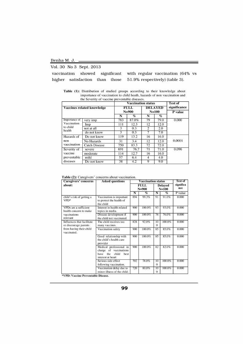
99
Benha M. J.
Vol. 30 No 3 Sept. 2013
vaccination showed significant
higher satisfaction than those
with regular vaccination (64% vs
51.9% respectively) (table 3).
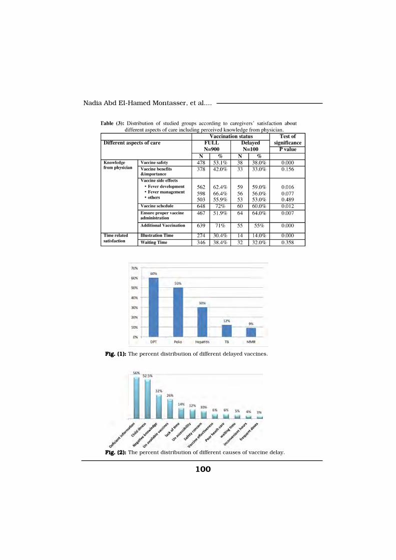
100
Nadia Abd El-Hamed Montasser, et al....
Fig. (1): Fig. (1): The percent distribution of different delayed vaccines.
Fig. (2): Fig. (2): The percent distribution of different causes of vaccine delay.
101
Benha M. J.
Vol. 30 No 3 Sept. 2013
Discussion Immunization is a proven tool
for controlling and eliminating life-
threatening infectious diseases
and is estimated to avert 2 to 3
million deaths each year. It is one
of the most cost-effective health
investments, with proven strate-
gies that make it accessible to
even the most hard-to-reach and
vulnerable populations (WHO,
2009). Despite this, vaccine pre-
ventable diseases remain the most
common cause of childhood mor-
tality with an estimated three mil-
lion deaths each year (Centre for
Global Development, 2005).
Uptake of vaccination services
is dependent not only on provision
of these services but also on other
factors including knowledge and
attitude of caregivers (Torun and
Bakirci, 2006), density of health
workers, accessibility to vaccina-
tion clinics and availability of safe
needles and syringes. Assessing
and evaluation of immunization
coverage helps to evaluate
progress in achieving program ob-
jectives and in improving service
delivery (Bonu et al, 2003).
Among the studied children,
90.0 % had their full scheduled
immunizations by the age of two
years. This rate is markedly high-
er than that reported by Odusan-
ya et al, (2008) and Jani et al,
(2008) who detected that the full
immunization coverage in children
below 2 years was only 61.9 %
and 71.8 % respectively. On the
other hand, the vaccination rate
reported by the present study is
lower than that reported in the
United States (94.8%) (Centers for
Disease Control and Prevention,
2012). This difference may be due
to different economic level be-
tween different countries that may
affect accessibility, availability of
vaccines and health care services.
In the present study, caregiv-
ers' perception of vaccination im-
portance for child health was sig-
nificantly associated with full
vaccine uptake. This is in accor-
dance with Wilson et al, (2008)
who concluded that the parental
decision to vaccinate was due to
recognizing the importance of pre-
venting disease.
We also found that, caregivers
who regularly immunized her
child had better knowledge than
102
Nadia Abd El-Hamed Montasser, et al....
caregivers who delayed child im-
munization. This is in agreement
with Joseph et al, (2011) who
found that parents’ knowledge
about the disease and the vaccine
is a predictor of higher vaccination
compliance. Consistently, De
Courval et al, (2003) and Davis et
al, (2001) declared that lack of
knowledge about the importance
of vaccines has been identified as
a main barrier to immunization,
and the provision of information
about a disease, the adverse se-
quelae of the disease, and the ef-
fectiveness of the vaccine have
been shown to increase uptake, so
receiving vaccine-information ma-
terials during pregnancy or at a
well-child visit before the vaccina-
tion visit is very essential.
In our study, the delay vaccina-
tion was mainly due to deficient
information about the importance
of vaccination at time or the tim-
ing of vaccination, however there
were other causes of delayed vac-
cination as fear of child illness as
a side effect of vaccine. Consis-
tently, Ozkaya et al, (2010), de-
clared that some mothers refused
to complete child vaccination due
to high anxiety levels about vac-
cine side effect. This agreed with
Ritvo et al, (2003), who revealed
that fear of vaccination side-
effects may be a barrier for immu-
nization. We also found that de-
layed vaccination may be due to
vaccine deficiency in the health of-
fices. In agreement with us, De
Serres et al, (2002) reported that
the unavailability of the vaccines
may be a cause of delayed immu-
nization.
We have declared that, caregiv-
ers who delayed vaccination were
significantly less likely to agree
with the statement, “vaccines are
safe” this matched with Freed et
al, (2010) who declared that, par-
ents who delayed vaccine were
significantly anxious about vac-
cines safety . They believed that if
they vaccinated their child, he/
she might have serious side ef-
fects.
Limitation of study:Limitation of study:
Nevertheless, potential biases
should be considered before gen-
eralizing these results to all care-
givers of Egyptian young children.
The most important possible bias
is that the caregivers who agreed
to participate in the study may be
103
Benha M. J.
Vol. 30 No 3 Sept. 2013
those who were most in favour of
vaccinations and therefore the
most inclined to vaccinate their
children with recommended vacci-
nations.
Conclusion and recommenda-Conclusion and recommenda-
tion:tion:
Our results suggest that modi-
fiable determinants for a negative
attitude could probably be based
on a lack of specific knowledge. In
fact, there is still uncertainty sur-
rounding the safety of recom-
mended vaccines, so it is recom-
mended to minimize different
barriers to vaccinations by im-
proving health education of care-
givers in the vaccination programs
and stepping up awareness cam-
paigns using known and effective
communication channels to con-
vey messages to communities with
a large number of at-risk families.
Clearly, the Ministry of health
(MOH) needs to do more in inform-
ing people about the process of
immunization in order to allay
fears (e.g., information in the an-
tenatal period may enable caregiv-
ers to make more informed deci-
sions about recommended
vaccinations) and develop ways of
addressing common uncertainties
about immunization, including
the safety of combining antigens
and the need for boosters. Train-
ing of the physician about proper
communication with caregivers
and response to their needs and
concerns may increase caregivers’
satisfaction.
References1. Ashley H.S., Cynthia S.M.,1. Ashley H.S., Cynthia S.M.,
Donna M.S. and Bernard G.Donna M.S. and Bernard G.
(2007):(2007): Parental satisfaction with
early pediatric care and immuni-
zation of young children, the me-
diating role of age-appropriate
well-child care utilization. Arch
Pediatr Adolesc Med. 161(1):50-56.
2. Bonu S., Rani M. and Bak-2. Bonu S., Rani M. and Bak-
er T.D. (2003):er T.D. (2003): The impact of the
national polio immunization cam-
paign on levels and equity in im-
munization coverage: evidence
from rural North India. Soc Sci
Med; 57:1807-19.
3. Centers for Disease Control3. Centers for Disease Control
and Prevention (CDC) (2012):and Prevention (CDC) (2012):
Vaccination coverage among chil-
dren in kindergarten--United
States, 2011-12 school year.
MMWR Morb Mortal Wkly Rep.;
61(33):647-5.
104
Nadia Abd El-Hamed Montasser, et al....
4. Centre for Disease control4. Centre for Disease control
and prevention (CDC) (2010):and prevention (CDC) (2010):
Global routine vaccination cover-
age, 2009. Morbidity and Mortality
Weekly Report. 59:1367-71.
5. Centre for Global Develop-5. Centre for Global Develop-
ment (2005):ment (2005): Making Markets for
vaccines: from ideas to actions.
Centre for Global Development;
Washington DC.
6. Davis T.C., et al. (2001):6. Davis T.C., et al. (2001):
Childhood vaccine risk/benefit
communication in private practice
office settings: a national survey.
Pediatrics, 107:e17.
7. De Courval F.P., De Serres7. De Courval F.P., De Serres
G. and Duval B. (2003):G. and Duval B. (2003): Varicella
vaccine: Factors influencing uptake.
Can J Public Health; 94: 268-71.
8. De Serres G., Duval B. and8. De Serres G., Duval B. and
Boulianne N. (2002):Boulianne N. (2002): Impact of
vaccine cost and information
about complications of varicella
on parental decision regarding
varicella vaccine. Can J Public
Health; 93:114-16.
9. Division of Communicable9. Division of Communicable
Disease control (DCD) (2005):Disease control (DCD) (2005):
Future directions for achieving the
communicable disease control tar-
gets of DCD Newsl., 2005; 5: 1-12.
10. Freed G.L., Clark S.J.,10. Freed G.L., Clark S.J.,
Butchart A.T., Singer D.C. andButchart A.T., Singer D.C. and
Davis M.M. (2010):Davis M.M. (2010): Parental vac-
cine safety concerns in 2009. Pe-
diatrics; 125: 654-9.
11. Gentile A., Bhutta Z.,11. Gentile A., Bhutta Z.,
Bravo L., Samy A.G., GarciaBravo L., Samy A.G., Garcia
R.D., Hoosen A., et al. (2010):R.D., Hoosen A., et al. (2010):
Pediatric disease burden and vac-
cination recommendation: under-
standing local differences. Int J
Infect Dis.; 14(8): e649-58.
12. Glanz K., Lewis F.M. and12. Glanz K., Lewis F.M. and
Rimer B.K. (1997): Rimer B.K. (1997): Linking theo-
ry, research, and practice. In:
Glanz K, Lewis FM and Rimer BK.
Health behavior and health educa-
tion: theory, research, and prac-
tice. San Francisco: Jossey-Bass.
13. Jani J.V., De Schacht C.,13. Jani J.V., De Schacht C.,
Jani I.V. and Bjune G. (2008):Jani I.V. and Bjune G. (2008):
Risk factors for incomplete vacci-
nation and missed opportunity for
immunization in rural Mozam-
bique. BMC Public Health. May
16; 8: 161.
14. Joseph N., Subba Sh.,14. Joseph N., Subba Sh.,
105
Benha M. J.
Vol. 30 No 3 Sept. 2013
Nelliyanil M., Kotian S., Hari-Nelliyanil M., Kotian S., Hari-
dath A.N.K, Attavar S., et al.dath A.N.K, Attavar S., et al.
(2011):(2011): A study of the knowledge
and attitude towards pulse polio
immunization in semi urban areas
of South India. Australas Med J.;
4(2):81-6.
15. Ministry Of Health and15. Ministry Of Health and
Population (MOHP) (2005):Population (MOHP) (2005):
Achievements of the Expanding
Program of Immunization 1996-
2005. MOHP/ EPI: Egypt.
16. Odusanya O.O., Alufohai16. Odusanya O.O., Alufohai
E.F., Meurice F.P. and AhonkhaiE.F., Meurice F.P. and Ahonkhai
V.I. (2008):V.I. (2008): Determinants of vac-
cination coverage in rural Nigeria.
BMC Public Health.; 8: 381.
17. Ozkaya E., Eker H.H., Ay-17. Ozkaya E., Eker H.H., Ay-
can N. and Samanci N. (2010):can N. and Samanci N. (2010):
Impact of maternal anxiety level
on the childhood vaccination cov-
erage. Eur J Pediatr. Nov; 169(11):
1397-401.
18. Ritvo P., et al. (2003):18. Ritvo P., et al. (2003): A
Canadian national survey of atti-
tudes and knowledge regarding
preventive vaccines. Journal of
Immune Based Therapies and
Vaccines, 1:3.
19. Strecher V.J. and Rosen-19. Strecher V.J. and Rosen-
stock I.M. (1997):stock I.M. (1997): The Health Be-
lief Model. In: Glanz K,Lewis FM
and Rimer BK, editors. Health be-
havior and health education: theo-
ry, research, and practice. San
Francisco: Jossey-Bass.
20. Torun S.D. and Bakirci N.20. Torun S.D. and Bakirci N.
(2006):(2006): Vaccination coverage and
reasons for non-vaccination in a
district of Istanbul. BMC Public
Health.; 6:125.
21. Wilson K., Barakat M.,21. Wilson K., Barakat M.,
Vohra S., Ritvo P. and Boon H.Vohra S., Ritvo P. and Boon H.
(2008):(2008): Parental views on pediatric
vaccination: the impact of competing
advocacy coalitions. Public Un-
derst Sci. Apr; 17(2): 231-43.
22. World Health Organiza-22. World Health Organiza-
tion (WHO) (2009):tion (WHO) (2009): State of
world’s vaccines and immuniza-
tion. 3rd ed. Geneva: World Health
Organization, 1-169.
23. World Health Organiza-23. World Health Organiza-
tion (WHO) (2010):tion (WHO) (2010): Department of
Immunization, Vaccines and Bio-
logicals.. Vaccines for Preventable
Diseases: Monitoring System. glo-
ba summary. Geneva: WHO.

106
Nadia Abd El-Hamed Montasser, et al....
BELOW 2 YEARS CHILDRENBELOW 2 YEARS CHILDRENCAREGIVERS’ KNOWLEDGE,CAREGIVERS’ KNOWLEDGE,
ATTITUDE AND BELIEFS TOWARDSATTITUDE AND BELIEFS TOWARDSIMMUNIZATIONIMMUNIZATION
Nadia Abd El-Hamed Montasser MD,Nadia Abd El-Hamed Montasser MD,Randah Mohamad Helal MD, Noha El-Adawi MD,Randah Mohamad Helal MD, Noha El-Adawi MD,
Eman Mostafa, Fatma Abd El-Rahman,Eman Mostafa, Fatma Abd El-Rahman,Maged Saad and Soha Hamza Maged Saad and Soha Hamza
BENHAMEDICALJOURNAL
REPRINT
Published byPublished by
Benha Faculty of MedicineBenha Faculty of Medicine Volume 30 Number 3Sept. 2013
107
Benha M. J.
Vol. 30 No 3 Sept. 2013
THE ROLE OF MR DIFFUSION INTHE ROLE OF MR DIFFUSION INDIFFERENTIATION OF MALIGNANT ANDDIFFERENTIATION OF MALIGNANT AND
BENIGN HEPATIC FOCAL LESIONSBENIGN HEPATIC FOCAL LESIONS
Mahmoud Abd El-Latif El-Shewail MD*, Galal El HawaryMahmoud Abd El-Latif El-Shewail MD*, Galal El HawaryMD*, Adel El-Badrawy MD*, Hatem El-Alfy MD**MD*, Adel El-Badrawy MD*, Hatem El-Alfy MD**
Departments of Radio Diagnosis* and Tropical Medicine**
Faculty of Medicine, Mansoura University
AbstractAim: Aim: The objective of this study was to determine if focal liver mass-
es could be differentiated as benign or malignant on the basis of diffu-sion-weighted MR imaging (DWMRI) and ADC maps.
Methods and Materials:Methods and Materials: Between June 2011 and December 2012, atotal of 60 consecutive patients (43 men, 17 women; age range 20-70,mean age, 45 years) with 60 focal liver lesions were scanned using 1.5 Tmagnetic resonance imaging (MRI). Respiratory-triggered single-shotecho-planar diffusion weighted imaging (DWI) was performed with b 0, b500 and b 1000 gradients with ADC measurements. Comparison ofmean ADC values between each benign and malignant lesion was done .Reference standard of diagnosis was obtained by correlating DWI withhistopathologic findings, characteristic MR sequences and imaging fol-low-up. The accuracies of DWI and ADC values in differentiating benignand malignant focal liver lesions were assessed with the Student t test,and cut-off values were determined with receiver operating characteris-tic curve analysis (ROC). The analyzed lesions were hemangioma (n=8),cysts (n=8), adenoma (n=5), focal nodular hyperplasia (FNH) (n=5), he-patocellular cacinoma (HCC) (n=20), cholangiocarcinoma (n=5) and me-tastases (n=9).
Results: Results: The mean ADC value (at b1000) of malignant focal liver le-sions was 0.858±0.18X 10⁻³mm2/sec and of benign focal lesions was1.55±0.0.43X 10-3mm2/sec. There was statistically difference in meanADC values between malignant and benign focal liver lesions(p<0.0001). When apparent diffusion coefficient value of 1.0 X 10-
108
Mahmoud Abd El-Latif El-Shewail, et al....
IntroductionThe differential diagnosis be-
tween malignant and benign focal
liver lesions remains a diagnostic
challenge for every radiologist. For
detection and characterization of
focal liver lesions, many different
modalities have been proposed; in-
cluding multi-phase contrast-
enhanced CT[1] and MRI [2], CT
portography[3] and perfusion
studies using dedicated ultra-
sound or Computed tomography(
CT) or MRI contrast agents[4]. Of
these modalities, magnetic reso-
nance imaging is considered the
most accurate imaging technology
because it has high resolution for
soft tissue and has the potential
to characterize a lesion on various
data acquired, such as T1, T2,
and early and late post-
gadolinium images[5] and[6].
Magnetic resonance imaging
(MRI) has been used in both the
detection and characterization of
focal hepatic lesions. With the ad-
vent of the echo-planar MR imag-
ing technique, diffusion weighted
imaging (DWI) of the abdomen has
become possible with fast imaging
times which minimize the effect of
gross physiologic motion from res-
piration and cardiac movement[7].
Thus, DWI became a valuable
technique for evaluating focal he-
patic lesions in addition to con-
ventional MRI sequences[8]. More
recently, apparent diffusion coeffi-
3mm2/sec was used as a threshold value for differentiation of malig-nant tumors from benign lesions, sensitivity was 90.3%, specificity78.57%, accuracy 86.7%, positive predictive value 90.3% and negativepredictive value of 78.6%. The best result was obtained with the use ofADC cut off value (at b500) of 1.5 x10⁻³mm2/sec and ADC cut off value(at b1000) of 1.0 x10⁻³mm2/sec, with 90.3% sensitivity, 92.86% speci-ficity, 91.1% accuracy, 96.6 % positive predictive value and 81.3 % neg-ative predictive value.
Conclusions:Conclusions: adding DWI to routine abdominal MRI and ADC meas-urements at least at 2 different gradients is a useful tool in differentialdiagnosis of malignant from benign liver lesions and may be useful fordifferentiation of different subtypes of either benign and malignant le-sions.
109
Benha M. J.
Vol. 30 No 3 Sept. 2013
cient (ADC) value has been intro-
duced in quantitative measure-
ments as an adjunct to DWMRI.
ADC is a quantitative parameter
measuring the rate of diffusion of
water molecules in biological tis-
sues. There are several reports re-
garding the use of ADC in diagno-
sis and characterization of focal
hepatic lesions[7,8,9], and[10].
However, the efficacy of ADC val-
ues in diagnosing and characteri-
zation of solid benign and solid
malignant lesions has not been
well described. Similarly, lesion
and diffusion gradient variabilities
were also limited in these studies[8,9,10]. Usage of ADC measure-
ments in various types of focal he-
patic lesions at different diffusion
gradient values may define the
role of ADC values in radiological
evaluation of focal hepatic lesions[11].
The expanding role of DWI in
evaluation of liver lesions can in-
crease confidence in differentia-
tion between benign and malig-
nant lesions. DW sequences can
be performed on most modern
MRI machines with relative ease,
in a short time period and without
the need for contrast medium[12].
Patients and Methods1. Patients:1. Patients:
Between June 2011 and De-
cember 2012, a total of 60 focal
hepatic lesions in 60 consecutive
patients were evaluated by ab-
dominal MRI. Informed consent
was taken from all. Patients with
hepatic neoplasms who had un-
dergone chemotherapy or radia-
tion therapy within the last 3
months prior to the MR examina-
tion were excluded from our anal-
ysis in order to ensure that ADC
measurements were reflective of
the natural state of the liver le-
sions. In addition, patients with-
out sufficient confirmation of the
nature of the lesions were exclud-
ed. The final study population
consisted of 60 patients (43 men,
17 women; age range 20–70 years,
mean age 45 years).
There were 9 patients with his-
tory of an extra-hepatic primary
malignancy and suspected liver
metastases (colorectal cancer
[n=3], breast cancer [n=2], gastric
cancer [n=1], and pancreatic can-
cer [n=3]. There were 28 patients
with chronic liver disease (includ-
ing chronic hepatitis and cirrhosis
related to hepatitis C and old bil-

110
Mahmoud Abd El-Latif El-Shewail, et al....
harziasis) with suspected malig-
nancy. Finally, there were 23 pa-
tients with no history of malignan-
cy or chronic liver disease who
underwent MR imaging for evalua-
tion of presumably benign or inde-
terminate, incidentally diagnosed
focal liver lesion( FLL). In patients
with multiple FLL, the largest
ones of each lesion type were ran-
domly selected for further analysis
by the study coordinator.
Thirty four lesions were malig-
nant tumors (20 hepatocellular
carcinoma (HCCs), 5 cholangiocar-
cinomas & 9 metastases). Benign
liver lesions were twenty six (8
cysts, 8 hemangiomas, 5 adeno-
mas and 5 focal nodular hyperple-
sia (FNH).
The standard of reference for
characterizing FLL was evaluated
by two radiologists (GE & MA),
with experience in MR imaging of
20 and 10 years respectively. Ma-
lignant nature of lesions was con-
firmed by pathologic findings fol-
lowing biopsy or surgery for 13
HCCs, 5 cholangiocarcinomas and
5 metastases. The diagnosis of the
remaining 7 HCCs lesions was
confirmed by using the estab-
lished imaging criteria and follow
up after chemoembolization. The
remaining 4 metastatic lesions
were confirmed on the basis of
new occurrence of a lesion com-
pared to a prior MR study, follow
up of lesion size after the start of
chemotherapy in patients with
known extra-hepatic primary ma-
lignancies. Three FNHs and two
adenomas were confirmed patho-
logically. The remaining benign le-
sions were diagnosed by using es-
tablished imaging criteria[13,14,15]
in conjunction with stable appear-
ance and size at follow-up imaging
in equivocal cases with a mini-
mum follow-up interval of six
months ( range: 6-12 months).
2. MR imaging & image analy-2. MR imaging & image analy-
sis:sis:
Magnetic resonance imaging
examinations were performed on
1.5 tesla (T) system (Philips,
Achieva) with a sixteen-channel
body coil.
Before DWI, breath-hold T1-
weighted image, fat-saturated fast
spin echo T2 weighted image, dual
echo fast spoiled gradient-echo
(FSPGR) and single shot fast spin
echo (SSFSE) T2 weighted images
111
Benha M. J.
Vol. 30 No 3 Sept. 2013
were obtained in axial and coronal
images. Diffusion weighted images
were obtained before contrast ad-
ministration with b values of 0,
500, and 1000 s/mm2. Breath-
hold, dynamic 3D T1 weighted se-
quence was performed after DWIs
(bolus injection of 20 ml gadopen-
tetate dimeglumine 1.5 ml/s). All
DWIs were obtained in transverse
plane using single-shot echo-
planar spin echo sequences. Imag-
ing parameters for DWIs were:
Repetition time (TR): 1100 ms; TE:
67–91 ms; FOV: 35 Centimeter
(cm) x 35 cm (change according to
body size); number of excitation:
1; matrix size: 128 x 128; section
thickness: 5 mm; intersection gap:
none. DW sequences required a
total of 96 s to scan on MR. Array
spatial sensitivity encoding tech-
nique (ASSET) was used as paral-
lel imaging technique.
Imaging findings were evaluat-
ed carefully, then, ADC values of
different masses detected on con-
ventional MRI sequences and DWI
were measured through gray-scale
ADC maps from each lesion at b
500, and b 1000 s/mm2 gradient
values by using 3 circumferential
region of interests (ROIs) and the
average ADC values were record-
ed. ADCs were measured over the
largest mass detected in patients
with multiple liver lesions. Necrot-
ic portions of solid lesions detect-
ed on contrast enhanced MRI were
excluded.
Mean ADC values of benign le-
sion group (FNH and other benign
liver masses) and malignant le-
sion group (HCC, cholangiocarcin-
oma and metastasis) at 2 different
diffusion gradients were com-
pared. Similarly, mean ADC val-
ues of each benign and malignant
lesion at 2 different gradients were
also recorded and compared in or-
der to determine whether it would
be possible to define the charac-
teristic or type of individual be-
nign and malignant lesion.
To validate our system ADC
values of water was measured on
phantoms one week before the
ADC measurements. The ADC val-
ue for water was 2.21 x10⁻³ mm2/
s, in agreement with other studies
in the literature[16].
Statistical Analysis Statistical analysis was carried
out via Statistical package for so-
112
Mahmoud Abd El-Latif El-Shewail, et al....
cial Science (SPSS) version 17 pro-
gram on windows XP. Qualitative
data were represented in the form
of number and frequency, while
quantitative data were represented
in the form of mean ± standard
deviation (mean±SD). Kolmogrov-
smirnov test was used to test nor-
mality of quantitative data. Stu-
dent’s t test, Mann-Whitney U and
Kruskal-Wallis Test were used to
compare groups. Receiver operat-
ing characteristic (ROC) curve was
computed to determine the cutoff
value for the malignancy. All tests
were considered significant if P
value equals or less than 0.05.
ResultsMean size of all 60 focal hepatic
lesions was 2.83±1.05 cm. Twenty
six of the 60 lesions were benign
and 34 were malignant. Benign le-
sions had a mean size of
2.96±0.77 cm (ranges 2–5 cm)
whereas mean size of malignant
lesions was 2.77±1.17 cm (ranges
1-6 cm) (table 1).
Mean ADC values of 26 benign
lesions at b 500 and b 1000 gra-
dients were 2.09±0.50x10⁻³,
1.55±0.43x10⁻³-3 mm2/s, respec-
tively. Mean ADC values of malig-
nant lesions at b 500 and b 1000
gradients were 1.20±0.18 x 10⁻³
and 0.85±0.18 x 10⁻³ mm2/s, re-
spectively. Mean ADC values of
benign lesions were higher than
malignant lesions and these differ-
ences were statistically significant
for the 2 diffusion gradients (P<
0.0001 & P<0.0001, respectively)
(table 1).
Mean ADC values of all lesions
at b 500 and b 1000 gradients
and differentiation between sub-
types of benign and malignant le-
sions are summarized in table 2.
An ADC cut-off value of 1.0x
10-3 mm2/s at b 1000 diffusion
gradient resulted in 90.3 % sensi-
tivity, 78.6 % specificity and
86.7% accuracy for differentiation
of benign and malignant focal he-
patic lesion groups . The best re-
sult was obtained with the use of
ADC cut off value of 1.5x10⁻³
mm2/sec at b 500 and ADC cut
off value of 1.0x10⁻³ mm2/sec at
b 1000, with 90.3% sensitivity,
92.86% specificity, and 91.1% ac-
curacy. The results of ROC curve
analyses, ADC cut off values for
the differentiation between benign
and malignant lesions at both
113
Benha M. J.
Vol. 30 No 3 Sept. 2013
b500 and b1000 diffusion gra-
dients are shown in table 3 and 4.
Seventeen of the 26 benign le-
sions show low SI and 9 benign le-
sions show intermediate SI on
DWI. While 26 of the 34 malignant
lesions show high SI and 8 malig-
nant lesions show intermediate SI
on DWI.
Cysts and hemangiomas
showed the highest mean ADC
values at b 500 and b 1000 gra-
dients in benign lesion group (fig-
ure 1). Hepatocellular carcinomas
showed highest mean ADC values
at b 500 and b1000 in malignant
lesion group (figure 2).
Comparison of ADC values re-
vealed that mean ADC value of all
benign hepatic focal lesions were
significantly higher than all malig-
nant focal lesions at b 500 and b
1000 gradients (P<0.0001) (figure
3 and 4).
Differentiation of benign and
malignant subtype lesions from
each other in their groups show
some promising results. There
was statistically significant differ-
ence between: the mean ADC val-
ues of cysts and adenoma at both
b 500 and b1000 (P value = 0.002,
< 0.001), the mean ADC values of
cysts and FNH at b 500 (P value
=0.001). However, there was no
significant difference between
mean ADC values of other benign
focal lesions from each others.
At b 500 gradient, HCCs had
significant high ADC value than
metastases and cholangiocarcino-
ma (P value = 0.034, 0.014 respec-
tively) with difficult differentiation
between metastases and cholan-
giocarcinoma. At b 1000 gradient,
there was no significant difference
between HCCs, metastases and
cholangiocarcinomas.
There was statistically signifi-
cant difference between ADC val-
ues of solid benign (adenoma ,
FNH) and solid malignant subtype
lesions, as there was significant
difference between: mean ADC
values of FNH and HCC at both
b500 and b1000 (P value =
<0.001, <0.001) (figure 5 and 6),
mean ADC values of FNH and me-
tastases at both b500 and b1000
(P value = <0.001, 0.004 ), mean
ADC values of FNH and cholangio-
carcinoma at both b500 and
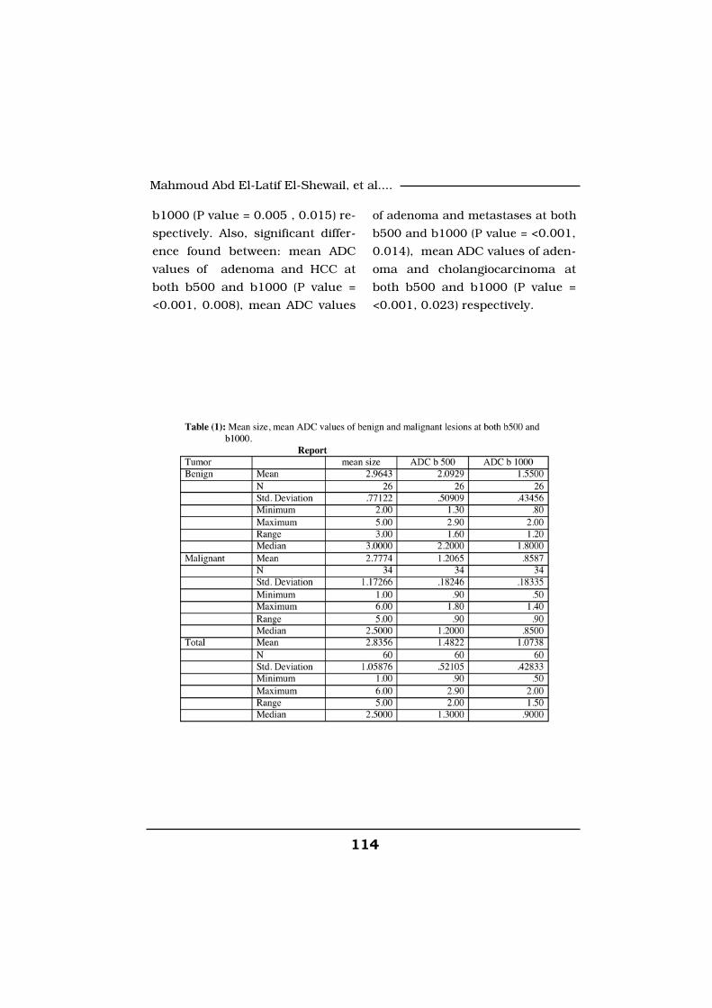
114
Mahmoud Abd El-Latif El-Shewail, et al....
b1000 (P value = 0.005 , 0.015) re-
spectively. Also, significant differ-
ence found between: mean ADC
values of adenoma and HCC at
both b500 and b1000 (P value =
<0.001, 0.008), mean ADC values
of adenoma and metastases at both
b500 and b1000 (P value = <0.001,
0.014), mean ADC values of aden-
oma and cholangiocarcinoma at
both b500 and b1000 (P value =
<0.001, 0.023) respectively.
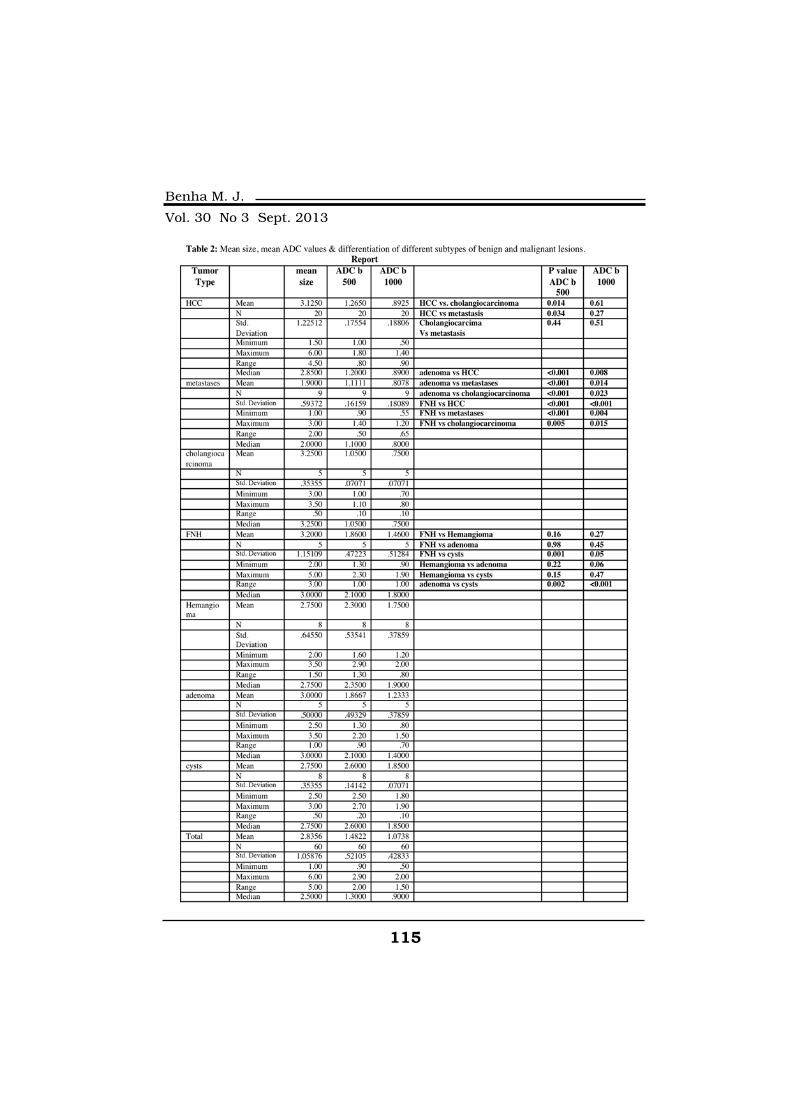
115
Benha M. J.
Vol. 30 No 3 Sept. 2013
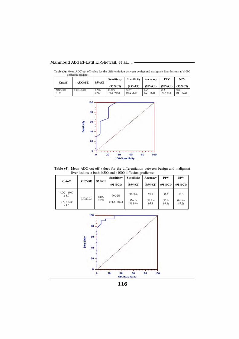
116
Mahmoud Abd El-Latif El-Shewail, et al....
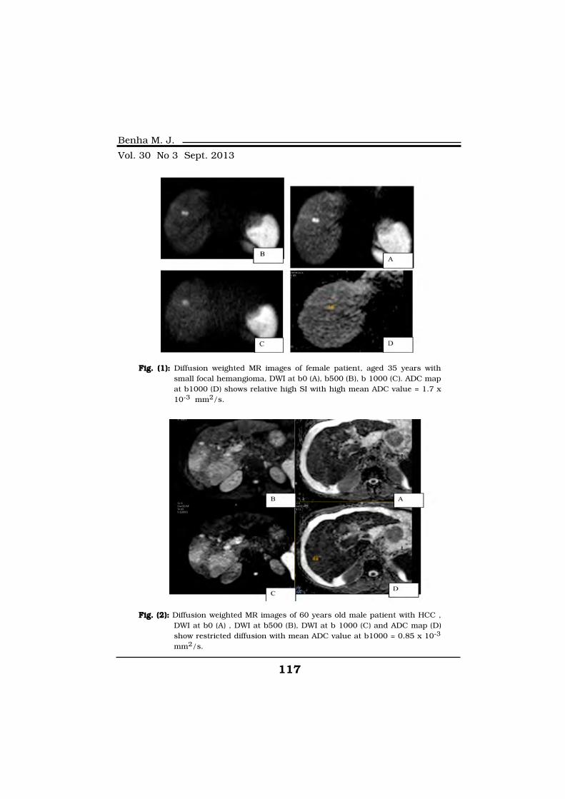
117
Benha M. J.
Vol. 30 No 3 Sept. 2013
Fig. (1):Fig. (1): Diffusion weighted MR images of female patient, aged 35 years withsmall focal hemangioma, DWI at b0 (A), b500 (B), b 1000 (C). ADC mapat b1000 (D) shows relative high SI with high mean ADC value = 1.7 x10⁻³ mm2/s.
Fig. (2):Fig. (2): Diffusion weighted MR images of 60 years old male patient with HCC ,DWI at b0 (A) , DWI at b500 (B), DWI at b 1000 (C) and ADC map (D)show restricted diffusion with mean ADC value at b1000 = 0.85 x 10⁻³
mm2/s.
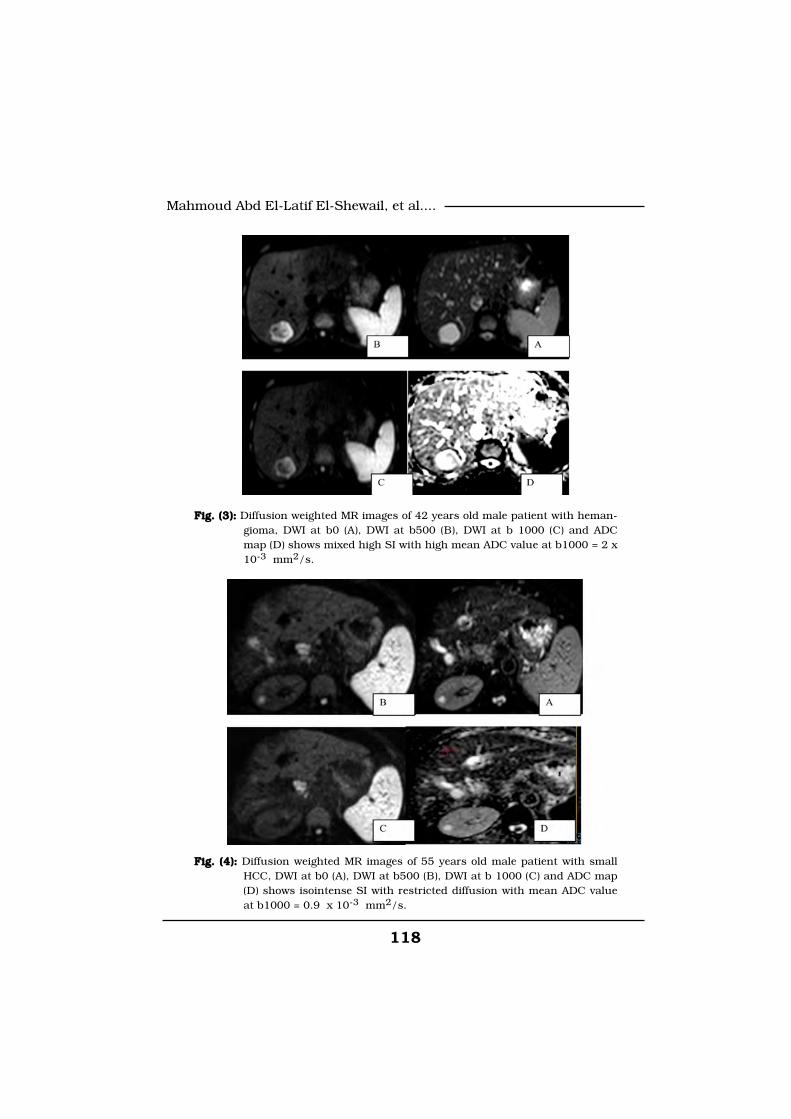
118
Mahmoud Abd El-Latif El-Shewail, et al....
Fig. (3):Fig. (3): Diffusion weighted MR images of 42 years old male patient with heman-gioma, DWI at b0 (A), DWI at b500 (B), DWI at b 1000 (C) and ADCmap (D) shows mixed high SI with high mean ADC value at b1000 = 2 x10⁻³ mm2/s.
Fig. (4):Fig. (4): Diffusion weighted MR images of 55 years old male patient with smallHCC, DWI at b0 (A), DWI at b500 (B), DWI at b 1000 (C) and ADC map(D) shows isointense SI with restricted diffusion with mean ADC valueat b1000 = 0.9 x 10⁻³ mm2/s.
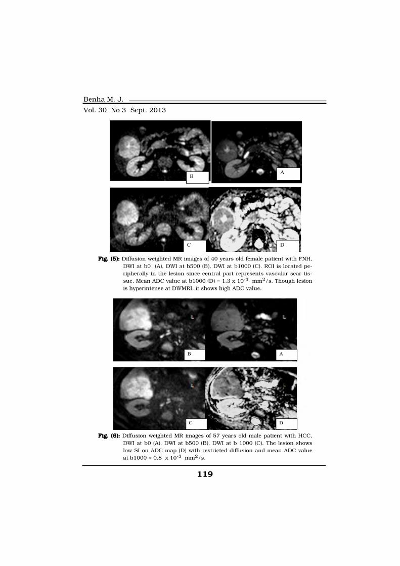
119
Benha M. J.
Vol. 30 No 3 Sept. 2013
Fig. (5):Fig. (5): Diffusion weighted MR images of 40 years old female patient with FNH,DWI at b0 (A), DWI at b500 (B), DWI at b1000 (C). ROI is located pe-ripherally in the lesion since central part represents vascular scar tis-sue. Mean ADC value at b1000 (D) = 1.3 x 10⁻³ mm2/s. Though lesionis hyperintense at DWMRI, it shows high ADC value.
Fig. (6):Fig. (6): Diffusion weighted MR images of 57 years old male patient with HCC,DWI at b0 (A), DWI at b500 (B), DWI at b 1000 (C). The lesion showslow SI on ADC map (D) with restricted diffusion and mean ADC valueat b1000 = 0.8 x 10⁻³ mm2/s.
120
Mahmoud Abd El-Latif El-Shewail, et al....
DiscussionReliable detection and charac-
terization of focal liver lesions is
critical for optimal patient man-
agement. Magnetic resonance im-
aging (MRI) has an established
role in focal liver lesion detection
and characterization and tradi-
tionally includes a combination of
unenhanced T1 and T2-weighted
sequences, in and out-of-phase
T1-weighted sequences, and en-
hanced T1-weighted sequences
after gadolinium administration or
other liver-specific contrast
agents. There is a good evidence
that diffusion-weighted (DW) MRI
has the potential to improve he-
patic lesion detection rates and
contribute to the differentiation of
benign from malignant hepatic le-
sions[12].
Appearance of focal hepatic le-
sions on DWI especially at high b
values was reported to be diagnos-
tic in several studies due to re-
stricted diffusion and increased
signal intensity on DW images [8]
and[17]. However this measure-
ment was a qualitative assess-
ment and represented a subjective
interpretation. On the contrary,
ADC value is a quantitative pa-
rameter of water diffusion. There
are several studies in the litera-
ture emphasizing diagnostic utility
of ADC measurement in the diffe-
rentiation of benign and malig-
nant focal hepatic lesions. Accord-
ing to these studies malignant
lesions had lower ADC values
compared to benign lesions which
was attributed to high cellularity
of malignant masses[7].
Chandarana and Taouli, 2010[18], said that , there is no consen-
sus in the scientific community
about which b-values are optimal
for liver imaging and when per-
forming DW-MRI in the liver, it is
advantageous to perform imaging
with at least 3 b-values including
both lower and higher b-values
(e.g. using b = 0, b ≤ 100, and b ≥
500 s/mm2). Goshima et al. 2008,[19] recommended DW-EPIs with
low and high b values as supple-
mentary sequences in the detec-
tion and characterization of be-
nign and malignant hepatic
lesions. In our study, DWI was
done with 3 diffusion gradients at
b0, b500 and b1000.
Most of the studies[1,20,10], in-
dicated that a region of interest
121
Benha M. J.
Vol. 30 No 3 Sept. 2013
(ROI) should be placed within the
confines of the lesion in image
analysis and put away from prom-
inent vascular structures to avoid
motion artifact. However, some
authors[1,20] placed ROI that cov-
ered entire lesions without separ-
ating component with various sig-
nal intensities for analysis of
heterogeneous lesions, whereas
Gourtsoyianni et al, 2008[10] put
the ROI in both sites in a lesion
with different signal behavior in
the periphery and center. In the
current study, ROI was placed
within the confines of the lesion
and put away from vascular and
necrotic portions of the lesion.
Kandpal et al, 2009[21] found
that respiratory-triggered DWI was
superior to breath-hold DWI for
hepatic imaging because it pro-
vides higher SNR. In our study,
DWI was done with respiratory
triggered technique.
Most of the studies included
hemangiomas and cysts in the be-
nign lesion group[7,22,23]. Thus it
was concluded that hypercellular
benign lesions like FNH should
also be studied in order to dis-
criminate benign and malignant
hepatic lesions more reliably[10].
While in the study of[11], solid
(high cellular) benign liver lesions
were included with exclusion of
cystic benign lesions. In our
study, cystic benign lesions as he-
mangiomas and cysts and solid
benign lesions as FNH and adeno-
ma were included in the benign le-
sion group.
Our results revealed that ADC
measurements at b 500 and b
1000 diffusion gradients were use-
ful in differential diagnosis of be-
nign and malignant lesions and
adding ADC cut off values at b
500 and b 1000 diffusion gra-
dients increase sensitivity and
specificity for differentiation.
In the present study, compari-
son of ADC values for individual
benign and malignant lesions
showed that there were statistical-
ly significant differences between
lesions at different gradients.
Mean ADC values at b 500 and
b1000 of different benign lesions
were as follow: FNH (1.86 and
1.46 x 10⁻³ mm2/s), adenoma
(1.86 and 1.23 x 10⁻³ mm2/s), he-
mangioma (2.30 x 10⁻³ mm2 and
1.75 x 10⁻³ mm2/s) and cysts
122
Mahmoud Abd El-Latif El-Shewail, et al....
(2.60 x 10⁻³ mm2/s and 1.85 x
10-3 mm2/s). While, mean ADC
values at b 500 and b1000 of dif-
ferent malignant lesions were:
HCC (1.26 and 0.89 x 10⁻³ mm2),
metastases (1.11 and 0.80 x 10⁻³
mm2) and cholangiocarcinoma
(1.05 and 0.75 x 10⁻³ mm2).
In the study of Onur et al, 2012[11], FNH was easily differentiated
from malignant lesions except
HCC at b 100 gradient. Other be-
nign solid liver lesions also could
be differentiated from metastases
and cholangiocarcinomas at all
gradients. They concluded that
solid benign liver lesions did not
show significant difference from
HCCs at all gradients. Also,[8,9]
said that The ADC values of these
solid benign lesions were similar
to ADC values of malignant le-
sions. Yet, in our study, FNH and
other benign lesions could be dif-
ferentiated from malignant lesions
including HCC at both b500 and
b1000 diffusion gradients.
Taouli et al, 2003[7], concluded
that some overlap is present be-
tween metastatic lesions and
FNHs, however, in our study, met-
astatic lesions had lower ADC val-
ues than FNH at both b500 and
b1000 diffusion gradients. This
matches with the results of Onur
et al, 2012[11], who observed that
ADC measurements were success-
ful in differentiating these lesions .
This may be due to exclusion of
necrotic components of metasta-
ses at ADC measurement which
may increased the ADC values of
metastases.
In the current study, the diffe-
rentiation between different sub-
types of malignant lesions with
mean ADC values showed some
difficulty at both gradients. At b
500 gradient, HCCs had signifi-
cant high ADC value than metas-
tases and cholangiocarcinoma
with difficult differentiation be-
tween metastases and cholangio-
carcinoma. At b 1000 gradient,
significant difference was only
found between HCCs and cholan-
giocarcinomas. This is similar to
the results of Gourtsoyianni et al,
2008, Kilickesmez et al, 2009[10,24] and Onur et al, 2012[11],
who concluded that the differenti-
ation of malignant lesions with
mean ADC values was difficult at
all gradients and similarity of ADC
values was found between malig-
123
Benha M. J.
Vol. 30 No 3 Sept. 2013
nant hepatic lesions. Onur et al,
2012[11] said that, The reason for
highest mean ADC values meas-
ured in HCCs among all malig-
nant lesions may be due to rela-
tively increased perfusion of HCCs
than hypovascular metastases
and cholangiocarcinomas and we
agree with that explanation.
Results of our study showed
that cysts and hemangioms have
high ADC value than adenoma
and FNH, however, (cysts and he-
mangiomas) and also (adenoma
and FNH) couldn't be differentiat-
ed well from each other at both
diffusion gradients, This is similar
to the results of Onur et al, 2012[11], as ADC measurements were
not helpful in differential diagno-
sis of different types of solid be-
nign lesions.
Sandrasegaran et al, 2009 and
Miller et al, 2010[25] and[26] sug-
gested that ADC values of solid
benign lesions (FNH and adeno-
ma) are similar to malignant le-
sions (metastasis and HCC) and
DW imaging is not helpful in dif-
ferentiating solid benign lesions
from solid malignant lesions. How-
ever, in our study, there was sig-
nificant difference between solid
benign ( adenoma, FNH) and solid
malignant lesions. This is may be
due to diminished perfusion effect
due to high diffusion gradient as
we used b 0, b 500 and b 1000
and previous studies used b 0, b
50 and b 500 as diffusion gra-
dients.
Different studies reported vari-
able ADC cut-off values for diffe-
rentiation of benign and malig-
nant focal hepatic lesions. Taouli
et al, 2003[7] offered a threshold
value as 1.5 x 10⁻³ at b 500 gradi-
ent with sensitivity of 84% and
specificity of 89%. Parikh et al,
2008[8] reported an ADC value of
1.6 x 10⁻³ as a cut off value with
sensitivity of 74.2% and specificity
of 77.3% at b 500 gradient. Onur
et al, 2012[11] concluded a cut-off
value of 1.23 x 10⁻³ at b 1000
with sensitivity of 83% and speci-
ficity of 76% for differentiation be-
tween benign and malignant le-
sions. In our study, using 1.0 x
10-3 mm2/s as an ADC cut-off
value at b 1000 diffusion gradient
resulted in 90.3% sensitivity,
78.6% specificity and 86.7%
accuracy for differentiation of
benign and malignant focal
124
Mahmoud Abd El-Latif El-Shewail, et al....
hepatic lesion groups. The best
result was obtained with the use
of ADC cut off value (at b500) of
1.5 x 10⁻³mm2/sec and ADC cut
off value (at b1000) of 1.0 x 10-
3mm2/sec, with 90.3% sensitivity,
92.86% specificity, and 91.1% ac-
curacy.
To best of our knowledge, only
few studies in the literature meas-
uring ADC values of different sub-
types of either benign or malig-
nant lesions. Our results showed
that HCC had high ADC value
than metastases and cholangio-
carcinima in the malignant group
liver masses. Cysts and heman-
giomas had high ADC values than
adenoma and FNH in the benign
group liver masses.
ConclusionSo, adding DWI to routine ab-
dominal MRI and ADC measure-
ments at least at 2 different gra-
dients is a useful tool in
differential diagnosis of malignant
from benign liver lesions and may
be useful for differentiation of dif-
ferent subtypes of either benign
and malignant lesions, further in-
vestigation in this point is recom-
mended.
Limitation of the study: Limitation of the study:
There was small number of cas-
es with FNHs and other benign liv-
er masses and as well as cholan-
giocarcinoma. However these
lesions were not seen as usually
as metastases or HCC and most of
these lesions (FNH, other benign
liver masses and cholangiocarcin-
oma) were rarely compared with
each other via ADC values in the
literature.
References1. Semelka C.R., Martin R.D.,1. Semelka C.R., Martin R.D.,
Balci C. and Lance T. (2001):Balci C. and Lance T. (2001): Fo-
cal liver lesions: comparison of
dual-phase CT and multisequence
multiplanar MR imaging including
dynamic gadolinium enhance-
ment. J Magn Reson Imaging, 13
(3), pp. 397-401.
2. Pirovano G., Vanzulli A.,2. Pirovano G., Vanzulli A.,
Marti-Bonmati L., et al. (2000):Marti-Bonmati L., et al. (2000):
Evaluation of the accuracy of ga-
dobenate dimeglumine-enhanced
MR imaging in the detection and
characterization of focal liver le-
sions. AJR Am J Roentgenol, 1175
(4), pp. 1111-112.
3. Matsuo M., Kanematsu M.,3. Matsuo M., Kanematsu M.,
125
Benha M. J.
Vol. 30 No 3 Sept. 2013
Inaba Y., et al. (2001):Inaba Y., et al. (2001): Pre-
operative detection of malignant
hepatic tumours: value of com-
bined helical CT during arterial
portography and biphasic CT dur-
ing hepatic arteriography. Clin Ra-
diol, 56 (2), pp. 138-145.
4. von Herbay A., Vogt C. and4. von Herbay A., Vogt C. and
Haussinger D. (2004):Haussinger D. (2004): Differenti-
ation between benign and malig-
nant hepatic lesions: utility of col-
or stimulated acoustice mission
with the microbubble contrast
agent Levovist. J Ultrasound Med,
23 (2), pp. 207-215.
5. Ramalho M., Altun E. and5. Ramalho M., Altun E. and
Heredia V. (2007):Heredia V. (2007): Zapparoli M.,
Semelka R. Liver MR imaging: 1.5
T versus 3 T. Magn Reson Imaging
Clin N Am, 15, pp. 321–347.
6. Motohara T., Semelka C.R.6. Motohara T., Semelka C.R.
and L. Nagase L. (2002):and L. Nagase L. (2002): MR im-
aging of benign hepatic tumors.
Magn Reson Imaging Clin N Am,
10, pp. 1-14.
7. Taouli B., Vilgrain V., Du-7. Taouli B., Vilgrain V., Du-
mont E., Daire L.J., Fan B. andmont E., Daire L.J., Fan B. and
Menu Y. (2003):Menu Y. (2003): Evaluation of liv-
er diffusion isotropy and charac-
terization of focal hepatic lesions
with two single-shot echo-planar
MR imaging sequences: prospec-
tive study in 66 patients. Radiolo-
gy, 226, pp. 71-78.
8. Parikh T., Drew J.S., Lee8. Parikh T., Drew J.S., Lee
S.V., et al. (2008):S.V., et al. (2008): Focal liver le-
sion detection and characteriza-
tion with diffusion-weighted MR
imaging: comparison with stan-
dard breath-hold T2- weighted im-
aging. Radiology, 246 (3), pp.
812-822.
9. Demir I.O., Obuz F., Sagol9. Demir I.O., Obuz F., Sagol
O. and O. Dicle O. (2007):O. and O. Dicle O. (2007): Con-
tribution of diffusion-weighted
MRI to the differential diagnosis of
hepatic masses. Diagn Interv Ra-
diol, 13, pp. 81-86.
10. Gourtsoyianni S., Papa-10. Gourtsoyianni S., Papa-
nikolaou N., Yarmenitis S., et al.nikolaou N., Yarmenitis S., et al.
(2008):(2008): Respiratory gated diffu-
sion- weighted imaging of the liv-
er: value of apparent diffusion co-
efficient measurements in the
differentiation between most com-
monly encountered benign and
malignant focal liver lesions. Eur
Radiol, 18, pp. 486-492.
11. Onur R.M., Çiçekçi M.,11. Onur R.M., Çiçekçi M.,
Kayalı A., Poyraz K.A. and Koca-Kayalı A., Poyraz K.A. and Koca-
126
Mahmoud Abd El-Latif El-Shewail, et al....
koç E. (2012):koç E. (2012): The role of ADC
measurement in differential diag-
nosis of focal hepatic lesions. Eur
J Radiol, 81 (3), pp.171-176.
12. Culverwell D.A., Sheridan12. Culverwell D.A., Sheridan
B.M., Guthrie A.J. and Scars-B.M., Guthrie A.J. and Scars-
brook F.A. (2013):brook F.A. (2013): Diffusion-
weighted MRI of the liver
—Interpretative pearls and pit-
falls. Clin Radiol, 68(4), pp. 406-
414.
13. Semelka C.R., Brown13. Semelka C.R., Brown
D.E., Ascher M.S., et al. (1994):D.E., Ascher M.S., et al. (1994):
Hepatic hemangiomas: a multi-
institutional study of appearance
on T2-weighted and serial gadolin-
ium-enhanced gradient-echo MR
images. Radiology, 192, pp. 401-
406.
14. Bartolozzi C., Cioni D.,14. Bartolozzi C., Cioni D.,
Donati F. and Lencioni R.Donati F. and Lencioni R.
(2001):(2001): Focal liver lesions: MR im-
aging-pathologic correlation. Eur
Radiol, 11, pp. 1374-1388.
15. Horton M.K., Bluemke15. Horton M.K., Bluemke
A.D., Hruban H.R., Soyer P. andA.D., Hruban H.R., Soyer P. and
Fishman K.E. (1999):Fishman K.E. (1999): CT and
MR imaging of benign hepatic and
biliary tumors. Radiographics, 19,
pp. 431-451.
16. Guo Y., Cai Q.Y. and Cai16. Guo Y., Cai Q.Y. and Cai
L.Z., et al. (2002):L.Z., et al. (2002): Differentiation
of clinically benign and malignant
breast lesions using diffusion-
weighted imaging. J Magn Reson
Imaging, 16 (2), pp. 172-178.
17. Bozgeyik Z., Kocakoc E.,17. Bozgeyik Z., Kocakoc E.,
Gul Y. and Dagli F.A. (2010):Gul Y. and Dagli F.A. (2010):
Evaluation of liver hemangiomas
using three different b values on
diffusion MR. Eur J Radiol, 75 (3),
pp. 360-363.
18. Chandarana H. and Taou-18. Chandarana H. and Taou-
li B. (2010):li B. (2010): Diffusion and perfu-
sion imaging of the liver. Eur J
Radiol, 76 (3), pp. 348-358.
19. Goshima S., Kanematsu19. Goshima S., Kanematsu
M., Kondo H., et al. (2008):M., Kondo H., et al. (2008): Dif-
fusion-weighted imaging of the liv-
er: optimizing b value for the de-
tection and characterization of
benign and malignant hepatic le-
sions. J Magn Reson Imaging, 28,
pp. 691-697.
20. Bruegel M., Holzapfel K.,20. Bruegel M., Holzapfel K.,
Gaa J., et al. (2008):Gaa J., et al. (2008): Characteri-
zation of focal liver lesions by ADC
measurements using a respiratory
triggered diffusion-weighted sin-
gle-shot echo-planar MR imaging
127
Benha M. J.
Vol. 30 No 3 Sept. 2013
technique. Eur Radiol, 18, pp.
477-485.
21. Kandpal H., Sharma R.,21. Kandpal H., Sharma R.,
Madhusudhan S.K. and KapoorMadhusudhan S.K. and Kapoor
S.K. (2009):S.K. (2009): Respiratory-triggered
versus breath-hold diffusion-
weighted MRI of liver lesions: com-
parison of image quality and ap-
parent diffusion coefficient values.
AJR Am J Roentgenol, 192 (4), pp.
915-922.
22. Sun J.X., Quan Y.X.,22. Sun J.X., Quan Y.X.,
Huang H.F. and Xu K.Y. (2005):Huang H.F. and Xu K.Y. (2005):
Quantitative evaluation of diffu-
sion-weighted magnetic resonance
imaging of focal hepatic lesions.
World J Gastroenterol, 11 (41),
pp. 6535-6537.
23. Moteki T. and Horikoshi23. Moteki T. and Horikoshi
H. (2006):H. (2006): Evaluation of hepatic
lesions and hepatic parenchyma
using diffusion-weighted echo-
planar MR with three values of
gradient b-factor. J Magn Reson
Imaging, 24 (3), pp. 637-645.
24. Kilickesmez O., Bayramo-24. Kilickesmez O., Bayramo-
glu S., Inci E. and Cimilli T.glu S., Inci E. and Cimilli T.
(2009):(2009): Value of apparent diffu-
sion coefficient measurement for
discrimination of focal benign and
malignant hepatic masses. J Med
Imaging. Radiat Oncol, 53, pp.
50-55.
25. Sandrasegaran K., Akisik25. Sandrasegaran K., Akisik
M.F., Lin C., Tahir B., Rajan J.M.F., Lin C., Tahir B., Rajan J.
and Aisen M.A. (2009):and Aisen M.A. (2009): The value
of diffusion weighted imaging in
characterizing focal liver masses.
Acad Radiol, 16 (10), pp. 1208-
1214.
26. Miller H.F., Hammond N.,26. Miller H.F., Hammond N.,
Sıddiqi A., et al. (2010):Sıddiqi A., et al. (2010): Utility of
diffusion weighted MRI distin-
guishing benign and malignant
hepatic lesions. J Magn Reson Im-
aging, 32 (1), pp. 138-147

128
Mahmoud Abd El-Latif El-Shewail, et al....
THE ROLE OF MR DIFFUSION INTHE ROLE OF MR DIFFUSION INDIFFERENTIATION OF MALIGNANTDIFFERENTIATION OF MALIGNANT
AND BENIGN HEPATICAND BENIGN HEPATICFOCAL LESIONSFOCAL LESIONS
Mahmoud Abd El-Latif El-Shewail MD, Galal El HawaryMahmoud Abd El-Latif El-Shewail MD, Galal El HawaryMD, Adel El-Badrawy MD, Hatem El-Alfy MDMD, Adel El-Badrawy MD, Hatem El-Alfy MD
BENHAMEDICALJOURNAL
REPRINT
Published byPublished by
Benha Faculty of MedicineBenha Faculty of Medicine Volume 30 Number 3Sept. 2013
129
Benha M. J.
Vol. 30 No 3 Sept. 2013
DOPAMINE TRANSPORTER 3'UTR VNTRDOPAMINE TRANSPORTER 3'UTR VNTRGENOTYPE AND WISCONSIN CARD SORTINGGENOTYPE AND WISCONSIN CARD SORTING
TEST IN CHILDREN WITH ADHD AMONGTEST IN CHILDREN WITH ADHD AMONGEGYPTIAN POPULATIONEGYPTIAN POPULATION
Mohamed Elwasify MD*, Zeinab Gomaa MD*,Mohamed Elwasify MD*, Zeinab Gomaa MD*,Elsayed ElNaggar Elsayed ElNaggar MD*, Farha Elshenawy MDMD*, Farha Elshenawy MD** and** and
Vishwaiit Nimgaonkar Vishwaiit Nimgaonkar MD***MD**** Department of Psychiatry, Mansoura University, Mansoura, Egypt.
**Department of Clinical Pathology, Mansoura University, Mansoura, Egypt.
*** Department of Psychiatry, UPMC, Pittsburgh, USA.
AbstractBackground: Background: ADHD is the most commonly diagnosed behavioral dis-
order of childhood, and that it occurs in 3 to 5 percent of school-agechildren, this means that it affects a great part of the Egyptian popula-tion. The etiology of ADHD is unknown. Therefore understanding theetiology and pathogenesis of ADHD is a key and important challenge inpsychiatry.
Method:Method: To investigate the relationship between the dopamine trans-porter gene (SLC6A3) 3'-UTR VNTR genotypes and Wisconsin Card Sort-ing Test in children with ADHD versus control, 50 children diagnosedwith ADHD and 50 of control children were sequentially recruited, geno-typed, and tested using neuropsychological tests .
Results:Results: There were significant differences in Total Category FirstCompleted (TCFC) and categories completed indices of WCST results be-tween cases and control. No significant difference in genotype of DAT3'UTR VNTR genotypes was found between cases and control. The mostcommon genotype among both ADHD cases and control was 9/10 whilethe least genotype was 9/9 among both groups. No significant geneticcorrelation and WCST indices in ADHD children.
Conclusion:Conclusion: There is impairment of set shifting domain of executivefunction in ADHD children. No significant genetic correlation and WCSTindices in ADHD children.
130
Mohamed Elwasify, et al....
IntroductionADHD, one of the most preva-
lent conditions in child psychiatry,
manifests as an unusually high
and chronic level of inattention
and/or impulsivity/hyperactivity.
ADHD is estimated to occur in
8–12% of children worldwide[1].
According to DSM-IV-TR, the
symptoms are categorized into ei-
ther inattention or hyperactivity-
impulsivity. The symptoms and
impairment of functioning should
be present in 2 or more settings.
The symptoms could be in either
categories, or in both, and so the
following subtypes are identified;
predominantly inattentive type,
predominantly hyperactive-
impulsive type, and combined
type[2].
The etiology of ADHD is un-
known. Therefore understanding
the etiology and pathogenesis of
ADHD is a key and important
challenge in psychiatry. A number
of recent studies, including family,
twin and adoption studies, have
provided various lines of evidences
that genetic factors play a sub-
stantial role in the etiology of
ADHD[3].
Dopamine transporter gene
(DAT1) regulates dopamine (DA)
transmission by removing DA
from extracellular spaces and re-
cycling it back into DA neurons.
Several lines of evidence imply
that dysregulation in DA trans-
mission, particularly altered DAT1
function, is likely to be involved in
the pathophysiology of ADHD: (1)
Increased striatal DAT1 density
has been found in imaging studies
in both adults and in children with
ADHD. (2) Stimulants are effective
in the treatment of the core
symptoms of ADHD. (3) Complete
disruption of the DAT1 gene in
mice alters DA tone and signaling[4].
The term executive functions
(EF) refer to a wide range of cen-
tral cognitive functions that play a
critical role for all individuals as
they manage multiple tasks of dai-
ly life. One model of EF includes
the following six clusters of cogni-
tive functions that tend to be im-
paired in individuals with ADHD
(activation, focus, effort, emotion,
memory, action)[5].
Finding an association between
executive function dysregulation
131
Benha M. J.
Vol. 30 No 3 Sept. 2013
and ADHD (with demonstration
that dopamine neurotransmission
is critical for normal executive
functions) would highlight the
plausibility of executive function
as an endophenotype in ADHD.
From a genetic standpoint, the
DAT1 gene is known to be prefe-
rentially expressed in the basal
ganglia and has been reported to
influence caudate volume and as-
pects of executive functioning in
normal subjects. Furthermore and
in keeping with the above, the
dopamine transporter has been
argued to play a critical role in
regulating cortical signal-to-noise
ratio during working memory via a
cortico- striato - thalamo - cortical
pathway[6].
Aim of the Work1- To assess of degree of im-
pairment in WCST in ADHD chil-
dren versus control.
2- Genetic correlation of WCST
in ADHD children.
Methodology:Methodology:
Assessment tools: Assessment tools:
1- Clinical assessment:1- Clinical assessment:
A) Complete thorough physical
and neurological examination.
B) Diagnostic assesement of the
patient: using Mini International
Neuropsychiatric Interview for
children and adolescent (M.I.N.I
KID)[7]. Arabic version of M.I.N.I
KID was used in study[8].
2- Psychometric assessment,2- Psychometric assessment,
for all participants using the fol-
lowing tools:
a) Arabic version of Child be-
havior check list[9,10].
b) Arabic version of Conner’s
global index, parent version[11,12].
c) Arabic version of Wechsler
Intelligence Scale for Children
(WISC)[13,14].
d) Executive function asses-
ment: By using Computerized
Wesconsin card sorting tests[15].
3-Molecular genetics study:3-Molecular genetics study:
- After consent, a blood sample
will be taken from child with
ADHD and will be investigated for
polymorphism of gene (5p region).
- The blood samples were col-
lected regularly in clinical patholo-
gy laboratory in Mansoura faculty
of medicine.
- 3 untranslated region VNTR
of dopamine transporter gene
(DAT) was chosen.
- Blood samples were undergo-
132
Mohamed Elwasify, et al....
ing the following steps:
1- DNA extraction.
2- PCR amplification (PCR).
3- Electrophoresis Gel.
4- Statistical Analysis:4- Statistical Analysis:
(B)- Control gruop:(B)- Control gruop:
This group will include 50 chil-
dren with the following criteria:
• No Psychiatric manifestations.
• Age and sex matched with the
participants’ sample.
• They will be subjected to the
same scales and tools.
ResultsOur results can be demonstrat-
ed in the following tables:
Table (1): showed that there
were no significant differences
among age, sex, education and
residence. The mean age of ADHD
cases was 8.18±1.48 and majority
were males. In the control group,
the mean age was 8.62±1.68 and
the majority were males too.
Table (2): There were significant
difference in Total Category First
Completed (TCFC) and categories
completed indices of WCST results
between cases and control.
Table (3): The most common
genotype among both ADHD cases
and control was 9/10 while the
least genotype was 9/9 among
both groups. No significant differ-
ence in genotype of DAT 3'UTR (9/
9,9/10,10/10) was found between
cases and control .
Table (4): No significant genetic
correlation among (9/9,9/10.10/
10) genotypes and WCST indices
in ADHD children.
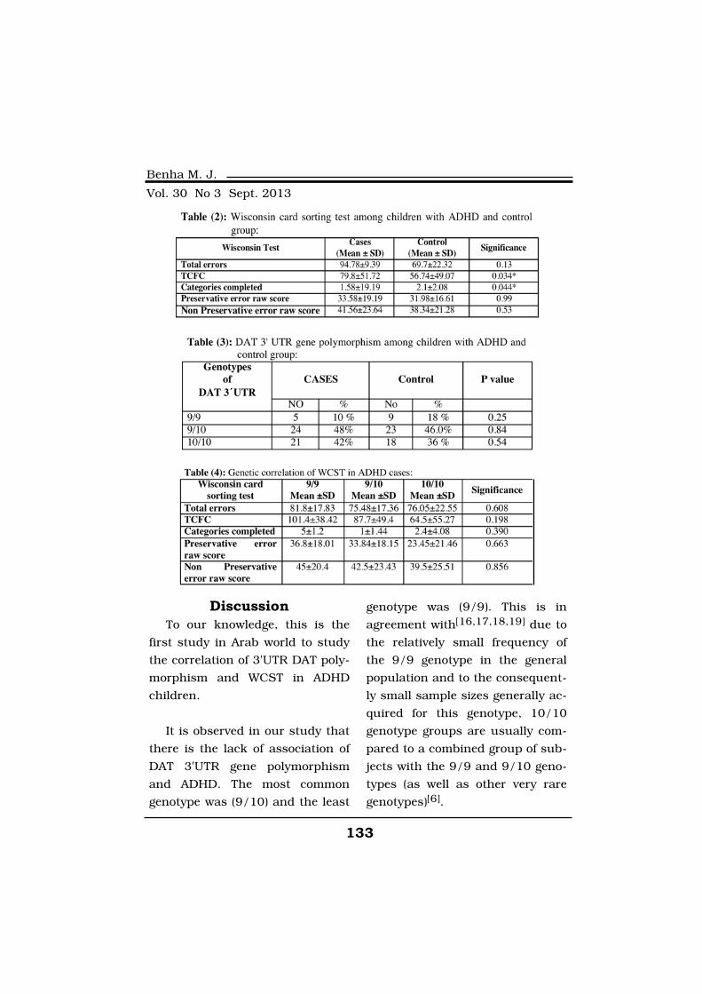
133
Benha M. J.
Vol. 30 No 3 Sept. 2013
DiscussionTo our knowledge, this is the
first study in Arab world to study
the correlation of 3'UTR DAT poly-
morphism and WCST in ADHD
children.
It is observed in our study that
there is the lack of association of
DAT 3'UTR gene polymorphism
and ADHD. The most common
genotype was (9/10) and the least
genotype was (9/9). This is in
agreement with[16,17,18,19] due to
the relatively small frequency of
the 9/9 genotype in the general
population and to the consequent-
ly small sample sizes generally ac-
quired for this genotype, 10/10
genotype groups are usually com-
pared to a combined group of sub-
jects with the 9/9 and 9/10 geno-
types (as well as other very rare
genotypes)[6].
134
Mohamed Elwasify, et al....
The dopamine transporter gene
(DAT1) has been reported to be as-
sociated with attention-deficit hy-
peractivity disorder (ADHD) in a
number of studies[20,21,22,6].
That is finding in our study
may be due to the following rea-
sons. First of all, small sample
size. The second one is the poly-
morphism that we studied in this
sample is located in the 3' un-
translated region of the SLC6A3
gene and thus does not change
the structure of the DAT1 protein.
Nonetheless, it has been suggest-
ed that it may affect the level of
expression of the DAT1 gene, re-
sulting in variable dopamine
transporter phenotypes[6]. While
this may be the case, the issue
still remains controversial. Indeed,
it has been reported that DAT
binding availability is significantly
lower for subjects who are homo-
zygous for the SLC6A3 VNTR 10-
repeat allele compared to carriers
of at least one 9-repeat allele
[23]while the opposite association
[24])was reported in another study
and yet no association [25]was
found in a third. The third expla-
nation may be related to the me-
thodological differences. The
Fourth one is ethnicity as this is
first study to be done on Egyptian
population.
In our study, there were a sig-
nificant difference in TCFC and
categories completed indices of
WCST results between ADHD cas-
es and control. On the other hand
no significant differences in total
errors, preservative errors and
non-preservative error raw score.
Concerning Wisconsin Card
Sorting Test, 17 out of 26 studies
using the WCST found significant
differences between ADHD and
normal controls[26]. Children with
ADHD complete fewer categories
than normal controls[27], and
showed more deficiency in flexibil-
ity and perseverative errors than
high functioning autistic and con-
trol groups[28].
Limitation of the presentLimitation of the present
studystudy
First,First, the sample size might
have limited our ability to detect
more 3'UTR DAT gene polymor-
phism and its correlation to WCST
in ADHD children. Second, Second, as-
sessment of executive functions
using WCST which represent one

135
Benha M. J.
Vol. 30 No 3 Sept. 2013
domain of executive function (set
shifting). Third,Third, the polymorphism
that we studied in this sample is
located in the 3' UTR DAT gene
polymophysism that doesn't
change structure of DAT protein.
References1- Biederman J. and Faraone1- Biederman J. and Faraone
S.V. (2005):S.V. (2005): Attention-deficit hy-
peractivity disorder. Lancet 366:
237-248.
2- American Psychiatric Asso-2- American Psychiatric Asso-
ciation (2000):ciation (2000): Diagnostic and
statistical manual of mental disor-
ders, (4th ed., text revision).
Washington, DC: American Psy-
chiatric Association.
3- Wang Y., Wang Z., Yao k.,3- Wang Y., Wang Z., Yao k.,
et al. (2007):et al. (2007): Lack of association
between the Dopamine transport-
er gene 3'VNTR polymorphism and
attention deficit hyperactivity dis-
order in Chinese Han children:
case-control and family-based
Studies. Kobe J. Med. Sci., 53(6):
327-33.
4- Friedel S., Saar K., Sauer4- Friedel S., Saar K., Sauer
S., et al. (2007):S., et al. (2007): Association and
linkage of allelic variants of the
dopamine transporter gene in
ADHD. Molecular Psychiatry. ; 12
(10): 923-933.
5- Brown T.E. (2009):5- Brown T.E. (2009): ADD/
ADHD and Impaired executive
function in clinical practice . Cur-
rent psychiatry reports 1:37-41.
6- Karama S., Grizenko N.,6- Karama S., Grizenko N.,
Barke E.S., et al. (2008):Barke E.S., et al. (2008): Dopa-
mine transporter 3'UTR VNTR
genotype is a marker of perfor-
mance on executive function tasks
in children with ADHD. BMC psy-
chiatry , 8:45.
7- Sheehan D., Shytle D.,7- Sheehan D., Shytle D.,
Milo K. and Lecrubier (1998):Milo K. and Lecrubier (1998):
Development and Validation of
M.I.N.I. J. Clinical psychiatry 59,
suppl (20).
8- Ibrahim M., Bishry Z. and8- Ibrahim M., Bishry Z. and
Hamed A. (2002):Hamed A. (2002): Comparison of
Mini International Neuropsychiat-
ric Interview for children (MINI-
KID) with the schedules for
affective disorders and schizophre-
nia for school aged children,
present and lifetime version
(KSADS-PL): In Egyptian sample
presenting with childhood
disorders. MD thesis, Ain Shams
University.
136
Mohamed Elwasify, et al....
9- Achenbach T. M. (1997):9- Achenbach T. M. (1997):
Child Behavior chick list and 1991
profiles for children and adoles-
cents. Department of Psychiatry,
Vermount University, USA.
10- Koura M.R. (1998):10- Koura M.R. (1998): Study
of the role of Alexandria primary
health care program in assess-
ment of behavioral disorder in pri-
mary school children thesis
,Alexandria university High Insti-
tute of public health.
11- Conners C.K. (1989):11- Conners C.K. (1989):
Manual for Conners' Rating
Scales. N.Tonawanda, NY: Multi-
Health Systems Inc.
12- El-Dafrawi M.H., Mahfouz12- El-Dafrawi M.H., Mahfouz
R. and Ragab L.A. (1990):R. and Ragab L.A. (1990): Relia-
bility and validity study of a rating
scale for ADHD in Egyptian chil-
dren and adolescents. Cairo,
Egypt: The 3rd International
Egyptian Congress of Psychiatry.
13- Wechsler D.I. (1991):13- Wechsler D.I. (1991):
scale for children, third edition.
Psychological Corporation, New
York Examiner's manual: Wech-
sler intelligence.
14- Ismael M. and Malekal14- Ismael M. and Malekal
(1993):(1993): The Wechsler Intelligence
Scale for Children (WISC) the Ara-
bic version. The Egyptian Anglo
library.
15- Heaton R.K., Chelune,15- Heaton R.K., Chelune,
G.J., Talley J.L., Kay G.G. andG.J., Talley J.L., Kay G.G. and
Curtiss G. (1993):Curtiss G. (1993): Wisconsin
Card Sorting Test Manual: Revised
and expanded. Odessa, FL: Psy-
chological Assessment Resources.
16- Zhou K., Chen W., Buite-16- Zhou K., Chen W., Buite-
laar J., Banaschewski T., et al.laar J., Banaschewski T., et al.
(2008a):(2008a): Genetic heterogeneity in
ADHD: DAT1 gene only affects
probands without CD. Am. J.
Med. Genet. B Neuropsychiatr.
Genet.; 147B: 1481-1487.
17- Bakker S.C., van der17- Bakker S.C., van der
Meulen E.M., Oteman N.,Meulen E.M., Oteman N.,
Schelleman H., Pearson P.L.,Schelleman H., Pearson P.L.,
Buitelaar J.K. and Sinke R.J.Buitelaar J.K. and Sinke R.J.
(2005):(2005): DAT1, DRD4, and DRD5
polymorphisms are not associated
with ADHD in Dutch families. Am.
J. Med. Genet. B Neuropsychiatr.
Genet.; 132B: 50-52.
18- Qian Q., Wang Y., Zhou18- Qian Q., Wang Y., Zhou
R., Yang L. and Faraone S.V.R., Yang L. and Faraone S.V.
(2004): (2004): Family based and case-
control association studies of
137
Benha M. J.
Vol. 30 No 3 Sept. 2013
DRD4 and DAT1 polymorphisms
in Chinese attention deficit hyper-
activity disorder patients suggest
long repeats contribute to genetic
risk for the disorder. Am J Med
Genet B Neuropsychiatr Genet;
128B(1): 84-89.
19- Cheuk D.K., Li S.Y. and19- Cheuk D.K., Li S.Y. and
Wong V. (2004):Wong V. (2004): No association
between VNTR polymorphisms of
dopamine transporter gene and
attention Molecular Psychiatry;
17: 1086-1092.
20- Genro J.P., Zeni C., Po-20- Genro J.P., Zeni C., Po-
lanczyk G.V., Roman T., Rohdelanczyk G.V., Roman T., Rohde
L.A. and Hutz M.H. (2007):L.A. and Hutz M.H. (2007): A
promoter polymorphism (839C >
T) at the dopamine transporter
gene is associated with attention
Am. J. Med. Genet. B Neuropsy-
chiatr. Genet.; 144B: 215-219.
21- Asherson P., Brookes K.,21- Asherson P., Brookes K.,
Franke B., Chen W., et al.Franke B., Chen W., et al.
(2007): (2007): Confirmation that a spe-
cific haplotype of the dopamine
transporter gene is associated
with combined-type ADHD. Am J
Psychiatry; 164: 674-677.
22- Barkley R.A., Smith K.M.,22- Barkley R.A., Smith K.M.,
Fischer M. and Navia B. (2006):Fischer M. and Navia B. (2006):
An examination of the behavioral
and neuropsychological correlates
of three ADHD candidate gene pol-
ymorphisms (DRD4 7_, DBH TaqI
A2, and DAT1 40 bp VNTR) in hy-
peractive and normal children fol-
lowed to adulthood. Am. J. Med.
Genet. B Neuropsychiatry Genet.
141B: 487-498.
23- El-Tarras A.E., Alsulai-23- El-Tarras A.E., Alsulai-
mani A.A., Awad N.S., Mitwalymani A.A., Awad N.S., Mitwaly
N., Said M.M. and Sabry A.M.N., Said M.M. and Sabry A.M.
(2012):(2012): Association study between
the dopamine-related candidate
gene polymorphisms and ADHD
among Saudi Arabia population
via PCR technique. Mol Biol Rep.;
39(12): 11081-6.
24- Jacobsen L.K., Staley24- Jacobsen L.K., Staley
J.K., Zoghbi S.S., Seibyl J.P.,J.K., Zoghbi S.S., Seibyl J.P.,
Kosten T.R., Innis R.B. and Ge-Kosten T.R., Innis R.B. and Ge-
lernter J. (2000):lernter J. (2000): Prediction of
dopamine transporter binding
availability by genotype: a prelimi-
nary report. Am J Psychiatry; 157:
1700-1703.
25- Heinz A., Goldman D.,25- Heinz A., Goldman D.,
Jones D.W., Palmour R., Hom-Jones D.W., Palmour R., Hom-
mer D., Lee K.S., Linnoila M.mer D., Lee K.S., Linnoila M.
and Weinberger D.R. (2000):and Weinberger D.R. (2000):
Genotype influences in vivo dopa-
138
Mohamed Elwasify, et al....
mine transporter availability in
human striatum. Neuropsycho-
pharmacology; 22: 133-139.
26- Martinez D., Gelernter J.,26- Martinez D., Gelernter J.,
Abi-Dargham A., van Dyck C.H.,Abi-Dargham A., van Dyck C.H.,
Innis R.B. and Laruelle M.Innis R.B. and Laruelle M.
(2001): (2001): The variable number of
tandem repeats polymorphism of
the dopamine transporter gene is
not associated with significant
change in dopamine transporter
phenotype in humans. Neuropsy-
chopharmacology; 24: 553-560.
27- Sergeant J.A., Geurts H.27- Sergeant J.A., Geurts H.
and Oosterlaan J. (2002):and Oosterlaan J. (2002): How
specific is a deficit of executive
functioning for attention-deficit/
hyperactivity disorder. Behav
Brain Res; 130: 3-28.
28- Doyle A.E., Faraone S.V.,28- Doyle A.E., Faraone S.V.,
Seidman L.J., et al. (2005):Seidman L.J., et al. (2005): Are
endophenotypes based on meas-
ures of executive functions useful
for molecular genetic studies of
ADHD? J Child Psychol Psychia-
try; 46: 774-803.

139
Benha M. J.
Vol. 30 No 3 Sept. 2013
DOPAMINE TRANSPORTER 3'UTRDOPAMINE TRANSPORTER 3'UTRVNTR GENOTYPE AND WISCONSINVNTR GENOTYPE AND WISCONSINCARD SORTING TEST IN CHILDRENCARD SORTING TEST IN CHILDREN
WITH ADHD AMONGWITH ADHD AMONGEGYPTIAN POPULATIONEGYPTIAN POPULATION
Mohamed Elwasify MD, Zeinab Gomaa MD,Mohamed Elwasify MD, Zeinab Gomaa MD,Elsayed ElNaggar MD, Farha Elshenawy MD andElsayed ElNaggar MD, Farha Elshenawy MD and
Vishwaiit Nimgaonkar MDVishwaiit Nimgaonkar MD
BENHAMEDICALJOURNAL
REPRINT
Published byPublished by
Benha Faculty of MedicineBenha Faculty of Medicine Volume 30 Number 3Sept. 2013
139
Benha M. J.
Vol. 30 No 3 Sept. 2013
EFFECT OF ORAL ANTICOAGULANTEFFECT OF ORAL ANTICOAGULANT(WARFARIN SODIUM -COUMADIN) ON(WARFARIN SODIUM -COUMADIN) ON
ARTERIAL BLOOD PRESSUREARTERIAL BLOOD PRESSURE(EXPERIMENTAL STUDY)(EXPERIMENTAL STUDY)
Ahmed A. El-Gendy Ph.D and Ghayaty E.A.D. MD*Ahmed A. El-Gendy Ph.D and Ghayaty E.A.D. MD*Departments of Medical Physiology and Clinical Pharmacology*
Faculty of Medicine Mansoura University. Egypt
AbstractBackground:Background: Warfarin sodium (coumadin) is an anticoagulant that
acts by inhibiting vitamin-K-dependent coagulation factors but there isno evidence that warfarin has a role in arterial blood pressure. Peoplewho suffer from high blood pressure and who also take coumadin forstroke prevention must always keep in mind that high blood pressureincreases the risk of strokes, or strokes caused by bleeding in the brain.
Aim of the work:Aim of the work: is to clarify the effect of warfarin sodium (couma-din) on arterial blood pressure in animals.
Material & Methods:Material & Methods: In our laboratories in Mansoura Universitypreliminary studies done to detect possible effect on blood pressure ofdog and it was surprising that this anticoagulant has antihypertensiveeffect on the blood pressure, this directs our attention to study the pos-sible site of action of warfarin sodium. Serial experiments were done onblood pressure of dog using different agonists and antagonists, also onisolated perfused hind limb of rat, also experiments were done on isolat-ed perfused rabbit's heart.
Results:Results: The study demonstrated that warfarin sodium has directvascular smooth muscle relaxant effect with no effect on the heart rateor cardiac contractility.
Conclusion:Conclusion: warfarin sodium in need of further clinical studies toformulate a related compound with both anticoagulant and antihyper-tensive effect which is valuble in many diseases in which hypercoagula-tion and hypertension occur concurrently.
140
Ahmed A. El-Gendy and Ghayaty E.A.D.
IntroductionThe oral anticoagulants are de-
rived from coumarin, which is
found in many plants. A promi-
nent member of this class is war-
farin(coumadin). It takes at least
48 to 72 hours for the anticoagu-
lant effect to develop. This antico-
agulants are used to treat patients
with deep-vein thrombosis (DVT),
pulmonary embolism (PE), atrial
fibrillation (AF), and mechanical
prosthetic heart valves(1).
Warfarin sodium was already
established experimentally to have
anticoagulant effect(2). Coumadin
(warfarin sodium) is an anticoagu-
lant that acts by inhibiting vita-
min K-dependent coagulation fac-
tors. Chemically, it is 3-(α-
acetonylbenzyl)-4 hydroxycoumar-
in and is a racemic mixture of the
R- and S-enantiomers. Crystalline
warfarin sodium is an isopropanol
clathrate. Its empirical formula is
C19H15NaO4(3).
Coumadin is a vitamin K antag-
onist indicated for prophylaxis
and treatment of venous thrombo-
sis and its extension, pulmonary
embolism. Prophylaxis and treat-
ment of thromboembolic complica-
tions associated with atrial fibril-
lation and/or cardiac valve re-
placement, reduction in the risk of
death, recurrent myocardial in-
farction, and thromboembolic
events such as stroke or systemic
embolization after myocardial in-
farction. Coumadin has no direct
effect on an established thrombus,
nor does it reverse ischemic tissue
damage. Once a thrombus has oc-
curred, however, the goals of anti-
coagulant treatment are to pre-
vent further extension of the
formed clot and to prevent secon-
dary thromboembolic complica-
tions that may result in serious
and possibly fatal sequelae(4).
As coumadin decreases the
body’s mechanisms which normal-
ly stop bleeding, people who take
coumadin must always keep their
blood pressure in check. In fact, a
study has shown that even small
reductions in systolic blood pres-
sure (as low as 12 points) can de-
crease the risk of bleeding in the
brain by almost 80%(5).
In humans, warfarin crosses
the placenta, and concentrations
in fetal plasma approach the ma-
ternal values(6).
141
Benha M. J.
Vol. 30 No 3 Sept. 2013
Coumarins have shown some
evidence of many biological activi-
ties, although they are approved
for few medical uses as pharma-
ceuticals. The activity reported for
coumarin and coumarins includes
anti-HIV, anti-tumor, anti-
hypertension, anti-arrhythmia, anti-
inflammatory, anti-osteoporosis,
antiseptic, and analgesic (pain re-
lief). It is also used in the treat-
ment of asthma(4). Coumarin has
been used in the treatment of lym-
phedema(4).
Coumarins have acquired in-
creasing significance as coumarin
moiety is biologically and phar-
macologically important it occurs
in several natural products and
especially in some antibiotics(4,8).
3-substituted-4- hydroxyl coumar-
ins are useful anticoagulants of
low toxicity(4,7). Dicoumarol or 3,
3- ethylene-bis (4-hydroxycoumarin)
is an anticoagulant and responsi-
ble for haemorrhagic sweet clover
disease of cattle(9). Several other
bis-compounds are similar to di-
coumarol in its action(10). Ethyl
bis (-4-hydroxy-3 coumarin) ace-
tate is physiologically very active
and has certain advantages over
dicoumarol as it is an anticoagu-
lant of short duration. They are
used instead of dicoumarol for re-
ducing the prothrombin index of
the blood and for its inhibitory ef-
fect on cholinesterase(11). Cyclo-
coumarol is one of the most active
anticoagulant among 106 synthet-
ic compounds tested for their ac-
tivity. In rabbits, dogs and healthy
men cyclocoumarol induced and
intense hypoprothrombinemia in
minimal doses(12). Same coumar-
in derivatives show antihyperten-
sive effect(4).
Many diseases have been found
to be associated with hypertension
and hypercoagulation. Hypercoa-
gulable state was found in pa-
tients with diabetes mellitus, preg-
nancy, induced hypertension and
glomerular disease especially in
those with uremia and nephritic
syndrome(13). Also erythrocyte
flexibility was impaired in diabetic
patients and was not influenced
by the duration of diabetes, its
type and treatment(7). Risk factors
as hypertension, arteriosclerosis
and smoking have an aggravating
effect.
The coagulation function were
studied in patients with myocar-
142
Ahmed A. El-Gendy and Ghayaty E.A.D.
dial infarction in relation to the
presence of essential hypertension,
hypercoagulation was founded to be
more pronounced in cases of asso-
ciated essential hypertension(14).
Also there is simultaneous ten-
dency to the decrease of blood
pressure and the inhibition of
the aggregation capacity of plate-
lets (15).
Aim of the WorkThe present work is an experi-
mental trial to obtain an effective
drug for treatment of both hyper-
tension and hypecoagulability as
these disease are commonly present
concurrently in the same patient
and to detect its site of action as
antihypertensive if possible.
Material and Methodsi) Chemicals used:i) Chemicals used:
1- Warfarin sodium (Vial con-
taining 5 mg lyophilized powder).
Crystalline warfarin sodium oc-
curs as a white, odorless,
crystalline powder that is
discolored by light.It is very
soluble in water, freely soluble in
alcohol, and very slightly soluble
in chloroform and ether (Initial
U.S. Approval: 1954).
2- Atropine powder obtained
from Macfarian Smith Lt Edin-
burg.
3- Adrenaline powder obtained
from G.H.Boehringer Sohn Com-
pany (Germany).
4- Norepinephrine powder ob-
tained from Sigma Company.
5- Phenylephrine powder ob-
tained from Sigma Company.
6- Acetylcholine was obtained
from Sigma Company.
ii) Animals used and meth-ii) Animals used and meth-
ods:ods:
20 Mongerl dogs of both sex,
weighing 15-20 kg, were used
throughout this study they are
prepared for recording blood pres-
sure and assessment of drug ef-
fects on it according to method of
Ghosh(16).
30 albino rats of either sex
weighing 200-250 gm were used,
they were prepared for assessment
of the effect of the drug of the per-
fused hind limb of rat according to
the method of Burn(1).
10 rabbits of either sex weigh-
ing 2 kg were used and prepared
for recording the effect of the drug
on the isolated rabbit's the heart
143
Benha M. J.
Vol. 30 No 3 Sept. 2013
according to the method of Lan-
gendorff(17).
iii) The following experimentsiii) The following experiments
were done:were done:
I. Effect of warfarin sodiumI. Effect of warfarin sodium
on blood pressure of dogs:on blood pressure of dogs:
1. Repeated doses of warfarin
sodium were administered IV (eve-
ry dose was dissolved in 100 ul
distilled water starting from 5 ug/
kg to 320 ug/kg according to the
dose response cure to choose the
effective dose.
2. Warfarin sodium was inject-
ed IV in a dose of 50 ug/kg and
the same dose was repeated after
5 minutes followed by acetyl cho-
line (1 ug/kg) then atropine (0.2
mg/kg) followed by acetyl choline
(1 ug/kg) then warfarin sodium
(50 ug/kg).
3. Warfarin sodium was admin-
istered IV in a dose of 50 ug/kg
followed by phenylephrine (1ug/
kg) then warfarin sodium (50 ug/
kg) followed by phenylephrine
(1ug/kg). The same steps were
done with adrenaline (1ug/k).
4. Induction of hypertension
was carried out according to the
method of EL-Tahir et al.,(3) using
norepinephrine (NE) infusion in
anaesthized dog over a period of
60 minutes in a dose of 4 ug/
minute. After initial elevation of
blood pressure (1 minute) NE
infusion then stopped and
warfarin sodium (50 ug/kg) IV,
then readministration of NE is
continued(3).
II. Effect of warfarin sodiumII. Effect of warfarin sodium
on perfused hind limb of rats:on perfused hind limb of rats:
30 albino rats of either sex
weighing 200-250 gm were used,
they were prepared for assessment
of the effect of the drug of
the perfused hind limb of rat
according to the method of Burn(1). Drugs were injected through
rubber part just proximal to the
canula. Control group composed
of six rats were given 1 cc
distilled water. The perfusate was
counted for five minutes. Warfarin
sodium was administered in
different doses ranging from
50-400 ug/kg, each dose was
administered in a separate
preparation and the perfusate was
collected every five minutes. The
maximum effect was obtained
after 1 hour and the mean was
determined.
144
Ahmed A. El-Gendy and Ghayaty E.A.D.
III. Effect of warfarin sodiumIII. Effect of warfarin sodium
on isolated perfused rabbit'son isolated perfused rabbit's
heart:heart: Isolated rabbit's heart was
prepared according to the method
of Langendorff(17). Warfarin sodi-
um was administered in different
doses ranging from 10 ug to 320
ug and every dose was dissolved
in 100 ul acetone.
The statisticsThe statistics were calculated
by T-test(18) comparing the mean
obtained by different dose of war-
farin sodium compared to the mean
of control saline treated group.
Results1. Effect of warfarin sodium1. Effect of warfarin sodium
on blood pressure of dog (figureson blood pressure of dog (figures
1 & 2):1 & 2): Warfarin sodium has hy-
potensive action on blood pres-
sure of dog.
2. Effect of atropine on the2. Effect of atropine on the
hypotensive action of warfarinhypotensive action of warfarin
sodium (Figure 3): sodium (Figure 3): Warfarin sodi-
um is administered IV , in a dose
of 50 ug/kg in the dog and the
same dose was repeated after 5
minutes, it produced a hypoten-
sive effect. Acetyl choline in a dose
of 1 ug/kg IV produced hypoten-
sion. Atropine IV, in a dose of 0.2
mg/kg produced hypotension due
to cutaneous vasodilatation. A test
dose of acetyl choline 1 ug/kg
produced no hypotensive effect de-
noting full atropinization and
blockade of muscarinic receptors.
The same dose of warfarin sodi-
um produced the same control hy-
potensive effect after blockade of
muscarinic receptors denoting
that this compound is not a mus-
carinic receptor agonist.
3. Effect of warfarin sodium3. Effect of warfarin sodium
on the hypertensive effect ofon the hypertensive effect of
adrenaline (figure 4):adrenaline (figure 4): Warfarin
sodium given IV to the dog in a
dose of 50 ug/kg produced hypo-
tension. Phenylephrine in a dose
of 1 ug/kg given IV, produced hy-
pertension indicating intact alpha
receptors in the dog. Warfarin so-
dium given IV to the dog in a dose
of 50 ug/kg given after phenyleph-
rine produced hypotension. Phen-
ylephrine in a dose of 1 ug/kg giv-
en IV, after warfarin sodium
produced the same hypertensive
effect denoting that warfarin sodi-
um has no alpha blocking effect.
Adrenaline in a dose of 1 ug/kg
after warfarin sodium injection
produced hypertensive effect due
to alpha and beta agonist effects.
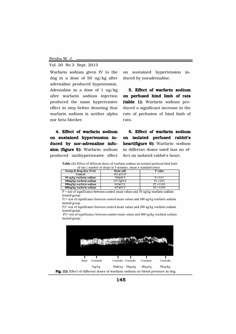
145
Benha M. J.
Vol. 30 No 3 Sept. 2013
Warfarin sodium given IV to the
dog in a dose of 50 ug/kg after
adrenaline produced hypotension.
Adrenaline in a dose of 1 ug/kg
after warfarin sodium injection
produced the same hypertensive
effect in step before denoting that
warfarin sodium is neither alpha
nor beta blocker.
4. Effect of warfarin sodium4. Effect of warfarin sodium
on sustained hypertension in-on sustained hypertension in-
duced by nor-adrenaline infu-duced by nor-adrenaline infu-
sion (figure 5):sion (figure 5): Warfarin sodium
produced antihypertensive effect
on sustained hypertension in-
duced by noradrenaline.
5. Effect of warfarin sodium5. Effect of warfarin sodium
on perfused hind limb of ratson perfused hind limb of rats
(table 1): (table 1): Warfarin sodium pro-
duced a significant increase in the
rate of perfusion of hind limb of
rats.
6. Effect of warfarin sodium6. Effect of warfarin sodium
on isolated perfused rabbit'son isolated perfused rabbit's
heart(figure 6): heart(figure 6): Warfarin sodium
in different doses used has no ef-
fect on isolated rabbit's heart.
Fig. (1):Fig. (1): Effect of different doses of warfarin sodium on blood pressure in dog.
↑ ↑ ↑ ↑ ↑ ↑
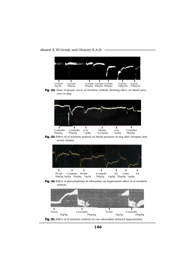
146
Ahmed A. El-Gendy and Ghayaty E.A.D.
Fig. (2): Fig. (2): Dose response curve of warfarin sodium showing effect on blood pres-sure in dog.
Fig. (3):Fig. (3): Effect of of warfarin sodium on blood pressure in dog after atropine andacetyl choline.
Fig. (4):Fig. (4): Effect of phenylephrine & adrenaline on hypotensive effect of of warfarinsodium.
Fig. (5):Fig. (5): Effect of of warfarin sodium on nor-adrenaline induced hypertension.
↑↑↑↑
↑ ↑ ↑ ↑ ↑ ↑
↑↑↑↑↑↑↑
↑↑↑↑↑↑↑
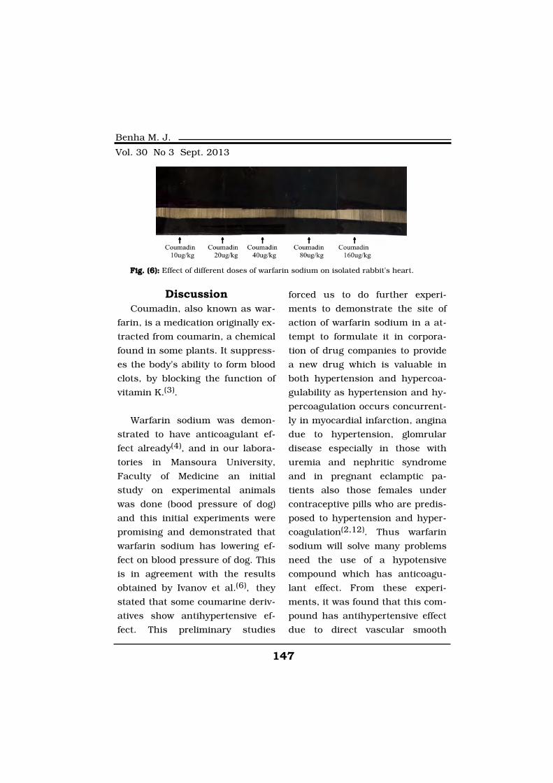
147
Benha M. J.
Vol. 30 No 3 Sept. 2013
DiscussionCoumadin, also known as war-
farin, is a medication originally ex-
tracted from coumarin, a chemical
found in some plants. It suppress-
es the body's ability to form blood
clots, by blocking the function of
vitamin K.(3).
Warfarin sodium was demon-
strated to have anticoagulant ef-
fect already(4), and in our labora-
tories in Mansoura University,
Faculty of Medicine an initial
study on experimental animals
was done (bood pressure of dog)
and this initial experiments were
promising and demonstrated that
warfarin sodium has lowering ef-
fect on blood pressure of dog. This
is in agreement with the results
obtained by Ivanov et al.(6), they
stated that some coumarine deriv-
atives show antihypertensive ef-
fect. This preliminary studies
forced us to do further experi-
ments to demonstrate the site of
action of warfarin sodium in a at-
tempt to formulate it in corpora-
tion of drug companies to provide
a new drug which is valuable in
both hypertension and hypercoa-
gulability as hypertension and hy-
percoagulation occurs concurrent-
ly in myocardial infarction, angina
due to hypertension, glomrular
disease especially in those with
uremia and nephritic syndrome
and in pregnant eclamptic pa-
tients also those females under
contraceptive pills who are predis-
posed to hypertension and hyper-
coagulation(2,12). Thus warfarin
sodium will solve many problems
need the use of a hypotensive
compound which has anticoagu-
lant effect. From these experi-
ments, it was found that this com-
pound has antihypertensive effect
due to direct vascular smooth
Fig. (6):Fig. (6): Effect of different doses of warfarin sodium on isolated rabbit's heart.
↑ ↑ ↑ ↑ ↑
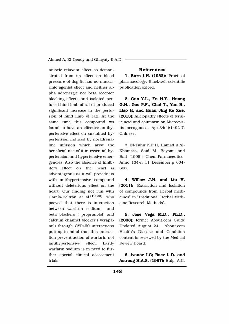
148
Ahmed A. El-Gendy and Ghayaty E.A.D.
muscle relaxant effect as demon-
strated from its effect on blood
pressure of dog (it has no musca-
rinic agonist effect and neither al-
pha adrenergic nor beta receptor
blocking effect), and isolated per-
fused hind limb of rat (it produced
significant increase in the perfu-
sion of hind limb of rat). At the
same time this compound ws
found to have an effective antihy-
pertensive effect on sustained hy-
pertension induced by noradrena-
line infusion which arise the
beneficial use of it in essential hy-
pertension and hypertensive emer-
gencies. Also the absence of inhib-
itory effect on the heart is
advantageous as it will provide us
with antihypertensive compound
without deleterious effect on the
heart. Our finding not run with
García-Beltrán at al.(19,20) who
pooved that there is interaction
between warfarin sodium and
beta blockers ( propranolol) and
calcium channel blocker ( verapa-
mil) through CYP450 interactions
putting in mind that this interac-
tion prevent action of warfarin not
antihypertensive effect. Lastly
warfarin sodium is in need to fur-
ther special clinical assessment
trials.
References1. Burn I.H. (1952):1. Burn I.H. (1952): Practical
pharmacology, Blackwell scientific
publication oxford.
2. Guo Y.L., Fu H.Y., Huang2. Guo Y.L., Fu H.Y., Huang
G.H., Gao P.F., Chai T., Yan B.,G.H., Gao P.F., Chai T., Yan B.,
Liao H. and Huan Jing Ke Xue.Liao H. and Huan Jing Ke Xue.
(2013):(2013): Allelopathy effects of ferul-
ic acid and coumarin on Microcys-
tis aeruginosa. Apr;34(4):1492-7.
Chinese.
3. El-Tahir K.F.H, Hamad A.Al-
Khamees, Said M. Bayomi and
Ball (1995): Chem.Farmaceutico-
Anno 134-n 11 December,p 604-
608.
4. Willow J.H. and Liu H.4. Willow J.H. and Liu H.
(2011):(2011): "Extraction and Isolation
of compounds from Herbal medi-
cines" in 'Traditional Herbal Medi-
cine Research Methods'.
5. Jose Vega M.D., Ph.D.,5. Jose Vega M.D., Ph.D.,
(2008):(2008): former About.com Guide
Updated August 24, About.com
Health's Disease and Condition
content is reviewed by the Medical
Review Board.
6. Ivanov I.C; Racv L.D. and6. Ivanov I.C; Racv L.D. and
Astroug H.A.S. (1987):Astroug H.A.S. (1987): Bulg. A.C.
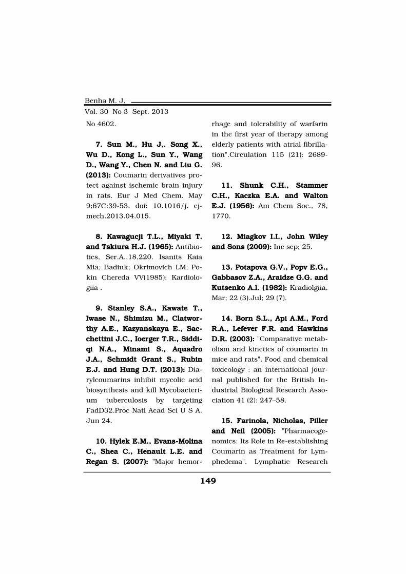
149
Benha M. J.
Vol. 30 No 3 Sept. 2013
No 4602.
7. Sun M., Hu J,. Song X.,7. Sun M., Hu J,. Song X.,
Wu D., Kong L., Sun Y., WangWu D., Kong L., Sun Y., Wang
D., Wang Y., Chen N. and Liu G.D., Wang Y., Chen N. and Liu G.
(2013):(2013): Coumarin derivatives pro-
tect against ischemic brain injury
in rats. Eur J Med Chem. May
9;67C:39-53. doi: 10.1016/j. ej-
mech.2013.04.015.
8. Kawagucji T.L., Miyaki T.8. Kawagucji T.L., Miyaki T.
and Tskiura H.J. (1965):and Tskiura H.J. (1965): Antibio-
tics, Ser.A.,18,220. Isanits Kaia
Mia; Badiuk; Okrimovich LM; Po-
kin Chereda VV(1985): Kardiolo-
giia .
9. Stanley S.A., Kawate T.,9. Stanley S.A., Kawate T.,
Iwase N., Shimizu M., Clatwor-Iwase N., Shimizu M., Clatwor-
thy A.E., Kazyanskaya E., Sac-thy A.E., Kazyanskaya E., Sac-
chettini J.C., Ioerger T.R., Siddi-chettini J.C., Ioerger T.R., Siddi-
qi N.A., Minami S., Aquadroqi N.A., Minami S., Aquadro
J.A., Schmidt Grant S., RubinJ.A., Schmidt Grant S., Rubin
E.J. and Hung D.T. (2013):E.J. and Hung D.T. (2013): Dia-
rylcoumarins inhibit mycolic acid
biosynthesis and kill Mycobacteri-
um tuberculosis by targeting
FadD32.Proc Natl Acad Sci U S A.
Jun 24.
10. Hylek E.M., Evans-Molina10. Hylek E.M., Evans-Molina
C., Shea C., Henault L.E. andC., Shea C., Henault L.E. and
Regan S. (2007):Regan S. (2007): "Major hemor-
rhage and tolerability of warfarin
in the first year of therapy among
elderly patients with atrial fibrilla-
tion".Circulation 115 (21): 2689-
96.
11. Shunk C.H., Stammer11. Shunk C.H., Stammer
C.H., Kaczka E.A. and WaltonC.H., Kaczka E.A. and Walton
E.J. (1956):E.J. (1956): Am Chem Soc., 78,
1770.
12. Miagkov I.I., John Wiley12. Miagkov I.I., John Wiley
and Sons (2009):and Sons (2009): Inc sep; 25.
13. Potapova G.V., Popv E.G.,13. Potapova G.V., Popv E.G.,
Gabbasov Z.A., Araidze G.G. andGabbasov Z.A., Araidze G.G. and
Kutsenko A.l. (1982):Kutsenko A.l. (1982): Kradiolgiia,
Mar; 22 (3).Jul; 29 (7).
14. Born S.L., Api A.M., Ford14. Born S.L., Api A.M., Ford
R.A., Lefever F.R. and HawkinsR.A., Lefever F.R. and Hawkins
D.R. (2003):D.R. (2003): "Comparative metab-
olism and kinetics of coumarin in
mice and rats". Food and chemical
toxicology : an international jour-
nal published for the British In-
dustrial Biological Research Asso-
ciation 41 (2): 247–58.
15. Farinola, Nicholas, Piller15. Farinola, Nicholas, Piller
and Neil (2005):and Neil (2005): "Pharmacoge-
nomics: Its Role in Re-establishing
Coumarin as Treatment for Lym-
phedema". Lymphatic Research
150
Ahmed A. El-Gendy and Ghayaty E.A.D.
and 16000056 PMID. 10.1089/
Irb. 2005.3.81:doi Biology3 (2):
81-86.
16. Ghosh M.N. (1971):16. Ghosh M.N. (1971): Fun-
damentals of experimental phar-
macology Calcutta Scientific Book
agency.
17. Langendorff pflugers17. Langendorff pflugers
Arch. (1913):Arch. (1913): Ges phys
18. Ron Winslow, Avery John-18. Ron Winslow, Avery John-
son (2007-12-10):son (2007-12-10): "Race Is on for
the Next Blood Thinner". Wall
Street Journal. p. A12. Retrieved
2008-01-06. "...in a market now
dominated by one of the oldest
mainstay pills in medicine: the
blood thinner warfarin. At least
five next-generation blood thin-
ners are in advanced testing to
treat or prevent potentially debili-
tating or life-threatening blood
clots in surgery and heart pa-
tients. First candidates could
reach the market in 2009."
19. García-Beltrán O., Mena19. García-Beltrán O., Mena
N., Yañez O., Caballero J., Var-N., Yañez O., Caballero J., Var-
gas V., Nuñez M.T., and Casselsgas V., Nuñez M.T., and Cassels
B.K. (2013):B.K. (2013): Design, synthesis
and cellular dynamics studies in
membranes of a new coumarin-
based "turn-off" fluorescent probe
selective for Fe<sup>2+</sup>Eur
J Med Chem. Jun 19;67C:60-63.
doi: 10.1016/j. ejmech. 2013.06.
022. [Epub ahead of print].
20. Wittkowsky A.K. (2001):20. Wittkowsky A.K. (2001):
"Drug interactions update: drugs,
herbs, and oral anticoagulation".
J. Thromb. Thrombolysis 12 (1): 67-
71. doi:10.1023/A:1012742628628.
PMID 11711691.
151
Benha M. J.
Vol. 30 No 3 Sept. 2013
EFFECT OF ORAL ANTICOAGULANTEFFECT OF ORAL ANTICOAGULANT(WARFARIN SODIUM -COUMADIN) ON(WARFARIN SODIUM -COUMADIN) ON
ARTERIAL BLOOD PRESSUREARTERIAL BLOOD PRESSURE(EXPERIMENTAL STUDY)(EXPERIMENTAL STUDY)
Ahmed A. El-Gendy Ph.D and Ghayaty E.A.D MD Ahmed A. El-Gendy Ph.D and Ghayaty E.A.D MD
BENHAMEDICALJOURNAL
REPRINT
Published byPublished by
Benha Faculty of MedicineBenha Faculty of Medicine Volume 30 Number 3Sept. 2013
151
Benha M. J.
Vol. 30 No 3 Sept. 2013
IntroductionIntroductionTyphoid fever is widely recog-
nized as a major public health
problem in developing countries.
It is a severe systemic infection
caused by Salmonella typhi. The
disease is endemic in the Indian
sub-continent, South- East Asia,
Africa, the Middle-East, South and
Central America, where provision
of pure water supplies and sewage
control are inadequate (Gillespie
BIOLOGICAL AND SEROLOGICAL STUDIES INBIOLOGICAL AND SEROLOGICAL STUDIES INSCHOOL CHILDREN TO EVALUATE WIDALSCHOOL CHILDREN TO EVALUATE WIDALTEST AS A SINGLE DIAGNOSTIC ONE INTEST AS A SINGLE DIAGNOSTIC ONE IN
TYPHOID FEVERTYPHOID FEVER
Soheir A. Abd El-Samie MD, Ibrahim M. Rageh MD,Soheir A. Abd El-Samie MD, Ibrahim M. Rageh MD,Mohammed E. Metwally Ph.DMohammed E. Metwally Ph.D
and Fatma A. M. Mohammed B.Scand Fatma A. M. Mohammed B.ScDepartments of Clinical Pathology and Zoology,
Faculty of Medicine and Faculty of Science, Benha University, Egypt
AbstractAbstractBackground:Background: The value of the Widal test for the diagnosis of typhoid
fever has been debated for as many years as it has been available. TheTheaimaim of this study was to to spot light on the typhoid fever problems asendemic disease and to detect the base line of Widal test in school chil-dren in Banha region. Materials and Methods:Materials and Methods: This study was con-ducted on children in Benha region during the period from January2013 to October 2013. The children were 250 Healthy child & 50 childwith typhoid symptoms with and without positive blood culture & 50child with fever (non typhoidal). Results: Results: In our study 23.7% gave posi-tive widal test, but 0% gave positive blood culture because of their anti-biotics intake& we found that the control group gave (13.2%) positivewidal test with titre (1/80) typhoidal group, (100%) gave positive widaltest with titre ranges from (1/80: 1/320).
Keywords:Keywords: Typhoid fever, Widal test, Agglutination test, Serologictests.
152152
Soheir A. Abd El-Samie, et al....
S. et al., (2003). It is less common
under 3 years of age. Every sys-
tem of our body bears the on-
slaught of typhoid fever. Typhoid
fever is atypical in nature in pre-
school children (Dr. K Jagdish Ku-
mar et al., (2010). Early symptoms
include fever, general ill-feeling,
and abdominal pain. A high (typi-
cally over 39.44 degrees) fever and
severe diarrhea occur as the dis-
ease gets worse. Some people with
typhoid fever develop a rash called
"rose spots," which are small red
spots on the abdomen and chest.
Other symptoms that occur include
Abdominal tenderness, Agitation,
Bloody stools, Chills, Confusion,
Difficulty paying attention (attention
deficit), Delirium, Fluctuating mood,
Hallucinations, Nosebleeds, Severe
fatigue, Slow, sluggish, lethargic
feeling, Weakness (Giannella Ra.
Et al., (2010). The infection is
transmitted by ingestion of food or
water contaminated with faeces.
Epidemiological data suggest that
water borne transmission of S.Typhi
usually involves small inocula,
whereas food borne transmission
is associated with large inocula
(Ivanoff B, et al., 1960). The defini-
tive diagnosis of typhoid fever de-
pends on the isolation of S. Typhi
from blood, bone marrow or a spe-
cific anatomical lesion. (Gasem
MH, et al., 1995; Hoffman SL, et
al., 1986; Soewandojo E, et al.,
1998; Wain J, et al., 1998). The
incubation period for Salmonella
gastroenteritis in humans is usu-
ally 12 hours to 3 days. Enteric fe-
ver usually appears after 10 to 14
days. (Isaza R, et al., 2000). The
diagnosis of typhoid fever on clini-
cal grounds is difficult, as the pre-
senting symptoms are diverse and
similar to those observed with oth-
er febrile illnesses. The definitive
diagnosis of typhoid fever requires
the isolation of Salmonella typhi
or paratyphi from the patient. Bac-
teria can be isolated from blood in
73 to 97% of cases before antibio-
tic use (Pearson RD, et al., 2000).
However, since patients often re-
ceive antibiotics prior to a medical
diagnosis, bacteria can be isolated
from the blood cultures in only 40
to 60% of the cases (Buke MG, et
al., 1987; Kalayci C, et al., 1987;
Willke ATH, et al., 1988). Table (1)
Laboratory diagnosis of typhoid
(Shirakawa T, et al., 2003).
Treatment of typhoid fever: The
incidence of typhoid fever can be
subsequently reduced by providing
153
Benha M. J.
Vol. 30 No 3 Sept. 2013clean water and proper hygienic
conditions to the population. The
traditional treatment for typhoid
fever was obtained with Chloram-
phenicol, Ampicillin, Trimetho-
prim and Sulphamethoxazole, the
so-called first line antibiotics. Pre-
vention of typhoid feve: The major
routes of transmission of typhoid
fever are through drinking water
or eating food contaminated with
Salmonella typhi. Prevention is
based on ensuring access to safe
water and by promoting safe food
handling practices. Health educa-
tion is paramount to raise public
awareness and induce behaviour
change. (Acharya VI, et al., 1987).
Material and MethodsMaterial and MethodsThis study was conducted on
children in Benha region during
the period from January 2013 to
October 2013. The children were:
- 250 Healthy child.
- 50 child with typhoid symp-
toms (high temperature, abdomi-
nal pain and headache) with and
without positive blood culture.
- 50 child with fever (non ty-
phoidal).
* Inclusion criteria:* Inclusion criteria:
1- Age from 5 years to 15 years.
2- School children.
3- The children were 179 males
and 171 female.
* Exclusion criteria:* Exclusion criteria:
1- Patients less than 5 years or
more than 15 years.
2- Children on antibiotics.
All patients were subjected to
the following:
A- Medical history:A- Medical history:
- Use of antibiotics.
- History about old infection.
- History of recurrent or pro-
longed hospitalization.
B- Clinical examination forB- Clinical examination for
looking signs of infection:looking signs of infection:
1-General look: pallor, profuse,
headache, sweating.
2- Signs of respiratory distress
(eg. Cough and feeling upset).
3-Abdominal examination: ab-
dominal pain, diarrhea, hepato-
splenomegaly and spots appear on
the bottom of the chest and abdo-
men in the second weak.
4- High temperature.
C- Investigations:C- Investigations:
1- Widal test.
2- Complete blood count (CBC).
3- Blood culture.
154154
Soheir A. Abd El-Samie, et al....
4- The erythrocyte sedimenta-
tion rate (ESR).
D- Methodology:D- Methodology:
- Blood culture: used automatic
monitoring blood culture system,
Bactec 9050(in cases of blood
samples).
Used BD BACTEC Mycosis- IC/
F culture vials which are selective
media for bacteria; and it is de-
signed for use with BACTEC
brand fluorescent series instru-
ments.
* Widal Test:* Widal Test:
- Used BioMed-Febrile Antigen
Kit.
- INTENDED FOR USE:- INTENDED FOR USE:
Rapid Slide test for the qualita-
tive and semi-quantitative deter-
mination of specific antibodies
were presented in serum against
Salmonellae, Rickettsiae and Bru-
cellae pathogens.
- PRINCIPLE:- PRINCIPLE:
Febrile antigens were suitable
for both the rapid slide and tube
agglutination tests against human
sera for the detection of these ag-
glutinins.
- SPECIMEN COLLECTION:- SPECIMEN COLLECTION:
- Clear fresh serum sample was
required and not exposed to ele-
vated temperature.
- The serum specimen was
stored refrigerated. If testing is to
be prolonged in excess of 24hours,
serum was frozen.
- PROCEDURE:- PROCEDURE:
1. All reagents were brought
and serum samples to room tem-
perature.
2. Pipette was used, added (20
µl) of the patient serum onto 4
cells of the glass slide.
3. Antigen vials were shaked
gently, expelled contents of drop-
per and refilled, then placed one
drop (50 µl) of each antigens sus-
pension to respective cells of the
glass slide.
4. Both were mixed together
with the flat end of the dispensing
pipettes.
5. The slide was rocked gently
for two minute. A rotary shaker
may also be used for rocking.
6. Results were observed at the
end of one minute under high in-
tensity light.
- FEBRILE ANTIGEN Kit is also
suitable for titration purposes.
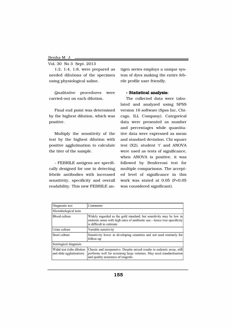
155
Benha M. J.
Vol. 30 No 3 Sept. 20131:2, 1:4, 1:8, were prepared as
needed dilutions of the specimen
using physiological saline.
Qualitative procedures were
carried-out on each dilution.
Final end point was determined
by the highest dilution, which was
positive.
Multiply the sensitivity of the
test by the highest dilution with
positive agglutination to calculate
the titer of the sample.
- FEBRILE antigens are specifi-
cally designed for use in detecting
febrile antibodies with increased
sensitivity, specificity and overall
readability. This new FEBRILE an-
tigen series employs a unique sys-
tem of dyes making the entire feb-
rile profile user friendly.
- Statistical analysis:- Statistical analysis:
The collected data were tabu-
lated and analyzed using SPSS
version 16 software (Spss Inc, Chi-
cago, ILL Company). Categorical
data were presented as number
and percentages while quantita-
tive data were expressed as mean
and standard deviation. Chi square
test (X2), student ‘t’ and ANOVA
were used as tests of significance,
when ANOVA is positive, it was
followed by Benferroni test for
multiple comparisons. The accept-
ed level of significance in this
work was stated at 0.05 (P<0.05
was considered significant).
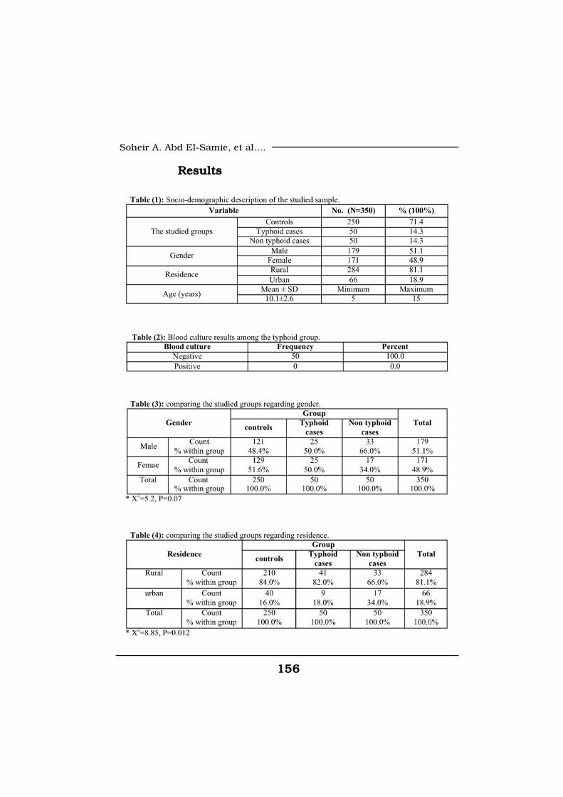
156156
Soheir A. Abd El-Samie, et al....
ResultsResults
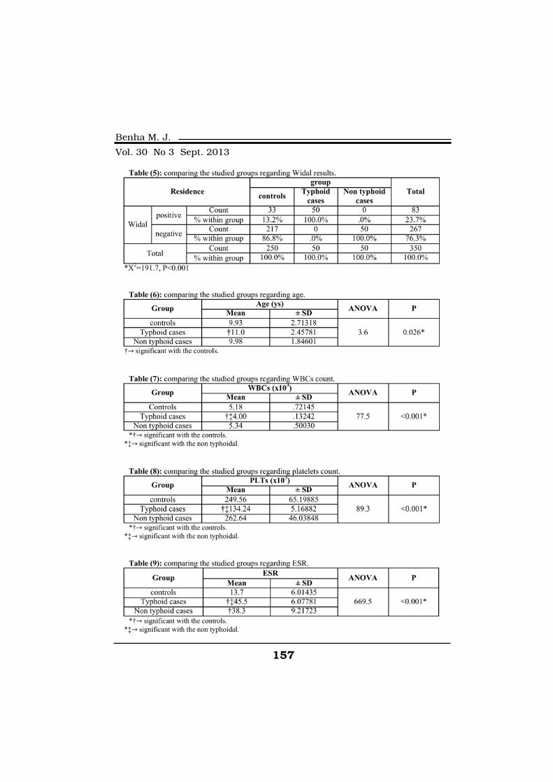
157
Benha M. J.
Vol. 30 No 3 Sept. 2013

158158
Soheir A. Abd El-Samie, et al....
DiscussionDiscussionThe value of the Widal test for
the diagnosis of typhoid fever has
been debated for as many years as
it has been available. The defini-
tive diagnosis of typhoid fever re-
quires the isolation of Salmonella
enterica subspecies enterica sero-
var Typhi from the patient by cul-
tures of blood. Our study was con-
ducted on 350 children from 5 to
15 years in Benha region Qalubia
during the period from January
2013 to October 2013. In our
study 23.7% gave positive widal
test, but 0% gave positive blood
culture because of their antibio-
tics intake. We found that typhoid
fever is more common in male
(51.1%) than female (48.9%)
which shows high resistance fe-
males in addition to the behavior
of male students in exposure to
injury-causing factors compared
to females. In our study, we found
that the control group gave
(13.2%) positive widal test with ti-
tre (1/80) typhoidal group, (100%)
gave positive widal test with titre
ranges from (1/80: 1/320). In the
present study, number of platelets
and white blood cells decrease in
typhoidal cases, while ESR values
increase in typhoidal cases. The
Widal test can be used as a com-
plimentary serological diagnostic
tool as and when it is required.
However, the importance of the
blood culture in the typhoid cases
cannot be ignored, as the antibio-
tic susceptibility testing of the iso-
lated strains is equally important).
ReferencesReferencesAcharya V.I., Lowe C.U., Tha-Acharya V.I., Lowe C.U., Tha-
pa R., Gurubacharya V.L.,pa R., Gurubacharya V.L.,
Shrestha M.B., Cadoz M.,Shrestha M.B., Cadoz M.,
Schulz D., Armand J., BrylaSchulz D., Armand J., Bryla
D.A., Trollfors B., Cramton T.,D.A., Trollfors B., Cramton T.,
Schneerson R. and Robbins J.B.Schneerson R. and Robbins J.B.
(1987):(1987): Prevention of typhoid fever
in Nepal with the Vi capsular poly-
saccharide of Salmonella typhi. A
preliminary report. New England
Journal of Medicine, 317: 4-1101.
Buck R.L., Escamilla J., San-Buck R.L., Escamilla J., San-
galang R.P., Cabanban A.B.,galang R.P., Cabanban A.B.,
Santiago L.T., Ranoa C.P. andSantiago L.T., Ranoa C.P. and
Cross J.H. (1987):Cross J.H. (1987): Diagnostic val-
ue of single, pre-treatment Widal
test in suspected enteric fever cas-
es in the Philippines. Trans. R.
Soc. Trop. Med. Hyg, 81: 871- 873.
Gasem M.H., Dolmans W.M.,Gasem M.H., Dolmans W.M.,
Isbandrio B.B., Wahyono H.,Isbandrio B.B., Wahyono H.,
Keuter M. and DjokomoeljantoKeuter M. and Djokomoeljanto
159
Benha M. J.
Vol. 30 No 3 Sept. 2013R. (1995):R. (1995): Culture of Salmonella
typhi and Salmonella paratyphi-
from blood and bone marrow in
suspected typhoid fever. Trop
Geog Med, 47: 7-164.
Giannella Ra. (2010):Giannella Ra. (2010): Infec-
tious enteritis and proctocolitis
and bacterial food poisoning. In:
Feldman, M; Friedman, L.S. and
Brandt, L.J. Sleisenger & Ford-
tran's Gastrointestinal and Liver
Disease.9th ed. Philadelphia, Pa:
Saunders Elsevier, 107.
Gillespie S. (2003):Gillespie S. (2003): Salonella
infection. In: Cook, G.C. andZum-
la, A. Manson's Tropical Diseases,
21st ed. London, UK: Elsevier Sci-
ence, Health Science Division,
937- 947.
Hoffman S.L., Edelman D.C.,Hoffman S.L., Edelman D.C.,
Punjabi N.H., Lesmana M.,Punjabi N.H., Lesmana M.,
Cholid A., Sundah S. and Hara-Cholid A., Sundah S. and Hara-
hap J. (1986):hap J. (1986): Bone marrow aspi-
rate culture superior to streptoki-
nase clot culture and 8 ml 1:10
blood-to-broth ratio blood culture
for diagnosis of typhoid fever. The
American Journal of Tropical Med-
icine and Hygiene, 35: 9-836.
Isaza R., Garner M. and Jacob-Isaza R., Garner M. and Jacob-
son E. (2000): son E. (2000): Proliferative osteoar-
thritis and osteoarthrosis in 15
snakes. J Zoo Wildl Med, 31: 7-20.
Ivanoff B., Martel J.L., HiretIvanoff B., Martel J.L., Hiret
D., Parado C., Normier G. andD., Parado C., Normier G. and
Dussourd D’Hinterland L.Dussourd D’Hinterland L.
(1960): (1960): Experimental infection by
“Salmonella typhi-murium”: pro-
tective role of homologus riboso-
mal extracts in calves. Ann Micro-
biol, Paris, 131: 163-74.
Jagdish Kumar K., KrishnaJagdish Kumar K., Krishna
Kumar H.C., Manjunath V.G.,Kumar H.C., Manjunath V.G.,
Umesh L. and Narayanappa D.Umesh L. and Narayanappa D.
(2010):(2010): Atypical typhoid fever in a
young child, 2(3):190-192.
Kalayci C., Karacadag S. andKalayci C., Karacadag S. and
Kansu, E. (1987):Kansu, E. (1987): Typhoid fever -
a report of 90 cases. Infeksiyon-
Dergisi, 3: 89-91.
Pearson R.D. and GuerrantPearson R.D. and Guerrant
R.L. (2000):R.L. (2000): Enteric fever and oth-
er causes of abdominal symptoms
with fever. In: Mandell, G.L; Ben-
nett, J.E; Dolin, R. Principles and
practice of infectious diseases, 5th
ed. Churchill Livingstone, New
York, 1136-1150.
Shirakawa T., Acharya B.,Shirakawa T., Acharya B.,
160160
Soheir A. Abd El-Samie, et al....
Kinoshita S., Kumagai S., GotohKinoshita S., Kumagai S., Gotoh
A. and Kawabata M. (2003): A. and Kawabata M. (2003): De-
creased susceptibility to fluoroqui-
nolones and gyrA gene mutation
in the Salmonella entericaserovar-
Typhi and Paratyphi A isolated in
Kathmandu, Nepal.
Soewandojo E., Suharto U.,Soewandojo E., Suharto U.,
Hadi U., Frans P. and PrihartiniHadi U., Frans P. and Prihartini
E. (1998):E. (1998): Comparative results
between bone marrow culture and
blood culture in the diagnosis of
typhoid fever. Medical Journal of
Indonesia, 7(S1): 209.
Wain J., Diep T.S., Ho V.A.,Wain J., Diep T.S., Ho V.A.,
Walsh A.M., Hoa T.T.N., ParryWalsh A.M., Hoa T.T.N., Parry
C.M. and White N.J. (1998):C.M. and White N.J. (1998):
Quantitation of bacteria in blood
of typhoid fever patients and rela-
tionship between counts and clini-
cal features, transmissibility, and
antibiotic resistance. Journal of
Clinical Microbiology, 36: 7-1683.
Willke A.T.H., Sozen K., GultanWillke A.T.H., Sozen K., Gultan
H., Kurt and Balik I. Tifo. (1988):H., Kurt and Balik I. Tifo. (1988):
100 hastaninklinik, laboratuvar-
vetedaviyonundendegerlendirilmes
i. Ankara, Tip Bulteni, 10: 53-62.

161
Benha M. J.
Vol. 30 No 3 Sept. 2013
Published byPublished by
Benha Faculty of MedicineBenha Faculty of Medicine
BIOLOGICAL AND SEROLOGICALBIOLOGICAL AND SEROLOGICALSTUDIES IN SCHOOL CHILDRENSTUDIES IN SCHOOL CHILDRENTO EVALUATE WIDAL TEST ASTO EVALUATE WIDAL TEST AS
A SINGLE DIAGNOSTIC ONEA SINGLE DIAGNOSTIC ONEIN TYPHOID FEVERIN TYPHOID FEVER
Soheir A. Abd El-Samie MD, Ibrahim M. Rageh MD,Soheir A. Abd El-Samie MD, Ibrahim M. Rageh MD,Mohammed E. Metwally Ph.DMohammed E. Metwally Ph.D
and Fatma A. M. Mohammed B.Scand Fatma A. M. Mohammed B.Sc
BENHAMEDICALJOURNAL
REPRINT
Volume 30 Number 3Sept. 2013
161
Benha M. J.
Vol. 30 No 3 Sept. 2013
ACUTE APPENDICITIS IN THE OVER-FIFTYACUTE APPENDICITIS IN THE OVER-FIFTYAGE GROUPAGE GROUP
Hasan I. Fadeel MD*, Mohan Patro MD*, Farag M. Mikaelasan I. Fadeel MD*, Mohan Patro MD*, Farag M. MikaelMD*, Othman issa Mohamed Abou Bakr MD*MD*, Othman issa Mohamed Abou Bakr MD*
and Yousef Thabet Ali MD**and Yousef Thabet Ali MD***Department of Surgery, **Department of Anaesthesia,
Faculty of Medicine, Omar Al Mukhtar Unversity, El Beida, Libya.
AbstractAbstractAbstract:Abstract: Acute appendicitis in elderly patients (ie. Patients above
the age of 50 years) is rare but often associated with complications, in-creased morbidity and mortality.
Objective:Objective: The aim of our study is to review and analyze the diagnos-tic challenges, treatment and outcome of cases of acute appendicitis inelderly patients at Al Thowra Teaching hospital, El Beida, Libya between1st January 2005 to 31st December 2009, over a period of 5 years.
Method:Method: 24 patients of acute appendicitis in elderly patients were re-viewed in details about their presentation, treatment offered and out-come. Out of them 66.6% were males and 33.3% females. Majority ofpatients were in the age group of 50 to 70 years.
Results:Results: Clinical presentation varied. Specific investigations includ-ing CBP, US Scan of abdomen, Plain X-Ray of the abdomen, were donefor all cases. CT Scan was done in 2 cases of confused diagnosis. Earlyoperation was performed in 29.16% cases and in 70.83% cases the sur-gery was delayed for more than 24 hours.
The complications encountered in our study were Chest, Urinarytract & wound infection, prolonged ileus, MI. fecal fistula, wound dehis-cence, septicemia and death.
Conclusion: Conclusion: Acute appendicitis is less common among elderly peo-ple. Often elderly patients have associated co-morbid conditions. Com-plications are comparatively more. Early diagnosis and surgical inter-vention reduces the morbidity and mortality considerably.
Keywords:Keywords: Appendicitis in elderly, complications, management.
162162
Hasan I. Fadeel, et al....
IntroductionIntroductionAcute appendicitis occurs in
7%(1) of cases out of which 90%
cases are seen among children and
young adults, where as 10% cases
seen in elderly patients(2,26). The
presentation in elderly may be
atypical or confused, hence surgi-
cal treatment is often delayed(3,4).
Hence, complications of appendi-
citis specially perforation is more
commonly observed; which may
be due to delayed intervention, co-
morbid conditions, immunosup-
pression and poor defense mecha-
nism(3). Early diagnosis and
prompt surgical management can
reduce complications, morbidity
and mortality rate(5,6,7,8).
The aim of this study was to re-
view the diagnostic, therapeutic
management and outcome of acute
appendicitis in elderly patients.
Material and MethodsMaterial and MethodsWe have reviewed medical
records of patients over 50 years
age, who underwent appendecto-
my at El Thowra Teaching Hospi-
tal during 5 years period from 1st
January 2004 to 31st December
2009. Total 24 cases were collect-
ed and analyzed retrospectively in-
cluding demographic data, symp-
toms & signs, onset and duration
of presentation, investigations,
timing of surgery, surgical ap-
proach, hospital stay, morbidity
and mortality.
ResultsResultsOut of 1160 appendectomies
performed during the period 1st
January 2005 to 31st December
2009; 24 cases were above the age
of 50 years, which constitutes
2.07%. The mean age was 58.87
years of which 50% cases in 6th
decade, 41.6%. In 7th decade,
8.3% in 8th decade. Male Female
ratio was 2:1. (Table 1).
Clinical presentation was typi-
cal abdominal pain with shift to
Rt.I.F. in 14(58.33%), atypical
presentation in 41.66%, anorexia
in 54.1%, nausea & vomiting in
50%, constipation in 25%, fever
with chills & rigor in 33.33% of
cases. Tenderness was elicited in
87.5%, rebound tenderness &
guarding in 41.66, features of gen-
eralized peritonitis in 16.6%, mass
163
Benha M. J.
Vol. 30 No 3 Sept. 2013
Rt.IF in 20.8% of cases.
The interval between the onset
of symptoms and hospitalization
varied from few hours to one
week; where as 45.8% cases were
admitted within 24 hours.
On investigation, leukocytosis
(>11,000 cumm) was found in 50%
cases. Chest X-Ray was done in all
cases. Abdominal X-Ray was done
in 41.66% cases. US Scan of ab-
domen was done in 66.6% cases
but was informative only in 50% of
them. CT Scan of abdomen was done
in 8.3% cases, and the diagnosis
was 100% accurate. (Table 2).
54.1% cases had associated co-
morbid conditions like Diabetes
mellitus in 29.1%, Hypertension
in 16.6% and Cardiac disease in
12.5% of cases. Only 7(29.16%)
cases were operated early. In rest,
the surgery was delayed due to
difficulty in diagnosis.
The morbidity rate was 37.5%.
The complications encountered
were appendicular mass and/or
abscess in 33.33%, perforation in
29.16%, localized peritonitis in
nearly 33.33%, generalized perito-
nitis in nearly 30% of cases. Nor-
mal appendix with mass in termi-
nal ileum (Crohn's disease) was
found in 1 case (4.16%); which
was also operated.
Post operative complications
encountered were wound infection
in 4(16.6%), chest infection in 4
(1606%), urinary tract infection in
2(8.3%), prolonged paralytic ileus
in 5(20.8%), MI in 1(4.16%), fecal
fistula in 1(4.16%), wound dehis-
cence in 1(4.16%).
Hospital stay varied from 5-15
days. 37.5% of cases over stayed,
more than one week. One patient
(4.1%) died following septicemia,
which got worsened due to asso-
ciated co-morbid conditions.
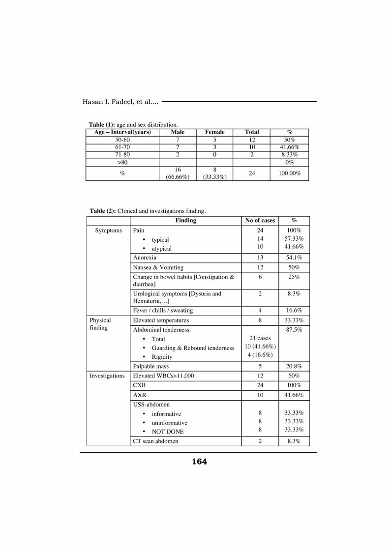
164164
Hasan I. Fadeel, et al....
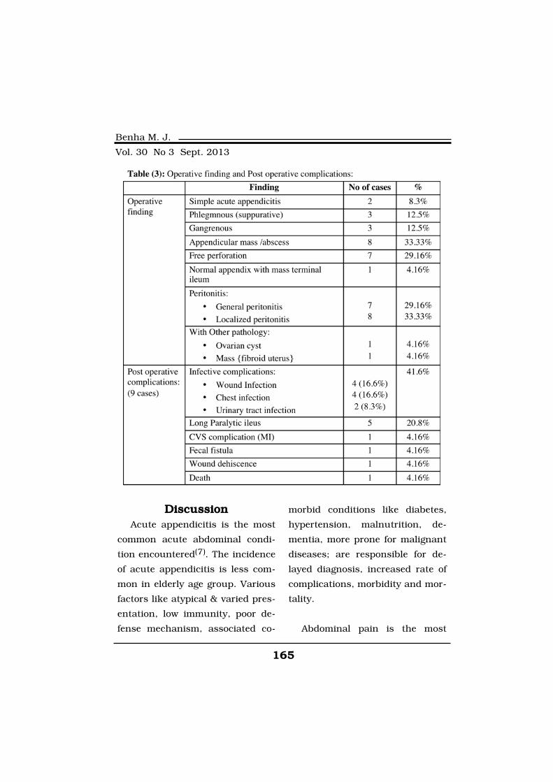
165
Benha M. J.
Vol. 30 No 3 Sept. 2013
DiscussionDiscussionAcute appendicitis is the most
common acute abdominal condi-
tion encountered(7). The incidence
of acute appendicitis is less com-
mon in elderly age group. Various
factors like atypical & varied pres-
entation, low immunity, poor de-
fense mechanism, associated co-
morbid conditions like diabetes,
hypertension, malnutrition, de-
mentia, more prone for malignant
diseases; are responsible for de-
layed diagnosis, increased rate of
complications, morbidity and mor-
tality.
Abdominal pain is the most
166166
Hasan I. Fadeel, et al....
common symptom of appendicitis(9). Classical history of shifting of
pain from umbilical region to Rt.IF
was seen in 50% of cases(7) and
20% of elderly patients presented
with anorexia, fever, right lower
quadrant pain and leukocytosis(8).
Abdominal tenderness is less lo-
calized in case of elderly patients(3). Elderly patients with acute ap-
pendicitis with peritonitis may not
have classical findings of rebound
tenderness and rigidity(5). In eld-
erly patients, the picture may be
confusing and may not have typi-
cal signs, symptoms and leukocy-
tosis in nearly 50% of cases(8).
Leukocytosis is also seen in 70%
of other abdominal conditions(10),
but serial repetition of leucocyte
count may help and increase
specificity. But there may be a ini-
tial fall of in leucocyte count in
case of perforation(11). Chest X-
Ray and abdominal X-Ray may
help to exclude other emergency
conditions. US Scan and CT Scan
specially Helical CT is more help-
ful to diagnose the confused and
difficult cases. CT Scan can ex-
clude other causes and more spe-
cific than US Scan of abdomen
(4,12,13,14,15). But routine use of
CT Scan in all cases in developing
countries is not cost effective, may
further delay the management
and thus increase the operative
risk(16,17,18).
Laparoscopic appendectomy is
associated with less pain, wound
infection and rapid recovery(19,20), variable results seen with
some surgeons(21,22). Patients
whose appendectomy was com-
pleted laparoscopically were
younger and less likely to have
perforation and abscess. The post
operative hospitalization of these
patients was shorter, reflecting
the selection of less complicated
cases for this subgroup(23). Late
presentation of patients to hospi-
tal, may be due to family negligence,
psychological factors, taking na-
tive treatment initially, consider-
ing other common diseases like
malignancy, pancreatitis, biliary
diseases, inflammatory bowel dis-
eases, bowel obstruction etc; may
delay in surgical management(24).
Local & generalized peritonitis
were seen in 2/3rd of our patients;
167
Benha M. J.
Vol. 30 No 3 Sept. 2013
mainly following perforation and
neglected appendicitis. The inci-
dence of perforation in elderly pa-
tients is progressively higher with
age and mainly due to delayed
presentation, extreme age, abnor-
mal location of appendix etc(1,24,25,26).
The morbidity rate in acute ab-
dominal conditions increases with
age, as was seen in 15% over the
age of 50 years and more than
70% over the age of 80 years(27).
Similarly is also 5% higher in eld-
erly patients, usually due to de-
layed diagnosis(1).
ConclusionConclusionAcute appendicitis is less com-
mon in elderly patients but often
associated with high morbidity and
mortality. All efforts should be
made for early and correct diagno-
sis, so that early surgical interven-
tion shall improve the out come.
ReferencesReferences1. Hardin D.M. Jr. (1999):1. Hardin D.M. Jr. (1999):
Acute appendicitis: review and up-
date. Am Fam Physician. Nov 1;
60(7): 2027-34.
2. Addiss D.G., Shaffer N.,2. Addiss D.G., Shaffer N.,
Fowler B.S. and Tauxe R.V.Fowler B.S. and Tauxe R.V.
(1990):(1990): The epidemiology of ap-
pendicitis and appendectomy in
the United States. Am J Epidemi-
ol; 132: 910-925
3. Kraemer M., Franke C.,3. Kraemer M., Franke C.,
Ohmann C. and Yang Q. (2000):Ohmann C. and Yang Q. (2000):
Acute Abdominal Pain Study
Group. Acute appendicitis in late
adulthood: incidence, presenta-
tion, and outcome. Results of a
prospective multicenter acute ab-
dominal pain study and a review
of the literature. Langenbecks
Arch Surg. Nov; 385(7): 470-81.
4. Shelton T., McKinlay R.4. Shelton T., McKinlay R.
and Schwartz R.W. (2003): and Schwartz R.W. (2003): Acute
appendicitis: current diagnosis
and treatment. Curr Surg. Sep-
Oct; 60(5): 502-5.
5. Wilcox R.T. and Traverso5. Wilcox R.T. and Traverso
L.W. (1997):L.W. (1997): Have the evaluation
and treatment of acute appendici-
tis changed with new technology?
Surg Clin North Am; 77: 1355-70.
6. Horattas M.C., Guyton6. Horattas M.C., Guyton
D.P. and Wu D. (1990):D.P. and Wu D. (1990): A reap-
168168
Hasan I. Fadeel, et al....
praisal of appendicitis in the eld-
erly. Am J Surg.; 160: 291-293
7. Liu C.D. and McFadden7. Liu C.D. and McFadden
D.W. (1997):D.W. (1997): Acute abdomen and
appendix. In: Greenfield LJ, et al.,
eds. Surgery: scientific principles
and practice. 2d ed. Philadelphia:
Lippincott-Raven, 1246-61.
8. E. David Bryan. (2008): 8. E. David Bryan. (2008): Ab-
dominal Pain in Elderly Persons.
Emergency Medicine.
9. Schwartz S.I. (1994):9. Schwartz S.I. (1994): Ap-
pendix. In: Schwartz SI, ed. Princi-
ples of surgery. 6th ed. New York:
McGraw Hill, 1307-18.
10. Calder J.D. and Gajraj H.10. Calder J.D. and Gajraj H.
(1995):(1995): Recent advances in the di-
agnosis and treatment of acute
appendicitis. Br J Hosp Med; 54:
129-33.
11. Graffeo C.S. and Counsel-11. Graffeo C.S. and Counsel-
man F.L. (1996):man F.L. (1996): Appendicitis.
Emerg Med Clin North Am; 14:
653-71.
12. Rao P.M., Feltmote C.M.,12. Rao P.M., Feltmote C.M.,
Rhea J.T., Schulick A.H. andRhea J.T., Schulick A.H. and
Novelline R.A. (1999):Novelline R.A. (1999): Helical
computed tomography in differen-
tiating appendicitis and acute gy-
necologic conditions. Obstet Gyne-
col; 93: 417-21.
13. Esses D., Birnbaum A.,13. Esses D., Birnbaum A.,
Bijur P., et al. (2004):Bijur P., et al. (2004): Ability of
CT to alter decision making in eld-
erly patients with acute abdominal
pain. Am J Emerg Med. Jul; 22(4):
270-2.
14. Hustey F.M., Meldon14. Hustey F.M., Meldon
S.W., Banet G.A., et al. (2005):S.W., Banet G.A., et al. (2005):
The use of abdominal computed
tomography in older ED patients
with acute abdominal pain. Am J
Emerg Med. May; 23(3): 259-65.
15. Rao P.M., Rhea J.T., No-15. Rao P.M., Rhea J.T., No-
velline R.A., McCabe C.J., Law-velline R.A., McCabe C.J., Law-
rason J.N., Berger D.L., et al.rason J.N., Berger D.L., et al.
(1997): (1997): Helical CT technique for
the diagnosis of appendicitis:
prospective evaluation of a fo-
cused appendix CT examination.
Radiology; 202: 139-44.
16. Stroman D.L., Bayouth C.V.,16. Stroman D.L., Bayouth C.V.,
Kuhn J.A., et al. (1999):Kuhn J.A., et al. (1999): The role
of the computed tomography in
169
Benha M. J.
Vol. 30 No 3 Sept. 2013
the diagnosis of acute appendici-
tis. Am J Surg.; 178: 485-489.
17. Rao P.M., Rhea J.T., No-17. Rao P.M., Rhea J.T., No-
velline R.A., Mostafavi A.A. andvelline R.A., Mostafavi A.A. and
McCabe C.J. (1998):McCabe C.J. (1998): Effect of
computed tomography of the ap-
pendix on treatment of patients
and use of hospital resources. N
Engl J Med.; 338: 141-146.
18. Peltokallio P. and Tykka H.18. Peltokallio P. and Tykka H.
(1981):(1981): Evolution of the age distri-
bution and mortality of acute ap-
pendicitis. Arch Surg.; 116: 153-156.
19. Frazee R.C., Roberts J.W.,19. Frazee R.C., Roberts J.W.,
Symmonds R.E., et al. (1994):Symmonds R.E., et al. (1994): A
prospective randomized trial com-
paring open versus laparoscopic
appendectomy. Ann Surg.; 219:
725-731
20. Golub R., Siddiqui F. and20. Golub R., Siddiqui F. and
Pohl D. (1998):Pohl D. (1998): Laparoscopic ver-
sus open appendectomy: a metaa-
nalysis. J Am Coll Surg.; 186:
545-553.
21. Fallahzadeh H. (1998):21. Fallahzadeh H. (1998):
Should a laparoscopic appendectomy
be done? Am Surg.; 64: 231-233.
22. Bonanni F., Reed III J.,22. Bonanni F., Reed III J.,
Hartzell G., et al. (1994):Hartzell G., et al. (1994): Lapar-
oscopic versus conventional ap-
pendectomy. J Am Coll Surg.;
179: 273-278.
23. Khalili T.M., Hiatt J.R.,23. Khalili T.M., Hiatt J.R.,
Savar A., Lau C., Phillips E.H.Savar A., Lau C., Phillips E.H.
and Margulies D.R. (1999):and Margulies D.R. (1999): Per-
forated appendicitis is not a con-
traindication to laparoscopy. Am
Surg.; 65: 965-967.
24. Temple C.L., Huchcroft24. Temple C.L., Huchcroft
S.A. and Temple W.J. (1995):S.A. and Temple W.J. (1995):
The natural history of appendicitis
in adults: a prospective study.
Ann Surg.; 221: 278-81.
25. Ricci M.A., Trevisani M.F.25. Ricci M.A., Trevisani M.F.
and Beck W.C. (1991):and Beck W.C. (1991): Acute ap-
pendicitis: a five year review. Am
Surg.; 57: 301-5.
26. Guidry S.P. and Poole26. Guidry S.P. and Poole
G.V. (1994):G.V. (1994): The anatomy of ap-
pendicitis. Am Surg.; 60: 68-71.
27. deDombal F.T. (1994):27. deDombal F.T. (1994):
Acute abdominal pain in the elder-
ly. J Clin Gastroenterol; 19: 331-
335.
170170
Hasan I. Fadeel, et al....
Published byPublished by
Benha Faculty of MedicineBenha Faculty of Medicine
ACUTE APPENDICITIS IN THEACUTE APPENDICITIS IN THEOVER-FIFTY AGE GROUPOVER-FIFTY AGE GROUP
Hasan I. Fadeel MD, Mohan Patro MD, Farag M. MikaelHasan I. Fadeel MD, Mohan Patro MD, Farag M. MikaelMD, Othman issa Mohamed Abou Bakr MDMD, Othman issa Mohamed Abou Bakr MD
and Yousef Thabet Ali MDand Yousef Thabet Ali MD
BENHAMEDICALJOURNAL
REPRINT
Volume 30 Number 3Sept. 2013
171
Benha M. J.
Vol. 30 No 3 Sept. 2013
PHYSICAL INTIMATE PARTNER VIOLENCE:PHYSICAL INTIMATE PARTNER VIOLENCE:PREVALENCE, PREDICTORS,PREVALENCE, PREDICTORS,
AND HEALTH IMPACTAND HEALTH IMPACT
Nesrine Saad Farrag M.Sc, Nadia Abd El-Hamed MontasserNesrine Saad Farrag M.Sc, Nadia Abd El-Hamed MontasserMD, Farida Abdel Wahab El-Sayed MDMD, Farida Abdel Wahab El-Sayed MD
and Ghada El-Khawaga MDand Ghada El-Khawaga MDPublic Health Department, College of Medicine, Mansoura University, Egypt
AbstractAbstractBackground: Background: Intimate partner violence (IPV) is an important public
health issue with severe adverse consequences. In Egypt, it is difficult tomake precise comparisons between the prevalence rates of IPV due to differ-ent data collections methods; sampling methods; and different approachesto asking the same questions.
Aim: Aim: to determine the prevalence of physical IPV against women in Man-soura centre and find out the predictors of exposure to violence and the im-pact violence on women health.
Methods:Methods: An observational community-based study was conducted us-ing a structured questionnaire developed by the WHO. Simple random sam-ple was selected form primary health centers and included 758 ever marriedwomen aged 15-49 years.
Results:Results: Self-reported past-year and lifetime prevalence of physical vio-lence was 28.8% and 34.3%, respectively. Female risk factors for physicalIPV included low socioeconomic standard, low income, justifying wife beat-ing, and exposure to IPV in childhood while husbands risk factors were ex-posure to IPV in childhood, low education, physical fighting with other peo-ple, alcohol and drug abuse. Physical IPV has poor physical (p≤ 0.001) andmental (p≤ 0.001) health oucome.
Conclusion:Conclusion: physical IPV is a common phenomenon in Mansoura dis-trict and it has poor consequences on women health and this requires im-mediate attention of policymakers.
Keywords:Keywords: intimate partner violence, domestic violence, Mansoura, gen-der inequality.
172172
Nesrine Saad Farrag, et al...
IntroductionIntroductionA report of WHO highlights vio-
lence against women as a ‘global
health problem of epidemic pro-
portions’. The study finds that in-
timate partner violence is the
most common type of violence
against women, affecting 30% of
women worldwide(1). The reality
and threat of violence exists in
women’s everyday lives, affecting
their ability to participate in their
societies, affecting their potential
to engage fully in the affairs of
their communities. It limits wom-
en’s choices directly by destroying
women’s health, disrupting their
lives and constricting the scope of
their activity, and indirectly by
eroding their self confidence and
self-esteem. The recognition of vio-
lence as a major risk factor for
women’s ill-mental health must be
fully integrated into mental health
policy. Resources must be allocat-
ed for preventing violence against
women and mitigating its conse-
quences in order for the mental
health needs of women to be effec-
tively addressed(2,3). In Egypt, it
is difficult to make precise com-
parisons between the prevalence
rates reported in the studies con-
ducted for a number of reasons.
Depending on how survey ques-
tions are formulated, women may
give different responses as to
whether they have experienced
physical assault perpetrated by
their partners. Interpretations of
physical or other forms of abuse
may also differ across sociocultu-
ral contexts. There is considerable
variation in how abuse is defined
across these studies(4,5).
Subjects and MethodsSubjects and MethodsStudy locality: Mansoura cen-
ter is one of 18 centers in Dakah-
lia governorate. The estimated
population number in Mansoura
in 2011 was 973152 according to
information center in Dakahlia
health administration. Mansoura
center comprises Mansoura city
and its suburbs which are 39 vil-
lages. Four urban primary health
care centers were randomly select-
ed, two were selected from East
Mansoura district and two were
selected from West Mansoura dis-
trict. Eight rural family health
centers were randomly selected.
The selected centers of mansoura
city serve attendants of different
socioeconomic standards and pro-
vide different preventive and cura-
tive services.
173
Benha M. J.
Vol. 30 No 3 Sept. 2013
Study design:Study design: A observational
descriptive and analytic study was
conducted using a standard ques-
tionnaire designed by WHO for
studying violence against women
(questionnaire version 10, 2003).
Operational definitions of violence
used in the WHO Multi-country
Study on Women’s Health and Do-
mestic Violence against Women
were used. All women in the child
bearing period from 15 to 49 years
attending the selected health cen-
ters for any reason were eligible
except women who are too ill to
participate. The females in the age
group (15-49) represent about
26% of the population in Dakahlia
governorate. So, the size of the
population is (253019 females).
Prevalence of physical IPV during
the past 12 months was 20.4%.
Sample size was calculated using
EpiInfo verion6 and the minimum
required sample is 691. Our sam-
ple included 785 women. Women
were interviewed using the ques-
tionnaire after giving verbal con-
sent to participate in the study.
The interview was conducted with
each woman separately to ensure
privacy. The women decisions and
choices was be respected. For so-
cioeconomic standard assessment,
social score modified after Fahmy
& El-Sherbini, 1983 was used.
Mental health was assessed by
use of a self-reporting question-
naire of 20 questions (SRQ-20),
developed by WHO to screen for
emotional distress and validated
in many settings (WHO, 1994).
Chi square test and Fisher’s Exact
tests were used were used for
comparing qualitative data. Statis-
tically significant variables in the
bivariate analyses were entered
into the multivariate model, one at
a time. Final models are dis-
played.
ResultsResultsTable (1) showed that the study
included 758 ever married fe-
males, 47.9% of them were in ur-
ban residence and 21.5% were (il-
literate, read and write) while
6.6% received basic education,
40.8% received secondary
education, and 31.1% received
higher education. The working
females represented 41.6% of the
sample and 29.7%, 54.7%, 15.6%
of females had insufficient,
sufficient income, and can save
money respectively. The study
found that (260) 34.3% and (218)
28.8% of women included in the
174174
Nesrine Saad Farrag, et al...
study, reported that they had
been exposed to some form of
physical IPV since marriage and
during the past year respectively.
The women who ever exposed to
sever violence were (141) 18.6%.
The prevalence of physical vio-
lence during pregnancy was
22.3%. The prevalence of specific
form of this violence which is
punching or kicking in abdomen
while pregnant is 13.5%. The most
prevalent forms of physical IPV
was slapping (85%) followed by
being pushing or shoving (66%).
The severe acts of violence, such
as being kicked, dragged (26%),
choked, burned (11.2%), or threat-
ened by a weapon (7.7%) were
less common (data not shown in
table).
We compared women exposed
to current violence (216 women)
with those not exposed to any type
of IPV (242 women) to find predic-
tors of violence. Table (2) shows
Logistic regression analysis of
women related predictors of physi-
cal IPV. On logistic regression, it
was found that only socioeconom-
ic standard, attitude toward wife
beating, residence, income, and
exposure to IPV in childhood are
the most significant predictors of
physical IPV. It was found that fe-
males with very low socioeconomic
are 4.7 times more likely to be
abused (P=0.001), Females with
attitude justifying wife beating are
2.7 times more likely to be abused
(P=0.001), also rural residence
made females 2 times more liable
to be abused (P=0.001). On the
other hand, females who were not
exposed to IPV in childhood and
those with sufficient income are
0.15, 0.3 times less likely to be
abused (P≤0.001). Table (3) shows
Logistic regression analysis of
husband related predictors of
physical IPV. On logistic regres-
sion, it was found that only expo-
sure to IPV in childhood, educa-
tion, history of physical fighting,
and alcohol and drug abuse are
the most significant predictors of
physical IPV. It was found that fe-
males whose husbands were not
exposed to IPV in childhood, with
higher education, and those with
no history of physical fighting with
other people are 0.1 times less
likely to be abused (P≤0.001), and
females whose husbands were not
alcohol or drug abuser are 0.2
times less likely to be abused
(P=0.001).
175
Benha M. J.
Vol. 30 No 3 Sept. 2013
Table (4) illustrated self-
reported physical health status of
women exposed to current physi-
cal IPV and non abused women.
Poor health was reported by
29.8% of abused women while this
was reported by 14.5% of non
abused women (p≤0.001). Acute
pain was reported by 23.9% of
abused women while 11.6% of non
abused women reported acute
pain (p≤0.001). Among abused
group, 29.4% of women reported
many problems that may prevent
them from performing usual activ-
ities while 11.6% of non abused
women reported so (p≤0.001). Diz-
ziness was reported by 54.6% of
abused women in the last 4
months compared to 45.5% of non
abused women (p=.05). Vaginal
discharge was reported by 48.2%
of abused women in the last 4
weeks compared to 30.6% of non
abused women (p≤0.001). About
two thirds of abused women said
they had medical consultation in
past 4 weeks compared to 56.2%
of non abused women (p=.031).
Among abused group, 16.1% of
women were hospitalized for at
least one night compared to 6.6%
of non abused group (p=.001).
There was more emotional distress
in abused group compared to the
other group and this was estimat-
ed using self reported question-
naire (SRQ20). In the abused
group 45.4% of women reported
score (0-10), 54.6% reported score
(11-20), while in the non abused
group 77.3% of women reported
score (0-10), and 22.8% of women
reported score (11-20) (p≤0.001).
Among abused group, 24.3% of
women reported many problems
with memory while 15.7% of non
abused women reported many
problems (p≤0.001). Among
abused group, 10.6% of women
had thought of committing suicide
in the previous 4 weeks while only
.4% of non abused women report-
ed so (p≤0.001) and 12.8% of
women in abused group ever at-
tempted suicide compared to only
.4% of non abused women (p≤
0.001).

176176
Nesrine Saad Farrag, et al...
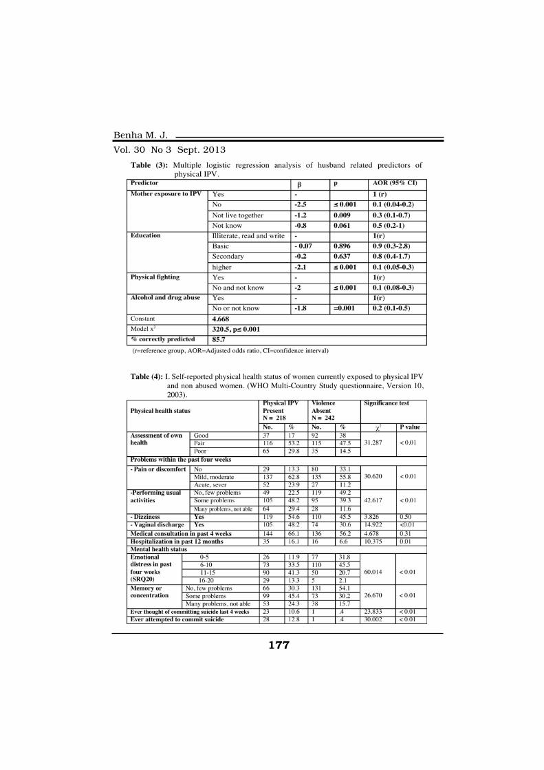
177
Benha M. J.
Vol. 30 No 3 Sept. 2013
178178
Nesrine Saad Farrag, et al...
DiscussionDiscussionEDHS-95 indicated that 34% of
women in this sample have ever
been beaten by their current hus-
band(6) while EDHS (2005) found
also that life time physical IPV
prevalence was 33.2%. both figure
are very proximate to our prevalence.
Our prevalence is higher than that
found by other studies(7,8,9)
which are only (11.1%), (29.9%)
and (20.5%) respectively for a life-
time prevalence of physical abuse.
Also our prevalence is higher than
that found by another study(10)
which found that physical IPV was
(40%) but the cause may be using
different questionnaire and the
sample that was taken from
Health Insurance Clinics in Alex-
andria that may over estimate the
problem. EDHS (2005) found that
the prevalence of physical violence
during pregnancy was 6.6% and
this figure is much lower than
that obtained by our result. Our
figures are higher than results of
population-based studies from
Canada, Chile and Nicaragua that
have found that 6-15% of ever-
partnered women have been phys-
ically abused during pregnancy,
usually by their partners(11,12).
Regarding the forms of violence,
our results are consistent with WHO
multicountry study which showed
that the most commonly experi-
enced acts of physical aggression
in most countries include being
slapped, having arms twisted, or
hair pulled. The more severe acts
of violence were less common(13).
The findings of this research as
well as many other studies sup-
port the view that poverty and its
associated stress are key contrib-
utors to intimate partner violence.
High levels of female empower-
ment seem to be protective against
intimate partner violence, but power
can be derived from many sources
such as education, income, and
community roles and not all of
these convey equal protection or
do so in a direct manner(14). Rural
women represented the larger part
of the women abused by any type
of violence. This could be ex-
plained through community-level
gender inequality (operationalized
as women’s autonomy, women’s
status, male patriarchal control,
and intimate partner violence).
Community-level gender inequali-
ty is associated with women’s own
experience of IPV(15). Women’s at-
titude to male dominance affects
179
Benha M. J.
Vol. 30 No 3 Sept. 2013
the level of their acceptance or tol-
erance of violence from their hus-
bands. Nearly all studies that
have included a variable on
witnessing interparental violence
have found this experience to be
a risk factor for women experi-
encing violence and men perpe-
trating physical IPV(16,17,18,20).
Husbands of abused women
were less educated. These finding
is in line with other studies which-
found that men with a higher edu-
cation were less likely to abuse their
wives physically(21,22). Alcohol
consumption has been positively as-
sociated with domestic violence in
several communities(19,23,24,25).
Findings from the WHO multi-
country study on women’s health
and domestic violence showed
also women whose partners were
involved in a fight with another
man in the past year experienced
higher levels of IPV than those
with partners who did not fight(25). A history of antisocial person-
ality and alcohol abuse among
men, may account for all or part
of this association(26).
Vives-Cases et al. found that
physical IPV is associated with a
greater likelihood of poor self-
perceived health(27). These results
are in agreement with the results
of WHO multicountry study. For
most specific health problems, in
most places, significant associa-
tions existed between reported vio-
lence and self-reported specific
health problems as women with
lifetime experiences of physical
IPV were significantly more likely
to report poor health, and that
within the past 4 weeks they had
experienced difficulties with daily
activities, pain, memory loss, diz-
ziness, and vaginal discharge(2).
The current study showed sig-
nificant association between IPV
and poor mental health. The study
showed that women who experi-
enced current IPV were more likely
to report emotional distress in the
past 4 weeks than non abused
women and this was measured by
SRQ20. Also IPV was significantly
associated with memory problems,
suicidal thoughts in the past 4
weeks, and suicidal attempts.
Physical IPV was associated with a
greater likelihood of psychological
distress(27). Another study found
that 10.4% of the physically abused
women had attempted suicide at least
180180
Nesrine Saad Farrag, et al...
once(19). Intimate partner violence
was among the most consistent
risk factors for suicide attempts
among women who participated in
WHO multicountry study(3).
ConclusionConclusionand Recommendationsand RecommendationsPhysical IPV is a common hid-
den problem. Those having the
least resources are most affected.
This situation requires rapid at-
tention at all levels of societal
organization, by policymakers,
political stakeholders, and profes-
sionals. Policy initiatives are need-
ed, as are legal actions, to crimi-
nalize men’s violence against
women. Basic education needs to
be made available for both girls
and boys, with special attention
placed on female education. Gen-
der equality teaching and training
should be included at different
levels in the school curriculum.
Mass media involvement is neces-
sary to create a debate on such
gender discrimination practices
and to encourage women’s em-
powerment in society.
ReferencesReferences1. WHO (2013): 1. WHO (2013): WHO report
highlights violence against women
as a ‘global health problem of epi-
demic proportions’ available at
http://www.who.int/mediacentre
/news/releases/2013/violence_
against_women_20130620/en/
2. Ellsberg M., Jansen H.A.,2. Ellsberg M., Jansen H.A.,
Heise L., Watts C.H., Garcia-Heise L., Watts C.H., Garcia-
Moreno C. (2008):Moreno C. (2008): WHO Multi-
country Study on Women’s Health
and Domestic Violence against
Women Study Team Collabora-
tors. WHO.
3. Devries K., Watts C., Yo-3. Devries K., Watts C., Yo-
shihama M., Kiss L., Schraibershihama M., Kiss L., Schraiber
L.B., Deyessa N. and Garcia-L.B., Deyessa N. and Garcia-
Moreno C. (2011): Moreno C. (2011): Violence
against women is strongly asso-
ciated with suicide attempts: evi-
dence from the WHO multi-
country study on women’s health
and domestic violence against
women. Social science & medi-
cine, 73(1), 79-86.
4. Boy A. and Kulczycki A.4. Boy A. and Kulczycki A.
(2008):(2008): What we know about inti-
mate partner violence in the Mid-
dle East and North Africa. Vio-
lence Against Women, 14(1), 53-
70.
5. Garcia-Moreno C. and5. Garcia-Moreno C. and
181
Benha M. J.
Vol. 30 No 3 Sept. 2013
Watts C. (2011):Watts C. (2011): Violence against
women: an urgent public health
priority. Bull World Health Organ.
1; 89(1):2. Retrieved May 25, 2011
from http://www.who.int/bulletin/
volumes/89/1/10-085217.pdf.
6. Diop-Sidibé N., Campbell6. Diop-Sidibé N., Campbell
J.C. and Becker S. (2006):J.C. and Becker S. (2006): Do-
mestic violence against women in
Egypt—wife beating and health
outcomes. Social Science & Medi-
cine, 62(5), 1260-1277.
7. Hassan F., Refaat A. and7. Hassan F., Refaat A. and
El Defrawi M. (2000):El Defrawi M. (2000): Domestic
violence against women in an ur-
ban area: Ismailia, Egypt. Egyp-
tian Journal of Psychiatry, 22.
8. Habib S.R., Abdel Azim8. Habib S.R., Abdel Azim
E.K., Fawzy I.A., Kamal N.N.E.K., Fawzy I.A., Kamal N.N.
and El Sherbini A.M. (2011):and El Sherbini A.M. (2011):
Prevalence and Effects of Violence
Against Women in a Rural Com-
munity in Minia Governorate,
Egypt. Journal of forensic scienc-
es, 56(6), 1521-1527.
9. Alsergany A.I., Abdul Aziz9. Alsergany A.I., Abdul Aziz
A., Mohammed D.H., IbrahimA., Mohammed D.H., Ibrahim
H.M., Kamel M.H. and IsmailH.M., Kamel M.H. and Ismail
M.A. (2012):M.A. (2012): Domestic violence
against married women in a rural
community: family based study-
Ismailia- Egypt. Eguptian journal
of community medicine, 30(3), 41-
50.
10. El Maqsoud S.A., Dab-10. El Maqsoud S.A., Dab-
bous N. and Nazel M.A. (2011):bous N. and Nazel M.A. (2011):
O2-3.2 Domestic violence against
women in Alexandria, Egypt: a de-
veloping country perspective.
Journal of Epidemiology and Com-
munity Health, 65(Suppl 1), A23-
A23.
11. Johnson H. (1996):11. Johnson H. (1996): Dan-
gerous domains: Violence against
women in Canada(No. s 50, p. 52).
Toronto: Nelson Canada.
12. Ellsberg M., Peña R., Her-12. Ellsberg M., Peña R., Her-
rera A., Liljestrand J. and Wink-rera A., Liljestrand J. and Wink-
vist A. (2000):vist A. (2000): Candies in hell:
women’s experiences of violence in
Nicaragua. Social Science & Medi-
cine, 51(11), 1595-1610.
13. Hindin M.J., Kishor S.13. Hindin M.J., Kishor S.
and Ansara D.L. (2008):and Ansara D.L. (2008): Intimate
partner violence among couples in
10 DHS countries: Predictors and
health outcomes. Macro Interna-
tional Incorporated.
14. Jewkes R. (2002):14. Jewkes R. (2002): Intimate

182182
Nesrine Saad Farrag, et al...
partner violence: causes and pre-
vention. The Lancet, 359(9315),
1423-1429.
15. Pallitto C.C. and O’Campo15. Pallitto C.C. and O’Campo
P. (2005):P. (2005): Community level effects
of gender inequality on intimate
partner violence and unintended
pregnancy in Colombia: testing
the feminist perspective. Social
Science & Medicine, 60(10), 2205-
2216.
16. Renner L.M. and Slack16. Renner L.M. and Slack
K.S. (2006):K.S. (2006): Intimate partner vio-
lence and child maltreatment: Un-
derstanding intra-and intergener-
ational connections. Child Abuse
& Neglect, 30(6), 599-617.
17. Jeyaseelan L., Kumar S.,17. Jeyaseelan L., Kumar S.,
Neelakantan N., Peedicayil A.,Neelakantan N., Peedicayil A.,
Pillai R. and Duvvury N. (2007):Pillai R. and Duvvury N. (2007):
Physical spousal violence against
women in India: some risk factors.
Journal of biosocial science, 39(5),
657.
18. Ansara D.L. and Hindin18. Ansara D.L. and Hindin
M.J. (2009):M.J. (2009): Perpetration of Inti-
mate Partner Aggression by Men
and Women in the Philippines
Prevalence and Associated Fac-
tors. Journal of Interpersonal Vio-
lence, 24(9), 1579-1590.
19. Ragheb K., Ismail R., At-19. Ragheb K., Ismail R., At-
tia H., Wahab M., El Missiry A.tia H., Wahab M., El Missiry A.
and Husein R. (2009):and Husein R. (2009): Violence
Against Women: Prevalence and
Psychiatric Consequences. Egypt.
Journal of Psychiatry, 29 (1), 15-25.
20. Roberts A.L., Gilman20. Roberts A.L., Gilman
S.E., Fitzmaurice G., DeckerS.E., Fitzmaurice G., Decker
M.R. and Koenen K.C. (2010):M.R. and Koenen K.C. (2010):
Witness of intimate partner vio-
lence in childhood and perpetra-
tion of intimate partner violence in
adulthood. Epidemiology (Cam-
bridge, Mass.), 21(6), 809.
21. Ali T.S., Asad N., Mogren21. Ali T.S., Asad N., Mogren
I. and Krantz G. (2011):I. and Krantz G. (2011): Intimate
partner violence in urban Paki-
stan: prevalence, frequency, and
risk factors. International journal
of women's health, 3, 105.
22. Babu B.V. and Kar S.K.22. Babu B.V. and Kar S.K.
(2010):(2010): Domestic violence in East-
ern India: factors associated with
victimization and perpetration.
Public Health, 124(3), 136-148.
23. Krishnan S. (2005):23. Krishnan S. (2005): Gen-
der, caste, and economic inequali-
ties and marital violence in rural
183
Benha M. J.
Vol. 30 No 3 Sept. 2013
South India. Health care for wom-
en international, 26(1), 87-99.
24.24. Xu X., Zhu F., O'CampoXu X., Zhu F., O'Campo
P., Koenig M.A., Mock V. andP., Koenig M.A., Mock V. and
Campbell J. (2005):Campbell J. (2005): Prevalence of
and risk factors for intimate part-
ner violence in China. American
Journal of Public Health, 95(1),
78-85.
25. Abramsky T., Watts C.H.,25. Abramsky T., Watts C.H.,
Garcia-Moreno C., Devries K.,Garcia-Moreno C., Devries K.,
Kiss L., Ellsberg M. and Heise L.Kiss L., Ellsberg M. and Heise L.
(2011):(2011): What factors are associat-
ed with recent intimate partner vi-
olence? findings from the WHO
multi-country study on women's
health and domestic violence.
BMC Public Health, 11(1), 109.
26. Fargo J.D. (2009):26. Fargo J.D. (2009): Path-
ways to Adult Sexual Revictimiza-
tion Direct and Indirect Behavioral
Risk Factors Across the Lifespan.
Journal of interpersonal violence,
24(11), 1771-1791.
27. Vives-Cases C., Torrubia-27. Vives-Cases C., Torrubia-
no-DomÍnguez J., Escribà-Agüirno-DomÍnguez J., Escribà-Agüir
V., Ruiz-Pérez I., Montero-PiñarV., Ruiz-Pérez I., Montero-Piñar
M.I. and Gil-González D. (2011):M.I. and Gil-González D. (2011):
Social determinants and health ef-
fects of low and high severity inti-
mate partner violence. Annals of
epidemiology, 21(12), 907-913.

184184
Nesrine Saad Farrag, et al...
PHYSICAL INTIMATE PARTNERPHYSICAL INTIMATE PARTNERVIOLENCE: PREVALENCE,VIOLENCE: PREVALENCE,
PREDICTORS, AND HEALTH IMPACTPREDICTORS, AND HEALTH IMPACT
Nesrine Saad Farrag M.Sc, Nadia Abd El-Hamed Montas-Nesrine Saad Farrag M.Sc, Nadia Abd El-Hamed Montas-ser MD, Farida Abdel Wahab El-Sayed MDser MD, Farida Abdel Wahab El-Sayed MD
and Ghada El-Khawaga MDand Ghada El-Khawaga MD
BENHAMEDICALJOURNAL
REPRINT
Published byPublished by
Benha Faculty of MedicineBenha Faculty of Medicine Volume 30 Number 3Sept. 2013
185
Benha M. J.
Vol. 30 No 3 Sept. 2013
IntroductionIntroductionLiver tumours represent about
11.75% of gastrointestinal malig-
nancies and about 1.68% of total
malignancies. Liver tumors in-
clude HCC represent about
70.48%, hepatoblastoma 10.24%,
non-Hodgkin’s lymphoma 4.21%
while unspecified adenocarcinoma
constitute 9.03% according to
INDUCTION OF CANCER STEM CELL ININDUCTION OF CANCER STEM CELL INHEPATOCELLULAR CARINOMA:HEPATOCELLULAR CARINOMA:
THIOACETAMIDE MODELTHIOACETAMIDE MODEL
Huda El-Tahry Ph.D, Omar Gabr Ph.D, Farha El-ChennawiHuda El-Tahry Ph.D, Omar Gabr Ph.D, Farha El-ChennawiPh.D*, Dalia Saleh Ph.D and Amira Othman M.ScPh.D*, Dalia Saleh Ph.D and Amira Othman M.Sc
Department of Anatomy and Clinical pathology*,
Mansoura Faculty of Medicine
AbstractAbstractIntroduction:Introduction: Hepatocellular carcinoma (HCC) is one of the most com-
mon tumours worldwide, and about 600,000 patients suffer from HCC year-ly, beside it is the third leading cause of cancer-related death worldwide andnow it represents the leading cause of death among most of the cirrhotic pa-tients. The term cancer stem cell (CSC) is used to define cancer cells thatpossess the same characters as normal stem cells, especially the ability ofdifferentiation to all cell types found in a particular tumor, which arethought to be associated with chemo-resistance and radio-resistance thatcomplicate the traditional therapy and leads to its failure. Identification crite-ria of CSC can be a valuable tool in early detection of the disease.
Aim: Aim: To establish a model for induction of CSC in albino rat.Material and Methods:Material and Methods: Thioacetamide (TAA) was used to induce HCC.
Immunoflourscent staining of CSC markers: CD133, CD90, CD44 were per-formed in control rats and in cancer stage to detect their progression.
Result:Result: CSCs were successfully induced in this model; three markersCD133, CD90, CD44, were used to identify CSCs, which were found to in-crease in number and percentage in the cancer stage.
Conclusion:Conclusion: HCC induced by TAA is a good model to study CSC.
186186
Huda El-Tahry, et al....
cancer institute pathology registry(1).
HCC is prevalent more between
rural resident and farmers espe-
cially those infected with hepatitis
C virus. The rate of liver cancer in
men is typically two to four times
higher than in women, but is
equal with women after meno-
pause(2,3).
The combined effects of hepati-
tis B and C virus infections ac-
count for more than 80% of liver
cancer cases worldwide(3).
The concept of tumor cells and
the stem cell origin of tumors are
closely linked. If tumors develop
from undifferentiated stem cells,
then the cancer stem cells are
probably the direct descendant of
the initiated population. It was hy-
pothesized that tumors may arise
from embryonic cells that stay
dormant throughout the whole
life, embryonic and neoplastic tis-
sue shared similar morphologic
and functional characteristics,
and a certain relationship may ex-
ist between both(4).
Various researches investigated
variations of the stem cell origin
hypothesis, however, no histologi-
cal evidence was found for "Em-
bryonic rests". With the technolog-
ical advances in immunophenotypic
characterization of cell lineages
and the tracking of transplanted
cells ultimately enabled the dis-
covery of stem cells within nearly
every adult tissue, which reopened
possibility of a stem cell origin of
tumors(5).
1.1. Cancer Stem Cell:1.1. Cancer Stem Cell:
Cancer stem cells (CSCs) are
defined as cancer cells that possess
the same characters as normal stem
cells, especially the ability of diffe-
rentiation to all cell types found in
a particular tumor. CSCs are
thought to be associated with
chemo-resistance and radio-
resistance that complicate the tra-
ditional therapy and leads to its
failure(6).
The cancer stem cell hypothesis
states that the origin of cancer
cells could be mutated normal
stem cells. Also there are subsets
of cancer cells which posses stem
cell criteria, and can give rise to a
cell linage having highly prolifera-
tive tumor cells, leading to tumor
187
Benha M. J.
Vol. 30 No 3 Sept. 2013
initiation, progression, and recur-
rence(7).
1.2. Criteria Of Cancer Stem1.2. Criteria Of Cancer Stem
cell:cell:
Normal stem cell (SC) and can-
cer stem cell share common crite-
ria that are targeted by the re-
searchers; one of these criteria is
their mode of proliferation. In
stem cell, scientists focus is on
certain type of cell proliferation
which is self-renewal type that
characterizes the stem cells. Self-
renewal indicated that the cells
have the ability to give rise to
daughter cells with the same de-
velopmental potential. They ob-
served that there is a link between
carcinogenesis and genetic dere-
gulation of cancer stem cells as
some of genes that regulate self-
renewal are oncogenes and on the
other hand some of the genes,
which inhibit self-renewal are tu-
mor suppressor genes(8,9).
These observations led to the
hypothesis that some cancers can
originate from cells that have in-
trinsic self-renewal activity (i.e.
stem cells) or in non-stem cells,
which acquire self-renewal criteria
by genetic mutations(10).
Progressive genetic instability
and/or environmental factors are
believed to result in sequential
mutations that lead to the malig-
nant tumors. Since the early
1990s, and scientists are trying to
find the link between stem cell
and CSCs; many clinical observa-
tions and genetic studies were
performed on variety of tumors
leading to evolution of a hypothe-
sis that six genetic mutations are
essential to convert a normal
somatic cell into a cancer cell,
which are (a) self-sufficiency for
growth signals, (b) insensitivity to
antigrowth signals, (c) evasion of
apoptosis, (d) limitless ability to
replicate, (e) sustained angiogene-
sis, and (f) tissue invasion and
metastasis(11).
1.3. Potential Markers Of He-1.3. Potential Markers Of He-
patocellular Carcinoma:patocellular Carcinoma:
1.3.1. CD133:1.3.1. CD133:
CD133 was first recognized as
a human hematopoietic stem cell
marker, and has since been
shown to distinguish stem cells
and tumors from several tissues(5). Investigation of liver cancer
cell lines denoted that they con-
tained a subpopulation of cells
that expressed CD133(12).
188188
Huda El-Tahry, et al....
1.3.2.CD90:1.3.2.CD90:
CD90 has emerged as a very
hopeful marker for isolation of hu-
man liver cancer stem cells. CD90
positive cells isolated from liver
cancer easily established tumors
in immunocompromised mice;
CD90 positive cells isolated from
these tumors were serially trans-
plantable into secondary and ter-
tiary recipients(5).
1.3.3.CD44:1.3.3.CD44:
CD44 is an adhesion molecule
that was proved to contribute to
tumor invasiveness, was also
shown to be greatly expressed by
the CD90 positive cells. Blockage
of this CD44 by neutralizing anti-
body meaningfully induced apop-
tosis of the CD90 positive cells in
vitro. The most interesting aspect
of these studies was the discovery
that the blood of hepatocellular
carcinoma patients contained a
population of CD44 negative,
CD90 positive cells(5).
This powerfully suggests that
CD90 positive cells may be blama-
ble for establishment of metasta-
sis, and the metastatic potential of
these cells, which is partially de-
pendent on functional CD44. Tak-
en these data together, it is con-
sidered that CD90 may potentially
be used clinically as a prognostic
marker, and CD44 as a therapeu-
tic target(5).
Understanding the criteria of
CSC will help to develop novel
therapies through targeting of
cancer stem cells in tumor bulk
which can be a promising cure for
the resistant and recurrent tu-
mors.
Material and methodsMaterial and methods2.1. Animal Used:2.1. Animal Used:
Twenty male albino rats were
used in this study, weighting
200-250 grams, they were housed
in stainless steel mesh cages un-
der temperature control (23 C ±
2), and fixed 12:12-hours light/
dark cycle. All the experiments
were carried out according to the
rules and regulations of Mansoura
University.
2.2. Induction of Hepatocellu-2.2. Induction of Hepatocellu-
lar carcinoma:lar carcinoma:
HCC was induced using Thioa-
cetamide (TAA) (Sigma -Aldrich, St
Louis, MO Lot # 163678) by being
added to drinking water at concen-
tration of 300 mg/L for 20 weeks.
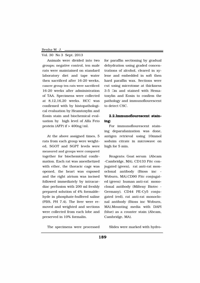
189
Benha M. J.
Vol. 30 No 3 Sept. 2013
Animals were divided into two
groups; negative control, ten male
rats were maintained on standard
laboratory diet and tape water
then sacrificed after 16-20 weeks,
cancer group ten rats were sacrificed
16-20 weeks after administration
of TAA. Specimens were collected
at 8,12,16,20 weeks. HCC was
confirmed with by histopathologi-
cal evaluation by Heamtoxylin and
Eosin stain and biochemical eval-
uation by high level of Alfa Feto
protein (AFP) if > 400ng/ml.
At the above assigned times, 5
rats from each group were weight-
ed, SGOT and SGPT levels were
measured and groups were compared
together for biochemichal confir-
mation. Each rat was anesthetized
with ether, the thoracic cage was
opened, the heart was exposed
and the right atrium was incised
followed immediately by intracar-
diac perfusion with 200 ml freshly
prepared solution of 4% formalde-
hyde in phosphate-buffered saline
(PBS, PH 7.4). The liver were re-
moved and weighted and sections
were collected from each lobe and
preserved in 10% formalin.
The specimens were processed
for paraffin sectioning by gradual
dehydration using graded concen-
trations of alcohol, cleared in xy-
lene and embedded in soft then
hard paraffin wax. Sections were
cut using microtome at thickness
3-5 µm and stained with Hema-
toxylin and Eosin to confirm the
pathology and immunoflourescent
to detect CSC.
2.2.Immunoflourescent stain-2.2.Immunoflourescent stain-
ing:ing:
For immunoflourescent stain-
ing deparafanization was done,
antigen retrieval using 10mmol
soduim citrate in microwave on
high for 5 min.
Reagents: Goat serum (Abcam
-Cambridge, MA), CD133 Fitc con-
jugated (green), rat anti-rat mon-
oclonal antibody (Bioss inc -
Woburn, MA).CD90 Fitc conjugat-
ed (green) human anti-rat mono-
clonal antibody (Milteny Biotec -
Germany). CD44 PE-Cy5 conju-
gated (red), rat anti-rat monoclo-
nal antibody (Bioss inc Woburn,
MA).Mounting media with DAPI
(blue) as a counter stain (Abcam,
Cambridge, MA).
Slides were marked with hydro-
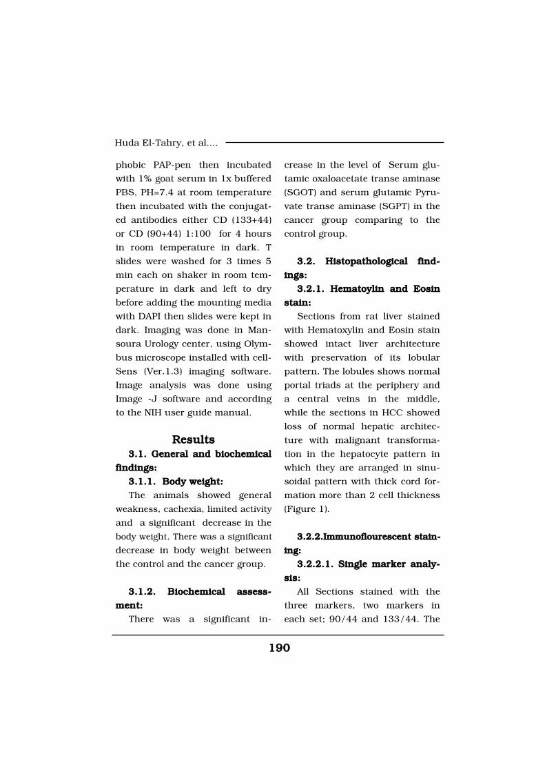
190190
Huda El-Tahry, et al....
phobic PAP-pen then incubated
with 1% goat serum in 1x buffered
PBS, PH=7.4 at room temperature
then incubated with the conjugat-
ed antibodies either CD (133+44)
or CD (90+44) 1:100 for 4 hours
in room temperature in dark. T
slides were washed for 3 times 5
min each on shaker in room tem-
perature in dark and left to dry
before adding the mounting media
with DAPI then slides were kept in
dark. Imaging was done in Man-
soura Urology center, using Olym-
bus microscope installed with cell-
Sens (Ver.1.3) imaging software.
Image analysis was done using
Image -J software and according
to the NIH user guide manual.
ResultsResults3.1. General and biochemical3.1. General and biochemical
findings:findings:
3.1.1. Body weight:3.1.1. Body weight:
The animals showed general
weakness, cachexia, limited activity
and a significant decrease in the
body weight. There was a significant
decrease in body weight between
the control and the cancer group.
3.1.2. Biochemical assess-3.1.2. Biochemical assess-
ment:ment:
There was a significant in-
crease in the level of Serum glu-
tamic oxaloacetate transe aminase
(SGOT) and serum glutamic Pyru-
vate transe aminase (SGPT) in the
cancer group comparing to the
control group.
3.2. Histopathological find-3.2. Histopathological find-
ings:ings:
3.2.1. Hematoylin and Eosin3.2.1. Hematoylin and Eosin
stain:stain:
Sections from rat liver stained
with Hematoxylin and Eosin stain
showed intact liver architecture
with preservation of its lobular
pattern. The lobules shows normal
portal triads at the periphery and
a central veins in the middle,
while the sections in HCC showed
loss of normal hepatic architec-
ture with malignant transforma-
tion in the hepatocyte pattern in
which they are arranged in sinu-
soidal pattern with thick cord for-
mation more than 2 cell thickness
(Figure 1).
3.2.2.Immunoflourescent stain-3.2.2.Immunoflourescent stain-
ing:ing:
3.2.2.1. Single marker analy-3.2.2.1. Single marker analy-
sis:sis:
All Sections stained with the
three markers, two markers in
each set; 90/44 and 133/44. The
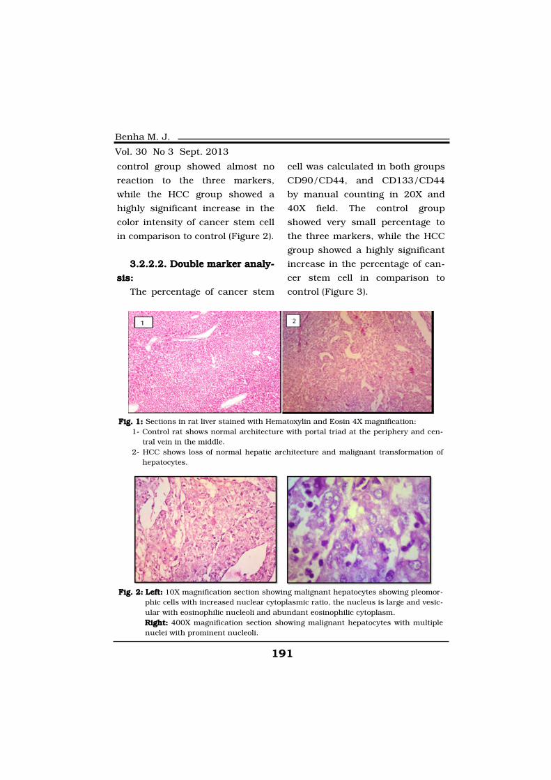
191
Benha M. J.
Vol. 30 No 3 Sept. 2013
control group showed almost no
reaction to the three markers,
while the HCC group showed a
highly significant increase in the
color intensity of cancer stem cell
in comparison to control (Figure 2).
3.2.2.2. Double marker analy-3.2.2.2. Double marker analy-
sis:sis:
The percentage of cancer stem
cell was calculated in both groups
CD90/CD44, and CD133/CD44
by manual counting in 20X and
40X field. The control group
showed very small percentage to
the three markers, while the HCC
group showed a highly significant
increase in the percentage of can-
cer stem cell in comparison to
control (Figure 3).
Fig. 1: Fig. 1: Sections in rat liver stained with Hematoxylin and Eosin 4X magnification:1- Control rat shows normal architecture with portal triad at the periphery and cen-
tral vein in the middle.2- HCC shows loss of normal hepatic architecture and malignant transformation of
hepatocytes.
Fig. 2: Fig. 2: Left:Left: 10X magnification section showing malignant hepatocytes showing pleomor-phic cells with increased nuclear cytoplasmic ratio, the nucleus is large and vesic-ular with eosinophilic nucleoli and abundant eosinophilic cytoplasm.Right:Right: 400X magnification section showing malignant hepatocytes with multiplenuclei with prominent nucleoli.

192192
Huda El-Tahry, et al....
DiscussionDiscussionThe main finding of this study
was that there was a significant
increase in the number and the
distribution of CSC was a dramatic
increase in the number of the CSC in
the hepatocellular carcinoma stage.
Thioacetamide was added to
drinking water in the dose of
300mg/L as mentioned(13). TAA at
this dose causes cirrhosis by in-
duction of chronic inflammation
through increase the oxidative
stress and up regulate the radioac-
tive oxygen species (ROS) which in
turn recruit large number of mac-
rophages and Kuffer cells leading
to fibrosis then cirrhosis(14).
The relation between inflamma-
tion and recruitment of CSC was
previously investigated(15), through
increasing the (ROS) which in-
Fig. 3: Fig. 3: Representative images for Immunoflourescnet staining of sections in rat livercomparing for the percentage of CSC double marked with CD90/CD44, in control(left) and cancer (right), yellow cells are the CSC.
Fig. 4: Fig. 4: Immunoflourscent staining in sections of rat liver comparing between control(left) and cancer (right), sectioned is double marked with CD133/CD44 the yellowcolor indicates the intensity of the reaction 4X magnification.
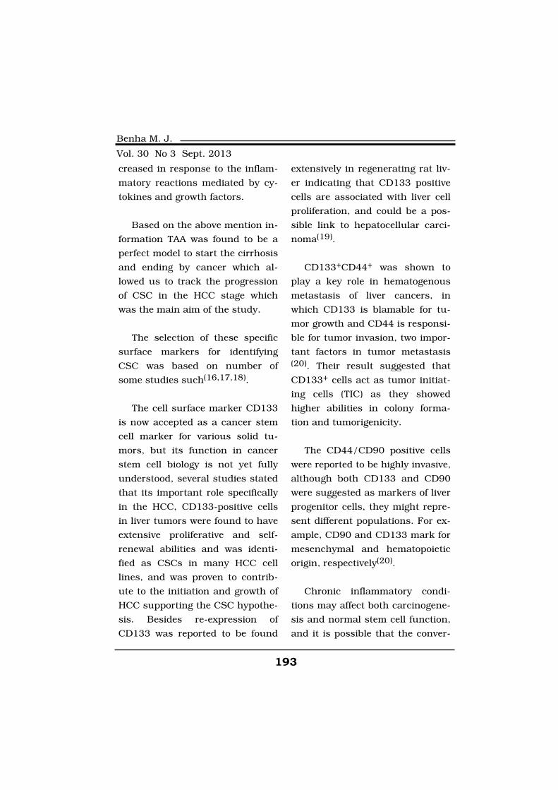
193
Benha M. J.
Vol. 30 No 3 Sept. 2013
creased in response to the inflam-
matory reactions mediated by cy-
tokines and growth factors.
Based on the above mention in-
formation TAA was found to be a
perfect model to start the cirrhosis
and ending by cancer which al-
lowed us to track the progression
of CSC in the HCC stage which
was the main aim of the study.
The selection of these specific
surface markers for identifying
CSC was based on number of
some studies such(16,17,18).
The cell surface marker CD133
is now accepted as a cancer stem
cell marker for various solid tu-
mors, but its function in cancer
stem cell biology is not yet fully
understood, several studies stated
that its important role specifically
in the HCC, CD133-positive cells
in liver tumors were found to have
extensive proliferative and self-
renewal abilities and was identi-
fied as CSCs in many HCC cell
lines, and was proven to contrib-
ute to the initiation and growth of
HCC supporting the CSC hypothe-
sis. Besides re-expression of
CD133 was reported to be found
extensively in regenerating rat liv-
er indicating that CD133 positive
cells are associated with liver cell
proliferation, and could be a pos-
sible link to hepatocellular carci-
noma(19).
CD133+CD44+ was shown to
play a key role in hematogenous
metastasis of liver cancers, in
which CD133 is blamable for tu-
mor growth and CD44 is responsi-
ble for tumor invasion, two impor-
tant factors in tumor metastasis(20). Their result suggested that
CD133+ cells act as tumor initiat-
ing cells (TIC) as they showed
higher abilities in colony forma-
tion and tumorigenicity.
The CD44/CD90 positive cells
were reported to be highly invasive,
although both CD133 and CD90
were suggested as markers of liver
progenitor cells, they might repre-
sent different populations. For ex-
ample, CD90 and CD133 mark for
mesenchymal and hematopoietic
origin, respectively(20).
Chronic inflammatory condi-
tions may affect both carcinogene-
sis and normal stem cell function,
and it is possible that the conver-
194194
Huda El-Tahry, et al....
gence of these behavior contrib-
utes to the formation or regulation
or both of CSCs. Increased intra-
cellular production of ROS pro-
duced by immune response or in-
flammatory cytokines are a
common feature of chronically in-
flamed tissues and may have mul-
tiple effects on CSCs(21).
Our finding agrees with the
above literature and can explain
the significant increase in the
number of the CSC in the active
stage of the cirrhosis where there
is a lot of inflammation and re-
placement by regenerating nod-
ules and increase in the cell divi-
sion.
In the late stage of the cirrhosis
the sections showed decrease in
the number of mononuclear cells
in filtrate which indicates that
there is no active inflammation,
and this explains the decrease in
the number of the CSC and that it
is strongly correlated to the in-
flammation.
Findings made in the CSC field
will feed back into other zones of
stem cell research because many
marker gene products found in
CSCs also share with the normal
stem cell population. It is also pre-
dictable that a better understand-
ing of the processes that control
autonomous growth, differentia-
tion and cell migration will con-
tribute to novel regenerative-
medicine-based treatments that
will transform therapeutic strate-
gies and bring renewed hope to
cancer patients.
ReferencesReferences1. Zakhary N., El-Merzabani1. Zakhary N., El-Merzabani
M., El-Sawi N., Saleh S., MoneerM., El-Sawi N., Saleh S., Moneer
M. and Mohamad R. (2011):M. and Mohamad R. (2011): Im-
pact of different biochemical
markers in serum of patients with
benign and malignant liver diseas-
es. J Advanced Res; 2(1): 49-55.
2. Abdel-Wahab M., El-2. Abdel-Wahab M., El-
Ghawalby N., Mostafa M., Sul-Ghawalby N., Mostafa M., Sul-
tan A., El-Sadany M., Fathy O.,tan A., El-Sadany M., Fathy O.,
Salah T. and Ezzat F. (2007):Salah T. and Ezzat F. (2007):
Epidemiology of hepatocellular
carcinoma in lower Egypt, Gas-
troenterology Center J. Hepatogas-
troenterol; 54(73): 157-62.
3. Whang-Peng J., Cheng3. Whang-Peng J., Cheng
A.L., Chiun Hsu and Chien-MingA.L., Chiun Hsu and Chien-Ming
Chen (2010):Chen (2010): Clinical Develop-
ment and Future Direction for the

195
Benha M. J.
Vol. 30 No 3 Sept. 2013
Treatment of Hepatocellular Carci-
noma, J Exp Clin Med 2(3): 93-
103.
4. Conheim J. (1875):4. Conheim J. (1875): Congen-
itales, quergestreiftes Muskelsar-
kon der Nireren. Virchows Arch
65:64 quoted by Shupe T, & Peter-
son B. (2009). Cancer Stem Cells:
Hepatocellular carcinoma. Stem
cell and Cancer, Humana press.
5. Shupe T. and Peterson B.5. Shupe T. and Peterson B.
(2009):(2009): Cancer Stem Cells: Hepat-
ocellular carcinoma. Stem cell and
Cancer, Humana press.
6. Moltzahn F.R., Volkmer6. Moltzahn F.R., Volkmer
J.P., Rottke D. and Acker- mannJ.P., Rottke D. and Acker- mann
R. (2008):R. (2008): Cancer stem cells-
lessons from Hercules to fight the
Hydra. Urol Oncol 26: 581-589.
7. Oliveira L.R., Fernandes A.7. Oliveira L.R., Fernandes A.
and Ribeiro-Silva A. (2011):and Ribeiro-Silva A. (2011):
Stem Cells and Cancer Stem
Cells- Cancer Stem Cells - The
Cutting Edge Edited by Stanley
Shostak Published by InTech, Ja-
neza Trdine 9, 51000 Rijeka,
Croatia 2011.
8. Lobo N.A., Shimono Y.,8. Lobo N.A., Shimono Y.,
Qian D. and Clarke M.F. (2007):Qian D. and Clarke M.F. (2007):
The biology of cancer stem cells.
Annu Rev Cell Dev Biol 23:675-
99.
9. He S., Nakada D. and Mor-9. He S., Nakada D. and Mor-
rison S.J. (2009):rison S.J. (2009): Mechanisms of
stem cell self-renewal. Annu Rev
Cell Dev Biol 25:377-406.
10. Shackleton M. (2010):10. Shackleton M. (2010):
Normal stem cells and cancer
stem cells: similar and different.
Seminars in Cancer Biology 20:
85-92.
11. Li L. and Neaves W.B.11. Li L. and Neaves W.B.
(2006):(2006): Normal Stem Cells and
Cancer Stem Cells: The Niche
Matters. Cancer Res; 66: 4553-
4557.
12. Suetsugu A., Nagaki M.,12. Suetsugu A., Nagaki M.,
Aoki H., Motohashi T., KunisadaAoki H., Motohashi T., Kunisada
T. and Moriwaki H. (2006):T. and Moriwaki H. (2006): Char-
acterization of CD133+ hepatocel-
lular carcinoma cells as cancer
stem/progenitor cells. Biochem
Biophys Res Commun; 351:820-
824.
13. Li X., Benjamin I. and Al-13. Li X., Benjamin I. and Al-
exander B. (2002):exander B. (2002): Reproducible
production of Thioacetamide in-
duced macronodular cirrhosis in
196196
Huda El-Tahry, et al....
the rat with no mortality. Journal
of Hepatology 36(4): 488-493.
14. Bird T., Lorenzini S. and14. Bird T., Lorenzini S. and
Forbes S. (2008):Forbes S. (2008): Activation of
stem cells in hepatic diseases. Cell
Tissue Res. 331:283-300.
15. Tanno T. and Matsui W.15. Tanno T. and Matsui W.
(2011):(2011): Development and mainte-
nance of cance stem cell under
chronic inflammation. J Nippon
MED. Sch 78:138-45.
16. Lingala S., Cui1Y., Chen16. Lingala S., Cui1Y., Chen
X., Boris H., et al. (2010):X., Boris H., et al. (2010): Immu-
nohistochemical Staining of Can-
cer Stem Cell markers in Hepato-
cellular Carcinoma Exp Mol
Pathol. 89(1): 27-35.
17. Yang Z.F., Ngai P., Yu17. Yang Z.F., Ngai P., Yu
W.C., Ng M.N., Lau C.K., et al.W.C., Ng M.N., Lau C.K., et al.
(2008b):(2008b): Identification of local and
circulating cancer stem cells in
human liver cancer. Hepatology
47:919-928.
18. Ma S., Hu L., Lee T.K.,18. Ma S., Hu L., Lee T.K.,
Wo J.Y., Ng I.O., et al. (2007):Wo J.Y., Ng I.O., et al. (2007):
Identification and characterization
of tumorigenic liver cancer stem/
progenitor cells. Gastroenterology
132:2542-2556.
19. Song W., Li H., Tao K.,19. Song W., Li H., Tao K.,
Song Z., Zhao Q., Zhang F. andSong Z., Zhao Q., Zhang F. and
Dou K. (2008):Dou K. (2008): Expression and
clinical significance of the stem
cell marker CD133 in hepatocellu-
lar carcinoma. Int J Clin Pract 62
(8): 1212-8.
20. Ying H., Qifei Z., Ruiliang20. Ying H., Qifei Z., Ruiliang
G., Feng S., Yizheng W. and CellG., Feng S., Yizheng W. and Cell
Research (2012):Research (2012): The critical role
of CD133+ CD44+/high tumor
cells in hematogenous metastasis
of liver cancers. Cell Research
22:259-272.
21. Miyamoto K., Araki K.Y.,21. Miyamoto K., Araki K.Y.,
Naka K., Araki K.Y., Naka K.,Naka K., Araki K.Y., Naka K.,
Arai F., Takubo K., et al. (2007):Arai F., Takubo K., et al. (2007):
Foxo3a is essential for mainte-
nance of the hematopoietic stem
cell pool. Cell Stem Cell 1:101-
112.

197
Benha M. J.
Vol. 30 No 3 Sept. 2013
INDUCTION OF CANCER STEM CELLINDUCTION OF CANCER STEM CELLIN HEPATOCELLULAR CARINOMA:IN HEPATOCELLULAR CARINOMA:
THIOACETAMIDE MODELTHIOACETAMIDE MODEL
Huda El-Tahry Ph.D, Omar Gabr Ph.D,Huda El-Tahry Ph.D, Omar Gabr Ph.D,Farha El-Chennawi Ph.D, Dalia Saleh Ph.DFarha El-Chennawi Ph.D, Dalia Saleh Ph.D
and Amira Othman M.Scand Amira Othman M.Sc
BENHAMEDICALJOURNAL
REPRINT
Published byPublished by
Benha Faculty of MedicineBenha Faculty of Medicine Volume 30 Number 3Sept. 2013
197
Benha M. J.
Vol. 30 No 3 Sept. 2013
EVALUATION OF DIFFERENT SCORINGEVALUATION OF DIFFERENT SCORINGSYSTEMS PREDICTING NONSENTINEL LYMPHSYSTEMS PREDICTING NONSENTINEL LYMPHNODE STATUS IN BREAST CANCER PATIENTSNODE STATUS IN BREAST CANCER PATIENTS
Magdy B. E-Moghazy MD*, Ashraf M. Shoma MD*,Magdy B. E-Moghazy MD*, Ashraf M. Shoma MD*,Abd El-Azeem El-Ganash MD*, Maha M. Abu Hashem MD**Abd El-Azeem El-Ganash MD*, Maha M. Abu Hashem MD**
and Ahmed Moatamed MD*and Ahmed Moatamed MD**Department of Surgery, Faculty of Medicine, Mansoura University
**Department of Pathology, Faculty of Medicine, Mansoura University
AbstractAbstractBackground:Background: Axillary lymph node dissection (ALND) performed after a
positive sentinel lymph node biopsy (SLNB) in breast cancer patients, oftenresults in no additional positive nodes. Scoring systems have been publishedto aid in the prediction of non sentinel lymph node (NSLN) metastasis. Ouraim was to assess the validity of these scoring systems in our patients.
Methods:Methods: This prospective study conducted on 48 patients who under-went ALND after a positive SLNB in Mansoura University Hospital fromMarch 2009 to December 2012. The accuracy of the Memorial Sloan-Kettering Cancer Center (MSKCC) nomogram, the MD Anderson scoring sys-tem and the Tenon scoring system were tested for the prediction of NSLNstatus. Receiver operating characteristics (ROC) curves were drawn, and theareas under the curves were calculated to assess the discriminative power ofeach system. Univariate analysis was performed to assess the predictabilityof individual patient and tumor characteristics.
Results:Results: Nonsentinel lymph nodes were positive in 38 (79%) patients.The areas under the ROC curves were 0.84, 0.90, and 0.91, respectively.The pathological tumor size was the only individual predictors of nonsentinelnode metastasis.
Conclusions:Conclusions: Scoring systems provide additional information regardingthe likelihood of metastasis in nonsentinel nodes, but their predictability re-mains less than optimal. The use of scoring systems must be applied withcaution until future studies provide a more accurate assessment of risk forpatients with a positive SLNB.
Key Words:Key Words: Scoring systems - Sentinel lymph node - Nonsentinel lymphnode - Breast cancer.
198198
Magdy B. El-Moghazy, et al....
IntroductionIntroductionThe introduction of SLNB for
nodal staging has revolutionized
the surgical approach for early
breast cancer. The goal of SLNB is
to reduce the morbidity of breast
cancer surgery by avoiding unnec-
essary ALND in patients with neg-
ative SLN. However, if a positive
SLN is found, it is currently rec-
ommended to continue with
ALND. In 40 - 70% of patients the
SLN is the only involved axillary
node, implying that these patients
undergo ALND unnecessarily(1,2,3).
Several studies have investigat-
ed clinicopathologic factors that
may predict which patients have a
higher risk of non sentinel lymph
node involvement, none of which
are sufficiently predictive when
used alone(4,5).
Scoring systems have been de-
veloped using a combination of
several factors, such as tumor
size, histology, hormone receptors,
presence of lymphovascular inva-
sion, the number of sentinel nodes
removed, as a guide to determine
which patients may forego ALND,
if they had a positive SLN(6,7).
Three scoring systems were
identified in the existing medical
literature using a Medline-based
search engine. The first scoring
system (MSK) is a nomogram from
Memorial Sloan-Kettering Cancer
Center (MSKCC) in New York,
USA, that includes eight charac-
teristics (nuclear grade, lympho-
vascular invasion, multifocality,
estrogen receptor status, number
of positive and negative sentinel
nodes, tumor size, and method of
detection of sentinel node metas-
tasis) that ultimately generates a
total point value, which then cor-
responds to a percentage of risk(8,9,10).
The second scoring system
[M.D. Anderson (MDA) score] was
developed at the M.D. Anderson
Cancer Center in Houston Texas,
USA and is based on four charac-
teristics (tumor size, number of
sentinel nodes, size of metastasis,
and lymphovascular invasion),
where a or coefficient was deter-
mined for each, and the sum of
rounded coefficients results in a
score ranging from -2 to 4(5,6,7).
The third scoring system (Ten-
on score) was derived at the Hos-
199
Benha M. J.
Vol. 30 No 3 Sept. 2013
pital Tenon in Paris, France, and
includes three characteristics (
size of metastasis, tumor size, and
proportion of sentinel nodes in-
volved) which are assigned point
values that, when added, result in
a score between 0 and 7(11,12).
The aim of our study is to eval-
uate available three scoring sys-
tems for accurate prediction of
metastasis in nonsentinel lymph
nodes in breast cancer patients
with a positive sentinel lymph
nodes biopsy.
Patients and MethodsPatients and MethodsThis prospective study was done
on all patients with invasive breast
cancer admitted to Mansoura Uni-
versity Hospital, from March 2009
to December 2012, where sentinel
lymph node biopsy will be chosen
as the procedure to deal with the
axillary lymph node status.
This study was carried on 58
patients with primary invasive
breast cancer with clinically nega-
tive axilla operated upon with
modified radical mastectomy in 53
cases (91%) or wide local excision
and axillary clearance in 5 cases
(9%).
Technique of SLNB:Technique of SLNB:
Sentinel lymph nodes were
mapped in the first 6 cases using
a subdermal injection of 0.5 mCi
of filtered 99mTc Sulfur Colloid in
5 ml of saline 2 hour before sur-
gery. In the next 52 cases sentinel
lymph nodes were mapped using
peritumeral injection of 5 ml of a
blue dye (Methylene blue in 50
cases and patent blue in 2 cases)
15 minutes before axillary dissec-
tion.
SLNs localization was under-
taken in the radioisotope group
using a hand held gamma probe
to determine radioactivity levels in
the axilla, looking for ‘hot’ nodes
(nodes with counts at least tenfold
those of the background), and in
the blue dye group as the most
proximal node the pathway of a
blue-stained lymphatic, or any
blue nodes.
After identification of SLNs,
complete axillary dissection was
done and all axillary lymph nodes
were taken as a separate speci-
men, with the breast tissue as the
third specimen and were fixed in
10% formalin saline. The three
specimens were sent for histo-
200200
Magdy B. El-Moghazy, et al....
pathological examination.
Pathological examination:Pathological examination:
Each primary tumor was evalu-
ated for size of the invasive com-
ponent, histological type, nuclear
grade, estrogen receptor status,
multifocality of the tumor, and
presence of lymphovascular inva-
sion.
Pathological SLN examination
includes routine hematoxylin and
eosin (HE), serial HE, or immuno-
histochemistry (IHC). Each half-
SN was sliced at 3-mm intervals.
All 58 cases were examined by
routine (HE) we found 38 cases
positive, (H&E) negative sections
(20 cases) were examined by serial
HE we found 10 cases positive,
and(serial H&E) negative sections
(10 cases) were examined by IHC
we found all 10 cases negative.
NSLN obtained from axillary
dissection specimen were analyzed
by HE staining only. The total
number and number of positive
NSLN were recorded.
All clinical data recorded in the
MSKCC, MD Anderson and Tenon
studies were prospectively collect-
ed as part of our clinical study
and then retrospectively reviewed.
All data were collected in a per-
formed sheet then entered into an
electronic spreadsheet (Microsoft
Excel) and transferred into SPSS
(Statistical Package for Social Sci-
ences).
Statistical AnalysisStatistical AnalysisStatistical analyses were per-
formed by using the SPSS (Statis-
tical Package for Social Sciences)
version 15, 2006. Qualitative data
was presented as number and
percent. Comparison between
groups was done by Chi-Square
test. Quantitative data was tested
for normality by Kolmogrov-
Smirnov test. Normally distributed
data was presented as means ±
standard deviations (SD). Student
t-test was used to compare be-
tween two or more groups.
The discriminatory accuracies
of the three scoring systems were
compared by constructing receiver
operating characteristics (ROC)
curves and measuring areas un-
der these curves (AUC). A model
with a ROC of 0.5 is equal to the
toss of a coin. A model with a ROC
201
Benha M. J.
Vol. 30 No 3 Sept. 2013
of 0.7- 0.8 is considered good,
whereas a ROC of 0.81 - 0.9 has
excellent discrimination. Categori-
cal data are reported as percent-
ages with 95% confidence inter-
vals (95% CI).
ResultsResultsThis prospective study compro-
mised of 58 patients underwent
SLNB, 10 (18%) of which had a
negative nodes, were excluded
from our study, leaving 48 (82%)
patients with positive SLNB, and
this group therefore comprised
our study sample.
All patients in the study were
female, with a mean age of 49
years (range, 30-74) years. The
mean pathological tumor size was
3.3 cm (range, 2-7 cm).
The mean pathological tumor
size was 3.35 cm (range, 2-7 cm).
There were no cases with tumor
size less than 2cm due to absence
of screening programs in Mansou-
ra University Hospital. There was
a statistical significance between
tumor size and NSLN status.
The mean total number of SLN
was 2.35 (range 1-4), the mean
number of positive SLN was 1.9
(range 1 - 4), and a median of 11
(range 5 - 24) nodes were dissect-
ed during ALND. Non sentinel
lymph nodes were positive in 38
(79.2%) patients and negative in
10 (20.8%) patients. There was no
statistical significance between to-
tal number of SLN, number of
positive SLN and NSLN status.
The MSKCC nomogram in our
patients shows all patients (4)
with score less than 20% have
negative NSLN (negative predictive
value=100%), and all patients (22)
with score more than 60% have
posative NSLN (posative predictive
value = 100%), so we can predict
the NSLN status in 55%of our pa-
tients. The areas under the curve
of MSKCC was 0.84 (exellent).
The MD Anderson score in our
patients shows all patients (6)
with score less than 0 have nega-
tive NSLN (negative predictive val-
ue = 100%), and all patients (21)
with score more than 2 have po-
sative NSLN (posative predictive
value = 100%), so we can predict
the NSLN status in 57 % of our
patients. The areas under the
curve of the MD Anderson score
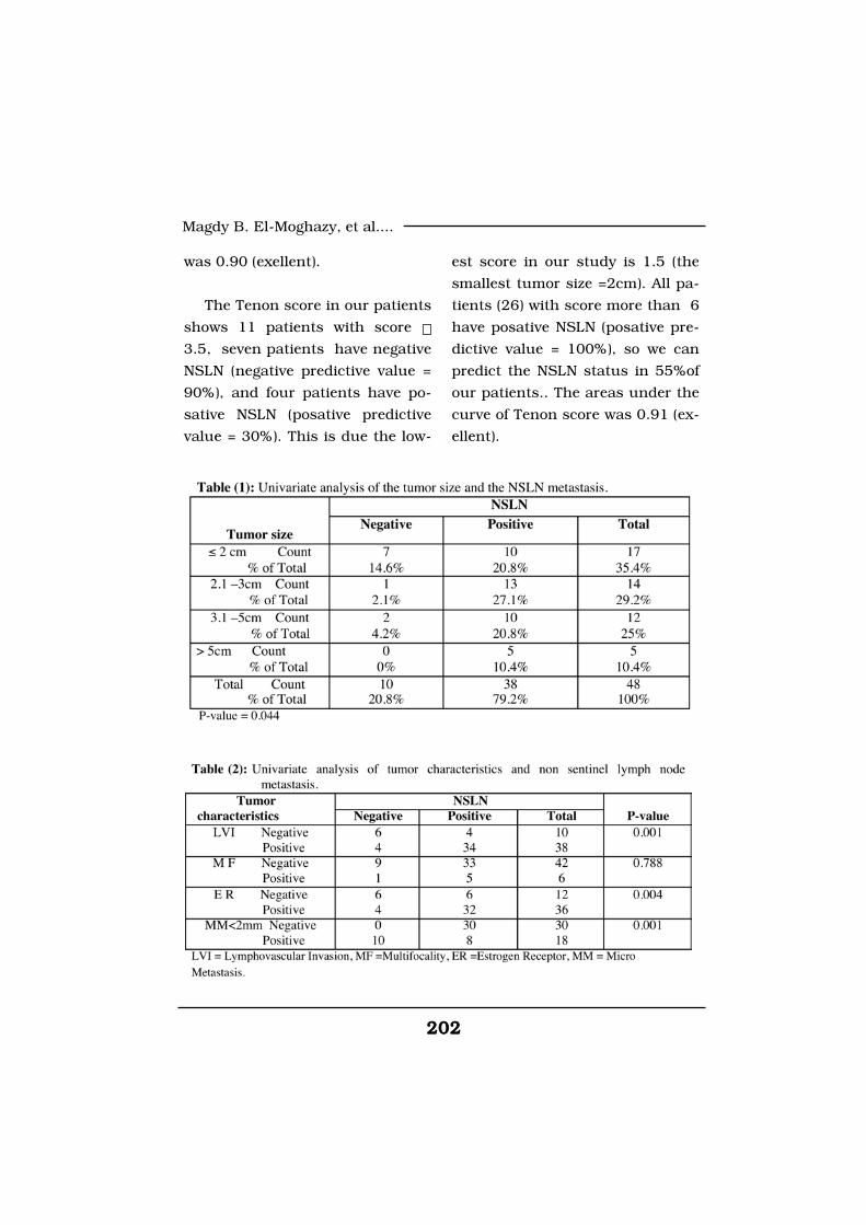
202202
Magdy B. El-Moghazy, et al....
was 0.90 (exellent).
The Tenon score in our patients
shows 11 patients with score
3.5, seven patients have negative
NSLN (negative predictive value =
90%), and four patients have po-
sative NSLN (posative predictive
value = 30%). This is due the low-
est score in our study is 1.5 (the
smallest tumor size =2cm). All pa-
tients (26) with score more than 6
have posative NSLN (posative pre-
dictive value = 100%), so we can
predict the NSLN status in 55%of
our patients.. The areas under the
curve of Tenon score was 0.91 (ex-
ellent).
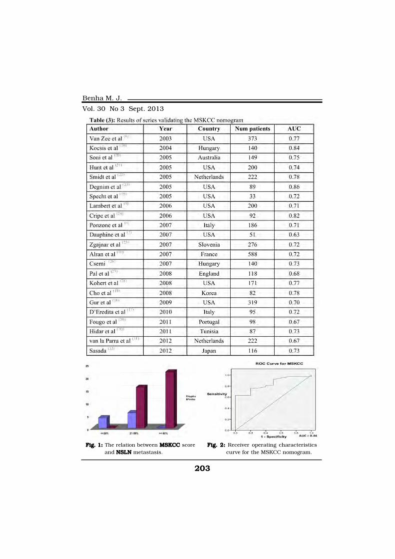
203
Benha M. J.
Vol. 30 No 3 Sept. 2013
Fig. 1: Fig. 1: The relation between MSKCC MSKCC scoreand NSLN NSLN metastasis.
Fig. 2:Fig. 2: Receiver operating characteristicscurve for the MSKCC nomogram.
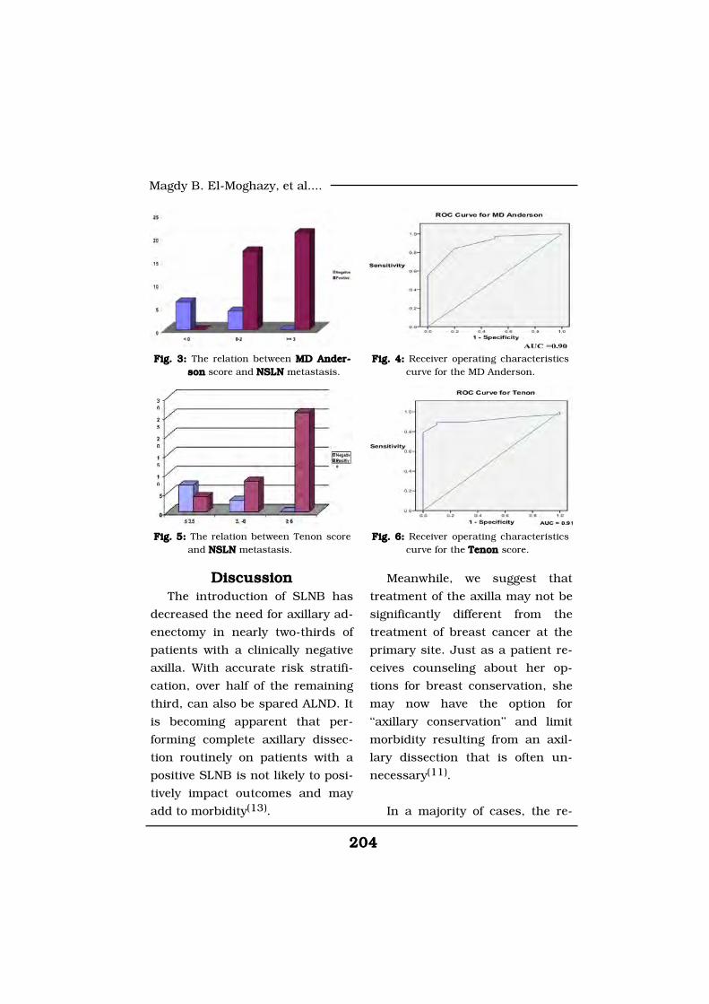
204204
Magdy B. El-Moghazy, et al....
DiscussionDiscussionThe introduction of SLNB has
decreased the need for axillary ad-
enectomy in nearly two-thirds of
patients with a clinically negative
axilla. With accurate risk stratifi-
cation, over half of the remaining
third, can also be spared ALND. It
is becoming apparent that per-
forming complete axillary dissec-
tion routinely on patients with a
positive SLNB is not likely to posi-
tively impact outcomes and may
add to morbidity(13).
Meanwhile, we suggest that
treatment of the axilla may not be
significantly different from the
treatment of breast cancer at the
primary site. Just as a patient re-
ceives counseling about her op-
tions for breast conservation, she
may now have the option for
‘‘axillary conservation’’ and limit
morbidity resulting from an axil-
lary dissection that is often un-
necessary(11).
In a majority of cases, the re-
Fig. 3: Fig. 3: The relation between MD Ander-MD Ander-sonson score and NSLN NSLN metastasis.
Fig. 4:Fig. 4: Receiver operating characteristicscurve for the MD Anderson.
Fig. 5: Fig. 5: The relation between Tenon scoreand NSLN NSLN metastasis.
Fig. 6:Fig. 6: Receiver operating characteristicscurve for the Tenon Tenon score.
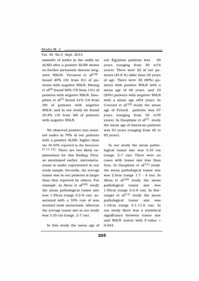
205
Benha M. J.
Vol. 30 No 3 Sept. 2013
mainder of nodes in the axilla on
ALND after a positive SLNB shows
no further metastatic disease (neg-
ative NSLN). Veronesi et al(14)
found 40% (32 from 81) of pa-
tients with negative NSLN, Hwang
et al(5) found 60% (78 from 131) of
patients with negative NSLN, Dau-
phine et al(7) found 41% (16 from
39) of patients with negative
NSLN, and in our study we found
20.8% (10 from 48) of patients
with negative NSLN.
We observed positive non senti-
nel nodes in 79% of our patients
with a positive SLNB, higher than
the 30-50% reported in the literature(7,11,12). There are two likely ex-
planations for this finding: First,
as mentioned earlier, micrometa-
stasis is under represented in our
study sample. Secondly, the average
tumor size in our patients is larger
than that reported by others. For
example, in Alran et al(10) study
the mean pathological tumor size
was 1.55cm (range 0.2-6 cm), as-
sociated with a 35% rate of non
sentinel node metastasis, whereas
the average tumor size in our study
was 3.35 cm (range, 2-7 cm).
In this study the mean age of
our Egyptian patients was 49
years (ranging from 30 to74
years). There were 22 of our pa-
tients (45.8 %) older than 50 years
of age. There were 38 (80%) pa-
tients with positive NSLN with a
mean age of 48 years, and 10
(20%) patients with negative NSLN
with a mean age of54 years. In
Coutant et al(12) study the mean
age of French patients was 57
years (ranging from 32 to78
years). In Dauphine et al(7) study
the mean age of American patients
was 53 years (ranging from 45 to
62 years).
In our study the mean patho-
logical tumor size was 3.35 cm
(range, 2-7 cm). There were no
cases with tumor size less than
2cm. In Dauphine et al(11) study
the mean pathological tumor size
was 2.5cm (range 1.7 - 4 cm). In
Alran et al(10) study the mean
pathological tumor size was
1.55cm (range 0.2-6 cm). In Bar-
ranger et al(11) study the mean
pathological tumor size was
1.44cm (range 0.1-11.6 cm). In
our study there was a statistical
significance between tumor size
and NSLN status with P-value =
0.044.
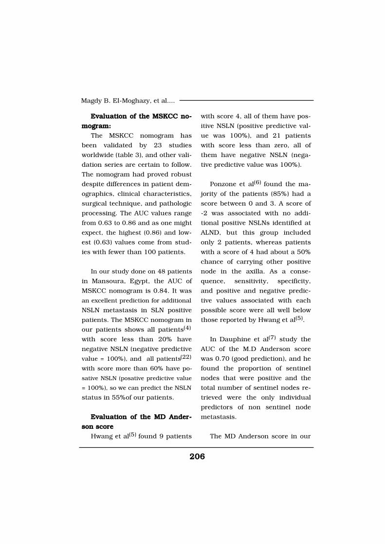
206206
Magdy B. El-Moghazy, et al....
Evaluation of the MSKCC no-Evaluation of the MSKCC no-
mogram:mogram:
The MSKCC nomogram has
been validated by 23 studies
worldwide (table 3), and other vali-
dation series are certain to follow.
The nomogram had proved robust
despite differences in patient dem-
ographics, clinical characteristics,
surgical technique, and pathologic
processing. The AUC values range
from 0.63 to 0.86 and as one might
expect, the highest (0.86) and low-
est (0.63) values come from stud-
ies with fewer than 100 patients.
In our study done on 48 patients
in Mansoura, Egypt, the AUC of
MSKCC nomogram is 0.84. It was
an excellent prediction for additional
NSLN metastasis in SLN positive
patients. The MSKCC nomogram in
our patients shows all patients(4)
with score less than 20% have
negative NSLN (negative predictive
value = 100%), and all patients(22)
with score more than 60% have po-
sative NSLN (posative predictive value
= 100%), so we can predict the NSLN
status in 55%of our patients.
Evaluation of the MD Ander-Evaluation of the MD Ander-
son scoreson score
Hwang et al(5) found 9 patients
with score 4, all of them have pos-
itive NSLN (positive predictive val-
ue was 100%), and 21 patients
with score less than zero, all of
them have negative NSLN (nega-
tive predictive value was 100%).
Ponzone et al(6) found the ma-
jority of the patients (85%) had a
score between 0 and 3. A score of
-2 was associated with no addi-
tional positive NSLNs identified at
ALND, but this group included
only 2 patients, whereas patients
with a score of 4 had about a 50%
chance of carrying other positive
node in the axilla. As a conse-
quence, sensitivity, specificity,
and positive and negative predic-
tive values associated with each
possible score were all well below
those reported by Hwang et al(5).
In Dauphine et al(7) study the
AUC of the M.D Anderson score
was 0.70 (good prediction), and he
found the proportion of sentinel
nodes that were positive and the
total number of sentinel nodes re-
trieved were the only individual
predictors of non sentinel node
metastasis.
The MD Anderson score in our
207
Benha M. J.
Vol. 30 No 3 Sept. 2013
patients shows all patients(6) with
score less than 0 have negative
NSLN (negative predictive value =
100%), and all patients(21) with
score more than 3 have posative
NSLN (posative predictive value =
100%), and only 45% of our pa-
tients had a score between 0 and
3. The areas under the curve 0.90
(exellent).
Evaluation of the Tenon score:Evaluation of the Tenon score:
In Brranger et al(11) study all
patients with a score of 3.5 or less
had a 97.3% chance of being free
of NSLN involvement. The main
limitation of this score is that is
does not take into account extra-
capsular extension of SN metasta-
sis, which is a powerful predictor
of non-SN metastasis.
In Coutant et al(12) study 120
(53.1%) of the 226 patients had a
Tenon score of 3.5 or less. Among
these patients, five had at least
one positive NSLN (negative pre-
dictive value was 95.8% and the
false-negative rate was 4.2%), with
an AUC of 0.82.
In Gur et al(16) study 115
(36.0%) of the 319 patients had a
score 3.5 according to the Tenon
model, and in this group, 20 had
at least 1 positive NSLN. With a
score cut-off of 3.5, the negative
predictive value was 72% and the
false negative rate was 17.3%,
with an AUC of 0.69.
In Hidar et al(30) study, 26
(30%) of the 87 cases were report-
ed with a Tenon score 3.5, and in
this subset of patients, 15 were
associated with a negative NSLN
(negative predictive value was 57%
and the false-negative rate was
43%), with an AUC of 0.75.
In our patients the Tenon score
shows 11 patients with score
3.5, seven patients have negative
NSLN (negative predictive value =
90%), and four patients have po-
sative NSLN (posative predictive
value = 30%). This is due the low-
est score in our study is 1.5 (the
smallest tumor size =2cm).All pa-
tients(26) with score 6 have po-
sative NSLN (posative predictive
value = 100%), so we can predict
the NSLN status in 55%of our pa-
tients.. The areas under the curve
of Tenon score was 0.91 (exellent).
ConclusionConclusionTo our knowledge, the current
208208
Magdy B. El-Moghazy, et al....
study is the first to report, to com-
pare and to validate NSLN metas-
tases predictive tools in Egyptian
population. The three scoring sys-
tems (MSK, MD Anderson, Tenon)
were comparable, as determined
by their AUC, 0.84, 0.90, 0.91 re-
spectively with sufficient predicta-
bility to be called valid.
The MSKCC nomogram was
outperformed in our study popula-
tion because it presents as graph-
ic nomogram and as digital availa-
ble at Web site, www.mskcc.org/
nomograms, and appears to pro-
vide a useful quantitative estima-
tion on NSLN involvement when
discussing the indication to com-
pletion ALND with the patient.
The use of scoring systems
must be applied with caution until
future studies provide a more ac-
curate assessment of the risk
NSLN metastases for patients with
a positive SLNB.
ReferencesReferences1. Veronesi U., Paganelli G.,1. Veronesi U., Paganelli G.,
Viale G., et al. (2003):Viale G., et al. (2003): A random-
ized comparison of sentinel-node
biopsy with routine axillary dis-
section in breast cancer. N Engl J
Med; 349(6):546-563.
2. Giullano A.E., Kirgan D.M.,2. Giullano A.E., Kirgan D.M.,
Guenther J.M., et al. (1994):Guenther J.M., et al. (1994):
Lymphatic mapping and sentinel
lymphadenectomy for breast can-
cer Ann Surg; 220: 391-398.
3. Chu Ku, Turner R.R., Han-3. Chu Ku, Turner R.R., Han-
sen N.M., et al. (1999):sen N.M., et al. (1999): Sentinel
node metastasis in patient with
breast carcinoma accurately pre-
dicts immunohistochemically de-
tectable nonsentinel node metas-
tasis. Ann Surg Oncol 6:756-761.
4. Yip C.H., Taib N.A., Tan G.4. Yip C.H., Taib N.A., Tan G.
H., et al. (2009):H., et al. (2009): Predictors of ax-
illary lymph node metastases in
breast cancer: Is there a role for
minimal axillary surgery? World J
Surg 33:54-57.
5. Hwang R.F., Krishna-5. Hwang R.F., Krishna-
murthy S., Hunt K.K., et al.murthy S., Hunt K.K., et al.
(2003):(2003): Clinicopathologic factors
predicting involvement of nonsen-
tinel axillary nodes in women with
breast cancer. Ann Surg Oncol
10:248-254.
6. Ponzone R., Maggiorotto F.,6. Ponzone R., Maggiorotto F.,
Mariani L., et al. (2007):Mariani L., et al. (2007): Com-
parison of two models for the pre-
209
Benha M. J.
Vol. 30 No 3 Sept. 2013
diction of non sentinel node me-
tastases in breast cancer. Am J
Surg 193:686-692.
7. Dauphine C.E., Haukoos7. Dauphine C.E., Haukoos
J.S., Vargas M.P., et al. (2007):J.S., Vargas M.P., et al. (2007):
Evaluation of three scoring sys-
tems predicting non-sentinel node
metastasis in breast cancer pa-
tients with a positive sentinel node
biopsy. Ann Surg Oncol 14:1014-
1019.
8. Van Zee K.J., Manasseh8. Van Zee K.J., Manasseh
D.M., Bevilacqua J.L., et al.D.M., Bevilacqua J.L., et al.
(2003):(2003): A nomogram for predict-
ing the likelihood of additional no-
dal metastases in breast cancer
patients with a positive sentinel
node biopsy. Ann Surg Oncol
10:1140-1151.
9. Lambert L.A., Ayers G.D.,9. Lambert L.A., Ayers G.D.,
Hwang R.F., et al. (2006): Hwang R.F., et al. (2006): Valida-
tion of a breast cancer nomogram
for predicting nonsentinel lymph
node metastases after a positive
sentinel node biopsy. Ann Surg
Oncol 13:310-320.
10. Alran S., De Rycke Y.,10. Alran S., De Rycke Y.,
Fourchotte V., et al. (2007):Fourchotte V., et al. (2007): Vali-
dation and limitations of use of a
breast cancer nomogram predict-
ing the likelihood of non-sentinel
node involvement after positive
sentinel node biopsy. Ann Surg
Oncol 14:2195-2201.
11. Barranger E., Coutant C.,11. Barranger E., Coutant C.,
Flahault A., et al. (2005):Flahault A., et al. (2005): An ax-
illa scoring system to predict non-
sentinel lymph node status in
breast cancer patients with senti-
nel lymph node involvement.
Breast Cancer Res Treat 91:113-
119.
12. Coutant C., Rouzier R.,12. Coutant C., Rouzier R.,
Fondrinier E. (2009):Fondrinier E. (2009): Validation
of the Tenon breast cancer score
for predicting non-sentinel lymph
node status in breast cancer pa-
tients with sentinel lymph node
metastasis: a prospective multi-
center study. Breast Cancer Res
Treat 113:537-543.
13. Cody H.S. III and Van Zee13. Cody H.S. III and Van Zee
K.J. (2008):K.J. (2008): Predicting non senti-
nel node metastases in sentinel
node positive breast cancer: what
have we learned, can we do better,
and do we need to? Ann Surg On-
col 15; 2998-3002.
14. Veronesi U., Paganelli G.,14. Veronesi U., Paganelli G.,
Galimberti V., et al. (1997):Galimberti V., et al. (1997): Sen-
210210
Magdy B. El-Moghazy, et al....
tinel-node biopsy to avoid axillary
dissection in breast cancer with
clinically negative lymph-nodes.
Lancet; 349: 1864-1867.
15. Specht M.C., Kattan M.W.,15. Specht M.C., Kattan M.W.,
Gonen M., et al. (2005):Gonen M., et al. (2005): Predict-
ing nonsentinel node status after
positive sentinel lymph biopsy for
breast cancer: clinicians versus
nomogram. Ann Surg Oncol 12:
654-659.
16. Gur A.S., Unal B., John-16. Gur A.S., Unal B., John-
son R., et al. (2009):son R., et al. (2009): Predictive
probability of four different breast
cancer nomograms for non senti-
nel axillary lymph node metasta-
sis in positive sentinel node biop-
sy. J Am Coll Surg 208:229-235.
17. D’Eredita G., Troilo V.L.,17. D’Eredita G., Troilo V.L.,
Giardina C., et al. (2010):Giardina C., et al. (2010): Senti-
nel lymph node micrometastasis
and risk of non sentinel lymph
node metastasis: validation of two
breast cancer nomograms. Clin
Breast Cancer; 10:455-551.
18. Cho J., Han W., Lee J.W.,18. Cho J., Han W., Lee J.W.,
et al. (2008):et al. (2008): A scoring system to
predict non sentinel lymph node
status in breast cancer patients
with metastatic sentinel lymph
nodes: a comparison with other
scoring systems. Ann Surg Oncol;
15:2278-2286.
19. Kocsis L., Svebis M., Bo-19. Kocsis L., Svebis M., Bo-
ross G., et al. (2004):ross G., et al. (2004): Use and
limitations of a nomogram predict-
ing the likelihood of non-sentinel
node involvement after a positive
sentinel node biopsy in breast
cancer patients. Am Surg;
70:1019-1024.
20. Soni N.K., Carmalt H.L.,20. Soni N.K., Carmalt H.L.,
Gillett D.J., et al. (2005):Gillett D.J., et al. (2005): Evalua-
tion of a breast cancer nomogram
for prediction of non-sentinel
lymph node positivity. Eur J Surg
Oncol; 31:958-964.
21.21. Hunt K.K., Hwang R.F.,Hunt K.K., Hwang R.F.,
Ross M.I., et al. (2005):Ross M.I., et al. (2005): Valida-
tion of a breast cancer nomogram
for predicting additional nodal me-
tastases after positive sentinel
node biopsy. Ann Surg Oncol;
12:347-352.
22. Smidt M.L., Kuster D.M.,22. Smidt M.L., Kuster D.M.,
van der Wilt G.J., et al. (2005):van der Wilt G.J., et al. (2005):
Can the Memorial Sloan-Kettering
Cancer Center nomogram predict
the likelihood of non sentinel
lymph node metastases in breast
211
Benha M. J.
Vol. 30 No 3 Sept. 2013
cancer patients in the Nether-
lands? Ann Surg Oncol ; 12:1066-
1072.
23. Degnim A.C., Reynolds23. Degnim A.C., Reynolds
C., Pantvaidya G., et al. (2005):C., Pantvaidya G., et al. (2005):
Non sentinel node metastasis in
breast cancer patients: assess-
ment of an existing and a new pre-
dictive nomogram. Am J Surg;
190:543-550.
24. Cripe M.H., Beran L.C.,24. Cripe M.H., Beran L.C.,
Liang W.C., et al. (2006):Liang W.C., et al. (2006): The
likelihood of additional nodal dis-
ease following a positive sentinel
lymph node biopsy in breast can-
cer patients: validation of a nomo-
gram. Am J Surg; 192:484-487.
25. Zgajnar J., Perhavec A.,25. Zgajnar J., Perhavec A.,
Hocevar M., et al. (2007):Hocevar M., et al. (2007): Low
performance of the MSKCC nomo-
gram in preoperatively ultrasoni-
cally negative axillary lymph node
in breast cancer patients. J Surg
Oncol; 96:547-553.
26. Cserni G. (2007):26. Cserni G. (2007): Compar-
ison of different validation studies
on the use of the Memorial Sloan-
Kettering Cancer Center nomo-
gram predicting non sentinel node
involvement in sentinel node-
positive breast cancer patients.
Am J Surg; 194:699-700.
27. Pal A., Provenzano E.,27. Pal A., Provenzano E.,
Duffy S.W., et al. (2008)Duffy S.W., et al. (2008): A mod-
el for predicting non-sentinel
lymph node metastatic disease
when the sentinel lymph node is
positive. Br J Surg; 95:302-309.
28. Kohrt H.E., Olshen R.A.,28. Kohrt H.E., Olshen R.A.,
Bermas H.R., et al. (2008):Bermas H.R., et al. (2008): New
models and online calculator for
predicting non-sentinel lymph
node status in sentinel lymph
node positive breast cancer pa-
tients. BMC Cancer; 8:66-71.
29. Fougo J.L., Senra F.S.,29. Fougo J.L., Senra F.S.,
Araújo C., et al. (2011):Araújo C., et al. (2011): Validat-
ing the MSKCC nomogram and a
clinical decision rule in the predic-
tion of non-sentinel node metasta-
ses in a Portuguese population of
breast cancer patients. The Breast
20; 134-140.
30. Hidar S., Harrabi I., Ben-30. Hidar S., Harrabi I., Ben-
regaya L., et al. (2011):regaya L., et al. (2011): Valida-
tion of nomograms to predict the
risk of non-sentinels lymph node
metastases in North African Tuni-
sian breast cancer patients with
sentinel node involvement . The
212212
Magdy B. El-Moghazy, et al....
Breast 20; 26-30.
31. Van la Parra R.F., Fran-31. Van la Parra R.F., Fran-
cissen G.M. and Peer P.G.,cissen G.M. and Peer P.G.,
(2012): (2012): Assessment of the Memo-
rial Sloan-Kettering Cancer Center
nomogram to predict sentinel
lymph node metastases in a
Dutch breast cancer population.
Eur J Cancer 8; 153-155.
32. Sasada T., Murakami S.32. Sasada T., Murakami S.
and Kataoka T. (2012):and Kataoka T. (2012): Memori-
al Sloan-Kettering Cancer Center
Nomogram to predict the risk of
non-sentinel lymph node metasta-
sis in Japanese breast cancer pa-
tients. Surg today 42: 245-249.
213
Benha M. J.
Vol. 30 No 3 Sept. 2013
EVALUATION OF DIFFERENTEVALUATION OF DIFFERENTSCORING SYSTEMS PREDICTINGSCORING SYSTEMS PREDICTING
NONSENTINEL LYMPH NODE STATUSNONSENTINEL LYMPH NODE STATUSIN BREAST CANCER PATIENTSIN BREAST CANCER PATIENTS
Magdy B. El-Moghazy MD, Ashraf M. Shoma MD,Magdy B. El-Moghazy MD, Ashraf M. Shoma MD,Abd El-Azeem El-Ganash MD, Maha M. Abu Hashem MDAbd El-Azeem El-Ganash MD, Maha M. Abu Hashem MD
and Ahmed Moatamed MDand Ahmed Moatamed MD
BENHAMEDICALJOURNAL
REPRINT
Published byPublished by
Benha Faculty of MedicineBenha Faculty of Medicine Volume 30 Number 3Sept. 2013
213
Benha M. J.
Vol. 30 No 3 Sept. 2013
IntroductionIntroductionAnemia is a condition in which
the number of red blood cells (and
consequently their oxygen-carrying
capacity) is insufficient to meet
the body’s physiologic needs (WHO,
2011). Normal range of hemoglo-
bin levels is defined by the World
Health Organization as a hemoglo-
bin concentration lower than 13
ANEMIA, PHYSICAL PERFORMANCEANEMIA, PHYSICAL PERFORMANCERELATIONSHIP AMONG ELDERY PATIENTSRELATIONSHIP AMONG ELDERY PATIENTS
ATTENDING GERIATRIC OUTPATIENT CLINICSATTENDING GERIATRIC OUTPATIENT CLINICSIN MANSOURA CITYIN MANSOURA CITY
Fatma Magdi Ibrahim M.Sc*, Soad Hassan Abd El Hamid MD*Fatma Magdi Ibrahim M.Sc*, Soad Hassan Abd El Hamid MD*and Farida Abdel-Wahab Ph.Dand Farida Abdel-Wahab Ph.D
*Gerontological Nursing, Faculty of Nursing, Mansoura University
Public Health, Faculty of Medicine, Mansoura University
AbstractAbstractBackground:Background: There is limited insight into the effect of anemia on func-
tional status of elderly person. Aim of work: Aim of work: To assess prevalence of anemiaand identify the effect of anemia on the physical performance of the elderlypatients attending the geriatric outpatient clinics in Mansoura hospitals.Method:Method: an observational study carried on 200 elderly patient's ageing≥ 60years attending geriatric outpatient clinics at the specialized medical hospi-tal and the general hospital in Mansoura city. Patients were interviewed indi-vidually by the researcher to collect data. Results: Results: The prevalence of anemiaat baseline was 30% according to WHO criteria (Hb<13 g/dl in males and<12 g/dl in females). Anemic elderly person had poorer performance andmore disability in activities of daily living, instrumental activity of daily living,short physical performance battery and International Physical Activity thanperson without anemia. Conclusion: Conclusion: Anemia in elderly persons appears tobe associated with disability and poorer physical performance. Recommen-Recommen-dation:dation: Health education programs to elders about the disease process andIn- service training programs to all nurses and health care providers in hos-pitals and outpatient clinics to update their knowledge, increase their abilityto care for elderly patients with anemia.
214214
Fatma Magdi Ibrahim, et al....
g/dL in elderly men and lowers
than 12 g/dL in women(3,10).
Anemia is a common problem
among elderly and it’s causes are
divided into three broad groups:
nutrient-deficiency anemia, most
often iron deficiency anemia; and/
or anemia of chronic disease, per-
haps better termed as anemia of
chronic inflammation; and unex-
plained anemia(13,33).
Anemia has been associated
with loss of physical function; in-
dependent of underlying disease
status(9,23) but the exact pathway
through which anemia may nega-
tively affect physical function has
not been studied extensively. It
could be hypothesized that ane-
mia results in fatigue and dimin-
ished muscular oxygenation,
which may affect muscle strength,
quality and subsequently physical
performance. In addition, because
older persons with anemia have
been shown to have higher serum
levels of C-reactive protein (CRP),
a state of underlying chronic in-
flammation may have caused
greater physical decline(9).
In Egypt generally and Dakah-
lia particularly there is lacking in
studies assessing the prevalence
of anemia among elderly and its
impact on their physical perfor-
mance.
Aim of the StudyAim of the StudyThis study was carried out to
assess the prevalence of anemia
and find the relationship between
presence of anemia and the physi-
cal performance of the elderly pa-
tients attending the outpatient
clinics hospital in Mansoura city.
Subjects and MethodSubjects and MethodStudy design: Study design: It is an observa-
tional study.
Settings: Settings: Carried out at geriat-
ric outpatient clinics at the spe-
cialized medical hospital and the
general hospital in Mansoura city
after taking the consent of the
managers.
Time of study: Time of study: from March
2012 to September 2013.
Subjects:Subjects:
Using DDS research .com soft
ware, for sample size calculation
and using percentage of anemia
among males 66.4% in 1986 and
77.4% in 2000 and Alfa error 5%,
Beta error 20%. The sample size is
215
Benha M. J.
Vol. 30 No 3 Sept. 2013
105+20% =126.
This study included 200 elderly
attendants of both sexes from the
geriatric outpatients clinics in the
above mentioned hospitals during
the time of study fulfilling the fol-
lowing criteria: aged 60-95 years,
able to communicate, willing to
participate in the study and avail-
able at the time of data collection.
Excluded elderly those who refuse
blood sample, who had blood
transfusions within12 weeks prior
to the beginning of the study, ac-
tive bleeding, severe cognitive im-
pairment, or subjects were over 95
years, elderly with neurological
disease and other neurodegenera-
tive diseases or severe organ in-
sufficiency (that limit the patient’s
autonomy) and elderly with termi-
nal illness.
Tools: Tools: six tools were used:
Tool I: A questionnaire aboutTool I: A questionnaire about
Socio demographic and medicalSocio demographic and medical
data :data :
It was developed by the re-
searcher after literature review
and it included:-
1: Socio-demographic charac-
teristics of the patients such as
age, sex, residence and co-
inhabitance marital status, level of
education, occupation before re-
tirement and income.
2: Dietary habits, feeding pat-
tern, daily fluid intake and appe-
tite changes.
3: Risk behaviors in the life
style such as smoking and caf-
feine consumption
4: Medical history of diseases (es-
pecially GIT, hepatic, renal, cardiac).
5: Medications and drugs used
& nutrient supplements,
6: Previous hospitalization and
Surgery conduction.
7: Medical history of anemia
(signs, symptoms and complica-
tions of anemia)
8: Family history of anemia.
Tool II: Mini Nutritional As-Tool II: Mini Nutritional As-
sessment short form scalesessment short form scale
(MNA):(MNA):
The MNA was developed by Vel-
las et al (2006)(31) for elderly; the
total score is 14 points which cat-
egorized into three levels; normal
nutritional status (12 to 14 points),
at risk of malnutrition (8 to 11
points), and malnourished (Less
than 7 points).
Tool III:Tool III:
Katz and Akpom scale (1976)
216216
Fatma Magdi Ibrahim, et al....
(20) was used to assess activities
of daily living. The total score of
the scale is 6-18. According to
Katz and Akpom scale elderly were
classified into three categories:
• Totally dependence: those who
scored 13 to 18.
• Partially dependence: score 7
to 12 points.
• Independence: those with
score of 6 points.
Tool IV:Tool IV:
Lawton and Brady scale of in-
strumental activities of daily living
(1969)(21). The scale includes eight
items: ability to use the telephone,
go shopping, food preparation,
housekeeping, laundry, transpor-
tation, responsibility for own med-
ication and ability to handle financ-
es. The answers were given a score:
Able (2) Unable (1)
The maximum score was 16 for
females and 10 for males. Six
points from the maximum score
were subtracted for males for gen-
der-specific questions. The score
achieved by the elder was calcu-
lated as a percentage. The degree
of the elder’s performance of IADL
was categorized as follows: totally
dependent (0-<25%), partially de-
pendent (25-<75%), independent
(≥75%) (Translated into Arabic and
tested for its validity and reliabili-
ty by Fatma Hallaj 2007 (Hallaj,
2007)(18).
Tool V: The short physicalTool V: The short physical
performance battery: performance battery:
It is developed by Guralnik, et
al (1999)(17) to assess walking
speed, standing balance, and abil-
ity to rise from a chair. Walking
speed was defined as the best per-
formance (time in seconds) of two
4-m walks along a corridor. For
standing balance, participants
were asked to stand in three pro-
gressively more-difficult positions
for 10 seconds each: a position
with the feet side by side, a semi-
tandem position, and a full-tandem
position. For the chair-stand test,
participants were asked to stand
up from and sit down in a chair
five times without using hands;
the performance was timed. Each
physical performance test was cat-
egorized into a five-level score,
with 0 representing inability to do
the test and 4 representing the
highest level of performance.
Tool VI: International Physi-Tool VI: International Physi-
cal Activity Questionnaire (IPAQ)cal Activity Questionnaire (IPAQ)
217
Benha M. J.
Vol. 30 No 3 Sept. 2013
short form: short form:
It is developed by Craig et al
(2003)(11). It was used to assess
the level of physical activity of the
elderly patients during the last
week. The tool include 7-items
that measure three specific types
of activity, namely walking such
as " How much time in total did
you usually spend walking on one
of those days?, moderate intensity
activity such as " How much time
in total did you usually spend on
one of those days doing moderate
physical activities, and vigorous-
intensity activity such as " How
much time in total did you usually
spend on one of those days doing
vigorous physical activities?
Method:Method:
1. Consent of the managers
was taken. Based on the schedule
of the outpatient clinics at special-
ized medical hospital and general
hospital. The researcher visited
each clinic twice/ week.
2. A verbal consent of every eld-
er included in the study was ob-
tained after explanation of the
purpose of the study. Patient's pri-
vacy & confidentiality was main-
tained.
3. For every elderly the ques-
tionnaires were fulfilled and the
six tools of the study were com-
pleted (by the researcher) then
physically examined, weight and
height were measured to calculate
the body mass index (BMI) and
blood sample was obtained for he-
moglobin estimation.
4. A pilot study was carried out
on 20 ( 10% ) of elderly patients at
the specialized medical hospital
before starting the data collection
to test the feasibility of the tools
and to make the necessary modifi-
cations.
5. Data obtained from this study
were analyzed using PC with sta-
tistical package for social science
(SPSS) version 13. The mean and
percentage were used for descrip-
tive statistics While for Analytical
statistics Chi square (χ2), Fisher
Exact Test probability (FET),and
student t-test were used. The 0.05
level was used as the cut off value
for statistical significance.
ResultsResultsData analysis of the socio-
demographic characteristics of the
218218
Fatma Magdi Ibrahim, et al....
studied subjects revealed that, the
age of the studied subjects ranged
from 60 to 85 years, (the mean
±SD = 65.74±5.57 years). Females
constituted 51.5% of the elders,
while 48.5% were males.
In this study the prevalence of
anemia among elderly persons
who attended the geriatric outpa-
tient's clinics of Mansoura hospi-
tals was 30% (Fig.I). As regard se-
verity of anemia 20.5% had mild
anemia with hemoglobin level be-
tween 10-12mg/dl while 9.5% had
moderate anemia with hemoglobin
level between 7-9mg/dl and no
one had severe anemia (Fig. II).
Comparison between anemic
and non anemic elderly as regard
personal Socio-demographic char-
acteristic, table(I)table(I) showed that the
mean age of non anemic elderly
was 64.8±4.8 years and it was
67.9±6.6 among anemic (t=3.3,
P=0.001). Concerning occupation
of elders, it was observed that
there was a statistically signifi-
cance difference between anemic
and non anemic elderly (Chi
square (χ2) = 123.6, P=0.018).
On studying medical history
and use of medications. Table (II)Table (II)
showed a statistical significant dif-
ference between the number of
chronic diseases affecting elders
and occurrence of anemia
(P=0.002). Also a significant rela-
tion was found between hyperten-
sion and diabetes mellitus to oc-
currence of anemia (P=0.035) and
(P=0.028) respectively. Moreover,
using antihypertensive drugs and
anti diabetic drugs affected signifi-
cantly anemia (P=0.010) and
(P=0.024) respectively. Not only
that but there were also a signifi-
cant difference between both
groups concerning smoking and
caffeine consumption (Chi square
(χ2)=8.3, p=0.015) and (χ2=4.4,
p=0.036) respectively.
Table III:Table III: Shows the relation
between the number of meals con-
sumed by the elderly and occur-
rence of anemia. Differences be-
tween groups of anemia was
statistically significance (Fisher
Exact Test probability (FET)
=42.333, p=0.000). Concerning
the type of food consumed by the
elderly, the differences between
groups regarding consumption of
enough milk (FET=32.737,
P=0.000), enough meat, poultry or
219
Benha M. J.
Vol. 30 No 3 Sept. 2013
Fish (FET=96.801, P=0.000), enough
fruits or vegetables, (FET=28.315,
P=0.000). And in relation to fluid
intake (FET=13.363, P=0.000).
Table V:Table V: It appears from the ta-
ble that there was significant dif-
ference between anemic and non
anemic elders as regard past his-
tory of anemia (χ2=37.8, P=0.000),
family history of anemia (χ2=33.7,
P=0.000), previous hospitalization
(χ2=6.585, P=0.010), previous sur-
gery (χ2=8.747, p=0.003), mani-
festation of easy fatigability and
palpitation (P=0.039 and P=0.024)
respectively and periodic follow up
examination(χ2= 5.5, p=0.019).
Results of using the mini nutri-
tional assessment tool (MNA) are
shown in Table VI: Table VI: There was a
significant difference in health
status of anemic and non anemic
elders (χ2= 101.2, p=0.0008). Also
There was statistically significant
difference between both groups
concerning the (IPAQ) (χ2=86.8,
p=0.000), ADLS (FEP, p=0.000),
IADLS (χ2=41.8, p=0.000) and
PPB (χ2=114.5, p=0.000).
After regression analysis TableTable
VII:VII: it appears that MNA score,
IADL score, physical function, age
and income were the most impor-
tant risk factors associated with
anemia. Regarding MNA score
(Odds Ratio=0.11;95% Confidence
interval:0.0-0.51),considering in-
come, those with insufficient in-
come had 12 times more risk for
having anemia than those with
sufficient income, for age (Odds
Ratio=0.70; 95% Confidence inter-
val: 0.5-0.90), Considering physi-
cal function (Odds Ratio= 0.15;
95% Confidenceinterval: 0.0-0.42)
and IADL score (Odds Ratio =0.70;
95% CI: 0.5-1.0).
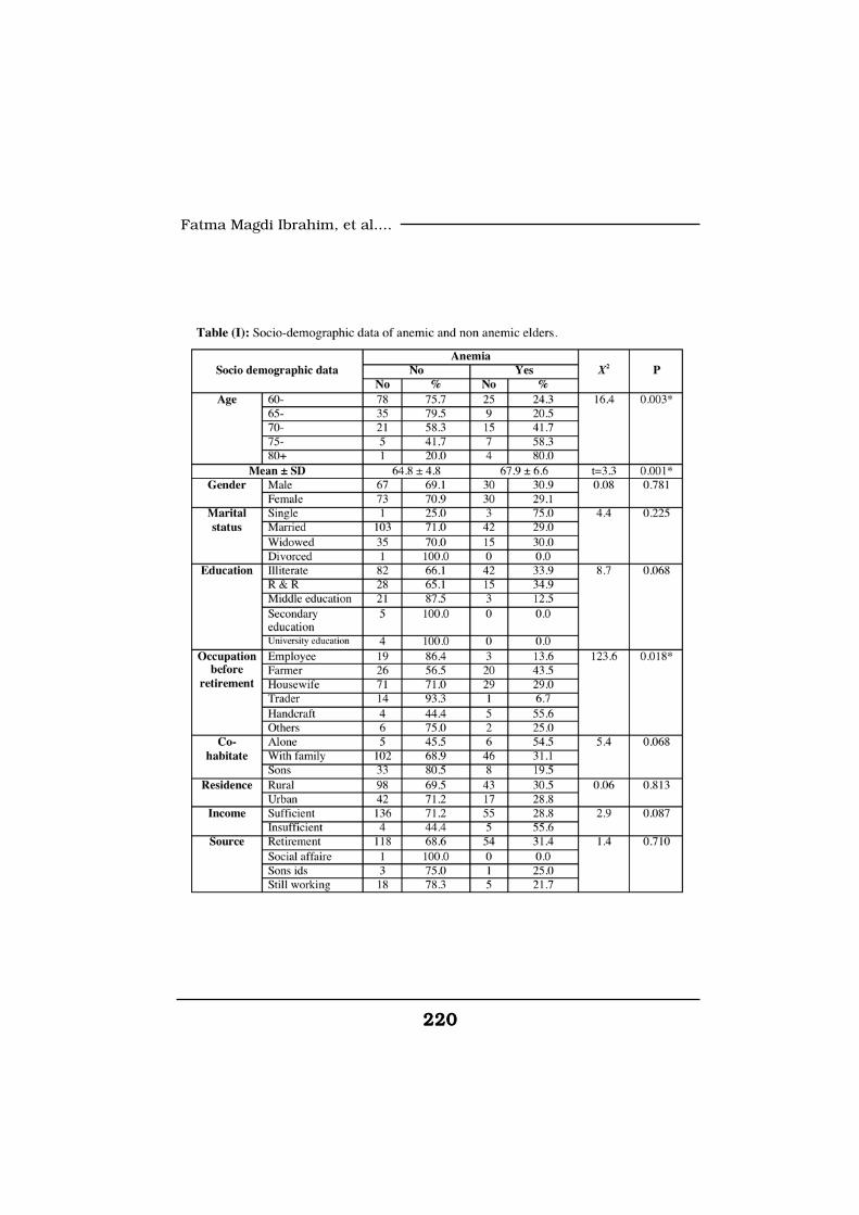
220220
Fatma Magdi Ibrahim, et al....
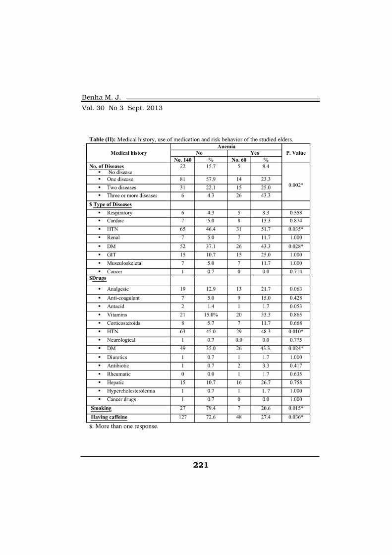
221
Benha M. J.
Vol. 30 No 3 Sept. 2013
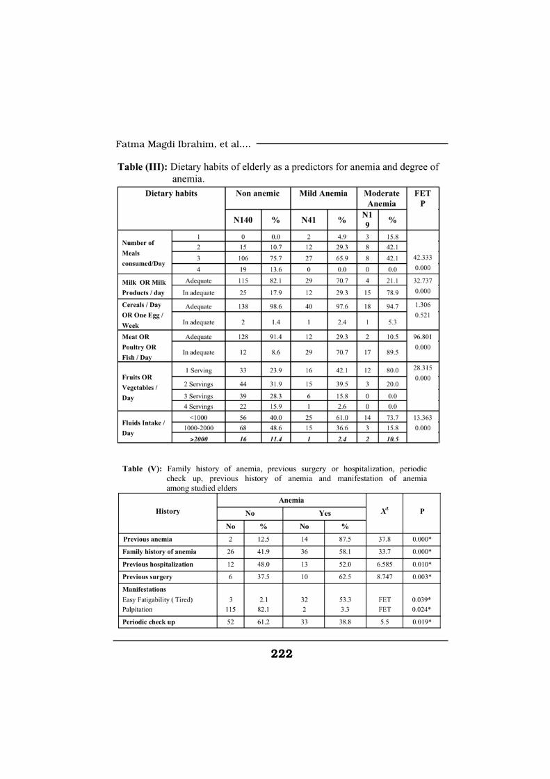
222222
Fatma Magdi Ibrahim, et al....
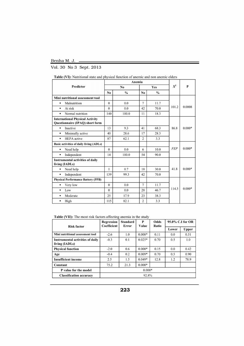
223
Benha M. J.
Vol. 30 No 3 Sept. 2013
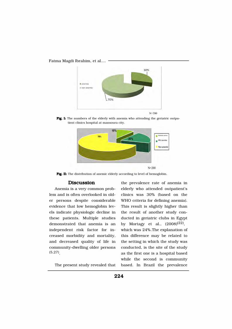
224224
Fatma Magdi Ibrahim, et al....
Discussion Discussion Anemia is a very common prob-
lem and is often overlooked in old-
er persons despite considerable
evidence that low hemoglobin lev-
els indicate physiologic decline in
these patients. Multiple studies
demonstrated that anemia is an
independent risk factor for in-
creased morbidity and mortality,
and decreased quality of life in
community-dwelling older persons(5,27).
The present study revealed that
the prevalence rate of anemia in
elderly who attended outpatient's
clinics was 30% (based on the
WHO criteria for defining anemia).
This result is slightly higher than
the result of another study con-
ducted in geriatric clubs in Egypt
by Mortagy et al., (2008)(22),
which was 24%.The explanation of
this difference may be related to
the setting in which the study was
conducted, is the site of the study
as the first one is a hospital based
while the second is community
based. In Brazil the prevalence
Fig. I: Fig. I: The numbers of the elderly with anemia who attending the geriatric outpa-tient clinics hospital at mansoura city.
Fig. II: Fig. II: The distribution of anemic elderly according to level of hemoglobin.
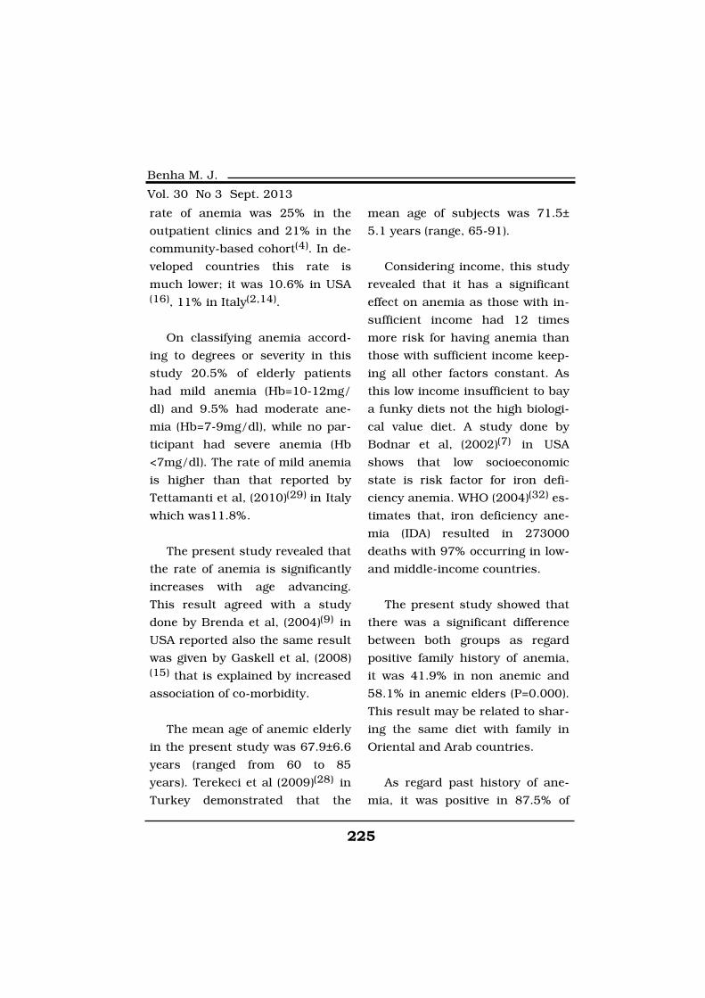
225
Benha M. J.
Vol. 30 No 3 Sept. 2013
rate of anemia was 25% in the
outpatient clinics and 21% in the
community-based cohort(4). In de-
veloped countries this rate is
much lower; it was 10.6% in USA(16), 11% in Italy(2,14).
On classifying anemia accord-
ing to degrees or severity in this
study 20.5% of elderly patients
had mild anemia (Hb=10-12mg/
dl) and 9.5% had moderate ane-
mia (Hb=7-9mg/dl), while no par-
ticipant had severe anemia (Hb
<7mg/dl). The rate of mild anemia
is higher than that reported by
Tettamanti et al, (2010)(29) in Italy
which was11.8%.
The present study revealed that
the rate of anemia is significantly
increases with age advancing.
This result agreed with a study
done by Brenda et al, (2004)(9) in
USA reported also the same result
was given by Gaskell et al, (2008)(15) that is explained by increased
association of co-morbidity.
The mean age of anemic elderly
in the present study was 67.9±6.6
years (ranged from 60 to 85
years). Terekeci et al (2009)(28) in
Turkey demonstrated that the
mean age of subjects was 71.5±
5.1 years (range, 65-91).
Considering income, this study
revealed that it has a significant
effect on anemia as those with in-
sufficient income had 12 times
more risk for having anemia than
those with sufficient income keep-
ing all other factors constant. As
this low income insufficient to bay
a funky diets not the high biologi-
cal value diet. A study done by
Bodnar et al, (2002)(7) in USA
shows that low socioeconomic
state is risk factor for iron defi-
ciency anemia. WHO (2004)(32) es-
timates that, iron deficiency ane-
mia (IDA) resulted in 273000
deaths with 97% occurring in low-
and middle-income countries.
The present study showed that
there was a significant difference
between both groups as regard
positive family history of anemia,
it was 41.9% in non anemic and
58.1% in anemic elders (P=0.000).
This result may be related to shar-
ing the same diet with family in
Oriental and Arab countries.
As regard past history of ane-
mia, it was positive in 87.5% of
226226
Fatma Magdi Ibrahim, et al....
anemic compared to 12.5% of non
anemic elders in the study. The
difference was significant (P=
0.000). This can be attributed to
the monotonous starchy diet upon
which the poor elders depend and
unable to change it. This agree
with study done in Egypt by Mor-
tagy et al., (2008)(22) which re-
vealed that there is statistical sig-
nificance difference between
anemia and past history of anemia
(P=0.001).
As regard manifestation of ane-
mia the study revealed that there
were statistically significant differ-
ence between two group as regard
easy fatigability and palpitation
(P=0.039 and P=0.024) respective-
ly. It agree with a study done by
Beghe et al, (2004)(6) in USA dem-
onstrated that anemia is associat-
ed with symptoms ranging from
weakness and fatigue to increased
falls and depression, and in severe
cases can lead to congestive heart
failure.
Concerning risk behavior and
periodic check up this study re-
vealed that there were statistically
significant reverse relations be-
tween smoking caffeine consump-
tion (p=0.036), adherence to peri-
odic check up (p=0.019) and oc-
currence of anemia. As 19.3% of
non anemic elders were smokers
compared to 11.6% smokers in
anemic elders (p=0.015). The rate
of caffeine consumption among non
anemic was 90.7% and it was 80%
in anemic elders (p=0.036). On the
contrary, a study done by Nelson
and Poulter (2004)(23) in UK re-
ported that tea drinking limits the
absorption of non haem iron.
The present study revealed that
there is a statistical significant in-
verse relation between dietary
habits and anemia regarding
number of meals (P=0.000),
enough consumption of: milk and
milk products (P=0.000), meat,
poultry or Fish (P=0.000), and
fruits or vegetables (P=0.000). Re-
garding consumption of cereals
the relation was statistically insig-
nificant (P=0.521). This result dis-
agree with the study done by Mor-
tagy et al., (2008)(22) in Egypt
which revealed that there is a sta-
tistically significance difference
between anemia and legume in-
take with (P-value=0.01).
Concerning medical history,
227
Benha M. J.
Vol. 30 No 3 Sept. 2013
the present study showed that
43.3% of anemic elders were com-
plaining of three or more chronic
illness compared to 4.3% of non
anemic (P=0.002). As regard the
relation between type of chronic
disease and anemia occurrence,
this work revealed that the signifi-
cant effect was detected only in di-
abetes and hypertension. As
43.3% of anemic elders have dia-
betes compared to 37.1% of non-
anemic (p=0.028). This agrees
with a study done by Anand et al,
(2005) in USA(1) on 5000 partici-
pants and found that 34% of ane-
mic patients were diabetics. Also
using of medications by elders in
this study revealed significant ef-
fect on occurrence of anemia only
when using hypoglycemic agents
(P=0.024) and antihypertensive
drug (P=0.010) There is good evi-
dence that many drugs used to
treat diabetes may exacerbate
anemia associated with diabetes.
Study done by Bolen et al, (2007)(8) in USA revealed that five dis-
tinct oral drug classes are now
available for the treatment of type
2 diabetes and most of these
agents lower hemoglobin levels ap-
proximately 1% to 2%. Also 51.7%
of anemic elders in this work had
hypertension compared to 46.4%
of non-anemic (p=0.035). The ex-
planation of this is that erythro-
poietin and anemia have impor-
tant interactions with blood
pressure control in both health
and disease. In patients with es-
sential hypertension, endogenous
erythropoietin levels are positively
correlated with blood pressure lev-
els and total peripheral resistance,
independent of hemoglobin levels.
It is conceivable that a reduction
in erythropoietin synthesis may
act to partly counterbalance blood
pressure elevation associated with
fluid retention in diabetes and
Chronic Kidney Disease(30).
The relation between anemia
and previous surgery is evident in
this work as 62.5% of those who
were anemic compared to 27.2%
of non anemic elderly conducted
previous surgery and the differ-
ence was significant (p=0.003)
This is in-agreement with a study
done by Jeong et al (2012)(19) in
Korea. Also previous hospitaliza-
tion was positive in 52.0% of those
who were anemic compared to
48.0% of non anemic elderly
(P=0.010). This is agree with a
study done by Riva et al (2009)(26)
228228
Fatma Magdi Ibrahim, et al....
in Italy and reported that the risk
of hospitalization was higher
among the mildly anemic non ane-
mic elderly subjects.
Concerning the relation be-
tween anemia and nutritional
status of the elders, in this work
using Mini Nutritional Assessment
Scale revealed that 11.7% of ane-
mic elders were mal-nourished
and 70% at risk of malnutrition
while all the non anemic were
well-nourished (P=0.000) After re-
gression analysis regarding MNA
score, the increase in the score by
one degree was associated with
decreased risk for anemia by
about 89% (Odds Ratio=0.11; 95%
Confidence Interval: 0.0-0.51).
Increasing functional deteriora-
tion is associated with decreasing
hemoglobin concentration in an
inverse and linear manner. It is
important to note, that even low
normal hemoglobin levels may be
a marker for declining function(10,25).
This study revealed that there
were significant relations between
anemia and ADLS result's where
10% of anemic elders need help
(P=0.000), IADLS result's revealed
that 30% of anemic elders need
help (P=0.000), Physical Perfor-
mance Battery (PPB) scale showed
that only 3.3% of them had high
performance (P=0.000) and for In-
ternational Physical Activity Ques-
tionnaire (IPAQ) scale it appeared
that 68.3% were inactive (P=0.000).
After regression analysis consider-
ing physical function, a 85.0%
lower risk for anemia was record-
ed for increased physical function
(Odds Ratio=0.15; 95% Confi-
dence Interval: 0.0-0.42). the in-
crease in IADL score by one more
degree was associated with de-
creased risk for anemia by about
30% (OR=0.70; 95% CI: 0.5-1.0) .
This is agree with another
study done by Penninx et al,
(2004)(25) in Italy which revealed
that anemic persons had poorer
performance (8.8 vs. 9.6, P=.003)
than persons without anemia. An-
other study done by Den Elzen et
al, (2009)(12) in Canada was simi-
lar to the present study which
demonstrated that Participants
with anemia had a greater in-
crease in disability in basic activi-
ties in daily living compared with
participants who did not have
229
Benha M. J.
Vol. 30 No 3 Sept. 2013
anemia during follow-up (differ-
ence in annual change in Groning-
en Activity Restriction Scale score
1.7, 95% CI 0.8 to 2.7, p<0.01).
ConclusionConclusionBased on findings of the
present study, it can be concluded
that anemia is a common problem
among elderly and it’s prevalence
increase with advancing age. The
most important predictor for ane-
mia in the present study was age,
income and nutritional habits.
Anemia has several adverse conse-
quences in the elderly; it increases
the incidence of fatigue, and has a
negative impact on cognitive and
physical function.
The present study revealed that
the increase in IADL score by one
more degree was associated with
decreased risk for anemia by
about 30% (OR=0.70; 95% CI: 0.5-
1.0). Considering physical func-
tion, a 85.0% lower risk for ane-
mia was recorded for increased
physical function (OR=0.15; 95%
CI: 0.0-0.42). Functional deterio-
ration is associated with decreas-
ing hemoglobin concentration in
an inverse and linear manner. Im-
paired physical function or disa-
bility lead to anemia as the elder
person become unable to do shop-
ping, preparing food or even feed
himself , this lead to anemia that
increases his disability.
RecommendationsRecommendationsBased on the results of this
study, the following recommenda-
tions are suggested:
• Health education programs to
elders about the disease process
and the importance of adoption
healthy lifestyle.
• Early identifying causes and
high risk behaviors conducive to
anemia by the caregivers is im-
portant in order to motivate elders
to modify them. It can be achieved
through comprehensive assess-
ment of elders which will provide
the nurse with baseline data, help
to identify high risk groups, plan
and implement appropriate nurs-
ing intervention.
• In- service training programs
to all nurses and health care pro-
viders in hospitals and outpatient
clinics to update their knowledge
increase their ability to care for
elderly patients with anemia, and
teach patients to modify their un-
230230
Fatma Magdi Ibrahim, et al....
healthy lifestyle behaviors.
• The evaluation of newly diag-
nosed anemia should not be de-
layed in patients with a life expec-
tancy of 1 year or longer, because
anemia may be the first sign of se-
rious underlying pathology.
• Follow up visits either to the
clinic or through home visits is
important in order to evaluate the
progress of patient's condition and
motivate them to adhere with pre-
ventive measures.
ReferencesReferences1. Anand I.S., Kuskowski1. Anand I.S., Kuskowski
M.A., Rector T.S., et al. (2005):M.A., Rector T.S., et al. (2005):
Anemia and change in hemoglobin
over time related to mortality and
morbidity in patients with chronic
heart failure. 112(8): 1121-7.
2. Artz A.S., Ershler W.B., et2. Artz A.S., Ershler W.B., et
al. (2009):al. (2009): Anemia in elderly per-
son. Monde Dent J .88(2):107-14 .
3. Balducci L., Ersher W.B.3. Balducci L., Ersher W.B.
and Bennett J.M. (2007):and Bennett J.M. (2007): Anemia
in elderly, 1st ed., Springer Com-
pany: New York, Chapter 2, 21-37.
4. 4. Barcellos D.S.E., Tello B.S.,Barcellos D.S.E., Tello B.S.,
Villacorta H., et al. (2010):Villacorta H., et al. (2010): Ane-
mia and Heart Failure in a Com-
munity-based Cohort: Comparison
with a Specialized Outpatient Clin-
ic. Arq Bras Cardiol; 94(1): 95-101.
5. Beattie W.S., Karkouti K.,5. Beattie W.S., Karkouti K.,
Wijeysundera D.N. and Tait G.Wijeysundera D.N. and Tait G.
(2009):(2009): Risk associated with preop-
erative anemia in noncardiac sur-
gery: a single-center cohort study.
Anesthesiology. 110(3):574-581.
6. Beghé C., Wilson A. and6. Beghé C., Wilson A. and
Ershler W.B. (2004):Ershler W.B. (2004): Prevalence
and outcomes of anemia in geriat-
rics: A systematic review of the lit-
erature. Am J Med. 116(7):3-10.
7. Bodnar L.M., Cogswell7. Bodnar L.M., Cogswell
M.E. and Scanlon K.S. (2002):M.E. and Scanlon K.S. (2002):
Low income postpartum women
are at risk of iron deficiency. J
Nutr. 132(2):298-302.
8. Bolen S., Leonard F., Ja-8. Bolen S., Leonard F., Ja-
son V., et al. (2007):son V., et al. (2007): Systematic
Review: Comparative Effectiveness
and Safety of Oral Medications for
Type 2 Diabetes Mellitus. Ann In-
tern Med. (147): 386-99.
9. Brenda W.J., Penninx H.,9. Brenda W.J., Penninx H.,
Pahor M., Cesari M., et al.Pahor M., Cesari M., et al.
231
Benha M. J.
Vol. 30 No 3 Sept. 2013
(2004): (2004): Anemia Is Associated with
Disability and Decreased Physical
Performance and Muscle Strength
in the Elderly. JAGS. (52):719-24.
10. Chaves P.H., Ashar B.,10. Chaves P.H., Ashar B.,
Guralnik J.M., et al. (2002):Guralnik J.M., et al. (2002):
looking at the relationship be-
tween hemoglobin concentration
and prevalent mobility difficulty in
older women. Should the criteria
currently used to define anemia in
older people be reevaluated? J Am
Geriatr Soc; (50):1257-64.
11. Craig C., Marshall A.,11. Craig C., Marshall A.,
Sjostrom M., Bauman A., BoothSjostrom M., Bauman A., Booth
M., Ainsworth B., Ekelund U., etM., Ainsworth B., Ekelund U., et
al. (2003):al. (2003): International physical
activity questionnaire: 12 country
reliability and validity. Medicine
and science in sports and exer-
cise. 35(8)1381-95.
12. Den Elzen W.P., Willems12. Den Elzen W.P., Willems
J.M., Westendorp R.G., et al.J.M., Westendorp R.G., et al.
(2009):(2009): Effect of anemia and co-
morbidity on functional status
and mortality in old age: results
from the Leiden 85-plus Study.
CMAJ. Aug 4; 181(34):151-7.
13. Eisenstaedt R., Penninx13. Eisenstaedt R., Penninx
B.W. and Woodman R.C. (2006):B.W. and Woodman R.C. (2006):
Anemia in the elderly: current un-
derstanding and emerging con-
cepts. Blood Rev; 20:213–26.
14. Ferrucci L., Maggio M.,14. Ferrucci L., Maggio M.,
Bandinelli S., Basaria S., Laure-Bandinelli S., Basaria S., Laure-
tani F., Ble A., et al. (2006):tani F., Ble A., et al. (2006): Low
testosterone levels and the risk of
anemia in older men and women.
Arch Intern Med; (166):1380-88.
Available on [PubMed: 16832003].
15. Gaskell H., Derry S.,15. Gaskell H., Derry S.,
Moore R.A., et al. (2008):Moore R.A., et al. (2008): Preva-
lence of anemia in older persons:
Systematic Review. BMC Geriat-
rics. 8:1.
16. Guralnik J.M., Eisen-16. Guralnik J.M., Eisen-
staedt R.S., Ferrucci L., et al.staedt R.S., Ferrucci L., et al.
(2004):(2004): Prevalence of anemia in
persons 65 years and older in the
United States: Evidence for a high
rate of unexplained anemia.
Blood; (104):2263-68.
17. Guralnik J.M., Simonsick17. Guralnik J.M., Simonsick
E.M., Ferrucci L., et al. (1999):E.M., Ferrucci L., et al. (1999): A
short physical performancebattery
assessing lower extremity func-
tion: Association with self-
reporteddisability and prediction
of mortality and nursing home ad-
mission. J Gerontol; (49): 85-94.
232232
Fatma Magdi Ibrahim, et al....
18. Hallaj A.F. (2007):18. Hallaj A.F. (2007): Activity
Patterns of Residents In Elderly
Homs. Unpublished Master The-
sis. Faculty of Nursing, University
of Alexandria.
19. Jeong O.H., Park Y.K. and19. Jeong O.H., Park Y.K. and
Ryu S.Y. (2012):Ryu S.Y. (2012): Prevalence, se-
verity, and evolution of postsurgi-
cal anemia after gastrectomy, and
clinicopathological factors affect-
ing its recovery. J Korean Surg
Soc. February; 82(2): 79-86.
20. Katz S. and Akpom C.20. Katz S. and Akpom C.
(1976):(1976): Index of ADL: A standard-
ized measure of biological and
psychological function. Interna-
tional Journal of Health Science.
(6):123-140.
21. Lawton H.P., Brody E.M.21. Lawton H.P., Brody E.M.
(1969):(1969): Assessment of older: self
maintaining and instrumental ac-
tivities of daily living. Gerontolo-
gist.1. (9):179-186.
22. Mortagy A.K., Farid M.T.22. Mortagy A.K., Farid M.T.
and Abd elrahman O. (2008):and Abd elrahman O. (2008):
Prevalence of Anemia Among
Elders Attending Elderly Clubs
in Cairo "Master thesis", Faculty
of Medicine, Ain Shams Universi-
ty.
23. Nelson M. and Poulter J.23. Nelson M. and Poulter J.
(2004):(2004): Impact of tea drinking on
iron status in the UK. The British
Dietetic Association. J Hum Nutr
Dietet. (17): 43-54.
24. Penninx B.W., Guralnik24. Penninx B.W., Guralnik
J.M., Onder G., et al. (2003):J.M., Onder G., et al. (2003):
Anemia and decline in physical
performance among older persons.
Am J Med. (115):104-10.
25. Penninx B.W., Pahor M.,25. Penninx B.W., Pahor M.,
Cesari M., et al. (2004):Cesari M., et al. (2004): Anemia
is associated with disability and
decreased physical performance
and muscle strength in the elder-
ly. J Am Geriatr Soc. (52):719-24.
26. Riva E., Tettamanti M.,26. Riva E., Tettamanti M.,
Mosconi P., Apolone G., GandiniMosconi P., Apolone G., Gandini
F., Nobili A., et al. (2009):F., Nobili A., et al. (2009): Asso-
ciation of mild anemia with hos-
pitalizationand mortality in the
elderly: the Health and Anemia-
population-based study. Haemato-
logica; (94):22-8.
27. Steensma D.P. and Tefferi27. Steensma D.P. and Tefferi
A. (2007): A. (2007): Anemia in the Elderly:
How Should We Define It, When
Does It Matter, and What Can Be
Done? Mayo Clin Proc. 82(8):958-
66.
233
Benha M. J.
Vol. 30 No 3 Sept. 2013
28. Terekeci H.M., Kucukar-28. Terekeci H.M., Kucukar-
dali Y., Onem Y., et al. (2010):dali Y., Onem Y., et al. (2010):
Relationship between anaemia
and cognitive functions in elderly
people. European Journal of Inter-
nal Medicine. (21): 87-90.
29. Tettamanti M., Lucca U.,29. Tettamanti M., Lucca U.,
Gandini F., et al. (2010):Gandini F., et al. (2010): Preva-
lence, incidence and types of mild
anemia in the elderly: the “Health
and Anemia population-based
study, (11): 1849-56.
30. Thomas M.C., MacIsaac30. Thomas M.C., MacIsaac
R.J., Tsalamandris C., Power D.R.J., Tsalamandris C., Power D.
and Jerums G. (2003):and Jerums G. (2003): Unrecog-
nized anemia in patients with dia-
betes: a cross-sectional survey.
Diabetes Care. 26(4): 1164-9.
31. Vellas B., Villars H., Abel-31. Vellas B., Villars H., Abel-
lan G., et al. (2006):lan G., et al. (2006): Overview of
MNA® - Its History and Challenges.
J Nut Health Aging. (10): 456-65.
32. World Health Organiza-32. World Health Organiza-
tion/UNICEF/UNU (2004):tion/UNICEF/UNU (2004): Iron
Deficiency Anaemia: Assessment,
Prevention, and Control. A Guide
for Programme Managers. Geneva,
Switzerland: World Health Organi-
zation.
33. Woodman R., Ferrucci L.33. Woodman R., Ferrucci L.
and Guralnik J. (2005):and Guralnik J. (2005): Anemia
in older adults. Curr Opin Hema-
tol; 12:123–8.
234234
Fatma Magdi Ibrahim, et al....
ANEMIA, PHYSICAL PERFORMANCEANEMIA, PHYSICAL PERFORMANCERELATIONSHIP AMONG ELDERYRELATIONSHIP AMONG ELDERY
PATIENTS ATTENDING GERIATRICPATIENTS ATTENDING GERIATRICOUTPATIENT CLINICS INOUTPATIENT CLINICS IN
MANSOURA CITYMANSOURA CITY
Fatma Magdi Ibrahim M.Sc, Soad Hassan Abd El HamidFatma Magdi Ibrahim M.Sc, Soad Hassan Abd El HamidMD and Farida Abdel-Wahab Ph.DMD and Farida Abdel-Wahab Ph.D
BENHAMEDICALJOURNAL
REPRINT
Published byPublished by
Benha Faculty of MedicineBenha Faculty of Medicine Volume 30 Number 3Sept. 2013
235
Benha M. J.
Vol. 30 No 3 Sept. 2013
EFFECT OF ANTICOAGULANT (WARFARIN)EFFECT OF ANTICOAGULANT (WARFARIN)AND L-CARNITINE ON HAEMOSTATICAND L-CARNITINE ON HAEMOSTATIC
FUNCTION AND OXIDATIVE STRESS INFUNCTION AND OXIDATIVE STRESS INSTREPTOZOTOCIN-INDUCED DIABETIC RATSSTREPTOZOTOCIN-INDUCED DIABETIC RATS
Ahmed A. El-Gendy Ph.D and Amr M. Abbas Ph.DAhmed A. El-Gendy Ph.D and Amr M. Abbas Ph.DDepartment of Medical Physiology, Faculty of Medicine,
Mansoura University, Egypt
AbstractAbstractBackground and Aim of Work: Background and Aim of Work: Diabetes mellitus (DM) is a complex pro-
gressive disease characterized by hyperglycemia and a high risk of athe-rothrombotic disorders affecting the coronary, cerebral and peripheralarterial trees. Oxidative stress is reported in diabetic patients. We investigat-ed the haemostatic functions and oxidative stress in streptozotocin (STZ)-induced diabetic rats and the effects of anticoagulant (warfarin) and L-carnitine on those parameters.
Materials and Methods: Materials and Methods: Forty male Sprague-Dawley rats were dividedinto four groups; control, DM, DM received warfarin or L-carnitine. In allrats, blood glucose, insulin, haemoglobin A1c (HBA1c), fibrinogen, factor VII(FVII), plasminogen activator inhibitor-1 (PAI-1), fibrin degradation products(FDP), protein C, malondialdehydes (MDA), and anti-oxidants (superoxidedismutase, catalase, glutathione peroxidase, glutathione) were measured.Also, prothrombin time (PT), activated partial thromboplastin time (aPTT)and platelet aggregation were evaluated.
Results:Results: In STZ-induced diabetic rats, plasma glucose, HBA1c, MDA, fi-brinogen, FVII, PAI-1 and platelet aggregation increased while insulin, PT,aPTT, FDP, protein C and anti-oxidants decreased. Warfarin administrationto diabetic rats decreased fibrinogen and FVII and increased PT and aPTTwith no effect on MDA, anti-oxidants, PAI-1, protein C, FDP and platelet ag-gregation. On the other hand L-carnitine decreased fibrinogen, FVII, PAI-1,MDA and platelet aggregation and increased PT, aPTT, protein C, FDP andanti-oxidants in diabetic rats.
Conclusion: Conclusion: Hyperglycemia plays an important role in hypercoagulationstate and oxidative stress in STZ-induced DM. L-carnitine improves oxida-
236236
Ahmed A. El-Gendy and Amr M. Abbas
IntroductionIntroductionAccording to the International
Diabetes Federation, percent of di-
abetes in Egypt was 11.4% in the
year 2010 and this likely to in-
crease to 13.7 % by the year 2030(1). Diabetes mellitus (DM) is a
complex progressive disease,
which is accompanied by multiple
complications. It has been recog-
nized as the sole independent risk
factor for the development of car-
diovascular disease(2). Adminis-
tration of streptozotocin (STZ)
causes pancreatic beta cell de-
struction that leads to the devel-
opment of hyperglycemia, dyslipi-
demia and renal dysfunction in
rats(3). The STZ animal model de-
velops characteristic symptoms of
diabetes such as hyperglycemia,
hyperlipidemia and increased wa-
ter and food intake without body
weight gain. Reactive oxygen spe-
cies (ROS), which cause cellular
damage by the oxidation ability,
have been implicated in the path-
ogenesis of diabetes mellitus(4).
During diabetes, persistent hyper-
glycemia increases the production
of ROS through glucose autoxida-
tion(5). In addition, oxidative
stress in diabetes mellitus results
from reduction in capacities of the
antioxidant defense system in-
cluding scavenging enzymes such
as superoxide dismutase and glu-
tathione reductase, and deficien-
cies of antioxidants such as vita-
min C and E(6). The oxidative
stress has also been associated
with diabetic states in animals
and humans(7). A study using
STZ-induced diabetic rats showed
that levels of lipid peroxidation
had increased, as indicated by thi-
obarbituric acid reactive sub-
stances (TBARS), which is one of
oxidative stress markers suggest-
ing the occurrence of oxidative
stress(8). Moreover, diabetic pa-
tients have significant defects in
antioxidant defense elements,
and enhanced ROS generation is
one of the major determinants of
diabetic complications(9). Admin-
istration of antioxidants as vita-
mins C and E has been reported
tive stress and decreases the hypercoagulation state in DM. On the otherhand, warfarin normalize the hypercoagulation state in DM with no effect onoxidative stress.
Key Words:Key Words: diabetes mellitus, coagulation, L-carnitine, warfarin, oxida-tive stress.

237
Benha M. J.
Vol. 30 No 3 Sept. 2013
to reduce the complications in DM
by arresting free radical damage(10).
Besides hyperglycemia, diabetic
patients also suffer from dyslipide-
mia(11), which can lead to in-
creased atherogenesis and heart
disease(12). In addition, clinical
and epidemiological observations
have led to the concept of a proco-
agulant state in type 2 diabetes.
Thrombophilia in diabetic patients
is a well recognized phenomenon
which contributes an additional
risk of coronary heart disease
(CHD). In a series of 1980 type 2
diabetic patients, both male and
female diabetic patients showed
significantly shorter activated par-
tial thromboplastin time (aPTT)(13). Acang and Jalil(14) have re-
ported significantly high fibrino-
gen and short prothrombin time
(PT) and aPTT in diabetic patients,
especially those who suffered from
diabetes for a long time. Increased
concentration of fibrinogen in un-
controlled non insulin dependent
diabetes mellitus (NIDDM) pa-
tients is implicated in vascular
damage induction(15). Yurekli et al(16) have concluded a subtle acti-
vation of extrinsic pathway with a
concomitant decrement in intrin-
sic pathway of the coagulation
cascade in type 2 diabetes. In ad-
dition, Bae et al(17) observed that
PT and aPTT did not differ signifi-
cantly between diabetic patients
and controls; however a signifi-
cant decrease was observed in fi-
brinogen levels in diabetic pa-
tients. Short aPTT and PT have
been associated with increased
risk for thromboembolism in renal
transplant patients(18). In a series
of 35 type 1 diabetic patients,
there was no significant difference
in coagulation parameters and
none of the patients developed
thrombosis or vascular disease in
a two year follow up(19).
L-carnitine is a naturally occur-
ring compound and an antiradical
widely distributed in the body,
and decreases lipid peroxidation(20). L-carnitine prevents TBARS
formation and increases the anti-
oxidants in aged rats’ brain(20).
Although these previous studies
suggest that L-carnitine possesses
antioxidative activity, it remains
unknown whether this compound
is able to affect coagulation asso-
ciated factors. It is hypothesized
that L-carnitine could attenuate
238238
Ahmed A. El-Gendy and Amr M. Abbas
diabetes associated haemostatic
disorder, at least via diminishing
oxidative stress. In addition, many
physiologic and pathologic condi-
tions affect blood, tissue and uri-
nary concentrations of carnitine in
both animal and human subjects.
In acute or chronic STZ-induced
diabetic rats, the pancreatic con-
tent of carnitine was found to be
significantly lower than in nondia-
betic control rats(21). Further-
more, studies on the antioxidant
effect of carnitine on diabetes mel-
litus are few. Therefore, we inves-
tigated the effect of L-carnitine on
the diabetogenic action of STZ in
rats.
Warfarin (Coumadin) is an anti-
coagulant normally used in the
prevention of thrombosis and
thromboembolism(22). Warfarin
inhibits the vitamin K-dependent
synthesis of biologically active
forms of the calcium-dependent
clotting factors II, VII, IX and X, as
well as the regulatory factors pro-
tein C, protein S, and protein Z(23). As there are few studies con-
sidering the anticoagulant effect of
warfarin and L-carnitine on
diabetes mellitus, therefore, the
aim of the present work was to
investigate the haemostatic
functions and oxidative stress in
streptozotocin (STZ)-induced dia-
betic rats, a well-characterized an-
imal model of type 1 diabetes, and
the effects of anticoagulant (war-
farin) and L-carnitine on those pa-
rameters.
Materials and MethodsMaterials and MethodsChemicals:Chemicals: STZ (streptozoto-
cin), warfarin [4-Hydroxy-3-(3-
oxo-1-phenylbutyl)coumarin] and
L-carnitine were obtained from
Sigma-Aldrich (St. Louis, MO,
U.S.A.).
Animals:Animals: Forty male Sprague
Dawley rats, weighing 220-240 g,
were used in the present study.
They were purchased from Vac-
cine and Immunization Authority
(Helwan, Cairo, Egypt) and housed
(Animal House, Medical Physiolo-
gy department, Faculty of Medi-
cine, Mansoura University, Egypt)
under controlled conditions (tem-
perature 23±1°C, and a 12:12
light/dark cycle).The animals were
allowed free access to food and tap
water. All animal procedures were
performed in accordance with pro-
tocols approved by the Medical Re-
search Ethics Committee of Man-
239
Benha M. J.
Vol. 30 No 3 Sept. 2013
soura University, Egypt and in
compliance with standards and
regulations for the care and use of
laboratory rats set by the National
Institutes of Health.
Experimental design:Experimental design:
The animals were randomly di-
vided into four groups of ten rats
each. First group (GI) consisted of
untreated control (normal) ani-
mals. The second group (GII)
served as untreated diabetic
group. The third group (GIII) in-
cluded the diabetic rats treated
with warfarin. The forth group
(GIV) included the diabetic rats
treated with L-carnitine. Diabetes
was induced in rats of second,
third and forth groups by single
intra-peritoneal (i.p) injection of
70 mg\kg streptozotocin(24) (dis-
solved in 0.1 M sodium-citrate
buffer, pH 4.5). The control rats
received an equivalent amount of
the sodium-citrate buffer. In order
to confirm diabetes, three days af-
ter streptozotocin injection, blood
glucose was measured using glu-
cometer instrument (Accu-check-
active, ROCHE, Germany) and
animals with blood glucose over
200 mg\ dL were considered as
diabetics (25). The rats with war-
farin treatment (GIII) were admin-
istered 0.25 mg/liter warfarin po-
tassium (26) in drinking water
from the day of STZ injection for
21 consecutive days. Because dia-
betic rats drink much more water,
the dosage of warfarin was re-
duced to 0.06 mg/liter(26) from 2
days after injection of STZ. Group
IV rats were injected intraperito-
neally with L-carnitine a dose of
500 mg/kg/day for 21 consecutive
days after STZ injection(27).
Sampling protocol:Sampling protocol:
Blood samples:Blood samples: At the end of
experimental period, blood sam-
ples were collected by cardiac
puncture into tubes containing
EDTA, mixed well and divided into
two tubes: the first one was used
for determination of hemoglobin
A1c (HBA1c), and the second tube
was centrifuged at 3000 r/min
(1000g) for 10 min to obtain plas-
ma samples which were used for
measurement of glucose, insulin,
malondialdehyde (MDA), and anti-
oxidants (reduced glutathione,
glutathione peroxidase, superoxide
dismutase, and catalase) levels.
Biochemical investigations: Biochemical investigations:
Plasma glucose was quantitat-

240240
Ahmed A. El-Gendy and Amr M. Abbas
ed by glucose oxidase-peroxidase
method using the kit supplied by
SPINREACT, Spain (Ref:1001190).
Plasma insulin was determined
using Ultra Sensitive Rat Insulin
ELISA Kit (Cat.No. 90060, Crystal
Chem INC, Spain) following manu-
facturer’s instructions. In addi-
tion, hemoglobin A1c (HBA1c) was
measured by a commercial kit
(Crystal Chem's rat HbA1c Kit,
Catalog. No. 80300).
Malondialdehyde (MDA) was
analyzed by measuring the pro-
duction of thiobarbituric acid re-
active substances (TBARS) accord-
ing to the method of Buege and
Aust(28) using TBARS assay kit
(Cat. No. 10009055, Cayman,
USA). Reduced glutathione (GSH)
(Cayman kit, Cayman, USA, Cat.
No. 703002), glutathione peroxi-
dase (GSH-Px) (Northwest Life Sci-
ence Specialties [NWLSSTM] kit,
Canada, Cat. No. NWK-GPX01),
superoxide dismutase (SOD) (Cay-
man kit, Cayman, USA, Cat. No.
706002), and catalase (CAT)
(Northwest Life Science Specialties
[NWLSSTM] kit, Canada, Cat. No.
NWK-CAT01) were measured ac-
cording to the manufacturer’s in-
structions.
Haemostatic functions: Haemostatic functions:
Assessment of prothrombinAssessment of prothrombin
time (PT) and activated partialtime (PT) and activated partial
thromboplastin time (aPTT):thromboplastin time (aPTT):
PT and APTT were measured
using calcium rabbit brain throm-
boplastin and kaolin platelet
substitute techniques (DIAGEN
DIAGNOSTIC REAGENT LTD, OXON,
U.K). Briefly, PT was assayed with
two hundred microlitre of calcium
rabbit brain thromboplastin rea-
gent placed in a clotting tube and
incubated in a water bath at 37°C
for 2 minutes. Hundred microlitre
of plasma is then added and a
stop watch started. The tube is
gently tilted at regular interval
and the watch was stopped when
the clot formation was observed.
For aPTT, two hundred microlitre
of kaolin platelet substitute mix-
ture was placed in a clotting tube
and incubated in a water bath at
37°C for 2 minutes. One hundred
microliter of plasma was then add-
ed and the tube was gently tilted
at interval for exactly two min-
utes. One hundred microliter of
calcium chloride (pre- incubated
at 37°C) was then added and a
stop watch started. The tube was
tilted at intervals and the time for
clot formation was recorded.
241
Benha M. J.
Vol. 30 No 3 Sept. 2013
Measurement of coagulationMeasurement of coagulation
and anticoagulation factors:and anticoagulation factors:
Coagulation factors, FI (fibrino-
gen) and FVII; anticoagulation fac-
tors, AT-III (antithrombin III) and
protein C; plasminogen activator
inhibitor-1 (PAI-1) and fibrin deg-
radation products (FDP) were
measured. Blood samples were
anticoagulated using sodium cit-
rate according to the protocols
provided by the manufacturers.
Fibrinogen level was measured us-
ing TEClot FIB kit (TECO, GmbH,
Germany). In addition, FVII activi-
ty was determined by a commer-
cial kit (CoasetQR FVII, Chromo-
genix, Lexington, Mass., U.S.A.).
The activity of AT-III and protein C
in plasma was measured by com-
mercial AT-III and protein C kits
(Sigma Chemical Co.), respective-
ly. The activity of AT-III and pro-
tein C was expressed as a percent-
age related to the activity of standard
plasma. PAI-1 activity (pg/ml) was
assayed by a commercial kit (Trin-
ity Biotech plc., Bray, C. Wicklow,
Ireland). Fibrin degradation prod-
ucts (FDP) is measured by Cusa-
bio rat FDP ELISA assay kit (Cata-
log No. CSB-E07942r).
Assessment of Platelet Aggre-Assessment of Platelet Aggre-
gation:gation:
Platelet rich plasma (PRP)Platelet rich plasma (PRP)
preparation: preparation: To obtain PRP, the
blood was centrifuged at about 250
g at 4°c for 10 min. Platelet count
was made by coulter LH 750 ana-
lyzer, (Beckman Coulter Inc., USA)
for PRP and for whole blood.
Platelet Aggregation:Platelet Aggregation: The
platelet aggregation profile was
evaluated by the method of Born(29) by measuring turbidity with a
Chronolog optical aggregometer
(AGGRO/LINK® Model 810-CA
software) using ADP at a concen-
tration of 5 and 10 µmol/l as an
agonist. The results were expressed
as percentage of aggregation.
Statistical analysis:Statistical analysis: The data
were expressed as mean ± standard
deviation of mean (Mean±SDM).
Data were processed and analyzed
using the Statistical Package of So-
cial Science version 10.0 (SPSS,
version 10.0). One way ANOVA was
done followed by Tukey’s post hoc
test. A minimum level of signifi-
cance is considered if P is ≤0.05.
ResultsResultsTable 1 shows the results of in-
sulin, glucose, and hemoglobin
242242
Ahmed A. El-Gendy and Amr M. Abbas
A1c (HbA1c). Plasma glucose and
HbA1c levels significantly increased
while insulin decreased in STZ-
induced diabetic rats as compared
to the control group (p<0.05).
Moreover, diabetic rats receiving
warfarin or L-carnitine showed no
changes in FBG, HbA1c or plasma
insulin levels (Table 1).
The changes in platelet count,
fibrinogen, plasminogen activator
inhibitor 1 (PAI-1) and fibrin deg-
radation products (FDP) concen-
trations, and prothrombin time
(PT) and activated partial throm-
boplastin time (aPTT) are shown in
Table (2). Streptozocin-induced di-
abetic rats showed a significant
increase in fibrinogen and PAI-1
and decrease in FDP, PT and aPTT
in comparison with control rats
(p<0.05). No significant change in
platelet count in the whole blood
or PRP was observed in STZ-
induced diabetic rats as compared
to control group (p>0.05). Warfar-
in administration to diabetic rats
significantly increased PT and
aPTT (p<0.05) and decreased fi-
brinogen concentration (p<0.05)
with no significant effect on FDP,
PAI-1 and platelet count (p>0.05)
compared with untreated diabetic
rats. Fibrinogen concentration, PT
and aPTT in diabetic rats treated
with warfarin showed a normal
values similar to those of control
group (p>0.05). On the other
hand, L-carnitine significantly in-
creased FDP, PT and aPTT (p<0.05)
and decreased fibrinogen concen-
tration and PAI-1 activity (p<0.05)
in diabetic group compared with
untreated diabetic rats, but their
values were significantly different
from those of control and warfarin
treated diabetic groups (p<0.05).
In addition, no significant effect
on platelet count was detected in
diabetic rats with L-carnitine
treatment (p>0.05) (Table 2).
The changes in factor VII, ATIII
and protein C are demonstrated in
figures 1, 2 and 3 respectively.
While ATIII, protein C significantly
decreased in STZ-induced diabetic
rats when compared with control
group (p<0.05), factor VII in-
creased. Warfarin significantly de-
creased factor VII (Figure 1)
(p<0.05) but had no effect on ATIII
(Figure 2) and protein C (Figure 3)
(p>0.05), in diabetic rats. On the
other hand, L-carnitine signifi-
cantly decreased factor VII (Figure
1) and increased ATIII (Figure 2)

243
Benha M. J.
Vol. 30 No 3 Sept. 2013
and protein C (Figure 3) activities
in diabetic rats but their values
were significantly different from
those of control and warfarin
treated diabetic groups (p<0.05).
Table 3 shows the effects of
warfarin and L-carnitine on ADP-
induced platelet aggregation in
control and STZ-induced diabetic
rats. ADP-induced platelet aggre-
gation was significantly increased
in STZ-induced diabetic rats when
compared with the control group
(p<0.05). Warfarin had no signifi-
cant effect on ADP-induced plate-
let aggregation in diabetic rats
(p>0.05). On the other hand, L-
carnitine significantly decreased
ADP-induced platelet aggregation
in diabetic rats when compared
with control and warfarin treated
diabetic groups (p<0.05).
The changes in GSH-Px, CAT,
and SOD activities and MDA and
GSH levels in the plasma of con-
trol and STZ-induced diabetic rats
are shown in Tables 4. GSH-Px,
CAT, and SOD activities and MDA
and GSH levels in the plasma
were affected by diabetes mellitus.
While GSH-Px, CAT, and SOD ac-
tivities and GSH levels in the
plasma were significantly de-
creased (p<0.05) in the STZ-
induced diabetic group compared
with the control group, MDA
levels increased (p < 0.05). L-
carnitine significantly increased
GSH levels and GSH-Px, CAT, and
SOD activities and decreased MDA
levels in the plasma of diabetic
rats compared with the control
group (p<0.05). On the other
hand, warfarin had no significant
effect on GSH-Px, CAT, and SOD
activities and MDA and GSH levels
in the plasma of diabetic rats
(p>0.05).
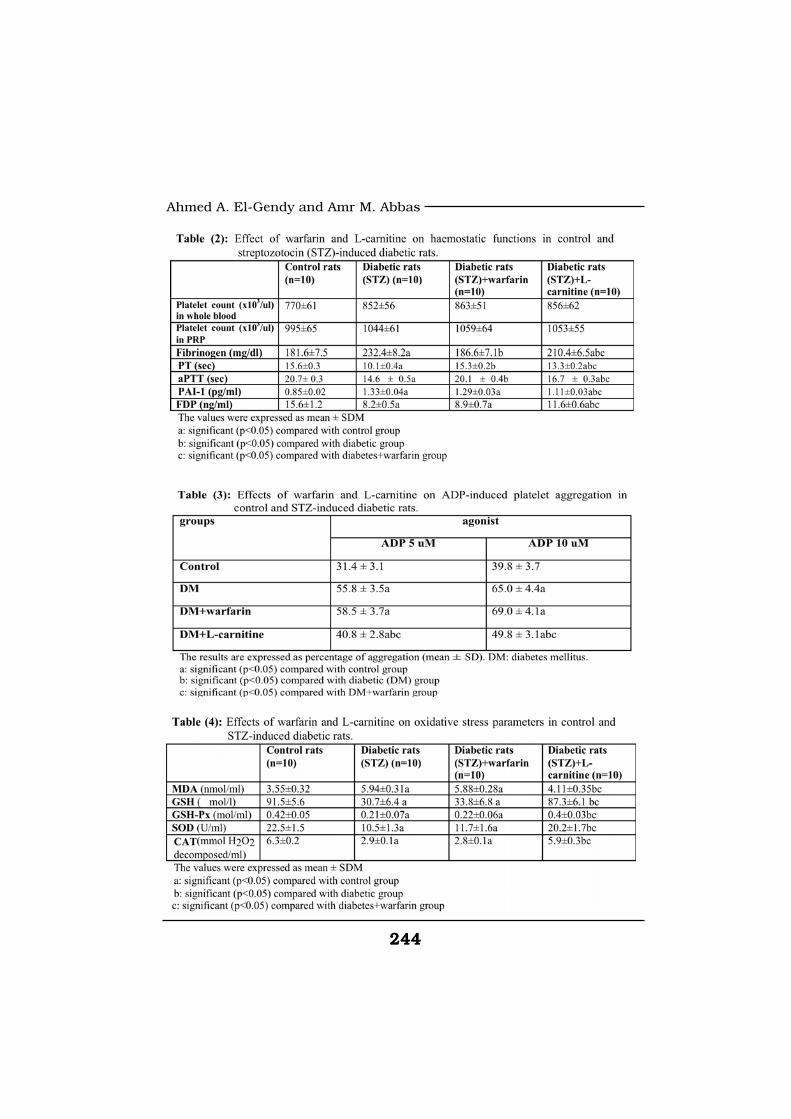
244244
Ahmed A. El-Gendy and Amr M. Abbas
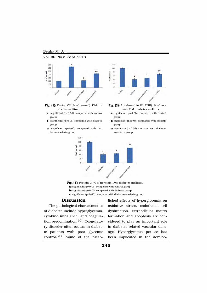
245
Benha M. J.
Vol. 30 No 3 Sept. 2013
DiscussionDiscussionThe pathological characteristics
of diabetes include hyperglycemia,
cytokine imbalance, and coagula-
tion predomination(30). Coagulato-
ry disorder often occurs in diabet-
ic patients with poor glycemic
control(31). Some of the estab-
lished effects of hyperglycemia on
oxidative stress, endothelial cell
dysfunction, extracellular matrix
formation and apoptosis are con-
sidered to play an important role
in diabetes-related vascular dam-
age. Hyperglycemia per se has
been implicated in the develop-
Fig. (1):Fig. (1): Factor VII (% of normal). DM: di-abetes mellitus.
a:a: significant (p<0.05) compared with control
group
b:b: significant (p<0.05) compared with diabetic
group
c:c: significant (p<0.05) compared with dia-
betes+warfarin group
Fig. (2):Fig. (2): Antithrombin III (ATIII) (% of nor-mal). DM: diabetes mellitus.
a:a: significant (p<0.05) compared with control
group
b:b: significant (p<0.05) compared with diabetic
group
c:c: significant (p<0.05) compared with diabetes
+warfarin group
Fig. (1):Fig. (1): Protein C (% of normal). DM: diabetes mellitus.a:a: significant (p<0.05) compared with control group
b:b: significant (p<0.05) compared with diabetic group
c:c: significant (p<0.05) compared with diabetes+warfarin group
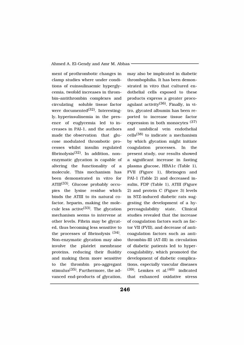
246246
Ahmed A. El-Gendy and Amr M. Abbas
ment of prothrombotic changes in
clamp studies where under condi-
tions of euinsulinaemic hypergly-
cemia, twofold increases in throm-
bin–antithrombin complexes and
circulating soluble tissue factor
were documented(32). Interesting-
ly, hyperinsulinemia in the pres-
ence of euglycemia led to in-
creases in PAI-1, and the authors
made the observation that glu-
cose modulated thrombotic pro-
cesses whilst insulin regulated
fibrinolysis(32). In addition, non-
enzymatic glycation is capable of
altering the functionality of a
molecule. This mechanism has
been demonstrated in vitro for
ATIII(33). Glucose probably occu-
pies the lysine residue which
binds the ATIII to its natural co-
factor, heparin, making the mole-
cule less active(33). The glycation
mechanism seems to intervene at
other levels. Fibrin may be glycat-
ed, thus becoming less sensitive to
the processes of fibrinolysis (34).
Non-enzymatic glycation may also
involve the platelet membrane
proteins, reducing their fluidity
and making them more sensitive
to the thrombin pro-aggregant
stimulus(35). Furthermore, the ad-
vanced end-products of glycation,
may also be implicated in diabetic
thrombophilia. It has been demon-
strated in vitro that cultured en-
dothelial cells exposed to these
products express a greater proco-
agulant activity(36). Finally, in vi-
tro, glycated albumin has been re-
ported to increase tissue factor
expression in both monocytes (37)
and umbilical vein endothelial
cells(38) to indicate a mechanism
by which glycation might initiate
coagulation processes. In the
present study, our results showed
a significant increase in fasting
plasma glucose, HBA1c (Table 1),
FVII (Figure 1), fibrinogen and
PAI-1 (Table 2) and decreased in-
sulin, FDP (Table 1), ATIII (Figure
2) and protein C (Figure 3) levels
in STZ-induced diabetic rats sug-
gesting the development of a hy-
percoagulability state. Clinical
studies revealed that the increase
of coagulation factors such as fac-
tor VII (FVII), and decrease of anti-
coagulation factors such as anti-
thrombin-III (AT-III) in circulation
of diabetic patients led to hyper-
coagulability, which promoted the
development of diabetic complica-
tions, especially vascular diseases(39). Lemkes et al.(40) indicated
that enhanced oxidative stress
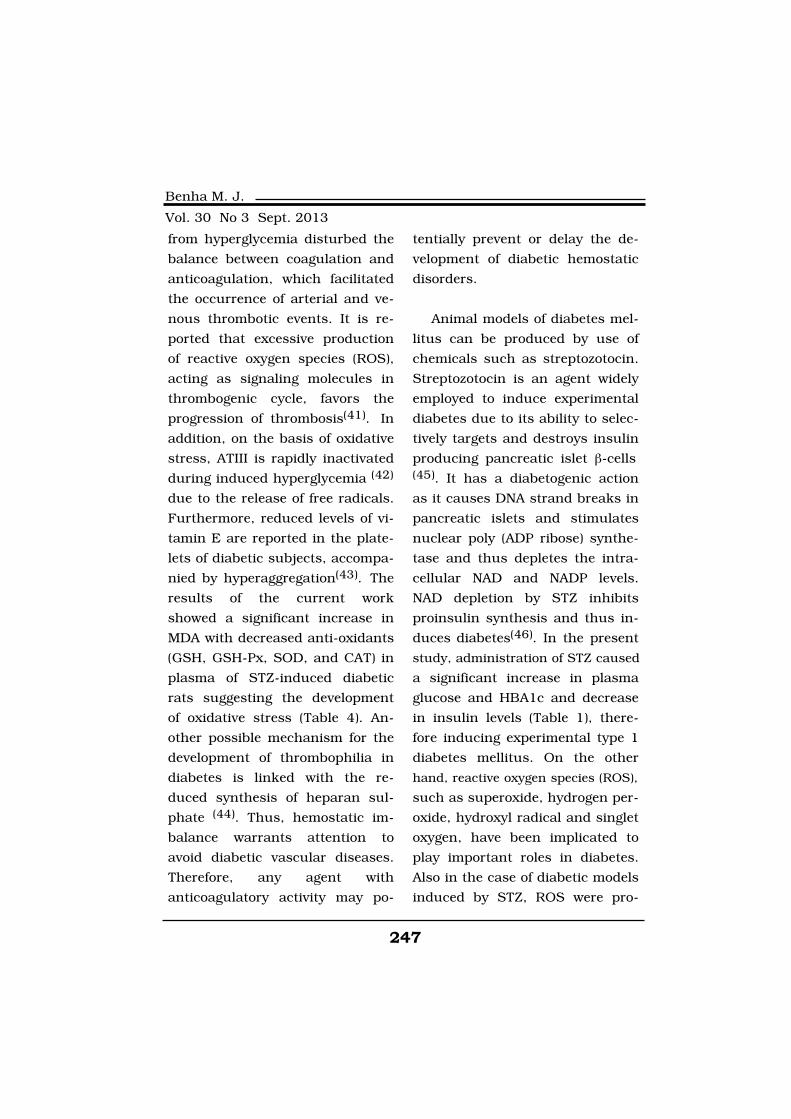
247
Benha M. J.
Vol. 30 No 3 Sept. 2013
from hyperglycemia disturbed the
balance between coagulation and
anticoagulation, which facilitated
the occurrence of arterial and ve-
nous thrombotic events. It is re-
ported that excessive production
of reactive oxygen species (ROS),
acting as signaling molecules in
thrombogenic cycle, favors the
progression of thrombosis(41). In
addition, on the basis of oxidative
stress, ATIII is rapidly inactivated
during induced hyperglycemia (42)
due to the release of free radicals.
Furthermore, reduced levels of vi-
tamin E are reported in the plate-
lets of diabetic subjects, accompa-
nied by hyperaggregation(43). The
results of the current work
showed a significant increase in
MDA with decreased anti-oxidants
(GSH, GSH-Px, SOD, and CAT) in
plasma of STZ-induced diabetic
rats suggesting the development
of oxidative stress (Table 4). An-
other possible mechanism for the
development of thrombophilia in
diabetes is linked with the re-
duced synthesis of heparan sul-
phate (44). Thus, hemostatic im-
balance warrants attention to
avoid diabetic vascular diseases.
Therefore, any agent with
anticoagulatory activity may po-
tentially prevent or delay the de-
velopment of diabetic hemostatic
disorders.
Animal models of diabetes mel-
litus can be produced by use of
chemicals such as streptozotocin.
Streptozotocin is an agent widely
employed to induce experimental
diabetes due to its ability to selec-
tively targets and destroys insulin
producing pancreatic islet β-cells(45). It has a diabetogenic action
as it causes DNA strand breaks in
pancreatic islets and stimulates
nuclear poly (ADP ribose) synthe-
tase and thus depletes the intra-
cellular NAD and NADP levels.
NAD depletion by STZ inhibits
proinsulin synthesis and thus in-
duces diabetes(46). In the present
study, administration of STZ caused
a significant increase in plasma
glucose and HBA1c and decrease
in insulin levels (Table 1), there-
fore inducing experimental type 1
diabetes mellitus. On the other
hand, reactive oxygen species (ROS),
such as superoxide, hydrogen per-
oxide, hydroxyl radical and singlet
oxygen, have been implicated to
play important roles in diabetes.
Also in the case of diabetic models
induced by STZ, ROS were pro-
248248
Ahmed A. El-Gendy and Amr M. Abbas
posed to be formed and involved
in the death of β-cells(47). Super-
oxide radicals are converted to hy-
drogen peroxide by superoxide dis-
mutase (SOD) and hydrogen
peroxide is converted to water by
glutathione peroxidase (GPx) and/
or catalase(48). Hydrogen peroxide
can react with transition metals in
Fenton reactions, thereby being
converted into hydroxyl radicals.
Hydrogen peroxide can cross all
membranes and lead to hydroxyl
radical formation at more distant
sites. The hydroxyl radical can
cause lipid peroxidation, which in
turn leads to damage of cellular
organels and membranes, thus
causing cell death(48). With re-
spect to oxidative stress, an in-
creased free radical generation
was reported in diabetic plasma and
tissues. In diabetes, oxidative stress
seems mainly due to both in-
creased free radical concentrations
and sharp reduction of anti-
oxidative defenses(8). In consistent
with these studies, the current
work showed a significant increase
in MDA level and decrease in GSH
level and GSH-Px, CAT and SOD
activities (Table 4) in STZ-induced
diabetic rats indicating the devel-
opment of oxidative stress.
In the laboratory, measurement
of PT, APTT, and fibrinogen con-
centration are the most commonly
employed laboratory tests in pa-
tients with a suspected coagulopa-
thy(49). Prothrombin time is a la-
boratory screening test used to
detect disorders involving the ac-
tivity of the factors I, II, V, VII,
and X of the extrinsic and
common pathways(50). Activated
partial thromboplastin time is
used to screen for abnormalities of
the intrinsic and common clotting
systems and to monitor the anti-
coagulant effect of circulating hep-
arin. It measures the activities of
factors I, II, V, VIII, IX–XI, and XII
of the intrinsic and common path-
ways(51). Changes in these pro-
teins favour the development of
hypercoagulable and prothrombot-
ic state, which may in turn en-
hance cardiovascular risk by in-
creasing the possibility of
developing an occlusive thrombus
within a coronary/cerebral artery(52). PT and APTT can therefore be
used to assess the risk of clotting
complications in patients with dia-
betes mellitus although modern
coagulation diagnostic test are be-
coming more sophisticated, PT
and APTT are still important basic
249
Benha M. J.
Vol. 30 No 3 Sept. 2013
examinations in clinical laborato-
ries(53). In the current study, PT
and aPTT were evaluated in con-
trol and STZ-induced diabetic
rats. Prothrombin time and aPTT
were significantly decreased in
STZ-induced diabetic rats in com-
parison with control group (Table
2) suggesting the development of
prothrombotic state in diabetic
rats. In addition, administration of
L-carnitine to STZ-induced diabet-
ic rats significantly increased PT
and aPTT when compared with
control rats but their values were
significantly different from those
of control and warfarin treated di-
abetic groups (Table 2). On the
other hand, warfarin , a coumarin
derivative,normalized the values of
PT and aPTT (Table 2) in STZ-
induced diabetic rats.
Plasminogen activator inhibi-
tor-1 (PAI-1) is the primary physi-
ologic inhibitor of fibrinolysis(54).
Moreover, FVII is the first enzyme
in blood coagulation system for
triggering the clotting cascade(55).
Epidemiological studies have re-
ported that high levels of factor VII
are associated with a high mortali-
ty rate for cardiovascular events(56). In addition, fibrinogen, the
most interesting coagulation fac-
tor, is recognized as having an im-
portant predictive value as a
marker of cardiovascular risk(57).
Thus, the elevated FVII (Figure 1)
and PAI-1 (Table 2) activities and
fibrinogen level (Table 2) as we ob-
served in STZ-induced diabetic
rats reflected a disruption in the
balance of factors regulating coag-
ulation and fibrinolysis, and obvi-
ously promoted the progression of
thrombosis. However, we found
that L-carnitine administration
lowered FVII (Figure 1) and PAI-1
(Table 2) activities and fibrinogen
level (Table 2) level which contrib-
uted to attenuation of coagulatory
disorder. In addition, warfarin de-
creased fibrinogen (Table 2) level
and FVII (Figure 1) activity to nor-
mal control value with no effect on
PAI-1 (Table 2), thus reducing the
risk of developing a thrombus.
Warfarin produces an anticoagu-
lant effect by inhibiting the vita-
min K-dependent synthesis of bio-
logically active forms of the
calcium-dependent clotting factors
II, VII, IX and X, as well as the
regulatory factors protein C, pro-
tein S, and protein Z.(23). The pre-
cursors of these factors require
carboxylation of their glutamic

250250
Ahmed A. El-Gendy and Amr M. Abbas
acid residues to -y-
carboxyglutamates (Gla) on the
N-terminal regions to allow the
coagulation factors to bind to
phospholipid surfaces inside blood
vessels on the vascular endotheli-
um(58). The enzyme that carries
out the carboxylation of glutamic
acid is gamma-glutamyl carboxy-
lase. The carboxylation reaction
will proceed only if the carboxy-
lase enzyme is able to convert a
reduced form of vitamin K (vita-
min K hydroquinone) to vitamin K
epoxide at the same time. The vi-
tamin K epoxide is in turn recy-
cled back to vitamin K and vita-
min K hydroquinone by another
enzyme, the vitamin K epoxide re-
ductase (VKOR). Warfarin inhibits
epoxide reductase (specifically the
VKORC1 subunit), thereby dimin-
ishing available vitamin K and vi-
tamin K hydroquinone in the tis-
sues, which inhibits the
carboxylation activity of the gluta-
myl carboxylase(59). When this oc-
curs, the coagulation factors are
no longer carboxylated at certain
glutamic acid residues and are in-
capable of binding to the endothe-
lial surface of blood vessels, and
are thus biologically inactive. As
the body's stores of previously
produced active factors degrade
and are replaced by inactive fac-
tors, the anticoagulation effect be-
comes apparent. The coagulation
factors are produced, but have de-
creased functionality due to un-
dercarboxylation(60).
The pathogenesis of the vascu-
lar complications in diabetes is
complex and has several potential
contributors including alterations
in platelet morphology and func-
tion(61). In fact, growing evidence
suggests that platelets of diabetic
patients are larger and hyperreac-
tive, and consequently present
deregulation of several signaling
pathways leading to an increase of
adhesion, activation, and aggrega-
tion(61). Moreover, it has been
demonstrated that the platelets of
these patients are more prone to
spontaneous aggregation and are
highly hypersensitive to agonists
such as thrombin, collagen, and
ADP(62). Extracellular nucleotides
such as ATP, ADP, and their nu-
cleoside derivative, adenosine,
have become clearly recognized
for their important role in mod-
ulating processes linked to vas-
cular inflammation and throm-
bosis(63). In the vascular system,
251
Benha M. J.
Vol. 30 No 3 Sept. 2013
the nucleotide ADP acts upon
platelets promoting their aggrega-
tion and modifying their shape(64), while ATP has been postulat-
ed to be a competitive inhibitor of
ADP-induced platelet aggregation(65). In support for these observa-
tions, the results of the present
study showed that ADP-induced
platelet aggregation was signifi-
cantly increased in STZ-induced
diabetic rats as compared to con-
trol rats (Table 3). In addition,
adenosine, produced by the nucle-
otide catabolism, is recognized as
an important modulator of vascu-
lar tone and a potent inhibitor of
platelet aggregation(66). Schmatz
et al(67) reported that despite the
increase of ATP, ADP, and AMP
hydrolysis contributing to an in-
crease of adenosine production in
STZ-induced diabetic rats, an ele-
vation of the adenosine deaminase
(ADA) activity was found in the
platelets of those diabetic rats.
Adenosine deaminase (ADA) is an
important enzyme that degrades
adenosine into inosine, tightly reg-
ulating local extracellular concen-
trations of adenosine(68). There-
fore, a rise in the activity of this
enzyme may lead to increased
adenosine deamination, causing a
reduction of the levels of this nu-
cleoside in the circulation. Conse-
quently, this situation may pro-
duce a favorable scenario for the
development of vascular diseases
in diabetic state, since adenosine
has an important role in the pre-
vention of platelet aggregation and
atherothrombotic complications(66).
On the other hand, there is ac-
cumulating evidence that the hy-
perglycemia contributes to greater
reactivity and aggregability of
platelets, particularly through the
generation of reactive oxygen spe-
cies (ROS) and by the glycation of
platelet membrane proteins(61). In
this study, we found an increase
in the glucose levels in STZ-
induced diabetic rats, accompa-
nied by an elevation in the glycat-
ed hemoglobin levels (Table 1),
which may be an indicative that
the platelet proteins and vascular
wall protein can also be suffering
non-enzymatic glycosylation. In
addition, our results revealed in-
creased MDA level and decreased
GSH level, GSH-Px, CAT and SOD
activities in plasma of STZ-
induced diabetic rats indicating
the development of oxidative
252252
Ahmed A. El-Gendy and Amr M. Abbas
stress in those rats (Table 4).
In fact, it has been found that
polyphenols can inhibit the gly-
cation and autoxidation of glu-
cose, preventing the initiation
and propagation of protein modifi-
cation(69). Furthermore, it is well
known that the main polyphenols
present in red wine (RW) and
grape juice (GJ) are powerful anti-
oxidants, protecting many tissues
and cells, including platelets, of
damages caused by oxidative
stress(69,70). In addition, it has
been shown that the resveratrol
exerted a potent antioxidant effect
on the generation of different ROS
in activated platelets(70). Based on
these findings, the authors sug-
gest that the strong antioxidant
properties of RW and GJ may con-
tribute to the prevention of an in-
crease in the platelet aggregation
found in diabetic rats treated with
both beverages derived from
grape. In agreement with these
findings, the results of the current
work showed that administration
of L-carnitine, an anti-oxidant
that decreased MDA and in-
creased GSH level, GSH-Px, CAT
and SOD activities in plasma of
STZ-induced diabetic rats (Table
4), caused a significant decrease
in ADP-induced platelet aggrega-
tion in the diabetic group (Table
3). On the other hand, warfarin
treatment of diabetic rats caused
no change in ADP-induced platelet
aggregation (Table 3).
Hyperglycemia has been ac-
cepted as an essential factor in
the development of diabetic com-
plications(71). Oxidative stress
plays an important role in the de-
velopment of diabetes complica-
tions, both microvascular and car-
diovascular(72). It is known that
hyperglycemia induced oxidative
stress causes endothelial dysfunc-
tion, and enhances the release of
PAI-1 and vWF(73). vWF is in-
volved in platelet adhesion and ag-
gregation(73). Furthermore, ROS
could stimulate platelet hyperac-
tivity, and facilitate coagulation(74). In agreement with these stud-
ies, ADP-induced platelet aggrega-
tion (Table 3), and PAI-1 (table 2)
significantly increased in STZ-
induced diabetic rats. Thus, low-
ering ROS production could bene-
fit hemostatic balance. The antiox-
idative property of L-carnitine
under diabetic condition has been
observed(75), and those research-
253
Benha M. J.
Vol. 30 No 3 Sept. 2013
ers reported that this compound
could decrease ROS generation
and improve redox imbalance. In
accord with these studies, the re-
sults of the current work revealed
that the intake of this compound
effectively reduced MDA as well as
increased GSH level and GSH-Px,
SOD and CAT activities in the
plasma (Table 4). These findings
agreed that L-carnitine via its an-
tioxidative activity alleviated redox
disorder in circulation of diabetic
rats. Thus, it is possible that L-
carnitine diminished oxidative
stress, which consequently re-
versed the imbalance of hemostat-
ic factors, and abated hypercoagu-
latory risk in the diabetic rats. In
support for this suggestion, the
present work showed that L-
carnitine significantly decreased
ADP-induced platelet aggregation
(Table 3), fibrinogen level and PAI-
1 (Table 2) and FVII (Figure 1) ac-
tivities in STZ-induced diabetic
rats. In addition, AT-III and pro-
tein C are anticoagulation factors
because AT-III inhibits the activity
of several proteases in the coagu-
lation cascade, and protein C in-
activates coagulation factors such
as FVII(39). The decreased biologi-
cal activity of ATIII, due to hyper-
glycemia, results in a reduced
thrombin-antithrombin complex
formation with consequent hyper-
activity of the thrombin(42). A de-
crease in protein C has been re-
ported in diabetes(76). This
decrease is directly caused by hy-
perglycemia, and affects the entire
molecule, involving both the bio-
logical activity of the protein
and its antigenic concentration(76). Our results showed a signifi-
cant decrease in ATIII (Figure 2)
and protein C (Figure 3) in STZ-
induced diabetic rats when com-
pared with control rats. In addi-
tion, the current work revealed
that L-carnitine treatment of dia-
betic rats elevated AT-III (Figure 2)
and protein C (Figure 3) activities.
Apparently, the anticoagulatory ef-
fects of this agent could be partial-
ly ascribed to enhancement of
thrombolysis. These findings sug-
gested that L-carnitine was able to
reduce the risk of diabetes asso-
ciated thrombosis.
A considerable body of evidence
indicates that the generation of
ROS may mediate the cytotoxic ef-
fect of streptozotocin on pancreat-
ic β-cell. An increase of ROS pro-
duction has also been pointed out
254254
Ahmed A. El-Gendy and Amr M. Abbas
as a main instrument of destruc-
tion in streptozotocin-damaged
pancreatic islets(47). In agreement
with these studies, the current
work revealed that streptozotocin
causes an increase of MDA level
and decreased the anti-oxidants
(GSH level, and GSH-Px, SOD and
CAT activities) (Table 4). Diabetes-
related increase in lipid peroxida-
tion product (MDA) might be the
reflection of the decrease in enzy-
matic and non- enzymatic antioxi-
dant protection. L-carnitine ad-
ministration caused a decrease in
lipid peroxidation (MDA) (Table 4)
in STZ-induced diabetic rats. This
may be due to its active role in the
transport of fatty acids for energy
production, thereby lowering the
availability of lipids for peroxida-
tion(77). In addition, the produc-
tion of highly reactive oxygen spe-
cies such as O2•–, H2O2 and OH•
are also catalyzed by free iron
through Haber-Weiss reaction (78).
Carnitine and its esters have been
shown to partially inhibit iron-
induced lipid peroxidation in lipo-
somes(79) by forming complexes
with free iron. Thus, the reduction
in lipid peroxidation in the present
study is due to the iron-chelating
property of L-carnitine. In addi-
tion, the anti-oxidants (GSH level,
and GSH-Px, SOD and CAT activi-
ties) (Table 4) were increased in
STZ-induced diabetic rats with L-
carnitine administration which
could be the result of decreased
ROS. These results indicate the
important role of L-carnitine in
the improvement of oxidative
stress in STZ-induced diabetic
rats. On the other hand, no
change in lipid peroxidation and
anti-oxidants (GSH level, and
GSH-Px, SOD and CAT activities)
(Table 4) was observed in diabetic
rats with warfarin administration.
ConclusionConclusionHyperglycemia plays an impor-
tant role in hypercoagulation state
and oxidative stress in streptozo-
cin-induced diabetes mellitus. L-
carnitine improves oxidative stress
and decreases the hypercoagula-
tion state in diabetes mellitus. On
the other hand, warfarin normaliz-
es the hypercoagulation state in
diabetes mellitus with no effect on
oxidative stress.
References References 1. I.D.F. (2009): 1. I.D.F. (2009): International
Diabetes Federation. Diabetes At-
las. 4th ed ©
255
Benha M. J.
Vol. 30 No 3 Sept. 2013
2. Jay D., Hitomi H. and2. Jay D., Hitomi H. and
Griendling K.K. (2006): Griendling K.K. (2006): Oxidative
stress and diabetic cardiovas-
cular complications. Free Radic.
Biol. Med. 40:183-192.
3. Clozel M., Hess P., Qiu C.,3. Clozel M., Hess P., Qiu C.,
Ding S.S. and Rey M. (2006):Ding S.S. and Rey M. (2006):
The urotensin-II receptor antago-
nist palosuran improves pan-
creatic and renal function in dia-
betic rats. J Pharmacol Exp Ther;
316: 1115-1121.
4. Kowluru R.A. and Chan4. Kowluru R.A. and Chan
P.S. (2007):P.S. (2007): Oxidative stress and
diabetic retinopathy. Exp Diabetes
Res: 43603.
5. Wolff S.P., Jiang Z.Y. and5. Wolff S.P., Jiang Z.Y. and
Hunt J.V. (1991):Hunt J.V. (1991): Protein glyca-
tion and oxidative stress in dia-
betes mellitus and ageing. Free
Radic Biol Med 10: 339-352.
6. Abou-Seif M.A. and Yous-6. Abou-Seif M.A. and Yous-
sef A.A. (2004):sef A.A. (2004): Evaluation of
some biochemical changes in dia-
betic patients. Clin. Chem. Acta.;
346:161-70.
7.7. Davi G., Falco A. and Pa-Davi G., Falco A. and Pa-
trono C. (2005):trono C. (2005): Lipid peroxida-
tion in diabetes mellitus. Antioxid
Redox Signal 7: 256-268.
8. Kakkar R., Kalra J., Man-8. Kakkar R., Kalra J., Man-
tha S.V. and Prasad K. (1995):tha S.V. and Prasad K. (1995):
Lipid peroxidation and activity of
antioxidant enzymes in diabetic
rats. Mol Cell Biochem 151: 113-
119.
9. Opara E.C. (2002):9. Opara E.C. (2002): Oxida-
tive stress, micronutrients, dia-
betes mellitus and its complica-
tions. J R Soc Promot Health 122:
28-34.
10. Kukner A., Colakoglu N.,10. Kukner A., Colakoglu N.,
Ozogul C., Naziroglu M. and Fi-Ozogul C., Naziroglu M. and Fi-
rat T. (2009):rat T. (2009): The Effects of Com-
bined Vitamin C and Vitamin E
in Streptozotocin- Induced Dia-
betic Rat Kidney. Research Jour-
nal of Medical Sciences Volume: 3
(6) : 214-220.
11. Khan H.A., Sobki S.H.11. Khan H.A., Sobki S.H.
and Khan S.A. (2007):and Khan S.A. (2007): Associa-
tion between glycaemic control
and serum lipids profile in type 2
diabetic patients: HbA1c predicts
dyslipidaemia. Clin Exp Med; 7:
24-29.
12. Stamler J., Vaccaro O.,12. Stamler J., Vaccaro O.,
Neaton J.D. and Wentworth D.Neaton J.D. and Wentworth D.
256256
Ahmed A. El-Gendy and Amr M. Abbas
(1993):(1993): Diabe- tes, other risk fac-
tors and 12-yr cardiovascular
mortality for men screened in the
Multiple Risk Factor Interven- tion
Trial. Diab Care; 16: 434-444.
13. Chan P. and Pan W.H.13. Chan P. and Pan W.H.
(1995):(1995): Coagulation activation in
type 2 diabetes mellitus: the high-
er coronary risk of female diabet-
ic patients. Diabet Med; 12: 504-
507.
14. Acang N. and Jalil F.D.14. Acang N. and Jalil F.D.
(1993):(1993): Hypercoagulation in dia-
betes melli- tus. Southeast Asian
J Trop Med Public Health; 24:
263-266.
15. Zachary T. and Bloom-15. Zachary T. and Bloom-
garden M.D. (2011):garden M.D. (2011): Diabetes
and cardiovascular disease. Dia-
betes Care, 34: 24-30.
16. Yurekli B.P., Ozcebe O.I.,16. Yurekli B.P., Ozcebe O.I.,
Kirazli S. and Gurlek A. (2006):Kirazli S. and Gurlek A. (2006):
Global assessment of the coagula-
tion status in type 2 diabetes mel-
litus using rotation thromboe-
lastography. Blood Coagul
Fibrinolysis; 17: 545-549.
17. Bae S.H., Lee J., Roh K.H.17. Bae S.H., Lee J., Roh K.H.
and Kim J. (2003):and Kim J. (2003): Platelet acti-
vation in patients with diabetic re-
tinopathy. Korean J Ophthal-
mol; 17: 140-144.
18. Guirguis N.G., Eicher C.,18. Guirguis N.G., Eicher C.,
Hock L., Lynch J., Graham V.D.,Hock L., Lynch J., Graham V.D.,
Rajagopalan P.R., et al. (2003):Rajagopalan P.R., et al. (2003):
Thromboembolic risk factors in
patients undergoing kidney trans-
plant: implication of abnormally
short activated partial thrombo-
plastin time. Ann Clin Lab Sci; 33:
396-400.
19. Zeitler P., Thiede A. and19. Zeitler P., Thiede A. and
Muller H.L. (2001):Muller H.L. (2001): Prospective
study on plasma clotting parame-
ters in diabetic children - no evi-
dence for specific changes in co-
agulation system. Exp Clin
Endocrinol Diab; 109: 146-150.
20.20. Rani P.J.A. and Pan-Rani P.J.A. and Pan-
neerselvam C. (2001):neerselvam C. (2001): Carnitine
as a free radical scavenger in ag-
ing. Experimental Gerontology;
36:1713-26.
21. Reddi A.S., Jyothirmayi21. Reddi A.S., Jyothirmayi
G.N., DeAngelis B., Frank O.G.N., DeAngelis B., Frank O.
and Baker H. (1991):and Baker H. (1991): Effect of
short- and long-term diabetes on
carnitine and myo-inositol in rats.
Comperative Biochemistry and
257
Benha M. J.
Vol. 30 No 3 Sept. 2013
Physiology A; 98:39-42.
22. Holbrook A.M., Pereira22. Holbrook A.M., Pereira
J.A., Labiris R., McDonald H.,J.A., Labiris R., McDonald H.,
Douketis J.D., Crowther M. andDouketis J.D., Crowther M. and
Wells P.S. (2005): Wells P.S. (2005): “Systematic
overview of warfarin and its drug
and food interactions". Arch. In-
tern. Med. 165 (10): 1095-106.
23.23. Ansell J., Hirsh J., Hy-Ansell J., Hirsh J., Hy-
lek E., et al. (2008):lek E., et al. (2008): "Pharmacol-
ogy and management of the vita-
min K antagonists: American
College of Chest Physicians evi-
dence-based clinical practice
guidelines (8th Edition)". Chest
133 (6 Suppl): 160S-198S.
24. Tikoo K., Tripathi D.N.,24. Tikoo K., Tripathi D.N.,
Kabra D.G., Sharma V. andKabra D.G., Sharma V. and
Gaikwad A.B. (2007): Gaikwad A.B. (2007): “Intermit-
tent fasting prevents the progres-
sion of type I diabetic nephropathy
in rats and changes the expres-
sion of Sir2 and p53,” FEBS Let-
ters, vol. 581, no. 5, pp. 1071-
1078.
25. Zangiabadi N., Sheibani25. Zangiabadi N., Sheibani
V., Asadi-Shekaari M., ShabaniV., Asadi-Shekaari M., Shabani
M., Jafari M., Asadi A.R., Tajadi-M., Jafari M., Asadi A.R., Tajadi-
ni H. and Jarahi M. (2011):ni H. and Jarahi M. (2011): Ef-
fects of Melatonin in Prevention of
Neuropathy in STZ-Induced Dia-
betic Rats. Am. J. Pharmacol. Tox-
icol. 6 (2): 59-67.
26. Yanagita M., Arai H., Ishii26. Yanagita M., Arai H., Ishii
K., Nakano T., Ohashi K., Mizu-K., Nakano T., Ohashi K., Mizu-
no K., Varnum B., Fukatsu A.,no K., Varnum B., Fukatsu A.,
Doi T. and Kita T. (2001):Doi T. and Kita T. (2001): Am.
J. Pathol. 158, 1423-1432.
27. Soneru I.L., Khan T., Or-27. Soneru I.L., Khan T., Or-
falian Z. and Abraira C. (1997):falian Z. and Abraira C. (1997):
Acetyl-L-carnitine effects on nerve
conduction and glycemic regula-
tion in experimental diabetes. En-
docrine Research; 23:27-36.
28. Buege J.A. and Aust S.D.28. Buege J.A. and Aust S.D.
(1972):(1972): Microsomal lipid peroxida-
tion. Methods Enzymol., 52:302-
10.
29. Born G.V.R. (1962):29. Born G.V.R. (1962): Aggre-
gation of blood platelets by adeno-
sine diphosphate and its reversal.
Nature, 194, 927–929.
30. Brownlee M. (2001):30. Brownlee M. (2001): Bio-
chemistry and molecular cell biol-
ogy of diabetic complications. Na-
ture (Lond.) 414:813-20.
31. Nwose E.U., Jelinek H.F.,31. Nwose E.U., Jelinek H.F.,
Richards R.S., Tinley P. andRichards R.S., Tinley P. and
258258
Ahmed A. El-Gendy and Amr M. Abbas
Kerr P.G. (2009): Kerr P.G. (2009): Atherothrombo-
sis and oxidative stress: the con-
nection and correlation in dia-
betes. Redox Rep 14:55-60.
32. Stegenga M., van der32. Stegenga M., van der
Crabben S., Levi M., et al.Crabben S., Levi M., et al.
(2006): (2006): Hyperglycemia stimulates
coagulation, whereas hyperinsuli-
naemia impairs fibrinolysis in
healthy individuals. Diabetes; 55:
1807-12.
33. Villanueva G.B. and Allen33. Villanueva G.B. and Allen
N. (1988):N. (1988): Demonstration of al-
tered anti-thrombin III activity due
to non enzymatic glycosylation at
glucose concentration expected
to be encountered in severely
diabetic patients. Diabetes 37:
1103-1107.
34. Brownlee M., Vlassara H.34. Brownlee M., Vlassara H.
and Cerami A. (1983):and Cerami A. (1983): Nonenzy-
matic glycosylation reduces the
susceptibility of fibrin to degrada-
tion by plasmin. Diabetes 32: 600-
604.
35. Winocour P.D., Watala C.35. Winocour P.D., Watala C.
and Kinlough-Rathbone R.L.and Kinlough-Rathbone R.L.
(1992): (1992): Mem- brane fluidity is re-
lated to the extent of glycation of
proteins, but not to alterations in
the cholesterol to phospholipid
molar ratio in isolated platelet
membranes from diabetic and
control subjects. Thromb Haemos-
tas 65:567-571.
36. Esposito C., Gerlach H.,36. Esposito C., Gerlach H.,
Brett J., Stern D. and VlassaraBrett J., Stern D. and Vlassara
H. (1989):H. (1989): Endothelial receptor-
mediated binding of glucose-
modified albumin is associated
with increased monolayer permea-
bility and modulation of cell sur-
face coagulant properties. J Exp
Med 170: 1387-1407.
37. Khechai F., Ollivier V.,37. Khechai F., Ollivier V.,
Bridey F., et al. (1997):Bridey F., et al. (1997): Effect of
advanced gly- cation end product-
modified albumin on tissue factor
expression by monocytes. Arteri-
oscler Vasc Biol; 17: 2885-90.
38. Min C., Kang E., Yu S.H.,38. Min C., Kang E., Yu S.H.,
Shinn S.H. and Kim Y.S. (1999):Shinn S.H. and Kim Y.S. (1999):
Advanced glycation end products
induce apoptosis and procoagu-
lant activity in cultured human
umbilical vein endothelial cells.
Diabetes Res Clin Pract; 46: 197-
202.
39. Asakawa H., Tokunaga K.39. Asakawa H., Tokunaga K.
and Kawakami F. (2000):and Kawakami F. (2000): Eleva-
259
Benha M. J.
Vol. 30 No 3 Sept. 2013
tion of fibrinogen and thrombin-
antithrombin III complex levels of
type 2 diabetes mellitus patients
with retinopathy and nephropa-
thy. J Diabet Complicat 14:121-6.
40. Lemkes B.A., Hermanides40. Lemkes B.A., Hermanides
J., Devries J.H., Holleman F.,J., Devries J.H., Holleman F.,
Meijers J.C. and Hoekstra J.B.Meijers J.C. and Hoekstra J.B.
(2010):(2010): Hyperglycemia: a pro-
thrombotic factor? J Thromb Hae-
most 8:1663-9.
41. Herkert O., Djordjevic T.,41. Herkert O., Djordjevic T.,
BelAiba R.S. and Go¨ rlach A.BelAiba R.S. and Go¨ rlach A.
(2004):(2004): Insights into the redox
control of lood coagulation: role of
vascular NADPH oxidase-derived
reactive oxygen species in the
thrombogenic cycle. Antioxid Re-
dox Signal 6:765-76.
42. Ceriello A., Giugliano D.,42. Ceriello A., Giugliano D.,
Quatraro A., et al. (1987):Quatraro A., et al. (1987): In-
duced hyperglycemia alters anti-
thrombin III activity but not its
plasma concentration in healthy
normal subjects. Diabetes 36:
320-323.
43. Karpen C.W., Pritchard43. Karpen C.W., Pritchard
K.A. Jr, Arnold J.H., CornwellK.A. Jr, Arnold J.H., Cornwell
D.G. and Panganamala R.V.D.G. and Panganamala R.V.
(1982):(1982): Restoration ofprostacyclin
thromboxane A2 balance in the
diabetic rat: influence of dietary
vitamin E. Diabetes 31: 947-951.
44. Rohrbach D.H., Wagner44. Rohrbach D.H., Wagner
C.W., Star V.L., Martin G.R.,C.W., Star V.L., Martin G.R.,
Brown K.S. and Yoon J.W.Brown K.S. and Yoon J.W.
(1983):(1983): Reduced synthesis of
basement membrane heparan-
sulfate proteoglycan in streptozot-
ocin-induced diabetic mice. J Bioi
Chern 258: 11672-11677.
45. Gonzalez E., Rosello-45. Gonzalez E., Rosello-
Catafau J., Jawerbaum A., Sin-Catafau J., Jawerbaum A., Sin-
ner D., Pustovrh C., Vela J., etner D., Pustovrh C., Vela J., et
al. (2000):al. (2000): Pancreatic nitric oxide
and oxygen free radicals in the
early stages of streptozotocin-
induced diabetes mellitus in the
rat. Brazilian Journal of Medical
and Biology Research 2000,
33:1335-42.
46. Oberley L.W. (1988):46. Oberley L.W. (1988): Free
radicals and diabetes. Free Radi-
cal Biology and Medicine; 5:113-
24.
47. Ohkuwa T., Sato Y. and47. Ohkuwa T., Sato Y. and
Naoi M. (1995):Naoi M. (1995): Hydroxyl radical
formation in diabetic rats induced
by streptozotocin. Life Science;
56:1789-98.
260260
Ahmed A. El-Gendy and Amr M. Abbas
48. Halliwell B. (1986):48. Halliwell B. (1986): Oxi-
dants and the central nervous
system: some fundamental ques-
tions. Is oxidant damage relevant
to Parkinson’s disease, Alzhei-
mer’s disease, traumatic injury
or stroke? Acta Neurologica Scan-
dinavica; 126:23-33.
49. Furlanello T., Caldin M.49. Furlanello T., Caldin M.
and Stocco A. (2006):and Stocco A. (2006): Stability of
stored canine plasma for hemosta-
sis testing. Veterinary Clinical Pa-
thology, 35:204-207.
50. Hinchcliff K.W., Kaneps50. Hinchcliff K.W., Kaneps
A.J. and Geor R.J. (2004):A.J. and Geor R.J. (2004):
Equine sports medicine and sur-
gery. In: Basic and clinical sci-
ence of the equine athlete. WB
Saunders, Philadelphia, PA Pp
1295-1302.
51. Iazbik C., Couto G., Gray51. Iazbik C., Couto G., Gray
T.L. and Kociba G. (2001): T.L. and Kociba G. (2001): Effect
of storage conditions on hemostat-
ic parameters of canine plasma
obtained for transfusion. Am. J.
Veterin. Res. 62:734-735.
52. Dunn E.J. and Grant P.J.52. Dunn E.J. and Grant P.J.
(2005):(2005): Type 2 diabete: an athe-
rothrombotic syndrome. Current
Molecular Medicine, 5(3): 323-322.
53. Ng V.L. (2009):53. Ng V.L. (2009): Prothrom-
bine Time and partial thrombo-
plastin time assay considerations.
Clinical Laboratory Medicine, 29:
253-263.
54. Urano T., Ihara H., Suzu-54. Urano T., Ihara H., Suzu-
ki Y., Takada Y. and Takada A.ki Y., Takada Y. and Takada A.
(2000): (2000): Coagulation-associated
enhancement of fibrinolytic activi-
ty via a neutralization of PAI-1 ac-
tivity. Semin Thromb Hemost 26:
39-42.
55.55. Eigenbrot C. and Kirch-Eigenbrot C. and Kirch-
hofer D. (2002):hofer D. (2002): New insight into
how tissue factor allosterically reg-
ulates factor VIIa. Trends Cardio-
vasc Med 12:19-26.
56. Meade J.W., Brozovic M.,56. Meade J.W., Brozovic M.,
Chakrabarti R.R., et al. (1986):Chakrabarti R.R., et al. (1986):
Haemostatic function and ischem-
ic heart disease: principal results
of the Northwick Park Heart
Study. Lancet II: 533-537.
57. Wilhelmsen L., Svard-57. Wilhelmsen L., Svard-
sudd K., Korsan-Bengtensen K.,sudd K., Korsan-Bengtensen K.,
Larsson B., Welin L. and TibblinLarsson B., Welin L. and Tibblin
G. (1984):G. (1984): Fibrinogen as a risk
factor for stroke and myocardial
infarction. N Engl. J Med 311:
501-505.

261
Benha M. J.
Vol. 30 No 3 Sept. 2013
58. Choonara I.A., Malia58. Choonara I.A., Malia
R.G., Haynes B.P., et al. (1988):R.G., Haynes B.P., et al. (1988):
The relationship between inhibi-
tion of vitamin K1 2,3-epoxide re-
ductase and reduction of clotting
factor activity with warfarin. Br J
Clin Pharmacol. 1988;25:1-7.
59. Li T., Chang C.Y., Jin59. Li T., Chang C.Y., Jin
D.Y., Lin P.J., Khvorova A. andD.Y., Lin P.J., Khvorova A. and
Stafford D.W. (2004):Stafford D.W. (2004): "Identifica-
tion of the gene for vitamin K
epoxide reductase". Nature 427
(6974): 541-4.
60. Malhotra O.P., Nesheim60. Malhotra O.P., Nesheim
M.E. and Mann K.G. (1985):M.E. and Mann K.G. (1985): The
kinetics of activation of normal
and gamma carboxy glutamic acid
deficient prothrombins. J Biol
Chem. 1985;260:279-287.
61. Morel O., Kessler L., Ohl-61. Morel O., Kessler L., Ohl-
mann P. and Bareiss P. (2010):mann P. and Bareiss P. (2010):
Diabetes and the platelet: Toward
new therapeutic paradigms for di-
abetic atherothrombosis. Athero-
sclerosis, 212(2), 367-376.
62. Haouari M.E. and Rosado62. Haouari M.E. and Rosado
J.A. (2008): J.A. (2008): Platelet signaling ab-
normalities in patients with type 2
diabetes mellitus: A review. Blood
Cells, Molecules, & Diseases, 41
(1), 119-123.
63. Di Virgilio F., Chiozzi P.,63. Di Virgilio F., Chiozzi P.,
Ferrari D., Falzoni S., SanzFerrari D., Falzoni S., Sanz
J.M., Morelli A., et al. (2001):J.M., Morelli A., et al. (2001):
Nucleotide receptors: An emerg-
ing family of regulatory molecules
in blood cells. Blood, 97(3), 587-
600.
64. Rozalski M., Nocun M.64. Rozalski M., Nocun M.
and Watala C. (2005):and Watala C. (2005): Adenosine
diphosphate receptors on blood
platelets: Potential new targets for
antiplatelet therapy. Acta Bio-
chimica Polonica, 52(2), 411-415.
65. Birk A.V., Broekman65. Birk A.V., Broekman
M.J., Gladek E.M., RobertsonM.J., Gladek E.M., Robertson
H.D., Drosopoulos J.H., MarcusH.D., Drosopoulos J.H., Marcus
A.J., et al. (2002):A.J., et al. (2002): Role of extra-
cellular ATP metabolism in regula-
tion of platelet reactivity. Journal
of Laboratory and Clinical Medi-
cine, 140(3), 166-175.
66. Anfossi G., Russo I., Mas-66. Anfossi G., Russo I., Mas-
succo P., Mattiello L., Cavalotsucco P., Mattiello L., Cavalot
F., Balbo A., et al. (2002):F., Balbo A., et al. (2002): Aden-
osine increases human platelet
levels of cGMP through nitric ox-
ide: Possible role in its antiaggre-
gating effect. Thrombosis Re-
search, 105(1), 71-78.
262262
Ahmed A. El-Gendy and Amr M. Abbas
67. Schmatz R., Mann T.R.,67. Schmatz R., Mann T.R.,
Spanevello R., Machado M.M.,Spanevello R., Machado M.M.,
Zanini D., Pimentel V.C., Stefa-Zanini D., Pimentel V.C., Stefa-
nello N., Martins C.C., Cardosonello N., Martins C.C., Cardoso
A.M., Bagatini M., Gutierres J.,A.M., Bagatini M., Gutierres J.,
Leal C.A.M., Pereira L.B., Maz-Leal C.A.M., Pereira L.B., Maz-
zanti C., Schetinger M.R. andzanti C., Schetinger M.R. and
Morsch V.M. (2013):Morsch V.M. (2013): Moderate
red wine and grape juice con-
sumption modulates the hydroly-
sis of the adenine nucleotides and
decreases platelet aggregation in
streptozotocin-induced diabetic
rats. Cell Biochem Biophys 65:
129-143.
68. Franco R., Casado V., Ci-68. Franco R., Casado V., Ci-
ruela F., Saura C., Mallol J., Ca-ruela F., Saura C., Mallol J., Ca-
nela E., et al. (1997):nela E., et al. (1997): Cell surface
adenosine deaminase: Much more
that an ectoenzyme. Progress in
Neurobiology, 52, 283-294.
69. Wu C.H. and Yen G.C.69. Wu C.H. and Yen G.C.
(2005):(2005): Inhibitory effect of natu-
rally occurring flavonoids on the
formation of advanced glycation
endproducts. Journal of Agricul-
ture and Food Chemistry, 53(8),
3167-3173.
70. Olas B. and Wachowicz B.70. Olas B. and Wachowicz B.
(2005):(2005): Resveratrol, a phenolic
antioxidant with effects on blood
platelet functions. Platelets, 16(5),
251-260.
71. Rösen P., Nawroth P.P.,71. Rösen P., Nawroth P.P.,
King G., Möller W., TritschlerKing G., Möller W., Tritschler
H.J. and Packer L. (2001):H.J. and Packer L. (2001): The
role of oxidative stress in the on-
set and progression of diabetes
and its complications: a summary
of a Congress Series sponsored by
UNESCO-MCBN, the American
Diabetes Association and the Ger-
man Diabetes Society. Diabetes
Metab. Res. Rev. 17: 189-212.
72. Jay D., Hitomi H. and72. Jay D., Hitomi H. and
Griendling K.K. (2006):Griendling K.K. (2006): Oxidative
stress and diabetic cardiovascular
complications. Free Radic. Biol.
Med. 40:183-192.
73. Lee E.A., Seo J.Y., Jiang73. Lee E.A., Seo J.Y., Jiang
Z., Yu M.R., Kwon M.K., Ha H.Z., Yu M.R., Kwon M.K., Ha H.
and Lee H.B. (2005):and Lee H.B. (2005): Reactive ox-
ygen species mediate high glu-
cose-induced plasminogen activa-
tor inhibitor-1 up-regulation in
mesangial cells and in diabetic
kidney. Kidney Int 67:1762-71.
74. Meerarani P., Badimon74. Meerarani P., Badimon
J.J., Zias E., Fuster V. and Mo-J.J., Zias E., Fuster V. and Mo-
reno P.R. (2006):reno P.R. (2006): Metabolic syn-
drome and diabetic atherothrom-
263
Benha M. J.
Vol. 30 No 3 Sept. 2013
bosis: implications in vascular
complications. Curr Mol Med
6:501-14.
75. Uysal N., Yalaz G., Acik-75. Uysal N., Yalaz G., Acik-
goz O., Gonenc S. and Kayatekingoz O., Gonenc S. and Kayatekin
B.M. (2005):B.M. (2005): Effect of L-carnitine
on diabetogenic action of strepto-
zotocin in rats. Neuroendocrinolo-
gy Letters No.4 August Vol. 26,
419-422.
76. Vukovich T.C. and76. Vukovich T.C. and
Schernthaner G. (1986):Schernthaner G. (1986): De-
creased protein C levels in pa-
tients with insulin-dependent
type I diabetes mellitus. Diabetes
35: 617-619.
77. Kalaiselvi T. and Panneer-77. Kalaiselvi T. and Panneer-
selvam C. (1998):selvam C. (1998): Effect of L-
carnitine on the status of lipid
peroxidation and antioxidants in
aging rats. Journal of Nutritional
Biochemistry; 9:575-81.
78. Halliwell B. and Gutte-78. Halliwell B. and Gutte-
ridge J.M.C. (1998):ridge J.M.C. (1998): Free radicals
in biology and medicine. In: The
chemistry of free radicals and re-
leated ‘reactive species’. 3rd ed. Lon-
don: Oxford press; 1998. p.36-104.
79. Arduini A. (1992):79. Arduini A. (1992): Carni-
tine and its acetyl esters as secon-
dary antioxidants? American Heart
Journal; 123:1726-7.

264264
Ahmed A. El-Gendy and Amr M. Abbas
EFFECT OF ANTICOAGULANTEFFECT OF ANTICOAGULANT(WARFARIN) AND L-CARNITINE ON(WARFARIN) AND L-CARNITINE ON
HAEMOSTATIC FUNCTION ANDHAEMOSTATIC FUNCTION ANDOXIDATIVE STRESS INOXIDATIVE STRESS IN
STREPTOZOTOCIN-INDUCEDSTREPTOZOTOCIN-INDUCEDDIABETIC RATSDIABETIC RATS
Ahmed A. El-Gendy Ph.D and Amr M. Abbas Ph.DAhmed A. El-Gendy Ph.D and Amr M. Abbas Ph.D
BENHAMEDICALJOURNAL
REPRINT
Published byPublished by
Benha Faculty of MedicineBenha Faculty of Medicine Volume 30 Number 3Sept. 2013
265
Benha M. J.
Vol. 30 No 3 Sept. 2013
OUTCOME OF BAHA SURGERY USING THEOUTCOME OF BAHA SURGERY USING THEINFERIORLY BASED PARTIAL THICKNESSINFERIORLY BASED PARTIAL THICKNESS
SKIN FLAP SKIN FLAP
Mahmoud El-Sayed Ali, MBBCh, MSc, FRCS, MDMahmoud El-Sayed Ali, MBBCh, MSc, FRCS, MDAssistant Professor, Department of Otolaryngology,
Mansoura University Hospital, Mansoura University, Egypt
AbstractObjective:Objective: To evaluate the outcome of bone anchored hearing aid
(BAHA) surgery using an inferiorly based partial thickness skin flap. Introduction:Introduction: The wide application of BAHA surgery and the differ-
ent surgical techniques necessitate the analysis of the outcome of thissurgery and its safety and effectiveness as an auditory rehabilitationmodality.
Methods:Methods:Study design:Study design: Retrospective chart review.Patients and methods: Patients and methods: Patients subjected for BAHA surgery for the
rehabilitation of their deafness. Clinical records were reviewed from Au-gust 2007 to April 2011. Skin healing was reviewed and post operativecomplications were analysed.
Main outcome measures:Main outcome measures: The incidence and grading of complica-tions and their temporal development following BAHA surgery. A modi-fied Holger’s classification was used to measure post BAHA surgerycomplications.
Results: Results: BAHA surgery was performed for 45 patients, with a male tofemale ratio of 3:5. No major complications were encountered in thefirst post operative month. Afterwards, complication rates decrease withtime. Early complications were usually minor and medically treatablewhereas late complications were major and required surgical interven-tion.
Discussion:Discussion: Most of early complications of BAHA surgery are relatedto defective healing of skin flaps. These were mostly minor and reversi-ble and did not disturb osseointegration. Long term complications aremostly related to defective osseointegration, were usually major and
266
Mahmoud El-Sayed Ali
IntroductionThe bone-anchored hearing aid
BAHA) surgery is a well-accepted
auditory rehabilitation modality
for patients who can’t tolerate the
traditional air conduction hearing
aids[1-3]. The BAHA is also better
than bone conductor aids as it
eliminates trans-cutaneous damp-
ing of sound transmission, asso-
ciated with bone conductor aids,
resulting in better aided thresh-
olds[4,5]. The main principle of
BAHA is the transmission of
sound from the hearing aid (bone
conduction vibrator) to skull bone
and hence to the functioning
cochlea. This is achieved via im-
planting a titanium fixture into
temporal bone and allowing ade-
quate time for osseointegration to
take place.
The main component of surgi-
cal intervention is the preparation
of implantation site by thinning
the soft tissue down to the perios-
teum so that the trans-cutaneous
abutment, implanted on top of the
fixture, stands above skin level.
This results in a percutaneous im-
plant surrounded by thin, hairless
and immobile skin adherent to the
underlying periosteum[6]. This
helps to reduce mechanical energy
loss from the titanium implant to
the surrounding myofascial tis-
sues. It also creates hairless skin
to enable easy use and cleaning of
the BAHA.
There have been several surgi-
cal techniques for skin prepara-
tion for BAHA implantation. These
include free retro-auricular full-
thickness skin grafts[7,8], split-
thickness skin grafts[9], single or
multiple pedicled local flaps[8,10,11]. Simple vertical post au-
ral skin incision and subcutane-
ous tissue reduction have also
been proposed[12,13]. Graft and or
flap raising was conducted either
manually[8,12,14] or using derma-
tomes[8,9,11]. The choice of surgi-
cal technique depends mainly of
the surgeon’s experience and pref-
erence. Reported complications of
BAHA surgery include soft tissue
complications such as skin flap
would mostly require surgical intervention to correct.Conclusion: Conclusion: BAHA surgery is safe and effective for auditory rehabili-
tation. The skin flap raising technique applied in this group of patientsis safe and applicable compared to other skin preparation techniques.
267
Benha M. J.
Vol. 30 No 3 Sept. 2013
necrosis, infection of the flap, skin
growth over the abutment, and
bone complications such as fail-
ure of osseointegration and im-
plant extrusion[15,16]. The overall
patients’ satisfaction after BAHA
surgery has been reported to be
high[17].
The multitude of different tech-
niques utilised for skin prepara-
tion for BAHA implantation indi-
cate that none of these is ideal for
all cases. There is continuous
search of BAHA surgeons to estab-
lish a less complicated surgical
approach for BAHA fitting. In this
paper, an inferiorly based skin
flap technique for BAHA surgery is
described and the outcome of 45
adult BAHA implantations is ana-
lysed.
MethodsA retrospective review of clini-
cal records of patients implanted
with a BAHA from August 2007 to
April 2011 was conducted. The in-
dications for BAHA surgery were
recorded. Patients were usually
referred for audiological assess-
ment for their suitability for BAHA
before being considered for sur-
gery.
Operative TechniqueOperative Technique
The surgical technique used in
these cases included the creation
of a partial thickness post auricu-
lar inferiorly based skin flap which
was manually fashioned. Depend-
ing of the thickness of the retro-
auricular subcutaneous tissue,
the flap dimensions were 4-5x3-
4cm and the flap is centred on the
point of optimal insertion 55mm
behind and 30mm above the cen-
tre of external auditory canal
(EAC) at the level of the supra
meatal crest. A 15 blade was used
to make an inverted U incision
and the inferior based skin flap
was reflected as thin as possible
and kept moist with saline swabs
to avoid tissue dehydration. Sub-
cutaneous soft tissue was reduced
down to periosteum and the exci-
sion edges are slopped to allow a
smooth closure. A cruciate inci-
sion was then made to raise peri-
osteum and expose the underlying
bone for drilling and placement of
the counter sink self tapping fix-
ture followed by the abutment.
This was performed following
standard Branemark technique[6]
and as recommended by the man-
ufacturer. Then the periosteum
was replaced and incision closed.
268
Mahmoud El-Sayed Ali
A linear incision was made in the
skin flap to deliver the abutment.
The abutment was a closed with a
healing cap and mastoid dressing
was applied to be removed the fol-
lowing day. The healing cap was
removed after 5 days. Patients
were reviewed in BAHA dressing
clinic as and when required and
routinely reviewed in BAHA clinic
run by the surgeon in 3 weeks
and the after 2 months. Further
clinical visits were arranged as
and when needed. In all cases, the
operation was done in one stage.
At least 3 moths were allowed for
complete osseointegration before
loading the implant.
Overall results and complica-
tions of BAHA surgery were stud-
ied. The main outcome measures
were post BAHA implantation
complications including soft tissue
complications and defective osse-
ointegration. Patients’ satisfaction
using the BAHA was also as-
sessed.
Assessment of post operativeAssessment of post operative
complicationcomplication
The author modified Holgers
classification[18] to grade skin re-
actions around the BAHA implant
as follows:
Grade 1:Grade 1: redness with slight
swelling around the abutment
Grade 2:Grade 2: moist skin and mod-
erate swelling – slight infections,
minimal crustation, raw skin,
slight skin gapping.
Grade 3:Grade 3: tissue granulation
around the abutment – treatable
infection.
Grade 4:Grade 4: overt signs of infec-
tion/pain resulting in removal of
the implant – overt skin deficien-
cy/overgrowth, excess scarring.
Grade 5: Grade 5: abutment or fixture
extrusion.
Generally, grades 1 and 2 were
considered minor, grades 3 inter-
mediate and grades 4 and 5 ma-
jor.
Temporal classification of post
operative complications defined
early complications as developing
within the first 1 month, interme-
diate complications developing af-
ter one month up to one year and
late complications developing after
one year.
Results The study included 45 patients,
16 men and 29 women with male:
269
Benha M. J.
Vol. 30 No 3 Sept. 2013
female ratio ~5:9. BAHA was in-
serted for 26 right and 19 left
ears. Demographic data are pre-
sented in table 1.
BAHA indications BAHA indications
The most common clinical indi-
cation for BAHA surgery was re-
current ear infections (24 pa-
tients, 53%) with which the
patient was unable to wear an or-
dinary hearing aid. Nearly half of
these had middle ear surgeries in-
cluding 4 bilateral, 4 right and 3
left mastoidectomies. One patient
had severely stenotic external au-
ditory canal and one could not tol-
erate the ordinary behind-the-ear
hearing aid. Nine patients had lost
hearing due to acquired aetiolo-
gies (post infective or post trau-
matic, accidental or surgical) and
7 had either congenital or degen-
erative aetiologies of deafness.
Three patients had otosclerosis.
The most predominant type of
hearing loss was mixed encoun-
tered in over one third of patients
(38%), whereas conductive and
sensorineural loss together pre-
sented over one fourth of cases
(27%). Profound hearing loss and
dead ears were found in over one
third of patients (35%). Of the 11
patients with dead ears 6 had
dead right and 5 had dead left
ears.
All cases were done under GA.
No dural exposure, venous hae-
morrhage, mastoid cells exposure
or skin flap damage was seen in
any case. All implants osseointe-
grated and all patients proceeded
to BAHA fitting. Follow up dura-
tion ranged from 7 to 56 months
(mean 17.6 ± 23 months). No pa-
tient required removal of the abut-
ment or fixture during the obser-
vation period of this study.
One patient had excess skin
around the abutment which did
not respond to local steroid appli-
cation and required excision of the
overgrowing skin. Abutment ex-
truded in 2 patients after an aver-
age 22.3 months and fixture ex-
truded in 3 patients after an
average 43.6 months. In all cases,
the cause of extrusion minor trau-
ma. Extruded fixtures were rein-
serted under local anaesthesia,
with limited exploration of the op-
erative site. Incidence of post
BAHA surgery complications,
based on complication onset and
270
Mahmoud El-Sayed Ali
overall grading, is summarised in
Table 2 and Figure 1.
Early complications developed
within 1 month after surgery, in-
termediate complications devel-
oped from 1-12 months and late
complications after 1 year. Com-
plications were classified minor,
intermediate and major based on
a modified Holger's classification.
The % quoted is calculated in rela-
tion to the total number of compli-
cations not the number of surgical
procedures. Minor complications
were reversible and treated con-
servatively. Major complications
required either long term medical
treatment or surgical intervention.
Normal skin healing was re-
ported in 25 patients (56%) where-
as complications developed in 20
patients (44%). In 3 patients, vari-
ous onset complications developed
and these were counted as multi-
ple complications to result in a
total of 21(47%) complications
rate.
Considering the general grad-
ing of complications, minor com-
plications represented more than
half of the total complications
(57%). Moderate and major com-
plications represented 24% and
19% of total complications respec-
tively. Minor complications re-
quired 1 or 2 clinical visits to re-
solve whereas intermediate
complications required 1-3 visits
(p>0.05). Major complications re-
quired either a prolonged medical
or surgical management to cor-
rect. Considering the onset of
complications, more than one
fourth of complications (29%) de-
veloped in the first month and two
thirds of these were minor. In the
following 11 months (intermedi-
ate), 57% of total complications
developed and more than 1/2 of
these were minor complications.
Late onset complications devel-
oped in 3 cases representing 14%
of total complications and these
developed after 13-52 months
(mean 30.8 months). Two out of
the three late complications were
major and these represented 50%
of major complications and the
other 50% developed in the inter-
mediate period.
Patient's satisfaction was as-
sessed qualitatively not quantita-
tively based on patients' comment
and impressions about the impact
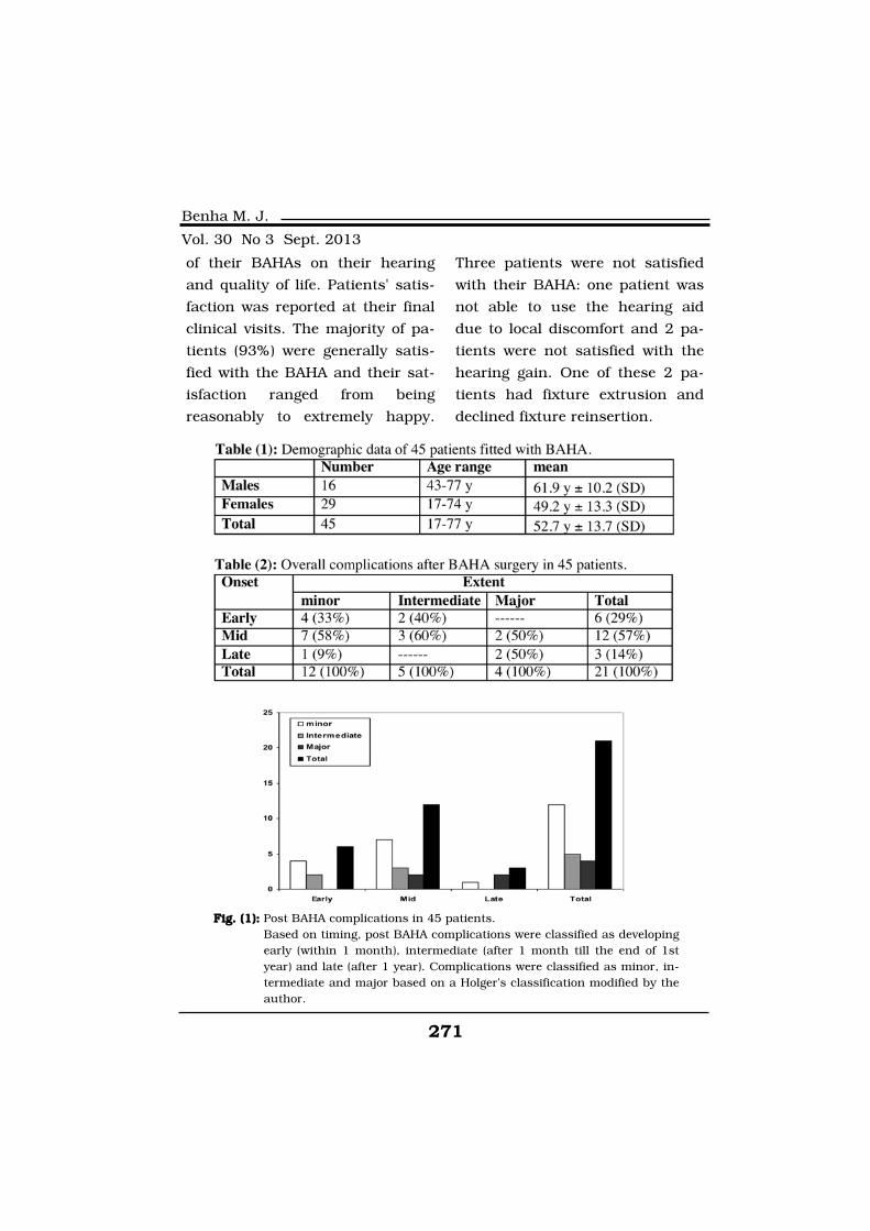
271
Benha M. J.
Vol. 30 No 3 Sept. 2013
of their BAHAs on their hearing
and quality of life. Patients' satis-
faction was reported at their final
clinical visits. The majority of pa-
tients (93%) were generally satis-
fied with the BAHA and their sat-
isfaction ranged from being
reasonably to extremely happy.
Three patients were not satisfied
with their BAHA: one patient was
not able to use the hearing aid
due to local discomfort and 2 pa-
tients were not satisfied with the
hearing gain. One of these 2 pa-
tients had fixture extrusion and
declined fixture reinsertion.
Fig. (1):Fig. (1): Post BAHA complications in 45 patients. Based on timing, post BAHA complications were classified as developingearly (within 1 month), intermediate (after 1 month till the end of 1styear) and late (after 1 year). Complications were classified as minor, in-termediate and major based on a Holger’s classification modified by theauthor.

272
Mahmoud El-Sayed Ali
DiscussionThe technique used in this se-
ries involves manual thinning of
the skin flap. The author found
that technique reasonably easy
and safe. There was no occasion of
flap damage or penetration due to
careful flap thinning with frequent
inspection of the skin side and as-
sessment of flap thickness as the
flap raising progresses. In the first
few cases, flap raising was rela-
tively slow paying more attention
to avoid flap over thinning and/or
penetration. As the technique was
mastered, less time was required
to raise the flap with the required
thickness. The flap was centred on
the supra-meatal crest. At this
point, a suitable bone thickness
can be found and a good sound
conduction to the cochlea is
achieved[12]. Dehydration of the
flap was avoided by applying
moist saline swabs on the under
surface of the flap. This seems to
help as there were no records of
flap dryness or ischemia. Based
on general surgical principles, a
skin flap technique is expected to
secure quicker and less complicat-
ed healing compared to a skin
graft[7,16]. The linear incision
technique has been reported to
provide minimal disturbance to
the skin and its blood supply with
complication rates and long-term
results comparable to other re-
ported skin flap techniques[12,19]. However, soft tissue reduc-
tion and surgical exposure of peri-
osteum and underlying skull bone
can be relatively difficult. The
creation of an inferiorly based flap
allows wider exposure of the surgi-
cal field and easier and more con-
trolled soft tissue reduction with
no apparent compromise of flap
vascularity.
All patients had their BAHA fit-
ting as planned. Over half of pa-
tients enjoyed uneventful post op-
erative course with complete
wound healing and implant fitting
as planned. In those who devel-
oped post operative complications,
most of complications (81%) were
either minor or intermediate and
all these were reversible with care-
ful medical treatment and did not
interfere with the hearing aid use.
This is comparable to previously
reported BAHA outcome[7,11,15,
20,21].
The first postoperative month is
the critical period for wound heal-
273
Benha M. J.
Vol. 30 No 3 Sept. 2013
ing and infection at the site of im-
plant could endanger osseointe-
gration resulting in surgical fail-
ure[22]. No major complication
was recorded in this period and
only minor or intermediate compli-
cations were encountered and
these represent over one fourth of
total complications. After complete
wound healing, the chance of de-
veloping minor complications de-
creased through the first year and
became progressively less after-
wards. Over the following 11
months there were increased ab-
solute numbers minor and inter-
mediate complications in addition
to 2 major complications. Howev-
er, considering the timescale and
taking the average complication
rate/month, the chance of devel-
oping complications after the first
post operative month was less
that one fifth that in the first post-
operative month. After the first
post operative year, the chance of
developing a complication was nil
as only one minor and 2 major
complications were encountered
with a mean follow up of 17.6
months.
Major complications in this se-
ries developed in 9% of patients
which represented 19% of total
complications. All major complica-
tions developed after complete os-
seointegration and sound proces-
sor fitting. The one patient who
developed excess skin growth over
the abutment received prolonged
medical treatment with local ster-
oids to try and reverse skin
growth over the abutment as rec-
ommended before[23]. This patient
required surgical intervention to
excise the excess skin. The other 3
patients required reinsertion of
the extruded fixture and the 2 pa-
tients who had this done proceed-
ed to sound processor fitting and
enjoyed satisfactory hearing reha-
bilitation.
Fixture extrusion occurred in 3
patients (7%) in this study and
this is comparable to that reported
in other studies[11,15,24]. It would
be expected that fixture extrusion
occurred much later than abut-
ment extrusion as the latter is not
implanted in skull bone and is
more superficial and prone to ex-
ternal forces. The abutment is
likely to fall out much easier and
earlier than the bone anchored ti-
tanium fixture particularly after
complete osseointegration. Howev-
274
Mahmoud El-Sayed Ali
er, fixture extrusion upon expo-
sure to repeated external forces
might indicate delayed failure or
gradual loss of osseointegration.
In these circumstances, the fix-
ture would come out after a trivial
trauma such as manipulation of a
jammed sound processor or hit-
ting the bed. Excessive external
force could, on the other hand,
force the fixture to extrude partic-
ularly in the presence of abutment
with or without the sound proces-
sor as the latter 2 components
would act then as a lever increas-
ing the effect of applied external
force. None of the patients had re-
ported symptoms or signs of late
infection. Whether fixture extru-
sion was due to gradual loss of os-
seointegration, subtle local inflam-
matory changes or a direct effect
of external trauma can not be de-
termined.
The majority of patients in this
series were satisfied with their
BAHA. As the vast majority of pa-
tients had previously worn air or
bone conduction hearing aids, the
BAHA relieved these patients, par-
ticularly those with recurrent ear
infections, from the former trou-
bles they had with the air or bone
conduction aids. The hearing gain,
improved communication and
quality of life as well as the lack of
inconvenience of fitting and re-
moving the ordinary haring aids
were among the satisfying factors
for these patients. This is compar-
able to previous results reported
before[17].
Although early BAHA fitting
has been advocated in some stud-
ies[12,19,25-27], in this series, os-
seointegration was considered to
be complete in 3 months and pa-
tients were fitted afterwards as
traditionally followed[6]. Following
this policy, none of the patients
had failure of osseointegration
and all patients were finally load-
ed with the sound processor as
planned. The author thinks that
early fitting should be taken with
great caution given the fact that
more than half of BAHA patients
had already suffered from recur-
rent ear infections with or without
hearing aids. After the first 3
months, the chance of developing
post operative complications
which could fail osseointegration
decreases and it would be safer to
allow complete wound healing and
treat any post operative complica-
275
Benha M. J.
Vol. 30 No 3 Sept. 2013
tions before fitting the patient with
the sound processor. The author,
however, has no reasons not to
consider earlier fitting for selected
patients when there is adequate
supportive evidence to the safety
and suitability of early fitting in
BAHA surgery.
In this series, one fourth of pa-
tients had dead one ear and would
not benefit from the ordinary
hearing aids. They would only
benefit from the contra lateral re-
routing of sound (CROS) hearing
aids or BAHA. Previous studies
had reported greater improvement
in speech intelligibility in noise
from BAHA compared with CROS
hearing aids[28]. Furthermore,
CROS aids have the inherent in-
convenience for patients in the
presence of recurrent ear infec-
tions. Furthermore, as the sound
is directly transmitted from the
BAHA fixture to skull bone, there
is no trans-cutaneous damping of
sound transmission inherent in
the bone conductor aids resulting
in better aided thresholds using
the BAHA[4,5]. It was not, there-
fore, the author's practice to try
CROS hearing aids before consid-
ering the patients for BAHA sur-
gery although all the options were
always discussed with patients to
chose from and to give an in-
formed consent.
Weakness of the study: This is
a retrospective study looking at a
single operative technique prac-
tised by the author. The number
of patients involved in this study
is not large enough and the follow
up period of this group of patients
is not long enough to obtain an
adequate insight to the potential
triggering or precipitating factors
involved in the development of
post BAHA surgery complications.
Further studies with large pa-
tients' number and long enough
follow up are required to evaluate
the long term clinical effectiveness
of this surgical technique com-
pared to other techniques em-
ployed in BAHA surgery.
ConclusionThe inferiorly based partial
thickness skin flap technique util-
ised in this series of 45 patients is
easy to master for skin prepara-
tion prior to BAHA implantation. It
is a safe technique and with fairly
satisfactory outcome (post opera-
tive complications and overall pa-
276
Mahmoud El-Sayed Ali
tients' satisfaction) comparable to
other skin preparation techniques
employed in BAHA surgery.
References1- George A., Coulson C.,1- George A., Coulson C.,
Ross E. and De R. (2012):Ross E. and De R. (2012): Sin-
gle-Stage BAHA and Mastoid
Obliteration. Int J Otol Laryn-
gol.12:1-3.
2- Pai I., Kelleher C., Nunn2- Pai I., Kelleher C., Nunn
T., Pathak N., Jindal M., O'Con-T., Pathak N., Jindal M., O'Con-
nor A.F. and Jiang D. (2012):nor A.F. and Jiang D. (2012):
Outcome of bone-anchored hear-
ing aids for single-sided deafness:
a prospective study. Acta Otola-
ryngol. 132(7):751-5.
3- Rasmussen J., Olsen S.Ø.3- Rasmussen J., Olsen S.Ø.
and Nielsen L.H. (2012):and Nielsen L.H. (2012): Evalua-
tion of long-term patient satisfac-
tion and experience with the
Baha® bone conduction implant.
Int J Audiol. 51(3):194-9.
4- Browning G.G. and Gate-4- Browning G.G. and Gate-
house S. (1994):house S. (1994): Estimation of
the benefit of bone-anchored hear-
ing aids. Ann Otol Rhinol Laryn-
gol.103(11):872-8.
5- Heywood R.L., Patel P.M.5- Heywood R.L., Patel P.M.
and Jonathan D.A. (2011):and Jonathan D.A. (2011): Com-
parison of hearing thresholds ob-
tained with Baha preoperative as-
sessment tools and those obtained
with the osseointegrated implant.
Ear Nose Throat J.; 90(5):E21-7.
6- Tjellström A., Rosenhall6- Tjellström A., Rosenhall
U., Lindström J., Hallén O., Al-U., Lindström J., Hallén O., Al-
brektsson T. and Brånemark P.I.brektsson T. and Brånemark P.I.
(1983):(1983): Five-year experience with
skin-penetrating bone-anchored
implants in the temporal bone.
Acta Otolaryngol.95(5-6):568-75.
7- Lekakis G.K., Najuko A.7- Lekakis G.K., Najuko A.
and Gluckman P.G. (2005):and Gluckman P.G. (2005):
Wound related complications fol-
lowing full thickness skin graft
versus split thickness skin graft
on patients with bone anchored
hearing aids. Clin Otolaryngol. 30
(4):324-7.
8- van de Berg R., Stokroos8- van de Berg R., Stokroos
R.J., Hof J.R. and ChenaultR.J., Hof J.R. and Chenault
M.N. (2010):M.N. (2010): Bone-anchored hear-
ing aid: a comparison of surgical
techniques. Otol Neurotol. 31
(1):129-35.
9- D'Eredita R. and Cenzi M.9- D'Eredita R. and Cenzi M.
(2010):(2010): TriVerse versus molecular
resonance-harvested grafts in
single-stage Baha surgery.
277
Benha M. J.
Vol. 30 No 3 Sept. 2013
Otolaryngol Head Neck Surg. 142
(4):560-4.
10- Persaud R.A., Papadimit-10- Persaud R.A., Papadimit-
riou N., Siva T., Kothari P. andriou N., Siva T., Kothari P. and
Quinn J.S. (2006):Quinn J.S. (2006): How we do it:
A novel approach to the skin--
abutment interface of the bone-
anchored hearing aid: use of local
skin flaps. Clin Otolaryngol. 31
(6):540-2.
11- Van Rompaey V., Claes11- Van Rompaey V., Claes
G., Verstraeten N., van DintherG., Verstraeten N., van Dinther
J., Zarowski A., Offeciers E. andJ., Zarowski A., Offeciers E. and
Somers T. (2011):Somers T. (2011): Skin reactions
following BAHA surgery using the
skin flap dermatome technique.
Eur Arch Otorhinolaryngol.268
(3):373-6.
12- Bovo B. (2008):12- Bovo B. (2008): Simplified
technique without skin flap for the
bone-anchored hearing aid
(BAHA®) implant. Acta Otorhinol-
aryngol Ital. October; 28(5): 252-
255.
13- Wilkinson E.P., Luxford13- Wilkinson E.P., Luxford
W.M., Slattery W.H. 3rd, De laW.M., Slattery W.H. 3rd, De la
Cruz A., House J.W. and FayadCruz A., House J.W. and Fayad
J.N. (2009):J.N. (2009): Single vertical inci-
sion for Baha implant surgery:
preliminary results. Otolaryngol
Head Neck Surg. 140(4):573-8.
14- Tamarit Conejeros J.M.,14- Tamarit Conejeros J.M.,
Dalmau Galofre J., Murcia Pu-Dalmau Galofre J., Murcia Pu-
chades V., Pons Rocher F.,chades V., Pons Rocher F.,
Fernández Martínez S. and Es-Fernández Martínez S. and Es-
trems Navas P. (2009):trems Navas P. (2009): Compari-
son of kin complications between
dermatome and U-graft technique
in BAHA surgery. Acta Otorrinola-
ringol Esp. 60(6):422-7.
15- Proops D.W. (1996):15- Proops D.W. (1996): The
Birmingham bone anchored hear-
ing aid programme: surgical meth-
ods and complications. J Laryngol
Otol Suppl. 21:7-12.
16- Snyder M.C., Moore G.F.16- Snyder M.C., Moore G.F.
and Johnson P.J. (2003):and Johnson P.J. (2003): The
use of full-thickness skin grafts
for the skin-abutment interface
around bone-anchored hearing
aids. Otol Neurotol. 24(2):255-8.
17- Rasmussen J., Olsen S.Ø.17- Rasmussen J., Olsen S.Ø.
and Nielsen L.H. (2012):and Nielsen L.H. (2012): Evalua-
tion of long-term patient satisfac-
tion and experience with the
Baha® bone conduction implant.
Int J Audiol. 51(3):194-9.
18- Holgers K.M., Tjellström18- Holgers K.M., Tjellström
A., Bjursten L.M. and Erlands-A., Bjursten L.M. and Erlands-
278
Mahmoud El-Sayed Ali
son B.E. (1988):son B.E. (1988): Soft tissue reac-
tions around percutaneous im-
plants: a clinical study of soft tis-
sue conditions around skin-
penetrating titanium implants for
bone-anchored hearing aids. Am J
Otol. 9(1):56-9.
19- D'Eredità R., Caroncini19- D'Eredità R., Caroncini
M. and Saetti R. (2012):M. and Saetti R. (2012): The new
Baha implant: a prospective osse-
ointegration study. Otolaryngol
Head Neck Surg. 146(6):979-83.
20- Håkansson B., Lidén G.,20- Håkansson B., Lidén G.,
Tjellström A., Ringdahl A., Ja-Tjellström A., Ringdahl A., Ja-
cobsson M., Carlsson P. and Er-cobsson M., Carlsson P. and Er-
landson B.E. (1990):landson B.E. (1990): Ten years of
experience with the Swedish bone-
anchored hearing system. Ann
Otol Rhinol Laryngol Suppl.
151:1-16.
21- Tjellström A. and21- Tjellström A. and
Granström G. (1994):Granström G. (1994): Long-term
follow-up with the bone-anchored
hearing aid: a review of the first
100 patients between 1977 and
1985. Ear Nose Throat J. 73
(2):112-4.
22- Gristina A.G. (1987): 22- Gristina A.G. (1987): Bio-
material-centered infection: Micro-
bial adhesion versus tissue inte-
gration. Science. 237:1588.
23- Falcone M.T., Kaylie23- Falcone M.T., Kaylie
D.M., Labadie R.F. and HaynesD.M., Labadie R.F. and Haynes
D.S. (2008):D.S. (2008): Bone-anchored hear-
ing aid abutment skin overgrowth
reduction with Clobetasol. Otola-
ryngology-Head and Neck surgery.
139:829-832.
24- Mylanus E.A.M. and24- Mylanus E.A.M. and
Cremers C.W. (1994):Cremers C.W. (1994): A one
stage surgical procedure for place-
ment of percutaneous implants for
the bone anchored hearing aid. J
Laryngol Otol. 108:1031-5.
25- Wazen J.J., Gupta R.,25- Wazen J.J., Gupta R.,
Ghossaini S., Spitzer J., Farru-Ghossaini S., Spitzer J., Farru-
gia M. and Tjellstrom A. (2007):gia M. and Tjellstrom A. (2007):
Osseointegration timing for Baha
system loading. Laryngoscope.
2007 May;117(5):794-6.
26- Lekue A., Lassaletta L.,26- Lekue A., Lassaletta L.,
Sánchez-Camón I., Pérez-MoraSánchez-Camón I., Pérez-Mora
R. and Gavilán J. (2012):R. and Gavilán J. (2012): Quality
of life in patients implanted with
the BAHA device depending on the
aetiology. Acta Otorrinolaringol
Esp. 9.
27- McLarnon C.M., Johnson27- McLarnon C.M., Johnson
I., Davison T., Hill J., Hender-I., Davison T., Hill J., Hender-
279
Benha M. J.
Vol. 30 No 3 Sept. 2013
son B., Leese D. and Marley S.son B., Leese D. and Marley S.
(2012):(2012): Evidence for early loading
of osseointegrated implants for
bone conduction at 4 weeks. Otol
Neurotol. 2012;33(9):1578-82.
28- Wazen J.J., Spitzer J.B.,28- Wazen J.J., Spitzer J.B.,
Ghossaini S.N., Fayad J.N., Ni-Ghossaini S.N., Fayad J.N., Ni-
parko J.K., Cox K., Brackmannparko J.K., Cox K., Brackmann
D.E. and Soli S.D. (2003):D.E. and Soli S.D. (2003): Trans-
cranial contralateral cochlear
stimulation in unilateral deafness.
Otolaryngol Head Neck Surg. 129
(3):248-54.
280
Mahmoud El-Sayed Ali
OUTCOME OF BAHA SURGERYOUTCOME OF BAHA SURGERYUSING THE INFERIORLY BASEDUSING THE INFERIORLY BASED
PARTIAL THICKNESSPARTIAL THICKNESSSKIN FLAPSKIN FLAP
Mahmoud El-Sayed Ali, MBBCh, MSc, FRCS, MDahmoud El-Sayed Ali, MBBCh, MSc, FRCS, MD
BENHAMEDICALJOURNAL
REPRINT
Published byPublished by
Benha Faculty of MedicineBenha Faculty of Medicine Volume 30 Number 3Sept. 2013
281
Benha M. J.
Vol. 30 No 3 Sept. 2013
EVALUATION OF THE ROLE OF LAPAROSCOPYEVALUATION OF THE ROLE OF LAPAROSCOPYIN THE MANAGEMENT OF BLUNTIN THE MANAGEMENT OF BLUNT
ABDOMINAL TRAUMAABDOMINAL TRAUMA
Abu-Sheashaa M.S. MRCS, Dawood I. MD, El-Sedek M. MD,Abu-Sheashaa M.S. MRCS, Dawood I. MD, El-Sedek M. MD,Nashat Noaman MD and Ahmed Negm MDNashat Noaman MD and Ahmed Negm MD
Department of & General Surgery, Faculty of Medicine,
Mansoura University, Mansoura, Egypt
AbstractIntroduction:Introduction: In the setting of blunt abdominal trauma, laparoscopy
is used mainly for diagnosis, and its role in definitive operative repair isstill debated.
Aim of the work:Aim of the work: The aim of our work was evaluation of the role oflaparoscopy in the management of blunt abdominal trauma.
Patients and Methods: Patients and Methods: This is an interventional, prospective, non-randomized clinical study conducted on twenty patients with blunt ab-dominal trauma admitted at Mansoura emergency hospital over the pe-riod between April 2011 and April 2013.
Results:Results: It was evident that the largest number of patients was be-tween 20-30 years (40%) and the least number of patients between 50-55 years (10%). No patients were below 10 nor above 55 years. Ten cas-es had hollow viscus injuries four of them were ileal, two of them werejejunal, three of them were colonic injuries and one was duodenal. Wehad two cases of active internal bleeding, one was splenic and the otherwas hepatic injury. We had one case of diaphragmatic injury. Therewere 2 cases of missed injuries discovered during delayed laparotomydone 2 days or more after diagnostic laparoscopy. The sensitivity of lap-aroscope in detection of hollow viscus injury was 80% and detection ofdiaphragmatic injury was100% and in the detection of solid organ inju-ries was 80%.
Conclusion:Conclusion: The diagnostic and therapeutic role of laparoscopy isprogressing with time but is mainly directed towards hollow vicera anddiaphragmatic injuries.The role of laparoscopy in control of intra-abdominal bleeding is limited and should not be on the expense of safe-ty.
282
Abu-Sheashaa M.S., et al....
IntroductionDespite improved diagnostic
tools such as computerized tomog-
raphy (CT scan), conventional
treatment of patients with abdom-
inal trauma injures often requires
exploratory laparotomy proce-
dures to accurately diagnose and
treat patients, injures. Studies
showed that nontherapeutic (i.e.,
negative) laparotomy rates range
from 5% to 40%, depending on the
clinical situation. Many surgeons
now perform diagnostic laparos-
copic procedures in haemodynam-
ically stable patients with abdomi-
nal trauma injuries. Although
laparoscopy in patient with ab-
dominal trauma injury does have
limitations, it is an effective tool
for preventing negative laparoto-
mies.(1)
In the setting of blunt abdomi-
nal trauma, laparoscopy is used
mainly for diagnosis, and its role
in definitive operative repair is still
debated.(2)
Aim of the Work The aim of our work was evalu-
ation of the role of laparoscopy in
the management of blunt abdomi-
nal trauma.
Patients and MethodsThis is an interventional, pros-
pective, non-randomized clinical
study conducted on twenty pa-
tients with blunt abdominal trau-
ma admitted at Mansoura emer-
gency hospital over the period
between April 2011 and April
2013.
Patients' population:Patients' population:
Twenty patients (14 males and
6 females) were included in the
study. Their ages ranged between
13 and 55 years with mean age 26
years.
Our inclusion criteria wereOur inclusion criteria were
blunt abdominal mono-trauma,
haemodynamically stable and pos-
itive clinical or radiological signs
for hollow viscus or diaphragmatic
injuries.
Exclusion criteria were Exclusion criteria were poly-
traumatized patient, haemody-
namically unstable and general
contraindications of laparoscopy.
Patients' evaluation:Patients' evaluation:
All patients were subjected to
primary survey, then secondary
survey which localized the trauma
to the abdomen.
283
Benha M. J.
Vol. 30 No 3 Sept. 2013
All patients were investigatedAll patients were investigated
as follows:as follows:
A- Laboratory in the form of;A- Laboratory in the form of;
CBC, serum creatinine, liver func-
tion studies, Coagulation profile,
RBS, serum electrolytes and arte-
rial blood gases.
B- B- Radiological studies includ-Radiological studies includ-
ed;ed; Abdominal X-ray supine and
erect showing the lower chest, FAST
and aspiration from the free fluid
or collection if accessible and CT
abdomen with oral and IV contrast.
Follow up studies:Follow up studies:
A- ClinicalA- Clinical
• Vital signs every 3 hours, Se-
rial abdominal examination every
6 hours.
B- LaboratoryB- Laboratory
• Hemoglobin, hematocrit val-
ue, TLC and ABG every 6 hours.
C- RadiologicalC- Radiological
• Serial FAST every 6 hours,
which could be modulated accord-
ing to the clinical situation.
• Follow up abdominal X-ray
when the clinical picture was
equivocal.
The study was conducted on
one group including 20 patients
who were subjected to a diagnos-
tic laparoscopy after receiving the
conservative measures done to
stable patients with blunt abdomi-
nal trauma. Management was
then tailored according to the la-
paroscopic findings.
We had 4 sub-groups:We had 4 sub-groups:
a- Non therapeutic (only diag-a- Non therapeutic (only diag-
nostic) sub-groupnostic) sub-group in which the
laparoscopic findings did not ne-
cessitate therapeutic intervention.
b- Totally laparoscopic sub-b- Totally laparoscopic sub-
groupgroup in which the laparoscopic
findings were managed totally by
laparoscopy.
c- Laparoscopically assistedc- Laparoscopically assisted
sub-groupsub-group in which the laparos-
copic findings were managed par-
tially by laparoscopy and the pro-
cedure was completed through a
small laparotomy incision (3-4 cm).
d- Laparoscopic converted tod- Laparoscopic converted to
laparotomy sub-grouplaparotomy sub-group in which
laparoscope failed to manage the
injury so converted to laparotomy.
Diagnostic laparoscopy underDiagnostic laparoscopy under
general anesthesia with endotra-
284
Abu-Sheashaa M.S., et al....
cheal intubation was used in all
cases.
Steps:Steps:
• 10 mm trocar was inserted in
the left iliac fossa or peri-umblical
region using an open technique
(Hasson technique).
• A pneumo-peritoneum was
produced using CO2 insufflation
through the trocar at a rate of
about 0.5ml per minute to a maxi-
mum intra-abdominal pressure of
9-12 mmHg.
• A 30 degree telescope was in-
serted in the abdominal cavity.
• An initial rapid tour was
made inside the abdominal cavity
searching for active bleeding
which took priority in manage-
ment.
• We determined the sites of
port placement according to the
operative findings and needs.
• For handling of the small in-
testine to explore, two additional 5
mm trocars were placed in the
upper left and the lower right
quadrants and 2 atraumatic gras-
pers were inserted through the
two trocars.
• One side of the bowel wall is
observed by lifting up the bowel
with the 2 bowel forceps the other
side of the bowel is observed by
turning over the bowel.
• The stomach, duodenum, co-
lon and small intestine were care-
fully examined for perforation or
ischaemic changes.
• If subserosal hematoma or
dirty exudate was not seen in the
retroperitoneal parts of the duode-
num or the colon, the patient was
judged not to have perforation.
• The transverse colon, splenic
flexure and descending colon were
inspected while the surgeon
standing on the right side.
• The liver, stomach spleen and
diaphragm were inspected while
the patient in the reversed Tren-
dlenburg position.
Therapeutic laparoscopic pro-Therapeutic laparoscopic pro-
cedures:cedures:
Laparoscopic intestinal repairLaparoscopic intestinal repair
and resection anastomosis:and resection anastomosis:
Cases with small intestinal or
colonic injuries which were docu-
mented by laparoscopy were re-
paired either totally laparoscopi-
cally by intra-corporeal suturing
technique or by laparoscopic as-
sisted techniques in which com-
plete laparoscopic mobilization of
the small intestine was performed
and the injured segments a were
285
Benha M. J.
Vol. 30 No 3 Sept. 2013
pulled out through a small lapa-
rotomy incision (3-4cm) then the
resection of the injured segment
and anastomosis of the two ends
of the small intestine was per-
formed using conventional tech-
niques (single layer extra-mucosal).
Then we pushed the anastomotic
segment back into the abdomen.
Laparoscopic repair of dia-Laparoscopic repair of dia-
phragmatic injury; phragmatic injury;
We had one case of diaphragmat-
ic repair done by the use of non ab-
sorbable sutures (Prolene 0) using
intra corporeal suturing technique.
Follow up of the patients inFollow up of the patients in
the post-operative periodthe post-operative period with re-
cording of the morbidity and mor-
tality for 6 months after discharge.
All data were collected in special
preformed sheet for statistical
analysis. The data included pa-
tient demographics, co-morbidities,
mechanisms of injuries, indication
of laparoscopy, findings in labora-
tory and radiological investiga-
tions, different types of laparos-
copic intervention, hospital stay,
number of units of blood transfu-
sion, intensive care unit period,
time of intervention and postoper-
ative morbidity and mortality.
Statistical AnalysisData were analyzed using SPSS
(Statistical Package for Social Sci-
ences) version 15. Qualitative data
were presented as number and
percent. Quantitative data were test-
ed for normality by Kolmogrov-
Smirnov test. Normally distributed
data were presented as mean ±
SD.
ResultsPatients' profile:Patients' profile:
This prospective study was
conducted on twenty patients who
had blunt abdominal trauma.
These patients were managed by
the usual conservative measures
done to patients who are haemod-
ynamically stable (admission in
HDU after starting resuscitative
measures then follow up using
clinical, laboratory and radiologi-
cal parameters) then diagnostic la-
paroscopy was done and manage-
ment differed according to the
laparoscopic finding.
Causes of conversion of lapa-Causes of conversion of lapa-
roscopy to laparotomy (2 cases):roscopy to laparotomy (2 cases):
Failure of control of bleeding 1
case (50%).
Technical difficulties 1 case
(50%).
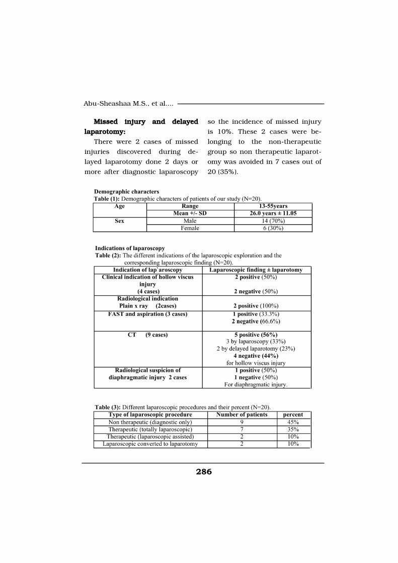
286
Abu-Sheashaa M.S., et al....
Missed injury and delayedMissed injury and delayed
laparotomy:laparotomy:
There were 2 cases of missed
injuries discovered during de-
layed laparotomy done 2 days or
more after diagnostic laparoscopy
so the incidence of missed injury
is 10%. These 2 cases were be-
longing to the non-therapeutic
group so non therapeutic laparot-
omy was avoided in 7 cases out of
20 (35%).
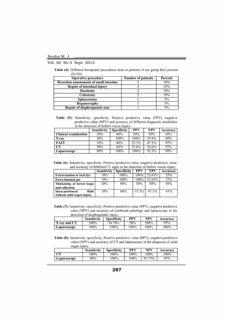
287
Benha M. J.
Vol. 30 No 3 Sept. 2013
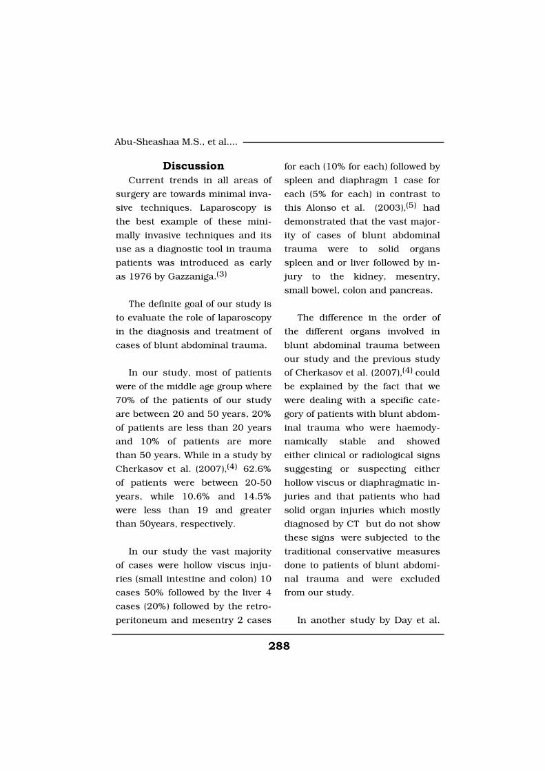
288
Abu-Sheashaa M.S., et al....
DiscussionCurrent trends in all areas of
surgery are towards minimal inva-
sive techniques. Laparoscopy is
the best example of these mini-
mally invasive techniques and its
use as a diagnostic tool in trauma
patients was introduced as early
as 1976 by Gazzaniga.(3)
The definite goal of our study is
to evaluate the role of laparoscopy
in the diagnosis and treatment of
cases of blunt abdominal trauma.
In our study, most of patients
were of the middle age group where
70% of the patients of our study
are between 20 and 50 years, 20%
of patients are less than 20 years
and 10% of patients are more
than 50 years. While in a study by
Cherkasov et al. (2007),(4) 62.6%
of patients were between 20-50
years, while 10.6% and 14.5%
were less than 19 and greater
than 50years, respectively.
In our study the vast majority
of cases were hollow viscus inju-
ries (small intestine and colon) 10
cases 50% followed by the liver 4
cases (20%) followed by the retro-
peritoneum and mesentry 2 cases
for each (10% for each) followed by
spleen and diaphragm 1 case for
each (5% for each) in contrast to
this Alonso et al. (2003),(5) had
demonstrated that the vast major-
ity of cases of blunt abdominal
trauma were to solid organs
spleen and or liver followed by in-
jury to the kidney, mesentry,
small bowel, colon and pancreas.
The difference in the order of
the different organs involved in
blunt abdominal trauma between
our study and the previous study
of Cherkasov et al. (2007),(4) could
be explained by the fact that we
were dealing with a specific cate-
gory of patients with blunt abdom-
inal trauma who were haemody-
namically stable and showed
either clinical or radiological signs
suggesting or suspecting either
hollow viscus or diaphragmatic in-
juries and that patients who had
solid organ injuries which mostly
diagnosed by CT but do not show
these signs were subjected to the
traditional conservative measures
done to patients of blunt abdomi-
nal trauma and were excluded
from our study.
In another study by Day et al.
289
Benha M. J.
Vol. 30 No 3 Sept. 2013
(2002),(6) they postulated that the
sensitivity of initial abdominal ex-
amination was 16% and specificity
was 52%in determining the need
of laparotomy.
In our study we had 4 cases
suspected to have hollow viscus
injury by serial clinical abdominal
examination depending on the
previous signs. Laparoscopic eval-
uation revealed 2 positive cases
for this while the other 2 cases
were negative for this. Sensitivity
was 20% and specificity was 62%
which was higher than the previ-
ous study which could be ex-
plained by that in our study we
evaluated serial examination not
initial examination only. The prob-
lem in clinical examination is that
it depends on the experience of
the examiner also it needs serial
abdominal examination also initial
evaluation is not so helpful be-
cause contusion of the abdominal
wall may produce signs similar to
that of peritonitis also may be af-
fected by drugs, analgesics, nar-
cotics or diminished level of con-
sciousness because of trauma.
Liu et al. (2003),(7) had stated
that CT is a relatively non-invasive
diagnostic modality. Its complica-
tion reported to date has been so
infrequent. Both intra peritoneal
and retroperitoneal structures are
visualized and the vascular integ-
rity of the solid organs (spleen- liv-
er- kidney) can be evaluated but
the sensitivity of CT remains ques-
tionable especially for hollow vis-
cus injury.
Alan et al (2008),(8) had dem-
onstrated that CT has a signifi-
cant false negative rate in the di-
agnosis of small bowel injuries,
findings suggestive of small bowel
injury include fluid collection
without solid organ injury, bowel
wall thickening, mesenteric infil-
tration, free intra-peritoneal air
and oral dye extravasation.
In our study we found that the
sensitivity and specificity of CT in
the diagnosis of solid organs was
100% in agreement with most of
the literature but its sensitivity for
hollow viscus injury was 50% but
its accuracy in detection of the ex-
act site is questionable. In our study
it was 20% where only one case
out of five show CT findings simi-
lar to laparotomy regarding the site
of leak and was the duodenum.

290
Abu-Sheashaa M.S., et al....
In a study by Feliz et al. (2006),(9) there were 7127 trauma admis-
sions, of which 113 had abdomi-
nal explorations for blunt (88%)
and penetrating (12%) trauma.
Thirty-two (28%) patients had la-
paroscopy performed. Laparotomy
was avoided in 56% of these pa-
tients.
In our study, laparotomy was
avoided in 70% of patients 35%
was because of laparoscopic find-
ings that do not necessitate thera-
peutic intervention and 35% was
because laparoscopic findings that
were managed totally by laparos-
copy but the ratio is higher in our
study because all our patients
are blunt abdominal trauma un-
like the previous study which in-
cluded patients with blunt and
penetrating abdominal trauma
and it is well established that the
incidence of intra-abdominal inju-
ry is less in blunt abdominal trau-
ma than that of penetrating abdom-
inal trauma and so conservative
management is more applicable in
blunt than in penetrating.
In the previous study by Feliz
et al. (2006),(9) Laparoscopy was
negative in 9 (28%) patients, 5 of
whom had blunt trauma and 4
had penetrating trauma. Laparos-
copy was diagnostic and nonther-
apeutic in 3 (10%) patients with
nonexpanding hematomas. Lapar-
oscopic therapeutic interventions
were performed in 6 (19%) pa-
tients. Laparotomy was avoided in
17 (56%) patients. Laparoscopy
assisted in the diagnosis and sub-
sequent conventional repair of 10
cases of perforated viscera, dia-
phragmatic ruptures, and 1 distal
pancreatic injury. Thirteen (68%)
of the injuries diagnosed by lapa-
roscopy were an injury to a hollow
viscus. No injuries were missed, or
technical complications occurred,
as a result of laparoscopic explo-
rations. No patients who under-
went laparoscopy died.
In our study, we had no nega-
tive cases by laparoscopy. This
could be explained by the fact that
we dealt with patients already
having either clinical or radiologi-
cal signs suggesting hollow viscus
or diaphragmatic injuries we did
not do laparoscopy for all patients
with blunt abdominal trauma. Ini-
tially we had 9 (45%) cases with
nontherapeutic (only diagnostic
laparoscopy) with liver, retroperi-
291
Benha M. J.
Vol. 30 No 3 Sept. 2013
toneal and mesenteric injuries
with no active bleeding. laparosco-
py assisted in the diagnosis of 2
cases (10%) of intestinal injuries
which enabled us to make a small
laparotomy incision and complet-
ing the procedure (laparoscopic
assisted). We made diagnostic la-
paroscopy then completing the
therapeutic intervention totally by
laparoscopy in 7cases (35%). We
had 10 cases (50%) of the whole
group with intestinal injury 8 cas-
es (80%) of them were diagnosed
by laparoscopy 2 cases (20%) were
missed by laparoscope and detect-
ed during delayed laparotomy, 6
cases (60%) of them were man-
aged totally laparoscopic, 2 cases
(20%) were managed by laparos-
copic assisted technique.
Laparoscopy determined the
need for laparotomy and conven-
tional repair was done after lapa-
rotomy in 2 cases (10%) and
missed injuries occurred in 2 cas-
es out of 20 which is the total
number of the patients in our
study (10%).
In a study by Elliot et al.
(1998),(10) they assessed 47 pa-
tients selected for laparotomy after
trauma all patients underwent di-
agnostic laparoscopy followed by
laparotomy. Sensitivity was poor
less than 50% for injuries of hol-
low viscera however laparoscopy
had excellent sensitivity 96.2%
and specificity 100% for determin-
ing the need for therapeutic lapa-
rotomy.
In our study, the sensitivity of
laparoscope for detection of hollow
viscus injury was 80% and speci-
ficity for this was 100%.
In a study by Chersakov et al.
(2007),(4) conversion to laparoto-
my was performed in 26.7% of pa-
tients, massive solid organ in-
volvement multiple abdominal
organs injuries hemorrhage great-
er than 1500ml, haemoperitoneum
greater than 500 ml and continu-
ing heavy internal bleeding were
the absolute indications for con-
version to laparotomy in haemody-
namically unstable subjects. Co-
morbid conditions such as respir-
atory cardiovascular and renal in-
sufficiency were relative indications
for conversion to laparotomy.
In our study, conversion to lap-
arotomy immediately after lapa-
292
Abu-Sheashaa M.S., et al....
roscopy was done in 2 cases only
(10%) there were two reasons for
this conversion, the first was ina-
bility to control bleeding for more
than 10 minutes.
The second reason was techni-
cal difficulties where duodenal in-
jury was suspected and mobiliza-
tion of the duodenum was
required and could not be done by
laparoscopy.
In the previous study by Cher-
sakov et al. (2007),(4) the average
time for diagnostic laparoscopy
was 22±8 minutes and that of la-
paroscopic surgery was 60±12
minutes the average postoperative
hospital stay following video as-
sisted laparoscopy 12.2±3.8 days
and 18.44±3.8 days after laparoto-
my.
In our study, the average time
for diagnostic laparoscopy was
40±8minutes which is more than
that of the previous study which
may be due to our starting limited
experience in the use of laparo-
scope in blunt abdominal trauma
and this was reflected on our re-
sults as the starting limited expe-
rience made us more meticulous
during exploration so the sensitiv-
ity and specificity of laparoscope
was better than other studies. The
post-operative hospital stay was
variable between the different sub-
groups of our study it was 7.2±1.9
days for the non-therapeutic sub-
group it was 10±2.8 days for total-
ly laparoscopic sub-group and it
was 8.1±1.7 it was 12±2.5days for
the subgroup in which laparosco-
py is converted to laparotomy and
all these values were less than
that of the previous study. We do
not have clear explanation for this
but could be due to the use of
principles of fast track surgery in
most cases of our study.
In the study by Cherkasov et al
(2007),(4) the complications of la-
paroscopy group were as follows
chest infection was 1.2% of pa-
tients, wound infection 2.8% of
patients, evisceration 0% intesti-
nal bands and obstruction 0%
and the complications of the oth-
er group who underwent laparoto-
my were as follows chest infection
5.3% wound infection3.8% bowel
evisceration 0.4% intestinal ob-
struction 0.6%.
In our study, wound infection
293
Benha M. J.
Vol. 30 No 3 Sept. 2013
occurred only in the 2 subgroups
in which a laparotomy incision
was done. This can be explained
by the fact that incisions of totally
laparoscopic subgroups mostly
are very small 1cm or less, but in-
tra-peritoneal collection occurred
only in other 2 subgroups which
were done totally laparoscopically
this could be attributed to that la-
paroscopy is less efficient in drain-
age of intra-peritoneal collection
than open drainage and breaking
of septa by hand dissection. We
had only one case of chest infec-
tion in the subgroup in which la-
paroscopy converted to laparoto-
my. This can be explained by that
laparotomy incision made patient
feel postoperative pain that limits
respiratory movement predispos-
ing to chest infection. This could
be considered one of the major ad-
vantages of laparoscopy. No other
chest complications related to
CO2 insufflation in the studied
group were detected.
No cases of evisceration nor ad-
hesive intestinal obstruction were
detected in our studied group un-
like the previous study because of
the small number of patients in
subgroups subjected to laparoto-
my and relatively short period of
follow up and can be explained
also as an advantage of laparosco-
py. We had 2 cases of intestinal
injury which were missed by diag-
nostic laparoscopy. There was 1
more case of intestinal leak in the
subgroup which was done totally
laparoscopic which may be ex-
plained by error in the technique
of repair because of our limited
experience in intra-corporeal su-
turing technique.
Laurance and Gerard (2003).(11) stated that blunt abdominal
injuries is usually associated with
serious complications and high
mortality of about 25- 65% as a
result of difficulty in prompt diag-
nosis and management of other
injuries.
In our study, the mortality rate
was 0% in our patients' popula-
tion, this can be explained by the
fact that we dealt with a specific
category of patients in which the
abdomen was only involved in
trauma and all patients were hae-
modynamically stable. Extra-
abdominal injuries were absent in
our study. It is well known that
extra-abdominal injuries account
294
Abu-Sheashaa M.S., et al....
for a significant share of mortality
rate among trauma patient.
Conclusion and Recommen-Conclusion and Recommen-
dations:dations:
The diagnostic role of laparos-
copy is progressing with time but
mainly directed towards diagnosis
of hollow vicera and diaphragmat-
ic injuries.
The role of laparoscopy in con-
trol of intra-abdominal bleeding is
limited and should not be on the
expense of safety
References1- Fabien T.C. and Croce1- Fabien T.C. and Croce
M.A. (2000):M.A. (2000): Prospective analysis
of diagnostic laparoscopy in trau-
ma. Am surg, 217-(5): 557-64.
2- Iannelli A., Fabiani P., Ka-2- Iannelli A., Fabiani P., Ka-
rimdjee B.S., et al. (2003):rimdjee B.S., et al. (2003): Ther-
apeutic laparoscopy for blunt ab-
dominal trauma with bowel
injuries. J Laparo-endoscopic Ad-
vanced Surgical Technique A 13
(3): 189-91.
3- Gazzaniga A.B., Slanton3- Gazzaniga A.B., Slanton
W.W. and Bartlett R.H. (1976):W.W. and Bartlett R.H. (1976):
Laparoscopy in the diagnosis of
blunt and penetrating injuries to
abdomen. Am J Surg; 131: 315-
31833. 39.
4- Cherkasov M., Sitnikov V.,4- Cherkasov M., Sitnikov V.,
Bsarkisyn, Degtriev O., et al.Bsarkisyn, Degtriev O., et al.
(2008):(2008): Laparoscopy versus lapa-
rotomy in management of abdomi-
nal trauma. Journal of Surgical
Endoscopy. 22, 228-231.
5- Alonso M., Brathwaite C.5- Alonso M., Brathwaite C.
and Garcia V. (2003): and Garcia V. (2003): Practice
management guidelines for the
non-operative management of
blunt injury to the liver and
spleen. Eastern Association for
the Surgery of Trauma.
6- Day A.C., Rankin N. and6- Day A.C., Rankin N. and
Charlesworth P. (2002):Charlesworth P. (2002): Diagnos-
tic Peritoneal Lavage: integration
with clinical information to im-
prove diagnostic performance. J
trauma. 32:52-57.
7- Liu M., Lee C.H. and Peng7- Liu M., Lee C.H. and Peng
F.K. (2003):F.K. (2003): Prospective compari-
son of diagnostic peritoneal lav-
age, computed tomographic scan-
ning, and ultrasonography for the
diagnosis of blunt abdominal trau-
ma. J Trauma 35:267-70.
8- Alan A., Simeone, Heidi L.,8- Alan A., Simeone, Heidi L.,
295
Benha M. J.
Vol. 30 No 3 Sept. 2013
et al. (2008):et al. (2008): Abdominal injury In
the Trauma Manual: Trauma and
acute care surgery. Edited by:
Peitzmam AB, Rhodes M, Schwab
CW, Donald MY and Fabian TC.
Lippincott William and Wilkins
3rd edition: 243-72.
9- Feliz A., Shultz B., McKen-9- Feliz A., Shultz B., McKen-
na C., et al. (2006): na C., et al. (2006): Diagnostic
and therapeutic laparoscopy in
pediatric abdominal trauma. Jour-
nal of Pediatric Surgery. 41, 7-77.
10- Elliot D.C., Rodriguez A.,10- Elliot D.C., Rodriguez A.,
Moncure M., et al. (1998):Moncure M., et al. (1998): The
accuracy of diagnostic laparosco-
py in trauma patients: a prospec-
tive, controlled study. Int Surg.
83:294-8.
11- Laurance W. and Gerard11- Laurance W. and Gerard
M. (2003):M. (2003): Management of injured
patients In: James R, William C,
Frank R (eds) Current surgical di-
agnosis and treatment. Lange,
New York, pp. 230-255.
296
Abu-Sheashaa M.S., et al....
EVALUATION OF THE ROLE OFEVALUATION OF THE ROLE OFLAPAROSCOPY IN THE MANAGEMENTLAPAROSCOPY IN THE MANAGEMENT
OF BLUNT ABDOMINAL TRAUMAOF BLUNT ABDOMINAL TRAUMA
Abu-Sheashaa M.S. Abu-Sheashaa M.S. MRCSMRCS, Dawood I. MD, El-Sedek M. MD,, Dawood I. MD, El-Sedek M. MD,Nashat Noaman MD and Ahmed Negm MDNashat Noaman MD and Ahmed Negm MD
BENHAMEDICALJOURNAL
REPRINT
Published byPublished by
Benha Faculty of MedicineBenha Faculty of Medicine Volume 30 Number 3Sept. 2013
297
Benha M. J.
Vol. 30 No 3 Sept. 2013
IntroductionIntroductionRecently more light has been
shed on the overlap in biologic
and morphologic features between
classic Hodgkin lymphoma (cHL)
and mature B-cell lymphoma[1].
This overlap was further substan-
tiated by analyzing the neoplastic
IMMUNOHISTOCHEMICAL PROFILING OFIMMUNOHISTOCHEMICAL PROFILING OFB-CELL LYMPHOMA INTERMEDIATEB-CELL LYMPHOMA INTERMEDIATEBETWEEN DIFFUSE LARGE B-CELLBETWEEN DIFFUSE LARGE B-CELL
LYMPHOMA AND CLASSICALLYMPHOMA AND CLASSICALHODGKIN LYMPHOMAHODGKIN LYMPHOMA
Nirmeen Megahed M.Sc, Azmy Abdelhameed MD,Nirmeen Megahed M.Sc, Azmy Abdelhameed MD,Asmaa Gado MD, Maha Amin MD, Naoko Asano MD*,Asmaa Gado MD, Maha Amin MD, Naoko Asano MD*,
Seiichi Kato MD*, Katsuyoshi Takata MD**,Seiichi Kato MD*, Katsuyoshi Takata MD**,Aki Mitsuda MD*** and Shigeo Nakamura MD*Aki Mitsuda MD*** and Shigeo Nakamura MD*
Departments of Pathology, Mansoura University, Faculty of Medicine, Egypt
*Nagoya University Hospital, Japan, **Okayama University Graduate School of
Medicine, Dentistry and Pharmaceutical, Sciences, Japan
*** Toho University School of Medicine, Japan.
AbstractAbstractAim:Aim: Currently, WHO classification 2008 approved the term B-cell lym-
phoma intermediate between diffuse large B-cell lymphoma (DLBCL) andclassical Hodgkin lymphoma (cHL).This study was urged by scarcity of dataabout such cases to explore their immunohistochemical features.
Methods and Results: Methods and Results: Thirteen cases of B-cell lymphoma cases with in-termediate features were identified over a 9-year period. HRS-like cells wereCD30+ with a high rate of CD20+ (100%) and lower rates of CD15+ (25%)and Fascin+ (50%).
Conclusion:Conclusion: B-cell lymphoma intermediate between diffuse large B-celllymphoma (DLBCL) and classical Hodgkin lymphoma (cHL) is a distinct enti-ty of lymphoma characterized by intermediate histological and immuno-phenotypical features between cHL and DLBCL.
Key words:Key words: Lymphoma, Grey zone, Hodgkin.
298298
Nirmeen Megahed, et al....
nature of the malignant cells of
cHL, which were proven to be of
B-cell origin in nearly all cases[2].
This advocated the introduction of
the new term ‘‘grey zone lympho-
ma’’, which was used for the first
time in 1998 in the ‘‘Workshop on
Hodgkin’s disease and related dis-
eases’’[3].
According to WHO classifica-
tion 2008, the term “grey zone
lymphoma” designates anterior
mediastinal involvement with lym-
phoma intermediate between
DLBCL and cHL[4]. On morpholog-
ic basis this lesion is character-
ized by sheet like growth of pleo-
morphic tumor cells with
Hodgkin-Reed-Sternberg (HRS)-
like morphology admixed with a
varying degree of inflammatory
background formed of mature
lymphocytes, eosinophils and
plasma cells[5]. The tumor charac-
teristically shows fibrous stroma
reminiscent of nodular sclerosis
variant of Hodgkin lymphoma[1].
Geographic necrosis is usually
present which, unlike necrosis of
cHL, do not show neutrophilic in-
filtrate[4]. Immunophenotypically,
the tumor cells show combined
expression of B-cell markers
(CD20, CD79a, PAX5) and Hodg-
kin markers (CD30, CD15)[6].
ALK-1 is consistently negative.
Surface or cytoplasmic immuno-
globulins are also absent. EBV
has been detected in less than
20% of cases[4].
Materials and MethodsMaterials and MethodsPatient samples:Patient samples:
Thirteen consultation cases of
B-cell lymphoma intermediate be-
tween diffuse large B-cell lympho-
ma (DLBCL) and classical Hodgkin
lymphoma (cHL) lymphoma diag-
nosed between 2003 and 2011
were retrieved from the files of the
Department of Pathology and La-
boratory Medicine, Nagoya Univer-
sity hospital, Japan. This study
was approved by the institutional
review board of Nagoya University
Hospital.
All the examined slides were
from excisional biopsies. The his-
tological features were evaluated
in 4µm thick hematoxylin and eo-
sin-stained sections of formalin-
fixed paraffin-embedded tissue.
Immunohistochemistry:Immunohistochemistry:
Tissue sections were stained
with antibodies directed against
299
Benha M. J.
Vol. 30 No 3 Sept. 2013
CD20, CD79a, CD15, CD30,
PAX5, MUM1, Fascin, ALK1, CD3,
CD10, BCL-2, BCL-6, P53, Ki-67,
CD45RO, EMA, κ and λ. Appropri-
ate positive and negative controls
for all the selected immunostains
were used. Histological and immu-
nohistochemical data were assessed
by three pathologists of the au-
thors (Megahed, Asano and Kato).
In situ hybridization study:In situ hybridization study:
The presence of EBV small ri-
bonucleic acids was examined by
in situ hybridization using EBV-
encoded small nuclear early re-
gion (EBER) oligonucleotides on
formalin- fixed, paraffin-embedded
sections. Briefly, a DAKO hybridi-
zation kit was used with a cocktail
of fluorescein isothiocyanate-
labeled EBER oligonucleotides
(one oligonucleotide corresponding
to EBER1 and the other to
EBER2, both 30 bases long;
DAKO A/S code Y 017). Hybridiza-
tion products were detected with a
mouse monoclonal anti- fluoresce-
in isothiocyanate (DAKO M878), a
Vectastain ABC Kit (Vector, Bur-
lingame, CA) using immunoperoxi-
dase techniques and 3, 3’-
diamino- benzidine (DAB) as the
chromogen. RNase A or DNase I
pre- treatment was used for the neg-
ative controls, and EBER-positive
Hodgkin disease specimens were
used as positive controls.
Statistical AnalysisStatistical AnalysisDifferences in characteristics
between the two groups were ex-
amined by the chi-squared test,
Fisher exact test, Student t test,
or Mann-Whitney U test as appro-
priate. Patient survival data were
analyzed by the Kaplan-Meier
method. Differences in survival
were tested by the log-rank test.
Survival for this study was evalu-
ated in terms of disease-specific
survival (DSS), measured from the
date of diagnosis to the date of
death due to a lymphoma-related
cause. In DSS analysis, patients
were examined at the time of
death if this was from a lympho-
ma-unrelated cause, while deaths
from treatment- related causes
were classified as death from lym-
phoma. All data were analyzed
with the aid of STATA software
(version 10.0; Stata Corp., College
Station, Texas).
ResultsResultsHistopathological examination
of the cases showed 4 cases re-
300300
Nirmeen Megahed, et al....
sembling DLBCL while 9 cases
were closer in their histopathology
to cHL. All the cases were charac-
terized by sheets of highly pleo-
morphic cells; some of them were
Hodgkin-Reed- Sternberg like cells
(HRS-like cells). The background
showed mixed inflammatory infil-
trate formed mainly of lympho-
cytes, plasma cells and eosino-
phils (figure 1; A, B). The density
of the inflammatory infiltrate was
mild in 4 cases, moderate in 2
cases and severe in 7 cases. All
the cases showed sclerosis in the
form of admixture of thick com-
partmentalizing (Figure 1; C) and
thin mesh-like fibrous bands (Fig-
ure 1; D).
Necrosis was recognized in all
the cases and constituted less
than 20% of the tumor area ex-
cept one case in which necrosis
reached up to 40% of the tumor
area (Figure 1; E). In contrast to
cHL, the necrosis in all the cases
was devoid of any neutrophilic in-
filtrate.
The pattern of the tumor
growth was diffuse in 7 cases, si-
nusoidal in 1 case, nodular in 4
cases and vaguely nodular in 1
case. Two cases showed associat-
ed granulomatous response in the
form of multiple epithelioid granu-
lomas.
Immunohistochemical staining
summarized in Table (1) revealed
that CD20 was positive in more
than 30% of the malignant cells in
all the examined cases (figure 1;F).
Other B-cell markers also, namely
CD79a and PAX5, were positive in
all cases. CD30 was positive in all
but 2 cases (figure1; G). CD15 was
positive in 4 cases including the
two CD30-negative cases (figure1;
H). Fascin was positive in 7 cases
including one of the CD30-
negative cases. Mum1 was posi-
tive in all but one case (case no.8).
None of the examined cases
showed EBV, ALK-1, CD10, κ or λ
positivity. P53 was positive in 8
cases. EMA was detected in less
than 10% of the tumor cells in 3
cases. Ki-67 index ranged from
40% to 90% (mean=64%). Three
cases showed Bcl-6 positivity.
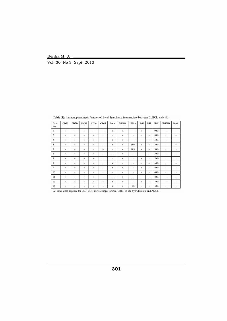
301
Benha M. J.
Vol. 30 No 3 Sept. 2013
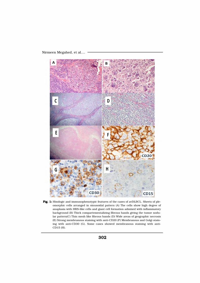
302302
Nirmeen Megahed, et al....
Fig. 1:Fig. 1: Hisologic and immunophenotypic features of the cases of avDLBCL. Sheets of ple-omorphic cells arranged in sinusoidal pattern (A) The cells show high degree ofanaplasia with HRS-like cells and giant cell formation admixed with inflammatorybackground (B) Thick compartementalizing fibrous bands giving the tumor nodu-lar pattern(C) Thin mesh like fibrous bands (D) Wide areas of geographic necrosis(E) Strong membranous staining with anti-CD20 (F) Membranous and Golgi stain-ing with anti-CD30 (G). Some cases showed membranous staining with anti-CD15 (H).
303
Benha M. J.
Vol. 30 No 3 Sept. 2013
DiscussionDiscussionDistinguishing between Hodg-
kin and non-Hodgkin lymphoma
has always been the main task of
hematopathologists for ages. Their
decision is the main guide for cli-
nicians to implement the strategy
plan of chemotherapy. Although
clearer cut criteria are currently
used in the WHO lymphoma clas-
sification 2008; which is attribut-
ed to the advances in immunohis-
tochemistry, cytogenetic and
molecular pathology; segregation
between Hodgkin and Non-
Hodgkin lymphoma cases has not
always been an easy job. Cases
with intermediate histologic and
immunophenotypic features are
now on the record. Many of these
cases have been noticed in the
mediastinum and the term “medi-
astinal grey zone lymphoma”
(MGZL) is now officially applied in
the WHO classification 2008 to
designate such intermediate cas-
es. In the study performed by Tra-
verse-Glehen et al about MGZL
they reported 6 cases of mediasti-
nal composite lymphoma of cHL
and mediastinal large B-cell lym-
phoma (MLBCL), and 9 cases of
sequential cHL and MLBCL from
which they concluded that such
cases might represent a continu-
um[8]. Gracia et al postulated that
the recognition of such cases in
the mediastinum might signify ex-
istence of favorable mediastinal
microenvironment for their devel-
opment[5].
However, cases of such morpho-
logic and immunophenotypic features
have been recognized recently at
extramediastinal sites[5]. Gracia et
al reported 9 cases of DLBCL with
Hodgkin features of which 3 cases
occurred extramediastinally. Port-
lock et al also reported a series of
25/248 cases of cHL showing
more than 50% positivity of the
tumor cells for CD20[9].
The main pathological charac-
teristic feature of our cases was
the discrepancy between morpho-
logic and immunophenotypic fea-
tures; cases of cHL-like morphology
showed pan B-cell marker positivity
(CD20, CD79a, PAX5) while cases
morphologically closer to DLBCL
showed CD30 and CD15 positivi-
ty[10,11]. In contrast to cHL[12,13,14], our series showed P53
and Bcl-6 positivity in a number
of cases which are usually detect-
ed among high-grade B-cell tu-
304304
Nirmeen Megahed, et al....
mors or among cases of aggressive
transformation of cHL[15].
The underlying pathogenesis of
evolution of grey zone lymphoma
is still mysterious. However, re-
cent reports analyzing molecular
biology of cHL and MLCL revealed
similarities in genetic alterations.
This was explained by Traverse-
Glehen et al that B-cells are capa-
ble of transformation to either
HRS cells or neoplastic large B-
cell[1]. The precise molecular
events deciding such transforma-
tion are yet undetermined. During
such complex steps of transforma-
tion the progression of B-cell
might stop in an intermediate-
stage giving rise to grey zone lym-
phoma. On the other hand, Gracia
et al suggested that such interme-
diate lymphomas might arise by
aggressive progression from cHL[5]. Further study of such cases in
the future will contribute to lift
the curtain on the biologic links
between Hodgkin and non-
Hodgkin lymphoma.
In conclusion, B-cell lymphoma
intermediate between diffuse large
B-cell lymphoma (DLBCL) and
classical Hodgkin lymphoma (cHL)
represents a distinctive subtype of
lymphoma showing histological
and immunophenotypic features
intermediate between DLBCL and
cHL. Further studies of these cas-
es will reveal more links between
Hodgkin and non-Hodgkin lym-
phoma and offer better treatment
strategies.
Conflict of interest:Conflict of interest:
The authors declare to have no
conflict of interest.
ReferencesReferences1- Traverse-Glehen A., Pitta-1- Traverse-Glehen A., Pitta-
luga S., Gaulard P., et al.luga S., Gaulard P., et al.
(2005): (2005): Mediastinal Gray Zone
Lymphoma.The Missing Link Be-
tween Classic Hodgkin’s Lympho-
ma and Mediastinal Large B-Cell
Lymphoma. Am J Surg Pathol 29
(11): 1411-1421.
2- Jox A., Wolf J. and Diehl2- Jox A., Wolf J. and Diehl
V. (1997):V. (1997): Hodgkin’s disease biol-
ogy: recent advances. Hematol
Oncol 15:165-171
3- Rudiger T., Jaffe E.S., Del-3- Rudiger T., Jaffe E.S., Del-
sol G., et al. (1998):sol G., et al. (1998): Workshop
report on Hodgkin’s disease and
related diseases (gray zone lympho-
ma). Ann Oncol 9(suppl 5):31-38.
305
Benha M. J.
Vol. 30 No 3 Sept. 2013
4- Jaffe E.S., Stein H., Swerd-4- Jaffe E.S., Stein H., Swerd-
low S.H., et al. (2008):low S.H., et al. (2008): B-cell
lymphoma, unclassifiable, with
features intermediate between dif-
fuse large B-cell lymphoma and
classical Hodgkin lymphoma. In:
Swerdlow SH, Campo E, Harris NL
et al (eds) WHO classification of
tumors of hematopoetic and lym-
phoid tissues, 4th edn. Interna-
tional agency for research on can-
cer, Lyon, pp 267-268.
5- Gracia J.F., Mollejo M.,5- Gracia J.F., Mollejo M.,
Fraga M., et al. (2005):Fraga M., et al. (2005): Large B-
cell lymphoma with Hodgkin’s fea-
tures. Histopathol 47:101-110.
6- Schmid C., Pan .L, Diss T.,6- Schmid C., Pan .L, Diss T.,
et al. (1991):et al. (1991): Expression of B-cell
antigens by Hodgkin’s and Reed-
Sternberg cells. Am J Pathol
139:701-707.
7- Jaffe E.S. and Wilson W.H.7- Jaffe E.S. and Wilson W.H.
(2004):(2004): Gray zone, synchronous,
and metachronous lymphomas:
diseases at the interface of non-
Hodgkin’s lymphomas and Hodg-
kin’s Lymphoma. In: Mauch PM,
Armitage JO, Coiffier B, et al (eds)
Non-Hodgkin’s Lymphoma. Lip-
pincott Williams and Wilkins, Phil-
adelphia, pp 69-80.
8- Traverse-Glehen A., Pitta-8- Traverse-Glehen A., Pitta-
luga S., Wilson W.H., et al.luga S., Wilson W.H., et al.
(2004):(2004): Mediastinal gray zone
lymphoma: the missing link be-
tween classical Hodgkin’s lympho-
ma and mediastinal large B-cell
lymphoma. Eur J Haematol 73:
15.
9- Portlock C., Donnelly G.,9- Portlock C., Donnelly G.,
Qin J., Straus D., Yahalom J.,Qin J., Straus D., Yahalom J.,
Zelenetz A., Noy A., O’ConnorZelenetz A., Noy A., O’Connor
O., Horwitz S., Moskowitz C.O., Horwitz S., Moskowitz C.
and Filipp D. (2004):and Filipp D. (2004): Adverse
prognostic significance of CD20
positive Reed–Sternberg cells in
classical Hodgkin’s disease. BJH
125: 701-708.
10- Parrens M., Vergier B.,10- Parrens M., Vergier B.,
Fitoussi O., et al. (2002):Fitoussi O., et al. (2002): Se-
quential development of Hodgkin’s
disease and CD30+ diffuse large
B-cell lymphoma in a patient with
MALT-type lymphoma: evidence of
different clonal origin of single mi-
crodissected Reed-Sternberg cells.
Am J Surg Pathol 26:1634-1642.
11- Schmid C., Pan L., Diss11- Schmid C., Pan L., Diss
T., et al. (1991):T., et al. (1991): Expression of B-
cell antigens by Hodgkin’s and
Reed-Sternberg cells. Am J Pathol
139:701-707.
306306
Nirmeen Megahed, et al....
12- von Wasielewski R., Men-12- von Wasielewski R., Men-
gel M., Fischer R., et al. (1997):gel M., Fischer R., et al. (1997):
Classical Hodgkin’s disease: clini-
cal impact of the immunopheno-
type. Am J Pathol 151:1123-1130.
13- Wlodarska I., Nooyen P.,13- Wlodarska I., Nooyen P.,
Maes B., et al. (2003):Maes B., et al. (2003): Frequent
occurrence of BCL6 rearrange-
ments in nodular lymphocyte pre-
dominance Hodgkin lymphoma
but not in classical Hodgkin lym-
phoma. Blood 101: 706-710.
14- Maggio E.M., Stekelen-14- Maggio E.M., Stekelen-
burg E., Van den Berg A. andburg E., Van den Berg A. and
Poppema S. (2001):Poppema S. (2001): TP53 gene
mutations in Hodgkin lymphoma
are infrequent and not associated
with absence of Epstein–Barr vi-
rus. Int. J. Cancer 94: 60-66.
15- Sanchez-Beato M., San-15- Sanchez-Beato M., San-
chez-Aguilera A. and Piris M.A.chez-Aguilera A. and Piris M.A.
(2003):(2003): Cell cycle deregulation in
B-cell lymphomas. Blood 101:
1220-1235.

307
Benha M. J.
Vol. 30 No 3 Sept. 2013
IMMUNOHISTOCHEMICALIMMUNOHISTOCHEMICALPROFILING OF B-CELL LYMPHOMAPROFILING OF B-CELL LYMPHOMAINTERMEDIATE BETWEEN DIFFUSEINTERMEDIATE BETWEEN DIFFUSE
LARGE B-CELL LYMPHOMA ANDLARGE B-CELL LYMPHOMA ANDCLASSICAL HODGKIN LYMPHOMACLASSICAL HODGKIN LYMPHOMA
Nirmeen Megahed M.Sc, Azmy Abdelhameed MD,Nirmeen Megahed M.Sc, Azmy Abdelhameed MD,Asmaa Gado MD, Maha Amin MD, Naoko Asano MD,Asmaa Gado MD, Maha Amin MD, Naoko Asano MD,
Seiichi Kato MD, Katsuyoshi Takata MD,Seiichi Kato MD, Katsuyoshi Takata MD,Aki Mitsuda MD and Shigeo Nakamura MDAki Mitsuda MD and Shigeo Nakamura MD
BENHAMEDICALJOURNAL
REPRINT
Published byPublished by
Benha Faculty of MedicineBenha Faculty of Medicine Volume 30 Number 3Sept. 2013
307
Benha M. J.
Vol. 30 No 3 Sept. 2013
EFFECT OF ERYTHROPOIETIN AND STEMEFFECT OF ERYTHROPOIETIN AND STEMCELLS ON ANIMAL MODEL OF CHRONICCELLS ON ANIMAL MODEL OF CHRONIC
NEPHROPATHYNEPHROPATHY
Mohammed E. Sarhan MD, Hanaa G. El-Serougy MD,Mohammed E. Sarhan MD, Hanaa G. El-Serougy MD,Mohammed A. Sobh MD*, Abdel Aziz M. Hussein MDMohammed A. Sobh MD*, Abdel Aziz M. Hussein MD
and Mohammed E. Salama M.Scand Mohammed E. Salama M.ScDepartments of Medical physiology, Internal Medicine; Nephrology and
Urology Center*, Faculty of Medicine, Mansoura University
AbstractAbstractAim: Aim: the purpose of this study was to assess the effects of bone marrow
derived mesenchymal stem cells and Darbopoietin alpha on adriamycin- in-duced chronic renal disease in rats. Methodology: Methodology: 80 male Sprague Dwaleyrats divided into 4 groups (20 rats each); (Group I): normal (negative) controlgroup received saline as a vehicle, (Group II): positive control (twice intrave-nous injection of ADR via penile vein at fourteen days interval with no treat-ment given. (Group III): twice injection of ADR plus darbepoetin alpha (Ara-nesp) subcutaneously once weekly for twelve weeks. (Group IV): twiceinjection of ADR plus twice intravenous injection of MSCs each one was ad-ministrated 5 days after each adriamycin injections. Results:Results: Administrationof subcutaneous darbepoietin alpha favored survival of adriamycin nephro-sis in rats and significantly improved animal body weight when comparedwith positive control group and also had beneficial effect on histolopathologi-cal and biochemical parameters such as blood hemoglobin level, renal func-tions manifested by decrease in BUN, prevented glomerular filtration barrierdamage by adriamycin as shown by amelioration of proteinuria, while, twiceintravenous injection of MSCs produces non significant improvement of ani-mal survival rate, body weight, blood hemoglobin level, proteinuria, hypoal-buminemia of adriamycin nephropathy model in rats while, it produced sig-nificant improvement in BUN and hyperlipidemia. Conclusion:Conclusion: Concomitantadministration of darbepoietin alpha with adriamycin treatment from thefirst day had a protective effect manifested by improvement in both biochem-ical and histopathological parameters. On the other hand, MSCs injectionsignificantly produces histological protection on both glomeruli and tubu-

308308
Mohammed E. Sarhan, et al....
IntroductionIntroductionThe prevalence of chronic kid-
ney disease has been growing con-
sistently for the past decades.
This alarming increase in chronic
and end-stage renal disease is ac-
companied and promoted by a
growing prevalence of cardiovas-
cular risk factors such as obesity,
diabetes, and hypertension that
increase the overall morbidity and
mortality in these patients. There-
fore, there is an urgent need to
identify the mechanisms that per-
petuate and aggravate renal dys-
function and scarring, and to de-
velop strategies to prevent and
attenuate them(1). ADR-induced
renal failure is a well-accepted
chronic disease model of progres-
sive glomerulosclerosis in rats(2)
which mirrors that seen in human
CKD due to primary focal segmen-
tal glomerulosclerosis(3). It is
characterized by rapid develop-
ment of proteinuria and glomeru-
losclerosis and evolves to renal fi-
brosis and renal failure. The use
of cell therapy has been suggested
as a potential modality to improve
the course and outcome of kidney
injury(4). Bone marrow derived
mesenchymal stem cells(MSCs)
are a source of multipotent cells
having the potential of tissue re-
generation in experimental models
of myocardial infarction(5) neuro-
logical disease(6), and acute kid-
ney injury(7); infusion of murine(8)
and human MSCs(9) in mice with
acute kidney injury decreased re-
nal tubular injury and ameliorat-
ed renal function impairment,
which translated into reduced ani-
mal mortality. But, the therapeu-
tic potential of MSCs in animal
models of chronic nephropathies
has not been completely estab-
lished so far(10). EPO is a glyco-
protein hormone, primarily produced
by renal cortical and outer medul-
lary fibroblasts in response to hy-
poxia(11). The expression of EPO R
in non-erythroid tissues such as
the brain(12), retina(13), heart(14),
kidney(15), smooth muscle cells(16),
myoblasts(17) and vascular endo-
thelium(18) has been associated
with the discovery of novel biologi-
cal functions of endogenous Epo
lointerstitial region of the kidney but couldn’t modify clinical parameters as(body weight, animal mortality, anemia, proteinuria) indicating that MSCsonly provide partial protection that did not modify outcome of adriamycinnephrosis.
309
Benha M. J.
Vol. 30 No 3 Sept. 2013
signalling in non-haematopoietic
tissues. Studies on acute renal failure
have demonstrated the beneficial
effect of erythropoietin on renal
function(19) However, the effects
of EPO on chronic renal failure
have been poorly investigated(20).
So, the present work was con-
ducted to investigate the possible
protective effects of darbepoietin
alpha and mesenchymal stem cells
on the outcome of chronic adriam-
ycin nephropathy model in rats.
Materials and MethodsMaterials and Methods1. Experimental Animals:1. Experimental Animals:
The materials of this work com-
prised of eighty male Sprague-
Dwaley rats weighing 200-250 gm,
aging 2-3 months, bred and
housed in the animal house of
Mansoura Experimental Research
Center (MERC). These animals
were housed at temperature 20°C-
25°C and fed standard laboratory
chow, and had free access to tap
water. The experimental protocol
was approved by the Local Ethical
Committee, Faculty of Medicine,
Mansoura University.
Study Design:Study Design:
Eighty rats were assigned to
the following four experimental
groups (n = 20/group):-
1. Group A: vehicle (negative1. Group A: vehicle (negative
control):control):
Rats received tail-vein injection
of comparable volume of 0.9% sa-
line.twice at a 14-day interval.
2. Group B: Adriamycin (posi-2. Group B: Adriamycin (posi-
tive control): tive control):
Rats received tail-vein injection
of ADR (pharmacia Italia,SPA) (4
mg/kg body weight saline) twice at
a 14-day interval.
3. Group C:(ADR+darbepoetin3. Group C:(ADR+darbepoetin
alpha): alpha):
Rats received tail-vein injection
of ADR (4 mg/kg body weight)
twice at a 14-day interval plus 0.3
µg/kg body weight darbepoetin al-
pha (Aranesp) (Amgen Europe
B.V Breda, Netherlands) subcuta-
neously once weekly for four
weeks.
4. Group D: (ADR + MSC):4. Group D: (ADR + MSC):
Rats received tail-vein injection
of ADR (4 mg/kg body weight)
twice at a 14-day interval) plus
twice injection of MSCs (2x10⁶)
cells at a 14-day interval, each
one administrated 5 days after ad-
riamycin injections.
310310
Mohammed E. Sarhan, et al....
Collection of blood and urineCollection of blood and urine
samples:samples:
By the end of the experiment, rats
were weighed and sacrificed using
an overdose of thiopental (75 mg/kg
body weight, i.p.). Blood samples were
taken by heart puncture then placed
into 2 sets of tubes; one set is
EDTA-containing tubes for assess-
ment of haemoglobin and the other
set are tubes without anticoagulant
that were centrifuged at 3000x g for
15 minutes, and stored at -20°C till
the time of biochemical analysis.
Hemoglobin content was as-
sessed at the time of sacrifice for
each group, but the other investi-
gated biochemical parameters such
as serum albumin, creatinine,
BUN, serum triglyceride and ser-
um cholesterol were assessed for
all groups by the end of the study.
The rats were placed in a me-
tabolism cage for 24 hours in or-
der to collect 24-hour urine from 8
a.m. to 8 a.m. of the next day. The
urine volume was measured, and
then a sample was taken for esti-
mation of urine protein.
Harvesting kidney specimens:Harvesting kidney specimens:
By the end of the experiment
both kidneys were removed and
bisected into 2 equal halves. Then
they rapidly fixed into 10% neu-
tral formalin.
Histopathological examina-Histopathological examina-
tion:tion:
The kidney specimens were
processed for paraffin blocks and
sections of 3-µm thickness were
made and stained with haemtoxy-
lin and eosin and prussian blue.
Results Results Effect of DPO and MSC onEffect of DPO and MSC on
animal survival:animal survival:
Nineteen of twenty rats (95%)
in the negative control group
(group I) survived 12 weeks, while
survived rats were eight of twenty
(37%) in positive control group
(group II), fifteen of twenty (75%)
in DPO-treated group (group III)
and eleven of twenty (55%) in MSC
treated group (group III) (Fig.1).
Compared with negative control
group, positive control group showed
significant increase in animal mor-
tality (p=0.001). Treatment with DPO
caused significant attenuation in
animal mortality rate (p=0.02),
while MSCs therapy caused non-
significant attenuation in animal
mortality rate (p=0.3).
311
Benha M. J.
Vol. 30 No 3 Sept. 2013
Effect of DPO and MSC onEffect of DPO and MSC on
animal weight:animal weight:
Compared to negative control
group (group I), positive control
group (group II) and MSCs treated
group (group IV) showed signifi-
cant decrease in body weight
(P<0.001) respectively, while DPO
treated group (group III) showed
no significant decrease in body
weight.
As regard to the effect of treat-
ment on body weight, DPO treated
group showed statistically significant
increase in body weight (P<0.001)
when compared with positive con-
trol group while MSCs treated
group showed non-significant in-
crease in body weight. In addition,
there was statistically significant
difference in body weight between
DPO treated group and MSCs
treated group (P=0.01) being high-
er in DPO treated group (table 1).
Effect of DPO and MSC onEffect of DPO and MSC on
blood hemoglobin level:blood hemoglobin level:
There was statistically signifi-
cant decrease in haemoglobin lev-
el in positive control group (group
II) (P<0.001), DPO treated group
(group III) (P=0.004) and MSCs
treated group (group IV) (P<0.001)
when compared to negative con-
trol group (group I).
Compared with positive control
group (Group II), it was shown
that, there was statistically signifi-
cant increase in haemoglobin level
in DPO treated group (group III)
(0.002), while MSCs treated group
(group IV) showed no significant
increase in hemoglobin level, in
addition, there was no statistically
significant difference between
MSCs treated group (group IV)
and DPO treated group (Group III)
(Table 2).
Effect of DPO and MSC onEffect of DPO and MSC on
serum BUN & creatinine:serum BUN & creatinine:
Positive control group (group II)
had significant higher BUN level
(P<0.001) when compared to nega-
tive control group (group I). As re-
gard to effect of DPO injection
(group III), there was statistically
significant decrease in BUN level
when compared to positive control
group (group II) and to MSCs
treated group (group IV) (P<0.001
and P=0.016) respectively with no
statistically significant difference
DPO treated group (group III) and
negative control group(group I). As
regard to effect of MSCs injection,

312312
Mohammed E. Sarhan, et al....
there was statistically significant
decrease in BUN level when com-
pared to positive control group
(group II) (P=0.014) but still signif-
icantly higher than DPO treated
group (group III) (P=0.016), while,
there was no statistical significant
difference among all groups as re-
gard to serum creatinine(table 3) .
Effect of DPO and MSC onEffect of DPO and MSC on
Serum cholesterol and serumSerum cholesterol and serum
triglycerides:triglycerides:
Positive control group (group II)
showed significant increase in ser-
um triglycerides (P<0.001) and
cholesterol (P<0.001) when com-
pared to negative control group
(group I).
As regard to effect of subcuta-
neous DPO treatment, DPO treat-
ed group (Group III) showed signif-
icant reduction in serum
triglycerides (P<0.001) and serum
cholesterol (P=0.002) when com-
pared with positive control group
(group II) but when compared with
negative control group (group I),
serum triglycerides still had signif-
icant higher value (P=0.005) while
there was no statistically signifi-
cant difference as regard to serum
cholesterol.
In MSCs treated group (group
IV), there was significant reduction
in serum triglycerides (P=0.017)
and serum cholesterol (P=0.011)
when compared with positive con-
trol group (group II) but when
compared with negative control
group (group I), serum cholesterol
still had significant higher value
(P=0.005) while there was no sta-
tistically significant difference as
regard to serum triglycerides. In
addition, there was statistically
significant difference between ser-
um triglycerides or cholesterol be-
tween DPO treated group (Group
III) and MSCs treated group
(group IV).
Effect of DPO and MSC onEffect of DPO and MSC on
urinary protein excretion (mg/urinary protein excretion (mg/
24 hr) and serum albumin (g/24 hr) and serum albumin (g/
dl):dl):
Positive control group (group II)
had marked and significant in-
crease in urinary protein excretion
(P<0.001) when compared to nega-
tive control group (group I).
As regard to effect of subcuta-
neous DPO treatment, DPO treat-
ed group (group III) showed signifi-
cant reduction in urinary protein
excretion (P<0.001) when com-
313
Benha M. J.
Vol. 30 No 3 Sept. 2013
pared with positive control group
(group II) but when compared with
negative control group (group I)
there was no statistically signifi-
cant difference.
In MSCs treated group (group
IV), there was significant reduc-
tion in urinary protein excretion
(P<0.001) when compared with
positive control group (Group II)
but when compared with negative
control group (group I) and DPO
treated group (Group III), urinary
protein excretion still had signifi-
cant higher value (P<0.001 and
0.004) respectively (table 5).
As regard to serum albumin,
positive control group (group II)
had statistically lower serum albu-
min level (P<0.001) when com-
pared with negative control group
(group I).
As regard to effect of subcuta-
neous DPO treatment, DPO treat-
ed group (group III) showed statis-
tically higher serum albumin level
(P=0.012) when compared with pos-
itive control group (group II) but
when compared with negative con-
trol group (group I) there was no
statistically significant difference.
In MSCs treated group (group
IV), there was no significant re-
duction in serum albumin level
(P<0.001) when compared with
negative control group (group I).
Also, there was no statistically sig-
nificant difference in serum albu-
min level when compared with ei-
ther positive control group (Group
II) or DPO treated group (Group
III).
Effect of DPO and MSC on re-Effect of DPO and MSC on re-
nal morphology:nal morphology:
Light microscopic examination
revealed that kidney lesions are
present mainly in the cortex, outer
strip of the outer medulla and to
less extent in inner strip of outer
medulla and inner medulla.
In the cortex, the lesion was
mainly glomerular where the posi-
tive control group (group II)
showed significant higher number
of podocyte detachment (P<0.001),
podocyte vacuolization (P<0.001),
capillary dilatation and also, pa-
rietal epithelial cell vacuolization
of bowman's capsule (P<0.001)
when compared to negative con-
trol group (group I) (Fig.2).
As regard to changes in tubulo-
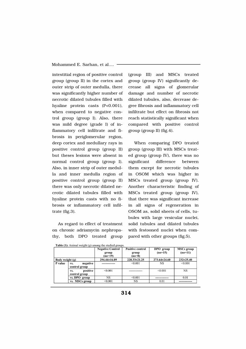
314314
Mohammed E. Sarhan, et al....
intestitial region of positive control
group (group II) in the cortex and
outer strip of outer medulla, there
was significantly higher number of
necrotic dilated tubules filled with
hyaline protein casts (P<0.001),
when compared to negative con-
trol group (group I). Also, there
was mild degree (grade I) of in-
flammatory cell infiltrate and fi-
brosis in periglomerular region,
deep cortex and medullary rays in
positive control group (group II)
but theses lesions were absent in
normal control group (group I).
Also, in inner strip of outer medul-
la and inner medulla region of
positive control group (group II)
there was only necrotic dilated ne-
crotic dilated tubules filled with
hyaline protein casts with no fi-
brosis or inflammatory cell infil-
trate (fig.3).
As regard to effect of treatment
on chronic adriamycin nephropa-
thy, both DPO treated group
(group III) and MSCs treated
group (group IV) significantly de-
crease all signs of glomerular
damage and number of necrotic
dilated tubules, also, decrease de-
gree fibrosis and inflammatory cell
infiltrate but effect on fibrosis not
reach statistically significant when
compared with positive control
group (group II) (fig.4).
When comparing DPO treated
group (group III) with MSCs treat-
ed group (group IV), there was no
significant difference between
them except for necrotic tubules
in OSOM which was higher in
MSCs treated group (group IV).
Another characteristic finding of
MSCs treated group (group IV),
that there was significant increase
in all signs of regeneration in
OSOM as, solid sheets of cells, tu-
bules with large vesicular nuclei,
solid tubules and dilated tubules
with festooned nuclei when com-
pared with other groups (fig.5).
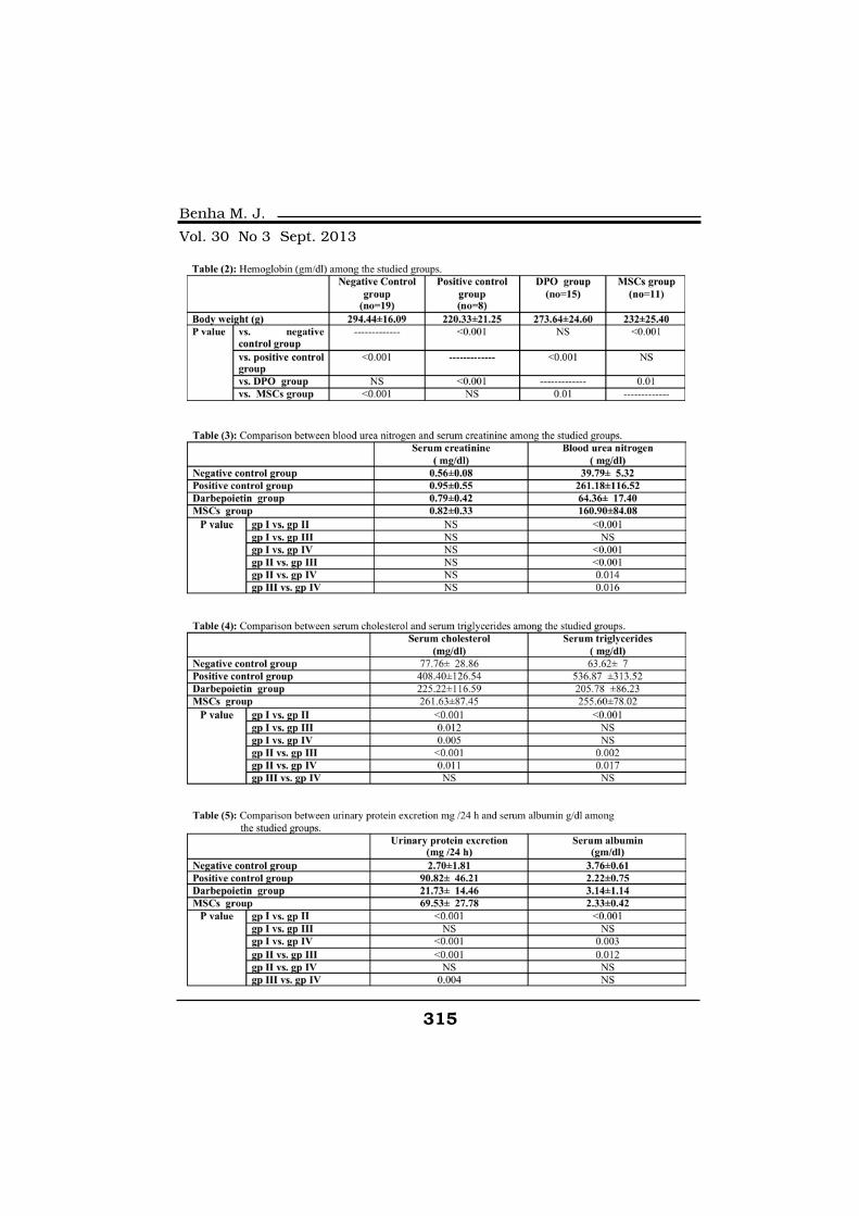
315
Benha M. J.
Vol. 30 No 3 Sept. 2013
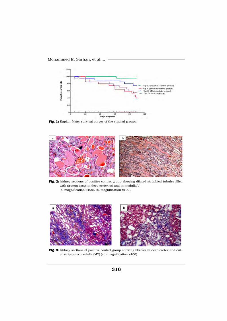
316316
Mohammed E. Sarhan, et al....
Fig. 1: Fig. 1: Kaplan-Meier survival curves of the studied groups.
Fig. 2: Fig. 2: kidney sections of positive control group showing dilated atrophied tubules filledwith protein casts in deep cortex (a) and in medulla(b) (a, magnification x400), (b, magnification x100).
Fig. 3: Fig. 3: kidney sections of positive control group showing fibrosis in deep cortex and out-er strip outer medulla (MT) (a,b magnification x400).
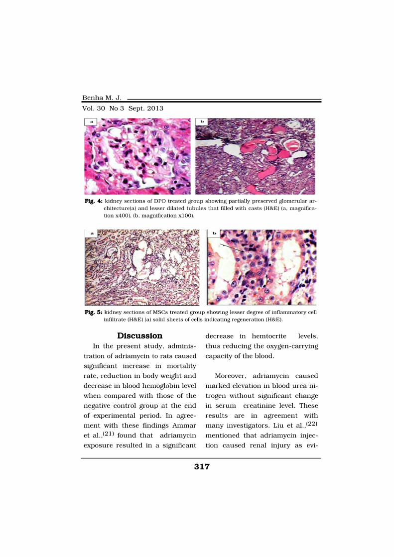
317
Benha M. J.
Vol. 30 No 3 Sept. 2013
DiscussionDiscussionIn the present study, adminis-
tration of adriamycin to rats caused
significant increase in mortality
rate, reduction in body weight and
decrease in blood hemoglobin level
when compared with those of the
negative control group at the end
of experimental period. In agree-
ment with these findings Ammar
et al.,(21) found that adriamycin
exposure resulted in a significant
decrease in hemtocrite levels,
thus reducing the oxygen-carrying
capacity of the blood.
Moreover, adriamycin caused
marked elevation in blood urea ni-
trogen without significant change
in serum creatinine level. These
results are in agreement with
many investigators. Liu et al.,(22)
mentioned that adriamycin injec-
tion caused renal injury as evi-
Fig. 4: Fig. 4: kidney sections of DPO treated group showing partially preserved glomerular ar-chitecture(a) and lesser dilated tubules that filled with casts (H&E) (a, magnifica-tion x400), (b, magnification x100).
Fig. 5: Fig. 5: kidney sections of MSCs treated group showing lesser degree of inflammatory cellinfiltrate (H&E) (a) solid sheets of cells indicating regeneration (H&E).
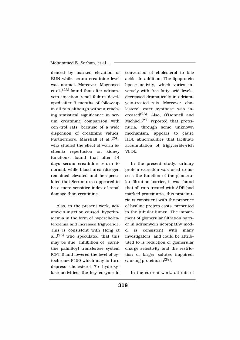
318318
Mohammed E. Sarhan, et al....
denced by marked elevation of
BUN while serum creatinine level
was normal. Moreover, Magnasco
et al.,(23) found that after adriam-
ycin injection renal failure devel-
oped after 3 months of follow-up
in all rats although without reach-
ing statistical significance in ser-
um creatinine comparison with
con¬trol rats, because of a wide
dispersion of creatinine values.
Furthermore, Marshall et al.,(24)
who studied the effect of warm is-
chemia reperfusion on kidney
functions, found that after 14
days serum creatinine return to
normal, while blood urea nitrogen
remained elevated and he specu-
lated that Serum urea appeared to
be a more sensitive index of renal
damage than creatinine.
Also, in the present work, adi-
amycin injection caused hyperlip-
idemia in the form of hypercholes-
terolemia and increased triglyceride.
This is consistent with Hong et
al.,(25) who speculated that this
may be due inhibition of carni-
tine palmitoyl transferase system
(CPT I) and lowered the level of cy-
tochrome P450 which may in turn
depress cholesterol 7α hydroxy-
lase activities, the key enzyme in
conversion of cholesterol to bile
acids. In addition, The lipoprotein
lipase activity, which varies in-
versely with free fatty acid levels,
decreased dramatically in adriam-
ycin-treated rats. Moreover, cho-
lesterol ester synthase was in-
creased(26). Also, O'Donnell and
Michael,(27) reported that protei-
nuria, through some unknown
mechanism, appears to cause
HDL abnormalities that facilitate
accumulation of triglyceride-rich
VLDL.
In the present study, urinary
protein excretion was used to as-
sess the function of the glomeru-
lar filtration barrier, it was found
that all rats treated with ADR had
marked proteinuria, this proteinu-
ria is consistent with the presence
of hyaline protein casts presented
in the tubular lumen. The impair-
ment of glomerular filtration barri-
er in adriamycin nepropathy mod-
el is consistent with many
investigators and could be attrib-
uted to is reduction of glomerular
charge selectivity and the restric-
tion of larger solutes impaired,
causing proteinuria(28).
In the current work, all rats of
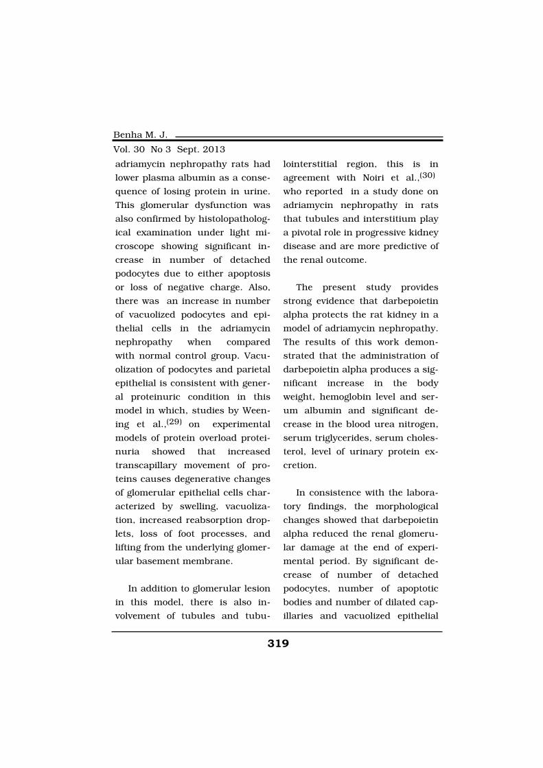
319
Benha M. J.
Vol. 30 No 3 Sept. 2013
adriamycin nephropathy rats had
lower plasma albumin as a conse-
quence of losing protein in urine.
This glomerular dysfunction was
also confirmed by histolopatholog-
ical examination under light mi-
croscope showing significant in-
crease in number of detached
podocytes due to either apoptosis
or loss of negative charge. Also,
there was an increase in number
of vacuolized podocytes and epi-
thelial cells in the adriamycin
nephropathy when compared
with normal control group. Vacu-
olization of podocytes and parietal
epithelial is consistent with gener-
al proteinuric condition in this
model in which, studies by Ween-
ing et al.,(29) on experimental
models of protein overload protei-
nuria showed that increased
transcapillary movement of pro-
teins causes degenerative changes
of glomerular epithelial cells char-
acterized by swelling, vacuoliza-
tion, increased reabsorption drop-
lets, loss of foot processes, and
lifting from the underlying glomer-
ular basement membrane.
In addition to glomerular lesion
in this model, there is also in-
volvement of tubules and tubu-
lointerstitial region, this is in
agreement with Noiri et al.,(30)
who reported in a study done on
adriamycin nephropathy in rats
that tubules and interstitium play
a pivotal role in progressive kidney
disease and are more predictive of
the renal outcome.
The present study provides
strong evidence that darbepoietin
alpha protects the rat kidney in a
model of adriamycin nephropathy.
The results of this work demon-
strated that the administration of
darbepoietin alpha produces a sig-
nificant increase in the body
weight, hemoglobin level and ser-
um albumin and significant de-
crease in the blood urea nitrogen,
serum triglycerides, serum choles-
terol, level of urinary protein ex-
cretion.
In consistence with the labora-
tory findings, the morphological
changes showed that darbepoietin
alpha reduced the renal glomeru-
lar damage at the end of experi-
mental period. By significant de-
crease of number of detached
podocytes, number of apoptotic
bodies and number of dilated cap-
illaries and vacuolized epithelial
320320
Mohammed E. Sarhan, et al....
cells. Also, the morphological
changes showed that darbepoietin
alpha reduced the renal tubular-
interstistial damage in the form of
reducing urinary protein cast in
tubules, inflammatory cell infiltrate
and fibrosis though not reaching
statistitically significant difference
when compared with adriamycin
non treated group (group II).
These findings are in agreement
with those reported by others who
demonstrated that EPO treatment
improved the functional and mor-
phologic glomerular and tubular
injury in rats subjected to adriam-
ycin nephropathy(31,32).
The improvement of body
weight, hemoglobin level and BUN
in adriamycin model by DPO is
consistent with Noiri et al.,(33)
who found similar results against
adriamycin induced cardio renal
injury in rats. Furthermore spec-
ulated that correction of anemia to
the normal level by DA is an im-
portant mechanism of action be-
cause it retards the progression of
CKD and cardiovascular diseases.
In the current work, in order to
elucidate the effect of darbepoetin
on the glomerular barrier func-
tion, urine protein level was as-
sessed, it was found that darbe-
poetin ameliorates proteinuria
when comparing DPO treated
group with positive control group.
The effect of DPO on proteinuria
may be indirect effect due to an
increase in oxygen-carrying capac-
ity might improve structures and
functions of podocytes,or direct ef-
fect as suggested by Eto et al.,(34)
who reported that darbepoietin
may provide direct protection on
podocytes, the major culprit ac-
counting for proteinuria through
acting on EPO-R in podocytes and
that DPO treatment ameliorated
podocyte injury and reduced pro-
teinuria by preventing the disrup-
tion of actin cytoskeleton and the
reduction of nephrin through the
binding to the EPOR to activate
Janus-tyrosine kinase 2, phos-
phoinositide 3 kinase, and protein
kinase B (Akt). It is noteworthy
that several reports showed the di-
rect interaction of Akt and actin.
Twice intravenous injection of
MSCs produced improvement in,
not reach statistically significant
value, animal survival rate, body
weight. blood hemoglobin level,
proteinuria, hypoalbuminemia of
321
Benha M. J.
Vol. 30 No 3 Sept. 2013
adriamycin nephropathy model in
rats while, it produced significant
improvement in BUN and hyper-
lipidemia. Also, histipathlogical
finding revealed that MSCs provid-
ed histological protection of glome-
ruli manifested by decrease in de-
tached podocytes per glomerulus,
apoptotic bodies, aneurysmal di-
lated capillaries and vacuolized
podocyte or parietal epithelial
cells, while in tubulointerstitial re-
gion. Also, there was a significant
decrease in number of necrotic tu-
bules. Inflammatory cell infiltrate
showed non-significant decrease
in MSC-treated rats. As regard to
fibrosis in this group it was mini-
mal or absent, with appearance of
histological findings of regenera-
tion as Solid sheets, tubules with
large vesicular nuclei and solid tu-
bules.
This might be attributed to ei-
ther failure of anemia correction
by MSCs, time of stem cells ad-
ministration or due to decrease
number of engrafted cells in kid-
ney after injection. Noiri et al.,(36)
mentioned that anemia can share
in progression of CKD, in the
present study it was chosen to ad-
ministrate cells five days post ad-
riamycin injection to provide suffi-
cient time for injury process to oc-
cur, it is known that injury is es-
sential for homing to occur and
also MSC and also in a more clini-
cally relevant setting of intrave-
nous infusion via intravenous
route at the time of evident protei-
nuria.
Adequate homing of MSC to the
injured tissue is important for ef-
fective therapy. As regard to effi-
cient homing iron labeled cells
technique was used to track cells
in the kidney, it was found that
homing occurred but few MSCs
was detected in the glomeruli and
tubulointerstitial region.
As regard route of administra-
tion, in the current, MSC is ad-
ministered through a standard in-
travenous route. A disadvantage
of the systemic intravenous deliv-
ery of MSC can be low uptake at
the site of injury. Indeed, signifi-
cant engraftment of injured tissue
was observed in some studies(37,38), but Schrepfer et al.,(39)
demonstrated that the systemic
intravenous route of administra-
tion was not appropriate for MSC
to reach their site of activation.
322322
Mohammed E. Sarhan, et al....
Zonta et al.,(40) showed that the
intra-arterial administration of
MSC were the most effective route
to achieve immunomodulating ef-
fects in experimental kidney
transplantation, which primarily
occurs because large MSC (15 mm
to 19 mm) remain trapped in the
capillaries of the small lung filter,
which in turn causes the inade-
quate homing of MSC to the in-
jured tissue. However, using the
renal artery as the injection route
to administer MSC to treat DN
may be associated with the follow-
ing 2 major complications: (1) re-
nal infarcts and loss of function,
and (2) ectopic differentiation into
adipocytes within glomeruli(41).
ConclusionConclusionConcomitant administration of
darbepoietin alpha with adriamy-
cin treatment from the first day
has a protective effect manifested
by improvement in both biochemi-
cal and histopathological parame-
ters,while MSCs injection signifi-
cantly produces histological
protection on both glomeruli and
tubulointerstitial region of the kid-
ney but couldn't modify clinical
parameters as animal mortality,
anemia, proteinuria and hyperlipi-
demia indicating that MSCs only
provide partial protection that did
not modify outcome of adriamycin
nephrosis.
ReferencesReferences1.1. Lerman L.O. and ChadeLerman L.O. and Chade
A.R. (2009):A.R. (2009): Angiogenesis in the
kidney: a new therapeutic target.
Curr Opin Nephrol Hypertens.; 18
(2):160-5.
2. Okuda S., Oh Y., Tsuruda2. Okuda S., Oh Y., Tsuruda
H., Onoyama K., Fujimi S. andH., Onoyama K., Fujimi S. and
Fujishima M. (1986):Fujishima M. (1986): Adriamy-
cin-induced nephropathy as a
model of chronic progressive glo-
merular disease. Kidney Int. Feb;
29(2):502-10.
3. Lee V.W. and Harris D.C.3. Lee V.W. and Harris D.C.
(2011):(2011): Adriamycin nephropathy:
a model of focal segmental glomer-
ulosclerosis. Nephrology. 2011;
16: 30-38.
4. Gupta S., Verfaillie C.,4. Gupta S., Verfaillie C.,
Chmielewski D., Kim Y. and Ro-Chmielewski D., Kim Y. and Ro-
senberg M.E. (2002):senberg M.E. (2002): A role for
extrarenal cells in the regenera-
tion following acute renal failure.
Kidney Int. 62: 1285-1290.
5. Tang J., Xie Q., Pan G.,5. Tang J., Xie Q., Pan G.,
323
Benha M. J.
Vol. 30 No 3 Sept. 2013
Wang J. and Wang M. (2006):Wang J. and Wang M. (2006):
Mesenchymal stem cells partici-
pate in angiogenesis and improve
heart function in rat model of
myocardial ischemia with reperfu-
sion. Eur J Cardiothorac Surg 30:
353-361.
6. Torrente Y. and Polli E.6. Torrente Y. and Polli E.
(2008):(2008): Mesenchymal stem cell
transplantation for neurodegener-
ative diseases. Cell Transplant 17:
1103-1113, 2008.
7. Imberti B., Morigi M., Tom-7. Imberti B., Morigi M., Tom-
asoni S., Rota C., Corna D.,asoni S., Rota C., Corna D.,
Longaretti L., Rottoli D., Valsec-Longaretti L., Rottoli D., Valsec-
chi F., Benigni A., Wang J., Ab-chi F., Benigni A., Wang J., Ab-
bate M., Zoja C. and Remuzzi G.bate M., Zoja C. and Remuzzi G.
(2007): (2007): Insulin-like growth factor-
1 sustains stem cell mediated re-
nal repair. J Am Soc. Nephrol 18:
2921-2928.
8. Morigi M., Imberti B., Zoja8. Morigi M., Imberti B., Zoja
C., Corna D., Tomasoni S., Ab-C., Corna D., Tomasoni S., Ab-
bate M., Rottoli D., Angioletti S.,bate M., Rottoli D., Angioletti S.,
Benigni A., Perico N., Alison M.Benigni A., Perico N., Alison M.
and Remuzzi G. (2004):and Remuzzi G. (2004): Mesen-
chymal stem cells are renotropic,
helping to repair the kidney and
improve function in acute renal
failure. J Am Soc Nephrol 15:
1794-1804.
9. Morigi M., Introna M., Im-9. Morigi M., Introna M., Im-
berti B., Corna D., Abbate M.,berti B., Corna D., Abbate M.,
Rota C., Rottoli D., NankivellRota C., Rottoli D., Nankivell
B.J., Borrows R.J., Fung C.L.,B.J., Borrows R.J., Fung C.L.,
O'Connell P.J., Allen R.D. andO'Connell P.J., Allen R.D. and
Chapman J.R. (2003):Chapman J.R. (2003): The natu-
ral history of chronic allograft
nephropathy. N Engl J Med 349:
2326-2333.
10. Johnson D.W., Vesey D.A.10. Johnson D.W., Vesey D.A.
and Gobe G.C. (2010): and Gobe G.C. (2010): “Erythro-
poietin protects against acute kid-
ney injury and failure,” Open
Drug Discovery Journal, vol. 2,
no. 1, pp. 8-17.
11. Liu C., Shen K., Liu Z.11. Liu C., Shen K., Liu Z.
and Noguchi C.T. (1997):and Noguchi C.T. (1997): Regu-
lated human erythropoietin recep-
tor expression in mouse brain.
Journal of Biological Chemistry,
272, 32395-32400.
12. Grimm C., Wenzel A.,12. Grimm C., Wenzel A.,
Groszer M., Mayser H., SeeligerGroszer M., Mayser H., Seeliger
M., Samardzija M., Bauer C.,M., Samardzija M., Bauer C.,
Gassmann M. and Reme C.E.Gassmann M. and Reme C.E.
(2002): (2002): HIF-1-induced erythropoietin
in the hypoxic retina protects again-
stlight-induced retinal degenera-
tion. Nature Medicine, 8,718-724.
13. Wu H., Lee S.H., Gao J.,13. Wu H., Lee S.H., Gao J.,

324324
Mohammed E. Sarhan, et al....
Liu X. and Iruela-Arispe M.L.Liu X. and Iruela-Arispe M.L.
(1999):(1999): Inactivation of erythro-
poietin leads to defects in cardiac
morphogenesis.Development, 126,
3597-3605.
14. Westenfelder C., Biddle14. Westenfelder C., Biddle
D.L. and Baranowski R.L.D.L. and Baranowski R.L.
(1999): (1999): Human, rat and mouse
kidney cells express functional
erythropoietin receptors. Kidney
International, 55, 808-820.
15. ammarguellat F., Gogusev15. ammarguellat F., Gogusev
J. and Drueke T.B. (1996):J. and Drueke T.B. (1996): Di-
rect effect of erythropoietin on rat
vascular smooth-muscle cell via a
putative erythropoietin receptor.
Nephrology, Dialysis, Transplanta-
tion, 11,687-692.
16. Anagnostou A., Liu Z.,16. Anagnostou A., Liu Z.,
Steiner M., Chin K., Lee E.S.,Steiner M., Chin K., Lee E.S.,
Kessimian N. and Noguchi C.T.Kessimian N. and Noguchi C.T.
(1994):(1994): Erythropoietin receptor
mRNA expression in human endo-
thelial cells. Proceedings of the
National Academy of Sciences of
the United States of America, 91,
3974-3978.
17. Ogilvie M., Yu X., Nicolas-17. Ogilvie M., Yu X., Nicolas-
Metral V., Pulido S.M., Liu C.,Metral V., Pulido S.M., Liu C.,
Ruegg U.T. and Noguchi C.T.Ruegg U.T. and Noguchi C.T.
(2000):(2000): Erythropoietin stimulates
proliferation and interferes with
differentiation of myoblasts. Jour-
nal of Biological Chemistry, 275,
39754-39761.
18. Yang W., Wang J., Shi L.,18. Yang W., Wang J., Shi L.,
Yu L., Qian Y., Liu Y., Wang W.Yu L., Qian Y., Liu Y., Wang W.
and Cheng S. (2012):and Cheng S. (2012): Podocyte
injury and overexpression of vas-
cular endothelial growth factor
and transforming growth factor-
beta 1 in adriamycin-induced
nephropathy in rats. Cytokine;
(59):370-376.
19. Yalcin S., Müftüoglu S.,19. Yalcin S., Müftüoglu S.,
Cetin E., Sarer B., Yildirim B.A.,Cetin E., Sarer B., Yildirim B.A.,
Zeybek D. and Orhan B. (2003):Zeybek D. and Orhan B. (2003):
Protection against cisplatin-
induced nephrotoxicity by recom-
binant human erythropoietin.Med
Oncol.; 20(2):169-174.
20. Modelli de Andrade L.G.,20. Modelli de Andrade L.G.,
Viero R.M. and Cordeiro de Car-Viero R.M. and Cordeiro de Car-
valho M.F. (2013):valho M.F. (2013): Treatment of
adriamycin-induced nephropathy
with erythropoietin and G-CSF. J
Nephrol.; 26(3):534-9.
21. Ammar H.I., Saba S., Am-21. Ammar H.I., Saba S., Am-
mar R.I., Elsayed L.A., Ghalymar R.I., Elsayed L.A., Ghaly
W.B. and Dhingra S. (2011):W.B. and Dhingra S. (2011):
325
Benha M. J.
Vol. 30 No 3 Sept. 2013
Erythropoietin protects against
doxorubicin-induced heart failure.
Am J Physiol Heart Circ Physiol.;
301(6): 2413-21.
22. Liu H.F., Guo L.Q.,22. Liu H.F., Guo L.Q.,
Huang Y.Y., Chen K., Tao J.L.,Huang Y.Y., Chen K., Tao J.L.,
Li S.M. and Chen X.W. (2010):Li S.M. and Chen X.W. (2010):
Thiazolidinedione attenuate pro-
teinuria and glomerulosclerosis in
adriamycin-induced nephropathy
rats via slit diaphragm protection.
Nephrology; 15(1):75-83.
23. Magnasco A., Corselli M.,23. Magnasco A., Corselli M.,
Bertelli R., Ibatici A., Peresi M.,Bertelli R., Ibatici A., Peresi M.,
Gaggero G., Cappiello V., Chiav-Gaggero G., Cappiello V., Chiav-
arina B., Mattioli G., Gusmanoarina B., Mattioli G., Gusmano
R., Ravetti J.L., Frassoni F. andR., Ravetti J.L., Frassoni F. and
Ghiggeri G.M. (2008):Ghiggeri G.M. (2008): Mesenchy-
mal stem cells protective effect in
adriamycin model of nephropathy.
Cell Transplant.; 17(10-11):1157-
67.
24. Marshall V., Jablonski24. Marshall V., Jablonski
P., Howden B., Leslie E., Rae D.P., Howden B., Leslie E., Rae D.
and Tange J. (1982):and Tange J. (1982): Recovery of
renal function in the rat after
warm ischaemia: functional and
morphological changes. Organ
Preservation: 69-76.
25. Hong Y.M., Kim H.S. and25. Hong Y.M., Kim H.S. and
Yoon H. (2002):Yoon H. (2002): Serum lipid and
fatty acid profiles in adriamycin-
treated rats after administration of
L-carnitine. Pediatr. Res.; 51: 249-
55.
26. Malarkodi K.P. and Vara-26. Malarkodi K.P. and Vara-
lakshmi P. (2003):lakshmi P. (2003): Lipid acid as a
rescue agent for adriamycin-
induced hyperlipidemic nephropa-
thy in rats. Nutr. Res.; 23:539-48.
27. O'Donnell and Michael P.27. O'Donnell and Michael P.
(2001):(2001): Mechanisms and clinical
importance of hypertriglyceride-
mia in the nephrotic syndrome.
Kidney Int; 59(1): 380-382.
28. Jeansson M., Bjorck K.,28. Jeansson M., Bjorck K.,
Tenstad O. and Haraldsson B.Tenstad O. and Haraldsson B.
(2009):(2009): Adriamycin alters glomer-
ular endothelium to induce protei-
nuria. J. Am. Soc. Nephrol.; 20:
114-22.
29. Weening J.J., Van Gul-29. Weening J.J., Van Gul-
dener C., Daha M.R., Klar N.,dener C., Daha M.R., Klar N.,
van der Wal A. and Prins F.A.van der Wal A. and Prins F.A.
(1987): (1987): The pathophysiology of
protein-overload proteinuria. Am J
Pathol.; 129(1):64-73.
30. Noiri E., Nagano N., Negi-30. Noiri E., Nagano N., Negi-
shi K., Doi K., Miyata S., Abeshi K., Doi K., Miyata S., Abe
326326
Mohammed E. Sarhan, et al....
M., Tanaka T., Okamoto K.,M., Tanaka T., Okamoto K.,
Hanafusa N., Kondo Y., IshizakaHanafusa N., Kondo Y., Ishizaka
N. and Fujita T. (2006):N. and Fujita T. (2006): Efficacy
of darbepoetin in doxorubicin in-
duced cardiorenal injury in rats.
Nephron Exp Nephrol;104(1):6-14.
31. Nakazawa Y., Nishino T.,31. Nakazawa Y., Nishino T.,
Obata Y., Nakazawa M., FurusuObata Y., Nakazawa M., Furusu
A., Abe K., Miyazaki M., Koji T.A., Abe K., Miyazaki M., Koji T.
and Kohno S. (2013):and Kohno S. (2013): Recombi-
nant human erythropoietin atten-
uates renal tubulointerstitial injury
in murine adriamycin-induced neph-
ropathy. J Nephrol.; 26 (3): 527-33.
32. Modelli de Andrade L.G.,32. Modelli de Andrade L.G.,
Viero R.M. and Cordeiro de Car-Viero R.M. and Cordeiro de Car-
valho M.F. (2013):valho M.F. (2013): Treatment of
adriamycin-induced nephropathy
with erythropoietin and G-CSF. J
Nephrol.; 26(3):534-9.
33. Noiri E., Nagano N., Negi-33. Noiri E., Nagano N., Negi-
shi K., Doi K., Miyata S., Abeshi K., Doi K., Miyata S., Abe
M., Tanaka T., Okamoto K.,M., Tanaka T., Okamoto K.,
Hanafusa N., Kondo Y., IshizakaHanafusa N., Kondo Y., Ishizaka
N. and Fujita T. (2006):N. and Fujita T. (2006): Efficacy
of darbepoetin in doxorubicin in-
duced cardiorenal injury in rats.
Nephron Exp Nephrol;104(1):6-14.
34. Eto N., Wada T., Inagi R.,34. Eto N., Wada T., Inagi R.,
Takano H., Shimizu A., Kato H.,Takano H., Shimizu A., Kato H.,
Kurihara H., Kawachi H.,Kurihara H., Kawachi H.,
Shankland S.J., Fujita T. andShankland S.J., Fujita T. and
Nangaku M. (2007):Nangaku M. (2007): Podocyte
protection by darbepoetin: preser-
vation of the cytoskeleton and
nephrin expression. Kidney Int;
72:455-463.
35. O'Donnell and Michael P.35. O'Donnell and Michael P.
(2001):(2001): Mechanisms and clinical
importance of hypertriglyceride-
mia in the nephrotic syndrome.
Kidney Int; 59(1): 380-382.
36. Noiri E., Nagano N., Negi-36. Noiri E., Nagano N., Negi-
shi K., Doi K., Miyata S., Abeshi K., Doi K., Miyata S., Abe
M., Tanaka T., Okamoto K.,M., Tanaka T., Okamoto K.,
Hanafusa N., Kondo Y., IshizakaHanafusa N., Kondo Y., Ishizaka
N. and Fujita T. (2006):N. and Fujita T. (2006): Efficacy
of darbepoetin in doxorubicin in-
duced cardiorenal injury in rats.
Nephron Exp Nephrol;104(1):6-14.
37. Ezquer F.E., Ezquer M.E.,37. Ezquer F.E., Ezquer M.E.,
Parrau D.B., Carpio D., YanezParrau D.B., Carpio D., Yanez
A.J. and Conget P.A. (2009):A.J. and Conget P.A. (2009):
Systemic administration of multi-
potent mesenchymal stromal cells
reverts hyperglycemia and pre-
vents nephropathy in type 1 dia-
betic mice. Biol. Blood Marrow
Transplant.; 14, 631-640.
38. Semedo P., Correa-Costa38. Semedo P., Correa-Costa
327
Benha M. J.
Vol. 30 No 3 Sept. 2013
M., Cenedeze M.A., Maria Avan-M., Cenedeze M.A., Maria Avan-
cini Costa Malheiros D., Antoniacini Costa Malheiros D., Antonia
dos Reis M., Shimizu M.H., Se-dos Reis M., Shimizu M.H., Se-
guro A.C., Pacheco-Silva A. andguro A.C., Pacheco-Silva A. and
Saraiva Camara N.O. (2009):Saraiva Camara N.O. (2009): Me-
senchymal stem cells attenuate
renal fibrosis through immune
modulation and remodeling prop-
erties in a rat remnant kidney
model. Stem Cells; 27(12): 3063.
39. Schrepfer S., Deuse T.,39. Schrepfer S., Deuse T.,
Reichenspurner H., FischbeinReichenspurner H., Fischbein
M.P., Robbins R.C. and PelletierM.P., Robbins R.C. and Pelletier
M.P. (2007):M.P. (2007): Stem cell transplan-
tation: the lung barrier. Trans-
plant Proc.; 39:573-576.
40. Zonta S., De Martino M.,40. Zonta S., De Martino M.,
Bedino G., Piotti G., RampinoBedino G., Piotti G., Rampino
T., Gregorini M., Frassoni F.,T., Gregorini M., Frassoni F.,
Dal Canton A., Dionigi P. andDal Canton A., Dionigi P. and
Alessiani M. (2010):Alessiani M. (2010): Which is the
most suitable and effective route
of administration for mesenchy-
mal stem cell-based immunomod-
ulation therapy in experimental
kidney transplantation: endove-
nous or arterial? Transplant Proc.;
42:1336-1340.
41. Kunter U., Rong S., Boor41. Kunter U., Rong S., Boor
P., Eitner F., Müller-Newen G.,P., Eitner F., Müller-Newen G.,
Djuric Z., van Roeyen C.R., Ko-Djuric Z., van Roeyen C.R., Ko-
nieczny A., Ostendorf T. and Vil-nieczny A., Ostendorf T. and Vil-
la L. (2007):la L. (2007): Mesenchymal stem
cells prevent progressive experi-
mental renal failure but maldiffer-
entiate into glomerular adipocytes.
J Am Soc Nephrol; 18(6): 1754-64.
328328
Mohammed E. Sarhan, et al....
EFFECT OF ERYTHROPOIETIN ANDEFFECT OF ERYTHROPOIETIN ANDSTEM CELLS ON ANIMAL MODEL OFSTEM CELLS ON ANIMAL MODEL OF
CHRONIC NEPHROPATHYCHRONIC NEPHROPATHY
Mohammed E. Sarhan MD, Hanaa G. El-Serougy MD,Mohammed E. Sarhan MD, Hanaa G. El-Serougy MD,Mohammed A. Sobh MD, Abdel Aziz M. Hussein MDMohammed A. Sobh MD, Abdel Aziz M. Hussein MD
and Mohammed E. Salama M.Scand Mohammed E. Salama M.Sc
BENHAMEDICALJOURNAL
REPRINT
Published byPublished by
Benha Faculty of MedicineBenha Faculty of Medicine Volume 30 Number 3Sept. 2013
329
Benha M. J.
Vol. 30 No 3 Sept. 2013
IntroductionAsthma is associated with a
wide range of symptoms and
signs, including wheezing, cough,
chest tightness, shortness of
breath and sputum production.
The symptoms and signs evolve
from three basic characteristics
that underlie the disease: airway
obstruction, AHR, airway inflam-
mation(1). Airway inflammation in
asthma may represent a loss of
normal balance between two “op-
posing” populations of Th lympho-
cytes. A cytokine imbalance tow-
ardTh2 will set the stage to pro-
mote the production of IgE anti-
body and subsequent allergic
inflammation(2).
Toll-like receptors (TLRs) are a
type of pattern recognition recep-
tor (PRR) that recognize foreign
substances, activate immune cell
responses and play a key role in
the innate immune system, and
receive its name from their simi-
larity to the Toll gene identified in
EFFECT OF ISS-ODN-INDUCED TLR9EFFECT OF ISS-ODN-INDUCED TLR9STIMULATION ON EXPERIMENTALLY INDUCEDSTIMULATION ON EXPERIMENTALLY INDUCED
AIRWAY INFLAMMATION IN MICEAIRWAY INFLAMMATION IN MICE
Mona M. El-Haroun M.Sc.*, Ali M. Yousef MD**Mona M. El-Haroun M.Sc.*, Ali M. Yousef MD**and Tarek M. Ibrahim Ph.D***and Tarek M. Ibrahim Ph.D***
Faculty of Pharmacy, Pharos University*, Benha Educational Hospital**,
Faculty of Pharmacy, Mansoura University***
AbstractWe have used a mouse model of allergen-induced airway inflamma-
tion to demonstrate that ISS-ODN inhibit airway inflammation and re-modeling in acute and chronic asthma. Inflammatory cells in micetreated with ISS-ODN decreases in acute and chronic asthma com-pared to positive control mice. Also, more than one dose of ISS-ODN de-crease IL-4. While one dose increases Th1 cytokines (IFN-γ). ISS-ODNdecreases airway remodeling such as thickness of epithelium and peri-bronchial fibrosis which is related to the increase of IFN-γ but not thedecrease of IL-4 as other studies. So ISS-ODN before allergen exposureprovide a new immunotherapy in asthma.
330
Mona M. El-Haroun, et al....
Drosophila(3). TLRs Form a recep-
tor superfamily with interleukin-
1receptors (Interleukin-1 recep-
tor/Toll-like receptor superfamily)
so called TIR (Toll-IL-1 receptor).
TIR domain recruits four adapters'
molecules which are MyD88, Ti-
rap, Trif and Tram in order to
propagate a signal. These adapt-
ers activate other molecules in-
cluding certain protein kinases
(IRAK1, IRAK, TBK1, and IKKi)
leading to the activation of two
pathways both lead to nuclear
translocation of transcription fac-
tor, NF-?B which lead to induction
of inflammatory cytokines such as
TNF-α and IL-12(4,5).
Synthetic (ODN) bearing un-
methylated CpG motifs can mimic
the immune-stimulatory effects af-
ter recognized by TLR9. Strong im-
munostimulatory effects are driv-
en by sequences containing
unmethylated CpG motifs these
motifs appear to function as Th1-
promoting adjuvants capable of
switching the usual Th2 response
toward a Th1 response(6). CpG
DNA directly activates denteritic
cells and B-cells that express
TLR9, binds CpG DNA and trans-
duces its immune-stimulatory ef-
fects and indirectly activates other
cells of the immune system such
as T- lymphocytes cell, Natural
killer cells and neutrophils, by al-
tering the expression of specific
cytokines, receptors, and adhe-
sion molecules(7). ISS-ODN acti-
vates the TLR- 9 pathway and pro-
motes the development of a Th1
response and generates cytokines,
such as TNF-α, IL-12, IFN-α and
indirectly, IFN-γ. Long term treat-
ment with ISS through induction
of IL-12 and IFNs would lead to a
rebalancing of Th1/Th2 response(8), which leads to decrease in air-
way inflammation Th2cytokine ex-
pression and airway hyperrespon-
siveness, and increases the
expression of Th1 cytokines (IFN-
γ, IL-12). It has also been shown
that ISS-ODN attenuate allergen-
induced airway remodeling in mice
as manifested by decreased subep-
ithelial collagen deposition(9).
Materials and Methods1. Materials1. Materials
A. Drug: OligonucleotidesA. Drug: Oligonucleotides
Endotoxin-free (<1ng/mg DNA)
phosphorothioate ISS-ODN (5'-
TGACTGTGAACGTTCGAGATGA-3')
(Trilink, San Diego, CA) were used
in the in vivo experiments.
331
Benha M. J.
Vol. 30 No 3 Sept. 2013
B. AnimalsB. Animals
One hundred and twenty Fe-
male BALB/c mice were obtained
from Theodor Bilharz Research In-
stitute, Cairo and were used when
reached 8-10 weeks of age (25-30
gm weight) and fed normal diet.
C. Reagent: Imject alumC. Reagent: Imject alum
Aqueous solution of aluminum
hydroxide (40 mg/ml) and magne-
sium hydroxide (40 mg/ml), (50 ml)
(Pierce Biotechnology, Inc., USA).
2. Experimental Design2. Experimental Design
A. Induction of acute asthmaA. Induction of acute asthma
A total number of sixty BALB/c
mice were randomly divided into
the following three groups (20
mice per group).
Group A: Control GroupGroup A: Control Group
(n=20)(n=20)
Mice were sensitized subcuta-
neously on Days 0, 7, 14, and 21
with 100µl PBS to which 100µl im-
ject alum is added. The inhala-
tion challenge (days 27,29 and 31)
consisted of three 30 minutes in-
halations separated by 30 minutes
rest intervals of PBS in an inhala-
tion chamber, the nebulizer was set
up to aerosolize 100 ml in the 30
minutes inhalation time period(10).
Group B: Positive ControlGroup B: Positive Control
Group (n =20)Group (n =20)
Mice were sensitized subcuta-
neously on Days 0, 7, 14, and 21
with 100 µg of OVA(OVA, grade V;
Sigma, St. Louis, MO) per mouse
in 100µl PBS to which 100µl im-
ject alum is added drop wise and
mixed for 30 minutes to allow ad-
sorption of OVA. The OVA inhala-
tion challenge (days 27,29 and 31)
consisted of three 30 minutes in-
halations separated by 30 minutes
rest intervals of OVA at a concen-
tration of 10 mg/ml in an inhala-
tion chamber the nebulizer was
set up to aerosolize 100 ml in the
30 minutes inhalation time period(10).
Group C: Treated Mice groupGroup C: Treated Mice group
(n=20)(n=20)
Mice were administered intra-
peritoneal ISS-ODN (100 µg in 100
µl of PBS) starting one day before
the first intranasal OVA challenge
on day 26(10).
B. Induction of chronic Asth-B. Induction of chronic Asth-
mama
A total number of sixty BALB/c
mice were randomly divided into
the following three groups (20
mice per group).
332
Mona M. El-Haroun, et al....
Group A: Control GroupGroup A: Control Group
(n=20)(n=20)
Mice were sensitized subcuta-
neously on Days 0, 7, 14, and 21
with 100µl PBS to which 100µl im-
ject alum is added. Intranasal
challenges with PBS were admin-
istered on Days 27, 29, and 31 un-
der ether anesthesia. Intranasal
OVA challenges were then repeated
twice a week for six months(11).
Group B: Positive ControlGroup B: Positive Control
Group (n=20)Group (n=20)
Mice were sensitized subcuta-
neously on Days 0, 7, 14, and 21
with 100 µg of OVA per mouse in
100µl PBS to which
100µlimjectalum is added drop
wise and mixed for 30 minutes to
allow adsorption of OVA. Intrana-
sal OVA challenges (20 µg/50 µl in
PBS) were administered on Days
27, 29, and 31 under ether anes-
thesia. Intranasal OVA challenges
were then repeated twice a week
for six months(11).
Group C: Treated Mice groupGroup C: Treated Mice group
(n=20)(n=20)
Mice were administered intra-
peritoneal ISS-ODN (100 µg in
100 µl of PBS) starting one day be-
fore the first intranasal OVA chal-
lenge on Day 27, and then contin-
uing every other week one day be-
fore intranasal challenges for six
months(11).
3- Methods3- Methods
Histopathological StudyHistopathological Study
For histopathological examina-
tion lungs were collected 24 hours
after the last ovalbumin exposure.
Animals were anesthetized with
ether and dissected then lungs
were collected. Sections were
stained with haematoxylin and eo-
sin (H&E)(12). Quantitive analysis
of inflammatory response was ex-
amined using Olympus camedia
(Olympus imaging corp, japan),
this include number of inflamma-
tory cells, thickness of epithelium
and thickness of collagen layer.
Blood collectionBlood collection
Blood was collected from the
heart into open dry tube kept for
thirty minutes at room tempera-
ture to clot and centrifuged at
4000 rpm for fifteen minutes then
the serum was collected and
stored at -70°C to be used for
measurement of IL-4 and IFN-γ.
Tissue HomogenizationTissue Homogenization
Lung tissue homogenized ac-
333
Benha M. J.
Vol. 30 No 3 Sept. 2013
cording to ELISA kits manufactur-
er method, Tissue samples are col-
lected, weighed (400-500mg) and
added to lysis buffer (100 mM so-
dium phosphate, 150 mMNaCl,
pH 7.4 at 1 ml PBS/100 mg tis-
sue) then tissues are homogenized
using glass grinder, Following ho-
mogenization, the tissue prepara-
tion is clarified by centrifugation
for 15 minutes at 1,500 x . The
supernatant should be removed
from the pellet, aliquot and stored
at –70°C or below until it is ana-
lyzed for cytokine content.
Measurement of Serum and-Measurement of Serum and-
Lung Cytokines (IFN-Lung Cytokines (IFN-γ, IL-4) , IL-4)
The concentrations of IFN-γ
and IL-4 were assayed in serum
and lung tissue by enzyme-linked
immunosorbent assay (ELISA) ac-
cording to the manufacturer's in-
structions (Pierce biotechnology,
Rockford, USA).
Statistical AnalysisStatistical calculations were
carried out using GraphPad In-
stat3 computer program (Graph-
Pad software Inc., version 3.05,
2000 California, USA).
Statistical analysis was carried
out using analysis of variance
(ANOVA) followed by Tukey-
Kramer multiple comparison test.
Results1-1- Effect of ISS-ODNEffect of ISS-ODN
(100(100µg/100g/100µl) on IFN-l) on IFN-γ and IL-4 and IL-4
levels in both serum and lunglevels in both serum and lung
homogenate in acute cases.homogenate in acute cases.
- Table (1) Figure (2) and (3)
show that in control mice IFN-γ
serum level was (213.4±0.7 pg/ml)
and in lung homogenate was
(132.3±1 pg/ml).
- The concentration of IFN-γ in
mice treated with ISS-ODN in-
creased significantly in serum
compared to control mice
(610±39.8 pg/ml vs 213.4±20.8
pg/ml) and increased significantly
in lung homogenate compared to
control mice (552±53 pg/ml vs
132.3± 23.2 pg/ml).
- The concentration of IFN-γ in
mice treated with ISS-ODN in-
creased significantly in serum
compared to positive control mice
(administered ovalbumin)
(610±39.8 pg/ml vs 269.4±32.3
pg/ml) and increased significantly
in lung homogenate compared to
positive control mice (adminis-
334
Mona M. El-Haroun, et al....
tered ovalbumin) (552 ±53 pg/ml
vs 207±15.8 pg/ml).
- And in figure (4) and (5) show
that in control mice IL-4 serum
level was (5.7±1.8 pg/ml) and in
lung homogenate was (15.5±3.1
pg/ml).
- Concentration of IL-4 in posi-
tive group (administered ovalbu-
min) increased insignificantly
compared to control group in ser-
um (6.3±1.1 vs 5.7±1.8 pg/ml) but
increased significantly compared
to control group in homogenate
(113±28.5 vs 15.5±3.1 pg/ml).
- Concentration of IL-4 in mice
treated with ISS-ODN decreased
insignificantly compared to posi-
tive group in serum (6.2±0.9 vs
6.3±1.1 pg/ml) and decreased in-
significantly compared to positive
group in lung homogenate
(102±15 vs 113.7±28.5 pg/ml).
- Concentration of IL-4 in mice
treated with ISS-ODN was insig-
nificantly higher than control
group in serum (6.2±0.9 vs 5.7±
1.8 pg/ml), and significantly higher
than control group in lung homog-
enate (102±15 vs 15.5±3.1 pg/ml).
2-2- Effect of ISS-ODNEffect of ISS-ODN
(100(100µg/100g/100µl) on IFN-l) on IFN-γ and IL-4 and IL-4
levels in both serum and lunglevels in both serum and lung
homogenate in chronic cases.homogenate in chronic cases.
- Table (2), Figure (6) and (7)
show that serum level of IFN-γ in
control mice was (213.4 ± 20.8
pg/ml) and in lung homogenate
was (132.3±23.2 pg/ml).
- The concentration of IFN-γ in
mice treated with ISS-ODN in-
creased significantly compared to
positive group (administered oval-
bumin) in serum (1313.4±229.5
pg/ml vs 213±27 pg/ml) and in-
creased significantly compared to
positive group in lung homogenate
(386.5±55.3 pg/ml vs 148.7±2.8
pg/ml).
- The concentration of IFN-γ in
mice treated with ISS-ODN in-
creased significantly compared to
control group in serum (1313.4
±229.5 pg/ml vs 213±27 pg/ml)
and increased significantly com-
pared to control group in lung ho-
mogenate (386.5 ±55.3 pg/ml vs
132.3±23.2 pg/ml).
- While in figure (8) and (9)
show that level of IL-4 in control
mice was in serum (5.7±1.8 pg/
335
Benha M. J.
Vol. 30 No 3 Sept. 2013
ml) and in lung homogenate
(15.5±3.1 pg/ml).
- Concentration of IL-4 in posi-
tive group (administered ovalbu-
min) increased significantly com-
pared to control mice in serum
(57.1±27.vs 5.7±1.8 pg/ml) and
increased significantly compared
to control mice in lung homogen-
ate (35.2±3.4 vs 15.5±3.1 pg/ml).
- Treatment with ISS-ODN de-
creased significantly the concen-
tration of IL-4 compared to posi-
tive group in serum (6.8±1 vs
57.1±27.4 pg/ml) and decreased
significantly the concentration of
IL-4 compared to positive group in
lung homogenate (19.5±4 vs
35.2±3.4 pg/ml).
3- Effect of ISS-ODN (1003- Effect of ISS-ODN (100µm/m/
100100µl) on airways inflammatoryl) on airways inflammatory
and epithelial changes in miceand epithelial changes in mice
with acute asthma.with acute asthma.
- During one month of sensiti-
zation and challenge with ovalbu-
min and treatment with ISS-ODN
the mortality rate was 30%.
As shown in table (3) and in fig-
ure (10) sensitization and inhala-
tion challenge with ovalbumin for
one month increase the number of
inflammatory cells significantly in
this positive group (171.6±3.3)
compared to control group
(23.2±4.1); these inflammatory
cells are mainly neutrophils , few
eosinophils are found. A single
dose of ISS-ODN (100µm/100 µl)
decreases the number of inflam-
matory cells significantly (94.8±4)
compared to positive group (ad-
ministered ovalbumin) (171.6±3.3).
As shown in table (3) and in fig-
ure (11) sensitization and chal-
lenge with ovalbumin increase the
thickness of epithelial layer signif-
icantly (39±3.3 µm) compared to
control group (19±1.9µm). Treat-
ment with ISS-ODN decreases the
thickness of epithelial layer signif-
icantly (25.1±6.2µm) compared to
positive group (39±3.3µm).
As shown in table (3) and figure
(12) peri-bronchial fibrosis in-
creases significantly in positive
group mice which sensitized and
challenged with ovalbumin
(10.4±3µm) compared to control
group mice (3±0.25µm). Single
dose of ISS-ODN (100?g/100µl)
decreases fibrosis to normal thick-
ness.

336
Mona M. El-Haroun, et al....
4- Effect of ISS-ODN (1004- Effect of ISS-ODN (100µm/m/
100100µl) on airways inflammatoryl) on airways inflammatory
and epithelial changes in miceand epithelial changes in mice
with chronic asthmawith chronic asthma
During six months of sensitiza-
tion and inhalation challenge with
ovalbumin and treatment with
ISS-ODN the mortality rate was
25%.
As shown in table (4) and figure
(13) number of inflammatory cells
increases significantly in mice
sensitized and challenged with
ovalbumin for six months
(175.8±11.2) compared to control
mice (23.2±3.6) while in mice
treated with ISS-ODN, the number
of inflammatory cells decreases
significantly (59±2.3) compared to
mice sensitized and challenged
with ovalbumin (175.8±11.2),
These inflammatory cells are
mainly lymphocyte and macro-
phages (foam cells). Most mice ad-
ministered ovalbumin for six
weeks form lymphoid follicles
which are aggregation of inflam-
matory cells.
As shown in table (4) and figure
(14) the mean thickness of epithe-
lium in positive group mice (ad-
ministered ovalbumin) (57.3
±7.4µm) increases significantly
compared to control mice
(19±1.9µm). on the other hand
mice treated with ISS-ODN shows
significant decrease in the thick-
ness of epithelium (31.3±1 µm)
than in positive group mice
(57.3±7.4µm).
As shown in table (4) and figure
(15)sensitization followed by inha-
lation challenge with ovalbumin
for six months results in collagen
layer formation either peri-
bronchial or parenchymal fibrosis.
The thickness of sub-epithelial
layer of collagen in mice treated
with ISS-ODN (12.6±1.9 µm) de-
creases significantly compared to
positive group mice (27.7±4 µm).
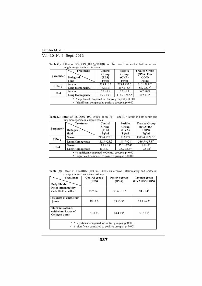
337
Benha M. J.
Vol. 30 No 3 Sept. 2013
µ µ
µ µ
µ µ
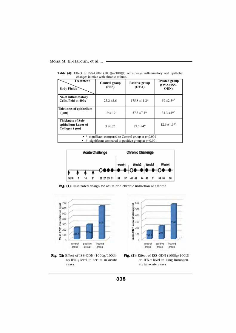
338
Mona M. El-Haroun, et al....
Fig. (1):Fig. (1): Illustrated design for acute and chronic induction of asthma.
Fig. (2):Fig. (2): Effect of ISS-ODN (100µg/100µl)on IFN-γ level in serum in acutecases.
Fig. (3):Fig. (3): Effect of ISS-ODN (100µg/100µl)on IFN-γ level in lung homogen-ate in acute cases.
µ µ

339
Benha M. J.
Vol. 30 No 3 Sept. 2013
Fig. (4):Fig. (4): Effect of ISS-ODN (100µg/100µl)on IL-4 level in serum in acutecases.
Fig. (5):Fig. (5): Effect of ISS-ODN (100µg/100µl)on IL-4 level in lung homogenatein acute cases.
Fig. (6):Fig. (6): Effect of ISS-ODN (100µg/00µl)on IFN-γ level in serum inchronic cases.
Fig. (7):Fig. (7): Effect of ISS-ODN (100µg/100µl)on IFN-γ level in lung homogen-ate in chronic cases.
Fig. (8):Fig. (8): Effect of ISS-ODN (100µg/100µl)on IL-4 level in serum in chroniccases.
Fig. (9):Fig. (9): Effect of ISS-ODN (100µg/100µl)on IL-4 level in lung homogenatein chronic cases.
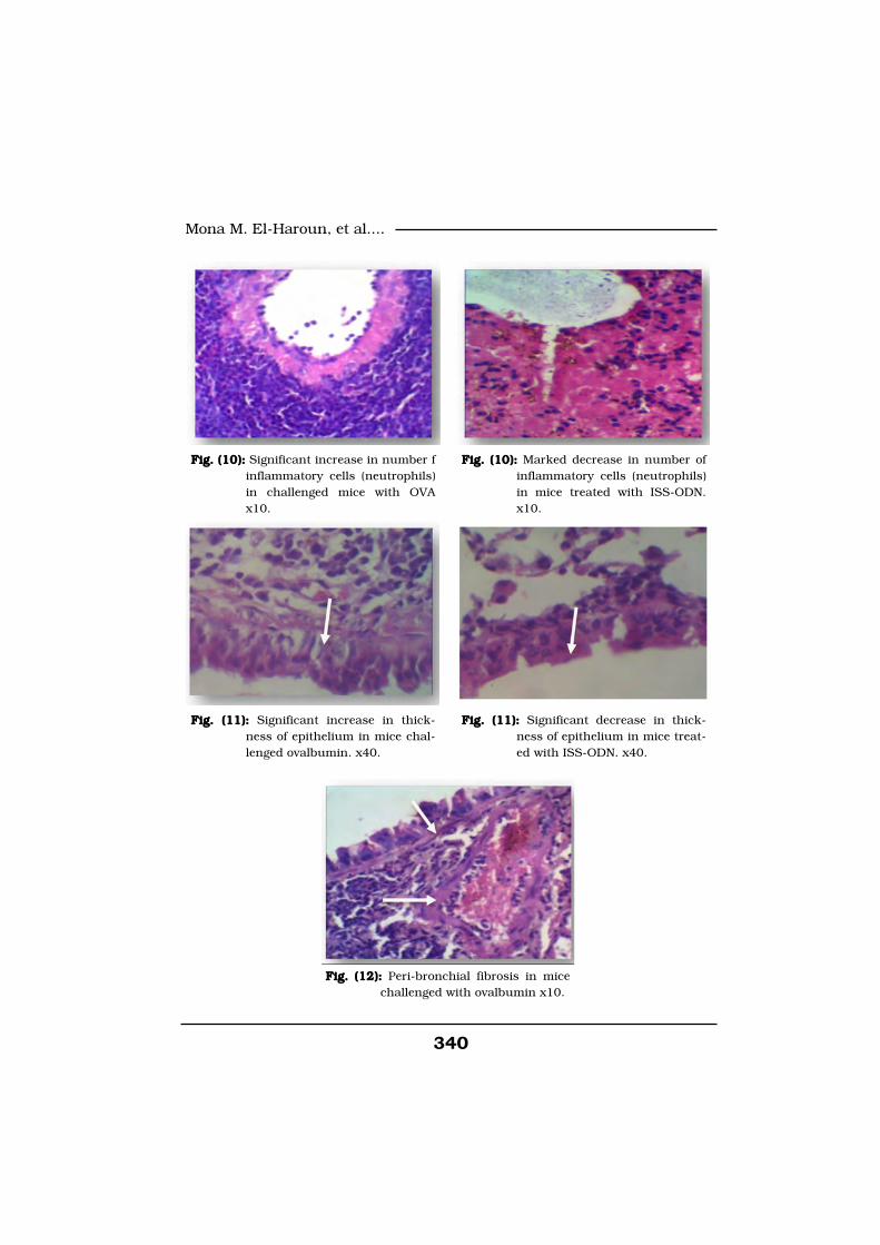
340
Mona M. El-Haroun, et al....
Fig. (10):Fig. (10): Significant increase in number finflammatory cells (neutrophils)in challenged mice with OVAx10.
Fig. (10):Fig. (10): Marked decrease in number ofinflammatory cells (neutrophils)in mice treated with ISS-ODN.x10.
Fig. (12):Fig. (12): Peri-bronchial fibrosis in micechallenged with ovalbumin x10.
Fig. (11):Fig. (11): Significant increase in thick-ness of epithelium in mice chal-lenged ovalbumin. x40.
Fig. (11):Fig. (11): Significant decrease in thick-ness of epithelium in mice treat-ed with ISS-ODN. x40.
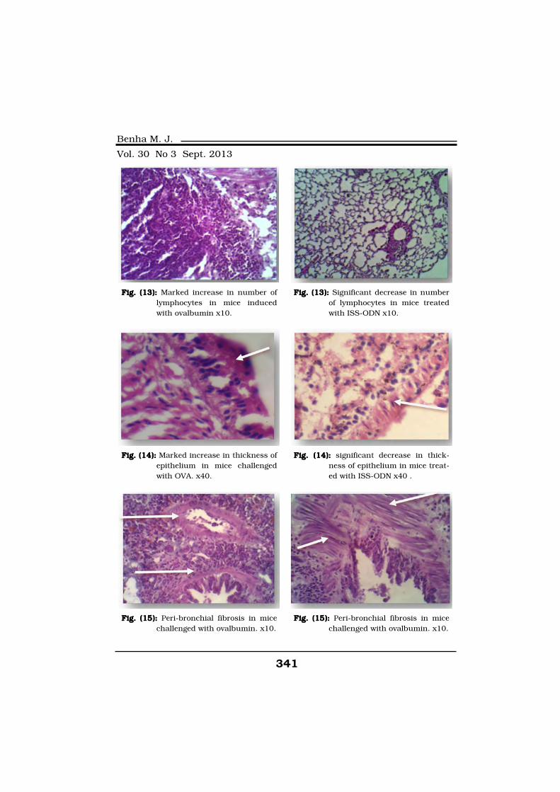
341
Benha M. J.
Vol. 30 No 3 Sept. 2013
Fig. (13):Fig. (13): Marked increase in number oflymphocytes in mice inducedwith ovalbumin x10.
Fig. (13):Fig. (13): Significant decrease in numberof lymphocytes in mice treatedwith ISS-ODN x10.
Fig. (14):Fig. (14): Marked increase in thickness ofepithelium in mice challengedwith OVA. x40.
Fig. (14):Fig. (14): significant decrease in thick-ness of epithelium in mice treat-ed with ISS-ODN x40 .
Fig. (15):Fig. (15): Peri-bronchial fibrosis in micechallenged with ovalbumin. x10.
Fig. (15):Fig. (15): Peri-bronchial fibrosis in micechallenged with ovalbumin. x10.
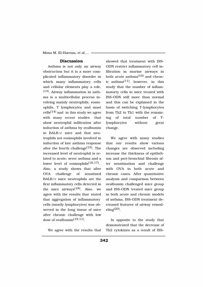
342
Mona M. El-Haroun, et al....
Discussion Asthma is not only an airway
obstruction but it is a more com-
plicated inflammatory disorder in
which many inflammatory cells
and cellular elements play a role,(13). Airway inflammation in asth-
ma is a multicellular process in-
volving mainly neutrophils, eosin-
ophils, T lymphocytes and mast
cells(14) and in this study we agree
with many recent studies that
show neutrophil infiltration after
induction of asthma by ovalbumin
in BALB/c mice and that neu-
trophils not eosinophils involved in
induction of late asthma response
after the fourth challenge(15). The
increased level of neutrophil is re-
lated to acute; sever asthma and a
lower level of eosinophils(16,17).
Also, a study shows that after
OVA challenge of sensitized
BALB/c mice neutrophils are the
first inflammatory cells detected in
the mice airways(18). Also, we
agree with the results that stated
that aggregation of inflammatory
cells (mostly lymphocytes) was ob-
served in the lung tissue of mice
after chronic challenge with low
dose of ovalbumin(19,11).
We agree with the results that
showed that treatment with ISS-
ODN restrict inflammatory cell in-
filtration in murine airways in
both acute asthma(10) and chron-
ic asthma(11), however, in this
study that the number of inflam-
matorty cells in mice treated with
ISS-ODN still more than normal
and this can be explained in the
basis of switching T-lymphocytes
from Th2 to Th1 with the remain-
ing of total number of T-
lymphocytes without great
change.
We agree with many studies
that our results show various
changes are observed including
increase the thickness of epitheli-
um and peri-bronchial fibrosis af-
ter sensitization and challenge
with OVA in both acute and
chronic cases. After quantitative
analysis and comparison between
ovalbumin challenged mice group
and ISS-ODN treated mice group
in both acute and chronic models
of asthma, ISS-ODN treatment de-
creased features of airway remod-
eling(20).
In opposite to the study that
demonstrated that the decrease of
Th2 cytokines as a result of ISS-
343
Benha M. J.
Vol. 30 No 3 Sept. 2013
ODN drug was related to the de-
crease in airway remodeling and not
the increase in Th1 cytokines(10)
our study demonstrated that in
acute model of asthma ISS-ODN
decrease features of airways re-
modeling despite the high level of
IL-4 which concurrent with a high
level of Th1 cytokines IFN-γ. While
in chronic model of asthma the
decrease in airway remodeling was
related to both decreases in Th2
cytokines and increase in Th1 cy-
tokines and as conclusion this un-
derlies a suggestive direct role of
IFN-γ against airway remodeling.
In this study, ISS-ODN de-
creased Th2 cytokines IL-4 in
chronic model of asthma only,
while in acute model of asthma
the level of IL-4 in mice treated
with ISS-ODN was still more than
normal significantly. It could be
explained that one only dose of
ISS-ODN in acute asthma could
not affect the highly proliferated
inflammatory cells while more
than one dose of ISS-ODN (in
chronic model of asthma) could af-
fect inflammatory cells activation
and Th2 cytokines production.
Also, ISS-ODN increased Th1 cyto-
kines IFN-γ in acute model asthma
which means that one only dose
(acute case) can give effect on rest-
ing naïve T lymphocytes (Th 0) to be
activated to Th1. Also, several doses
of ISS-ODN increase IFN-γ level
than normal in chronic model of
asthma. We agree with the results
that explained that it is evident
that ISS both induces Th1 re-
sponses and inhibits Th2 respons-
es in chronic model of asthma(11).
Summary We can conclude our results
that ISS-ODN can restrict inflam-
matory cell infiltration, decrease
airway remodeling, increase Th-1
cytokines production (IFN-γ) in
both acute and chronic models of
asthma while more than one dose
of ISS-ODN can decrease Th-2 cy-
tokines production (IL-4) so more
studies are needed to explain the
suggestive direct role of IFN-γ in
treatment of airway inflammation
and remodeling.
References1- Wenzel S.E. (1994):1- Wenzel S.E. (1994): Asthma
as an inflammatory disease. An-
nual Allergy. 72: 261-271.
2- Bousquet J., Jeffery P.K.,2- Bousquet J., Jeffery P.K.,
Busse W.W., Johnson M. andBusse W.W., Johnson M. and
344
Mona M. El-Haroun, et al....
Vignola A.M. (2000):Vignola A.M. (2000): Asthma.
From bronchoconstriction to air-
ways inflammation and remodel-
ing. American Journal of Respira-
tory and Critical Care Medicine.
161(5):1720-1745.
3- Dabbagh K. and Lewis3- Dabbagh K. and Lewis
D.B. (2003):D.B. (2003): Toll-like receptors
and T-helper-1/T-helper-2 re-
sponses. Current Opinion in Infec-
tious Diseases. 16(3): 199-204.
4- Bhattacharjee R.N. and4- Bhattacharjee R.N. and
Akira S. (2005):Akira S. (2005): Toll- like Recep-
tor signaling: Emerging Opportu-
nities in Human Diseases and
Medicine. Current Immunology
Reviews. 1:81-90.
5- Heine H. and Lien E.5- Heine H. and Lien E.
(2003):(2003): Toll-Like Receptors and
their Function in Innate and
Adaptive Immunity. International
Archives of Allergy and Immunolo-
gy. 130: 180-92.
6- Holtzman M.J. (2003):6- Holtzman M.J. (2003):
Drug Development for Asthma.
American Journal of Respiratory,
Cell and Molecular Biology.
29:163-171.
7- Silverman E.S. and Drazen7- Silverman E.S. and Drazen
J.M. (2003):J.M. (2003): Immunostimulatory
DNA for Asthma. Better than Eat-
ing Dirt. American Journal of Res-
piratory Cell and Molecular Biolo-
gy. 28: 645-647.
8- Hessel E.M., Chu M., Liz-8- Hessel E.M., Chu M., Liz-
cano J.O., Chang B., Hermancano J.O., Chang B., Herman
N., Kell S.A., Marsha Wills-KarpN., Kell S.A., Marsha Wills-Karp
M., and Coffman R.L. (2005):M., and Coffman R.L. (2005): Im-
munostimulatory oligonucleotides
block allergic airway inflammation
by inhibiting Th2 cell activation
and IgE-mediated cytokine induc-
tion. Journal of Experimental
Medicine. 202(11): 1563-1573.
9- Ikeda R.K., Cho N.J.Y.,9- Ikeda R.K., Cho N.J.Y.,
Miller M., Rodriguez M., Raz E.Miller M., Rodriguez M., Raz E.
and Broide D.H. (2003):and Broide D.H. (2003): Resolu-
tion of airway inflammation follow-
ing ovalbumin inhalation: compar-
ison of ISS DNA and
corticosteroids. American Journal
of Respiratory, Cell Molecular Bi-
ology. 28:655-663.
10- Broide D.H., Schwarze J.,10- Broide D.H., Schwarze J.,
Tighe H., Gifford T., Nguyen M.,Tighe H., Gifford T., Nguyen M.,
Malek S., Uden J.V., Martin-Malek S., Uden J.V., Martin-
Orozco E., Gelfand E.W. and RazOrozco E., Gelfand E.W. and Raz
E. (1998):E. (1998): Immunostimulatory
DNA Sequences Inhibit IL-5, Eo-
sinophilic Inflammation, and Air-
345
Benha M. J.
Vol. 30 No 3 Sept. 2013
way Hyperresponsiveness in Mice.
The Journal of Immunology. 161:
7054-7062.
11- Cho J.Y., Miller M., Baek11- Cho J.Y., Miller M., Baek
K.J., Han J.W., Nayar J., Rodri-K.J., Han J.W., Nayar J., Rodri-
guez M., Lee S.Y., McElwain K.,guez M., Lee S.Y., McElwain K.,
McElwain S., Raz E. and BroideMcElwain S., Raz E. and Broide
D.H. (2004):D.H. (2004): Immunostimulatory
DNA Inhibits Transforming
Growth Factor-β Expression and
Airway Remodeling. American
Journal of Respiratory, Cell and
Molecular Biology. 30: 651-661.
12- Temelkovski J., Hogan12- Temelkovski J., Hogan
S.P., Shepherd D.P., Foster P.S.S.P., Shepherd D.P., Foster P.S.
and Kumar R. (1998):and Kumar R. (1998): An im-
proved murine model of asthma:
selective airway inflammation, epi-
thelial lesions and nincreasedme-
thacholine responsiveness follow-
ingchronic exposure to aerosolised
allergen. Thorax. 53:849-856.
13- Humbert M. and Kay A.B.13- Humbert M. and Kay A.B.
(2003):(2003): Chronic inflammation in
asthma. European Respiratory
Monograph. 23: 126-137.
14- Kay A.B. (2005):14- Kay A.B. (2005): The role
of eosinophils in the pathogenesis
of asthma. Molecular Medicine.
11: 148-52.
15- Nabe T., Hosokawa F.,15- Nabe T., Hosokawa F.,
Matsuya K., Morishita T., IkedoMatsuya K., Morishita T., Ikedo
A., Fujii M., Mizutani N., Yoshi-A., Fujii M., Mizutani N., Yoshi-
no S. and Chaplin D. (2011):no S. and Chaplin D. (2011): Im-
portant role of neutrophils in the
late asthmatic response in mice.
Life Science.; 88 (26): 1127-1135.
16- Fahy J.V., Woo K., Liu J.16- Fahy J.V., Woo K., Liu J.
and Boushey H.A. (1995):and Boushey H.A. (1995): Promi-
nent neutrophilic inflammation in
sputum from subjects with asth-
ma exacerbation. Journal of Aller-
gy and Clinical Immunology.
95:843-852.
17- Sur S., Crotty T.B.,17- Sur S., Crotty T.B.,
KeKephart G.M., Hyma B.A., Colbyphart G.M., Hyma B.A., Colby
T.V., Reed C.E., Hunt L.W. andT.V., Reed C.E., Hunt L.W. and
Gleich G.J. (1993):Gleich G.J. (1993): Sudden-onset
fatal asthma. A distinct entity with
few eosinophils and relatively
more neutrophils in the airway
submucosa. American Review of
Respiratory Disease. 148:713-719.
18- Monteseirin J. (2009):18- Monteseirin J. (2009):
Neutrophils and asthma. Journal
of investigational allergology &
clinical immunology. 19(5): 340-
354.
19- Siegle J.S., Hasbro N.,19- Siegle J.S., Hasbro N.,
Herbert C., Yang M., FosterHerbert C., Yang M., Foster
346
Mona M. El-Haroun, et al....
P.S. and Kumar R.K. (2006):P.S. and Kumar R.K. (2006):
Airway Hyperreactivity in Exacer-
bation of Chronic Asthma Is Inde-
pendent of Eosinophilic Inflamma-
tion. American Journal of
Respiratory, Cell and Molecular
Biology. 35 (5):565-570.
20- Jain V.V., Kitagaki K.,20- Jain V.V., Kitagaki K.,
Businga T., et al. (2002):Businga T., et al. (2002): CpG-
oligodeoxynucleotides inhibit air-
way remodeling in a murine model
of chronic asthma. Journal of Al-
lergy and Clinical Immunology.
110:867-872.
347
Benha M. J.
Vol. 30 No 3 Sept. 2013
EFFECT OF ISS-ODN-INDUCED TLR9EFFECT OF ISS-ODN-INDUCED TLR9STIMULATION ON EXPERIMENTALLYSTIMULATION ON EXPERIMENTALLY
INDUCED AIRWAYINDUCED AIRWAYINFLAMMATION IN MICEINFLAMMATION IN MICE
Mona M. El-Haroun M.Sc., Ali M. Yousef MDMona M. El-Haroun M.Sc., Ali M. Yousef MDand Tarek M. Ibrahim Ph.Dand Tarek M. Ibrahim Ph.D
BENHAMEDICALJOURNAL
REPRINT
Published byPublished by
Benha Faculty of MedicineBenha Faculty of Medicine Volume 30 Number 3Sept. 2013
347
Benha M. J.
Vol. 30 No 3 Sept. 2013
IntroductionIntroductionDiffuse Large B-cell Lymphoma
(DLBCL) is the most common sub-
type of Non-Hodgkin’s Lymphoma
(NHL), comprising about 30% of
all NHL cases in all epidemiologi-
cal reports1,2 and it accounts for
80% of aggressive lymphomas 3.
PROGNOSTIC AND PREDICTIVE VALUES OFPROGNOSTIC AND PREDICTIVE VALUES OFKI67 PROLIFERATIVE INDEX IN DIFFUSEKI67 PROLIFERATIVE INDEX IN DIFFUSE
LARGE B -CELL LYMPHOMALARGE B -CELL LYMPHOMA
Seham El-Sayed Abdel-Khalek MD and Azza Abdel-Aziz MD*Seham El-Sayed Abdel-Khalek MD and Azza Abdel-Aziz MD*Clinical Oncology & Nuclear Medicine and *Pathology Department,
Faculty of Medicine, Mansoura University, Egypt
AbstractAbstractPurpose:Purpose: To evaluate role and prognostic significance of Ki-67 prolifera-
tion index (PI) in diffuse large B cell lymphoma. Methods: Expression of Ki-67 protein was examined immunohistochemically in 34 tumor specimensfrom patients newly diagnosed with DLBCL and treated with CHOP (cyclo-phosphamide, doxorubicin, vincristine, and prednisone) regimen. Results:Univariate analysis showed that a high Ki-67 PI (≥60%) was found in 11 pa-tients (42.3%) whose age was ≤ 60 years compared with 62.5% of older pa-tients (age > 60 years) (P=0.025), and in 46.2% of patients with PS=0-1 (0–1)compared with 71.4% of patients with PS=2 (P=0.03). Three-year survivalwas 52.8%±7% in the patients with a high index (mean OS 48.3%±2.8months, median 76 months) and 76%±4.7% in those with a low index (meanOS 76.8±3.6, median not reached, P=0.013). Disease free survival was high-er in patients with Ki67<60% than those with Ki67≥60%, and the differencewas statistically significant (p=0.042). Multivariate analysis performed byCox model revealed that IPI≥3 and high Ki-67PI had a significant indepen-dent prognostic value concerning overall survival (p<0.05).
Conclusion: Conclusion: Initial high Ki 67≥60% associated with high IPI score couldrepresent possible predictive factors of poor prognosis, which would help toidentify a high risk subgroup of newly diagnosed DLBCL.
Keywords: Keywords: Diffuse large B cell lymphoma, IPI, prognostic factors, Ki67,immunohistochemistry.
348348
Seham El-Sayed Abdel-Khalek and Azza Abdel-Aziz
The CHOP (cyclophosphamide,
doxorubicin, vincristine, and pred-
nisone) regimen alone or with ri-
tuximab (CHOP/R) is standard
therapeutic approach for the most
patients who have DLBCL. Al-
though DLBCL can be cured with
the current chemotherapy regi-
mens, the long-term survival is es-
timated to be only 50% for high-
risk patients 3, a substantial mi-
nority of patients (about 30%) are
not cured4,5.
While International Prognostic
Index (IPI) was developed to pro-
vide a model system for predicting
the outcome of patients with ag-
gressive NHL depending on some
clinical parameters (age, stage, per-
formance status, number of extra-
nodal sites and serum level of lac-
tic acid dehydrogenase)6, asubstantial
variability in outcome has been
observed despite IPI subgroups7.
Thus, identifying new prognostic
parameters might contribute to-
wards better prediction of out-
come and the development of ef-
fective risk-adaptive strategies8.
Patients with similar DLBCL di-
agnoses can have varied molecu-
lar profiles, heterogeneous clinical
presentations, and clinical out-
comes. Several immunohistochem-
ical algorithms and gene profiling
sets have been developed to iden-
tify DLBCL subgroups with unfa-
vorable prognosis9,10. The hall-
mark features of the tumor cell
phenotype, which contribute to
aggressive tumor behavior,are: its
capacity for sustained prolifera-
tion,disregard of signals to stop
proliferation and differentiation
and the capacity to invade and
promote angiogenesis11. Despite
the sustained research in recent
years, risk-adapted therapies based
on DLBCL phenotype are still in
the development stage.
The Ki67 antigen plays a pivot-
al role in maintaining cell prolife-
ration and is expressed in all
phases of the cell cycle except G0
and has been extensively used as
a marker of cell proliferation in a
variety of neoplastic12 and non-
neoplastic disorders13. The mono-
clonal antibody anti Ki67 was the
only available till the early 90s
and it has the inherited drawback
that it could be applied only to
sections from fresh frozen tissues.
Recently using a recombinant
parts of Ki67 protein as an immu-
349
Benha M. J.
Vol. 30 No 3 Sept. 2013
nogen, an equivalent monoclonal
antibody, MIB-1 has been devel-
oped12 and can be utilized as a
routine stain on paraffin -embedded
sections from fixed tissues through
microwave process14 providing an
interest in the prognostic valida-
tion of MIB-1in different human
tumors including NHL.
The aim of this study was to
assay Ki-67 immunohistochemi-
cally in patients with newly diag-
nosed diffuse large B cell lympho-
ma, correlated with IPI regarding
to clinical course and outcome.
Material and MethodsMaterial and MethodsThirty four patients were en-
rolled in the current studyat the
Clinical Oncology and Nuclear
Medicine Department, and De-
partment of Pathology, Mansoura
University Hospital in a period be-
tweenJanuary 2004 and Decem-
ber 2008. The eligibility criteria for
the patients in this study were as
follows: age between 18 and 70
years, Eastern Cooperative Oncol-
ogy Group (ECOG) performance
status 0-2, histologically con-
firmed diffuse large B cell Lym-
phoma.Adequate hematologic pro-
file and organ function were
required for study entry: a neu-
trophil count greater than 2000/
mm3, platelet count greater than
100 000/mm3, serum bilirubin,
SGOT, SGPT, serum creatinine
within normal limit and normal
electrocardiogram. No other histo-
ry of active malignancy and no
other serious medical disease.
Central nervous system lym-
phoma, post-transplant lymphop-
roliferative disorders, cutaneous
lymphomas, T-cell/histiocyte-rich
large B-cell lymphoma and Burkitt
lymphoma were excluded.
Patients included in the study
had an initial diagnosis of DLBCL
according to the World Health Or-
ganization (WHO) classification15.
In all patients; standard clinical,
radiological and laboratory data
were collected: age, gender, Ann
Arbor stage, extranodal sites,
bulky disease, clinical stage, B
symptoms and LDH13. Bulky dis-
ease was defined as a mediastinal
mass larger than one third of the
maximum thoracic diameter and/
or any node over 10cm. Nodal or-
gan was assigned to those cases
with a clinical presentation in
lymph nodes, waldeyer’s ring,
350350
Seham El-Sayed Abdel-Khalek and Azza Abdel-Aziz
spleen or bone marrow by the cri-
teria of Kramer et al.16. Extrano-
dal organ was defined as presen-
tation in other sites with or
without local lymph node involve-
ment. The IPI was calculated ac-
cording to the five high risk fea-
tures: age >60 years, performance
status (PS) >2, Ann Arbor tumor
stage 3 or 4, LDH >460 IU/µL,
and number of extranodal sites >1
while patients were divided into a
low risk group (0-2 factors) and a
high-risk group (3 or 4 factors)17.
All patients treated with CHOP
(cyclophosphamide, doxorubicine,
vincristine, and prednisone) regi-
men. Complete re-staging was
scheduled at the end of the treat-
ment program and periodic follow
up examinations were performed
every 3 to 4 months for the first 2
years after completion of therapy
and every 6 months thereafter.
Tissue Microarray Construc-Tissue Microarray Construc-
tion:tion:
Tissue microarrays (TMAs) were
prepared at the Department of Pa-
thology,Mansoura University Hos-
pital from 34 representative pa-
tient samples with DLBCL with
adequate archival formalin-fixed
and paraffin-embedded material.
Five-µm thick paraffin sections
from the samples were mounted
onto poly-L-lysine coated slides
(Sigma, Milan, Italy) and dried
over-night at 37˚C. Subsequently,
sections were de-waxed in xylene,
rehydrated according to histopath-
ological standards. Slides were im-
mersed in antigen re-trieval solu-
tion (BioGenex Cat. No. HK 090-
5K) at a dilution of 1:4 with deion-
ized water. The slides were pro-
cessed for two cycles of 5 minutes
at maximum power (1000w) in a
mi-crowave oven (Goldstar, USA).
Sections were never allowed to
dry. The sections were incubated
with the monoclonal antibody MIB-1
(Bi-oGenex ready-to-use) at room
temperature overnight in a humid
air and successively with a biotin-
ylated rabbit anti-mouse IgG ser-
um. Then, treated with an avidin-
biotin system (No-vostain super
ABC kit, NovocastraLaborato-ries,
Newcastle, UK) for 30 minutes fol-
lowed by rinsing with buffer for 10
minutes. The anti- gen-antibody
complex was visualized using DAB
(3, 3`-diaminobenzidine tetrahy-
drocloride) and then counter-
stained with haematoxylin. A reac-
351
Benha M. J.
Vol. 30 No 3 Sept. 2013
tive LN with follicular hyperplasia
was used as positive control; neg-
ative controls were obtained by
omission of the primary monoclo-
nal antibody. The cell Image ana-
lyzer system was used for Ki-
67quantitation. A cell was consid-
ered positive when it exhibits a
weak, moderate or strong nuclear
and/or nuclear staining. At least
1000 cells from the most repre-
sentative areas of each tumor
were scored and the MIB-1 index
was evaluated as the ratio be-
tween positive cells and total tu-
mor cells. The percentage of tumor
cells with Ki-67+ nuclear staining
on 10 different high power micros-
copy fields (HPF, 400x) was deter-
mined.
Statistical analysis:Statistical analysis:
SPSS version 10.0 was used for
data analysis. Chi-square and
Fisher exact test were used for
testing proportions independence.
Disease-free survival (DFS) was
measured from the start of treat-
ment to the date of primary treat-
ment failure, relapse, or the date
of last follow-up. Overall survival
(OS) was measured from the be-
ginning of treatment to the time of
last follow-up (censored patients)
or time of death. Both were stud-
ied by the Kaplan Meier method
and the survival curves were com-
pared by the log-rank test18. Cox
backward proportional hazard
model was performed for multivar-
iate analysis of the factors that
might be of independence signifi-
cance in influencing the overall
survival. Factors included in the
maximum model were IPI [low risk
group (0-2 factors) and a high-risk
group (3 or 4 factors)]. A Ki-67 PI
cutoff of 60% distinguished the tu-
mors with a favorable or poor
prognosis. The response rates
were analyzed according to widely
accepted international Cheson cri-
teria19. Complete remission (CR)
was defined as the resolution of
clinical and radiological evidence
of disease for a minimum of 4
weeks. For the purpose of statisti-
cal analysis, partial remission,
non-response and progressive dis-
ease were considered as treatment
failures. All p values were always
two tailed and values of 0.05 or
less were considered statistically
significant.
ResultsResultsThe cell proliferation marker
Ki-67 was observed as nuclear
352352
Seham El-Sayed Abdel-Khalek and Azza Abdel-Aziz
staining in tumor cells and in lym-
phocytes within the tumor tissues
in all 34 cases. The staining inten-
sity and number of tumor cells
positive to Ki-67 varied from case
to case, ranging from 30% to 95%.
None of the patients had more
than 95% Ki-67 positive tumor
cells.
On ROC curve analysis (Receiv-
er Operating Characteristic curve),
a Ki-67 PI of 60% was found to
significantly discriminate patients
with DLBCL who had a good or
bad prognosis (Area under curve =
0.64, P = 0.003). Univariate analy-
sis showed a significant associa-
tion between the Ki-67 PI (≤60%
vs. >60%) and patient age and
ECOG PS. A high Ki-67 PI was
found in the 11patients (42.3%)
whose agewas ≤60 years com-
pared with 62.5% of the older pa-
tients (age >60 years) (P=0.025),
and in 46.2% of the patients with
a good PS (0-1) compared with
71.4% of the patients with a poor
PS (2) (P=0.03).
The value of Ki-67 positive tu-
mor cells follows the values of IPI
scores, meaning that patients with
a high proliferation rate also had
high IPI value (IPI > 3). We found
statistically significant positive
correlation between low and high
Ki-67+ and IPI scores (χ2, p<
0.05).
None of the other factors exam-
ined yielded a significant associa-
tion with Ki-67 PI: stage (I, II vs.
III, IV) (P=0.6); number of disease-
involved extranodal sites (≤1 vs.
≥2) (P=0.57); b2 microglobulin lev-
el (P=0.3); B symptoms (present
vs. absent) (P=0.56) and LDH
(high vs. normal) (P=0.45).
After the first line therapy,
there were significant difference in
complete remission (CR) for pa-
tients with high and low Ki67 (ta-
ble 2).
Using the cut-off value of 60%,
we found a significant association
between Ki-67 PI and overall sur-
vival in patients with DLBCL (Fig.
1). Three-year survival was
52.8%±7% in the patients with a
high index (mean overall survival
48.3%±2.8 months, median 76
months) and 76%±4.7% in those
with a low index (mean overall
survival 76.8±3.6, median not
reached, P=0.013). Disease free
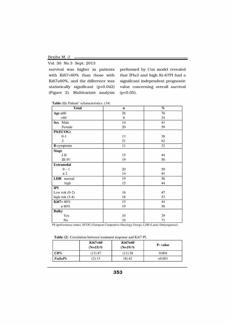
353
Benha M. J.
Vol. 30 No 3 Sept. 2013
survival was higher in patients
with Ki67<60% than those with
Ki67≥60%, and the difference was
statistically significant (p=0.042)
(Figure 2). Multivariate analysis
performed by Cox model revealed
that IPI≥3 and high Ki-67PI had a
significant independent prognostic
value concerning overall survival
(p<0.05).
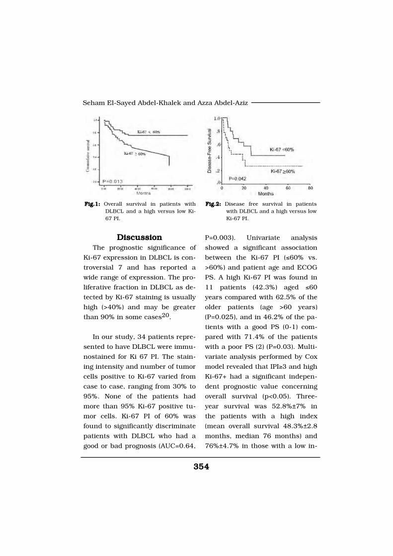
354354
Seham El-Sayed Abdel-Khalek and Azza Abdel-Aziz
DiscussionDiscussionThe prognostic significance of
Ki-67 expression in DLBCL is con-
troversial 7 and has reported a
wide range of expression. The pro-
liferative fraction in DLBCL as de-
tected by Ki-67 staining is usually
high (>40%) and may be greater
than 90% in some cases20.
In our study, 34 patients repre-
sented to have DLBCL were immu-
nostained for Ki 67 PI. The stain-
ing intensity and number of tumor
cells positive to Ki-67 varied from
case to case, ranging from 30% to
95%. None of the patients had
more than 95% Ki-67 positive tu-
mor cells. Ki-67 PI of 60% was
found to significantly discriminate
patients with DLBCL who had a
good or bad prognosis (AUC=0.64,
P=0.003). Univariate analysis
showed a significant association
between the Ki-67 PI (≤60% vs.
>60%) and patient age and ECOG
PS. A high Ki-67 PI was found in
11 patients (42.3%) aged ≤60
years compared with 62.5% of the
older patients (age >60 years)
(P=0.025), and in 46.2% of the pa-
tients with a good PS (0-1) com-
pared with 71.4% of the patients
with a poor PS (2) (P=0.03). Multi-
variate analysis performed by Cox
model revealed that IPI≥3 and high
Ki-67+ had a significant indepen-
dent prognostic value concerning
overall survival (p<0.05). Three-
year survival was 52.8%±7% in
the patients with a high index
(mean overall survival 48.3%±2.8
months, median 76 months) and
76%±4.7% in those with a low in-
Fig.1: Fig.1: Overall survival in patients withDLBCL and a high versus low Ki-67 PI.
Fig.2: Fig.2: Disease free survival in patientswith DLBCL and a high versus lowKi-67 PI.
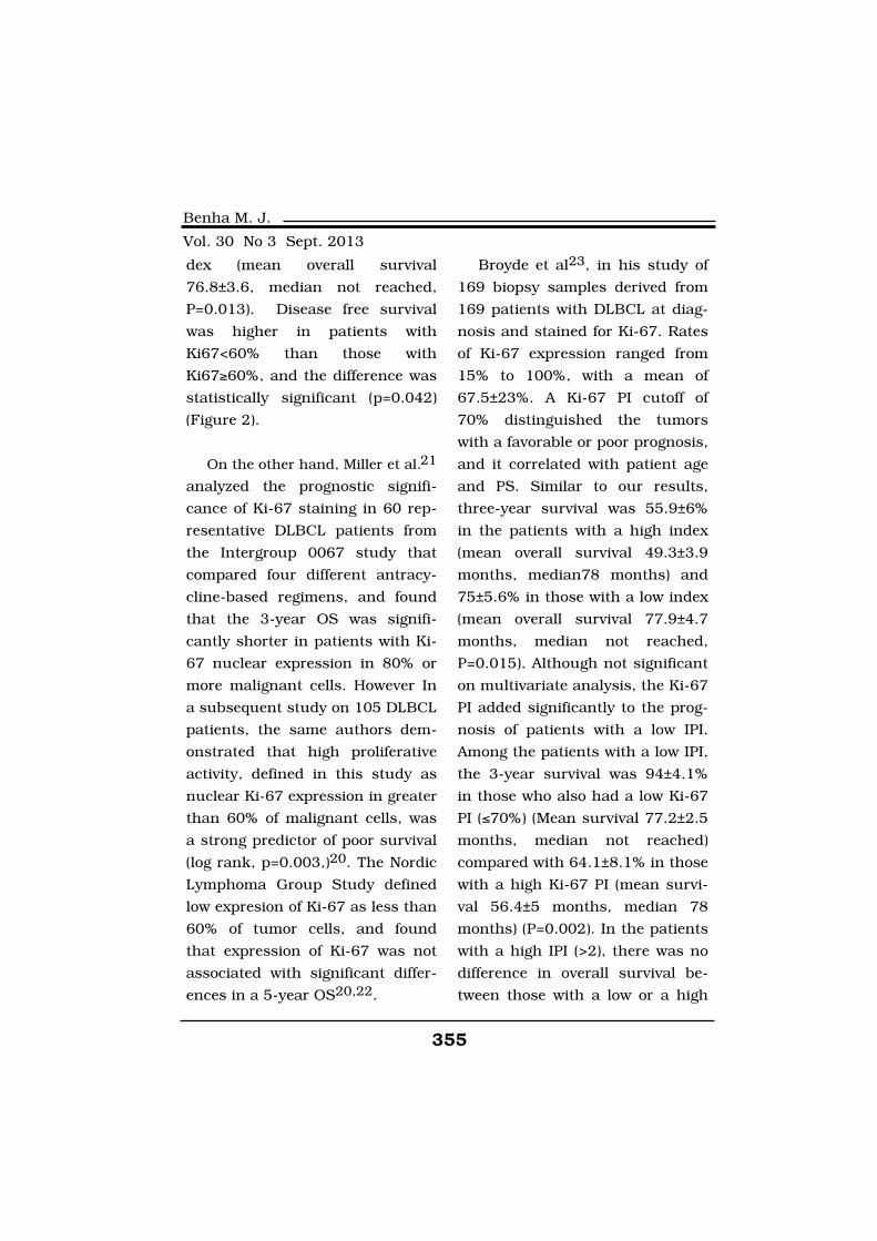
355
Benha M. J.
Vol. 30 No 3 Sept. 2013
dex (mean overall survival
76.8±3.6, median not reached,
P=0.013). Disease free survival
was higher in patients with
Ki67<60% than those with
Ki67≥60%, and the difference was
statistically significant (p=0.042)
(Figure 2).
On the other hand, Miller et al.21
analyzed the prognostic signifi-
cance of Ki-67 staining in 60 rep-
resentative DLBCL patients from
the Intergroup 0067 study that
compared four different antracy-
cline-based regimens, and found
that the 3-year OS was signifi-
cantly shorter in patients with Ki-
67 nuclear expression in 80% or
more malignant cells. However In
a subsequent study on 105 DLBCL
patients, the same authors dem-
onstrated that high proliferative
activity, defined in this study as
nuclear Ki-67 expression in greater
than 60% of malignant cells, was
a strong predictor of poor survival
(log rank, p=0.003,)20. The Nordic
Lymphoma Group Study defined
low expresion of Ki-67 as less than
60% of tumor cells, and found
that expression of Ki-67 was not
associated with significant differ-
ences in a 5-year OS20,22.
Broyde et al23, in his study of
169 biopsy samples derived from
169 patients with DLBCL at diag-
nosis and stained for Ki-67. Rates
of Ki-67 expression ranged from
15% to 100%, with a mean of
67.5±23%. A Ki-67 PI cutoff of
70% distinguished the tumors
with a favorable or poor prognosis,
and it correlated with patient age
and PS. Similar to our results,
three-year survival was 55.9±6%
in the patients with a high index
(mean overall survival 49.3±3.9
months, median78 months) and
75±5.6% in those with a low index
(mean overall survival 77.9±4.7
months, median not reached,
P=0.015). Although not significant
on multivariate analysis, the Ki-67
PI added significantly to the prog-
nosis of patients with a low IPI.
Among the patients with a low IPI,
the 3-year survival was 94±4.1%
in those who also had a low Ki-67
PI (≤70%) (Mean survival 77.2±2.5
months, median not reached)
compared with 64.1±8.1% in those
with a high Ki-67 PI (mean survi-
val 56.4±5 months, median 78
months) (P=0.002). In the patients
with a high IPI (>2), there was no
difference in overall survival be-
tween those with a low or a high
356356
Seham El-Sayed Abdel-Khalek and Azza Abdel-Aziz
Ki-67 PI. The Ki-67 PI also had
prognostic value in patients with
bulky disease (diameter >10 cm).
Three-year survival was signifi-
cantly better in patients with
bulky disease who had a low Ki-67
PI (100%) than in patients with a
high Ki-67 PI (25±12%; mean sur-
vival 14.5 months, median 9
months) (P=0.012). These data
suggest that tumors that reach
bulkiness with a low Ki-67 PI still
have a very good prognosis.
In the largest study, which was
performed by Jerkeman et al20,
included 185 cases. Ki-67 expres-
sion was found to be low (<60%)
in 116 tumors (63%), moderate
(60-90%) in 59 (32%), and high
(>90%) in 10 (5%), and it correlat-
ed with performance status (PS)
(P=0.0005). However, a low Ki-67
PI (<60%) was associated with a
low failure-free survival (RR 1.7,
95% CI 1.1-2.6%) compared with
a moderate or high Ki- 67 PI.
Moreover, patients with either low
or high Ki-67 expression demon-
strated a trend toward lower over-
all survival than patients with
moderate expression. On multi-
variate analysis, high bcl-2 and
low Ki-67 levels added prognostic
information to the clinical Interna-
tional Prognostic Index (IPI)24 for
aggressive lymphoma in terms of
failure-free survival. However in
later series performed byHassel-
bladet al25 no correlation was ob-
served between low Ki 67 PI and
subgroups of DLBCL.
Others analyzed DLBCL pa-
tients treated by R-CHOP as Yoon
DH et al26 analyzed Ki-67 expres-
sion and its correlation with prog-
nosis in 144 patients with DLBCL
treated with rituximab plus CHOP
(R-CHOP). They found the com-
plete response (CR) rates following
R-CHOP administration were not
significantly different, based on
Ki-67 expression status (P=0.104).
However, higher rates of relapse
were observed in the high Ki-67
expression group (Ki-67 ≥85%, n=46)
with 25.0%, compared to 10.0% in
the low Ki-67 expression group
(Ki-67<85%, n=88) (P=0.040). The
2-yr event-free survival (EFS) rates
were 44.3% and 74.1% in the high
and low Ki-67 expression groups,
respectively (P=0.011). The 2-yr
overall survival (OS) rate was
66.4% in the high Ki-67 expres-
sion group and 82.2% in the low
Ki-67 expression group (P=0.016).
357
Benha M. J.
Vol. 30 No 3 Sept. 2013
In multivariate analysis, Ki-67 ex-
pression was a significant prog-
nostic factor for EFS [hazard ratio
(HR)=2.909; 95% confidence inter-
val (CI) 1.261-6.708; P=0.012]. Ki-
67 was associated with higher OS
rate but with borderline signifi-
cance (HR=2.876; 95% CI, 0.972-
8.508; P=0.056).
Li ZM et al27 in their study,
evaluated whether Ki-67 expres-
sion is an indicator of outcome in
DLBCL patients (especially non-
GCB DLBCL patients) treated with
standard chemotherapy combined
with rituximab. They foundoverall
survival (OS) and progression-free
survival (PFS) were lower in pa-
tients with high Ki-67 expression
than in those with low Ki-67 ex-
pression (3-year OS: 65.2% vs.
81.7%, P=0.030; 3-year PFS:
56.4% vs. 73.3%, P=0.020), simi-
lar in patients with GCB subtype
and those with the non-GCB sub-
type (OS: P=0.330; PFS: P=0.287).
According to Ki-67 expression
status by immunophenotype sub-
groups, patients with high Ki-67
expression in non-GCB subgroup
had the most unfavorable PFS and
OS, comparing with the other
three subgroups (P=0.004 and
P=0.002, respectively). In multi-
variate analysis, non-GCB with
high Ki-67 expression was an in-
dependent prognostic predictor of
inferior survival in DLBCL pa-
tients treated with R-CHOP.
As we see Ki-67 immunostain-
ing has several advantages: it can
be performed on routinely pro-
cessed tissues, permitting the si-
multaneous evaluation of the mor-
phology and tumor cell kinetics; it
stains the sections homogeneous-
ly; and there is no background in-
terference, making it suitable for
automated image analysis. Fur-
thermore, it can be applied even
on small biopsy specimens and
core needle biopsies. There are
several possible explanations for
the discrepancies in the prognos-
tic significance of Ki-67 expression
in DLBCL among the studies.
First, the cutoff value used to de-
fine a high Ki-67 PI ranged from
20% to 80%; some of the studies
applied the median value. Second,
some studies used manual tech-
niques to assess the Ki-67 PI,
whereas others used quantitative
image analysis with Cell Analysis
Systems (CAS), which are more
objective and reproducible28.
358358
Seham El-Sayed Abdel-Khalek and Azza Abdel-Aziz
Third, a recent study from the in-
ternational Lunenburg Lymphoma
Biomarker Consortium29 on tech-
nical and inter-observer variations
in scoring different immunohisto-
chemical prognostic markers in
DLBCL specimens found poor re-
producibility among laboratories
for Ki-67 PI staining. They con-
cluded that clinical decisions
based on immunohistochemical
stratification should be performed
only in the context of clinical trials
with centralized consensus review.
Lossos IS et al.7 support this
opinion as they stated that tumors
with low Ki-67 index may exhibits
resistance to chemotherapy, be-
cause the majority of the malig-
nant cells are in G0/G1 and thus
are resistant to cycle specific cyto-
toxic chemotherapy. Furthermore,
G0/G1 arrested cells have time to
repair DNA damage induced by
the chemotherapy and thus sur-
vive7.
ConclusionConclusionInitial high Ki 67≥60% associat-
ed with high IPI score could repre-
sent possible predictive factors of
poor prognosis, which would help
to identify a high risk subgroup of
newly diagnosed DLBCL.
Acknowledgements Acknowledgements Disclosure: The authors declare
no conflict of interest.
ReferencesReferences1. Anderson J.R., Armitage1. Anderson J.R., Armitage
J.O. and Weisenburger D.D.J.O. and Weisenburger D.D.
(1998):(1998): Epidemiology of the non-
Hodgkin's lymphomas: distribu-
tions of the major subtype differ
by geographic locations. Non-
Hodgkin's Lymphoma Classifica-
tion Project, Ann Oncol; 9: 717-
720
2. Morton L.M., Purdue M.P.,2. Morton L.M., Purdue M.P.,
Zheng T., et al. (2009):Zheng T., et al. (2009): Risk of
non-Hodgkin lymphoma with
germline variation in genes that
regulate the cell cycle, apoptosis,
and lymphocyte development.
Cancer Epidemiol Biomarkers
Prev;18:1259-1270 313.
3. Sehn L.H., Berry B., Chha-3. Sehn L.H., Berry B., Chha-
nabhai M., et al. (2007):nabhai M., et al. (2007): The re-
vised International Prognostic In-
dex (R-IPI) is a better predictor of
outcome than the standard IPI for
patients with diffuse large B-cell
lymphoma treated with R-CHOP.
Blood; 109: 1857-1861
4. Mounier N., Briere J., Gis-4. Mounier N., Briere J., Gis-
359
Benha M. J.
Vol. 30 No 3 Sept. 2013
selbrecht C., et al. (2003):selbrecht C., et al. (2003): Ritux-
imab plus CHOP (R-CHOP) over-
comes bcl-2-- associated resis-
tance to chemotherapy in elderly
patients with diffuse large B-cell
lymphoma (DLBCL). Blood; 101
(11): 4279-84.
5. Sehn L.H. (2006):5. Sehn L.H. (2006): Optimal
use of prognostic factors in non-
Hodgkin lymphoma. Hematology
Am SocHematolEduc Program:
295-302.
6. Harrs N.L., Jaffe E.S.,6. Harrs N.L., Jaffe E.S.,
Stein H., et al. (1994):Stein H., et al. (1994): A revised
European American classification
of lymphoma neoplasm: A propo-
sal from the International Lym-
phoma Study Group. Blood,
84:1361.
7. Lossos I.S. and Morgensz-7. Lossos I.S. and Morgensz-
tern D. (2006):tern D. (2006): Prognostic bio-
markers in diffuse large B-cell
lymphoma. J Clin Oncol; 24(6):
995-1007.
8. Biasoli I., Morais J.C.,8. Biasoli I., Morais J.C.,
Scheliga A., et al. (2005):Scheliga A., et al. (2005): CD10
and Bcl-2 expression combined
with the International Prognostic
Index can identify subgroups of
patients with diffuse large-cell
lymphoma with very good or very
poor prognoses. Histopathology;
46(3): 328-33.
9. Choi W.W., Weisenburger9. Choi W.W., Weisenburger
D.D., Greiner T.C., et al. (2009):D.D., Greiner T.C., et al. (2009):
A new immunostain algorithm
classifies diffuse large B-cell lym-
phoma into molecular subtypes
with high accuracy. Clin Cancer
Res; 15: 5494-5502
10. Hans C.P., Weisenburger10. Hans C.P., Weisenburger
D.D., Greiner T.C., et al. (2004):D.D., Greiner T.C., et al. (2004):
Confirmation of the molecular
classification of diffuse large B-cell
lymphoma by immunohistochem-
istry using a tissue microarray.
Blood; 103: 275-282
11. Hanahan D. and Wein-11. Hanahan D. and Wein-
berg R.A. (2000):berg R.A. (2000): The hallmarks
of cancer. Cell; 100(1): 57-70.
12. Franklin W.A., McDonald12. Franklin W.A., McDonald
G.B. and Stein H.O. (1985):G.B. and Stein H.O. (1985): Im-
munohistological demonstration of
colonic crypt cell kinetics in ulcerative
colitis. Hum. Pathology, 16: 1129.
13. Barnard M.J., Hall P.A.,13. Barnard M.J., Hall P.A.,
Lemoine N.R., et al. (1987):Lemoine N.R., et al. (1987): Pro-
liferative index in breast carcino-
ma determined in situ by Ki 67
360360
Seham El-Sayed Abdel-Khalek and Azza Abdel-Aziz
immunostaining and its relation-
ship to clinical and pathological
variables. J. Pathology, 152:287.
14. Cattoretti G., Beker14. Cattoretti G., Beker
M.H.G. and Key G. (1992):M.H.G. and Key G. (1992): Mon-
oclonal antibodies against recom-
binant parts of the Ki67 antigen
(MIB1-MIB3) detect proliferating
cell in microwave- processed for-
maline-fixed paraffin sections. J.
Pathology, 168:357.
15. Harris N.L., Jaffe E.S.,15. Harris N.L., Jaffe E.S.,
Diebold J., et al. (1999):Diebold J., et al. (1999): World
Health Organization classification
of neoplastic diseases of the he-
matopoietic and lymphoid tissues:
report of the Clinical Advisory
Committee meeting-Airlie House,
Virginia, November 1997. J Clin
Oncol; 17(12): 3835-49.
16. Kramer M.H.H., Hermans16. Kramer M.H.H., Hermans
J. and Parker J. (1996):J. and Parker J. (1996): Clinical
significance of bcl2 and p53 pro-
tein expression in diffuse large B-
cell lymphoma: A population-based
study.J.Clin. Oncology, 14; 2131.
17. A predictive model for ag-17. A predictive model for ag-
gressive non-Hodgkin's lympho-gressive non-Hodgkin's lympho-
ma (1993):ma (1993): The International
Non-Hodgkin's Lymphoma Prog-
nostic Factors Project. N Engl J
Med; 329(14): 987-94.
18. Kleinbaum D.G. (1994):18. Kleinbaum D.G. (1994):
Survival analysis. A self-learning
text: Springer-Verlage, New York.
19. Cheson B.D., Horning19. Cheson B.D., Horning
S.J., Coiffier B., et al. (2000):S.J., Coiffier B., et al. (2000):
Report of an international work-
shop to standardize response cri-
teria for non-Hodgkin's lympho-
mas. NCI Sponsored International
Working Group. J Clin Oncol Jun;
18(11): 2351.
20. Jerkeman M., Anderson20. Jerkeman M., Anderson
H., Dictor M., et al. (2004): H., Dictor M., et al. (2004): As-
sessment of biological prognostic
factors provides clinically relevant
information in patients with dif-
fuse large B-cell lymphoma-a Nor-
dic Lymphoma Group study. Ann
Hematol. 83: 414-419.
21. Miller T.P., Grogan T.M.,21. Miller T.P., Grogan T.M.,
Dahlberg S., Spier C.M., BrazielDahlberg S., Spier C.M., Braziel
R.M., Banks P.M., et al. (1994):R.M., Banks P.M., et al. (1994):
Prognostic significance of the Ki-
67-associated proliferative antigen
in aggressive non-Hodgkin's lym-
phomas: a prospective Southwest
Oncology Group trial. Blood; 83
(6): 1460-6.

361
Benha M. J.
Vol. 30 No 3 Sept. 2013
22. Maja Perunicic Jovanovic,22. Maja Perunicic Jovanovic,
Ljubomir Jakovic, Andrija Bog-Ljubomir Jakovic, Andrija Bog-
danovic, et al. (2009):danovic, et al. (2009): Poor out-
come in patients with diffuse large
B-cell lymphoma is associated
with high percentage of bcl-2 and
Ki 67-positive tumor cells. Vojno-
sanitetskiPregled; 66(9): 538-743.
23. Broyde A., Boycov O.,23. Broyde A., Boycov O.,
Strenov Y., et al. (2009):Strenov Y., et al. (2009): Role
and prognostic significance of the
Ki-67 index in non-Hodgkin's lym-
phoma. Am J Hematol.; 84: 338-
343.
24. The International Non-24. The International Non-
Hodgkin's Lymphoma PrognosticHodgkin's Lymphoma Prognostic
Factors Project (1993):Factors Project (1993): A predic-
tive model for aggressive non-
Hodgkin's lymphoma. N Engl J
Med.; 329: 987-994.
25. Hasselblom S., Ridell B.,25. Hasselblom S., Ridell B.,
Sigurdardottir M., et al. (2008):Sigurdardottir M., et al. (2008):
Low rather than high Ki-67 protein
expression is an adverse prognostic
factor in diffuse large B-cell lym-
phoma. Leuk Lymph, 49, 1501-9.
26. Yoon D.H., Choi D.R.,26. Yoon D.H., Choi D.R.,
Ahn H.J., et al. (2010):Ahn H.J., et al. (2010): Ki-67 ex-
pression as a prognostic factor in
diffuse large B-cell lymphoma pa-
tients treated with rituximab plus
CHOP. Eur J Haematol. Aug; 85
(2):149-57. doi: 10.1111.
27. Li Z.M., Huang J.J., Xia27. Li Z.M., Huang J.J., Xia
Y., et al. (2012):Y., et al. (2012): High Ki-67 ex-
pression in diffuse large B-cell
lymphoma patients with non-
germinal center subtype indicates
limited survival benefit from R-
CHOP therapy.Eur J Haematol.
Jun; 88(6):510-7. doi: 10.1111
28. Lejeune M., Jae´n J., Pons28. Lejeune M., Jae´n J., Pons
L., et al. (2008):L., et al. (2008): Quantification of
diverse subcellular immunohisto-
chemical markers with clinicobio-
logical relevancies: validationof a
new computer-assisted image
analysis procedure. J Anat.; 212
(6): 868-878.
29. de Jong D., Rosenwald A.,29. de Jong D., Rosenwald A.,
Chhanabhai M., et al. (2007):Chhanabhai M., et al. (2007):
Immunohistochemical prognostic
markers in diffuse large B-cell
lymphoma: validation of tissue mi-
croarray as a prerequisite for broad
clinical applications. A study from
the Lunenberg Lymphoma Bio-
marker Consortium.J Clin Oncol.;
25: 805-812.

362362
Seham El-Sayed Abdel-Khalek and Azza Abdel-Aziz
PROGNOSTIC AND PREDICTIVEPROGNOSTIC AND PREDICTIVEVALUES OF KI67 PROLIFERATIVEVALUES OF KI67 PROLIFERATIVE
INDEX IN DIFFUSE LARGE B -CELLINDEX IN DIFFUSE LARGE B -CELLLYMPHOMALYMPHOMA
Seham El-Sayed Abdel-Khalek MDSeham El-Sayed Abdel-Khalek MDand Azza Abdel-Aziz MDand Azza Abdel-Aziz MD
BENHAMEDICALJOURNAL
REPRINT
Published byPublished by
Benha Faculty of MedicineBenha Faculty of Medicine Volume 30 Number 3Sept. 2013
363
Benha M. J.
Vol. 30 No 3 Sept. 2013
IntroductionIntroductionDiabetes mellitus is one of the
most common endocrine metabol-
ic disorders. It is a chronic disease
that characterized by hyperglycae-
mia(1). Hyperglycemic in long time
have side effect in other tissues
especially in liver. Liver dysfunc-
PROTECTIVE EFFECT OF QUERCETIN ONPROTECTIVE EFFECT OF QUERCETIN ONLIVER DAMAGE IN STREPTOZOTOCIN-LIVER DAMAGE IN STREPTOZOTOCIN-
INDUCED DIABETIC RATSINDUCED DIABETIC RATS
Ola A. El Gohary MD and Abeer A. Shoman MDOla A. El Gohary MD and Abeer A. Shoman MDPhysiology Department, Faculty of Medicine, Banha Univeristy
AbstractAbstractThe negative impact of diabetes on the liver is well recognized. This study
was designed to evaluate the hepatoprotective properties of Quercetin instreptozotocin-induced diabetes in rats. Male Wistar rats were made diabeticwith a single injection of STZ (40 mg/kg i.p.). Rats were randomly dividedinto four groups 8 animals each: Group 1, healthy control rats; Group 2non-diabetic rats treated with 15 mg/kg/day i.p. injection of Quercetin;Group 3, diabetic rats; Group 4, diabetic rats treated with Quercetin (15mg/kg/day, i.p.) for 8 weeks. Finally, serum ALT, AST, ALP and albumin lev-els as well as liver MDA contents and activities of GSH-Px, were measured toassess hepatic injury. Liver tissues of Rat in whole groups were removedthen prepared for Apoptosis analysis. Liver MDA content and serum ALT,AST, ALP and bilirubin levels in Groups 3 were found to be significantly in-creased as compared to Group 1 (P<0.001) and these parameters in Group 4were significantly decreased as compared to Group 3 (P<0.001). Liver GSH-Px contents and serum albumin level in Group 3 was significantly decreasedas compared to Group 1 (P<0.001) and were found to be significantly in-creased in Group 4 as compared to Groups 3 (P<0.001). Histopathologicalexamination revealed that diabetes increased apoptotic index in liver tissuewhile the treatment of diabetic rats with Quercetin was shown to have anti-apoptosis effect. This study showed that Quercetin have hepatoprotective ef-fects in experimentally induced diabetic rats.
Keywords: Keywords: Apoptosis, Diabetic, Quercetin, Streptozotocin, Liver, Rat.
364364
Ola A. El Gohary and Abeer A. Shoman
tional has seen Indirectly or di-
rectly, the liver is a major target of
insulin action. The onset of dia-
betes is accompanied by develop-
ment of major biochemical and
functional abnormalities in the liv-
er, including alterations in carbo-
hydrate, lipid, and protein metab-
olism, and changes in antioxidant
status(2). On the other hand, it
was established that hyperglyce-
mia increases mitochondrial reac-
tive oxygen species (ROS) produc-
tion, which could represent a key
event in the development of dia-
betes complications(3,4). The ini-
tial cellular response to high glu-
cose challenge is the generation of
ROS, which rapidly induces apop-
totic cell death(5). The balance of
ROS and antioxidant is a major
mechanism in preventing damage
by oxidative stress. However, al-
though it may not be possible to
completely reverse diabetic com-
plications, antioxidants could be
useful in preventing or attenuat-
ing the adverse effects of chronic
hyperglycemia(6). Therefore, the
dietary supplement of antioxi-
dants such as vitamins, flavonoids
has been used to prevent the oc-
currence of many chronic diseas-
es(7,8). Flavonoids are a large
group of natural polyphenolic sub-
stances widely distributed in the
plant kingdom(9). They are impor-
tant constituents of the non ener-
getic part of the human diet and
are thought to promote optimal
health, via their antioxidant ef-
fects in protecting cellular compo-
nents against ROS(10). Quercetin
(3,5,7,3'4'-pentahydroxy flavon) is
one of the most widely distributed
flavonoids, present in fruit, vege-
tables, tea olive oil and many oth-
er dietary sources(11). It is a
strong antioxidant and it has been
shown to reduce oxidative stress(12,13). It has been demonstrated
that quercetin exhibits its thera-
peutic potential against many dis-
eases, including ischemic heart
diseases, atherosclerosis, liver fi-
brosis, renal injury, and chronic
biliary obstruction(14,15,16).
Because liver is subjected to
ROS-mediated injury in diabetes(17), our experiments were per-
formed to investigate the potential
protective effects of quercetin
treatment on liver oxidative stress.
Material and MethodsMaterial and MethodsAnimals:Animals:
This study was conducted on

365
Benha M. J.
Vol. 30 No 3 Sept. 2013
32 adult Wistar albino male rats
6-8 weeks old, weighing between
170 and 200g. Animals were
housed in the animal laboratory at
the medical research center at
Benha faculty of medicine. They
were housed at room temperature
(25°C) and 12h/12h light/ dark
cycle. All Rats were fed a standard
diet and water.
Groups of the experiment:Groups of the experiment:
The animals were randomly di-
vided into 4 groups each consisted
of 8 rats as follow:
Group (I): Control group inject-
ed with citrate buffer daily, intra-
peritoneal (IP).
Group (II): Quercetin (QR) group
that received 15 mg/kg QR (IP).
Group (III): Diabetic group that
received 40 mg/kg streptozotocin
(IP).
Group (IV): Treatment group re-
ceived 40mg/kg (IP) STZ plus
15mg/kg QR (IP).
Induction and diagnosis of di-Induction and diagnosis of di-
abetes mellitus:abetes mellitus:
Diabetes was induced by intra-
peritoneal (ip) injection of a single
dose of STZ (40 mg/kg in freshly
prepared citrate buffer pH 4.5).
The animals were allowed to drink
5% glucose solution overnight to
overcome drug induced hypoglyce-
mia. Control rats were injected by
the buffer alone(18).
Diabetes was verified 72 hours
later by measuring blood glucose
levels (after an overnight fasting)
with the use of glucose oxidase re-
agent strips. Rats having blood
glucose level of ≥250 mg/dl were
considered to be diabetic.
Quercetin administration:Quercetin administration:
Quercetin (QR) treatment was
initiated 5 days after the adminis-
tration of streptozotocin. Quercetin
(QR) injections of 15mg/kg intra-
peritoneal (IP)(19) were continued
daily to the end of the study (for 8
weeks)(20).
Chemicals used:Chemicals used:
*Streptzotocin drug:*Streptzotocin drug:
It was purchased from Sigma-
Aldrich Company (USA). It is pre-
sented in powder form, purity
more than 99% to be dissolved in
freshly prepared sodium citrate
buffer pH 4.5.
* Sodium citrate buffer pH 4.5:* Sodium citrate buffer pH 4.5:
Preparation of 0.1MCitratePreparation of 0.1MCitrate
Buffer: Buffer:
366366
Ola A. El Gohary and Abeer A. Shoman
Weigh accurately citric acid
10.5 gm and sodium citrate 14.7
gm. Mix it with 500 ml water.
Make up volume to 1000 ml with
distilled water. Adjust pH 4.5 by
sodium hydroxide(21).
* Quercetin drug:* Quercetin drug:
Quercetin powder was obtained
from Sigma Chemical Company
(St. Louis, MO, USA). It was dis-
solved and diluted with 20% glyce-
rol in 0.9% normal saline, mixed
vigorously and stored in a dark
bottle at 4°C. The quercetin solu-
tion was freshly prepared each
week(19).
Procedure of the experiments:Procedure of the experiments:
At the end of the treatment pe-
riod, the animals were anesthe-
tized after 12 hour fasting by in-
halation of diethyl ether. The
animals were fixed on operating
table and the blood samples were
taken as follow:
Blood sample collection:Blood sample collection:
A craniocaudal incision of
about 2 cm is made, parallel and
with slightly to the left of the ster-
num through the skin and pecto-
ral muscles to expose the ribs. A
blunt curved forceps is then
binged between the 5th and 6th
ribs, through the intercostals mus-
cles. The gap is widened so that
the rapidly beating heart becomes
visible, then the blood sample were
taken from the right ventricle.
Biochemical assessment:Biochemical assessment:
Plasma activities of Alanine Trans-
aminase (ALT), Aspartate Transam-
inase (AST), alkaline phosphatase
(ALP) and concentration of glucose,
albumin and total bilirubin were
determined by a standard auto-
mated technique using Hitachi
Analyzer Model 911 and adequate
kits from Roche Company (Swit-
zerland)(22). These were investigat-
ed in Banha faculty of medicine at
biochemistry analysis unit.
Tissue preparation:Tissue preparation:
A midline laparotomy was per-
formed to remove the liver. The liv-
er was dissected and fixed in 10%
formalin solution at room temper-
ature. Slices of liver tissue were
processed for histopathological &
immunohistochemical studies.
Immunohistochemical analy-Immunohistochemical analy-
sis:sis:
Paraffin embedded tissue sec-
tions of 5Mm were prepared on
367
Benha M. J.
Vol. 30 No 3 Sept. 2013
positively- charged slides to be
stained with antiBCL-X antibody
using Biotin streptavidin immune-
peroxidase technique.
Interpretation for immunos-Interpretation for immunos-
taining:taining:
BCL-X was detected as cyto-
plasmic brown staining in exam-
ined tissue.
Stained sections were classified
as; Mild intensity for weak brown
cytoplasmic stain. Moderate inten-
sity for moderate brown cytoplasmic
stain. Strong intensity for strong
brown cytoplasmic stain(23).
Measurement of antioxidantMeasurement of antioxidant
activity:activity:
The rat's Liver were removed
immediately and washed in normal
saline and homogenate 10% prepared
in 1.15% w/v of potassium chloride.
The homogenate was centrifuged
in 7000 x g for 10 minutes at 4˚C
and supernatant were used for
measurement of oxidative stress
by determination of malondialde-
hyde (MDA) as well as estimation
of antioxidant enzymes such as
glutathione peroxidase (GSH-Px)(24). Tissue MDA levels were deter-
mined by the thiobarbituric acid
(TBA) method and expressed as
nmol MDA formed/mL. Plasma
MDA concentrations were deter-
mined with spectrophotometer(25).
Glutathione peroxidase (GSH-Px)
activity was measured by NADPH
oxidation, using a coupled reac-
tion system consisting of glutathi-
one, glutathione reductase, and
cumene hydroperoxide . One unit
of enzyme activity is defined as
the amount of enzyme that trans-
forms 1 µmol of NADPH to NADP
per minute. Results are expressed
as units/mg protein(26).
Statistical analysis:Statistical analysis:
All data were expressed as
mean±S.D; data were evaluated by
the one way analysis of variance.
Difference between groups were
compared by Student's t-test with
P<0.05 selected as the level of sta-
tistical significance.
ResultsResultsResults of the effect of daily
treatment of Quercetin at a dose
of 15mg/kg for 8 weeks on blood
glucose levels of experimental rats
are presented in Table 1. The
Quercetin treatment produced hy-
poglycemic effect in both normal
and diabetic rats after 8 weeks of
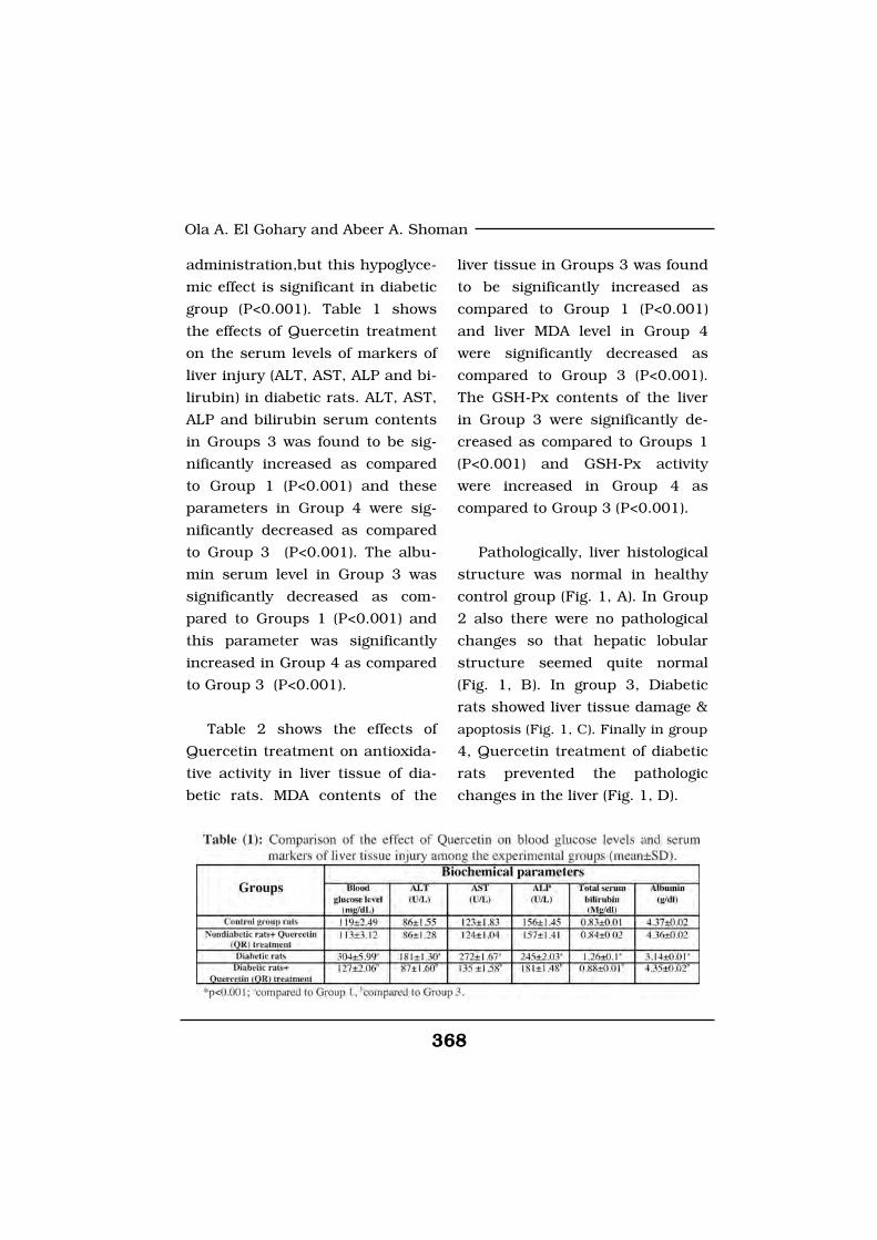
368368
Ola A. El Gohary and Abeer A. Shoman
administration,but this hypoglyce-
mic effect is significant in diabetic
group (P<0.001). Table 1 shows
the effects of Quercetin treatment
on the serum levels of markers of
liver injury (ALT, AST, ALP and bi-
lirubin) in diabetic rats. ALT, AST,
ALP and bilirubin serum contents
in Groups 3 was found to be sig-
nificantly increased as compared
to Group 1 (P<0.001) and these
parameters in Group 4 were sig-
nificantly decreased as compared
to Group 3 (P<0.001). The albu-
min serum level in Group 3 was
significantly decreased as com-
pared to Groups 1 (P<0.001) and
this parameter was significantly
increased in Group 4 as compared
to Group 3 (P<0.001).
Table 2 shows the effects of
Quercetin treatment on antioxida-
tive activity in liver tissue of dia-
betic rats. MDA contents of the
liver tissue in Groups 3 was found
to be significantly increased as
compared to Group 1 (P<0.001)
and liver MDA level in Group 4
were significantly decreased as
compared to Group 3 (P<0.001).
The GSH-Px contents of the liver
in Group 3 were significantly de-
creased as compared to Groups 1
(P<0.001) and GSH-Px activity
were increased in Group 4 as
compared to Group 3 (P<0.001).
Pathologically, liver histological
structure was normal in healthy
control group (Fig. 1, A). In Group
2 also there were no pathological
changes so that hepatic lobular
structure seemed quite normal
(Fig. 1, B). In group 3, Diabetic
rats showed liver tissue damage &
apoptosis (Fig. 1, C). Finally in group
4, Quercetin treatment of diabetic
rats prevented the pathologic
changes in the liver (Fig. 1, D).
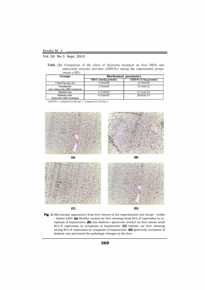
369
Benha M. J.
Vol. 30 No 3 Sept. 2013
(A)(A) (B)(B)
(C)(C) (D)(D)
Fig. 1:Fig. 1: Microscopic appearance from liver tissues of the experimental rats (strept - avidin- biotin) x200. (A)(A) Healthy control rat liver showing weak BCL-X expression in cy-toplasm of hepatocytes. (B) (B) non-diabetic+ Quercetin treated rat liver shows weakBCL-X expression in cytoplasm of hepatocytes. (C)(C) Dabetic rat liver showingstrong BCL-X expression in cytoplasm of hepatocytes. (D) D) Quercetin treatment ofdiabetic rats prevented the pathologic changes in the liver.
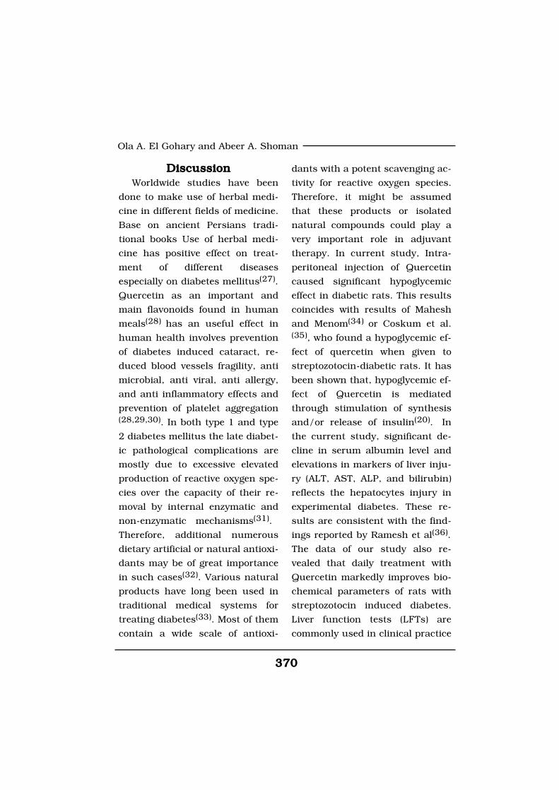
370370
Ola A. El Gohary and Abeer A. Shoman
DiscussionDiscussionWorldwide studies have been
done to make use of herbal medi-
cine in different fields of medicine.
Base on ancient Persians tradi-
tional books Use of herbal medi-
cine has positive effect on treat-
ment of different diseases
especially on diabetes mellitus(27).
Quercetin as an important and
main flavonoids found in human
meals(28) has an useful effect in
human health involves prevention
of diabetes induced cataract, re-
duced blood vessels fragility, anti
microbial, anti viral, anti allergy,
and anti inflammatory effects and
prevention of platelet aggregation(28,29,30). In both type 1 and type
2 diabetes mellitus the late diabet-
ic pathological complications are
mostly due to excessive elevated
production of reactive oxygen spe-
cies over the capacity of their re-
moval by internal enzymatic and
non-enzymatic mechanisms(31).
Therefore, additional numerous
dietary artificial or natural antioxi-
dants may be of great importance
in such cases(32). Various natural
products have long been used in
traditional medical systems for
treating diabetes(33). Most of them
contain a wide scale of antioxi-
dants with a potent scavenging ac-
tivity for reactive oxygen species.
Therefore, it might be assumed
that these products or isolated
natural compounds could play a
very important role in adjuvant
therapy. In current study, Intra-
peritoneal injection of Quercetin
caused significant hypoglycemic
effect in diabetic rats. This results
coincides with results of Mahesh
and Menom(34) or Coskum et al.(35), who found a hypoglycemic ef-
fect of quercetin when given to
streptozotocin-diabetic rats. It has
been shown that, hypoglycemic ef-
fect of Quercetin is mediated
through stimulation of synthesis
and/or release of insulin(20). In
the current study, significant de-
cline in serum albumin level and
elevations in markers of liver inju-
ry (ALT, AST, ALP, and bilirubin)
reflects the hepatocytes injury in
experimental diabetes. These re-
sults are consistent with the find-
ings reported by Ramesh et al(36).
The data of our study also re-
vealed that daily treatment with
Quercetin markedly improves bio-
chemical parameters of rats with
streptozotocin induced diabetes.
Liver function tests (LFTs) are
commonly used in clinical practice
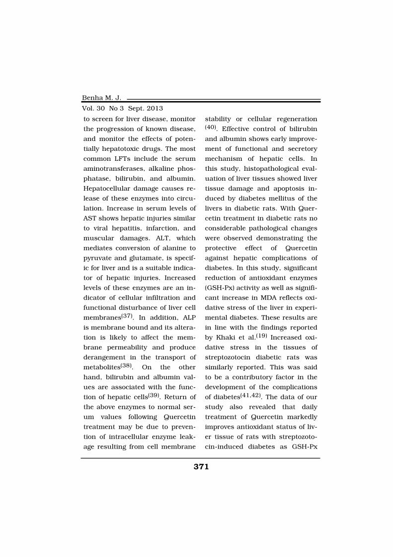
371
Benha M. J.
Vol. 30 No 3 Sept. 2013
to screen for liver disease, monitor
the progression of known disease,
and monitor the effects of poten-
tially hepatotoxic drugs. The most
common LFTs include the serum
aminotransferases, alkaline phos-
phatase, bilirubin, and albumin.
Hepatocellular damage causes re-
lease of these enzymes into circu-
lation. Increase in serum levels of
AST shows hepatic injuries similar
to viral hepatitis, infarction, and
muscular damages. ALT, which
mediates conversion of alanine to
pyruvate and glutamate, is specif-
ic for liver and is a suitable indica-
tor of hepatic injuries. Increased
levels of these enzymes are an in-
dicator of cellular infiltration and
functional disturbance of liver cell
membranes(37). In addition, ALP
is membrane bound and its altera-
tion is likely to affect the mem-
brane permeability and produce
derangement in the transport of
metabolites(38). On the other
hand, bilirubin and albumin val-
ues are associated with the func-
tion of hepatic cells(39). Return of
the above enzymes to normal ser-
um values following Quercetin
treatment may be due to preven-
tion of intracellular enzyme leak-
age resulting from cell membrane
stability or cellular regeneration(40). Effective control of bilirubin
and albumin shows early improve-
ment of functional and secretory
mechanism of hepatic cells. In
this study, histopathological eval-
uation of liver tissues showed liver
tissue damage and apoptosis in-
duced by diabetes mellitus of the
livers in diabetic rats. With Quer-
cetin treatment in diabetic rats no
considerable pathological changes
were observed demonstrating the
protective effect of Quercetin
against hepatic complications of
diabetes. In this study, significant
reduction of antioxidant enzymes
(GSH-Px) activity as well as signifi-
cant increase in MDA reflects oxi-
dative stress of the liver in experi-
mental diabetes. These results are
in line with the findings reported
by Khaki et al.(19) Increased oxi-
dative stress in the tissues of
streptozotocin diabetic rats was
similarly reported. This was said
to be a contributory factor in the
development of the complications
of diabetes(41,42). The data of our
study also revealed that daily
treatment of Quercetin markedly
improves antioxidant status of liv-
er tissue of rats with streptozoto-
cin-induced diabetes as GSH-Px
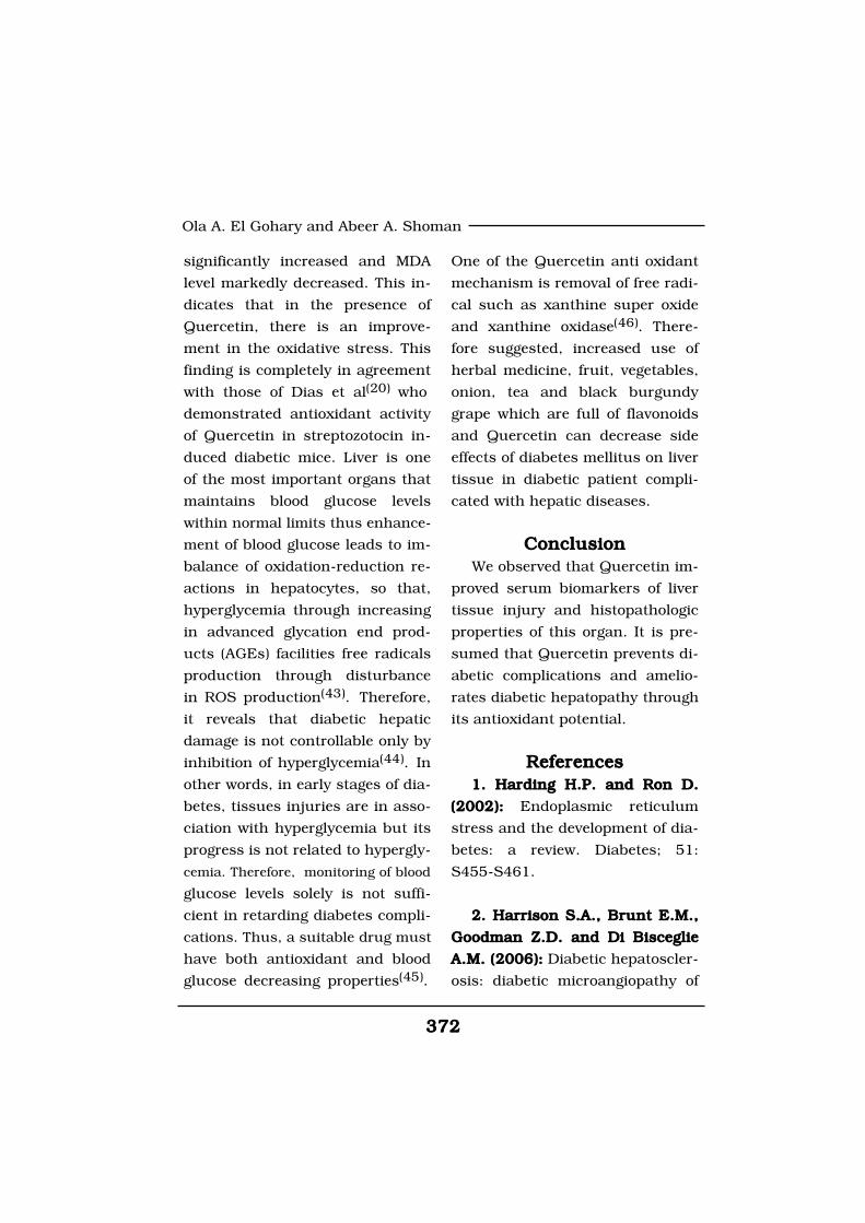
372372
Ola A. El Gohary and Abeer A. Shoman
significantly increased and MDA
level markedly decreased. This in-
dicates that in the presence of
Quercetin, there is an improve-
ment in the oxidative stress. This
finding is completely in agreement
with those of Dias et al(20) who
demonstrated antioxidant activity
of Quercetin in streptozotocin in-
duced diabetic mice. Liver is one
of the most important organs that
maintains blood glucose levels
within normal limits thus enhance-
ment of blood glucose leads to im-
balance of oxidation-reduction re-
actions in hepatocytes, so that,
hyperglycemia through increasing
in advanced glycation end prod-
ucts (AGEs) facilities free radicals
production through disturbance
in ROS production(43). Therefore,
it reveals that diabetic hepatic
damage is not controllable only by
inhibition of hyperglycemia(44). In
other words, in early stages of dia-
betes, tissues injuries are in asso-
ciation with hyperglycemia but its
progress is not related to hypergly-
cemia. Therefore, monitoring of blood
glucose levels solely is not suffi-
cient in retarding diabetes compli-
cations. Thus, a suitable drug must
have both antioxidant and blood
glucose decreasing properties(45).
One of the Quercetin anti oxidant
mechanism is removal of free radi-
cal such as xanthine super oxide
and xanthine oxidase(46). There-
fore suggested, increased use of
herbal medicine, fruit, vegetables,
onion, tea and black burgundy
grape which are full of flavonoids
and Quercetin can decrease side
effects of diabetes mellitus on liver
tissue in diabetic patient compli-
cated with hepatic diseases.
ConclusionConclusionWe observed that Quercetin im-
proved serum biomarkers of liver
tissue injury and histopathologic
properties of this organ. It is pre-
sumed that Quercetin prevents di-
abetic complications and amelio-
rates diabetic hepatopathy through
its antioxidant potential.
ReferencesReferences1. Harding H.P. and Ron D.1. Harding H.P. and Ron D.
(2002):(2002): Endoplasmic reticulum
stress and the development of dia-
betes: a review. Diabetes; 51:
S455-S461.
2. Harrison S.A., Brunt E.M.,2. Harrison S.A., Brunt E.M.,
Goodman Z.D. and Di BisceglieGoodman Z.D. and Di Bisceglie
A.M. (2006):A.M. (2006): Diabetic hepatoscler-
osis: diabetic microangiopathy of
373
Benha M. J.
Vol. 30 No 3 Sept. 2013
the liver. Archives of Pathology
and Laboratory Medicine; 130: 27-
32.
3. Nishikawa T., Edelstein D.,3. Nishikawa T., Edelstein D.,
Du X.L., et al. (2000):Du X.L., et al. (2000): Normaliz-
ing mitochondrial superoxide pro-
duction blocks three pathways of
hyperglycaemic damage. Nature;
404: 787-790.
4. Kiritoshi S., Nishikawa T.,4. Kiritoshi S., Nishikawa T.,
Sonoda K., et al. (2003):Sonoda K., et al. (2003): Reactive
oxygen species from mitochondria
induce cyclo oxygenase-2gene ex-
pression in human mesangial
cells: potential role in diabetic
nephropathy. Diabetes; 52: 2570-
2577.
5. Park K.S., Kim J.H., Kim5. Park K.S., Kim J.H., Kim
M.S., et al. (2003): M.S., et al. (2003): 8-
hydroxyguanine and tissue 8-
hydroxydeoxyguanosine in strep-
tozotocin-induced diabetic rats.
Diabetes; (50): 2837-2841.
6. King G.L. and Loeken M.R.6. King G.L. and Loeken M.R.
(2004):(2004): Hyperglycemia-induced
oxidative stress in diabetic compli-
cations. Histochem Cell Biol.; 122:
333-338.
7. Peluso M.R. (2006):7. Peluso M.R. (2006): Flavo-
noids attenuate cardiovascular dis-
ease, inhibit phosphodiesterase,
and modulate lipid homeostasis in
adipose tissue and liver. J. of Exp
Biol. Med. (Maywood).; 231 (8):
1287-99.
8. Mahesh T. and Menon V.P.8. Mahesh T. and Menon V.P.
(2004):(2004): Quercetin alleviates oxida-
tive stress in streptozotocin-
induced diabetic rats. J. of Photo-
therapy Res.; 18:123-7.
9. David C., Rodrigues G.,9. David C., Rodrigues G.,
Bona S., et al. (2011):Bona S., et al. (2011): Role of
quercetin in preventing thioaceta-
mide-induced liver injury in rats.
Toxicol. Pathol.; 39 (6): 949-957.
10. Khaki A.A., Khaki A.,10. Khaki A.A., Khaki A.,
Nouri M., et al. (2009):Nouri M., et al. (2009): Evalua-
tion effects of quercetin on liver
apoptosis in streptozotocin - in-
duced diabetic rat.J. of Medicinal
Plants.; 8 (5): 70-78.
11. Pawlikowska-Pawlega B.,11. Pawlikowska-Pawlega B.,
Guszecki W.I., Misiak L.E. andGuszecki W.I., Misiak L.E. and
Gawron A. (2003):Gawron A. (2003): The study of
the quercetin action on human
erythrocyte membranes. Biochem
Pharmacol.; 66: 605-612.
12. Shrilatha B. (2007):12. Shrilatha B. (2007): Mura-
374374
Ola A. El Gohary and Abeer A. Shoman
lidhara early oxidative stress in
testis and epididymal sperm in
streptozotocin-induced diabetic
mice: its progression and genotox-
ic consequences. J. of Reprod Tox-
icol.; 23 (4): 578-87.
13. Khaki A., Fathiazad F.,13. Khaki A., Fathiazad F.,
Nouri M., et al. (2009):Nouri M., et al. (2009): Evalua-
tion of Androgenic Activity of Alli-
um cepa on Spermatogenesis in
Rat. J. of Folia Morphol. (Warsz).;
68: 45-51.
14. Peres W., Tunon M.J.,14. Peres W., Tunon M.J.,
Collado P.S., et al. (2000):Collado P.S., et al. (2000): The
flavonoid quercetin ameliorates
liver damage in rats with biliary
obstruction. J Hepatol; 33(5), 742-
50.
15. Singh D., Chander V. and15. Singh D., Chander V. and
Chopra K. (2004):Chopra K. (2004): The effect of
quercetin, a bioflavonoid on ische-
mia/reperfusion induced renal in-
jury in rats. Arch Med Res; 35(6),
484-94.
16. Tieppo J., Vercelino R.,16. Tieppo J., Vercelino R.,
Dias A.S., et al. (2007):Dias A.S., et al. (2007): Evalua-
tion of the protective effects of
quercetin in the hepatopulmonary
syndrome. Food Chem Toxicol; 45
(7), 1140-6.
17. Seven A., Guzel S., Sey-17. Seven A., Guzel S., Sey-
men O., et al. (2004):men O., et al. (2004): Effect of vi-
tamin E supplementation on oxi-
dative stress in streptozotocin
induced diabetic rats: investiga-
tion of liver and plasma. Yonsei
Med J.; 45:703-710.
18. Gojo A., Utsunomiya K.18. Gojo A., Utsunomiya K.
and Taniguchi K. (2007):and Taniguchi K. (2007): The
Rhokinase inhibitior, fasudil, at-
tenuates diabetic nephropathy in
strepazotocin-induced diabetic
rats. Eur J Pharm.; 568: 242-7.
19. Khaki A.A., Khaki A.,19. Khaki A.A., Khaki A.,
Nouri M., et al. (2009):Nouri M., et al. (2009): Evalua-
tion Effects of Quercetin on Liver
Apoptosis in Streptozotocin-
induced Diabetic Rat. 8 (5); 70-78.
20. Dias A.S., Porawski M.,20. Dias A.S., Porawski M.,
Alonso M., et al. (2005):Alonso M., et al. (2005): Querce-
tin Decreases Oxidative Stress,
NF-κ B Activation, and iNOS Over-
expression in Liver of Streptozoto-
cin-Induced Diabetic Rats. 135
(10); 2299-2304.
21. Rajurkar B.M. (2011):21. Rajurkar B.M. (2011):
Phyto pharmacological investigations
of clerodendrum infrotunatum
gartn.international research jour-
nal of pharmacy.; 2 (11): 130-132.
375
Benha M. J.
Vol. 30 No 3 Sept. 2013
22. ULICNÁ O., VANCOVÁ O.,22. ULICNÁ O., VANCOVÁ O.,
BOZEK P., et al. (2006):BOZEK P., et al. (2006): Rooibos
Tea (Aspalathus linearis) Partially
Prevents Oxidative Stress in
Streptozotocin-Induced Diabetic
Rats Physiol. Res.; 55: 157-164.
23. Holland T.A., Elder J.,23. Holland T.A., Elder J.,
McCloud J.M., et al. (2001):McCloud J.M., et al. (2001):
Subcellular localisation of cyclin
D1 protein in colorectal tumors is
associated with p21 WAF1/CIP1
expression and correlates with pa-
tient survival. Int J Cancer; 95:
302-6
24. Tabrizi B.A. and Mohaje-24. Tabrizi B.A. and Mohaje-
ri D. (2012):ri D. (2012): Protective effect of
Aloe Vera Leaves Extract on He-
patic Tissue Injury in Streptozoto-
cin-induced Diabetic Rats. Ad-
vances in Bioresearch; 3 (3): 67-
73.
25. Quintanilha A.T., Packer25. Quintanilha A.T., Packer
L., Davies J.M., et al. (1982):L., Davies J.M., et al. (1982):
Membrane effects of vitamin E de-
ficiency: bioenergetics and surface
charge density studies of skeletal
muscle and liver mitochondria. J.
of Ann. NY Acad. Sci.; 393: 32-47.
26. Rotruck J.T., Pope A.L.,26. Rotruck J.T., Pope A.L.,
Ganther H.E., et al. (1973):Ganther H.E., et al. (1973): “Se-
lenium: biochemical role as a
component of glatathione peroxi-
dase,” Science; 179 (73): 588-590.
27. Jiang G.Y. (1996):27. Jiang G.Y. (1996): Practi-
cal Diabetes 1st Edition. Beijing:
People's Health Publishing House:
295.
28. Formica J.V. and Regel-28. Formica J.V. and Regel-
son W. (1995):son W. (1995): Review of the biol-
ogy of Quercetin and related bio-
flavonoids. Food Chem. J. of
Toxicol.; 33: 1061-80.
29. Hertog M.G. and Hollman29. Hertog M.G. and Hollman
P.C. (1996):P.C. (1996): Potential health ef-
fects of the dietary flavonol quer-
cetin. Eur. J. of Clin. Nutr.; 50:
63-71.
30. Dok-Go H., Lee K.H., Kim30. Dok-Go H., Lee K.H., Kim
H.J., et al. (2003): H.J., et al. (2003): Neuroprotective
effects of antioxidative flavonoids,
quercetin, (1)-dihydroquercetin
and quercetin 3-methyl ether, iso-
lated from Opuntia ficus-indica
var. saboten. J. of Brain. Res.;
965: 130-6.
31. 31. BONNEFONT-ROUSSELOTBONNEFONT-ROUSSELOT
D. (2002):D. (2002): Glucose and reactive
oxygen species. Curr Opin Nutr
Metab Care; 5: 561-568,.
376376
Ola A. El Gohary and Abeer A. Shoman
32. RUHE R.C. and MCDO-32. RUHE R.C. and MCDO-
NALD R.B. (2001):NALD R.B. (2001): Use of an-
tioxidant nutrients in the preven-
tion and treatment of type 2
diabetes. J Am Coll Nutr; 20:
363S-369S.
33. SHAPIRO K. and GONG33. SHAPIRO K. and GONG
W.C. (2002):W.C. (2002): Natural products
used for diabetes. J Am Pharm As-
soc; 42: 217-226.
34. Mahesh T. and Menom34. Mahesh T. and Menom
V.P. (2004):V.P. (2004): Quercetin alleviates
oxidative stress instreptozotocin-
induced diabetic rats. Phytother
Res.; 18: 123-127.
35. Coskum O., Kanter M.,35. Coskum O., Kanter M.,
Korkmaz A. and Oter S. (2005):Korkmaz A. and Oter S. (2005):
Quercetin, a flavonoid antioxi-
dant, prevents and protects strep-
tozotocin-induced oxidative stress
and β-cell damage in rat pancreas.
Pharmacol Res.; 51:117-123.
36. Ramesh B., Viswanathan36. Ramesh B., Viswanathan
P. and Pugalendi K.V. (2007):P. and Pugalendi K.V. (2007):
“Protective effect of Umbelliferone
on membranous fatty acid compo-
sition in streptozotocin-induced
diabetic rats,” European Journal
of Pharmacology, 566: (1-3) 231-
239.
37. Drotman R. and Lawhorn37. Drotman R. and Lawhorn
G. (1978):G. (1978): “Serum enzymes as in-
dicators of chemically induced liv-
er damage,” Drug and Chemical
Toxicology, 1(2): 163-171.
38. Mehana E.E., Meki A.R.38. Mehana E.E., Meki A.R.
and Fazili K.M. (2012):and Fazili K.M. (2012): Amelio-
rated effects of green tea extract
on lead induced liver toxicity in
rats, Experimental and Toxicologic
Pathology, (64): 291-295.
39. Muriel P., Garciapina T.,39. Muriel P., Garciapina T.,
Perez-Alvarez V. and MourellePerez-Alvarez V. and Mourelle
M. (1992):M. (1992): “Silymarin protects
against paracetamol-induced lipid
peroxidation and liver damage,”
Journal of Applied Toxicology, 12:
(6) 439-442.
40. Thabrew M. and Joice P.40. Thabrew M. and Joice P.
(1992):(1992): “A comparative study of
the efficacy of Pavetta indica and
Osbeckia octandra in the treat-
ment of liver dysfunction,” Planta
Medica, 3:(3) 239-241, 1987.
41. Kakkar R., Kalra J., Man-41. Kakkar R., Kalra J., Man-
tha S.V. and Prasad K. (1995):tha S.V. and Prasad K. (1995):
“Lipid peroxidation and activity of
antioxidant enzymes in diabetic
rats,” Molecular and Cellular Bio-
chemistry, 151:(2) 113-119.
377
Benha M. J.
Vol. 30 No 3 Sept. 2013
42. Curcio F., Pegoraro L.,42. Curcio F., Pegoraro L.,
Dello Russo P., et al. (1995):Dello Russo P., et al. (1995):
“Sod and GSH inhibit the high
glucose-induced oxidative damage
and the PDGF increased secretion
in cultured human endothelial
cells,” Thrombosis and Haemosta-
sis. 74: (3) 969-973.
43. Cameron N.E., Gibson43. Cameron N.E., Gibson
T.M., Nangle M.R. and CotterT.M., Nangle M.R. and Cotter
M.A. (2005):M.A. (2005): Inhibitors of advanced
glycation end product formation
and neurovascular dysfunction in
experimental diabetes, Ann. N. Y.
Acad. Sci. 1043, 784-92.
44. Liu H.R., Tang X.Y., Dai44. Liu H.R., Tang X.Y., Dai
D.Z. and Dai Y. (2008):D.Z. and Dai Y. (2008): Ethanol
extracts of Rehmannia complex (Di
Huang) containing no corni fruc-
tus improve early diabetic neph-
ropathy by combining suppression
on the ET-ROS axis with modulate
hypoglycemic effect in rats, J. Eth-
nopharmacol.; 118: 466-472.
45. Ramesh B. and Pugalendi45. Ramesh B. and Pugalendi
K.V. (2006):K.V. (2006): Impact of umbellife-
rone (7-hydroxycoumarin) on he-
patic marker enzymes in strepto-
zotocin diabetic rats, Indian J.
Pharmacol. 38: 209-210.
46.46. Mi Y. and Zhang C. (2005): Mi Y. and Zhang C. (2005):
Protective Effect of Quercetin on
Aroclor 1254-Induced Oxidative
Damage in Cultured Chicken
Spermatogonial Cells. J. of Toxico-
logical Sci.; 88 (2): 545-50.

378378
Ola A. El Gohary and Abeer A. Shoman
PROTECTIVE EFFECT OFPROTECTIVE EFFECT OFQUERCETIN ON LIVER DAMAGE INQUERCETIN ON LIVER DAMAGE IN
STREPTOZOTOCIN-STREPTOZOTOCIN-INDUCED DIABETIC RATSINDUCED DIABETIC RATS
Ola A. El Gohary MD and Abeer A. Shoman MDOla A. El Gohary MD and Abeer A. Shoman MD
BENHAMEDICALJOURNAL
REPRINT
Published byPublished by
Benha Faculty of MedicineBenha Faculty of Medicine Volume 30 Number 3Sept. 2013
379
Benha M. J.
Vol. 30 No 3 Sept. 2013
IntroductionIntroductionPeptic ulcers are a common
disorder of the entire gastrointesti-
nal tract that occurs mainly in the
stomach and the proximal duode-
num. This disease is multifactorial
and its treatment faces great diffi-
culties due to the limited effective-
ness and severe side effects of the
currently available drugs. The use
of natural products for the preven-
tion and treatment of different pa-
thologies is continuously expand-
ing throughout the world[1]. It is
PROTECTIVE EFFECT OF QUERCETINPROTECTIVE EFFECT OF QUERCETINAGAINST INDOMETHACIN INDUCEDAGAINST INDOMETHACIN INDUCED
GASTRIC ULCER IN RATSGASTRIC ULCER IN RATS
Mona A. Said MD and Naglaa Y. Nafeh MDMona A. Said MD and Naglaa Y. Nafeh MDDepartment of Physiology, Benha Faculty of Medicine,
Benha University
AbstractAbstractAim: Aim: This study was designed to study the protective effects of Quercetin
against indomethacin induced gastric damage in rats.Materials and Methods: Materials and Methods: Adult male albino rats weighing 180 – 200 gm
were divided into 3 groups: group 1 (control group), group 2 received an ul-cerogenic dose of indomethacin (200 mg/kg body weight) in drinking water.Group 3: receives quercetin orally at a dose of 50mg/kg body weight for oneweek before induction of gastric ulcer with indomethacin.
Results:Results: A significant reduction in number and the mean area of gastriculcer, gastric tissue malondialdehyde (MDA) and the plasma levels of tumornecrotic factor alpha (TNF-α) and interleukin 1-B (IL-1B) and a significant in-creases in the PH, gastric tissue superoxide dismutase (SOD) and glutathi-one peroxidase (GSH-Px) were observed in quercetin treated rats comparedto indomethacin ulcerated rats.
Conclusion: Conclusion: Quercetin exerts a potent anti-inflammatory gastroprotec-tive effect besides its clear antioxidant effect against indomethacin inducedgastric ulcer.
380380
Mona A. Said and Naglaa Y. Nafeh
caused by many factors like
stress, drugs, alcohol, etc. and is
reported to be due to an imbal-
ance between offensive acid-
pepsin secretion and defensive
mucosal factors like mucin secre-
tion and cell shedding[2].
Non-steroidal anti-inflammatory
drugs (NSAIDs) are the most fre-
quently prescribed drugs world-
wide which are useful as analgesic
and anti-inflammatory agents. In-
domethacin (IND) is one of the
most popular NSAIDs which pre-
scribed extensively for to treatment
of rheumatoid arthritis, osteoar-
thritis, cervical spondylitis, anky-
losing spondylitis and acute mus-
culoskeletal disorders and infective
inflammation. A long-term use of
NSAIDs among patients is associated
with a range of oesophagho-gastro-
duodenal changes with a very high
morbidity and mortality rates[3]. It
accounts for gastro duodenal mucosal
erosions in approximately 35-60%
of patients, gastric or duodenal ul-
ceration in 10-25% of patients and
severe complications, such as gas-
trointestinal hemorrhage or perfo-
ration in 1% of patients[4].
The toxicity of NSAIDs is main-
ly attributed to inhibition of pros-
taglandin synthase activity that
inhibits prostaglandin production
in the GI tract resulting in accu-
mulation of intracellular arachi-
donic acid[5], induction of mito-
chondrial injury[6] and production
of reactive metabolites that cova-
lently bind to critical cellular Pro-
teins[7].
Flavonoids are a group of natu-
rally occurring compounds widely
distributed as secondary metabo-
lites in the plant kingdom found
mainly in fruits, vegetables, leaves
and grains. They have been recog-
nized for having interesting clini-
cal properties, such as anti-
inflammatory, antiallergic, antivi-
ral, antibacterial, and antitumoral
activities[8]. They are able to in-
hibit a series of enzymes which
are activated in inflammatory pro-
cess. Many studies support the
idea that reactive oxygen species
(ROS) generating in a situation of
oxidative stress plays an impor-
tant role in inflammation[9]. Quer-
cetin (3,5,7,3',4'-pentahydro- xy-
flavone) is one of these plant
derived flavonoids. Which is
founding black and green tea, ap-
ples, onion, red grapes, citrus
381
Benha M. J.
Vol. 30 No 3 Sept. 2013
fruits, tomato and leafy green veg-
etables[10].
Based on the previous data, the
present study was directed to-
wards assessment of the gastrop-
rotective efficacy of quercetin
against NSAID (indomethacin) in-
duced gastric ulcer and to clarify
the possible mechanisms underly-
ing that effect.
Materials and MethodsMaterials and MethodsI-Chemicals used:I-Chemicals used:
1- Indomethacin provided in
tablets, each one containing 25mg
and manufactured by Misr CO.,
Egypt. It was dissolved in drinking
water.
2- Quercetin provided as pow-
der manufactured by Sigma CO.,
USA.
3- Tissue MDA, SOD GSH-Px
kits (Ransod and Ransel and Ran-
dox Laboratories GmbH, Nether-
land).
4- IL-1 and TNF- were deter-
mined by ELISA according to the
manufacturer’s instructions (As-
say Designs, Ann Arbor, MI; Bend-
er MedSystems, SanDiego, CA).
5- Diethyl ether: available in
the form of solvent ether from la-
boratory Rasayan (1 L. M.W.
74.12).
II- Animals used: II- Animals used:
Experimental protocol for the
study was approved by the ethics
committee on animal experiments
in Benha University.
Thirty healthy adult male albi-
no rats weighting 180-200 g. aver-
aging 16 weeks old were brought
from Experimental Animal Breed-
ing Farm, Helwan - Cairo to be
utilized in this study. They were
housed in cages (5 rats/cage) un-
der standard laboratory condi-
tions (12h light/dark cycle, 20-
25°C, relative humidity 55%).The
animals were given commercial
standard caloric diet (El-Nasr
Company, Abou-Zaabal, Cairo,
Egypt) and tap water ad libitum.
All animals received human care
according to the criteria outlined
in the “Guide for the Care and Use
of Laboratory Animals” prepared
by the National Academy of Sci-
ences. After acclimatization for 1
week, the rats were randomly
classified into 3 equal experimen-
tal groups: Group I (Control
group): received no medication
and given free access to food and
water. Group II (Indomethacin ul-
382382
Mona A. Said and Naglaa Y. Nafeh
cerated group): received an ulcero-
genic dose of indomethacin (200
mg/kg body weight) in drinking
water. Group III (Quercetin and
indomethacin): receives quercetin
orally at a dose of 50mg/kg body
weight for one week before induc-
tion of gastric ulcer with indo-
methacin.
IV-Procedure of the experi-IV-Procedure of the experi-
ment: ment:
At the end of the experiment,
the rats were anaesthized by ether
and both chest and abdominal
wall were opened. Intracardiac
blood samples were collected then
put in the incubator till it is clot-
ted and the plasma was taken and
kept at -20˚C till the time of meas-
urement of plasma TNF-α and IL-
1B. The stomach of each rat was
removed after the lower oesopha-
geal[11] and the pyloric ends have
been ligated[12].
1- Measurement of the num-1- Measurement of the num-
ber and area of gastric ulcer asber and area of gastric ulcer as
well as gastric PH:well as gastric PH:
• The removed stomach was cut
open along the greater curvature
and the contents were collected in
centrifuge tubes and centrifuged
at 200 x g for 10 min. The resul-
tant supernatant fluid is transport-
ed to a test tube where PH was de-
termined by a PH-meter [13].
• The stomach was then washed
with warm saline, and the inner
surface was photographed and the
area of gastric ulcers in mm2 was
calculated. Next, the gastric mu-
cosal tissues were removed, frozen
in liquid nitrogen and stored at -
80˚C[14].
• Gastric tissue samples from
each group were fixed in 10% for-
malin for 24 h. The specimens
were then embedded in paraffin,
sectioned and stained with hema-
toxylin and eosin (H & E) before
being evaluated by light microsco-
py[15].
2- Measurement of gastric tis-2- Measurement of gastric tis-
sue MDA, SOD and GSH-Px lev-sue MDA, SOD and GSH-Px lev-
els:els:
• Tissue malondialdehyde (MDA)
(mmol/l) was determined by the
double heating method of Draper
and Hadley[16].
• Tissue SOD and GSH-Px ac-
tivities were measured by using
Ransod and Ransel and Randox
Laboratories GmbH commercial
kits, respectively with the Shimad-
zu UV-1601 spectrophotometer[17].
383
Benha M. J.
Vol. 30 No 3 Sept. 2013
3- Determination of plasma3- Determination of plasma
TNF-TNF-α and IL-1B levels: and IL-1B levels:
Blood samples in EDTA-
containing vials were centrifuged
at 1000 x g for 10 min at 4˚C. The
levels of IL-1 and TNF- were deter-
mined by ELISA according to the
manufacturer’s instructions from
Assay Designs, Ann Arbor, MI,
Bender Med Systems[19,20].
ResultsResultsTable (1) and figures (1a) clarify
that the number of gastric ulcer
increases from 0 in the control
group to 10±1.49 in the indometh-
acin ulcerated group (P<0.001)
while coadminstration of quercetin
with indomethacin decreases this
number to 1.4±0.84 (P<0.001).
The mean area of ulcers (mm2) in-
creases from 0 in the control
group to 28.8 mm2±2.53 in the in-
domethacin group (P<0.001). In-
domethacin-induced mean ulcer
area was decreased by coadmin-
stration of Quercetin with indo-
methacin to 5.14 mm2±1.75
(P<0.001). PH decreases from
3.6±0.62 in the control group to
1.53±0.36 in the indomethacin
group (P<0.001). Treatment with
quercetin together with indometh-
acin increases it to 3.66±0.57
(P<0.001).
The gastric damage was also
confirmed by macroscopic and
histological examination (figures 1b
and 1c). The gastric mucosa was
normal in control group. In indo-
methacin group, it was extensively
damaged involving the cells lining
the gastric pits or into the gland
area but this was partly protected
by administration of quercetin.
Table (2) and figure (2) clarify
that the gastric tissue malondialde-
hyde was increased from 78.9±4.95
µmol/g protein in the control group
to 152.9±5.57 in the indomethacin
ulcerated group (P<0.001), while
coadminstration of quercetin with
indomethacin decreases it to 83.7±5.4
(P<0.001). Superoxide dismutase
decreases from 28.95±2.29 U/mg
protein in the control group to
9.68±1.33 in the indomethacin group
(P<0.001). Coadminstration of quer-
cetin with indomethacin increases
it to 27.88±2.71 (P<0.001). Glutath-
ione peroxidase decreases from
0.412±0.043 U/mg protein in the
control group to 0.185±0.032 in
the indomethacin ulcerated group
(P<0.001), while coadminstration of
quercetin with indomethacin increas-

384384
Mona A. Said and Naglaa Y. Nafeh
es it to 0.385±0.044 (P<0.001).
Plasma TNF-α increases from
38.2±3.86 pg/ml in the control
group to 103.5±6.77 in the Indo-
methacin ulcerated group (P<0.001).
Treatment with quercetin decreas-
es it to 49.3±8.52 (P<0.001). Plasma
IL-1B increases from 119.3±3.74
pg/ml in the control group to
291.5±9.04 in the indomethacin
ulcerated group (P<0.001) and
this was decreased to 135.25±8.65
by coadminstration of Quercetin
with indomethacin (P<0.001).
Figure (1a):Figure (1a):
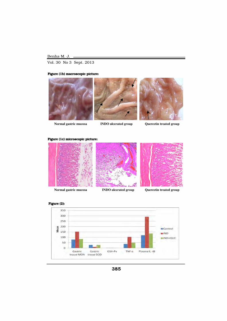
385
Benha M. J.
Vol. 30 No 3 Sept. 2013
Figure (1b) macroscopic picture:Figure (1b) macroscopic picture:
Figure (1c) microscopic picture: Figure (1c) microscopic picture:
Figure (2):Figure (2):
386386
Mona A. Said and Naglaa Y. Nafeh
DiscussionDiscussionOxygen derived free radicals
have been implicated in the patho-
genesis of a wide variety of clinical
disorders and gastric ulceration in
human and experimental animals[21].
Most of the available drugs are
thought to act on the offensive fac-
tors which neutralize acid secre-
tion like antacids, H2 receptor
blockers or interfere with acid se-
cretion as proton pump blockers[22].
Quercetin is a phenolic com-
pound widely distributed in the
plant kingdom. It is found in fre-
quently consumed foods as ap-
ples, berries, onions, tea and vege-
tables. Quercetin is reported to
have many beneficial effects on
human health including cardio-
vascular protection, anticancer ac-
tivity, antiulcer effects, antiallergic
activity, cataract prevention, antivi-
ral activity and anti-inflammatory
effects[23].
The present study was de-
signed to demonstrate gastric ul-
cer protective activity of quercetin
in rats and the possible mecha-
nisms implicated in it.
In the present study, the gastro
protective effectiveness of the
quercetin was evident from signifi-
cant reduction in the number and
mean area of the gastric ulcer in
quercetin treated rats as com-
pared with indomethacin ulcerat-
ed rats. The protective effect of
quercetin in preventing ulcer de-
velopment can be due to their pro-
tein precipitation as Quercetin
helps in precipitating microprot-
eins on the ulcer site. Therefore
forming a protective layer over the
lining mucosa thus protects the
underlying mucosa from gastric
acid secretion, toxins and other ir-
ritants[24].
Gastric acid plays an important
permissible role in NSAIDs asso-
ciated mucosal injury[25]. In our
study, there was a significant dec-
crease in gastric PH indicating in-
creased gastric acidity, and in ul-
cerated animals and gastric PH
was increased by treating the rats
with Quercetin which is a highly
desirable for gastroprotection and
antiulcer effect. This protective ef-
fect may be due to its direct inhib-
itory action on the acid producing

387
Benha M. J.
Vol. 30 No 3 Sept. 2013
cells. Quercetin also has antihis-
taminic properties preventing the
release of histamine from gastric
mast cells and inhibiting the gas-
tric H+ - K+ proton pump, dimin-
ishing acid gastric secretion. More-
over, it increases prostaglandins
E2 and I2 synthesis by gastric
mucosa. They inhibit the secretion
of gastric acid and stimulate mu-
cus and hydrophobic surfactant
like phospholipids secretion in
gastric epithelial cells [26,27].
Also it was reported that quer-
cetin exhibit ulcer healing effect in
indomethacin induced gastric ul-
cerated rats by several mechanisms
such as increasing mucous secre-
tion evidenced by higher hexosamine
and carbohydrate over protein ra-
tio, increasing mucosal resistance,
increasing mucosal blood flow and
epithelialization rates. These actions
were referred to the triterpenoid
saponins and flavonoids compo-
nents of quercetin. Quercetin in-
take also reduces cell shedding
and increase DNA content of gas-
tric mucosal cells indicating gas-
tric mucosal renewal ability[28].
In our study we tried to explore
the antioxidant effect of quercetin
in indomethacin ulcerated rats by
measuring gastric tissue content
of malondialdehyde (MDA) concen-
tration which is considered as an
indicator of lipid peroxidation
which is a well known example of
oxidative damage that affects cell
membranes, lipoproteins and oth-
er lipid containing structures un-
der conditions of oxidative stress
and gastric antioxidant enzyme
activity by measuring superoxide
dismutase (SOD) and glutathione
peroxidase (GSH-Px). Our results
showed that there was significant
increase in MDA concentration in
indomethacin ulcerated group
with reduction in the levels of an-
tioxidant enzyme (SOD, GSHPx).
We also found that pretreatment
with quercetin in indometheacin
ulcerated rats significantly de-
creased the elevated MDA and in-
creased the reduced antioxidant
enzyme activities. Our results
were agreed with other findings
that revealed that the preventive
effects of quercetin is due to inhi-
bition of lipid peroxidation by its
antioxidant nature indicating that
quercetin has an antioxidant ef-
fect[26,28,29].
NSAIDs induces gastric ulcer
388388
Mona A. Said and Naglaa Y. Nafeh
by inhibiting cyclooxygenase en-
zyme complex that convert free ei-
cosapolyenoic acids like arachi-
donic acid to cyclic endoperoxides
which is the key intermediate in
prostaglandin synthesis[29]. In an-
imal experiments, quercetin inhibits
production of inflammation producing
enzymes (cyclooxygenase (COX)[30]
and lipoxygenase (LOX)[31] and
decreasing NF-kappaB activation,
NOS (nitric oxide synthase) overex-
pression[32,33,34] and TNF-α[35,36].
So in our study we aimed to
find out the anti-inflammatory ef-
ficacy of quercetin against gastric
ulcer induced by indomethacin
that was assessed by measuring
plasma levels of TNF-α and IL-1B.
Our results showed that there was
a significant increase in both TNF-
α and IL-1B levels in the plasma
in indomethacin ulcerated group
and this was significantly de-
creased in quercetin group.
ConclusionConclusionFlavonoids as quercetin repre-
sent a highly diverse class of sec-
ondary metabolites with potential-
ly beneficial effects on human
health. These compounds protect
the gastrointestinal mucosa from
lesions produced by various ex-
perimental ulcer models and
against different necrotic agents.
Several mechanisms of action may
be involved in this protective effect
including antioxidant, anti-
secretory and anti-inflamatory ef-
fects. Quercetin may represent
an attractive therapeutic option
for preventing and healing NSAIDs
induced gastric ulcers but we rec-
ommend for further studies on its
anti-inflammatory effects against
gastric ulcer.
ReferencesReferences1. Mota K.S., Dias G.E., Pinto1. Mota K.S., Dias G.E., Pinto
M.E., Luiz-Ferreira A., Souza-M.E., Luiz-Ferreira A., Souza-
Brito A.R., Hiruma-Lima C.A.,Brito A.R., Hiruma-Lima C.A.,
Barbosa-Filho J.M. and BatistaBarbosa-Filho J.M. and Batista
L.M. (2009):L.M. (2009): Flavonoids with gas-
troprotective activity. Molecules;
14(3): 979-1012.
2. Mc Guigan J.E., Peptic ul-2. Mc Guigan J.E., Peptic ul-
cer and gastritis, in: Wilsoncer and gastritis, in: Wilson
J.D., Braunwald E., IsselbatcherJ.D., Braunwald E., Isselbatcher
K.J., Peterdorf R.G., MartinK.J., Peterdorf R.G., Martin
J.B., Fauchi A.S. and Root R.K.J.B., Fauchi A.S. and Root R.K.
(Eds.), (1991):(Eds.), (1991): Harrisons Principles
of Internal Medicine, 12th ed. Mc
Graw-Hill, New York, pp: 1229.
3. Hansen J.M., Hallas J.,3. Hansen J.M., Hallas J.,
389
Benha M. J.
Vol. 30 No 3 Sept. 2013
Lauritsen J.M. and Bytzer P.Lauritsen J.M. and Bytzer P.
(1997): (1997): Non-steroidal anti inflam-
matory drugs and ulcer complica-
tions: a factor analysis for clinical
decision making. Scand. J. Gas-
troenterol.; 31(2): 126-130.
4. Hawkey C.J. (1990):4. Hawkey C.J. (1990): Non-
steroidal anti-inflammatory drugs
and peptic ulcers. British. Med. J.;
300: 278-284.
5. Toborek M., Malecki A.,5. Toborek M., Malecki A.,
Garrido R., Mattson M.P., Hen-Garrido R., Mattson M.P., Hen-
nig B. and Young B. (1999):nig B. and Young B. (1999):
Arachidonic acid induced oxidative
injury to cultured spinal cord neu-
rons, J. Neurochem. 73: 684-692.
6. Somasundaram S., Sig-6. Somasundaram S., Sig-
thorsson G., Simpson R.J., Wattsthorsson G., Simpson R.J., Watts
J., Jacob M., Tavares I.A., RafiJ., Jacob M., Tavares I.A., Rafi
S., Roseth A., Foster R.A., PriceS., Roseth A., Foster R.A., Price
B., Wrigglesworth J.M. and Bjar-B., Wrigglesworth J.M. and Bjar-
nason I. (2000):nason I. (2000): Uncoupling of in-
testinal mitochondrial oxidative
phosphorylation an inhibition of
cyclooxygenases are required for
the development of NSAID ente-
ropathy in the rat. Aliment. Phar-
macol. Ther.; 14: 639-650.
7. Boelsterli U.A. (2002):7. Boelsterli U.A. (2002): Xen-
obiotic acyl glucuronides and acyl
CoA thioesters as protein-reactive
metabolites with the potential to
cause idiosyncratic drug reactions.
Curr. Drug Metab.; 3: 439-450.
8. Middleton E. Jr. (1998):8. Middleton E. Jr. (1998): Ef-
fect of plant flavonoids on immune
and inflammatory cell function.
Adv. Exp. Med. Biol.; 439: 175-82.
9. Gusdinar T., Herowati R.,9. Gusdinar T., Herowati R.,
Kartasasmita R.E. and AdnyanaKartasasmita R.E. and Adnyana
I.K. (2011):I.K. (2011): Anti-inflammatory and
antioxidant activity of Quercetin-3,
3', 4'-Triacetate. Journal of Pharma-
cology and Toxicology; 6: 182-188.
10. Goel R.K. and Bhatta-10. Goel R.K. and Bhatta-
charya S.K. (1991):charya S.K. (1991): Gastroduod-
enal mucosal defence and muco-
sal protective agents. Indian J.
Exp. Biol.; 29: 701-714.
11. Jainu M. and Devi C.S.11. Jainu M. and Devi C.S.
(2006):(2006): Gastroprotective action of
Cissus quadrangularis extract
against NSAID induced gastric ul-
cer: role of proinflammatory cyto-
kines and oxidative damage. Chem.
Biol. Interact.; 161: 262-270.
12. Khushtar M., Kumar V.,12. Khushtar M., Kumar V.,
Javed K. and Bhandari U.Javed K. and Bhandari U.
(2009): (2009): Protective effect of ginger

390390
Mona A. Said and Naglaa Y. Nafeh
oil on aspirin and pylorus ligation
induced gastric ulcer model in rats.
Ind. J. Pharm. Sci.; 71: 554-558.
13. Jainu M., Mohan K.V.13. Jainu M., Mohan K.V.
and Devi C.S.S. (2006):and Devi C.S.S. (2006): Gastroro-
tective effect of Cissus quadran-
gularis extract in rats with experi-
mentally induced ulcer. Indian J.
Med. Res.; 123: 799-806.
14. Wang G.Z., Huang G.P.,14. Wang G.Z., Huang G.P.,
Yin G.L., Zhou G., Guo C.J. andYin G.L., Zhou G., Guo C.J. and
Xie C.G. (2007):Xie C.G. (2007): Aspirin can elicit
the recurrence of gastric ulcer in-
duced with acetic acid in rats. Cell
Physiol. Biochem.; 20: 205-212.
15. Khan H.A. (2004):15. Khan H.A. (2004): Com-
puter-assisted visualization and
quantitation of experiment gastric
lesions in rats. J. Pharmacol. Tox-
icol. Methods; 49: 89-95.
16. Draper H.H. and Hadley M.16. Draper H.H. and Hadley M.
(1990):(1990): Malondialdehyde determi-
nation as index of lipid peroxidation.
Methods Enzymol.; 186: 421-31.
17. Lowry O.H., Rosebrough17. Lowry O.H., Rosebrough
N.J. and Randall R.J. (1951):N.J. and Randall R.J. (1951):
Protein measurement with the Fo-
lin phenol reagent. J. Biol. Chem.;
193: 265-72.
18. Aebi H. (1974):18. Aebi H. (1974): Catalase.
In Bergmeyer, H.U. textbook: Meth-
ods of enzymatic analysis. New
York, Academic Press; pp. 673-7.
19. Isa Y., Miyakawa Y., Ya-19. Isa Y., Miyakawa Y., Ya-
nagisawa M., Goto T., Kang M.S.nagisawa M., Goto T., Kang M.S.
and Kawada T. (2008):and Kawada T. (2008): 6-Shogaol
and 6-gingerol, the pungent of gin-
ger, inhibit TNF- mediated downreg-
ulation of adiponectin expression
via different mechanisms in 3T3-
L1 adipocytes. Biochem. Biophys.
Res. Comm.; 373: 429-434.
20. Odashima M., Otaka M.,20. Odashima M., Otaka M.,
Jin M., Komatsu K., Wada I. andJin M., Komatsu K., Wada I. and
Horikawa Y. (2006):Horikawa Y. (2006): Attenuation of
gastric mucosal inflammation induced
by aspirin through activation of A2A
adenosine receptor in rats. World
J. Gastroenterol.; 12: 568-573.
21. Rao Ch.V., Maiti R.N. and21. Rao Ch.V., Maiti R.N. and
Goel R.K. (1999):Goel R.K. (1999): Effect of mild ir-
ritant on gastric mucosal offensive
and defensive factors. Indian
Journal of Physiology and Phar-
macology; 44:185-191.
22. Sairam K., Rao Ch.V., Dora22. Sairam K., Rao Ch.V., Dora
Babu M. and Goel R.K. (2001):Babu M. and Goel R.K. (2001):
Prophylactic and curative effects of
Bacopa monnierain gastric ulcer
391
Benha M. J.
Vol. 30 No 3 Sept. 2013
models. Phytomedicine; 8: 423-30.
23. De-Whalley C., Rankin23. De-Whalley C., Rankin
S.M., Houct Jr.S., Jessup W.S.M., Houct Jr.S., Jessup W.
and Leake D.S. (1990):and Leake D.S. (1990): Flavo-
noids inhibit the oxidative modifi-
cation of low-density lipoproteins
by macrophages. Biochem. Phar-
macol.; 39: 1743-50.
24. Haslam E. (1996):24. Haslam E. (1996): Natural
polyphenols (vegetable tannins) as
drugs: possible modes of action. J.
Nat. Prod., 59: 205-215.
25. Scheiman J.M. (1992):25. Scheiman J.M. (1992):
Pathogenesis of gastroduodenal
injury due to nonsteroidal anti-
inflammatory drugs: implications
for prevention and therapy, Semin
Arthritis Rheum.; 21: 201-210.
26. Kahraman A., Erkasap N.,26. Kahraman A., Erkasap N.,
Koken T., Serteser M., Aktepe F.Koken T., Serteser M., Aktepe F.
and Erkasap S. (2003):and Erkasap S. (2003): The anti-
oxidative and antihistaminic prop-
erties of quercetin in ethanol-
induced gastric lesions. Toxicolo-
gy;183:133-142.
27. Shakeerabanu M., Suja-27. Shakeerabanu M., Suja-
tha K., Praveen Rajneesh C. andtha K., Praveen Rajneesh C. and
Manimaran A. (2011):Manimaran A. (2011): The defen-
sive effect of quercetin on indo-
methacin induced gastric damage
in rats. Advances in Biological Re-
search; 5(1): 64-70.
28. Martin M.J., La-Casa C.,28. Martin M.J., La-Casa C.,
Alarcon De La Lastra C., Cabeza J.,Alarcon De La Lastra C., Cabeza J.,
Villegas I. and Motilva V. (1998):Villegas I. and Motilva V. (1998):
Anti-oxidant mechanisms involved
in gastroprotective effects of quer-
cetin. Z. Naturforsch. C.J. Biosci.;
53:82-88.
29. Coskun O., Kanter M., Ar-29. Coskun O., Kanter M., Ar-
mutçu F., Çetin K., Kaybolmazmutçu F., Çetin K., Kaybolmaz
B. and Yazgan O. (2004):B. and Yazgan O. (2004): Pro-
tective effects of quercetin, a flavo-
noid antioxidant, in absolute etha-
nol induced acute gastric ulcer.
Eur. J. Gen. Med.; 1(3): 37-42.
30. Bakhle Y.S. (1983):30. Bakhle Y.S. (1983): Syn-
thesis and catabolism of cyclo-
oxygenase products. Br. Med.
Bull.; 39: 214-218.
31. Kim H.P., Mani I. and Ziboh31. Kim H.P., Mani I. and Ziboh
V.A. (1998):V.A. (1998): Effects of naturally
occurring flavonoids and bioflavonoids
on epidermal Cyclooxygenase from
guinea pigs. Prostaglandins Leukot.
Essent Fatty Acids; 58: 17-24.
32. Lee K.M., Hwang M.K.32. Lee K.M., Hwang M.K.
and Lee D.E. (2010):and Lee D.E. (2010): Protective
392392
Mona A. Said and Naglaa Y. Nafeh
effect of quercetin against arsenite
induced COX-2 expression by tar-
geting PI3K in rat liver epithelial
cells. J. Agric Food Chem.; 58:
5815-5820.
33. Comalada M., Camuesco33. Comalada M., Camuesco
D., Sierra S., Ballester I., Xaus J.,D., Sierra S., Ballester I., Xaus J.,
Galvez J. and Zarzuelo A. (2005):Galvez J. and Zarzuelo A. (2005):
In vivo quercitrin anti-inflammatory
effect involves release of quercetin,
which inhibits inflammation
through downregulation of the NF-
kappaB pathway. European J. of
Immunology; 35(2): 584-592.
34. Dias A.S., Porawski M.,34. Dias A.S., Porawski M.,
Alonso M., Marroni N., ColladoAlonso M., Marroni N., Collado
P.S. and Gonzalez-Gallego J.P.S. and Gonzalez-Gallego J.
(2005): (2005): Quercetin decreases oxi-
dative stress, NF-kappaB activa-
tion, and iNOS overexpression in
liver of streptozotocin-induced dia-
betic rats. Journal of Nutrition;
135(10): 2299-2304.
35. Ortega M.G., Saragusti35. Ortega M.G., Saragusti
A.C., Cabrera J.L. and Chia-A.C., Cabrera J.L. and Chia-
brando G.A. (2010):brando G.A. (2010): Quercetin
tetraacetyl derivative inhibits LPS-
induced Nitric oxide synthase
(iNOS) expression in J774A.1
cells. Arch. Biochem. Biophys.
2010; 498: 105-110.
36. Chuang C.C., Martinez36. Chuang C.C., Martinez
K., Xie G., Kennedy A., Bum-K., Xie G., Kennedy A., Bum-
rungpert A., Overman A., Jia W.rungpert A., Overman A., Jia W.
and McIntosh M.K. (2010): and McIntosh M.K. (2010): Quer-
cetin is equally or more effective
than resveratrol in attenuating
Tumor necrosis factor-{alpha}-
mediated inflammation and insu-
lin resistance inprimary human
adipocytes. Am. J. Clin. Nutr.; 92:
1511-1521.
393
Benha M. J.
Vol. 30 No 3 Sept. 2013
PROTECTIVE EFFECT OFPROTECTIVE EFFECT OFQUERCETIN AGAINSTQUERCETIN AGAINST
INDOMETHACIN INDUCEDINDOMETHACIN INDUCEDGASTRIC ULCER IN RATSGASTRIC ULCER IN RATS
Mona A. Said MD and Naglaa Y. Nafeh MDMona A. Said MD and Naglaa Y. Nafeh MD
BENHAMEDICALJOURNAL
REPRINT
Published byPublished by
Benha Faculty of MedicineBenha Faculty of Medicine Volume 30 Number 3Sept. 2013
393
Benha M. J.
Vol. 30 No 3 Sept. 2013
CAUDAL BUPIVACAINE, KETAMINE ANDCAUDAL BUPIVACAINE, KETAMINE ANDTHEIR COMBINATION FOR PEDIATRICTHEIR COMBINATION FOR PEDIATRIC
POSTOPERATIVE ANALGESIAPOSTOPERATIVE ANALGESIA
Gehan A. Tarbeeh MDGehan A. Tarbeeh MDDepartment of Anesthesia and Surgical Intensive Care, Faculty of Medicine,
Mansoura University, Egypt
AbstractAbstractBackground: Background: The aim of this study was to compare the analgesic efficacy
of ketamine either alone or in combination with bupivacaine for caudalblockade in pediatric surgery. Methods: Methods: After the induction of general anes-thesia without premedication sixty children, were allocated randomly into 3groups to receive single shot caudal blockade by bupivacaine 0.25% 1 ml/kg(group B, n=20), ketamine 1 mg/kg (group K, n 20), or a mixture of bupiva-caine 0.25% 1 ml/kg and preservative-free s-ketamine 0.5 mg/kg (group BKn=20). Postoperative pain was assessed using children’s and infant’s postop-erative pain scale (CHIPPS), 4-points sedation scores, analgesic require-ments and associated side effects were recorded for the first 24 hours afteroperation. Results: Results: The recovery time from general anesthesia in group-Kwas significantly longer (P>0.05). Sevoflurane concentration was significantlylower in group-K (P<0.001). The duration of absolute analgesia and the timeto first request for analgesic was significantly prolonged in Group-K. Patientsin Group-K had significantly less pain scale especially at the time to first re-quest for analgesic. The 4-points sedation score was significantly higher inGroup-K than the other two groups during the first three hours postopera-tive. Patients in Group-B were more wakeful with less sedation score andthe difference was statistically significant (P<0.05). Supplemental analgesiarequirements with intravenous paracetamol were significantly less in group-K (2 subjects). Five Patients in group-B, 3 in group-BK and only one ingroup-K experienced postoperative nausea and vomiting Postoperative uri-nary retention was noted in 10% in group-B and 5% in group-BK, while 0%in group-K, while Nystagmus occurred in two patients in group-K and one
394394
Gehan A. Tarbeeh
IntroductionIntroductionRegional anesthetic techniques
have gained considerable popular-
ity for use with pediatric patients.
The primary advantage of regional
supplementation are lowering gen-
eral anesthetic requirements in-
traoperatively and providing good
postoperative pain relief(1), Caudal
epidural analgesia is one of the
most popular and commonly per-
formed regional blocks in pediatric
anesthesia. It is a reliable and safe
technique that can be used
with.general anesthesia for intra-
¬and postoperative analgesia in
patients undergoing abdominal
and lower-limb surgery(2).
Many anesthetic agents have
been used for caudal analgesia in
pediatric patients, with lignocaine
and bupivacaine being most com-
mon(1), Bupivacaine has been in
clinical use for more than 30
years and is widely used for cau-
dal epidural analgesia in children
because of its long duration of ac-
tion and beneficial ratio of sensory
to motor blocks(3), The main dis-
advantage of caudal anesthesia is
the short duration of action after a
single injection of local anesthetic
solution. Prolongation of caudal
analgesia using a ‘single-shot’
technique has also been achieved
by the addition of various adju-
vants(4).
Ketamine is an anesthetic and
analgesic agent with a wide range
of applications in pediatric anes-
thesia(5), It exerts its effects by
binding non-competitively to a
subset of glutamate receptors
stimulated by the excitatory amine
N-methyl D-aspartate (NMDA),
blockade of which leads to a de-
crease in the activation of dorsal
horn neurons. These receptors are
located throughout the CNS as
well as ‘in the substantia gelatino-
patient in group-BK Conclusions: Conclusions: Caudal administration of ketamine alone(1 mg/kg) provided adequate postoperative analgesia of similar quality andslightly longer duration than caudal injection of 0.25% bupivacaine (1 ml/kg) with (ketamine 0.5 mg/kg), whereas it is proved to be superior from thecaudal administration of 1 ml/kg of bupivacaine 0.25% alone in pediatricsurgery without producing many side effects.
Keywords:Keywords: caudal analgesia, buvivacaine, s-ketamine.
395
Benha M. J.
Vol. 30 No 3 Sept. 2013
sa in the spinal cord and play an
important role in central pain pro-
cessing and in neural plasticity in
the spinal cord(6).
The aimThe aim of this study was to
compare the analgesic efficacy
and side effects of preservative
free ketamine either alone or in
combination with bupivacaine for
caudal blockade in pediatric pa-
tients after hypospadias surgery.
Materials and MethodsMaterials and MethodsUnpremedicated 60 male chil-
dren aged 2-5 years of ASA (Amer-
ican Society of Anesthesiologists
classification) physical status I
and II; undergoing hypospadias
surgery in Mansoura University
Hospital in plastic surgery unit,
were included in this study. Chil-
dren with history of allergic reac-
tion to bupivacaine or ketamine
and those with any contra-
indication for central neuroaxial
blockade (either absolute or rela-
tive), were excluded from the
study. After receiving informed
consent from parents, the children
were randomly allocated to receive
one of the three solutions for cau-
dal epidural injection after induc-
tion of general anesthesia.
Group-B:Group-B: were given 1 m1/kg
of 0.25% bupivacaine.
Group-K:Group-K: were given 1 mg/kg
of ketamine diluted with 0.9% sa-
line using weight-related volumes
1 ml/kg.
Group-BK:Group-BK: were given a mix-
ture of 0.25% bupivacaine and 0.5
mg/kg of ketamine at a volume of
1 ml/kg.
No, premedication was given
and all operation was performed
under general anesthesia. One
surgeon was performed all opera-
tions. IV cannula 22 guage was in-
serted in the dorsm of hand with
inhalation of oxygen and Sevoflu-
rane, tracheal intubation with
suitable size endotracheal tube.
Anesthesia was maintained using
oxygen 50% and Sevoflurane
spontaneous breathing. Intraoper-
ative Sevoflurane concentration
was adjusted to maintain ade-
quate depth of anaesthesia.
Caudal analgesia technique: Af-
ter induction of general anesthe-
sia, patients were given caudal in-
jections in left lateral position
using a 23-gauge needle. The area

396396
Gehan A. Tarbeeh
was carefully and thoroughly
cleaned with an antiseptic solu-
tion, sterile drapes were placed
around the site. The technique
was performed in the simplest way
to penetrate the sacrococcygeal
membrane by introducing a 23-
gauge hypodermic needle perpen-
dicular to it with the bevel parallel
to the long fibers of the mem-
brane. The needle was advanced
until there is loss of resistance as
it pierced the sacrococcygeal
membrane. Once the needle
crossed the membrane, it was di-
rected upwards so that it made an
angle of 20-30 degrees with the
skin. The needle was then advanced
for 2 to 3 mm so as to ensure that
the entire bevel was within the sa-
cral canal. The entire volume of
injection was made over a period
of 60 to 90 seconds and after com-
pletion of the injection the child
was placed supine after placing a
small Elastoplast dressing over
the site of the sacral hiatus.
No analgesic drugs used intra-
operative. Standard monitoring
was used during anesthesia and
surgery. The concentration of vol-
atile agent will be reduced towards
the end of surgery in order to
achieve rapid awakening before re-
turn to the recovery ward. All pa-
tients were admitted to the recov-
ery ward for at least one hour and,
when filly awake and pain free,
were returned to the ward.
Heart rate, mean arterial pres-
sure, respiratory rate and arterial
oxygen saturation were recorded
before operation and after caudal
block, these variables were record-
ed every 5 mm till the end of sur-
gery. The time from induction of
anesthesia to the end of surgery
when the anesthetic agent was
discontinued (Anesthesia dura-
tion) was recorded, and the time
from stoppage of anesthesia to
opening the eyes on calling the pa-
tient’s name or on tactile stimulus
(Recovery time) was noted. The
duration of caudal analgesia was
defined from the time of caudal in-
jection to the time the child first
complains of pain or time of first
postoperative analgesic require-
ment.
During the first 24 hours after
operation, the following variables
were recorded: heart rate, arterial
pressure, respiratory rate and ar-
terial oxygen saturation every
397
Benha M. J.
Vol. 30 No 3 Sept. 2013
hour. Sedation Was assessed us-
ing a four-point sedation score (0=
eyes open spontaneously; 1= eyes
open to speech; 2= eyes open
when shaken; 3= unrousable).
Postoperatively, a blinded Post-
Anesthesia Care Unit (PACU)
nurse assessed the quality of
analgesia with children’s and in-
fant’s postoperative pain scale
(CHIPPS) was recorded every 2
hours in the first 6 hours then
every 3 hours till 24 hours postop-
eratively(7) after recovery from an-
esthesia at rest and on movement.
Significant pain is defined as total
score above 3 and consequence
required a supplementary dose of
analgesia. Intra-venous paraceta-
mol 15 mg/kg.
The duration of absolute anal-
gesia was measured from the time
of caudal administration of the
drug to the time when the VAS
pain score was more than 0 (Abso-
lute analgesia). Number of painful
episodes, number of children re-
quiring analgesic and time to first
request for analgesic were also re-
corded. Motor block was assessed
by using a modified Bromage scale
4-points [0= flexion of knees and
feet, 1= flexion of knees, 2= little
movement of feet only, 3= no
movement of knees or feet]. The
incidence of adverse effects such
as nausea, vomiting, urinary re-
tention, nystagmus, behavior
changes (hallucination or agita-
tion), and any side effects related
to injection of ketamine or bupiva-
caine were recorded.
Data analysis: Data analysis: All statistical
analyses were carried out using SPSS
statistical package (SPSS 10.0 for
Windows). ANOVA with multiple
comparisons was used for com-
parisons between the groups. Us-
ing Chi squared test compared the
non-parametric data. P<0.05 was
regarded as statistically significant.
ResultsResultsThe demographic data in the
three groups were comparable in
age, height and weight (table 1).
There were no significant differ-
ences among groups for incidence
of hemodynamic changes (HR and
MABP) and SaO2 during the study
period. None of the children suf-
fered from hypotension or brady-
cardia and arteriel O2 saturation
(97%-100%).
398398
Gehan A. Tarbeeh
As shown in table 3 the opera-
tive data revealed that the dura-
tion of general anesthesia and the
duration of surgical procedure
were nearly similar in the three
groups, while the longest recovery
time from general anesthesia was
for Ketamine group (10.5±0.25
mm), which was significantly long-
er (P<0.05) than both Bupivacaine
and Bupivacaine with Ketamine
groups. Sevoflurane concentration
required was significantly lower in
group K (P<0.05) than other two
groups (B and BK).
According to the quality of
postoperative analgesia as shown
in table 4, both the duration of ab-
solute analgesia and the time to
first request for analgesic was sig-
nificantly prolonged in Group K
(210±30; 7.0±3.2 respectively)
compared with Group B (115±20
min; 4.20±3.1 hours respectively)
and Group BK (155±23 min;
6.50±2.11 hours respectively). Pa-
tients in Group K had significantly
less pain scale especially at the
time to first request for analgesic.
The 4-points sedation score (figure
1) was significantly higher in
Group K (0.200±0.41/4) than the
other two groups (Group B
0.02±0.22/4 and Group BK
0.150±0.36/4) during the first three
hours postoperative and more pa-
tients in Group B were more
wakeful with less sedation score
as compared with Group K and
Group BK and ~the difference was
statistically significant (P<0.05).
Supplemental analgesia re-
quirements with intravenous par-
acetamol within 24 hours postop-
erative were significantly less in
caudal Ketamine group (2 subjects
1 received once + 1 received twice)
when compared with Ketamine
plus Bupivacaine group (8 sub-
jects 5 received once, 3 received
twice), and Bupivacaine group (16
Subjects = 9 received once, 7 re-
ceived twice) (Table 5).
The incidence of postoperative
side effects was compared be-
tween the groups. Five patients in
the caudal bupivacaine group,
three patients in the caudal bupiv-
acaine-ketamine group and only
one patient in the caudal keta-
mine group experienced postoper-
ative nausea and vomiting. Post-
operative urinary retention was
noted in two patients in group B
and one patient in group BK.
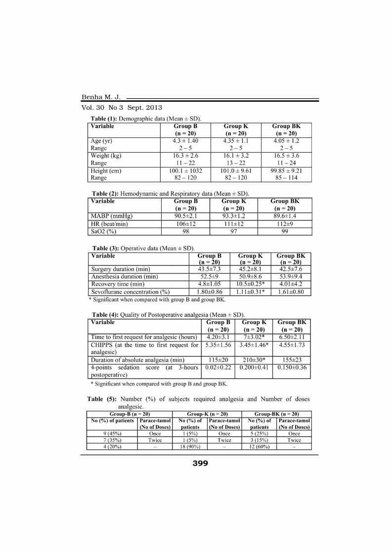
399
Benha M. J.
Vol. 30 No 3 Sept. 2013
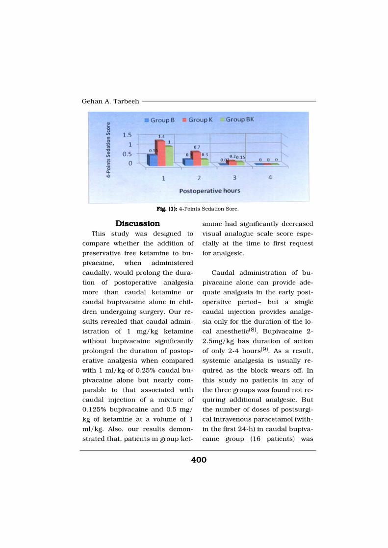
400400
Gehan A. Tarbeeh
DiscussionDiscussionThis study was designed to
compare whether the addition of
preservative free ketamine to bu-
pivacaine, when administered
caudally, would prolong the dura-
tion of postoperative analgesia
more than caudal ketamine or
caudal bupivacaine alone in chil-
dren undergoing surgery. Our re-
sults revealed that caudal admin-
istration of 1 mg/kg ketamine
without bupivacaine significantly
prolonged the duration of postop-
erative analgesia when compared
with 1 ml/kg of 0.25% caudal bu-
pivacaine alone but nearly com-
parable to that associated with
caudal injection of a mixture of
0.125% bupivacaine and 0.5 mg/
kg of ketamine at a volume of 1
ml/kg. Also, our results demon-
strated that, patients in group ket-
amine had significantly decreased
visual analogue scale score espe-
cially at the time to first request
for analgesic.
Caudal administration of bu-
pivacaine alone can provide ade-
quate analgesia in the early post-
operative period~ but a single
caudal injection provides analge-
sia only for the duration of the lo-
cal anesthetic(8). Bupivacaine 2-
2.5mg/kg has duration of action
of only 2-4 hours(9). As a result,
systemic analgesia is usually re-
quired as the block wears off. In
this study no patients in any of
the three groups was found not re-
quiring additional analgesic. But
the number of doses of postsurgi-
cal intravenous paracetamol (with-
in the first 24-h) in caudal bupiva-
caine group (16 patients) was
Fig. (1):Fig. (1): 4-Points Sedation Sore.
401
Benha M. J.
Vol. 30 No 3 Sept. 2013
significantly higher than that of
bupivacaine-ketamine group (8
patients) or that of caudal keta-
mine only 2 patients. This revealed
that caudal administration of ket-
amine (1 mg/kg) alone significant-
ly reduced the need for subse-
quent postoperative analgesia.
Oluklola and his colleagues
(2002) found that addition of 0.5
mg/kg of ketainine to caudal
0.25% plain bupivacaine signifi-
cantly decreased the need for res-
cue analgesics in the first 24 h
postoperative period in pediatric
patients undergoing elective ingui-
nal and lower abdominal opera-
tions, and revealed that caudal
administration of ketamine alone
provided analgesia of similar qual-
ity and slightly longer duration
than 0.25% bupivacaine but this
was not statistically significant(10)
Another study done by Frank We-
ber and Hinnerk Wulf (2003) and
they concluded that addition of
preservative free s- ketamine 0.5%
mg/kg to caudal bupivacaine 0.25%
1 ml/kg provides significant pro-
longation of analgesia without pro-
ducing negative side effects(11).
In our study, we increased the
dose of ketamine to 1 mg/kg,
while using it as a sole agent,
which perhaps increased the po-
tency of analgesia. One other
study using the same dose has
shown a median duration of 16.5
hours (in our study 7.05 hours)(9).
Ketamine, a phencyclidine de-
rivative, has structural similarities
to bupivacaine and has some local
anesthetic effects(12). The primary
mechanism of action is through
the blockade of N-methyl-d-
aspartate receptors situated in the
substantia gelatinosa of the spinal
cord(13). Ketamine also binds to
the opioid receptors, with a prefer-
ence for the µ receptors(14). Al-
though different doses of ketamine
(0.25%-1.0%) have been reported
in combination with local anes-
thetics to increase the duration of
analgesia, the optimal dose is
probably 0.5 mg/kg(15).
A potential advantage of central
neuraxial ketamine is that it may
counteract local anesthetic-
induced hypotension by an inhibi-
tory effect on the sympathetic
nerve activity and tends to in-
crease the respiratory rate(16). In
our study, the observed periopera-
402402
Gehan A. Tarbeeh
tive hemodynamic stability with
the use of caudal bupivacaine plus
ketamine and caudal ketamine
only supports this contention.
Kumar et al (2005) showed that
the incidence of vomiting was not
significantly different among
groups, two patients each in
group BK and B(17), but in our
study, 5 in group B, 3 in group K,
and only one in group K. Others
did not observe any negative side
effects attributable to the caudal
block of s-ketamine(11).
In this study, the patients who
received caudal epidural block
with ketamine had decreased In-
traoperative requirements for Se-
voflurane. This is in accordance
with the study of Gunduz et al
(2006) who found that Sevoflurane
concentration required was signifi-
cantly lower in group receiving
caudal ketamine(18). Also Joseph
Toblas (1996) concluded that pre-
incisional caudal epidural block
with ketamine had decreased In-
traoperative inhalational anesthet-
ic requirements during umbilical
herniorrhaphy in children(19).
In conclusion In conclusion this study dem-
onstrates that caudal administra-
tion of preservative-free ketarnine
alone (1 mg/kg) provided adequate
postoperative analgesia of similar
quality and slightly longer duration
than caudal injection of both 0.25%
bupivacaine 1 ml.kg-1 and ketarnine
0.5 mg/kg whereas it is proved to
be superior in those respects from
the caudal administration of 1 ml/
kg of bupivacaine 0.25% alone in
pediatric patients without produc-
ing many side effects.
ReferencesReferences1. Morgan G.E. and Mikhail1. Morgan G.E. and Mikhail
M.S. (1996):M.S. (1996): Pediatric Anesthesia.
In: Morgan GE, Mikhail MS, ed.
Clinical Anesthesiology. Appleton
& Lange; 726-742.
2. Dalens B. and Hasnaoui A.2. Dalens B. and Hasnaoui A.
(1989):(1989): Caudal anesthesia in pedi-
atric surgery: Success rate and
adverse effects in 750 consecutive
patients. Anesth Analg; 68: 83-9.
3. Gunter J.B., Dunn C.M.,3. Gunter J.B., Dunn C.M.,
Bennie J.B., Pentecost D.L.,Bennie J.B., Pentecost D.L.,
Bower R.J. and Tern berg J.L.Bower R.J. and Tern berg J.L.
(1991): (1991): Optimum concentration of
bupivacaine for combined caudal-
generai anesthesia in children.
Anesthesiology; 75: 57-61.
403
Benha M. J.
Vol. 30 No 3 Sept. 2013
4. Lioyd-Thomas A.R. (1990):4. Lioyd-Thomas A.R. (1990):
Pain management in paediatric
patients. Br J Anaesth; 64: 85-104.
5. Friesen R.H. and Morrison5. Friesen R.H. and Morrison
J.E. (1994):J.E. (1994): The role of ketamine
in the current practice of pediatric
anesthesia. Pediatr Anesth; 4: 79-
82.
6. Brockmeyer D.M. and6. Brockmeyer D.M. and
Kcndig J.J. (1995):Kcndig J.J. (1995): Selective ef-
fects of ketamine on amino acid
mediated pathways in neonatal
rat spinal cord. Br J Anesth; 74:
79-84.
7. Buttner W. and Finke W.7. Buttner W. and Finke W.
(2000):(2000): Analysis of behavioural
and physiological parameters for
the assessment of postoperative
analgesic demand in newborns,
infants and young children: a
comprehensive report on seven
consecutive studies. paediatric
Anaesth; 10: 303-318.
8. Quazi Siddiqui and Elahi8. Quazi Siddiqui and Elahi
Chowdhury (2006):Chowdhury (2006): Caudal Anal-
gesia In Paediatrics: A Compari-
son Between Bupivacaine And
Ketamine. The Internet Journal of
Anesthesiology. Volume 11 Num-
ber 1.
9. Cook B., Grubb D.J., Al-9. Cook B., Grubb D.J., Al-
dridge L.A. and Doyle E. (1995):dridge L.A. and Doyle E. (1995):
Comparison of the effects of
adrenaline, clonidine and keta-
mine on the duration of caudal
analgesia produced by bupiva-
caine in children. Br J Anaesth;
75: 698-701.
10. Olubukola O. Nafiu, Eni-10. Olubukola O. Nafiu, Eni-
tan O. Eelegbe and Kola A.tan O. Eelegbe and Kola A.
(2002):(2002): Kolawole, Richard A. Sal-
am Comarison efficacy of caudal
ketamine with or without bupiva-
caine in pediatric groin surgery.
Anesthesiology; 96: A 1295.
11. Frank Weher and Hinnerk11. Frank Weher and Hinnerk
Wulf (2003):Wulf (2003): Caudal bupivacaine
and s-ketamine for postoperative
analgesia in children. Pediatric
Anesthesia, 13 (3), 244-248.
12. Bräu M., Sander F., Vogel12. Bräu M., Sander F., Vogel
W. and Hempermann G. (1997):W. and Hempermann G. (1997):
Blocking mechanisms of ketamine
and its enantiomers in enzymati-
cally demyelinated peripheral
nerve as revealed by single chan-
nel experiments. Anesthesiology;
86: 394-406.
13. Smith D.J., Bouchal R.L.13. Smith D.J., Bouchal R.L.
and Desactis C.A. (1985):and Desactis C.A. (1985): Prop-
404404
Gehan A. Tarbeeh
erties of the interaction between
ketamine and opiate binding sites
in vivo and in vitro. Neuropharma-
cology; 24: 1253-60.
14. De Beer D.A.H. and Thom-14. De Beer D.A.H. and Thom-
asas M.L. (2003): M.L. (2003): Caudal additives
in children: solutions or prob-
lems? Br J Anaesth; 90: 487-98.
15. Panjabi N., Prakash S.,15. Panjabi N., Prakash S.,
Gupta P. and Gogia A.R. (2004):Gupta P. and Gogia A.R. (2004):
Efficacy of three doses of ketamine
with bupivacaine for caudal anal-
gesia in pediatric inguinal herniot-
omy. Reg Anesth Pain Med; 29:
28-31.
16. Abdulatif M. and E1-16. Abdulatif M. and E1-
Sanabary M. (2002):Sanabary M. (2002): Caudal neo-
stigmine, bupivacaine, and their
combination for postoperative
pain management after hypospa-
dias surgery in children. Anesth
Analg; 95: 1-4.
17. Kumar P., Rudra A., Pan17. Kumar P., Rudra A., Pan
A.K. and Acharya A. (200):A.K. and Acharya A. (200): Cau-
dal additives in pediatrics: a com-
parison among midazolam, keta-
mine, and neostigmine
coadministered with bupivacaine.
Anes Analg; 101: 69-73.
18. Gunduz M., Ozalevli M.,18. Gunduz M., Ozalevli M.,
Ozbek H. and Ozcengiz (2006):Ozbek H. and Ozcengiz (2006):
Comparison of caudal ketamine
with lidocaine or tramadol. Pediat-
ric Anesthesia; 6: 155-163.
19. Joseph D. Toblas (1996):19. Joseph D. Toblas (1996):
Postoperative analgesia and Intra-
operative inhalational anesthetic
requirements during umbilical hem-
iorrhaphy in children. Journal of
clinical Anesthesia; 8(8): 634-638.

405
Benha M. J.
Vol. 30 No 3 Sept. 2013
CAUDAL BUPIVACAINE, KETAMINECAUDAL BUPIVACAINE, KETAMINEAND THEIR COMBINATION FORAND THEIR COMBINATION FOR
PEDIATRIC POSTOPERATIVEPEDIATRIC POSTOPERATIVEANALGESIAANALGESIA
Gehan A. Tarbeeh MDGehan A. Tarbeeh MD
BENHAMEDICALJOURNAL
REPRINT
Published byPublished by
Benha Faculty of MedicineBenha Faculty of Medicine Volume 30 Number 3Sept. 2013
405
Benha M. J.
Vol. 30 No 3 Sept. 2013
EFFECT OF INTRAPERITONEALEFFECT OF INTRAPERITONEALINSTILLATION OF LORNOXICAM, AND BUPIV-INSTILLATION OF LORNOXICAM, AND BUPIV-
ACAINE COMBINATION ON PATIENTSACAINE COMBINATION ON PATIENTSOUTCOME AFTER LAPAROSCOPICOUTCOME AFTER LAPAROSCOPIC
CHOLECYSTECTOMYCHOLECYSTECTOMY
Ghada El-Rahmawy MD, Hosam Ghazy MDGhada El-Rahmawy MD, Hosam Ghazy MDand Amr Moawad MDand Amr Moawad MD
Department of Anesthesiology, General surgery and Chest Medicine
Faculty of Medicine, Mansoura University, Egypt
AbstractAbstractLaparoscopic Cholecystectomy(LC) is the gold stander technique for gall
bladder surgery. Aim: Aim: Evaluate the effect of intraperitoneal instillation ofLornoxicame combined with bupivacaine on postoperative pain and pulmo-nary function after laparoscopic cholecystectomy. Patients and Methods:Patients and Methods:forty four ASA 1 and 2 patients of either sex,aged between 20-50 years, un-dergoing laparoscopic cholecystectomy The patients were randomly assignedto either of the two groups: (group P) (n=22) received 25 ml of normal salineintraperitoneally or (group LB) received 20 ml of bupivacaine 0.5%combinedwith 5ml of Lornoxicame (16mg). All patients received general anesthesia.Intraoperative monitoring consisted of ECG, NIBP, end tidal CO2, pulse ox-imetry and intraabdominal pressure. Arterial blood gases (Ph, Pao2, paco2)and Spirometric values includes FEV1, FVC, FEVI/FVC were assessed. Post-operative pain was assessed by utilizing visual analogue scale (VAS) and Vis-ual Rating Prince Henry Scale Score (VRPHSS) and duration of analgesiawere recorded by determine the first request of analgesia . Results: Results: Bothgroups were comparable as regarded age, weight, height, sex, duration ofsurgery and days of hospital stay. There was insignificant differences in be-tween both groups intraoperative hemodynamic parameters and arterialblood gases preoperatively and postoperatively. There was significant in-creased in pulmonary function tests includes FEV1, FVC and FEV1/FVC ra-tio at 4 hours postoperatively in group (LB) in compared with group (P).
406406
Ghada El-Rahmawy, et al....
IntroductionIntroductionLaparoscopic Cholecystectomy
(LC) is more preferable technique
than open surgery. As LC strate-
gies is minimally invasive technique
with less tissue trauma than open
surgery, leading to less postopera-
tive pain with early home dis-
charge(1). However, prolonged hos-
pital stay was recorded in some
patient after (LC) due to pain or
postoperative pulmonary compli-
cations after laparoscopic surgery(2,3). Intraperitoneal instillation of
bupivacaine associated with good
pain relief after laparoscopic chol-
ecystectomy(4). Lornoxicam is a
new NSAID of the oxicam class with
analgesic, anti-inflammatory and
antipyretic properties(5), it has
non-selective inhibiting effect to
cyclooxygenases enzyme (COX2)(6). Lornoxicam has an analgesic
effect as morphine(6) meperidin,(7)
and tramadole.(8) but better toler-
ated than morphine when admin-
istered intravenously by patient-
controlled analgesia in the treat-
ment of moderate postoperative
pain.(9) Furthermore, The intraar-
ticular injection of ropivacaine,
morphine, and xefocam combina-
tion was superior to control or to a
combination of ropivacaine and
morphine alone in postoperative
pain control and in decreaseing
the need for opioid suggesting a
local effect.(10) Un fourintely,
there was no previous study to as-
sess the effect of intraperitoneal
analgesia on postoperative pulmo-
nary functions. Current study was
designed to assess the effect of in-
traperitoneal instillation of Lor-
noxicam combined with bupiva-
caine on postoperative pain and
Moreover, there was a highly significant increase in the analgesic require-ments in group (P) more than (LB) group. VASS was significantly decreasedin (LB) group more than (P) group. VRPHSS was significantly lower in (LB)group than group (P). There was two case of postoperative vomiting in(P)group. No recorded cases of dizziness postoperatively in both groups.Conclusion: Conclusion: Iintraperitoneal instillation of Lornoxicam in combination withbupivacaine after laparoscopic cholecystectomy had more better postopera-tive analgesic effect with less analgesic requirements and less affection ofthe pulmonary function( FEV1, FVC and FEV1/FVC ratio)
Keywords:Keywords: Laparoscopic Cholecystectomy, Lornoxicam, Intraperitonealinstillation, postoperative complications.
407
Benha M. J.
Vol. 30 No 3 Sept. 2013
pulmonary function after laparos-
copic cholecystectom.
Patients and MethodsPatients and MethodsThe current study was con-
ducted in a double blinded ran-
domized manner (closed envelope
technique) on forty four ASA 1 and
2 patients of either sex, the stud-
ied patients aged between 20-50
years, undergoing laparoscopic
cholecystectomy at Mansoura
Main University Hospital from
October 2012- to April 2013 under
general anesthesia. Informed con-
sent was taken and the study was
approved by the hospital ethics
committee. The exclusion criteria
included patients with acute chol-
ecystitis, who did not give consent
and who had any contraindica-
tions to NSAID. The patients were
randomly assigned to either of the
two groups according to the intra-
peritonal instilled solution which
prepared by the pharmacists in
coated suringes: Group (P) (n=22)
received 25 ml of normal saline or
group( LB) (n=22) received 20 ml
of bupivacaine 0.5% combined
with 5ml of Lornoxicame (Xefo)
(Necomed) (16mg) at the end of
surgery in the trendlenberg posi-
tion. The observer anesthetist and
surgeon who instilled the solu-
tions intraperitoneal were blinded
to the solution. All patients were
premedicated with diazepam 5mg
per orally at the morning of the
surgery. Induction was carried out
with 5 mg kg-1. thiopental sodium
and intubation achieved with 1.5
mg kg-1 of suxamethonium using
suitable size of endotracheal tube.
Intraoperative muscle relaxation
was achieved by atracuroium 0.05
mg kg-1. The anesthesia was
maintained by 02, intermittent
doses of atracroium, whereas in-
traoperative analgesia was achieved
with morphine sulphate 0.15 mg
kg-1.Ventilation was adjusted to
maintain end tidal C02 between
35-40 mmHg, whereas intra ab-
dominal pressure was maintained
between 10-12mmHg. The muscle
relaxation was reversed at the end
of surgery with neostigmine 0.05
mg kg-1 and atropine 0.025 mg
kg-1. Intraoperative monitoring
consisted of ECG, NIBP, end tidal
CO2, pulse oximetry and intraab-
dominal pressure. Either the study
or placebo solutions was sprayed
on the upper surface of the liver
and on right subdiaphragmatic
space, to allow it to diffuse into
the hepatodiaphragmatic space,
408408
Ghada El-Rahmawy, et al....
near and above the hepatoduod-
enal ligament and above gall blad-
der by the surgeon. This was done
using a catheter inserted into the
subcostal trocar under direct la-
paroscopic control. Arterial blood
gases (Ph, Pao2, paco2) are as-
sessed preoperatively, 2hours,12
hours and 24 hours postoperative-
ly. Spirometric values includes
FEV1, FVC, FEVI/FVC are record-
ed by (Smart PFT, Medical Equip-
ment Europe (MEE Spirometry)
Gmbh and vitalograph Copd-6 TM)
preoperatively, 4 hours and 24
hours postoperative. Postoperative
pain was assessed at immediate, 4
hours, 12hours and 24 hours by
utilizing visual analogue scale
(VAS), Visual rating Prince Henry
scale (VRS)(10) for shoulder pain
assessment The VAS consisted of
a 10 cm scale representing vary-
ing intensity of pain from 0 cm (no
pain) to 10 cm (worst imaginable
pain). The visual rating Prince
Henry pain scale(10) consisted of
0-4 grades with 0. No. pain no
cough, 1-pain on cough but not
on deep breathing 2-pain on deep
breathing but not on rest, 3-pain
on rest slight and 4-pain on rest-
severe. Duration of analgesia were
recorded by determine the first re-
quest of analgesia. Rescue analge-
sic consisted of intra muscular Di-
clofenac 75 mg at VAS more than
6 and VRS more than3. Number of
patient request Rescue analgesic
consisted were recorded. Dizziness
and postoperative nausea and
vomiting also were assessed
Statistical analysis:Statistical analysis:
Sample size was calculated by
using t test for mean in G *power
3.1 (Faul, Erdfelder, Lang, and
Buchner (2007) in Germany) pro-
gram. According to pilot study (5
patients in each group) we calcu-
lated that 19 patients per group
were give p<0.05 significant with
confidence interval of 95% with a
actual power of 95% when mean
value of FEV1/FVC ratio at the 4
hours postoperative in group(P)
was 0.74 and in group (LB) was
0.79. we added 3 cases for each
group to ensure more accuracy of
the statistical results.
Statistical analysis was carried
out using the Statistical Package
for Social Sciences 16 (SPSS Inc.,
Chicago, IL, USA). Data was pre-
sented as number, percentage,
means and standard deviations.
Parametric data were analyzed us-
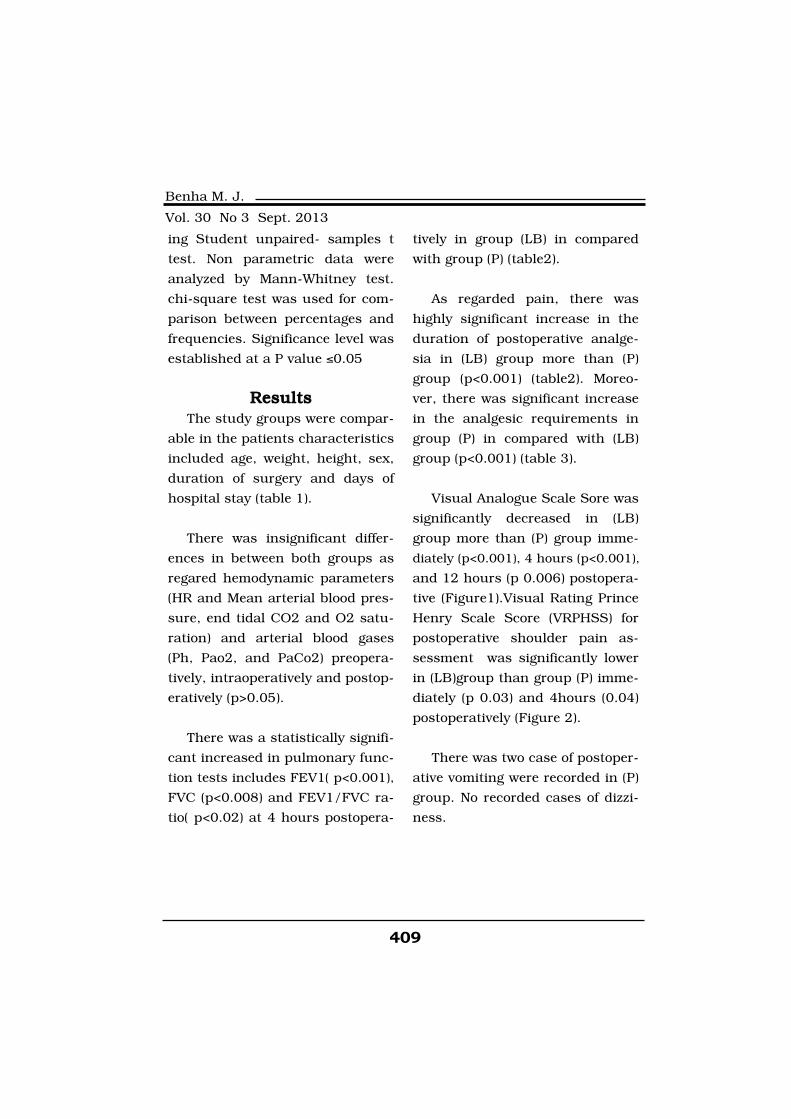
409
Benha M. J.
Vol. 30 No 3 Sept. 2013
ing Student unpaired- samples t
test. Non parametric data were
analyzed by Mann-Whitney test.
chi-square test was used for com-
parison between percentages and
frequencies. Significance level was
established at a P value ≤0.05
ResultsResultsThe study groups were compar-
able in the patients characteristics
included age, weight, height, sex,
duration of surgery and days of
hospital stay (table 1).
There was insignificant differ-
ences in between both groups as
regared hemodynamic parameters
(HR and Mean arterial blood pres-
sure, end tidal CO2 and O2 satu-
ration) and arterial blood gases
(Ph, Pao2, and PaCo2) preopera-
tively, intraoperatively and postop-
eratively (p>0.05).
There was a statistically signifi-
cant increased in pulmonary func-
tion tests includes FEV1( p<0.001),
FVC (p<0.008) and FEV1/FVC ra-
tio( p<0.02) at 4 hours postopera-
tively in group (LB) in compared
with group (P) (table2).
As regarded pain, there was
highly significant increase in the
duration of postoperative analge-
sia in (LB) group more than (P)
group (p<0.001) (table2). Moreo-
ver, there was significant increase
in the analgesic requirements in
group (P) in compared with (LB)
group (p<0.001) (table 3).
Visual Analogue Scale Sore was
significantly decreased in (LB)
group more than (P) group imme-
diately (p<0.001), 4 hours (p<0.001),
and 12 hours (p 0.006) postopera-
tive (Figure1).Visual Rating Prince
Henry Scale Score (VRPHSS) for
postoperative shoulder pain as-
sessment was significantly lower
in (LB)group than group (P) imme-
diately (p 0.03) and 4hours (0.04)
postoperatively (Figure 2).
There was two case of postoper-
ative vomiting were recorded in (P)
group. No recorded cases of dizzi-
ness.
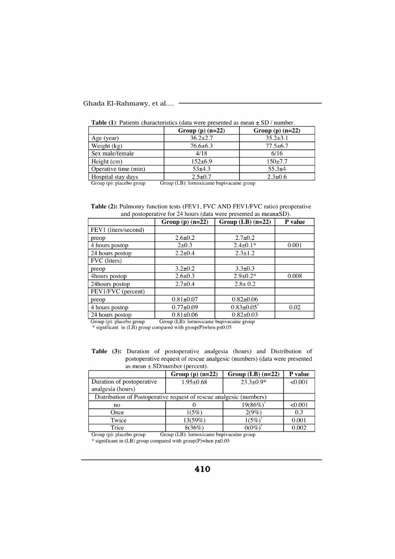
410410
Ghada El-Rahmawy, et al....
411
Benha M. J.
Vol. 30 No 3 Sept. 2013
DiscussionDiscussionPostoperative pain and pulmo-
nary complications are the major
causes of morbidity after laparos-
copic Cholecystectomy (LC)(11).
Previous studies reported that the
postoperative pain and diaphrag-
matic dysfunction were considered
the major contributing factors of
postoperative pulmonary compli-
cations(2,12). The possible cause
of postoperative shoulder pain af-
Fig.1:Fig.1: Post operative visual analogue scale score (Data expressed as Mean (SD).Group (p): placebo group Group (LB): lornoxicame bupivacaine group * significant in (LB) group compared with group(P) when p≤ 0.05.
Fig.2:Fig.2: Postoperative visual rating Prince Henry pain scale score(VRPHSS)(Data expressedas Mean (SD). Group (p): placebo group Group (LB): lornoxicame bupivacaine group * Significant in (LB) group compared with group (P) when p≤0.05
412412
Ghada El-Rahmawy, et al....
ter (LC) is the diaphragmatic irri-
tation by insufflated CO2. Carbon
dioxide may be transformed to ir-
ritative carbon dioxide by combi-
nation with the fluid in the perito-
neal cavity(13). Another possible
cause of shoulder pain after LC may
be due to overstretching of the di-
aphragmatic muscle fibers and
phrenic nerve neuropraxia a owing
to high rate of insufflations(14). In
present study shoulder pain was
low in all treatment groups due to
carful removal of residual intra-
peritoneal CO2 by the surgeon.
Moreover postoperative pulmonary
complications may be attributed
to manipulation and local stimula-
tion of the gallbladder and its bed
during laparoscopic Cholecystec-
tomy which may stimulate reflex
inhibition of the diaphragm as
shown in animal study(12).
Present study recorded that
there was significant increase in
the duration of postoperative anal-
gesia, reduction of the pain scores
and analgesic requirements with
significant improvements of the
pulmonary functions including
FEV1, FVC and FEV1/FVC post-
operatively in the group that re-
ceive intra peritoneal instillation
of (NSAID) lornoxicame combined
with bupivacaine than that receive
intra peritoneal instillation of sa-
line.
In agreement with current
study Memedov et al(15) found
that intraperitoneal instillation
and port site infiltration of ropiva-
caine and lornoxicam during la-
paroscopic cholecystectomy reduc-
es the postoperative pain.
Lornoxicam has been success-
fully used in prevention and treat-
mentof postoperative pain in pa-
tients undergoing laparoscopic
gynecological surgeries(16). Sen et
al(17) reported that lornoxicam
and lidocaine combination during
intravenous regional anesthesia
reduce sensory and motor block
onset times, prolongs sensory and
motor block recovery times, re-
duce tourniquet pain and increase
duration of analgesia with de-
creasing total amount of analgesic
requirements.
Lornoxicam is not a local anes-
thetic agent but it has a COX2 in-
hibitor effect leading to reduction
of secretion the of the pain media-
tors in the areas of surgical ma-
413
Benha M. J.
Vol. 30 No 3 Sept. 2013
nipulation and besides its analge-
sic effect, in addition to local anti-
nociceptive effect(6). NSAIDs may
induce predominant peripheral
antinociceptive effect by prevent
conduction of C Fibres and open-
ing of the K+ channels located in
the primary afferent nerve end-
ings(18). Also, Lornoxicam have a
peripheral analgesic effect via ac-
tiviation of NO-c GMP pathway
and the opening of K+ channels(17). Surprisingly, Lornoxicam has
antioxidative effects in rats de-
crease the dose of analgesics and
prevent the negative impact of re-
active oxygen species on nocicep-
tion(19). In correlation to present
study results of the effect of pain
management on the pulmonary
function, Spence and Smith(20)
documented that the continuous
extradural nerve block in patients
undergoing vagotomy with gas-
troentrostomy or pyloroplasty had
less effect on the postoperative
pulmonary function (FEV1/FVC
ratio) and arterial oxygenation
than intravenous morphine as the
result of better pain control.
The conclusion of current
study is the intraperitoneal instil-
lation of Lornoxicam combined-
with bupivacaine after laparoscop-
ic cholecystectomy had more better
postoperative analgesic effect with
less analgesic requirements and
less affection of the pulmonary
function (FEV1, FVC and FEV1/
FVC ratio).
ReferencesReferences1. Sharma A., Hayden J.D.,1. Sharma A., Hayden J.D.,
Reese R.A., Sedman P.C., Roys-Reese R.A., Sedman P.C., Roys-
ton C.M.S. and O'Boyle C.J.ton C.M.S. and O'Boyle C.J.
(2004): (2004): Prospective comparison of
ambulatory with inpatient lapar-
oscopic cholecystectomy: out-
come, patient preference and sat-
isfaction. J. of Ambulatory
Surgery April.1-4.
2. Joris J., Cigarini I., Le-2. Joris J., Cigarini I., Le-
grand M., Jacquet N., De Grootegrand M., Jacquet N., De Groote
D, Franchimont P., et al. (1992):D, Franchimont P., et al. (1992):
Metabolic and respiratory changes
after cholecystectomy performed
via laparotomy or laparoscopy. Br
J Anaesth; 69: 341-57.
3. Rademaker B.M., Ringers3. Rademaker B.M., Ringers
J., Odoom J.A., de Wit L.T.,J., Odoom J.A., de Wit L.T.,
Kalkman C.J. and Oosting J.Kalkman C.J. and Oosting J.
(1992):(1992): Pulmonary function and
stress response after laparoscopic
cholecystectomy: comparison with
subcostal incision and influence of
414414
Ghada El-Rahmawy, et al....
thoracic epidural analgesia.
Anesth Analg. Sep; 75(3): 381-5.
4. Bhardwaj N., Sharma V.4. Bhardwaj N., Sharma V.
and Chari P. (2002):and Chari P. (2002): Intraperito-
neal instillation for postoperative
pain relief after laparoscopic chol-
ecystectomy. Indian J. Anaesth.;
46 (1): 49-52.
5. Hein A., Norlander C.,5. Hein A., Norlander C.,
Blom L. and Jakobsson J.Blom L. and Jakobsson J.
(2001): I(2001): Is pain prophylaxis in mi-
nor gynaecological surgery of clini-
cal value? A double-blind placebo
controlled study of paracetamol 1
g versus lornoxicam 8 mg given
orally. Ambul Surg; 9: 91-94.
6. Norholt S.E., Sindet-6. Norholt S.E., Sindet-
Pedersen S., Larsen U., et al.Pedersen S., Larsen U., et al.
(1996):(1996): Pain control after dental
surgery: a double-blind, random-
ised trial of lornoxicam versus
morphine. Pain; 67: 335-343.
7. Rosenow D.E., VanKrieken7. Rosenow D.E., VanKrieken
F., Stolke D., et al. (1996):F., Stolke D., et al. (1996): Intra-
venous administration of lornoxi-
cam, a new NSAID, and pethidine
for postoperative pain - a placebo
- controlled pilot study. Clin Drug
Invest; 11: 11-19.
8. Staunstrup H., Ovesen J.,8. Staunstrup H., Ovesen J.,
Larsen U.T., et al. (1999): Larsen U.T., et al. (1999): Effica-
cy and tolerability of lornoxicam ver-
sus tramadol in postoperative pain.
J Clin Pharmacol; 39: 834-841.
9. Rosenow D.E., Albrechtsen9. Rosenow D.E., Albrechtsen
M. and Stolke D. (1998): M. and Stolke D. (1998): A com-
parison of patient controlled anal-
gesia with lornoxicarn versus mor-
phine in patients undergoing
lumber disk surgery. Anesth
Analg; 86: 1045-50.
10. Jiranantarat V., Rusha-10. Jiranantarat V., Rusha-
tamukayanunt W., Lert-akyamaneetamukayanunt W., Lert-akyamanee
N., Sirijearanai R., Piromrat I.,N., Sirijearanai R., Piromrat I.,
Suwannanonda P. and Muang-Suwannanonda P. and Muang-
kasem J. (2002):kasem J. (2002): Analgesic effect
of intraperitoneal instillation of bu-
pivacaine for postoperative laparos-
copic cholecystectomy. J Med Assoc
Thai. Sep; 85 Suppl 3: S897-903.
11. Hendolin H.I., Paakonen11. Hendolin H.I., Paakonen
M.E., Alhava E.M., Tarvainen R.,M.E., Alhava E.M., Tarvainen R.,
Kemppinen T. and Lahtinen P.Kemppinen T. and Lahtinen P.
(2000): (2000): Laparoscopic or open
cholecystectomy: a prospective
randomised trial to compare post-
operarative pain, pulmonary func-
tion, and stress response. Eur J
Surg; 166: 394-9.
415
Benha M. J.
Vol. 30 No 3 Sept. 2013
12. Karayiannakis A.J., Makri12. Karayiannakis A.J., Makri
G.G., Mantzioka A., Karousos D.G.G., Mantzioka A., Karousos D.
and Karatzas (1996): and Karatzas (1996): Postopera-
tive pulmonary function after la-
paroscopic and open cholecystectomy.
Br. J. Anaesth. Oct; 77(4): 448-52.
13. Tsimoyiannis E.C., Glant-13. Tsimoyiannis E.C., Glant-
zounis G., Lekkas E.T., Siakas P.,zounis G., Lekkas E.T., Siakas P.,
Jabarin M. and Tzourou H. (1998):Jabarin M. and Tzourou H. (1998):
Intraperitoneal normal saline and
bupivacaine infusion for reduction
of postoperative pain after lapar-
oscopic cholecystectomy. Surg
Laparosc Endosc. Dec; 8(6): 416-20.
14. Berberoglu M., Dilek14. Berberoglu M., Dilek
O.N., Ercan F., Kati I. and Oz-O.N., Ercan F., Kati I. and Oz-
men M. (1998):men M. (1998): The effect of CO2
insufflation rate on the postlapar-
oscopic shoulder pain. - J Lapa-
roendosc Adv Surg Tech A. Oct; 8
(5): 273-7.
15. MEMEDOV C., MENTES15. MEMEDOV C., MENTES
Ö, SIMSEKA, KEÇE C., YAGCIÖ, SIMSEKA, KEÇE C., YAGCI
G., HARLAK A., COSAR A. andG., HARLAK A., COSAR A. and
TUFAN T. (2010):TUFAN T. (2010): Comparison of
Analgesic Effects of Intraperitoneal
Lornoxicam and Ropivacaine Ad-
ministration in Laparoscopic Chol-
ecystectomy. Balkan Med J, Volume
27, Number 2, Page(s) 142-149.
16. Helvacioglu A. and Weis16. Helvacioglu A. and Weis
R. (1992):R. (1992): Operative laparoscopy
and postoperative pain relief. Fer-
til Steril; 57: 548-52.
17. Sen S., Ugur B., Aydın17. Sen S., Ugur B., Aydın
O.N., Ogurlu M., Gezer E. andO.N., Ogurlu M., Gezer E. and
Savk O. (2006): Savk O. (2006): The analgesic ef-
fect of lornoxicam when added to
lidocaine for intravenous regional
anaesthesia. British Journal of
Anaesthesia 97 (3): 408-13.
18. Deciga-Campos M. and18. Deciga-Campos M. and
Lopez Munoz F.J. (2004): Lopez Munoz F.J. (2004): Partici-
pation of the L-arginine-nitric ox-
idecyclic GMP-ATP-sensitive K2+
channel cascade in the antinoci-
ceptive effect of rofecoxib. Eur J
Pharmacol; 484: 193-9.
19. Rokyta R., Holecek V., Pe-19. Rokyta R., Holecek V., Pe-
karkova I., et al. (2003):karkova I., et al. (2003): Free
radicals after painful stimulation
are influenced by antioxidants
and analgesics. Neuro Endocrinol
Lett; 24: 304-9.
20. Spence A.A. and SMITH20. Spence A.A. and SMITH
G. (1998):G. (1998): Postoperative analgesia
and lung function: A comparison
of morphine With extradural block
Br. J. Anesth; 81: 984-988.
416416
Ghada El-Rahmawy, et al....
EFFECT OF INTRAPERITONEALEFFECT OF INTRAPERITONEALINSTILLATION OF LORNOXICAM,INSTILLATION OF LORNOXICAM,
AND BUPIVACAINE COMBINATIONAND BUPIVACAINE COMBINATIONON PATIENTS OUTCOME AFTERON PATIENTS OUTCOME AFTER
LAPAROSCOPIC CHOLECYSTECTOMYLAPAROSCOPIC CHOLECYSTECTOMY
Ghada El Rahmawy MD, Hosam Ghazy MDGhada El Rahmawy MD, Hosam Ghazy MDand Amr Moawad MDand Amr Moawad MD
BENHAMEDICALJOURNAL
REPRINT
Published byPublished by
Benha Faculty of MedicineBenha Faculty of Medicine Volume 30 Number 3Sept. 2013
417
Benha M. J.
Vol. 30 No 3 Sept. 2013
COLOR DOPPLER ECHOCARDIOGRAPHICCOLOR DOPPLER ECHOCARDIOGRAPHICCHANGES 2 YEARS AFTER 2D PLANNEDCHANGES 2 YEARS AFTER 2D PLANNEDRADIATION THERAPY FOR LEFT BREASTRADIATION THERAPY FOR LEFT BREAST
CANCER, ITS RELATION TO CARDIACCANCER, ITS RELATION TO CARDIACBIOMARKERSBIOMARKERS
Mona M. Halim MD and Shaheer K. George MDMona M. Halim MD and Shaheer K. George MDDepartments of Clinical Oncology and Nuclear Medicine & Internal Medicine
(Cardiovascular Unit), Mansoura University
AbstractAbstractIntroduction:Introduction: Radiation treatment has been associated with radiation in-
duced cardiotoxicity, especially with older, long-outdated, techniques. Suchcomplications include pericarditis, myocardial fibrosis, valvular injury, is-chemic heart disease, and myocardial infarction. Some of these complica-tions may be associated with changes in cardiac biomarkers.
Aim of the study: Aim of the study: The aim of this study is to assess the effect of 2Dplanned radiation therapy (RT) after 6 months and 2 years on cardiac func-tion using color Doppler echocardiography as well as to assess the relationof these changes to cardiac biomarkers in order to early detect and treatthese changes in the proper time.
Patients and methods:Patients and methods: 80 women under 65 years of age with stage IIIleft breast cancer who received postoperative radiotherapy were includedduring the time from January 2009 to January 2010. Each patient weresubjected to thorough history taking with special concentration upon cardio-vascular symptomatology, full cardiac examination, color Doppler echocardi-ographic examination as well as assessment of serum high sensitivity C-reactive protein (hs-CRP), cardiac troponin (cTn) and brain natriuretic poly-peptide (BNP) before radiation therapy (T1), 6 months after RT (T2) and 2years after radiation therapy (T3) for stage III left breast cancer patients inMansoura University Hospital, Egypt.
Results:Results: There was no significant change in color Doppler echocardio-graphic parameters after 6 months of radiation therapy, however, there was
418418
Mona M. Halim and Shaheer K. George
IntroductionIntroductionRadiation therapy (RT) in pa-
tients with breast cancer signifi-
cantly reduces locoregional recur-
rence in up to 60% of cases(1). The
most recent systematic overview,
conducted by the Early Breast
Cancer Trialists Collaborative
Group (EBCTCG), indicated that
post-mastectomy RT reduces ab-
solute breast cancer mortality by
an average of 5% at 10 years(2).
The relative benefits are similar af-
ter-mastectomy or breast conser-
vation surgery in the presence or
absence of adjuvant systemic
therapies as well as in axillary
node-negative and -positive pa-
tients.
However, RT has been associat-
ed with radiation-induced cardio-
toxicity, especially with older,
long-outdated techniques(1,3,4).
Such complications include peri-
carditis, myocardial fibrosis, val-
vular injury, ischemic heart dis-
ease, and myocardial infarction,
and have been observed particu-
larly in the literature on photon
decrease in L.V. ejection fraction at 2 years but the result was statisticallyinsignificant (P=0.095). Serum levels of hs-CRP, cTn and BNP were normalat baseline. There was no significant change in levels of cTn and BNP duringthe 2 years follow-up. However, serum level of hs-CRP was normal at 6month and significantly increased at 2 years follow-up (P=0.003) which wasassociated with insignificant decrease in left ventricular ejection fraction(LVEF).
Conclusion: Conclusion: There is limited value of color Doppler echocardiographic ex-amination in prediction of cardiac toxicity after RT for stage III left breastcancer except for LVEF assessment after 2 years which had insignificant val-ue. There was statistically significant role of hs-CRP in prediction of cardiactoxicity after RT for left breast cancer.
Recommendations: Recommendations: We recommend future studies including larger num-bers of patients, different stages of breast cancer, different radiation thera-peutic modalities as well as different tools for assessment of cardiac functionand prediction of cardiac toxicity of RT.
Keywords:Keywords: Echocardiography, Left breast cancer and cardiac biomark-ers.
419
Benha M. J.
Vol. 30 No 3 Sept. 2013
radiation of post-mastectomy
chest wall and direct internal
mammary lymph nodes(5,6).
At present, there is a controver-
sy as to whether modern RT tech-
niques are also cardiotoxic, espe-
cially in cases of left breast
cancer. Gustaysson et al.(7) found
that women younger than 5o
years at the time of adjuvant RT
following mastectomy for early
breast cancer had no serious car-
diac sequelae 13 years on. On the
contrary, Paszat et al.(8) suggested
that adjuvant RT for left-sided
breast cancer diagnosed in women
younger than 6o is associated
with a higher risk of fatal myocar-
dial infarction 10 to 15 years later
compared with adjuvant RT for
right-sided cases. Moreover, Har-
ris et al.(9) proposed that irradia-
tion to the left breast is not asso-
ciated with a higher risk of cardiac
death up to 20 years after treat-
ment, but is associated with an
increased rate of diagnoses of cor-
onary artery disease and myocar-
dial infarction compared with
right breast treatment.
Cardiac biomarkers are protein
molecules that are widely used in
the early detection of heart failure.
Serum levels of cardiac troponin
(cTn), C-reactive protein (hs-CRP)
and brain natriuretic polypeptide
(BNP) have been shown to be sen-
sitive markers for left ventricular
dysfunction and powerful markers
for morbidity and mortality in the
heart failure setting. All these bio-
markers have been evaluated in
prediction of early cardiac dys-
function after different chemother-
apeutic agents. However, their
ability to detect early cardiac dys-
function after radiation therapy
for stage III left breast cancer is
not still obvious(10,5,11).
The aim of this study is to ass-
es the color Doppler echocardio-
graphic changes after 6 months and
after 2 years of 2D planned radiation
therapy for women with stage III left
breast cancer and to assess the
relation to serum levels of some
cardiac biomarkers as hsCRP, car-
diac troponin (cTn) and brain nat-
riuretic polypeptide (BNP) in order
to evaluate the role of these bio-
markers for early prediction of ra-
diation induced cardiac toxicity.
Patients and MethodsPatients and MethodsEighty women under 65 years

420420
Mona M. Halim and Shaheer K. George
of age with stage III left breast
cancer who received postoperative
radiation therapy in Clinical On-
cology and Nuclear Medicine De-
partment, Mansoura University
Hospital, Egypt, were included
during the time from January
2009 to January 2010. Patients
with previous cardiovascular dis-
ease, thyroid disorder, significant
anemia with serum hemoglobin
<8.0 gm/dl, previous ECG abnor-
malities and previous echocardio-
graphic abnormalities were ex-
cluded from the study. 15 patients
(18.75%) were hypertensive and
10 patients (12.5%) were diabetic
type II. Each patient was subject-
ed to thorough history taking with
special concentration upon cardio-
vascular symptomatology, full
clinical cardiac examination, color
Doppler echocardiographic exami-
nation as well as assessment of
serum cardiac biomarkers (serum
hs-CRP, cardiac troponin cTn and
BNP) before start of radiation ther-
apy (T1), 6 months after radiation
therapy (T2) and 2 years after ra-
diation therapy (T3).
Color Doppler echocardiograph-
ic examination was performed us-
ing Vivid-5 ultrasound (GE-vingmed
ultrasound AS, Horten Norway)
with 2.5-3.5 MHz transducer in
left lateral decubitus position. Par-
asternal and apical projections
were obtained according to the
recommendations of American So-
ciety of Echocardiography(12).
Standard two-dimensional
echocardiographic evaluation for
left and right ventricular size and
function was performed. Left and
right ventricular diameters as well
as left atrial diameter were meas-
ured from a parasternal long-axis
view by M-mode examination at
the speed of 50 mm/s(13). Left
ventricular ejection fraction (EF)
was measured from the apical
four chamber view using biplane
Simpson's method(12).
hs-CRP, cTn, and BNP were
evaluated before, 6 months after
and 2 years after postoperative ra-
diation therapy. Quantitative de-
terminations of cTn levels were
performed with a third-generation
Roche Elecsys assay (Roche Diag-
nostics, Inc., Indianapolis, India-
na). The CRP levels were meas-
ured with the Immage 800
(Beckman Coulter, Brea, Califor-
nia) antigen-antibody precipitant
421
Benha M. J.
Vol. 30 No 3 Sept. 2013
rate reaction. The N-terminal pro-
BNP levels were measured with an
electrochemiluminescence sand-
wich immunoassay (Elecsys
ProBNP, Roche Diagnostics) with
the Roche 2010 system.
As regards radiotherapy tech-
nique, accurate patient-specific
anatomic information of the breast
is a prerequisite for planning and
implementing the delivery of radi-
ation to the entire breast while
minimising exposure to critical
structures such as lungs, ribs and
heart. The image data from the
patients' breasts were taken by a
diagnostic multi-slice CT scanner
with a flat couch, which could be
restrictive for setting up patients
with immobilization devices in the
treatment position. During RT all
patients were placed supine with
left hand up and attached to im-
mobilization devices in precise re-
producible position of treatment.
The anterior and lateral isometric
lines and other landmarks were
marked on the patient's skin by
radio-opaque markers or lead-
beads.
From all diagnostic CT slices,
the central one was chosen as the
one in where the disease was most
extensive. Thereafter, we delineat-
ed the target area as well as the
organs at risk, and these data
were input to a 2-dimensional (2D)
computer treatment planning. The
entire breast was included in the
planning target volume (PTV) with
a 1 cm margin around palpable
breast tissue. The physician and
the radiotherapist could then se-
lect the treatment beam direc-
tions. The objective was to treat
the PTV disease tissue plus a 1
cm margin to a tumoricidal dose
while limiting the dose to the sur-
rounding normal tissues. If critical
tissues were located nearby, the
aim was to keep the dose to these
organs to a level within the ac-
ceptable limit of complication. The
reference point (100% dose) was
located in the centre of the PTV
and in the junction of the axes of
the tangential fields. The dose
variation permitted inside the PTV
was between -5% and +l0%. In or-
der to further adjust doses and to
prevent radiation of sensitive
healthy tissues such as lungs and
pericardium, axes with a deviation
larger than 180° as well as wedges
of different angles (15°, 30°, 45°,
60°) were-employed.
422422
Mona M. Halim and Shaheer K. George
Radiotherapy was delivered with
a LA linear accelerator treatment
unit using a pair of opposed tan-
gential beams (medial and lateral).
The daily dose was 200 cGy, the
total dose on the PTV was 50 Gy
in 25 fractions over 5 weeks with-
out boost, while in some patients
a boost dose of 5 to 6 Gy was given
as necessary. None underwent RT
to the internal mammary chain.
Statistical analysis:Statistical analysis:
Data, analysis was performed
by using Statistical Package for
Social Sciences (SPSS) version
11.5 software (SPSS Inc., Chicago.
IL. USA). For the continuous vari-
ables, parametric test conditions
were tested. Descriptive statistics
were shown as mean ± standard
deviation or median (maximum-
minimum) where appropriate. While
the mean differences between
measurement times were com-
pared by repeated measures of
ANOVA. Degrees of association be-
tween continuous variables were
calculated by Spearman's correla-
tion analysis. A P=value <0.05 was
considered statistically significant.
ResultsResultsFiteen patients were hyperten-
sive (18.75%) and 10 patients were
having type II diabetes (12.5%) be-
fore start of radiation therapy (RT).
No new patients developed hyper-
tension nor diabetes during the 2
years follow-up. Mean age of pa-
tients was 47±8 years. Mean BMI
was 26±5. No history of smoking
and only 3 patients (3.75%) have
family history of CAD.
Table I summarizes echocardio-
graphic data of the studied pa-
tients. No significant changes in
echocardiographic parameters
were detected after 6 months and
after 2 years of RT apart from mild
decrease in LVEF after 2 years
and the results were statistically
insignificant (P=0.095).
Table II summarizes serum se-
rial levels of cardiac biomarkers in
the studied patients before start of
RT (T1), 6 months after RT (T2)
and 2 years after RT (T3). This ta-
ble showed that there was no sig-
nificant changes regarding serum
levels of cardiac troponin and BNP
after 6 months and after 2 years
of RT. However, there was in-
crease in hs-CRP after 2 years and
the result was statistically signifi-
cant (P=0.003).
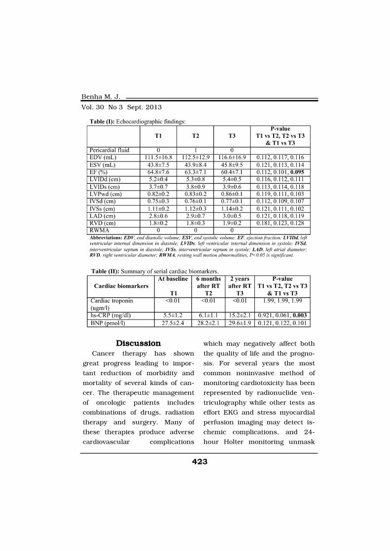
423
Benha M. J.
Vol. 30 No 3 Sept. 2013
DiscussionDiscussionCancer therapy has shown
great progress leading to impor-
tant reduction of morbidity and
mortality of several kinds of can-
cer. The therapeutic management
of oncologic patients includes
combinations of drugs, radiation
therapy and surgery. Many of
these therapies produce adverse
cardiovascular complications
which may negatively affect both
the quality of life and the progno-
sis. For several years the most
common noninvasive method of
monitoring cardiotoxicity has been
represented by radionuclide ven-
triculography while other tests as
effort EKG and stress myocardial
perfusion imaging may detect is-
chemic complications, and 24-
hour Holter monitoring unmask
424424
Mona M. Halim and Shaheer K. George
suspected arrhythmias(10). Also
biomarkers such as troponin I and
T and B-type natriuretic peptide
may be useful in early detection of
cardiotoxicity. The widely used
non-invasive method of monitor-
ing cardiotoxicity of cancer thera-
py is, however, represented by
Doppler-echocardiography which
allows to identify the main forms
of cardiac complications of cancer
therapy: left ventricular (systolic
and diastolic) dysfunction, valvu-
lar heart disease, pericarditis, per-
icardial effusion and carotid artery
lesions(17).
A meta-analysis by Cuzick(14)
showed a 62% increase in cardiac
death in women receiving RT.
Similarly, EBCTCG found a 30%
increase in vascular mortality in
women receiving RT for breast
cancer(15). Some authors reported
no increased risk for patients with
left breast disease treated with
techniques used approximately
since 1975(16,11), while others
claimed a 2-fold risk of fatal myo-
cardial infarction for left-sided
treatment compared with right-
sided(8).
A lot of data are available about
the cardiotoxic effect of different
chemotherapeutic agents. Howev-
er, the data about the cardiotoxic
effect of radiation therapy (RT) is
lacking.
Our study was designed to
evaluate the cardiotoxic effects of
postoperative radiation therapy at
6 months and 2 years in patients
with stage III left breast cancer
and its relation to serum levels of
some cardiac biomarkers as hs-
CRP, cTn and BNP that can allow
us for early detection and predic-
tion of cardiac toxic effects of RT.
Reported data from studies on
the comprehensive examination of
the long-term cardiac mortality
and morbidity after left breast ir-
radiation using contemporary RT
techniques indicate a significant
association with an increased inci-
dence of coronary artery disease
and myocardial infarction 20
years after RT treatment. Howev-
er, our study shows non signifi-
cant reduction of LVEF 2 years af-
ter RT. Furthermore, none of the
patients developed myocardial in-
farction or coronary artery dis-
ease. All other echocardiographic
parameters remained practically
425
Benha M. J.
Vol. 30 No 3 Sept. 2013
unchanged, showing that RT did
not affect them. Pericardial fluid
was present in only 1 case at 6
months after RT, an effect that
was temporary.
These results are contradictory
with those in most other publica-
tions. In the most recent popula-
tion-based case-cohort study(10),
the authors concluded that in ad-
dition to risk factors such as in-
creasing age at diagnosis, smok-
ing history, and history of acute
myocardial infarction before post-
operative RT, anatomic character-
istics of RT such as RT for left-
sided tumors, the use of an anteri-
or internal mammary field, and in-
creased area of an anterior left
breast boost field are associated
with increased risk of acute myo-
cardial infarction(10). The discrep-
ancy could most likely be ex-
plained by the small total number
of participants, the short follow-
up period, the absence of internal
mammary irradiation, and, espe-
cially, the younger mean age of
patients at the time of diagnosis.
At baseline, cardiac biomarkers
including hs-CRP, cTn and BNP
were within normal limits for the
entire patient population studied.
No changes in serum level of bio-
markers after 6 months. However,
there was increase in serum level
of hs-CRP after 2 years and the re-
sult was statistically significant
(P=0.003). Our data goes in contrast
with the data reported by Nazanin
et al.(17) who studied the utility of
cardiac biomarkers in predicting
early L.V. dysfunction in patients
with Human Epidermal Growth Factor
Receptor II breast cancer treated
with Trastuzumab therapy with or
without RT who reported no change
in serum biomarkers in one year
follow-up of their patients(17). The
difference in results may be attrib-
uted to different number of patients,
different ages included, different
geographic patient characteristics
as well as longer follow-up dura-
tion in our study.
In conclusion, color Doppler
echocardiography could be valuable
for detection of pre-clinical cardiac
toxicity in patients with stage III
left breast cancer after RT. Howev-
er, follow-up of serum hs-CRP lev-
els may be of more significant val-
ue before patients can go through
symptomatic cardiac toxicity and
HF. We recommend future studies

426426
Mona M. Halim and Shaheer K. George
with larger number of patients,
different patient characteristics,
different RT technique and doses
and different serum cardiac bio-
markers to be conducted to help
cardiologists and radiotherapists
for early detection and prediction
of cardiac toxicity following RT in
left breast cancer patients.
ReferencesReferences1. Gyenes G. (1998): 1. Gyenes G. (1998): Radia-
tion-induced ischemic heart dis-
ease in breast cancer. A review.
Acta Oncol.; 37: 241-6.
2. EBCTCG Early Breast Can-2. EBCTCG Early Breast Can-
cer Trialists Collaborative Groupcer Trialists Collaborative Group
Favourable and unfavourable ef-Favourable and unfavourable ef-
fects on long-term survival offects on long-term survival of
Radiotherapy for early breastRadiotherapy for early breast
cancer (2000):cancer (2000): an overview of the
randomised trials. Lancet.; 355:
1757-70.
3. Carlson R.G., Mayfield3. Carlson R.G., Mayfield
W.R., Norman S., et al. (1991):W.R., Norman S., et al. (1991):
Radiation-associated valvular dis-
ease. Chest.; 99: 538.
4. Cutuli B. (2000): 4. Cutuli B. (2000): Cancer of
the breast: results and toxicity of
locoregional irradiation after mas-
tectomy. Cancer Radiother.; 4:
167s-179s.
5. Rutqvist L.E., Lax I., For-5. Rutqvist L.E., Lax I., For-
nander T., et al. (1992): nander T., et al. (1992): Cardio-
vascular mortality in a random-
ized trial of adjuvant radiation
therapy versus surgery alone in
primary breast cancer. Int J Radi-
at Oncol Biol Phys.; 22: 887.
6. Gyenes G., Fornander T.,6. Gyenes G., Fornander T.,
Carlens P., et al. (1994):Carlens P., et al. (1994): Morbidi-
ty of ischaemic heart disease in
early breast cancer 15-20 years
after adjuvant radiotherapy. Int J
Radiat Oncol Biol Phys.; 28: 1235.
7. Gustaysson A., Bendahl7. Gustaysson A., Bendahl
P.O., Cwildel M., et al. (1999):P.O., Cwildel M., et al. (1999):
No serious late cardiac effects af-
ter adjuvant radiotherapy follow-
ing mastectomy in premenopausal
women with early breast cancer.
Int J Radiat Oncol Biol Phys.; 43:
745-54.
8. Paszat L.F., Mackillop8. Paszat L.F., Mackillop
W.J., Groome P.A., et al. (1998):W.J., Groome P.A., et al. (1998):
Mortality from myocardial infarc-
tion after adjuvant radiotherapy
for breast cancer in the surveil-
lance, epidemiology, and end-
results cancer registries. J Clin
Oncol.; 16: 2625-31.
427
Benha M. J.
Vol. 30 No 3 Sept. 2013
9. Harris E.E., Correa C.,9. Harris E.E., Correa C.,
Hwang W.T., et al. (2006): Hwang W.T., et al. (2006): Late
cardiac mortality and morbidity in
early-stage breast cancer patients
after breast-conservation treat-
ment. J Clin Oncol.; 24: 4100-6.
10. Paszat L.F., Vallis K.A.,10. Paszat L.F., Vallis K.A.,
Benk V.M.A., et al. (2007):Benk V.M.A., et al. (2007): A
population-based case cohort-
study of the risk of myocardial in-
farction following radiation thera-
py for breast cancer. Radiother
Oncol.; 82: 294-300.
11. Rutqvist L.E., Liedberg A.,11. Rutqvist L.E., Liedberg A.,
Hammar N., et al. (1998):Hammar N., et al. (1998): Myo-
cardial infraction among women
with early-stage breast cancer
treated with conservative surgery
and breast irradiation. Int J Radi-
at Oncol Biol Phys.; 40: 359-63.
12. Schiller N.B., Shan P.M.,12. Schiller N.B., Shan P.M.,
Crawford M., DeMaria A., Dever-Crawford M., DeMaria A., Dever-
eux R., Feigenbaum H., et al.eux R., Feigenbaum H., et al.
(1989):(1989): Recommendations for
quantitation of the left ventricle by
two-dimensional echocardiogra-
phy, American Society of Echocar-
diography Committee on Stan-
dards, Subcommittee on
Quantitation of Two-Dimensional
Echocardiograms. J Am Soc Echo-
cardiogr; 2: 358-67.
13. Quinones M.A., Otto13. Quinones M.A., Otto
C.M., Studdard Y., Waggoner A.C.M., Studdard Y., Waggoner A.
and Zoghbi W.A. (2002): and Zoghbi W.A. (2002): Dapple,
Quantification Task Force of the
Nomenclature and Standards
Committee of the American Socie-
ty of Echocardiography, Recom-
mendations for quantification of
Doppler echocardiography. A re-
port from the Doppler Quantifica-
tion Task Force of the Nomencla-
ture and Standards Committee of
the American Society of Echocar-
diography J Am Soc Echocardiogr;
15: 167-84.
14. Cuzick J. (2005): 14. Cuzick J. (2005): Radio-
therapy for breast cancer. J Natl
Cancer Inst.; 97: 406-7.
15. Clarke M., Collings R.,15. Clarke M., Collings R.,
Darby S., et al. (2005): Darby S., et al. (2005): Early
Breast Cancer Trialists Collabora-
tive Group (EBCTCG). Effects of
radiotherapy and of differences in
the extent of surgery for early
breast cancer on local recurrence
and 15-year survival: an overview
of the randomised trials. Lancet.;
366: 2087-106.
16. Hojris I., Overgaard M.,16. Hojris I., Overgaard M.,

428428
Mona M. Halim and Shaheer K. George
Christensen J.J., et al. (1999):Christensen J.J., et al. (1999):
Morbidity and mortality of is-
chaemic heart disease in high-risk
breast-cancer patients after adju-
vant postmastectomy systemic
treatment with or without radio-
therapy: analysis of DBCG 82b
and 82c randomized trials. Radio-
therapy Committee of the Danish
Breast Cancer Cooperative Group.
Lancet.; 354: 1425-30.
17. Nazanin F.A., Jonasan R.,17. Nazanin F.A., Jonasan R.,
Anthoni W., et al. (2011):Anthoni W., et al. (2011): Utility
of cardiac biomarkers, tissue ve-
locity, strain imaging and cardiac
magnetic resonance imaging in
predicting early left ventricular
dysfunction in patients with hu-
man epidermal growth factor posi-
tive breast cancer in patients
treated with adjuvant Trustuzumab
therapy. Jacc Journal 57: 1-13.
429
Benha M. J.
Vol. 30 No 3 Sept. 2013
COLOR DOPPLERCOLOR DOPPLERECHOCARDIOGRAPHIC CHANGES 2ECHOCARDIOGRAPHIC CHANGES 2
YEARS AFTER 2D PLANNEDYEARS AFTER 2D PLANNEDRADIATION THERAPY FOR LEFTRADIATION THERAPY FOR LEFT
BREAST CANCER, ITS RELATION TOBREAST CANCER, ITS RELATION TOCARDIAC BIOMARKERSCARDIAC BIOMARKERS
Mona M. Halim MD and Shaheer K. George MDMona M. Halim MD and Shaheer K. George MD
BENHAMEDICALJOURNAL
REPRINT
Published byPublished by
Benha Faculty of MedicineBenha Faculty of Medicine Volume 30 Number 3Sept. 2013
429
Benha M. J.
Vol. 30 No 3 Sept. 2013
METRONOMIC LOW DOSE CARBOPLATIN METRONOMIC LOW DOSE CARBOPLATIN AFTER RADIOTHERAPY IN STAGE III AFTER RADIOTHERAPY IN STAGE III
TESTICULAR SEMINOMATESTICULAR SEMINOMA
Mona M. Halim MD and Nazzem Shams MD*Mona M. Halim MD and Nazzem Shams MD*Departments of Clinical Oncology and Nuclear Medicine & Surgical Oncology*,
Mansoura University.
AbstractAbstractBackground: Background: Testicular germ cell tumor (TGCT) is the most common
cancer in men between age of 15 and 35 years, it represents 5% of urologictunmors. Extended field radiotherapy is a standard of care for stage II testic-ular seminoma, while stage III seminoma, neoadjuvant 3-4 cycles of Cispla-tin based chemotherapy followed by radiotherapy of residue (if residue ismore than 3 cms) but if residue less than 3 cm follow up by PET CT every 6months was recommended.
Patient and methods:Patient and methods: 51 patients with testicular seminoma stage IIIaand IIIb were treated between May 2009 to May 2010 with orchidectomy fol-lowed by 3 to 4 cycles of Carboplatin AUC-7 then by radiotherapy on resi-due, radiation dose ranged from 30-35 Gy. All patients received metronomiclow dose Carboplatin (not exceed 150 mg) weekly for 12 weeks after 3 weeksfrom end of radiotherapy.
Results:Results: After follow-up of 36 months with 74% of patients having beenfollowed for >1.5 years. There have been no metastatic nor local relapses(95% CI of relapse free survival of 93%). Toxicity has been low with grade 3toxicity limited to four patients with grade 3 haematological toxicity with (noclinical sequelae) and one patient with grade 3 nausea (during radiotherapy).No patients experienced grade 4 toxicity.
Conclusions:Conclusions: The results of this study suggest that a metronomic lowdose of Carboplatin after radiotherapy may reduce relapse rate comparedwith those who received radiotherapy alone and this approach is proposedfor further investigation.
Keywords:Keywords: seminoma, Carboplatin, radiotherapy
430430
Mona M. Halim and Nazzem Shams
IntroductionIntroductionTraditionally, stage II testicular
seminoma has been treated with
radiotherapy alone with an ex-
tended field including both para-
aortic and ipsilateral pelvic lymph
nodes. With this policy, there is a
relapse risk of 5%-11%[1,2].
Though these recurrences can
then be usually treated success-
fully with combination chemother-
apy[1], there is a concern over
long-term complications from the
intensity of their treatment, such
as cardiovascular disease or sec-
ond cancers[3]. A single dose of
Carboplatin (Hospira, UK) can re-
duce recurrence in stage I semino-
ma[4], and in-field recurrence is
rare after radiotherapy. While, in
stage III testicular seminoma, new
adjuvant 3-4 cycles of Cisplatin
based chemotherapy followed by
radiotherapy of residual lesion if
residual lesion is more than 3cm,
but if residue is smaller than 3
cm, follow up by PET CT every 6
months is recommended. We hy-
pothesized that a similar reduc-
tion in metastatic relapse could be
achieved in stage III seminoma if
metronomic low dose of Carbopla-
tin after radiotherapy was taken.
We used a metronomic low dose of
Carboplatin to achieve a blood
concentration x time of Carbopla-
tin (150 mg) weekly. This report is
on the 51 patients of testicular
seminoma treated in this way.
The initial strategy was to reduce
the risk of recurrence and thus
there is a need for salvage therapies.
Subsequently, we also reduce the
risk of metastatic relapse. The hy-
pothesis was that the addition of
metronomic Carboplatin would be
worthwhile to achieve these goals.
MethodsMethods51 patients with testicular sem-
inoma stage IIIa and IIIb were col-
lected from Clinical Oncology and
Nuclear Medicine Department and
Surgical Oncology Department, On-
cology Center, Mansoura Universi-
ty in period between May 2009 to
May 2010. All patients had a diag-
nosis of classical seminoma of the
testis. Staging was by sequential
analyses of blood levels of tumour
markers alphafetoprotein, and beta
human chorionic gonadotrophin
(HCG), and by computed tomogra-
phy (CT) scan of the thorax, abdo-
men and pelvis and classified by
the maximum axial diameter us-
ing The Royal Marsden Hospital

431
Benha M. J.
Vol. 30 No 3 Sept. 2013
Classification such that stage III=
infradiaphragmatic and supradi-
aphragmatic lymph nodes, which
classified into IIIa abdominal nodes
<2 cm, IIIb abdominal nodes 2-5 cm
and IIIc abdominal nodes >5 cm.
This analysis represents a ret-
rospective audit of the standard
treatment practice in our institu-
tion. Eligibility was generally that
men had stage III seminoma with
node metastasis up to 5 cm in di-
ameter. If node involvement was
equivocal, the scan was repeated
4-6 weeks later or was repeated
with the associated PET analysis
of fluorodeoxyglucose uptake
(PET-CT scan). Ultimately classifi-
cation as stage III disease was de-
termined by collective judgement
at a radiological nodes, supported
in equivocal cases by repeat CT
(n=5) or PET scans (n=5) or in one
case by excision biopsy. Once
classified as stage III, the number
of nodes >1 cm in diameter was
recorded as 'involved'. Orchidecto-
my was done then three to four
cycles of Carboplatin was admin-
istered via a 1h infusion at a dose
to achieve an area under the con-
centration x time curve of 7 mg.
min/ml (dose (mg) = (GFR + 25) x
7, where GFR = glomerular filtration
rate measured, by EDTA clearance).
Oral antiemetics were prescribed
before chemotherapy and during ra-
diotherapy. Radiotherapy was ini-
tiated 3-6 weeks following Carbop-
latin cycles, patients were treated
with anterior and posterior parallel-
opposed portals shaped on virtual
simulator CT planning. Initially, the
fields encompassed both para-aortic
and ipsilateral pelvic lymph nodes
(dogleg field) extending superiorly
to the lower border of the D10 ver-
tebral body and inferiorly to the
obturator fossa. This field was
treated to a dose of 30-35 Gy in 15
fractions following which the para
aortic nodes (D10-L5) were boost-
ed by a further 5 Gy in 3 fractions
for 12 cases while involved field
was used for 39 patients. The fol-
low-up was a 36 months. All pa-
tients received metronomic low
dose of Carboplatin (total 150 mg
weekly for 12 weeks) started three
weeks after end of radiotherapy.
Toxicity was recorded qualita-
tively and classified according to
its grade. Following treatment, pa-
tients were followed and confi-
dence intervals for relapse risks
were calculated[5,6].
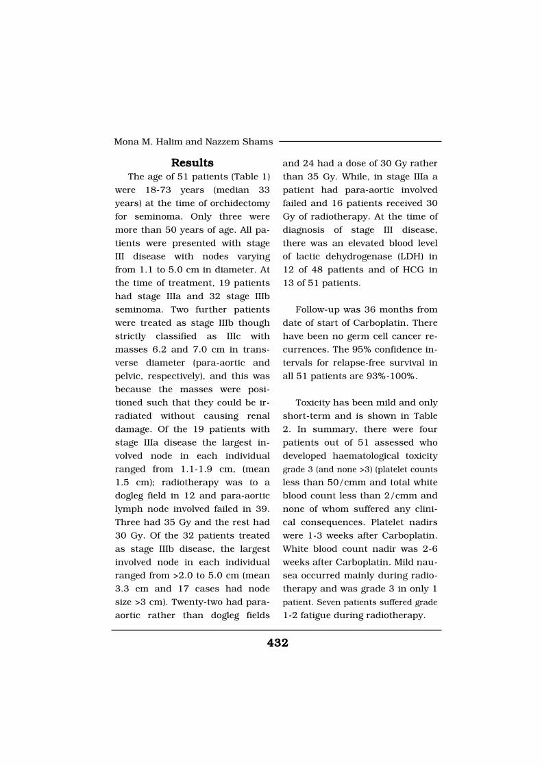
432432
Mona M. Halim and Nazzem Shams
ResultsResultsThe age of 51 patients (Table 1)
were 18-73 years (median 33
years) at the time of orchidectomy
for seminoma. Only three were
more than 50 years of age. All pa-
tients were presented with stage
III disease with nodes varying
from 1.1 to 5.0 cm in diameter. At
the time of treatment, 19 patients
had stage IIIa and 32 stage IIIb
seminoma. Two further patients
were treated as stage IIIb though
strictly classified as IIIc with
masses 6.2 and 7.0 cm in trans-
verse diameter (para-aortic and
pelvic, respectively), and this was
because the masses were posi-
tioned such that they could be ir-
radiated without causing renal
damage. Of the 19 patients with
stage IIIa disease the largest in-
volved node in each individual
ranged from 1.1-1.9 cm, (mean
1.5 cm); radiotherapy was to a
dogleg field in 12 and para-aortic
lymph node involved failed in 39.
Three had 35 Gy and the rest had
30 Gy. Of the 32 patients treated
as stage IIIb disease, the largest
involved node in each individual
ranged from >2.0 to 5.0 cm (mean
3.3 cm and 17 cases had node
size >3 cm). Twenty-two had para-
aortic rather than dogleg fields
and 24 had a dose of 30 Gy rather
than 35 Gy. While, in stage IIIa a
patient had para-aortic involved
failed and 16 patients received 30
Gy of radiotherapy. At the time of
diagnosis of stage III disease,
there was an elevated blood level
of lactic dehydrogenase (LDH) in
12 of 48 patients and of HCG in
13 of 51 patients.
Follow-up was 36 months from
date of start of Carboplatin. There
have been no germ cell cancer re-
currences. The 95% confidence in-
tervals for relapse-free survival in
all 51 patients are 93%-100%.
Toxicity has been mild and only
short-term and is shown in Table
2. In summary, there were four
patients out of 51 assessed who
developed haematological toxicity
grade 3 (and none >3) (platelet counts
less than 50/cmm and total white
blood count less than 2/cmm and
none of whom suffered any clini-
cal consequences. Platelet nadirs
were 1-3 weeks after Carboplatin.
White blood count nadir was 2-6
weeks after Carboplatin. Mild nau-
sea occurred mainly during radio-
therapy and was grade 3 in only 1
patient. Seven patients suffered grade
1-2 fatigue during radiotherapy.
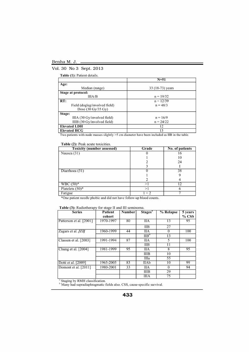
433
Benha M. J.
Vol. 30 No 3 Sept. 2013
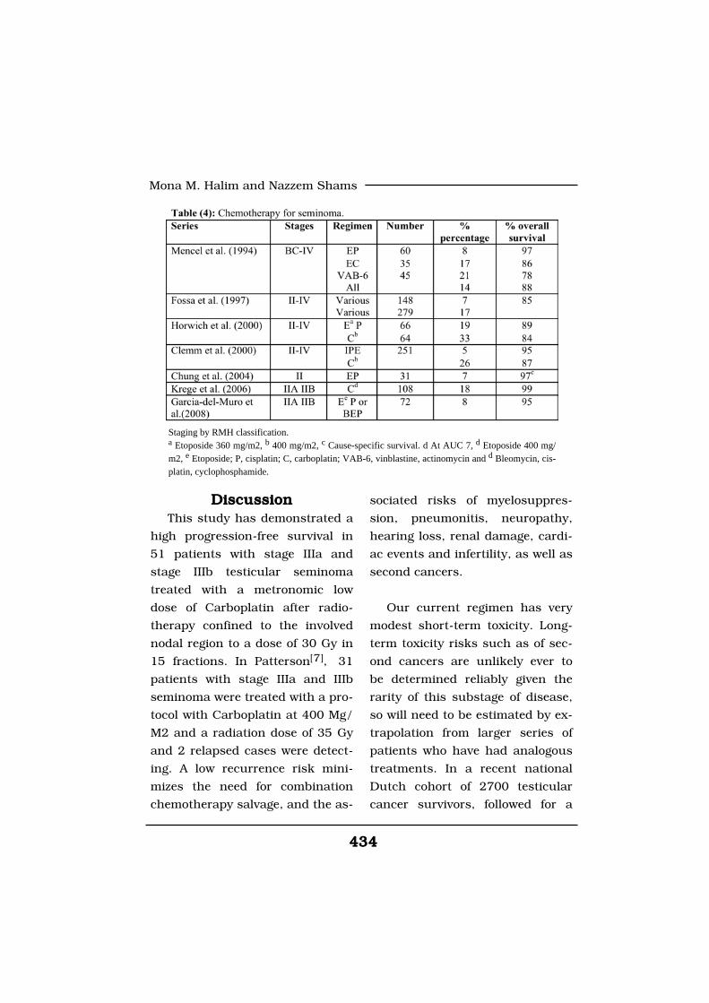
434434
Mona M. Halim and Nazzem Shams
DiscussionDiscussionThis study has demonstrated a
high progression-free survival in
51 patients with stage IIIa and
stage IIIb testicular seminoma
treated with a metronomic low
dose of Carboplatin after radio-
therapy confined to the involved
nodal region to a dose of 30 Gy in
15 fractions. In Patterson[7], 31
patients with stage IIIa and IIIb
seminoma were treated with a pro-
tocol with Carboplatin at 400 Mg/
M2 and a radiation dose of 35 Gy
and 2 relapsed cases were detect-
ing. A low recurrence risk mini-
mizes the need for combination
chemotherapy salvage, and the as-
sociated risks of myelosuppres-
sion, pneumonitis, neuropathy,
hearing loss, renal damage, cardi-
ac events and infertility, as well as
second cancers.
Our current regimen has very
modest short-term toxicity. Long-
term toxicity risks such as of sec-
ond cancers are unlikely ever to
be determined reliably given the
rarity of this substage of disease,
so will need to be estimated by ex-
trapolation from larger series of
patients who have had analogous
treatments. In a recent national
Dutch cohort of 2700 testicular
cancer survivors, followed for a
Staging by RMH classification.a Etoposide 360 mg/m2, b 400 mg/m2, c Cause-specific survival. d At AUC 7, d Etoposide 400 mg/m2, e Etoposide; P, cisplatin; C, carboplatin; VAB-6, vinblastine, actinomycin and d Bleomycin, cis-platin, cyclophosphamide.
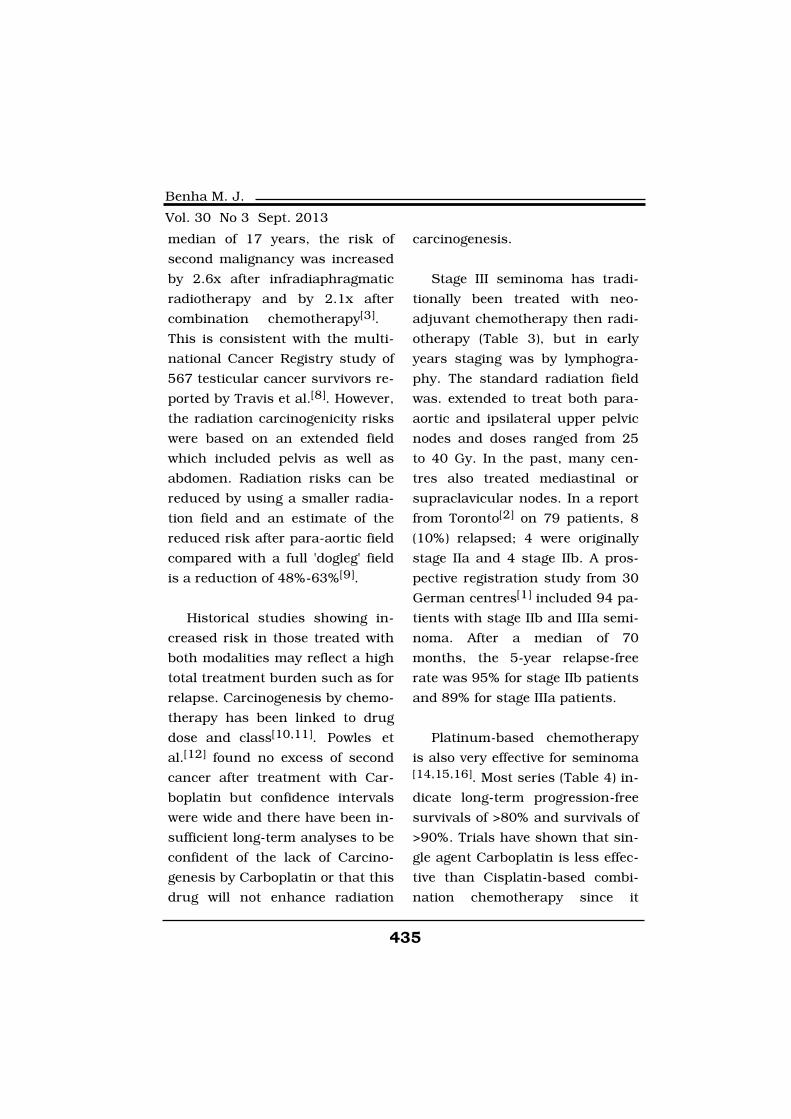
435
Benha M. J.
Vol. 30 No 3 Sept. 2013
median of 17 years, the risk of
second malignancy was increased
by 2.6x after infradiaphragmatic
radiotherapy and by 2.1x after
combination chemotherapy[3].
This is consistent with the multi-
national Cancer Registry study of
567 testicular cancer survivors re-
ported by Travis et al.[8]. However,
the radiation carcinogenicity risks
were based on an extended field
which included pelvis as well as
abdomen. Radiation risks can be
reduced by using a smaller radia-
tion field and an estimate of the
reduced risk after para-aortic field
compared with a full 'dogleg' field
is a reduction of 48%-63%[9].
Historical studies showing in-
creased risk in those treated with
both modalities may reflect a high
total treatment burden such as for
relapse. Carcinogenesis by chemo-
therapy has been linked to drug
dose and class[10,11]. Powles et
al.[12] found no excess of second
cancer after treatment with Car-
boplatin but confidence intervals
were wide and there have been in-
sufficient long-term analyses to be
confident of the lack of Carcino-
genesis by Carboplatin or that this
drug will not enhance radiation
carcinogenesis.
Stage III seminoma has tradi-
tionally been treated with neo-
adjuvant chemotherapy then radi-
otherapy (Table 3), but in early
years staging was by lymphogra-
phy. The standard radiation field
was. extended to treat both para-
aortic and ipsilateral upper pelvic
nodes and doses ranged from 25
to 40 Gy. In the past, many cen-
tres also treated mediastinal or
supraclavicular nodes. In a report
from Toronto[2] on 79 patients, 8
(10%) relapsed; 4 were originally
stage IIa and 4 stage IIb. A pros-
pective registration study from 30
German centres[1] included 94 pa-
tients with stage IIb and IIIa semi-
noma. After a median of 70
months, the 5-year relapse-free
rate was 95% for stage IIb patients
and 89% for stage IIIa patients.
Platinum-based chemotherapy
is also very effective for seminoma[14,15,16]. Most series (Table 4) in-
dicate long-term progression-free
survivals of >80% and survivals of
>90%. Trials have shown that sin-
gle agent Carboplatin is less effec-
tive than Cisplatin-based combi-
nation chemotherapy since it
436436
Mona M. Halim and Nazzem Shams
achieved only an —70% disease-
free survival[17,6]. The results of
Carboplatin in 108 patients with
low-volume stage II showed 80%
DFS[17]. In contrast, a trial of
more standard EP chemotherapy
in 72 patients showed a 90% 5-
year PFS[18].
Some centres have adopted a
selective approach employing radi-
otherapy for stage IIIa disease and
recommending chemotherapy for
those with more bulky nodes. For
example, a retrospective analysis
of 106 cases from Florence[1] in-
cluded 89 treated with radiothera-
py and the 5-year relapse-free sur-
vival was 94% for IIIa and 72.5%
for IIIb. However, the radiotherapy
included pelvis, abdomen, medias-
tinum and supraclavicular fossa
in 42 patients. The Institute Gus-
tave Roussy treated most stage IIa
and IIB patients with radiotherapy
and most stage IIc and IIIa with
chemotherapy[19].
A number of treatment guide-
line publications have addressed
management of stage II-III semino-
ma[20,21]. The European Associa-
tion of Urology guidelines[16] rec-
ommend that radiotherapy is the
standard treatment with para-
aortic and ipsilateral iliac nodes to
be treated to a dose of 30 Gy for
IIa and 36 Gy for IIb stages, while
neo-adjuvant 4 cycles of Cisplatin
based chemotherapy, followed by
radiotherapy of residual lesion
more than 3 cm in greatest dimen-
sion is advised.
ConclusionConclusionThe case for recommending our
approach for stage IIIa and IIIb
seminoma is not only the evidence
for efficacy, but also the expecta-
tion that long-term toxicity risks
will be low. Carboplatin is a low
toxicity drug with high level of ac-
tivity against seminoma[22,23] and
is used with a low total dose.
Though the radiotherapy compo-
nent has both short-term gas-
trointestinal toxicity and a long-
term risk of carcinogenesis, the
risk should be higher with more
extended radiation fields and with
higher radiation doses. Also, the
approach may allow in the future
evaluation of even further reduc-
tion of the extent of radiotherapy
to less than a standard para-
aortic field if post-radiotherapy
low dose of Carboplatin is used. A
recent node mapping study[24,25]
437
Benha M. J.
Vol. 30 No 3 Sept. 2013
has for example demonstrated the
rarity of node involvement above
the renal vessels and if this region
was not included there would be
less irradiation of stomach and
pancreas. The results presented
here demonstrate that our regi-
men is effective and has modest
toxicity so is worthy of additional
evaluation and development.
ReferencesReferences1. Classen J., Schmidberger1. Classen J., Schmidberger
H., Meisner C., et al. (2003):H., Meisner C., et al. (2003): Ra-
diotherapy for stages IIA/B testic-
ular seminoma: final report of a
prospective multicenter clinical
trial. J Clin Oncol; 21(6): 1101-
1106.
2. Chung P.W., Gospodarow-2. Chung P.W., Gospodarow-
icz M.K., Panzarella T., et al.icz M.K., Panzarella T., et al.
(2004): (2004): Stage II testicular semino-
ma: patterns of recurrence and
outcome of treatment. Eur Urol;
45(6): 754-759; discussion 759-
60.
3. Mencel P.J., Motzer R.J.,3. Mencel P.J., Motzer R.J.,
Mazumdar M., et al. (1994): Mazumdar M., et al. (1994): Ad-
vanced seminoma: treatment re-
sults, survival, and prognostic fac-
tors in 142 patients. J Clin Oncol;
12(1): 120-126.
4. Clemm C., Bokemeyer C.,4. Clemm C., Bokemeyer C.,
Geri A., et al. (2000):Geri A., et al. (2000): Random-
ized trial comparing Cisplatin/
Etoposide/Ifosfamide with Car-
boplatin monochemo-therapy in
patients with advanced metastatic
seminoma. Proc Am Soc Clin On-
col; 19: 328. (abstract 1283).
5. Garcia-del-Mum X., Maroto5. Garcia-del-Mum X., Maroto
P., Guma J., et al. (2008):P., Guma J., et al. (2008): Chem-
otherapy as an alternative to radi-
otherapy in the treatment of stage
IIA and 1113 testicular seminoma:
a Spanish Germ Cell Cancer
Group Study. J Clin Oncol; 26
(33): 5416-5421.
6. Dormont J., Massard C.,6. Dormont J., Massard C.,
Patrikidou A., et al. (2011): Patrikidou A., et al. (2011): A
risk-adapted strategy of radiother-
apy or Cisplatin-based chemother-
apy in stage II seminoma. Urol
Oncol June 10 [epub ahead of
print], http://
www.urologiconcology.org/article/
Sl078-1439(11)00133-5/ abstract.
7. Patterson H., Norman A.R.,7. Patterson H., Norman A.R.,
Mitra S.S., et al. (2001):Mitra S.S., et al. (2001): Combi-
nation Carboplatin and radiother-
apy in the management of stage II
testicular seminoma: comparison
with radiotherapy treatment alone.
438438
Mona M. Halim and Nazzem Shams
Radiother Oncol; 59(1): 5-11.
8. Travis L.B., Fossa S.O.,8. Travis L.B., Fossa S.O.,
Schonfeld S.J., et al. (2005):Schonfeld S.J., et al. (2005):
Second cancers among 40,576
testicular cancer patients: focus
on long-term survivors. J Natl
Cancer Inst; 97(18): 1354-1365.
9. Kollmannsberger C., Hart-9. Kollmannsberger C., Hart-
mann J.T., Kanz L., et al.mann J.T., Kanz L., et al.
(1999):(1999): Therapy-related malignan-
cies following treatment of germ
cell cancer. Int J Cancer; 83(6):
860-863.
10. Oliver R.T., Mead G.M.,10. Oliver R.T., Mead G.M.,
Rustin G.J., et al. (2011): Rustin G.J., et al. (2011): Ran-
domized trial of carboplatin versus
radiotherapy for stage I semino-
ma: mature results on relapse and
contralateral testis cancer rates in
MRC TE19/EORTC 30982 study
(ISRCTN27163214). J Clin Oncol;
29(8): 957-962.
11. Paly J.J., Efstathiou J.A.,11. Paly J.J., Efstathiou J.A.,
Hedgier S.S., et al. (2013): Hedgier S.S., et al. (2013): Map-
ping patterns of nodal metastases
in seminoma: rethinking radio-
therapy fields. Radiother Oncol;
106: 64-68.
12. Powles T., Robinson D.,12. Powles T., Robinson D.,
Shamash J., et al. (2008): Shamash J., et al. (2008): The
long-term risks of adjuvant Car-
boplatin treatment for stage I sem-
inoma of the testis. Ann Oncol;
19(3): 443-447,
13. Zagars G.K. and Pollack13. Zagars G.K. and Pollack
A. (2001):A. (2001): Radiotherapy for stage
II testicular seminoma. Int J Radi-
at Oncol Biol Phys; 51(3): 643-
649.
14. Krege S., Boergermann14. Krege S., Boergermann
C., Baschek R., et al. (2006):C., Baschek R., et al. (2006):
Single agent Carboplatin for CS
IVA testicular seminoma. A phase
II study of the German Testicular
Cancer Study Group (GTCSG).
Ann Oncol; 17(2): 276-280.
15. Detti B., Livi L., Scoccian-15. Detti B., Livi L., Scoccian-
ti S., et al. (2009): ti S., et al. (2009): Management
of stage II testicular seminoma
over a period of 40 years. Urol On-
col; 27(5): 534-538.
16. Albers P., Albrecht W., Al-16. Albers P., Albrecht W., Al-
gaba F., et al. (2011):gaba F., et al. (2011): update.
European Association of Urology.
Actas Urol Esp; 36(3): 127-145.
17. Horwich A., Oliver R.T.,17. Horwich A., Oliver R.T.,
Wilkinson P.M., et al. (2000): Wilkinson P.M., et al. (2000): A
medical research council random-
439
Benha M. J.
Vol. 30 No 3 Sept. 2013
ized trial of single agent Carbopla-
tin versus Etoposide and Cisplatin
for advanced metastatic semino-
ma. MRC Testicular Tumour
Working Party. Br J Cancer; 83
(12): 1623-1629.
18. Norwich A., Dearnaley18. Norwich A., Dearnaley
D.P., Duchesne G.M., et al.D.P., Duchesne G.M., et al.
(1989):(1989): Simple nontoxic treatment
of advanced metastatic seminoma
with Carboplatin. J Olin Oncol; 7:
1150-1156.
19. Fossa S.D., Oliver R.T.,19. Fossa S.D., Oliver R.T.,
Stenning S.P., et al. (1997):Stenning S.P., et al. (1997):
Prognostic factors for patients with
advanced seminoma treated with
platinum-based chemotherapy.
Eur J Cancer; 33(9): 1380-1387.
20. Gilbert D.C., Van as N.J.,20. Gilbert D.C., Van as N.J.,
Beesley S., et al. (2009): Beesley S., et al. (2009): Treating
IIA/B seminoma with combination
carboplatin and radiotherapy. J
Clin Oncol; 27(12): 2101-2102.
21. Schmoll H.J., Jordan K.,21. Schmoll H.J., Jordan K.,
Huddart R., et al. (2010): Huddart R., et al. (2010): Testic-
ular seminoma: ESMO Clinical
Practice Guidelines for diagnosis,
treatment and follow-up. Ann On-
col; 21 (Suppl 5): v140-v146.
22. van den Belt-Dusebout22. van den Belt-Dusebout
A.W., de Wit R., Gietema J.A., etA.W., de Wit R., Gietema J.A., et
al. (2007): al. (2007): Treatment-specific
risks of second malignancies and
cardiovascular disease in 5-year
survivors of testicular cancer. J
Clin Oncol; 25(28): 4370-4378.
23. van As N.J., Gilbert D.C.,23. van As N.J., Gilbert D.C.,
Money-Kyrie J., et al. (2008): Money-Kyrie J., et al. (2008): Ev-
idence-based pragmatic guidelines
for the follow-up of testicular can-
cer: optimising the detection of re-
lapse. Br J Cancer; 98(12): 1894-
1902.
24. Wood L., Kollmannsberger24. Wood L., Kollmannsberger
C., Jewett M., et al. (2010): C., Jewett M., et al. (2010): Ca-
nadian consensus guidelines for
the management of testicular
germ cell cancer. Can Urol Assoc
J; 4(2): e19-e38.
25. Wardle P., Huddart R.,25. Wardle P., Huddart R.,
Bolton D., et al. (2011): Bolton D., et al. (2011): Manage-
ment of localized seminoma, stage
III: SIU/ICUD Consensus Meeting
on Germ Cell Tumors (GCT),
Shanghai 2009. Urology; 78 (4
Suppl): S435-S443.

440440
Mona M. Halim and Nazzem Shams
METRONOMIC LOW DOSEMETRONOMIC LOW DOSECARBOPLATIN AFTERCARBOPLATIN AFTER
RADIOTHERAPY IN STAGE III RADIOTHERAPY IN STAGE III TESTICULAR SEMINOMATESTICULAR SEMINOMA
Mona M. Halim MD and Nazzem Shams MDMona M. Halim MD and Nazzem Shams MD
BENHAMEDICALJOURNAL
REPRINT
Published byPublished by
Benha Faculty of MedicineBenha Faculty of Medicine Volume 30 Number 3Sept. 2013
441
Benha M. J.
Vol. 30 No 3 Sept. 2013
COMPARISON OF NERVE STIMULATORCOMPARISON OF NERVE STIMULATORVERSUS ULTRASOUND GUIDED BRACHIALVERSUS ULTRASOUND GUIDED BRACHIAL
PLEXUS BLOCK FOR UPPERPLEXUS BLOCK FOR UPPEREXTREMITY SURGERYEXTREMITY SURGERY
Aly E. Rashad MD*, Salwa M. Sabry MD*,Aly E. Rashad MD*, Salwa M. Sabry MD*,Ahmed G. Sadek MD**, Olfat M. Ismail MD*Ahmed G. Sadek MD**, Olfat M. Ismail MD*
and Amer Abd A. Attia MD*and Amer Abd A. Attia MD**Anesthesia and Surgical Intensive Care Department, Faculty of Medicine,
Mansoura University
** Prof. of Diagnostic Radiology Faculty of Medicine, Mansoura University
AbstractAbstractObjectives:Objectives: The aim of this prospective study is to compare ultrasound
guided and nerve stimulator guided supraclavicular block as regard theblock performance times, the spread of sensory block, the intensity and du-ration of motor block and the safety of both approaches.
Methods:Methods: After obtaining the research ethics board approval and writteninformed consent from 80 patients of either sex aged 20-40 years, ASA I & II,scheduled for surgery of the distal arm, forearm, or hand were included inthis study and they were randomized into two equal groups. Group 1: ultra-sound guided supraclavicular block and Group 2: nerve stimulator guidedsupraclavicular block.
Results: Results: Patients in ultrasound guided block showed rapid onset of sen-sory and motor block, more easier, less needle puncture and less complica-tion than nerve stimulator guided block.
Conclusion:Conclusion: Ultrasound guided block confers confidence and accuracy ofneedle placement for nerve localization and examines the pattern of local an-esthetic spread.
Keywords: Keywords: ultrasound, nerve stimulator, brachial plexus block, lido-caine, bupivacaine.

442442
Aly E. Rashad, et al....
IntroductionIntroductionBrachial plexus block is one of
the most commonly used periph-
eral nerve blocks in clinical prac-
tice. It can be used as the sole an-
aesthetic technique or in
combination with general anaes-
thesia for intraoperative and post-
operative analgesia.(1) The suprac-
lavicular approach to the brachial
plexus is anesthetically efficient; a
small volume of solution can be
delivered at a point in which the
three trunks are compactly ar-
ranged, resulting in rapid onset of
reliable blockage of the brachial
plexus.(2)
The key steps in any successful
regional anesthetic technique in-
volve identifying the exact position
of the nerve, reaching it with a
precisely placed needle (without
damage to any adjacent struc-
tures), and carefully injecting right
dose of the right drug.(3)
The success of the classical su-
praclavicular block depends on a
good understanding of anatomy.
Because of the blind nature of this
technique, unpredictable block
failure, inadvertent puncture of
adjacent structures (blood vessels,
pleura and nerves) may occur
leading to complications or, frus-
trating and time-consuming trial,
and error attempts.(4) The intro-
duction of the peripheral nerves-
timulator into clinical practice was
a major advance. Unfortunately,
performance is still far from per-
fect because of the blind nature of
this technique.(5)
Ultrasound-facilitated nerve
blocks were first reported in 1978,
and interest has increased owing
to pogress in transducer technolo-
gy, image processing, portability
and cheaper equipments.(6) High
frequency ultrasound can be used
to identify the brachial plexus be-
fore the block, guide the block
needle, and visualize the pattern
of local anesthetic spread.(7-8)
Patients and MethodsPatients and MethodsAfter approval of Local Ethics
Committee and the patient's writ-
ten informed consent was ob-
tained, the study was carried out
over eighty patients of either sexes
aged from 20-40 years who were
presented for surgery of the distal
arm, forearm, or hand. The pa-
tients were randomly allocated
into two equal groups (40 patients
443
Benha M. J.
Vol. 30 No 3 Sept. 2013
each) according to the used meth-
od of nerve localization by using a
computer-generated sequence of
random numbers. Group 1, Ultra-
sound guided Supraclavicular
block (US group), in which nerve
localization was detected by ultra-
sound. Group 2, Nerve Stimulator
guided supraclavicular block (NS
group), in which nerve localization
was detected by nerve stimulator.
Exclusion criteria were patients
with clinically significant coagu-
lopathy, infection at the injection
site, allergy to local anesthetics,
evident pulmonary pathology,
mental incapacity, a body mass
index more than 35, and preexist-
ing motor or sensory deficit in the
operative limb.
Preoperative investigation in-
cludes coagulation profile, com-
plete blood picture, liver enzymes
(SGOT and SGPT), creatinine level,
serum electrolytes, plain chest X-
ray preoperatively and postopera-
tively.
Basal monitoring of ECG, blood
pressure in the contralateral limb,
pulse oxymetry. At the night be-
fore surgery, patients had been
visited for evaluation (history of
any exclusion criteria, medical ill-
ness, the current drugs, clinical
examination of various body sys-
tems and laboratory investiga-
tions, explain the technique of the
desired block and Visual Analogue
Scale (VAS) for patients, take an
informed written consent and give
instructions about the fasting pe-
riod (at least 6 hours before sur-
gery).
After arrival to anaesthetic
room, 18 gauge IV cannula was
inserted at the contralateral upper
limb to the operated one and
blood pressure. Cuff applied to
this side also, ECG leads and
pulse oximetry probe applied to
the chest and bigtoe of one of the
lower limb respectively for moni-
toring throughout procedure.
Midazolam 0.5 -2 mg and fen-
tanyl 25-50µg will be provided as
needed before performance of the
block. No other sedation will be
administered until the evaluation
of the block will be completed. All
patients placed supine with the
head turned to the side away from
the side to be blocked, the arm ad-
ducted and the hand extended
with the head of the patient slight-
444444
Aly E. Rashad, et al....
ly raised from the operating table.
The block was performed using 40
ml of mixture of lidocaine (2%)
and bupivacaine (0.5%) in equal
volume. In ultrasound group (US
group) nerve location was done
using 38 mm, 7-12 MHz linear
probe and a mindry digital ultra-
sonic diagnostic imaging system
(DP-1100Plus China). The probe
surface was covered by a sterile
transparent sheath and a sterile
gel was applied prior to scanning.
After skin and transducer prepar-
ation, the probe was placed firmly
over the supraclavicular fossa to
obtain the transverse view of the
subclavian artery as a landmark
where the subclavian vein lies
more medially and the anterior
scalene muscle inserts onto the
first rib between these 2 vessels.
The hyperechoic first rib lying
deep to the vessels, where the bra-
chial plexus trunks toward lateral
and posterior to the subclavian ar-
tery and above the first rib. In-
Plane (IP) approach is highly rec-
ommended for this block to track
the needle tip in real time to avoid
inadvertent pleural puncture. The
insulated needle was inserted on
the lateral end of ultrasound
transducer after skin local anes-
thetic infiltration, where the nee-
dle will be advanced along the
long axis of the transducer. To op-
timize visualization of the needle
shaft and tip, the needle was in-
serted at the skin on the outer
end of the ultrasound probe (cra-
niolateral to the probe) so that the
path of needle advancement would
be in-line with and in the same
plane as the ultrasound beam.
Once the needle was judged in
satisfactory position (in contact
with the brachial plexus in the su-
praclavicular location, the local
anesthetic was injected under di-
rect visualization and its distribu-
tion was confirmed when encir-
cling the target nerves. The
injection was over 1 min, with re-
peated aspirations every 5 ml.
In nerve Stimulating group (NS
group) the blocks were performed
with peripheral nerve stimulator
and 22 G x 1/4 insulated short
beveled electric needle (Stimuplex
B Braun NEL Singen, Germany.
The operator stand lateral to
the patient at the level of the pa-
tient's upper arm and feel the in-
terscalene groove between the pos-
terior border of the sternomastoid
445
Benha M. J.
Vol. 30 No 3 Sept. 2013
muscle and the anterior scalene
muscle and then feel the groove
between the anterior scalene mus-
cle and the middle scalene muscle
at the level of cricoid cartilage (C6)
where the external jugular vein is
located. The subclavian artery is
felt above the mid-point of the
clavicle and lateral to the artery
22 G x 2 inch insulated needle in-
serted caudally aiming the ipsilat-
eral nipple or big toe. If the first
rib was encountered, walking the
needle antero-posterior on the rib
until the plexus was encountered,
taking care from air aspiration
due to pleural puncture, nerve
stimulation was set as follows: 1
mA initial current intensity, 2 Hz
frequency and 0.1 millisecond
pulse duration till the desired mo-
tor response achieved (contraction
of deltoid and biceps muscles),
then the current intensity de-
creased to reach the least current
achieving visible motor response
(0.5 mA) or less than 1 ml of local
anesthetic was injected (Raj test) if
the desired motor response
stopped, then the target nerve or
nerve location considered ade-
quate and the remaining local an-
esthetic volume was injected. Pa-
tient showed discomfort and
irritability were given 1µg/kg fen-
tanyl.
The following parameters were
detected and recorded in each
group:
(1) Block performance time:
Time between the start of needle
insertion to the end of local anes-
thetic injection.
(2) Number of needle punctures
and redirection.
(3) Ease of block by doctor,
score consistes of: 1= not diffcult,
2=moderate diffculty, 3= extremly
diffcult.
(4) The onset of sensory block:
It was defined as the time to di-
minished response to pinch in all
dermatomes.
(5) The onset of motor block:
The time of motor weakness of
three joints.
(6) Complete sensory block on-
set: It is the time from the mo-
ment of local anesthetic injection
to the moment of complete loss of
cold sensation (in minutes), it was
graded as follow (normal sensa-
446446
Aly E. Rashad, et al....
tion= 0, reduced sensation= 1 and
no sensation= 2).(9)
(7) Complete motor block:
It is the time from the moment
of local anesthetic injection to the
moment of complete paralysis of
shoulder, elbow and or wrest.
(8) Assessment of quality of
block:
It done after 30 minutes of the
block,was made on a three point
scale: (0= complete failure, 1= un-
satisfactory block inadequate
analgesia, inadequate relaxation
or patient required general anes-
thesia because of agitation or rest-
lessness, 2= satisfactory block).(9)
(9) Patient satisfaction:
It was assessed using, Visual
analog score of 0-10 (0= not satis-
fied, 10=entirely satisfied) after 24
hours postoperatively.(10)
(10) Incidence of complication:
Painful paraesthesia, vascular
puncture, local anesthetic toxici-
ty., subcutaneous haematoma
and bruises, postoperative neu-
ropathy (neurological examination
was performed in the first day af-
ter surgery to assess new tran-
sient or permanent nerve dam-
age), cardiovascualr changes (HR-
MAP) (baseline, skin incision, in-
traoperative and skin closure),
pneumothorax, horner's syn-
drome.
Statistical analysis:Statistical analysis:
Statistical analysis was done by
using statistical package for social
scientists (SPSS) program version
16. Data was proved parametric
by using kolmogro-Smimov test.
The quantitative data was present-
ed in the form of mean and stan-
dard Deviation, median and range
and frequncies. Independent sam-
ple T test was used to compare be-
tween quantitative data of the two
groups. Visual analog score and
quality of block were represented
by median and range and were
analyzed by Chi-square test to
compare between the two groups.
Significance was considered when
P-value is less than 0.05.
ResultsResultsPatient's demographic data,
there were no significant differenc-
es between the study groups with
respect to age, weight, sex (table 1).
Characteristics of supraclavic-
ular block in the studied groups
447
Benha M. J.
Vol. 30 No 3 Sept. 2013
including the time to perform
block was significantly shorter in
US group than nerve stimulator
group (5.13±0.36 vs 7.43±1.32
minutes respectively) (p.value
<0.001) (table 2), the onset of sen-
sory block was significantly short-
er in US group than NS group.
(8.84±0.75 vs 10.35±0.47 minutes
respectively) (p value <0.001) (ta-
ble 2), the time of complete senso-
ry block was significant shorter in
US group than NS group
(18.38±1.00 vs 22.25±1.01 respec-
tively) (p.value <0.001) (table 2).
The onset of motor block wad
significantly shorter in US group
than NS group (11.09±0.60 vs
12.85±1.05 respectively) (p.value
<0.001) (table 2). The time of com-
plete motor block was significantly
shorter in US group than NS
group (20.23±1.14 vs 23.28±0.78)
(p.value <0.001) (table 2).
There was no statistically sig-
nificance difference between the 2
groups as regard Patient satisfac-
tion score, in US group the score
ranging from 5-10 (median 9.5)
while in NS group ranging from 5-
10 (median 9) (table 2).
At 30 minutes after injection of
local anaesthetic solution in US
group there was 3 cases failure of
block, while in nerve stimulator
group there were 5 cases failure of
the block. There were 4 cases in
US group showed incomplete
block while 33 cases showed com-
plete block, in NS group 5 cases
showed incomplete block while 30
cases showed complete block, sta-
tistically there were significant dif-
ference between the 2 groups as
regard quality of block (P value
less than 0.05). (table 3)
As regard ease of block by the
doctor it was significantly easier in
US than NS group (P value less
than 0.001) (table 4).
As regard number of needle
puncture and needle passen
,there was significant difference
between the two groups, in US
group all the 40 cases showed one
needle puncture while in NS group
28 cases showed double needle
puncture and 12 cases showed 3
needle puncture (P value less than
0.001). (table 5)
Regarding heart rate, mean ar-
terial blood pressure, and pulse
oximetry. There were no signifi-
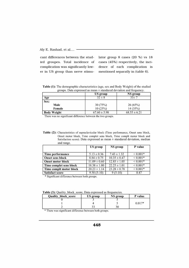
448448
Aly E. Rashad, et al....
cant differences between the stud-
ied groupes. Total incidence of
complication was significantly low-
er in US group than nerve stimu-
lator group 8 cases (20 %) vs 18
cases (45%) respectively, the inci-
dence of each complication is
mentioned separatly in (table 6).
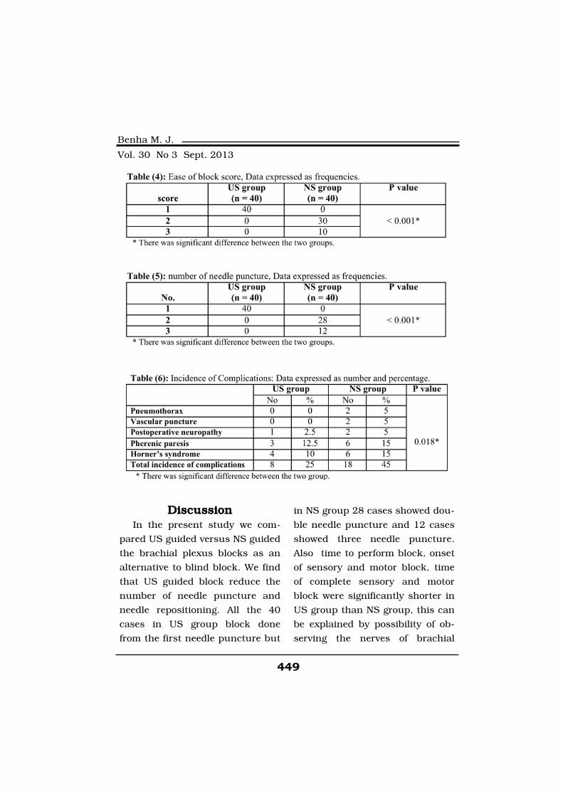
449
Benha M. J.
Vol. 30 No 3 Sept. 2013
DiscussionDiscussionIn the present study we com-
pared US guided versus NS guided
the brachial plexus blocks as an
alternative to blind block. We find
that US guided block reduce the
number of needle puncture and
needle repositioning. All the 40
cases in US group block done
from the first needle puncture but
in NS group 28 cases showed dou-
ble needle puncture and 12 cases
showed three needle puncture.
Also time to perform block, onset
of sensory and motor block, time
of complete sensory and motor
block were significantly shorter in
US group than NS group, this can
be explained by possibility of ob-
serving the nerves of brachial
450450
Aly E. Rashad, et al....
plexus and distribution of local
anaesthic liquid and whether the
consequently applied local anaes-
thic liquid had completely reached
the targeted tissues or not can
also be monitored.
Marhofer et al.(11) stated that
ultrasound visualization of ana-
tomical structures is the only
method offering safe blocks of su-
perior quality of optimal needle
positioning. In addition the
amount of local anesthetic needed
for effective nerve block can be
minimized by directly monitoring
its distribution. They also demon-
strated that ultrasound guidance
significantly improved the punc-
ture-to-onset interval and the
quality of sensory and motor block
while avoiding complications, be-
cause local anesthetics could be
applied more accurately with ul-
trasound guidance compared with
the blind classical technique and
these results were parallel to the
present study.
Also in parallel to the present
study Soeding et al.(12) detected
that ultrasonography application
significantly reduced the starting
time of sensory and motor block
and that it significantly increased
the block quality. Kefalianakis et
al.(13) stated that ultraso-
nography application decreases
the starting of block. In the
present study, we have identified
that sensory block start was earli-
er in the ultrasonography-applied
group.
According to Liu et al.(14) ultra-
sonography application provides more
accomplished sensory and motor
blocks. Also William et al.(15) com-
pared the same technique with
nerve stimulation guidance and
found that ultrasound guidance
was superior, they concluded that,
while inexperienced users may
prefer to use both nerve stimula-
tion and ultrasound to verify the
position of the needle, it is gener-
ally better to avoid nerve stimula-
tion, thereby sparing the patient
the painful muscle contractions
associated with this approach.
In the present study there was
no difference between US group
and NS group as regard satisfac-
tion of patients. A successful
block may be defined as one pro-
viding complete anesthesia of all
target nerves. In the present study
451
Benha M. J.
Vol. 30 No 3 Sept. 2013
3 cases in US group and 5 cases
in NS group showed complete fail-
ure of block, 4 cases in US group
and 5 cases in NS group showed
in complete block and theses cas-
es needed analgesia or sedation
only and the operation was com-
pleted.
Sandhu and Capan(5), found
that the use of ultrasound guided
brachial plexus block, appears to
permit accurate deposition of the
local anesthetic perineurally and
to be associated with a high suc-
cess rate, short onset time, easy
placement of catheter, low compli-
cation rate, and excellent analge-
sia.
As regard complications; in the
present study there were no re-
ported cases of pneumothorax
or vascular injury in US group
while there was two cases of
pneumothorax and two cases of
vascular injury in NS group, these
cases were observed clinically and
by postoperative radiology and
these cases not need any treat-
ment.
Fanelli et al.(16), reported a rate
of 1.7% transient neurological
complications using a multiple in-
jection technique for peripheral
nerve blockade. Liu et al.(17), ob-
served that ultrasound guidance
for interscalene block does not ap-
pear to offer major advantages
over nerve stimulator guidance.
However, the exact pathophysiolo-
gy of postoperative neurological
symptoms is unclear. Other pro-
posed etiologies for postoperative
nerve injury after shoulder ar-
throscopy include patient position(18), compression due to fluid ex-
travasation.(19) amount of trac-
tion(20), selection of arthroscopy
ports.(21) or toxic effects of local
anesthetics.(22)
In the present study overall in-
cidence of complications in US
group were 8 cases (20%), but in
NS group were 18 cases (45%) so
US showed less incidence of com-
plications than NS guided block.
This can be explained that the ul-
trasound approach identifies nerves,
vessels, muscles, and septa.
ConclusionConclusionWe found that ultrasound guid-
ance is clinically useful for suprac-
lavicular brachial plexus block. It
confers confidence and accuracy
452452
Aly E. Rashad, et al....
of needle placement for nerve lo-
calization and examines the Pat-
tern of local anesthetic spread.
ReferencesReferences1. Fisher L. and Gordon M.1. Fisher L. and Gordon M.
(2011):(2011): In Wolfe, SW; Hotchkiss
RN; Pederson WC et al. Green's
Operative Hand Surgery (6th ed.).
Philadelphia: Elsevier/Churchill
Livingstone. pp. 25-40.
2. Malhotra V. and Tjan J.2. Malhotra V. and Tjan J.
(2003):(2003): Brachial plexus blocks. In:
Yao FF, Arusio JF, ed Anesthesio-
logy, problem-oriented patient
management, 5th ed. Philadelphia
Lippincott Williams & Wilkins.
629-42.
3. Denny N.M. and Harrop-3. Denny N.M. and Harrop-
Griffiths W. (2005):Griffiths W. (2005): Location lo-
cation, location! Ultrasound imag-
ing in regional anaesthesia. Br J
Anaesth; 94: 1093.
4. Parlas A., Chan V.W. and4. Parlas A., Chan V.W. and
Simons M. (2003):Simons M. (2003): Brachial plex-
us examination and localization
using ultrasound and electrical
stimulation, a volunteer study.
Anesthesiology; 81, 429-35.
5. Sandhu N.S. and Capan5. Sandhu N.S. and Capan
L.M. (2002):L.M. (2002): Ultrasound-guided
infraclavicular brachial plexus
block. Br J Anaesth; 89: 254-259.
6. Peterson M.K., Millar F.A.6. Peterson M.K., Millar F.A.
and Sheppard D.G. (2002): and Sheppard D.G. (2002): Ul-
trasound-guided nerve blocks. Br
J Anaesth; 88: 621-624.
7. Marhofer P., Greher M. and7. Marhofer P., Greher M. and
Kapral S. (2005):Kapral S. (2005): Ultrasound
guidance in regional anesthesia.
Br J Anaesth; 94: 7-17.
8. Chan V.W., Parlas A., Raw-8. Chan V.W., Parlas A., Raw-
son R. and Odukoya O. (2003):son R. and Odukoya O. (2003):
Ultrasound-guided supraclavicu-
lar brachial plexus block. Anesth
Analg; 97: 1514-17.
9. Moataz M.E. and Mohamed9. Moataz M.E. and Mohamed
Y.E. (2006):Y.E. (2006): High-Resolution Ul-
trasound Imaging Guidance In-
creases Efficacy and Safety of Su-
praclavicular Brachial Plexus
Block. Alexandria Journal of An-
esthesia and Intensive Care; 9
Sept: 26-33.
10. DeLoach L.J., Higgins10. DeLoach L.J., Higgins
M.S., Caplan A.B. and Stiff J.L.M.S., Caplan A.B. and Stiff J.L.
(1998): (1998): The visual analog scale in
the immediate postoperative peri-
od: intrasubject variability and
453
Benha M. J.
Vol. 30 No 3 Sept. 2013
correlation with a numeric scale
Anesth. Analg. 86: 102-106.
11. Marhofer P. and Chan11. Marhofer P. and Chan
V.W. (2007):V.W. (2007): Ultrasound-guided
regional anesthesia: current con-
cepts and future trends. Anesth
Analg; 104: 1265-1269.
12. Soeding P.E., Sha S.,12. Soeding P.E., Sha S.,
Royse C.E., et al. (2005):Royse C.E., et al. (2005): A ran-
domized trial of ultrasound-guided
brachial plexus anaesthesia in
upper limb surgery Anaesth Inten-
sive Care; 33: 719-725
13. Kefalianakis F. and Spoh-13. Kefalianakis F. and Spoh-
ner F. (2005): ner F. (2005): Ultrasound-guided
blockade of axillary plexus brachi-
alis for hand surgery. Handchir
Mikrochir Plast Chir.; 37: 344-348.
14. Liu F.C., Liou J.T., Tsai14. Liu F.C., Liou J.T., Tsai
Y.F., et al. (2005):Y.F., et al. (2005): Efficacy of ul-
trasound-guided axil-lary brachial
plexus block: a comparative study
with nerve stimulator-guided
method. Chang Gung Med J.; 28:
396-402.
15. Williams S.R., Chouinard15. Williams S.R., Chouinard
P., Arcand G., et al. (2003):P., Arcand G., et al. (2003): Ul-
trasound guidance speeds execu-
tion and improves the quality of
supraclavicular block. Anesth
Analg; 97: 1518-23.
16. Fanelli G., Casati A., Ga-16. Fanelli G., Casati A., Ga-
rancini P. and Torri G. (1999):rancini P. and Torri G. (1999):
Nerve stimulator an multiple in-
jection technique for upper and
lower limb blockade: failure rate,
patient acceptance, and neurolog-
ic complications Study Groupon-
regional Anesthesia. Anesth
Analg; 88 (4): 847-852.
17. Liu S.S., Ngeow J. and17. Liu S.S., Ngeow J. and
Yadeau J.T. (2009):Yadeau J.T. (2009): Ultrasound
guided regional anesthesia and
analgesia: a qualitative systematic
review. Reg Anesth Pain Med.; 34:
47-59.
18. Borgeat A., Dullenkopf A.,18. Borgeat A., Dullenkopf A.,
Ekatodramis G. and Nagy L.Ekatodramis G. and Nagy L.
(2003):(2003): Evaluation of the lateral
modified approach for continuous
interscalene block after shoulder
surgery. Anesthesiology; 99: 436-42.
19. Coppieters M.W., Van de19. Coppieters M.W., Van de
Velde M. and Stappaerts K.H.Velde M. and Stappaerts K.H.
(2002):(2002): Positioning in anesthesio-
logy: toward abetter understand-
ing of stretch-nduced periopera-
tive neuropathies. Anesthesiology;
97: 75-81.
454454
Aly E. Rashad, et al....
20. Weber S.C., Abrams J.S.20. Weber S.C., Abrams J.S.
and Nottage W.M. (2002):and Nottage W.M. (2002): Com-
plications associated with arthros-
copic shoulder surgery. Arthrosco-
py; 18: 88-95.
21. Rodeo S.A., Forster R.A.21. Rodeo S.A., Forster R.A.
and Weiland A.J. (1993): and Weiland A.J. (1993): Neuro-
logical complications due to ar-
throscopy. J Bone Joint Surg Am.;
75: 917-26.
22. Hogan Q.H. (2008):22. Hogan Q.H. (2008): Pa-
thophysiology of peripheral nerve
injury during regional anesthesia.
Reg Anesth Pain Med.; 33: 435-41.

455
Benha M. J.
Vol. 30 No 3 Sept. 2013
COMPARISON OF NERVECOMPARISON OF NERVESTIMULATOR VERSUS ULTRASOUNDSTIMULATOR VERSUS ULTRASOUNDGUIDED BRACHIAL PLEXUS BLOCKGUIDED BRACHIAL PLEXUS BLOCKFOR UPPER EXTREMITY SURGERYFOR UPPER EXTREMITY SURGERY
Aly E. Rashad, Salwa M. Sabry MD,Aly E. Rashad, Salwa M. Sabry MD,Ahmed G. Sadek MD, Olfat M. Ismail MDAhmed G. Sadek MD, Olfat M. Ismail MD
and Amer Abd A. Attia MDand Amer Abd A. Attia MD
BENHAMEDICALJOURNAL
REPRINT
Published byPublished by
Benha Faculty of MedicineBenha Faculty of Medicine Volume 30 Number 3Sept. 2013

455
Benha M. J.
Vol. 30 No 3 Sept. 2013
IntroductionIntroductionLow back pain (LBP) is one of
the most frequent musculoskeletal
disorders in daily practice. It is de-
fined as pain between the costal
margins and inferior gluteal folds,
with or without referred leg pain(1). Low back pain is a major
health and socioeconomic problem
and is associated with high costs,
work absenteeism and disability
worldwide(2). There is a high prev-
STUDY OF CAUSES OF LOW BACK PAIN ANDSTUDY OF CAUSES OF LOW BACK PAIN ANDITS PREVALENCE IN REPRESENTATIVEITS PREVALENCE IN REPRESENTATIVECOHORT OF EGYPTIAN POPULATIONCOHORT OF EGYPTIAN POPULATION
El-Boghdady I. MD, El-Kady B. MD, Onsy N. MDEl-Boghdady I. MD, El-Kady B. MD, Onsy N. MDand Seif El-Dein S. M.Scand Seif El-Dein S. M.Sc
Rheumatology and Rehabilitation Department,
Mansoura Faculty of Medicine, Mansoura University
AbstractAbstractAim: Aim: To determine the prevalence and causes of LBP in outpatient clinic
in Egyptian population of various age, sex and occupation.Methods: Methods: 600 patients randomly collected from OPD within a period of 2
years to estimate the prevalence and causes of LBP in both male and femalewith variable occupations and age ranged from 17 to 70 years old. Radio-graphs, CT or MRI were done.
Results: Results: 60 % diagnosed as non specific LBP while the most common di-agnosis was herniated disc (15%), spinal canal stenosis found in (6%), anky-losing spondylitis in (4%), visceral disease (3.5%), malignancy (1%), infection(1%), osteoporosis (1.5%), fractures (1%), spondylolisthesis (1.5%), congeni-tal anomalies (2%), fibromyalgia (1.5%) and (3%) others. The point preva-lence was 58% while the one year prevalence was 60% in this study.
Conclusions: Conclusions: Most cases of LBP diagnosed as non specific LBP. Individu-al, psychosocial and occupational factors are risk factor for LBP develop-ment. LBP is one of the most common problems and one of the most com-mon causes of work absence with high prevalence between population ofboth sex and variable occupation.
Keywords:Keywords: back pain -prevalence-occupational LBP-MRI spine.
456456
El-Boghdady I., et al....
alence of LBP in all western indus-
trial countries(3) as well as in
Egypt.
Several classification schemes,
each with its own philosophy and
categorizing method, subgroup
low back pain (LBP) patients with
the intent to guide treatment. Pre-
vious research suggests that treat-
ing patients based on a classifica-
tion approach results in better
clinical outcomes than non classi-
fication-based treatment strate-
gies(4).
Non-specific low back pain is
defined as low back pain not at-
tributable to a recognisable, known
specific pathology (eg, infection,
tumour, osteoporosis, fracture,
structural deformity, inflammato-
ry disorder, radicular syndrome,
or cauda equina syndrome). Disc
is the most common aetiology of
chronic specific low back pain in
adults(5).
Prevalence measures the pro-
portion of the population that ex-
periences low back pain at a given
time, which can be at any speci-
fied point (point prevalence) or in
a past period such as 1 month, 1
year, or a lifetime(6).
Individual, psychosocial and
occupational factors are the com-
monest risk factors of LBP al-
though varies between studies(7).
Diagnostic triage is used to dis-
tinguish those patients with non-
spinal or serious spinal disorders
from those with pain of musculos-
keletal origin, by means of history
and examination, with particular
emphasis on red flags(8). Once se-
rious disease has been ruled out,
the next priority is to identify pa-
tients with radicular pain. All oth-
er cases are classified as non-
specific(9).
Most guidelines advise that all
imaging studies should be re-
served for patients with progres-
sive neurological deficit, or when
serious underlying causes are
suspected. MRI of the lumbar
spine has become the initial imag-
ing modality of choice in compli-
cated LBP, displacing myelogra-
phy and CT in recent years(10).
Most people with low back pain
do not seek medical care. Many
self treat with the counter medica-
457
Benha M. J.
Vol. 30 No 3 Sept. 2013
tions and lifestyle changes. There
is a wide range of treatment possi-
bilities for patients with low back
pain, including education behavi-
oural therapy, pharmacotherapy,
physical modalities, manual ther-
apy, exercise, spinal injections,
surgery and others(11).
Patients and MethodsPatients and Methodsepidemiological study, based on
a prospective analysis of the col-
lected data conducted on 600 pa-
tients (male and female) visited
the back pain clinic, rheumatology
and rehabilitation outpatient clin-
ic in Mansoura University Hospi-
tal, from the period of June 2011
till June 2013 with age range from
17-70 years. All patients were as-
sessed according to spine disorder
sheet edited in the department in-
cluding personal, past and
present history, local, general and
neurological examination, and in-
vestigations required. Most pa-
tients performed X ray while some
underwent CT and others exposed
to MRI.
The Statistical Package for So-
cial Sciences (SPSS) version 17
was used for statistical analyses of
data. Descriptive statistics includ-
ing means, standard deviation,
frequency and percent were used
to describe sociodemographic data.
Comparisons between both groups
(worker, employee) (analytic meas-
ures) were done using chi-square.
The P-values ≤0.05 was consid-
ered statistically significant.
Exclusion criteria:Exclusion criteria:
Any person younger than (17)
years of age as these patients usu-
ally referred to pediatric hospital,
pregnant ladies and cases of re-
ferred and psychological pain.
ResultsResultsFrom the period of June 2011
till June 2013, 350 patients of
variables age, gender and occupa-
tion were diagnosed to have LBP
from the total 600 patients under-
went this study.
Sociodemographic data of LBP
in workers (including house wives)
versus LBP in employee (including
computer workers) were more
common (71.2%) among workers
compared to (52.8%) of employee
in younger age group (<40 years).
However, higher prevalence (47%)
of LBP was among employees
compared to (28.8%) among work-
458458
El-Boghdady I., et al....
ers in older age group ≥40 years.
Age, gender, occupation, smoking
and obesity were statistically sig-
nificant value in prevalence of LBP
(p<0.001) table (1).
Also, Sociodemographic data of
150 university students visited the
clinic with age range (17-24)
shown in table (2) and the point
prevalence was (40%).
LBP related absence was (42%)
in workers, (38%) in employee and
(32%) in students.
Point prevalence was (58%) in
studied population, while year
prevalence was 60% in the last
year, table (3).
X ray:X ray:
From the 350 patients with
LBP, 245 (70%) patients did plain
x ray as initial investigation, most
of the other 105 improved with in-
itial treatment and some refused
to do so. 47% of the performed x
rays were within normal with no
positive findings. While (53%)
showed variable findings as shown
in table (4).
CT:CT:
From the 245 patients did x
ray, 100 patient underwent lum-
bosacral CT scan. (24%) of these
CT were free from findings while
the other (76%) showed different
findings as in table (4). Total num-
ber of patient did CT was100
(28.5%).
MRI:MRI:
145 patients with LBP under-
went MRI scan, (23%) of them
were normal while (77%) showed
the following changes in table (4).
Total number of patient did MRI
was 145 (41%).
Final diagnosis:Final diagnosis:
Nearly (60%) diagnosed as non
specific LBP while the most com-
mon diagnosis was herniated disc
(15%) with (30%) of them associat-
ed with sciatica, spinal canal sten-
osis found in (6%) of patients, ar-
thropathies in (4%), nearly half cases
are ankylosing spondylitis, viscer-
al disease in (3.5%), other diag-
noses are shown as in table (5).
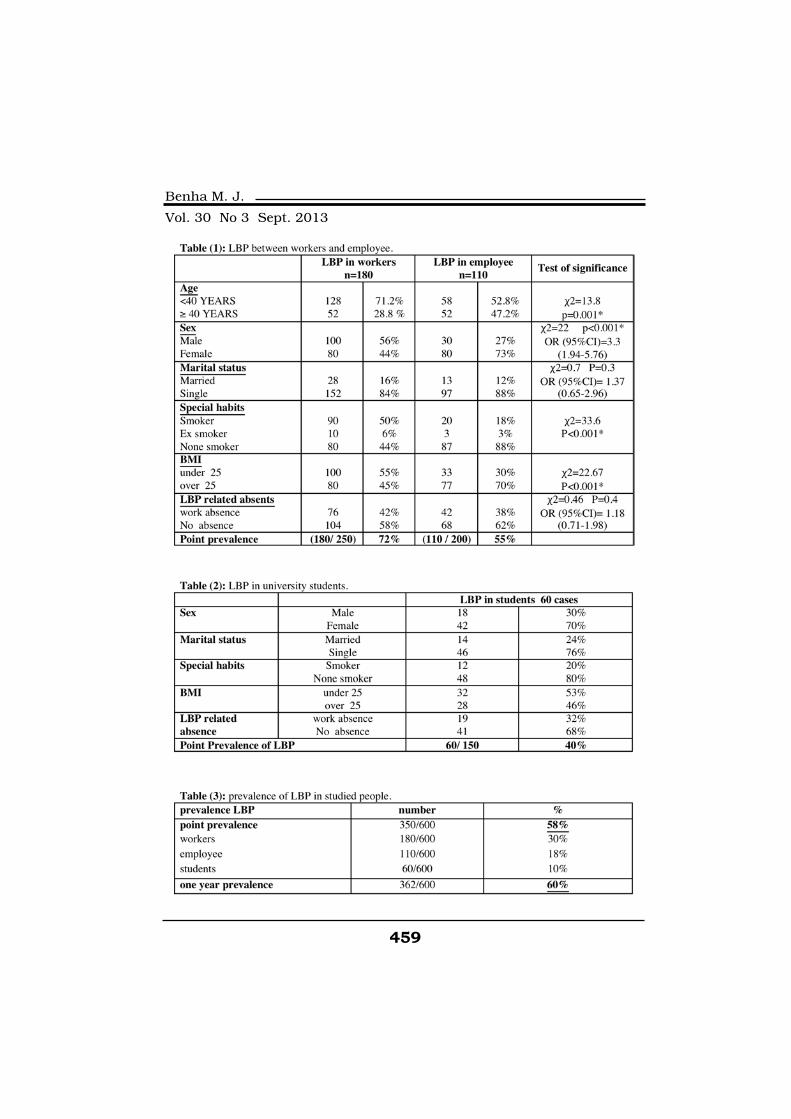
459
Benha M. J.
Vol. 30 No 3 Sept. 2013
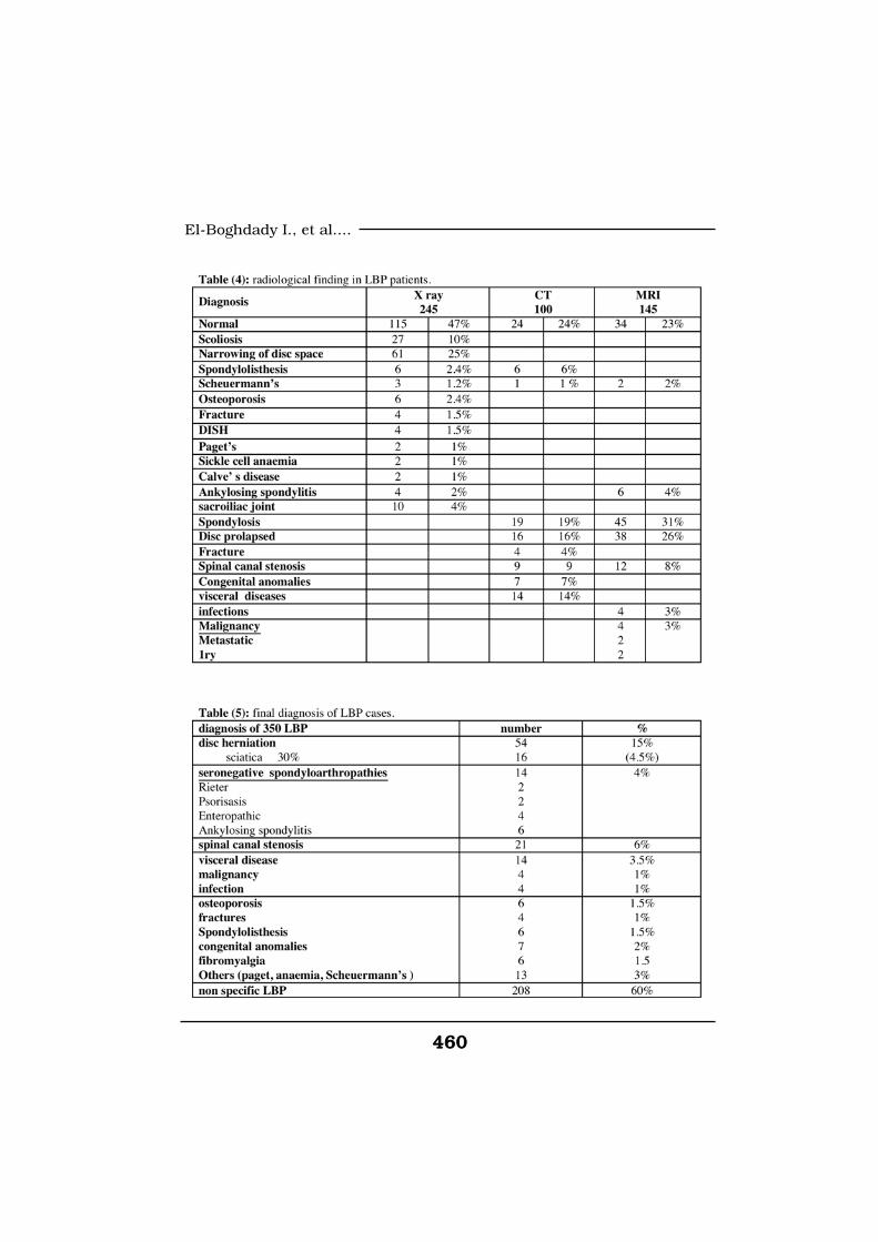
460460
El-Boghdady I., et al....
461
Benha M. J.
Vol. 30 No 3 Sept. 2013
DiscussionDiscussionStudies have shown that low
back pain occurs most often in
those between the ages of 20 to
50; an age group that corresponds
to the largest component of work-
ers(12).
The findings in the current
study had shown that there was
statistically significant (p=.01) as-
sociation between age and low
back pain. Low back pain was pre-
dominant at the middle age group
between 30 and 49 in agree with(13,14).
Some studies have reported a
higher proportion of women re-
porting back pain and sciatica
than men(15,16). In contrast(17),
found the opposite with more males
having sciatica. It was found that
there was no statistical difference
in male: female ratio(18), like in
this study.
Obesity or high body mass in-
dex (BMI) (>30 BMI) are associated
with an increased occurrence of
low back pain(19), in agree with
this study. The prevalence of LBP
was statistically significant higher
in overweight and obese patients
(BMI >25) (p<.001).
Smoking was associated with LBP
and there was significant correlation
between disc herniation and smok-
ing(20). In this study there was
none statistically significant corre-
lation between smoking and LBP.
The prevalence and risk of oc-
cupational low back pain in the
United States with high physical
demands are high(21), in agree
with our study. The association
between low back pain and bend-
ing, twisting, lifting and vibration
was established(22).
Individuals with sitting or
standing jobs occupying most of
the workday had an increased risk
of low back pain(23), in agree with
this study.
In this study, lack of regular
exercise and fitness is strong pre-
dictor for LBP development
(p<0.001), in agree with(24).
Approximately ≥90% of patients
who present to primary care has
nonspecific LBP(25).
In this study nearly (60%) of
462462
El-Boghdady I., et al....
cases are non specific LBP; which
is considered lower than most
studies; this may be due to that
the main sample of study was col-
lected from specialized back pain
and rheumatology outpatient clin-
ic.
Osteoporotic fractures of the
spine are represented by (1%)(26),
this agrees with our study (1.5%).
While it was (4%)(2), most of our
patients was postmenopausal fe-
males in agree with(27).
Frequency of arthropathies was
(3-5%)(2). Like in our study, (4%)
were seronegative spondyloar-
thropathies most of them were An-
kylosing spondylitis.
Malignant neoplasm accounts
for less than (1%) of episodes of
low back pain. However, metastat-
ic cancer should be considered as
a potential aetiology in any patient
with a previous history of cancer,
until proved otherwise(26). (1%) of
our LBP patients were diagnosed
as malignancy.
Lumbar disc herniations are
one of the most common causes of
low back pain. The one year prev-
alence of low back pain which was
diagnosed as herniated disc is
(12%-15%) of all adults(28), in
agree with our study (15%) and
(20%) in(14). However(3) has de-
scribed up to (25%) with LBP had
herniated disc.
The prevalence of lumbar spi-
nal stenosis (LSS) in association
with LBP was varied from (4%-
20%), but the actual ratio remains
unclear(29). This was agreed with
our study in which LSS preva-
lence was (6%).
The prevalence of spondylolis-
thesis in this study was (1.5%).
While(30) found incidence of
(2.7%). Most studies revealed inci-
dence of visceral disease about
(2%)(31). Visceral diseases were di-
agnosed in (3.5%) in our patients.
The point prevalence in this
study was (58%), this is consid-
ered high prevalence, but this is
due to that most cases were
caught from back pain clinic, in
agree with(12) (57%) and(32) (58%).
The prevalence was divided into 3
categories: physical worker (in-
clude house wives) (30%), employ-
ee (static job) (18%) and students
463
Benha M. J.
Vol. 30 No 3 Sept. 2013
(10%). The one year prevalence in
this study was (60%) in agree
with(33) (61.3%).
ReferencesReferences1. Chou R., Qaseem A., Snow1. Chou R., Qaseem A., Snow
V., Casey D., Cross J. and She-V., Casey D., Cross J. and She-
kelle P. (2007): kelle P. (2007): Diagnosis and
treatment of low back pain: a joint
clinical practice guideline from the
American College of Physicians
and the American Pain Society.
Ann Intern Med; 147: 478-91.
2. Chou R., Fu R. and Carri-2. Chou R., Fu R. and Carri-
no J. (2009): no J. (2009): Imaging strategies
for low-back pain: systematic re-
view and meta-analysis. Lancet;
373: 463-72.
3. Waddell G. (2005):3. Waddell G. (2005): Sub-
groups within nonspecific low
back pain. J Rheumatol, 32: 395-
6.
4. Fritz J., Cleland J. and4. Fritz J., Cleland J. and
Childs J. (2007):Childs J. (2007): Subgrouping
patients with low back
pain:evolution of a classification
approach to physical therapy. J
Orthop Sports Phys Ther; 37: 290-
302.
5. Rubin D. (2007):5. Rubin D. (2007): Epidemiol-
ogy and risk factors for spine
pain. Neurol Clin; 25: 353-71.
6. Guo H., Chang Y., Yeh W.,6. Guo H., Chang Y., Yeh W.,
et al. (2004):et al. (2004): Prevalence of mus-
culoskeletal disorder among work-
ers in Taiwan: a nationwide study.
J Occup Health; 46: 26-36.
7. Currie S. and Wang J.7. Currie S. and Wang J.
(2005):(2005): More data on major de-
pression as an antecedent risk
factor for first onset of chronic
back pain. Psychological Medicine;
35(9): 1275-82.
8. Rubinstein S. and van Tul-8. Rubinstein S. and van Tul-
der M. (2008):der M. (2008): A best-evidence re-
view of diagnostic procedures for
neck and low-back pain. Best
Pract Res Clin Rheumatol; 22:
471-82.
9. Dagenais S., Tricco A. and9. Dagenais S., Tricco A. and
Haldeman S. (2010):Haldeman S. (2010): Synthesis of
recommendations for the assess-
ment and management of low
back pain from recent clinical
practice guidelines. Spine J.; 10:
514-29.
10. Lurie J., Tosteson A. and10. Lurie J., Tosteson A. and
Tosteson T. (2008): Tosteson T. (2008): Reliability of
magnetic resonance imaging read-
464464
El-Boghdady I., et al....
ings for lumbar disc herniation in
the Spine Patient Outcomes Re-
search Trial (SPORT). Spine; 33(9):
991-98
11. Daffner S. and Wang J.11. Daffner S. and Wang J.
(2009):(2009): The pathophysiology and
nonsurgical treatment of lumbar
spinal stenosis. Instr Course Lect;
58: 657-68.
12. Hoy D., Brooks P., Blyth12. Hoy D., Brooks P., Blyth
F. and Buchbinder R. (2010):F. and Buchbinder R. (2010):
The Epidemiology of low back
pain. Best Practice & Research
Clinical Rheumatology; 24: 769-
81.
13. Plenet A., Gourmelen J.,13. Plenet A., Gourmelen J.,
Chastang J., Ozguler A., LanoeChastang J., Ozguler A., Lanoe
J. and Leclerc A. (2010):J. and Leclerc A. (2010): Seeking
care for lower back pain in the
French population aged from 30 to
69: the results of the 2002–2003
Decennale Sante survey. Ann
Phys Rehabil Med; 53: 224-31.
14. de Schepper E., Damen J.14. de Schepper E., Damen J.
and van Meurs J. (2010):and van Meurs J. (2010): The as-
sociation between lumbar disc de-
generation and low back pain: the
influence of age, gender, and indi-
vidual radiographic features.
Spine (Phila Pa 1976); 35: 531-36.
15. Bejia I., Younes M., Zrour15. Bejia I., Younes M., Zrour
S., Touzi M. and Bergaoui N.S., Touzi M. and Bergaoui N.
(2004):(2004): Factors predicting out-
comes of mechanical sciatica: a
review of 1092 cases. Joint Bone
Spine; 71: 567-71.
16. Nordeman L., Gunnars-16. Nordeman L., Gunnars-
son R. and Mannerkorpi K.son R. and Mannerkorpi K.
(2012): (2012): “Prevalence and charac-
teristics of widespread pain in fe-
male primary health care patients
with chronic low back pain.” Clin
J Pain.; 28(1): 65-72.
17. Atlas S., Deyo R., Keller17. Atlas S., Deyo R., Keller
R. and Chang Y. (2001):R. and Chang Y. (2001): Evaluat-
ing and managing acute low back
pain in the primary care setting. J
Gen Intern Med; 16: 120-31.
18. Hartvigsen J. and Chris-18. Hartvigsen J. and Chris-
tensen K. (2008): tensen K. (2008): Pain in the
back and neck are with us until
the end: a nationwide interview-
based survey of Danish 100-year-
olds. Spine (Phila Pa 1976); 33:
909-13
19. Mirtz T. and Greene L.19. Mirtz T. and Greene L.
(2005):(2005): Is obesity a risk factor for
low back pain? An example of us-
ing the evidence to answer a clini-
cal question. Chiropractic & Oste-
465
Benha M. J.
Vol. 30 No 3 Sept. 2013
opathy; 13(2): 1340-46.
20. Poddubnyy D., Haibel H.,20. Poddubnyy D., Haibel H.,
Listing J., Marker-Hermann E.,Listing J., Marker-Hermann E.,
Braun J. and Sieper J. (2012):Braun J. and Sieper J. (2012):
Baseline radiographic damage, el-
evated acute phase reactant lev-
els, and cigarette smoking status
predict spinal radiographic progres-
sion in early axial spondylarthri-
tis. Arthritis Rheum; 64: 1388-98.
21. Wai E., Roffey D., Bishop21. Wai E., Roffey D., Bishop
P., Kwon B. and Dagenais S.P., Kwon B. and Dagenais S.
(2010):(2010): Causal assessment of oc-
cupational bending or twisting
and carrying and low back pain:
results of a systematic review.
Spine J; 10: 628-38.
22. Thorbjrnsson C., Alfreds-22. Thorbjrnsson C., Alfreds-
son L. and Fredriksson K.son L. and Fredriksson K.
(2000): (2000): Physical and psychosocial
factors related to low back pain
during a 24-year period. A nested
case-control analysis. Spine; 25:
369-75.
23. Turner L. and Reed D.23. Turner L. and Reed D.
(2011):(2011): Exercise among commer-
cial truck drivers. AAOHN J.; 59:
429-36.
24. Standaert C. (2002): 24. Standaert C. (2002): New
strategies in the management of
low back injuries in gymnasts.
Curr Sports Med Rep.; 1(5): 293-
300.
25. Raspe H., Matthis C.,25. Raspe H., Matthis C.,
Croft P. and O’Neill T. (2004):Croft P. and O’Neill T. (2004):
Variation in back pain between
countries: the example of Britain
and Germany. Spine; 29: 1017-21.
26. Henschke N., Maher C.,26. Henschke N., Maher C.,
Refshauge K., Herbert R., Cum-Refshauge K., Herbert R., Cum-
ming R. and Bleasel J. (2009):ming R. and Bleasel J. (2009):
Prevalence of and screening for se-
rious spinal pathology in patients
presenting to primary care set-
tings with acute low back pain.
Arthritis Rheum; 60: 3072-80.
27. Miyakoshi N., Itoi E., Mu-27. Miyakoshi N., Itoi E., Mu-
rai H., Wakabayashi I., Ito H.rai H., Wakabayashi I., Ito H.
and Minato T. (2003): and Minato T. (2003): Inversed
relation between osteoporosis and
spondylosis in postmenopausal
women as evaluated by bone min-
eral density and semiquantitative
scoring of spinal degeneration.
Spine; 28 (5): 492-95.
28. Van der Windt D., Simons28. Van der Windt D., Simons
E. and Riphagen I. (2010):E. and Riphagen I. (2010): Physi-
cal examination for lumbar radic-
ulopathy due to disc herniation in
466466
El-Boghdady I., et al....
patients with low back pain. Co-
chrane Database Syst Rev; 2:
CD007431.
29. Kalichman L. and Hunter29. Kalichman L. and Hunter
D. (2008):D. (2008): The genetics of inter-
vertebral disc degeneration. Famil-
ial predisposition and heritability
estimation. Joint Bone Spine; 75:
383-87.
30. Jacobsen S., Sonne-Holm30. Jacobsen S., Sonne-Holm
S., Rovsing H., Monrad H. andS., Rovsing H., Monrad H. and
Gebuhr P. (2007):Gebuhr P. (2007): Degenerative
lumbar spondylolisthesis: an epi-
demiological perspective: the Co-
penhagen Osteoarthritis Study.
Spine.; 32(1): 120-25.
31. Patel A. and Ogle A.31. Patel A. and Ogle A.
(2000):(2000): Diagnosis and manage-
ment of acute low back pain. Am
Fam Physician; 61: 1779-86.
32. Landry M., Raman S.,32. Landry M., Raman S.,
Sulway C., Golightly Y. andSulway C., Golightly Y. and
Hamdan E. (2008):Hamdan E. (2008): Prevalence
and risk factors associated with
low back pain among health care
providers in a Kuwait hospital.
Spine (Phila Pa 1976); 33: 539-45.
33. Karahan A., Kav S., Abba-33. Karahan A., Kav S., Abba-
soglu A. and Dogan N. (2009):soglu A. and Dogan N. (2009):
Low back pain: prevalence and as-
sociated risk factors among hospi-
tal staff. J Adv Nurs; 65: 516-24.

467
Benha M. J.
Vol. 30 No 3 Sept. 2013
STUDY OF CAUSES OF LOW BACKSTUDY OF CAUSES OF LOW BACKPAIN AND ITS PREVALENCE INPAIN AND ITS PREVALENCE INREPRESENTATIVE COHORT OFREPRESENTATIVE COHORT OF
EGYPTIAN POPULATIONEGYPTIAN POPULATION
El-Boghdady I. MD, El-Kady B. MD, Onsy N. MDEl-Boghdady I. MD, El-Kady B. MD, Onsy N. MDand Seif El-Dein S. M.Scand Seif El-Dein S. M.Sc
BENHAMEDICALJOURNAL
REPRINT
Published byPublished by
Benha Faculty of MedicineBenha Faculty of Medicine Volume 30 Number 3Sept. 2013
467
Benha M. J.
Vol. 30 No 3 Sept. 2013
VINORELBINE AND 5 FLUORO-URACIL /VINORELBINE AND 5 FLUORO-URACIL /FOLINIC ACID VERSUS DOCETAXEL AS FIRSTFOLINIC ACID VERSUS DOCETAXEL AS FIRST
LINE TREATMENT FOR PATIENTS WITHLINE TREATMENT FOR PATIENTS WITHMETASTATIC BREAST CANCERMETASTATIC BREAST CANCER
I. Abdel Halim MD*, E. El-Sherbini MD*I. Abdel Halim MD*, E. El-Sherbini MD*and N. Haddad MD**and N. Haddad MD**
*Department of Clinical Oncology, Mansoura University, Egypt.
**Department of Medical Affairs, Pierre Fabre Médicament, Lebanon.
AbstractAbstractBackground:Background: Vinorelbine (V) and 5-Fluorouracil (5FU) is an effective
combination for the treatment of metastatic breast cancer (MBC). AvailablePhase II clinical data reports response rates ranging from 60-64% in firstline MBC. Single agent Docetaxel (D) is also an effective treatment for MBC,demonstrating an objective response rate of 48% in a pivotal phase III trial.We evaluated the efficacy and safety of V + 5FU (Arm A) versus D (Arm B) inpatients (pts) with MBC relapsing after adjuvant anthracycline based treat-ment.
Materials & Methods:Materials & Methods: 100 pts (50% Arm A, 50% Arm B) were enrolledbetween Jul 2003 and March 2005. All pts had measurable MBC after adju-vant anthracycline treatment, WHO PS 1, adequate bone marrow, renal andhepatic functions. Pts were randomized to Arm A: Vinorelbine i.v. 25 mg/mD1, D3 + folinic acid 100 mg/m D1, D2, D3 + 5FU 350 mg/m D1, D2, D3 orArm B: Docetaxel 100 mg/m D1 with optional prophylactic G-CSF. Cycleswere repeated every 3weeks. Pts with PD went off study while those with CR,PR, or SD continued treatment for a maximum of 8 cycles.
Results: Results: Median age (Arm A; Arm B): 53 & 50 years; the majority hadWHO PS 0 in both arms. Previous adjuvant therapy: anthracycline (100%),hormonotherapy (60% & 46% in Arms A & B respectively). The majority ofpatients, in both arms, presented with more than one metastatic site andtwo thirds had visceral disease. Liver was the most commonly involved or-gan. Total number of cycles delivered (Arm A: 281, Arm B: 282). Mediannumber of cycles per patient: 6 in both arms. An objective tumor response of
468468
I. Abdel Halim, et al....
IntroductionIntroductionBreast cancer is the most com-
mon malignancy among women
and is the second cause of cancer
deaths in female population(1).
Despite the proven benefit of
adjuvant systemic therapy in re-
ducing the risk of recurrence(2,3),
a significant number of patients
with operable breast carcinoma
will eventually develop metastatic
disease, and ultimately die of ad-
vanced metastatic breast carcino-
ma (MBC)(4).
Many chemotherapeutic agents
have shown antitumor activity in
MBC, among which the anthracy-
clines have been considered stan-
dard therapy(5,6).
Vinorelbine, a semisynthetic
vinca alkaloid, has been consid-
ered one of the most active cyto-
toxic drugs against MBC, with a
low toxicity profile. Vinorelbine
has been found to yield response
rates of 34-50% as a single agent
when used as first-line therapy,
and 15-30% as second-line thera-
py(7-13).
The combination of vinorelbine
and infused 5-fluorouracil (5-FU)
has been acknowledged as an ef-
fective palliative regimen for MBC,
especially in Europe, and has
been tested in several phase II
studies(14-16). Although this regi-
men showed high response rates,
up to 70%, as first-line therapy,
treatment tolerance was not satis-
factory.
This regimen has also been em-
ployed as front-line treatment for
MBC, alone or in combination
64% & 68% and a complete response of 26% & 22% were achieved in armsA & B respectively. Median time to progression & overall survival: Arm A: 15& 27 months, Arm B: 15 & 30 months. No WHO grade Gr 3-4 toxicities werenoted in Arm A. Gr 3 alopecia (18%) & Gr 3 liver enzymes elevation (2%)were noted in Arm B.
Conclusions:Conclusions: Our results suggest that Vinorelbine-5FU combination andsingle agent Docetaxel demonstrate similar efficacy as first line treatment forMBC. Vinorelbine-5FU is however better tolerated besides being a less costlytherapeutic option in Egypt. A large prospective randomized trial is neededto confirm these results.

469
Benha M. J.
Vol. 30 No 3 Sept. 2013
with FA, with a 60% overall re-
sponse rate(17,18).
Docetaxel, an antimitotic agent
that blocks cells in the M phase of
the cell cycle it is recognised as
one of the most active agents cur-
rently available for the treatment
of breast cancer(19).
In previously untreated pa-
tients, single-agent docetaxel pro-
vides ORR of 40% to 68%(20,21).
The first schedules of adminis-
tration of docetaxel employed dos-
es ranging from 75 to 100 mg/m2
as a 1-hour intravenous infusion
every three weeks. The 3-week sched-
ule of docetaxel 100 mg/m2, although
extremely active, showed an impor-
tant myelosuppression with more
than 90% of cases experiencing
grade (G) 3-4 neutropenia(21,22).
Our aim in this study is to
compare the clinical efficacy and
safety of the VNR plus 5FU/FA
regimen versus docetaxel when
given as first-line treatment in pa-
tients with MBC.
Patients and MethodsPatients and MethodsThis study included 100 female
patients with metastatic breast
cancer (50 in Arm A and 50 in
Arm B) and were enrolled between
Jul 2003 and March 2005
Patient population:Patient population:
Key Eligibility Criteria:Key Eligibility Criteria:
- Female Patients between 18
and 65 years-old.
- Performance Status (WHO) <2.
- Life expectancy ≥12 weeks.
- Histologically confirmed meta-
static breast cancer.
- At least one bi-dimensionally
measurable lesion.
- Prior neo/adjuvant chemo-
therapy with anthracycline was al-
lowed.
- Previous hormonal therapy for
adjuvant or metastatic disease
was also allowed.
- Interval between last course
of chemotherapy and the treat-
ment protocol should exceed 3
months.
- No previous treatment with
chemotherapy for metastatic dis-
ease.
- Adequate bone marrow, he-
patic & renal functions. (WBCs
≥3.5*109/L, platelets ≥100*109/
L), renal function (creatinine clea-
ralance >60 mL/min) and liver
function (serum bilirubin <1.5mg/

470470
I. Abdel Halim, et al....
dL, transaminases <3 times the
upper limit of normal).
- Oral informed consent.
Exclusion criteria:Exclusion criteria:
No symptom or sign of brain
metastasis. Patients with known
brain or leptomeningial infiltration
were excluded. Pregnant or lactat-
ing women & male patients. Pa-
tients were ineligible if they had
local relapse only. Another malig-
nancy within the previous 5 years,
except cured basal cell carcinoma
of the skin or excised carcinoma
in situ of the cervix, were also ex-
cluded. Bone metastasis or malig-
nant pleural effusion as only site
of metastasis were excluded. The
pretreatment evaluation included
medical history, physical examina-
tion, performance status, preg-
nancy test if needed, ECG, chest
X-rays, tumor measurements by
computed tomography (CT), and
bone scan if indicated.
Treatment Schedule:Treatment Schedule:
Pts were randomized to Arm A:
Vinorelbine i.v. 25 mg/m D1, D3 +
folinic acid 100 mg/ m D1, D2, D3
+ 5FU 350 mg/m D1, D2, D3 or
Arm B: Docetaxel 100 mg/mD1 with
optional prophylactic G-CSF. Cy-
cles were repeated every 3weeks.
Vinorelbine was administered
in a short infusion over 6 to 10
min, followed by a rapid infusion
of 500 ml of normal saline and
5FU 350 mg/m2 diluted in 250 cc
of 5% dextrose, folinic acid (FA)
100 mg/m2 diluted in 100 cc of
normal saline both administered
i.v. on days 1, 2 and 3.
Docetaxel 100 mg/m2 was ad-
ministered as a 1-hour intrave-
nous infusion. -Dexametazone
premedication starting one day
before & continued for two days
after the administration of docetax-
el. Secondary G-CSF prophylaxis
was administered when needed.
Dose modifications. The whole
treatment regimen was delayed for
1 week in case of grade 3 or more
toxicity.
Treatment assessment:Treatment assessment:
On day 1 of each cycle, physi-
cal condition and performance
status were assessed and hemato-
logical and blood chemistry meas-
urements were established.
Objective responses were as-
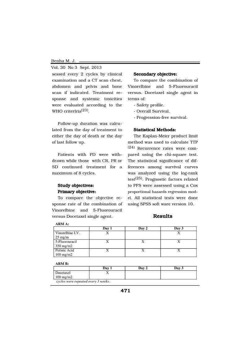
471
Benha M. J.
Vol. 30 No 3 Sept. 2013
sessed every 2 cycles by clinical
examination and a CT scan chest,
abdomen and pelvis and bone
scan if indicated. Treatment re-
sponse and systemic toxicities
were evaluated according to the
WHO criterlria(23).
Follow-up duration was calcu-
lated from the day of treatment to
either the day of death or the day
of last follow up.
Patients with PD were with-
drawn while those with CR, PR or
SD continued treatment for a
maximum of 8 cycles.
Study objectives:Study objectives:
Primary objective:Primary objective:
To compare the objective re-
sponse rate of the combination of
Vinorelbine and 5-Fluorouracil
versus Docetaxel single agent.
Secondary objective:Secondary objective:
To compare the combination of
Vinorelbine and 5-Fluorouracil
versus. Docetaxel single agent in
terms of:
- Safety profile.
- Overall Survival.
- Progression-free survival.
Statistical Methods:Statistical Methods:
The Kaplan-Meier product limit
method was used to calculate TTP(24) Recurrence rates were com-
pared using the chi-square test.
The statistical significance of dif-
ferences among survival curves
was analyzed using the log-rank
test(25). Prognostic factors related
to PFS were assessed using a Cox
proportional hazards regression mod-
el. All statistical tests were done
using SPSS soft ware version 10.
ResultsResults
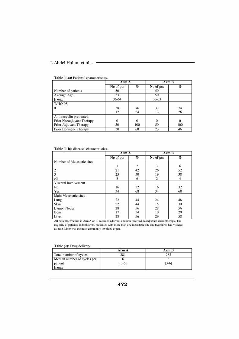
472472
I. Abdel Halim, et al....
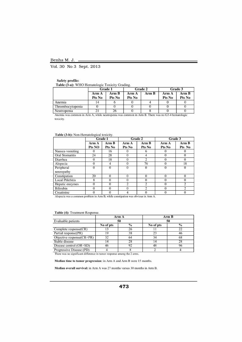
473
Benha M. J.
Vol. 30 No 3 Sept. 2013
474474
I. Abdel Halim, et al....
DiscussionDiscussionThe appropriate management
of patients with breast cancer af-
ter development of metastases
represents a therapeutic problem.
With the increasing use of an-
thracycline-based combinations in
the adjuvant setting, more pa-
tients with metastatic disease
have a prior history of anthracy-
cline exposure, thus limiting the
use of this relatively effective
group of drugs for palliation.
There is a need, therefore, for
the development of non-
anthracycline-based combinations
for the treatment of patients with
metastatic breast cancer.
The availability of several new
active chemotherapeutic agents
allows for different management
options. These drugs can be used
in combination regimens or as
single agents in sequence(26).
Chemotherapy for metastatic
breast cancer is, at best, a pallia-
tive measure and the main goal of
any treatment chosen for meta-
static disease is therefore to ob-
tain complete symptomatic relief,
ideally through tumor regression.
Vinorelbine is a semisynthetic
vinca alkaloid that has cytocidal
effect on a wide range of tumor
cell lines(27-29).
It is a mitotic inhibitor that has
a higher therapeutic index and less
neurotoxicity than other vinca al-
kaloids related to the lower degree
of damage of axonal microtubules(30). The dose limiting toxicity of
vinorelbine is granulocytopenia(31).
Given as single agent in breast
cancer the drug yields response
rates of 40% to 60% in previously
untreated patients(32-34), and of
about 30% when used as a sec-
ond- or third-line therapy(35-36).
The combination of two drugs,
such as vinorelbine and 5-FU,
without overlapping toxicity pro-
files seems particularly attractive
in this clinical setting.
The combination of vinorelbine
with 5-Fluorouracil (5-FU) achieved
high response rates of 60 to 64% and
a manageable toxicity profile, though
the tolerance profile of this regimen
depends on the schedule(13,14).
Docetaxel is regarded as the

475
Benha M. J.
Vol. 30 No 3 Sept. 2013
single most effective cytotoxic
agent for advanced breast cancer
with substantial objective re-
sponse rates in previously un-
treated patients (up to 68% in
phase II trials) and anthracycline-
pretreated patients (30% to 43%
in phase III trials)(37,38,39).
We conducted this trial to com-
pare the efficacy and toxicity pro-
file of those 2 regimens.
In our study, the objective re-
sponse rate was 64% which is
comparable to studies conducted
by (Nole et al(18), 1997 and Dieras
et al(14), 1996), who reported 62%
Objective response rate and slight-
ly better than Kornek et al(39)
(1998) who reported 59% objective
response.
After a median follow up of 30
months. The overall survival in
our study was 27 months and in
Dieras et al ”s study(14) it was 23
months.
The median time to tumor pro-
gression was better in our study
when compared to Nole et al(18),
(1997) (15 months versus 10
months) and this could be due to
inclusion of more patients with
multiple metastatic sites in Nole et
al study than ours.
In our study G1 neutropenia
was the commonest hematologic
toxicity (42%), which is less than
Nole et al(18) (1997) who reported
77% neutropenia.
In our study and in studies
conducted by (Nole et al, 1997
and Kornek et al, 1998), nonhe-
matologic toxicities were mild and
manageable.
In our study docetaxel achieved
68% the objective response, MTP
15 months and median OS 30
months, our results are compara-
ble to others who reported objec-
tive respone 58-68%, MTP (12-18
months) and median OS 25-30
months(39-43).
In our study grade 1 and grade
2 neutropenia were encountered
in 46% and 16 %, compared to
Joensuu et al, 2009(40) reported
99% hematologic toxicity.
The lower incidence of hemato-
logic toxicity in our trial could be
due to the frequent use of prophy-
476476
I. Abdel Halim, et al....
lactic granulocyte growth factors.
In our study and in others non
hematologic toxicity was mild and
manageable except for alopecia(39-
43).
We have observed from our
study that there is no significant
difference between the two regi-
mens in terms of response, while
the toxicity profile on navelbine is
much better than docetaxel.
ConclusionConclusionVinorelbine-5FU is better toler-
ated besides being a less costly
therapeutic option in Egypt. A
large prospective randomized trial
is needed to confirm these results.
ReferencesReferences1. Devita Vincent T., Law-1. Devita Vincent T., Law-
rence Theodore S., Rosenbergrence Theodore S., Rosenberg
Steven A. (2008):Steven A. (2008): Cancer: princi-
ples and practice of oncology, 8th
Edition Lippincott Williams and
Wilkins, 1606-9.
2. Harris J.R., Morrow M. and2. Harris J.R., Morrow M. and
Bonadonna G. (1993): Bonadonna G. (1993): Cancer of
the breast.In: Cancer. Principles
and Practice of Oncology, Fourth
edition. De Vita VT Jr, Hellman S,
Rosenberg SA (eds.). Philadelphia,
PA: Lippincott Company, pp.
1264-1332.
3. Sledge G.W. (2004): 3. Sledge G.W. (2004): Breast
cancer as a world challenge. Clin
Breast Cancer 5: 11.
4. Early Breast Cancer Trial-4. Early Breast Cancer Trial-
ist’s Collaborative Group (1992):ist’s Collaborative Group (1992):
Systemictreatment of early breast
cancer by hormonal, cytotoxic, or
immune therapy. Lancet 339: 1-
15.
5. Tormey C. (1975): 5. Tormey C. (1975): Adriamy-
cin (NSC 123-127) in breast can-
cer. An overview of studies. Can-
cer Chemother Rep; 3: 319-27.
6. Stewart D.J., Evans W.K.,6. Stewart D.J., Evans W.K.,
Shepherd F.A., Wilson K.S.,Shepherd F.A., Wilson K.S.,
Pritchard K.I., Trudeau M.E.,Pritchard K.I., Trudeau M.E.,
Wilson J.J. and Martz K. (1997):Wilson J.J. and Martz K. (1997):
Cyclophosphamide and fluoroura-
cil combined with mitoxantrone
versus doxorubicin for breast can-
cer:Superiority of doxorubicin. J
Clin Oncol; 15: 1897-905.
7. Budman D.R. (1997): 7. Budman D.R. (1997): Vino-
relbine (Navelbine): A third-
generation vinca alkaloid. Cancer
Invest; 15: 475-90.
477
Benha M. J.
Vol. 30 No 3 Sept. 2013
8. Johnson S.A., Harper P.,8. Johnson S.A., Harper P.,
Hortobagyi G.N. and Pouillart P.Hortobagyi G.N. and Pouillart P.
(1996):(1996): Vinorelbine: An overview.
Cancer Treat Rev; 22: 127-42.
9. Smith G.A. (1995): 9. Smith G.A. (1995): Current
status of vinorelbine for breast
cancer. Oncology; 9: 767-73.
10. Romero A., Rabinovich10. Romero A., Rabinovich
M.G., Vallejo C.T., Perez J.E.,M.G., Vallejo C.T., Perez J.E.,
Rodriguez R., Cuevas M.A., Ma-Rodriguez R., Cuevas M.A., Ma-
chiavelli M., Lacava J.A., Langhichiavelli M., Lacava J.A., Langhi
M. and Romero Acuna L. (1994):M. and Romero Acuna L. (1994):
Vinorelbine as first-line chemo-
therapy for metastatic breast car-
cinoma. J Clin Oncol; 12: 336-41.
11. Fumoleau P., Delgado11. Fumoleau P., Delgado
F.M., Delozier T., Monnier A.,F.M., Delozier T., Monnier A.,
Gil Delgado M.A., Kerbrat P.,Gil Delgado M.A., Kerbrat P.,
Garcia-Giralt E., Keiling R.,Garcia-Giralt E., Keiling R.,
Namer M. and Closon M.T.Namer M. and Closon M.T.
(1993):(1993): Phase II trial of weekly in-
travenous vinorelbine in first-line
advanced breast cancer chemo-
therapy. J Clin Oncol; 11: 1245-52.
12. Twelves C.J., Dobbs N.A.,12. Twelves C.J., Dobbs N.A.,
Curnow A., Coleman R.E., Stew-Curnow A., Coleman R.E., Stew-
art A.L., Tyrrell C.J., Canney P.art A.L., Tyrrell C.J., Canney P.
and Rubens R.D. (1994): and Rubens R.D. (1994): A
phase II multicentre, UK study of
vinorelbine in advanced breast
cancer. Br J Cancer; 70: 990-3.
13. Weber B.L., Vogel C.,13. Weber B.L., Vogel C.,
Jones S., Harvey H., HutchinsJones S., Harvey H., Hutchins
L., Bigley J. and Hohneker J.L., Bigley J. and Hohneker J.
(1995): (1995): Intravenous vinorelbine as
first-line and second-line therapy
in advanced breast cancer. J Clin
Oncol; 13: 2722-30.
14. Dieras V., Extra J.M.,14. Dieras V., Extra J.M.,
Bellissant E., Espie M., MorvanBellissant E., Espie M., Morvan
F., Pierga J.Y., Mignot L., TrescaF., Pierga J.Y., Mignot L., Tresca
P. and Marty M. (1996):P. and Marty M. (1996): Efficacy
and tolerance of vinorelbineand
fluorouracil combination as first-
line chemotherapy of advanced
breast cancer: Results of a phase
II study using a sequential group
method. J Clin Oncol; 14: 3097-
104.
15. Zambetti M., Demicheli15. Zambetti M., Demicheli
R., De Candis D., Antonelli G.,R., De Candis D., Antonelli G.,
Giacobone A., Terenziani M.,Giacobone A., Terenziani M.,
Laffranchi A., Garbagnati F., Bi-Laffranchi A., Garbagnati F., Bi-
asi S. and Bonadonna G. (1997):asi S. and Bonadonna G. (1997):
Five day infusion fluorouracil plus
vinorelbine i.v. in metastatic pre-
treated breast cancer patients.
Breast Cancer Res Treat; 44: 255-
60.
16. Froudarakis M.E., Cati-16. Froudarakis M.E., Cati-
478478
I. Abdel Halim, et al....
mel G., Guastalla J.P., Rebattumel G., Guastalla J.P., Rebattu
P. and Clavel M. (1998):P. and Clavel M. (1998): Phase II
trial of navelbine and fluorouracil
as second-line chemotherapy in
metastatic breast carcinoma. On-
cology; 55: 87-8.
17. Cany L., Toulouse C., Ra-17. Cany L., Toulouse C., Ra-
vaud A., Durand M. and Mauri-vaud A., Durand M. and Mauri-
ac L. (1996): ac L. (1996): Vinorelbine/5-FU
combination in metastatic breast
cancer chemotherapy: A retro-
spective study of 63 cases. Eur J
Cancer; 32A: 370-1.
18. Nole F., deBraud F., Aa-18. Nole F., deBraud F., Aa-
pro M., Minchella I., De pas M.,pro M., Minchella I., De pas M.,
Zampino M.G., Monti S., Andro-Zampino M.G., Monti S., Andro-
ni G. and Goldhirsch A. (1997):ni G. and Goldhirsch A. (1997):
Phase I-II study of vinorelbine in
combination with 5-fluorouracil
and folinic acid as first-line chem-
otherapy in metastatic breast can-
cer : a regimen with a low subjec-
tive toxic burden. Ann Oncol 8:
865-870.
19. Crown J. (2001):19. Crown J. (2001): Docetax-
el: overview of an active drug for
breast cancer. Oncologist; 6
Suppl. 3: 1-4
20. Cortes J.E. and Pazdur R.20. Cortes J.E. and Pazdur R.
(1995):(1995): Docetaxel. J Clin Oncol.;
13: 2643-55.
21. Valero V. (1997):21. Valero V. (1997): Docetax-
el as single-agent therapy in meta-
static breast cancer: clinical effica-
cy. Semin Oncol.; 24(Suppl 13):
S13-1-S-8.
22. Ravdin P.M., Burris H.A.,22. Ravdin P.M., Burris H.A.,
Cook G., et al. (1995): Cook G., et al. (1995): Phase II
trial of docetaxel in advanced an-
thracycline-resistant or anthrace-
nedione-resistant breast cancer. J
Clin Oncol.; 13: 2879-85.
23. Miller A.B., Hoogstraten23. Miller A.B., Hoogstraten
B., Staquet M., et al. (1981):B., Staquet M., et al. (1981): Re-
porting results of cancer treat-
ment. Cancer, 47: 207-214.
24. Kaplan E.L. and Meier P.24. Kaplan E.L. and Meier P.
(1958):(1958): Non parametric estimation
from incomplete observation. J.
Am. Stat. Assoc., 53: 457-481.
25. Mantel N. (1966):25. Mantel N. (1966): Evalua-
tion of survival data and two rank
order statistics arising in its con-
sideration. Cancer Chemother
Rep., 50: 163-170.
26. Bernard-Marty C., Cardo-26. Bernard-Marty C., Cardo-
so F. and Piccart M.J. (2004):so F. and Piccart M.J. (2004):
Facts and controversies in system-
479
Benha M. J.
Vol. 30 No 3 Sept. 2013
ic treatment of metastatic breast
cancer. Oncologist 9: 617-632.
27. Maral N., Bourut C., Che-27. Maral N., Bourut C., Che-
nu E., et al. (1984): nu E., et al. (1984): Experimental
antitumor activity of 5'-nor-
anhydro-vinblastine (Navelbine).
Cancer Lett; 22: 49-54.
28. Cros S., Wright M., Mori-28. Cros S., Wright M., Mori-
moto M., et al. (1989):moto M., et al. (1989): Experi-
mental antitumor activity of Navel-
bine. Semin Oncol; 16 (Suppl 4):
15-20.
29. Ashizawa T., Miyoshi K.,29. Ashizawa T., Miyoshi K.,
Asada M., et al. (1993):Asada M., et al. (1993): Antitu-
mor activity of navelbine (vinorel-
bine ditartrate), a new vinca alka-
loid analog. Jpn J Cancer Chem;
59-66.
30. Binet S., Fellous A., La-30. Binet S., Fellous A., La-
taste H., et al. (1989):taste H., et al. (1989): In situ
analysis of the action of navelbine
on various types of microtubules
using immunofluorescence. Semin
Oncol; 16 (Suppl 4): 5-8.
31. Fumoleau P., Delozier T.,31. Fumoleau P., Delozier T.,
Extra J.M., et al. (1995): Extra J.M., et al. (1995): Vinorel-
bine (navelbine) in the treatment
of breast cancer: The European
experience. Semin Oncol; 22: 22-9.
32. Cannobbio L., Boccardo32. Cannobbio L., Boccardo
F., Pastorino G., et al. (1989):F., Pastorino G., et al. (1989):
Phase II study of Navelbine in ad-
vanced breast cancer. Semin On-
col; 16 (Suppl 4): 33-6.
33. Fumoleau P., Delgado F.,33. Fumoleau P., Delgado F.,
Delozier T., et al. (1993):Delozier T., et al. (1993): Phase II
trial of weekly intravenous vinorel-
bine in first line advanced breast
cancer chemotherapy. J Clin On-
col; 11: 1245-52.
34. Garcia-Conde J., Lluch34. Garcia-Conde J., Lluch
A., Martin M., et al. (1994):A., Martin M., et al. (1994):
Phase II trial of weekly i.v. vinorel-
bine in first-line advanced breast
cancer chemotherapy. Ann Oncol;
5: 854-7.
35. Marty M., Leandri S., Ex-35. Marty M., Leandri S., Ex-
tra J., et al. (1989):tra J., et al. (1989): A phase II
study of vinorelbine (NVB) in pa-
tients with advanced breast can-
cer (BQ. Proc Am Assoc Cancer
Res; 30: 256, A 1017.
36. Tresca P., Fumoleau P.,36. Tresca P., Fumoleau P.,
Roche H., et al. (1990): Roche H., et al. (1990): Vinorel-
bine an active new drug in breast
carcinoma: Results of an ARTAC
phase II trial. Proc 13th Ann San
Antonio Breast Cancer Sympo-
sium; 16: A66.
480480
I. Abdel Halim, et al....
37. Trudeau M.E. (1999):37. Trudeau M.E. (1999):
Phase I-II studies of docetaxel as a
single agent in the treatment of
metastatic breast cancer. Semin
Oncol.; 26 (3 Suppl 8): 21-6.
38. Burris H.A. (1999):38. Burris H.A. (1999): Single-
agent docetaxel (Taxotere) in ran-
domized phase III trials. Semin
Oncol.; 26 (3 Suppl 9): 1-6.
39. Kornek G.K., Haider K.,39. Kornek G.K., Haider K.,
Kwasny W., et al. (1998):Kwasny W., et al. (1998): Effec-
tive treatment of advanced breast
cancer with vinorelbine, 5-
fluorouracil and l-leucovorin plus
human granulocyte colony-
stimulating factor. Br J Ca; 78(5):
673-678.
40. Joensuu H., Sailas L.,40. Joensuu H., Sailas L.,
Alanko T., et al. (2009): Alanko T., et al. (2009): Docetax-
el versus docetaxel alternating
with gemcitabine as treatments of
advanced breast cancer: final
analysis of a randomised trial.
Ann Oncol; 21, (5): 968-973.
41. Fumoleau P., Chevallier41. Fumoleau P., Chevallier
B., Kerbrat P., et al. (1996):B., Kerbrat P., et al. (1996): A
multicentre phase II study of the
efficacy and safety of docetaxel as
first-line treatment of advanced
breast cancer: report of the Clini-
cal Screening Group of the
EORTC. Ann Oncol; 7: 165-171.
42. Krakowski I., Rios M., Fu-42. Krakowski I., Rios M., Fu-
moleau P., et al. (1995):moleau P., et al. (1995): Phase II
first line chemotherapy (CT) study
with docetaxel (Taxotere®) and
prophylactic premedication of
fluid retention (FR) in patients
(pts) with metastatic (mts) or local-
ly advanced breast cancer (ABC).
Proc Am Soc Clin Oncol; 14: 97a.
43. Trudeau M.E., Eisen-43. Trudeau M.E., Eisen-
hauer E.A., Higgins B.P., et al.hauer E.A., Higgins B.P., et al.
(1996): (1996): Docetaxel in patients
with metastatic breast cancer: a
phase II study of the National
Cancer Institute of Cana-
da—Clinical Trials Group. J Clin
Oncol; 14: 422-428.

481
Benha M. J.
Vol. 30 No 3 Sept. 2013
VINORELBINE AND 5VINORELBINE AND 5FLUORO-URACIL / FOLINIC ACIDFLUORO-URACIL / FOLINIC ACID
VERSUS DOCETAXEL AS FIRST LINEVERSUS DOCETAXEL AS FIRST LINETREATMENT FOR PATIENTS WITHTREATMENT FOR PATIENTS WITH
METASTATIC BREAST CANCERMETASTATIC BREAST CANCER
I. Abdel Halim MD*, E. El-Sherbini MD*I. Abdel Halim MD*, E. El-Sherbini MD*and N. Haddad MD**and N. Haddad MD**
BENHAMEDICALJOURNAL
REPRINT
Published byPublished by
Benha Faculty of MedicineBenha Faculty of Medicine Volume 30 Number 3Sept. 2013
481
Benha M. J.
Vol. 30 No 3 Sept. 2013
EFFECT OF PRESSURE VERSUS VOLUMEEFFECT OF PRESSURE VERSUS VOLUMECONTROLLED VENTILATION ON RESPIRATORYCONTROLLED VENTILATION ON RESPIRATORY
MECHANICS, HEMODYNAMICS ANDMECHANICS, HEMODYNAMICS ANDINTRA-ABDOMINAL PRESSUREINTRA-ABDOMINAL PRESSURE
DURING ABDOMINOPLASTYDURING ABDOMINOPLASTY
Medhat Mikhail Messeha MD, Tarek Abdel Aziz IbrahimMedhat Mikhail Messeha MD, Tarek Abdel Aziz IbrahimPh.D, Walaa Safaa Eldin Elkharboutly MD,Ph.D, Walaa Safaa Eldin Elkharboutly MD,
Ashraf Mohamed Wahba Wafa MDAshraf Mohamed Wahba Wafa MDand Saleh Ibrahim Elawady MDand Saleh Ibrahim Elawady MD
From Anaesthesia and Surgical Intensive Care Department*,
Faculty of Medicine, Mansoura University. Egypt.
AbstractAbstractBackground:Background: This prospective double blind randomized study was
designed to evaluate the effect of pressure versus volume controlledventilation on lung mechanics, gasometry, hemodynamics variables andintra-abdominal pressure in obese patients underwent abdominoplasty.
Methods: Methods: The study was carried out in Mansoura university hospitalincluded fifty patients ASA physical status I and II. Approval of thestudy protocol was obtained from the institutional ethics committee,and all patients gave written informed consent before inclusion. Inclu-sion criteria were age between twenty and fifty years old and body massindex (BMI) 30 -55 kg mg-2 All patients were randomly allocated in twogroups; each one was composed of twenty five patients, according to in-tra-operative ventilatory strategy. Group I: (PrePCV-PostVCV): Startedwith pressure controlled ventilation (PCV) during pre-pliclication perioduntil plication of rectus muscle then change into volume controlled ven-tilation (VCV) through post-pliclication period till the end of surgery.Group II: (PreVCV-PostPCV):Started with volume controlled ventilation(VCV) during pre-pliclication period until plication of rectus musclethen change into pressure controlled ventilation (PCV) through post-pliclication period till the end of surgery. Lung mechanics, gasometry,hemodynamics variables and intra-abdominal pressure were obtained
482482
Medhat Mikhail Messeha, et al....
IntroductionIntroductionAbdominoplasty is a surgical
procedure that effectively removes
a considerable amount of abdomi-
nal skin and fat with tightening
the abdominal wall. This is a very
popular procedure for obese wom-
en who have lost a considerable
amount of weight or have had
multiple pregnancies and there is
a loss of elasticity or looseness of
the abdominal wall. Another com-
mon reason to perform abdomino-
plasty is obesity. In some cases,
liposuction is an additional proce-
dure that may be performed dur-
ing or directly following abdomino-
plasty to remove fat that cannot
be removed by diet or exercise(1).
Difficulties in ventilation are
frequently encountered problems
during anesthesia in obese pa-
tients undergoing abdominoplas-
ty. Because of the restrictive ven-
tilatory effects of obesity; these
patients often show arterial hy-
poxemia, hypercapnia and ventila-
tion-perfusion mismatch(2).
During the procedure of abdo-
minoplasty there is an increase in
the intra-abdominal pressure (IAP)
to variable degree which is an ad-
ditional factor worsens the ventila-
tion(3). The effects of elevated (IAP)
and recorded. Results:Results: As regards hemodynamics (HR and MBP), gasometry our
results revealed that no significant difference between the studiedgroups. As regards lung mechanics, peak, plateau, mean airway pres-sures, static and dynamic lung compliance, there was a significant dif-ference when compared both PCV and VCV with tendency to be muchbetter during PCV. We also found insignificant difference between PCVand VCV on IAP
Conclusion:Conclusion: Regarding hemodynamics, oxygenation, gasometry andintra-abdominal pressure we believe that VCV or PCV appear to beequally suited in obese patients undergoing abdominoplasty with obser-vation that the PCV has the upper hand in improvement lung mechan-ics over VCV.
Keywords:Keywords: Obesity, Abdominoplasty, Intra-abdominal pressure, Ab-dominal compartmental syndrome, Pressure controlled ventilation, Vol-ume controlled ventilation.
483
Benha M. J.
Vol. 30 No 3 Sept. 2013
have been known since 1863,
when Marey of Paris highlighted
that “the effects that respiration
produces on the thorax are the in-
verse of those present in the abdo-
men"(4). Abdominal compartment
syndrome has been reported after
full abdominoplasty(5). Also, ele-
vated (IAP) has multi-systemic del-
eterious effects(6).
The operation of abdominoplas-
ty is usually carried out under
general anesthesia. The use of vol-
ume-controlled ventilation (VCV)
is common, despite frequently
seen high-pressure levels in obese
patients. This high airway pres-
sure may lead to volutrauma, bar-
otrauma and biotrauma. The me-
chanical consequences of reduced
lung compliance and chest wall
compliance, added to the reduc-
tion of functional residual capaci-
ty duo to increased intra-
abdominal pressure explain im-
paired alveolar ventilation and the
high airway pressures in those pa-
tients(7).
Pressure-controlled ventilation
(PCV) is a time-cycled mode in
which square waves of pressure
are applied and released by means
of a decelerating flow. The deceler-
ating flow often results in a higher
mean inflation pressure when
compared with constant flow of
VCV. PCV has been proposed as
an alternative to VCV in ICU pa-
tients with adult respiratory dis-
tress syndrome and in obese pa-
tients to achieve adequate
oxygenation and normocapnia(8).
Evidences have been accumulat-
ing that PCV compared with VCV
during anesthesia for bariatric
surgery improves gas exchanges
without increasing ventilation
pressures or causing any hemody-
namic side-effects(9).
Patients and MethodsPatients and MethodsThis prospective double blind
randomized study was carried out
in Mansoura university hospital
included fifty patients ASA physi-
cal status I and II subjected to ab-
dominoplasty. Approval of the
study protocol was obtained from
the institutional ethics committee,
and all patients gave written in-
formed consent before inclusion.
Inclusion criteria were age be-
tween twenty and fifty years old
and body mass index 30-55 kg
mg-2. All patients were randomly
allocated in two groups by closed
484484
Medhat Mikhail Messeha, et al....
envelope method according to in-
tra-operative ventilatory strategy;
each one was composed of twenty
five patients.
Exclusion criteria for this study
were patient refusal, ASA physical
status III and IV, history of OSA
and body mass index below 30
and above 55 kg mg-2. Intra-
operative exclusion criteria were
inability to perform tracheal intu-
bation in conditions of usual prac-
tice, inability to maintain stable
mechanical ventilation settings for
30 min, inability to maintain an
appropriate end tidal CO2 and in-
ability to remove the tracheal tube
in the operating room.
Patients preparation andPatients preparation and
monitoring:monitoring:
* One day before operation:* One day before operation:
• Complete medical history.
• Complete clinical examination
• Routine investigations were
done (CBC, ECG, liver function,
random blood glucose, serum
creatinine and coagulation pro-
file).
• Chest X-ray.
• Echocardiography.
* In the morning of the opera-* In the morning of the opera-
tion:tion:
After an overnight fasting, the
patients were transferred to the
anesthesia room, 20- gauge IV
cannula was placed. Midazolam
0.03 mg/kg I.V to alleviate anxie-
ty. Patient has been attached to
the monitor to display NIBP, HR,
SpO2 and ECG. An arterial sam-
ple was obtained for arterial blood
gases analysis as basal values.
Epidural catheter was inserted at
level L3-L4 for pain control intra-
operative and post-operative by in-
jection of 8-12 ml of bupivacaine
%0.25 according to patient height.
NIBP (systolic, diastolic and
mean), HR and SpO2, were meas-
ured automatically and recorded
just before induction of general
anesthesia as a basal guideline.
Induction of general anesthe-Induction of general anesthe-
sia was done as follow:sia was done as follow:
• After Pre-oxygenation with
100% oxygen for 5 minutes, 1 µg/
kg fentanyl followed by 2 mg/kg
propofol mixed with 2ml lidocaine
2% was injected to induce uncon-
sciousness.
• Endotracheal tube was insert-
ed using the direct laryngoscope
after 1 mg/kg succinylcholine and
485
Benha M. J.
Vol. 30 No 3 Sept. 2013
its correct position was confirmed
with the capneogram.
• Maintenance of anesthesia
was done using isoflurane with a
concentration titrated for each pa-
tient to ensure deep plain of anes-
thesia. Loading dose then incre-
mental doses of atracurium were
used to maintain muscle relaxa-
tion. Controlled ventilation
through the operation was done
as follow:
* Group I: (Pre* Group I: (PrePCVPCV-Post-PostVCVVCV):):
Started with pressure con-
trolled ventilation (PCV) during
pre-pliclication period until plica-
tion of rectus muscle then change
into volume controlled ventilation
(VCV) through post-pliclication pe-
riod till the end of surgery.
* Group II: (PreVCV-PostPCV):* Group II: (PreVCV-PostPCV):
Started with volume controlled
ventilation (VCV) during pre-
pliclication period until plication
of rectus muscle then change into
pressure controlled ventilation
(PCV) through post-pliclication pe-
riod till the end of surgery.
(PCV) parameters were as fol-(PCV) parameters were as fol-
low:low:
• Tidal volume: Pplateau has
been set so that tidal volume =10
ml/kg ideal
Weight: 50+ {0.91x (height in
cm-152.4)} for men.
and: 45.5+ {0.91x (height in
cm-152.4)} for women.
• Respiratory rate 12/min.
• The ratio of inspiratory-to-
expiratory time (I:E) was 1:2.
• FIO2 was 0.5 and PEEP =5
cm H2O.
(VCV) parameters were as fol-(VCV) parameters were as fol-
low:low:
• Tidal volume =10 ml/kg ideal
weight.
• Respiratory rate 12/min.
• (I:E) was 1:2.
• FIO2 was 0.5 and PEEP =5
cm H2O
• Plateau time was 20% of in-
spiratory time allowing the ventila-
tor to measure plateau pressure.
Continuation of mechanical
ventilation has been done accord-
ing to the following algorithm:(9)
Foley's urinary catheter was in-
serted just before induction of an-
esthesia and before the beginning
of the surgery. basal values of in-
tra-abdominal pressure were
measured through this Foley's
486486
Medhat Mikhail Messeha, et al....
catheter by measuring intra-
vesical pressure as shown in the
following figure:
Fluid maintenance was done
using 4-2-1 rule and according to
intraoperative patient need.
Intraoperative monitoring andIntraoperative monitoring and
recorded data:recorded data:
• NIBP (systolic, diastolic and
mean), HR and SpO2, were meas-
ured automatically and recorded
every15 minutes.
• Respiratory rate, tidal vol-
ume, minute volume, peak pres-
sure, plateau pressure, mean air-
way pressure, static and dynamic
compliance were recorded every
20 minutes.
• Arterial blood gas was done
after 40 minutes from induction
of anesthesia after establishment
of the ventilation mode. Another
ABG was done at 120 minutes
from beginning of the surgery af-
ter the plication of the rectus and
switch to the other mode to ob-
tain: PH, PaO2, PaCO2, PAO2-
PaO2 gradient and PaCO2-ÈTCO2gradient.
• Intra-abdominal pressure was
measured through Foley's cathe-
ter by measuring intra-vesical
pressure using technique de-
scribed by Kron and colleagues
every 20 minutes.
• Abdominal perfusion pressure
(APP) will be calculated and re-
corded every 20 minutes as follow:
APP = MAP - IAP.
MAP: mean arterial pressure.
IAP: intra-abdominal pressure.
• Rectus plication time was
marked as a target for switching
between the ventilation modes. Af-
ter termination of the surgery, dis-
continuation of the anesthesia
and reversal of muscle relaxant
was prepared and given to the pa-
tient in the form of atropine
(0.03mg/kg) and neostigmine
(0.06 mg/kg). Extubation was
done after fulfillment of the extu-
bation criteria. Patient was trans-
ferred to the recovery room and
the ward after fulfillment of dis-
charge criteria.
Postoperative monitoring:Postoperative monitoring:
After 6 hours the following data
have been recorded:
• NIBP (systolic, diastolic and
mean), HR, IAP and APP.
• Serum creatinine and ABG
were ordered for every patient
Statistical Analysis:Statistical Analysis:
Statistical analysis was per-
487
Benha M. J.
Vol. 30 No 3 Sept. 2013
formed using IBM SPSS Statistics
version 22 software. All data were
tested for normality using Kolmog-
orov-Simirnov test. Parameters
obtained during PCV and VCV
were collected and calculated as
mean values. Independent-Sample
T test was performed to compare
non-parametric values between
the two studied groups (inter-
group comparison). Paired-
Sample T test was used to com-
pare values within the two studied
groups (intra-group comparison).
All data were expressed as mean ±
SD. A value of p- value < 0.05 was
considered to be statistically sig-
nificant.
ResultsResultsThere were no significant differ-
ences between the 2 groups re-
garding demographic data (table
1).
The heart rate values were re-
corded as basal, means of intra-
operative values through pressure
controlled ventilation (PCV) or vol-
ume controlled ventilation (VCV)
according to the studied group
and 6 hours post-operative. Analy-
sis of data showed insignificant in-
ter-group and intra-group differ-
ences. (table 2).
Mean arterial blood pressure
values were recorded as basal,
means of intra-operative values
through (PCV) and (VCV) ventila-
tion according to the studied
group and 6 hours post-operative.
Analysis of data showed insignifi-
cant inter-group and intra-group
differences. (table 3).
PH (tables 4), PaCO2 (tables 5)
and HCO3 (tables 6) values from
arterial blood gases were recorded
as basal, intra-operative during
PCV and VCV according to the
studied group and 6 hours post-
operative. Arterial-end tidal CO2gradient values (tables 5) were re-
corded intra-operative during PCV
and VCV according to the studied
group. Analysis of data showed in-
significant inter-group and intra-
group differences.
Values of peripheral arterial ox-
ygen saturation (SPO2) (tables 7),
arterial oxygen tension (PaO2) (ta-
bles 8) and alveolar-arterial oxy-
gen gradient (A-a O2 g) (tables 9)
were recorded basal, intra-
operative during PCV and VCV ac-
cording to the studied group and

488488
Medhat Mikhail Messeha, et al....
6 hours post-operative. Analysis of
data showed the following:
a) Inter-group analysis:
showed insignificant differences
regarding comparison between the
basal values.
b) Intra-group analysis:
I. Group I:I. Group I: Concerning basal
SPO2, PaO2 and A-aO2g there was
a significant difference when com-
pared with both PrePCV and
PostVCV values and insignificant
difference when compared with of
post-operative values. There was
also a significant difference of A-
aO2g between both PrePCV and
PostVCV.
II. Group II:II. Group II: Concerning basal
SPO2, PaO2 and A-aO2g there
was a significant difference when
compared with both PreVCV and
PostPCV values and insignificant
difference when compared with of
post-operative values. There was
also a significant difference of A-
aO2g between both PreVCV and
PostPCV.
As regards respiratory mechan-
ics; peak, plateau, mean airway
pressures, static lung compliance
(St. comp) and dynamic lung com-
pliance (Dyn. comp) were moni-
tored and the mean values
through PCV and VCV were re-
corded according to the studied
group. Analysis of data showed
the following (table 10):
Intra-group analysis:Intra-group analysis:
Group I:Group I: Concerning peak, pla-
teau, mean airway pressures, (St.
comp) and (Dyn. comp) there was
a significant difference when com-
pared both PrePCV and PostVCVvalues.
Group II:Group II: Concerning peak,
plateau, mean airway pressures,
(St. comp) and (Dyn. comp) there
was a significant difference when
compared both PreVCV and
PostPCV values.
Intra-abdominal pressure (IAP)
(tables 11) and abdominal perfu-
sion pressure (APP) (tables
12)values were recorded as basal,
means of intra-operative values
through (PCV) and (VCV) ventila-
tion according to the studied
group and 6 hours post-operative.
Analysis of data showed the fol-
lowing:
a) Inter-group analysis:
showed insignificant differences
regarding comparison between the
basal values.
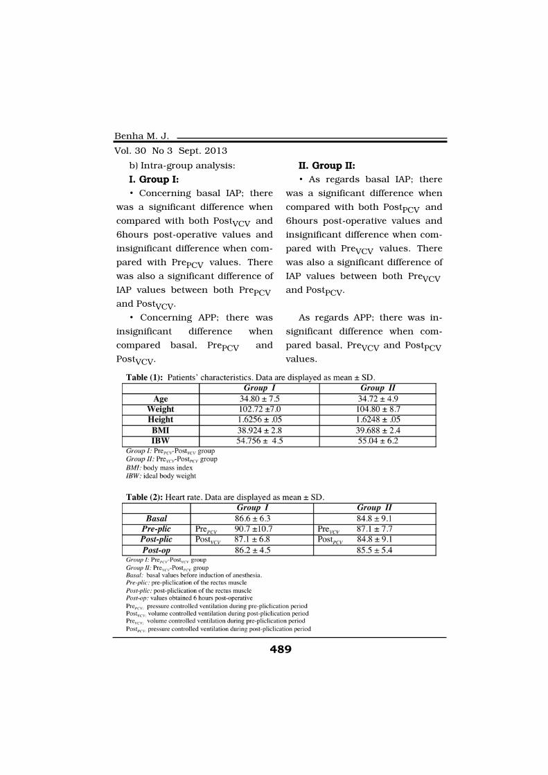
489
Benha M. J.
Vol. 30 No 3 Sept. 2013
b) Intra-group analysis:
I. Group I:I. Group I:
• Concerning basal IAP; there
was a significant difference when
compared with both PostVCV and
6hours post-operative values and
insignificant difference when com-
pared with PrePCV values. There
was also a significant difference of
IAP values between both PrePCVand PostVCV.
• Concerning APP; there was
insignificant difference when
compared basal, PrePCV and
PostVCV.
II. Group II:II. Group II:
• As regards basal IAP; there
was a significant difference when
compared with both PostPCV and
6hours post-operative values and
insignificant difference when com-
pared with PreVCV values. There
was also a significant difference of
IAP values between both PreVCVand PostPCV.
As regards APP; there was in-
significant difference when com-
pared basal, PreVCV and PostPCVvalues.
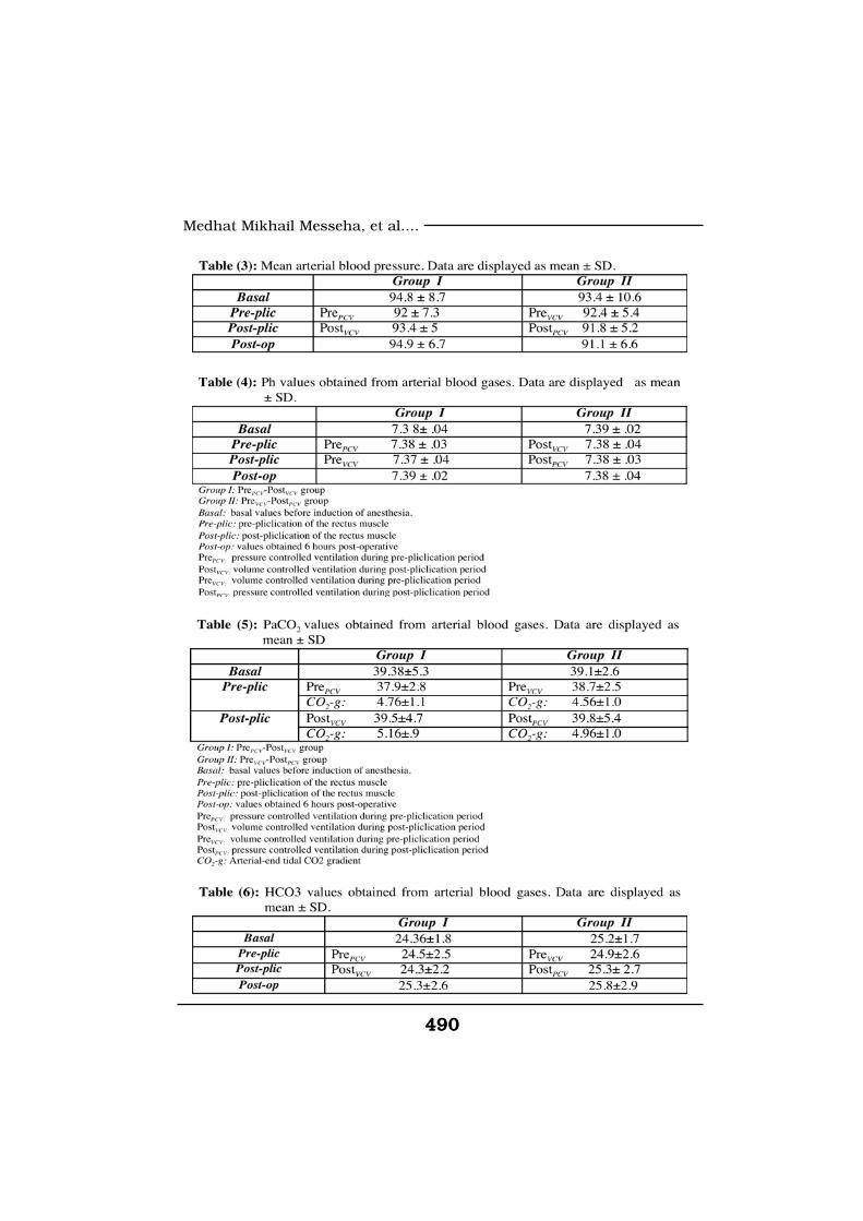
490490
Medhat Mikhail Messeha, et al....
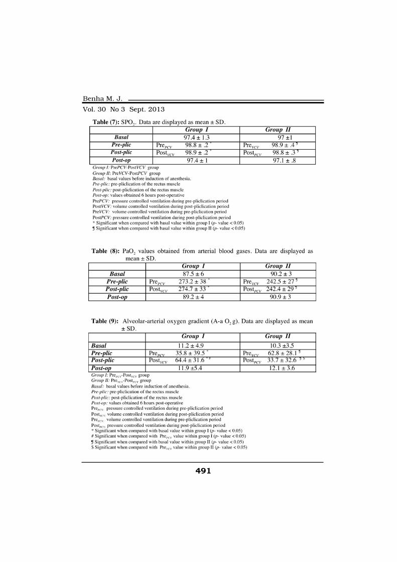
491
Benha M. J.
Vol. 30 No 3 Sept. 2013

492492
Medhat Mikhail Messeha, et al....
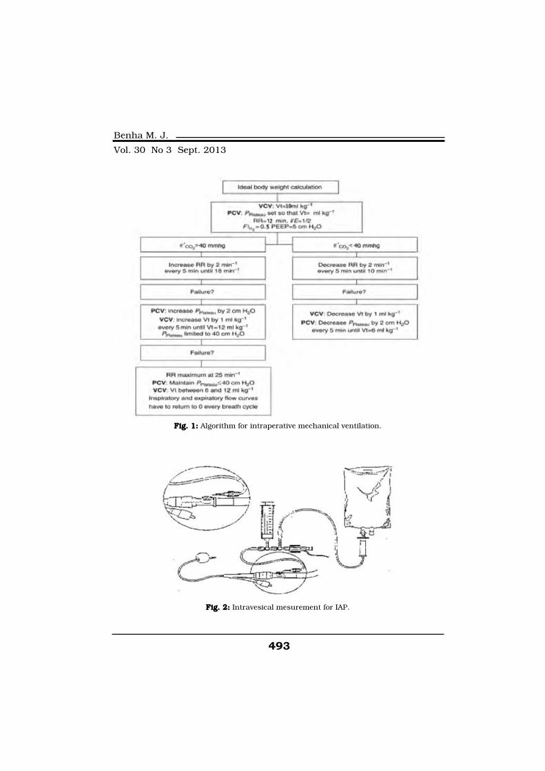
493
Benha M. J.
Vol. 30 No 3 Sept. 2013
Fig. 1:Fig. 1: Algorithm for intraperative mechanical ventilation.
Fig. 2:Fig. 2: Intravesical mesurement for IAP.
494494
Medhat Mikhail Messeha, et al....
DiscussionDiscussionAbdominal contouring proce-
dures have been performed since
the end of the nineteenth century.
In 1910 Kelly first described the
technique for large wedge excision
of the abdominal panniculus. The
modern abdominoplasty tech-
nique, including a low transverse
incision with wide undermining in
addition to rectus muscle plica-
tion, developed during the 60s.
Over time, many variations in the
technique have been described.
They all have three fundamental
objectives in common: resection of
excess skin and fat, correction of
the musculo-aponeurotic flaccidity
(diasthesis recti) and improvement
of appearance(10).
The tension caused by these
mechanical changes, leads to in-
creased intra-abdominal pressure
(IAP), which may impair ventilato-
ry function. In addition, other fac-
tors secondary to the procedure
may alter respiratory function such
as administration of anesthetic drugs
and the anesthesia itself(11).
The number of obese patients
undergoing surgery, either bariat-
ric or non-bariatric (e.g abdomino-
plasty) is steadily increasing. These
patients have a priori healthy lungs.
However, the pathophysiological
changes induced by obesity make
these patients prone to periopera-
tive complications, such as hypox-
emia, hypercapnia and atelectasis.
Immediately after the induction of
general anesthesia, atelectasis de-
velops, leading to a reduction in
both ventilation-perfusion ratio
and pulmonary compliance. It has
been demonstrate that in anesthe-
tized patients, PaO2 inversely re-
lated to BMI(12).
The selection of the optimal
ventilation mode or the optimal
control variable of ventilation for
the obese patient is of interest to
most of the anesthesiologists. Ac-
cording to Campbell and col-
leagues, VCV and PCV are not dif-
ferent ventilatory modes, but are
different control variables within a
mode. During VCV, airway pres-
sure increases in response to re-
duced compliance, increased re-
sistance, or active exhalation and
may increase the risk of ventila-
tor-induced lung injury. During
PCV, the inspiratory flow and flow
waveform are determined by the
ventilator as it attempts to main-
495
Benha M. J.
Vol. 30 No 3 Sept. 2013
tain an inspiratory pressure pro-
file. The clinician should titrate
the inspiratory pressure to the
measured tidal volume(13).
This study was designed to
evaluate the effect of pressure ver-
sus volume controlled ventilation
on hemodynamics, gasometry, lung
mechanics and intra-abdominal
pressure in obese patients under-
going abdominoplasty.
As regards hemodynamics (HR
and MBP), our results revealed
that no significant difference. This
result is in agreement with De
Baerdemaeker et al., (2008)(13)
who compared VCV and PCV dur-
ing laparoscopic gastric banding
in morbidly obese patients. The
same result was also reported by
Cadi et al., (2008)(9) who studied
the effect of VCV and PCV during
laparoscopic obesity surgery.
The effects of VCV and PCV on
gasometry have been studied and
showed controversial results. In
our current study, as regards PH,
PaCO2, arterial-end tidal CO2 gra-
dient and HCO3, our results
showed insignificant differences
between the two modes and many
studies had similar conclusions to
ours as De Baerdemaeker et al.,
(2008)(13), Esteban et al., (2000)(14) and Tugrul et al., (1997)(15).
In contrast to our results, Cadi et
al., (2008)(9) reported differences
PH, PaCO2 and arterial-end tidal
CO2 gradient with tendency of
PCV to be better.
As regards oxygenation param-
eters; SPO2 and PaO2; there were
significant differences when compared
basal values with intra-operative
values during both VCV and PCV.
This can be explained by the fact that
mechanical ventilation improves
oxygenation in obese patients(16).
Regarding the comparison between
VCV and PCV on oxygenation; our
results showed insignificant differ-
ences. These results were in
agreement with many studies. De
Baerdemaeker et al., (2008)(13) in
his previously mentioned study,
concluded that arterial oxygena-
tion remained unchanged. Karcz
et al., (2012)(17) concluded in his
systemic review "State-of-the-art
mechanical ventilation" that both
modes are likely equivalent in
supporting gas exchange.
Similarly, another systemic re-
496496
Medhat Mikhail Messeha, et al....
view and meta-analysis published
by Aldenkortt et al., (2012)(12)
confirmed our results and it was
performed for randomized trials
testing ventilation strategies in
obese patients undergoing sur-
gery. This review included all the
studies that were published be-
tween 1978 and 2011, and came
from 10 different countries. He
concluded that intraoperative oxy-
genation was similar with PCV
and VCV.
In contrast to our results, Cadi
et al., (2008)(9) reported that PCV
compared with VCV during anes-
thesia for laparoscopic bariatric
surgery improves gas exchange.
The same result was reported also
by Tugrul et al., (1997)(15) who
compared PCV and VCV during
one lung anesthesia.
In our results; A-a O2 gradient
showed significant difference as it
was higher during VCV compared
with PCV. Cadi et al., (2008)(9) re-
ported the same result and ex-
plained that by difference in the
flow pattern being constant flow in
VCV and decelerating in PCV
which leads to better gas distribu-
tion.
As regards lung mechanics,
peak, plateau, mean airway pres-
sures, static and dynamic lung
compliance, there was a signifi-
cant difference when compared
both PCV and VCV with tendency
to be much better during PCV.
These results are in agreement
with Davis et al., (1996)(18) who
compared PCV and VCV with dif-
ferent flow wave form. De Beer
and Gould, (2013)(19) published
review article about principles of
artificial ventilation and men-
tioned the same results. In con-
trast, De Baerdemaeker et al.,
(2008)(14), concluded in his study
that both VCV and PCV appear to
be equally suited in morbidly
obese patients. Also Karcz et al.,
(2012)(17) demonstrated that both
modes are likely equivalent in pul-
monary mechanics.
The possible explanation for
our results regarding lung me-
chanics is different inspiratory
flow wave form. PCV is character-
ized by decelerating pattern, while
VCV is characterized by constant
pattern. The ability of the deceler-
ating flow waveform to distend the
lung at the selected peak pressure
throughout the inspiratory time
497
Benha M. J.
Vol. 30 No 3 Sept. 2013
was described by Davis et al.,
(1996)(18). This may facilitate alve-
olar recruitment, enhance diffu-
sion, allow alveolar units with
slow time constants to fill while
preventing overdistension of nor-
mal alveoli and augment collateral
ventilation.
Body contouring surgery, spe-
cifically abdominoplasty, has be-
come increasingly popular; altera-
tion of intra-abdominal pressure
(IAP) during this procedure had
been investigated by many au-
thors. Talisman et al., (2002)(10),
Neto et al., (2006)(22) and Huang
et al., (2007)(20) showed increased
IAP at the end of abdominoplasty.
On the other hand, Al-Basti et al.,
(2004)(21) measured the IAP in
obese multiparous patients sub-
mitted to abdominoplasty and re-
ported that there was no signifi-
cant increase in IAP.
In our current study; we report-
ed significant increase in IAP after
placation of rectus muscle regard-
less the type of ventilatory mode.
This can be explained by the oper-
ative technique that has at least
two maneuvers that result in IAP
elevation: plication of the rectus
muscle and flap resection(22).
We believe that it is the first
study that compares PCV versus
VCV effect on IAP. Facing the fact
that the chest and abdomen are
two spaces separated by the dia-
phragm and the events occur in
one space do affect the other (Mal-
brain, 2006) and although we re-
ported that PCV has better effect
than VCV on lung mechanics; in
our study, we found insignificant
difference between PCV and VCV
on IAP. This could be hypothe-
sized by the dynamics of the ab-
dominal wall that allow great vol-
ume changes without a
proportional rise in IAP values
(Neto et al., 2006).
ReferencesReferences1. Bhargava D. (2008): 1. Bhargava D. (2008): Abdo-
minoplasty today .Indian J Plast
Surg. October; 41(Suppl): 20-26.
2. Candiotti K., Sharma S.2. Candiotti K., Sharma S.
and Shankar R. (2009): and Shankar R. (2009): Obesity,
obstructive sleep apnea and diabetes
mellitus: anesthetic implications.
Br J Anaesth; 249: 123-130.
3. Valladolid J.M.A., Ortiz S.,3. Valladolid J.M.A., Ortiz S.,
Orozcoand C.F., et al. (2004):Orozcoand C.F., et al. (2004):
498498
Medhat Mikhail Messeha, et al....
Variation of intra-abdominal pres-
sure caused by abdominoplasty in
healthy women. Rev Gastroenterol
Mex July; 69(3): 156-161.
4. Malbrain M.L. (2006): 4. Malbrain M.L. (2006): In-
tra-abdominal pressure: It is now
time to accept and promulgate. In-
tensive Care Med; 32:1722-1732.
5. Shen G.X., Gu B., Xie F.,5. Shen G.X., Gu B., Xie F.,
et al. (2007):et al. (2007): Three case reports
of abdominal compartment syn-
drome after full abdominoplasty.
Zhonghua Zheng Xing Wai Ke Za
Zhi. May 1; 23(3): 226-228.
6. Bailey J. and Shapiro J.M.6. Bailey J. and Shapiro J.M.
(2000):(2000): Abdominal compartment
syndrome. Crit Care.; 4(1): 23–29.
7. Adams J.P. and Murphy7. Adams J.P. and Murphy
P.G. (2000):P.G. (2000): Obesity in anesthesia
and intensive care. Br J Anaesth;
85: 91-108.
8. Prella M., Feihl F. and Do-8. Prella M., Feihl F. and Do-
menighetti G. (2002): menighetti G. (2002): Effects of
short-term pressure controlled
ventilation on gas exchange, air-
way pressures and gas distribu-
tion in patients with acute lung
injury/ARDS: comparison with
volume-controlled ventilation.
Chest; 122: 1382-1388.
9. Cadi P., Guenoun T., Jour-9. Cadi P., Guenoun T., Jour-
nois D., et al. (2008): nois D., et al. (2008): Pressure-
controlled ventilation improves ox-
ygenation during laparoscopic
obesity surgery compared with
volume- controlled ventilation. Br
J Anaesth; 100 (5): 709-716.
10. Talisman R., Kaplan B.,10. Talisman R., Kaplan B.,
Haik J., et al. (2002): Haik J., et al. (2002): Measuring
alterations in intra-abdominal
pressure during abdominoplasty
as a predictive value for possible
postoperative complications. Aesth
Plast. Surg; 26:189-192.
11. Rodrigues M.A., Nahas11. Rodrigues M.A., Nahas
F.X., Gomes H.C., et al. (2013):F.X., Gomes H.C., et al. (2013):
Ventilatory function and intra-
abdominal pressure in patients
who underwent abdominoplasty
with plication of the external
oblique aponeurosis. Aesth Plast
Surg; 37: 993-999.
12. Aldenkortt M., Lysakow-12. Aldenkortt M., Lysakow-
ski C., Elia N., et al. (2012): ski C., Elia N., et al. (2012): Ven-
tilation strategies in obese pa-
tients undergoing surgery: A
quantitative systematic review and
meta-analysis. Br J Anaesth; 109
(4): 493-502.
499
Benha M. J.
Vol. 30 No 3 Sept. 2013
13. De Baerdemaeker L.E.C.,13. De Baerdemaeker L.E.C.,
Van der Herten C., GillardinVan der Herten C., Gillardin
J.M., et al. (2008): J.M., et al. (2008): Comparison of
volume-controlled and pressure-
controlled ventilation during la-
paroscopic gastric banding in
morbidly obese patients. OBES
SURG; 18: 680-685.
14. Esteban A., Alía I., Gordo14. Esteban A., Alía I., Gordo
F., et al. (2000): F., et al. (2000): Prospective ran-
domized trial comparing pressure-
controlled ventilation and volume-
controlled ventilation in ARDS.
Chest; 117; 1690-1696.
15. Tugrul M., Camci E., Ka-15. Tugrul M., Camci E., Ka-
radeniz H., et al. (1997):radeniz H., et al. (1997): Com-
parison of volume-controlled and
pressure-controlled ventilation
during one lung anaesthesia. Br J
Anaesth; 79: 306-310.
16. Huschak G., Busch T.16. Huschak G., Busch T.
and Kaisers U.X. (2013):and Kaisers U.X. (2013): Obesity
in anesthesia and intensive care.
Best Practice & Research Clinical
Endocrinology & Metabolism; 27:
247-260.
17. Karcz M., Vitkus A., Peter17. Karcz M., Vitkus A., Peter
J., et al. (2012):J., et al. (2012): State-of-the-art
mechanical ventilation. Journal of
Cardiothoracic and Vascular An-
esthesia.; 26(3): 486-506.
18. Davis, Kenneth Jr., Bran-18. Davis, Kenneth Jr., Bran-
son, et al. (1996): son, et al. (1996): Comparison of
volume control and pressure con-
trol ventilation: Is flow waveform
the difference?. The Journal of
Trauma: Injury, Infection and
Critical Care; 41(5): 808-814
19. De Beer J.M. and Gould19. De Beer J.M. and Gould
T. (2013):T. (2013): Principles of artificial
ventilation. Anesthesia and inten-
sive care medicine; 14(3): 83-93.
20. Huang G.J., Bajaj A.K.,20. Huang G.J., Bajaj A.K.,
Gupta S., et al. (2007): Gupta S., et al. (2007): Increase
intra-abdominal pressure in abdo-
minoplasty: delineation of risk fac-
tors. Plast Reconstr Surg; 119:
1319-1325.
21. Al-Basti J.B., El-Khatib21. Al-Basti J.B., El-Khatib
H.A., Taha A., et al. (2004): H.A., Taha A., et al. (2004): In-
tra-abdominal pressure after full
abdominoplasty in obese multipa-
rous patients. Plast Reconstr
Surg; 113: 2145.
22. Neto L.G., Araujo L.R.,22. Neto L.G., Araujo L.R.,
Rudy M.R., et al. (2006): Rudy M.R., et al. (2006): Intra-
abdominal pressure in abdomino-
plasty patients. Aesthetic Plast
Surg; 30: 655-658.

500500
Medhat Mikhail Messeha, et al....
Published byPublished by
Benha Faculty of MedicineBenha Faculty of Medicine
EFFECT OF PRESSURE VERSUSEFFECT OF PRESSURE VERSUSVOLUME CONTROLLEDVOLUME CONTROLLED
VENTILATION ON RESPIRATORYVENTILATION ON RESPIRATORYMECHANICS, HEMODYNAMICS ANDMECHANICS, HEMODYNAMICS AND
INTRA-ABDOMINAL PRESSUREINTRA-ABDOMINAL PRESSUREDURING ABDOMINOPLASTYDURING ABDOMINOPLASTY
Medhat Mikhail Messeha MD, Tarek Abdel Aziz IbrahimMedhat Mikhail Messeha MD, Tarek Abdel Aziz IbrahimPh.D, Walaa Safaa Eldin Elkharboutly MD,Ph.D, Walaa Safaa Eldin Elkharboutly MD,
Ashraf Mohamed Wahba Wafa MDAshraf Mohamed Wahba Wafa MDand Saleh Ibrahim Elawady MDand Saleh Ibrahim Elawady MD
BENHAMEDICALJOURNAL
REPRINT
Volume 30 Number 3Sept. 2013
501
Benha M. J.
Vol. 30 No 3 Sept. 2013
COMPARISON OF THE EFFECTS OFCOMPARISON OF THE EFFECTS OFLORNOXICAM TO PARACETAMOL WHENLORNOXICAM TO PARACETAMOL WHEN
ADDED TO LIDOCAINE FOR INTRAVENOUSADDED TO LIDOCAINE FOR INTRAVENOUSREGIONAL ANESTHESIA IN PATIENTSREGIONAL ANESTHESIA IN PATIENTS
UNDERGOING HAND ANDUNDERGOING HAND ANDFOREARM SURGERYFOREARM SURGERY
Nasr Mahmoud Abdallah Sief El-Nasr MD,Nasr Mahmoud Abdallah Sief El-Nasr MD,Mohamed Mohamed Abd Elhaq MDMohamed Mohamed Abd Elhaq MD
and Ahmed Ragab Abd Elhakeim MDand Ahmed Ragab Abd Elhakeim MDDepartment of Anesthesia, Cairo University
AbstractAbstractBackground:Background: Lornoxicam is a new NSAID of the Oxicam class with
analgesic anti-inflammatory and antipyretic properties. Lornoxicam isavailable in oral and parental form and is recommended for short termuse in the postoperative period as it has a short term plasma elimina-tion half-life of 3-5h.Paracetamol (acetaminophen) is an analgesic whichrelieves pain and reduces fever. Several studies have demonstrated pe-ripheral antinoceptive properties of paracetamol in different pain mod-els. The study was planned to evaluate the effect of intravenous para-cetamol and lornoxicam when added to lidocaine in intravenousregional anesthesia for elective hand surgery regarding sensory and mo-tor block, tourniquet pain and postoperative analgesia.
Subjects and Methods: Subjects and Methods: This prospective double-blinded randomizedstudy was conducted in patients undergoing hand or forearm surgerywho were randomly assigned into 3 groups. The syringes in all groupscontained 3 mg/kg of lidocaine. In control group (C) patients received0.5% Lidocaine diluted with 0.9% normal saline to a total volume of 40ml (n=20) in Lornoxicam group (group L) received 0.5% lidocaine dilutedwith normal saline plus Lornoxicam 8 mg to a total volume of 40 ml (n =20) and Paracetamol group (group P) received 0.5% Lidocaine dilutedwith 250 mg of paracetamol (Perfalgan 10 mg/ml/Bristol-Myers-Squibb)diluted with saline to a total volume of 40 mL.
Results:Results: The primary outcome of our study the addition of Lornoxi-cam 8 mg or acetaminophen 250 mg to lidocaine for IVRA decreased
502502
Nasr Mahmoud Abdallah Sief El-Nasr, et al....
IntroductionIntroductionIntravenous regional anesthe-
sia (IVRA) was first used by August
Bier in 1908(1). This technique is
easy to administer, reliable and
cost-effective for short surgical
procedures of the extremities per-
formed on an ambulatory basis(2).
Delayed onset of action, poor
muscle relaxation and Lack of
postoperative analgesia are the
major limitations of this tech-
nique(3). Various additives have
been combined with local anes-
thetics (LAs) to improve the quali-
ty of block, including opioids,
clonidine, neostigmine and muscle
relaxants. Also various non-
steroidal anti-inflammatory drugs
(NSAIDs) have been demonstrated
to enhance analgesia such as ke-
torlac(4) tenoxicam(5) and aspirin(6) when added to local anesthet-
ics in IRVA.
Lornoxicam is a new NSAID of
the Oxicam class with analgesic
anti-inflammatory and antipyretic
properties. Lornoxicam is availa-
ble in oral and parental form and
is recommended for short term
use in the postoperative period as
it has a short term plasma elimi-
nation half-life of 3-5h(6,7) Lornox-
icam is also as effective as mor-
phine but better tolerated when
administered intravenously by pa-
tient-controlled analgesia for de-
creasing postoperative pain after
discectomy(8). Infiltration of the
wound with combination of lor-
noxicam and local anesthetic im-
proves the postoperative pain and
decreases the need for opioids
suggesting a local effect(9).
Paracetamol (acetaminophen) is
an analgesic which relieves pain
and reduces fever. Several studies
have demonstrated peripheral an-
tinoceptive(10,11) properties of
paracetamol in different pain mod-
els perflgan (10 mg/ml, Bristol-
Myers-Squibb, Anagni, Italy) is a
solution of acetaminophen admin-
istered intravenously in order to
relieve pain or reduce fever follow-
ing surgery and was introduced
into clinical practice in 2002.
tourniquet pain, improved the speed of onset of sensory and motorblock, prolonged the sensory and motor recovery times, decreased bothintraoprative and postoperative analgesic requirements and improvedthe quality of anesthesia without causing any side effects.
Keywords:Keywords: Lornoxicam, Paracetamol, Regional anesthesia, Lidocaine.
503
Benha M. J.
Vol. 30 No 3 Sept. 2013
We planned this study to evalu-
ate the effect of intravenous para-
cetamol and lornoxicam when
added to lidocaine in intravenous
regional anesthesia for elective
hand surgery regarding sensory
and motor block, tourniquet pain
and postoperative analgesia.
Subjects and MethodsSubjects and MethodsThis study was conducted at
Kasr El-Aini hospital, Cairo Uni-
versity after approval of local Ethi-
cal committee & obtaining informed
consent from all patients. Sixty
adult patients of ASA physical
status I and II, aged 20-60 years,
who were scheduled for surgery of
the hand or the forearm, were in-
cluded in this study. Patients with
Raynaud’s disease, history of drug
allergy, Sickle cell anemia, Liver
diseases and kidney diseases were
excluded from the study.
This prospective double-blinded
randomized study was conducted
in patients undergoing hand or
forearm surgery who were ran-
domly assigned into 3 groups. The
syringes in all groups contained 3
mg/kg of lidocaine (2% Lidocaine,
Rotexmedica, Trittan, Germany).
In control group (C) patients re-
ceived 0.5% Lidocaine diluted with
0.9% normal saline to a total vol-
ume of 40 ml (n=20) in Lornoxi-
cam group (group L) received 0.5%
lidocaine diluted with normal sa-
line plus Lornoxicam 8 mg (AUG
Pharma under license of Nycomed,
Austria) to a total volume of 40 ml
(n = 20) and Paracetamol group
(group P) received 0.5% Lidocaine
diluted with 250 mg of paraceta-
mol (Perfalgan 10 mg/ml/Bristol-
Myers-Squibb) diluted with saline
to a total volume of 40 mL.
Forty-five minutes before the
surgical procedures, patients were
premedicated with IM 0.07 mg/Kg
of midazolam. After the patient
had been taken to the operating
room, mean arterial blood pres-
sure (MAP), peripheral oxygen sat-
uration (SPO2) and heart rate (HR)
were monitored. Two cannulas
were placed, one 22-gauge intra-
venous (IV) cannula in the dorsum
of the hand of the surgical extrem-
ity and a second 20-gauge IV can-
nula in the non-operative arm for
crystalloid infusion.
The operative arm was elevated
for 2 min then exsanguinated with
an esmarch bandage. A pneumat-
ic double tourniquet was placed a
round upper arm, and the proxi-
504504
Nasr Mahmoud Abdallah Sief El-Nasr, et al....
mal cuff was inflated to 250 mmHg.
Circulatory isolation of the arm was
verified by inspection, absence of
radial pulse, and loss of pulse ox-
imetry tracing in the epsilateral
index finger. After the bandage
was removed, 40 ml of the respec-
tive solutions were injected over
20 seconds by an anesthesiologist.
The Sensory block was assessed
by pinprick performed with a 22-
gauge needle every 30 seconds un-
til the dermatomal sensory block
of medial and antebrachial cuta-
neous, ulnar, median and radial
nerves achieved. Motor function was
assessed by asking the patients to
flex and extend his/her wrist and
fingers and complete motor block
was noted when no voluntary
movement was possible. Sensory
block onset time was noted as the
time elapsed from drug injection
to complete sensory block
achieved in all dermatomes. Motor
block onset time was the time
elapsed from injection of study
drug to complete motor block.
After complete sensory and mo-
tor blocks were achieved, the distal
tourniquet was inflated to 250 mmHg,
and the proximal tourniquet was re-
leases and the surgery was started.
MAP, HR and SPO2 level were
recorded before application of
tourniquet and after the applica-
tion of tourniquet every five min-
ute and were measured after re-
lease of the tourniquet, and
postoperatively at 30 min, 1hr,
2hr, 3hr, 4hr, 6hr and 24 hours.
Pain due to the tourniquet was
assessed with visual analogue scale
(VAS) scores (0= no pain and 10=
worst pain imaginable). Levels of
sedation were assesses with the
Ramsey sedation scale as follows:
1) patient is anxious and agitated
or restless or both, 2) patients is
cooperative, oriented and tranquil,
3) patient responds to command only,
4) patient exhibits brisk response
to light glabellar tap or loud audi-
tory stimulus (5) patient exhibits a
sluggish response to light glabellar
tap or loud auditory stimulus, and
6) patients exhibits no response.
Both VAS and sedation levels
were recorded before and after the
application of tourniquet and dur-
ing the operation (10,15,20,
30,40,50 and 60 min). When pain
due to tourniquet was >3 on the
VAS, patients were given fentanyl
in 10 µg increments up to 1µg/kg
(Fentaryl-hmllin 50 mcg/ml, lan-
505
Benha M. J.
Vol. 30 No 3 Sept. 2013
gesfeid, Germany) and total ad-
ministered dose requirement time
were noted. Oxygen was adminis-
tered with face mask if SPO2 was
lower than 91%. At the end of the
operation patients were asked to
qualify the operative conditions
such as tourniquet pain or inci-
sional pain and the quality of the
anesthesia was graded by the an-
esthesiologist who was blinded to
the study drug as follows: (4) ex-
cellent = no complaint from the
patient; (3) good = minor com-
plaint with no need for supple-
mental analgesics; (2) moderate =
complaint that required a supple-
mental analgesic ……; (1) poor =
unsuccessful, patient was given
general anesthesia.
At the end of the operation the
surgeons, who was blinded to the
patient group, were asked to qual-
ity and score the operative condi-
tions such as excessive bleeding
and disturbing movement of the
arm as follows; 4 = excellent; 3 =
good; 2 = acceptable and 1 = poor.
The tourniquet was not deflated
before 30 min and was not inflat-
ed more than 1.5 hours. At the
end of the operation, the tourni-
quet deflation was performed by
cyclic deflation technique (the
tourniquet was deflated three
times in a cyclic manner with 10
seconds period of deflation). Sen-
sory recovery time was noted (time
elapsed after tourniquet deflation
up to recovery of pain in all inner-
vated areas determined by pin-
prick test). Motor block recovery
time was noted (the time elapsed
after tourniquet deflation up to
movement of fingers).
Also, first analgesic require-
ment time was noted (the time
elapsed after tourniquet release to
the first patient request of analge-
sic). In post-anesthetic care unit,
patients received tramadol 50 mg
when VAS >3 with maximum dose
of 600 mg/day and total amount
of tramadol administered to each
group were recorded.
During the first 2 hours in the
post-anesthetic care unit and later
in the surgical ward, patients were
questioned for circumoral numb-
ness and tingling, nausea and
vomiting, skin rash, tinnitus, gas-
tric discomfort and other side ef-
fects were noted if encountered
and MAP, HR and VAS scores
were assed every 2 hours postop-
eratively during the first 24 hours.
506506
Nasr Mahmoud Abdallah Sief El-Nasr, et al....
Statistical Methods:Statistical Methods:
Data was analyzed using IBM
SPSS Advanced Statistics version
20.0 (SPSS Inc., Chicago, IL). Chi-
square test (Fisher’s exact test)
was used to examine the relation
between qualitative variables. For
quantitative data, comparison be-
tween 3 groups was done using
ANOVA or Kruskal-Wallis test.
Comparison of repeated measures
was done using ANOVA for repeat-
ed measures. A p-value <0.05 was
considered significant.
ResultsResultsThe study included 3 groups,
Groups C (control), L (Lornoxicam
group) and P (paracetamol group).
Table 1 shows baseline and opera-
tive characteristics of the three
studied groups. The three groups
were comparable in age, sex and
body weight. There was no signifi-
cant difference in the operative
and tourniquet time between the
three groups.
Table 2 shows a comparison
between the three groups regard-
ing characteristics of anesthesia
and analgesia. The three groups
were significantly different from
each other regarding onset time of
sensory and motor block. Control
group had the longest onset time
of sensory and motor block. Also,
recovery time of sensory and mo-
tor block were significantly shorter
in control group compared to the
lornoxicam and paracetamol
groups. However, recovery time
was comparable between the latter
two groups. Time to fentanyl re-
quest was significantly shorter
and fentanyl dose was significant-
ly larger in control group com-
pared to the other two groups, al-
though both time to fentanyl
request and dose were comparable
between lornoxicam and paraceta-
mol groups. Similarly, a signifi-
cantly higher number of control
group patients needed fentanyl
(p=0.007) or tramadol (p<0.001)
for analgesia. Tramadol consump-
tion was significantly higher in
control group compared to lornox-
icam and paracetamol groups, the
latter two groups showed compar-
able tramadol consumption.
VAS score was comparable in
the three groups at the beginning
of surgery. Starting from 10 min-
utes up to 50 minutes intraopera-
tively, VAS score of lornoxicam
and paracetamol groups was sig-
nificantly lower than control group,
with no significant difference be-
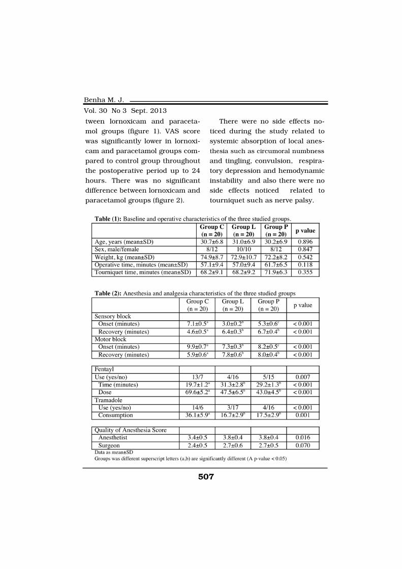
507
Benha M. J.
Vol. 30 No 3 Sept. 2013
tween lornoxicam and paraceta-
mol groups (figure 1). VAS score
was significantly lower in lornoxi-
cam and paracetamol groups com-
pared to control group throughout
the postoperative period up to 24
hours. There was no significant
difference between lornoxicam and
paracetamol groups (figure 2).
There were no side effects no-
ticed during the study related to
systemic absorption of local anes-
thesia such as circumoral numbness
and tingling, convulsion, respira-
tory depression and hemodynamic
instability and also there were no
side effects noticed related to
tourniquet such as nerve palsy.
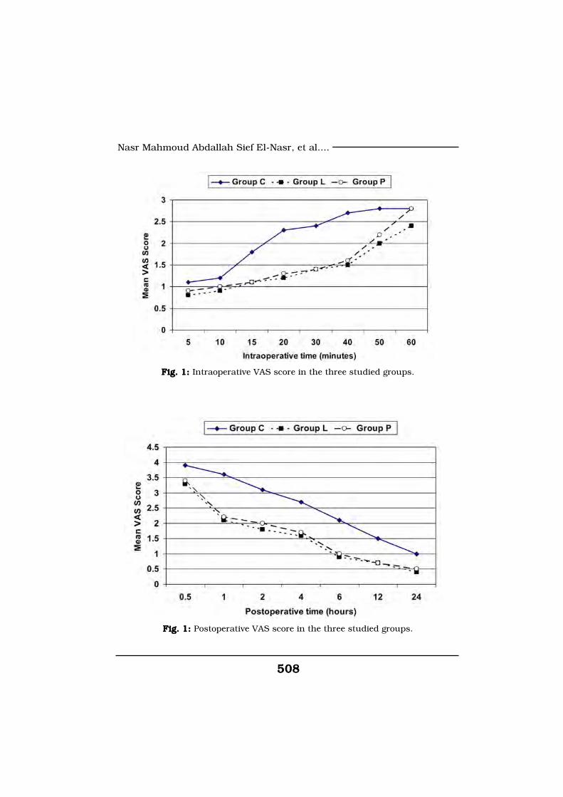
508508
Nasr Mahmoud Abdallah Sief El-Nasr, et al....
Fig. 1: Fig. 1: Intraoperative VAS score in the three studied groups.
Fig. 1: Fig. 1: Postoperative VAS score in the three studied groups.
509
Benha M. J.
Vol. 30 No 3 Sept. 2013
DiscussionDiscussionThe primary outcome of our
study the addition of Lornoxicam
8 mg or acetaminophen 250 mg to
lidocaine for IVRA decreased tour-
niquet pain, improved the speed of
onset of sensory and motor block,
prolonged the sensory and motor
recovery times, decreased both in-
traoprative and postoperative an-
algesic requirements and im-
proved the quality of anesthesia
without causing any side effects.
The addition of Lornoxicam
shortened the sensory block onset
time more than did addition of ac-
etaminophen. Various NSAIDs
such as lornoxicam(12,13), ace-
taminophen and ketorlac(4,15)
have been used to improve the
quality of analgesia in IVRA. It’s
known that increasing pH of local
anesthetics improves the nerve
penetration and onset of blockade(2). Alkalization with bicarbonate
has been found to be an effective
adjunct for IVRA(16).
It was found pH of lidocaine-
normal saline solution was 6.7,
pH of lidocaine- normal saline-
lornoxicam mixture was 7.6 and
that of lidocaine-normal saline-
acetaminophen mixture was 5.88.
So, alkalization of LA with lornoxi-
cam enhanced the sensory and
motor block onset times by in-
creasing the percentage of the free
base. This might explain the rapid
onset time of sensory block in
group (L) compared with group (P)
and group (C).
NSAIDs are largely used togeth-
er with local anesthetics in IVRA
to improve the quality of anesthe-
sia. A systematic review by Choyce
and Perg(2) suggested that
NSAIDs offer a good adjuncts to
IVRA. The major analgesic effect of
NSAIDs is attributed to isoenzyme
CoX-2 inhibition(17). Reuben and
Dupart(18) have found in another
study that NSAIDs decrease the
afferent nociceptive signals and
synthesis of inflammatory media-
tors arising at the site of surgery.
Reuben and Duprat(18) had
found that patients had less pain
with decreased analgesic require-
ments in first postoperative hour
when ketlorac was added to LA in
their study. This was attributed to
residual ketorlac in the operative
arm and to lesser extent redistri-
bution of ketoralc to systemic cir-
culation after deflation of the tour-
niquet(15). These findings are
510510
Nasr Mahmoud Abdallah Sief El-Nasr, et al....
related to our study as there was
prolonged postoperative analgesia
and decreased analgesic require-
ments with adding paracetamol or
lornoxicam to local anesthestics in
IVRA.
Paracetamol is generally con-
sidered as a week inhibitor of
prostaglandins synthesis. Several
studies have demonstrated several
mechanisms for anti-nociceptive
effect of paracetamol, including N-
methyl-D-aspartate(19), and the
effect on cannabinoid receptors(11,20).
A recent study done by Can
bay et al(21) have demonstrated
that paracetamol has peripheral
anti-nociceptive effect. They have
found also that pre-treatment with
50mg of IV paracetamol decreased
propofol-induced injection pain. In
another study acetaminophen
may reduce pain through interfer-
ence with delivery of peripheral B-
endorphins(22).
Buritova and Besson(23) have
demonstrated that Lornoxicam
shows anti-nociceptive effect at a
peripheral site and this action
may be medicated via No-CGMP
pathway and the opening of K+
channels.
It has been found that ischemia
and oxidation stress may lead to
tourniquet pain(24) Coderre and
Colleagues(25) have demonstrated
that antioxidant therapy my de-
crease ischemic tourniquet pain
due to oxidative damage. Lornoxi-
cam exhibits antioxidant effects in
rats(26), so when lornoxicam was
added to LA in IVRA might attenu-
ate the tourniquet pain by antioxi-
dant mechanism.
A Limitation of this study de-
sign was attributed to the fixed
dose of intravenous acetamino-
phen 250 mg. the effective dose
has yet to be determined. As when
doses over 250 mg of paracetamol
were mixed, the total volume of
IVRA was too large and the control
of the content of LA would be very
difficult.
In conclusion, this study indi-
cated (has found) that addition of
lornoxicam to lidocaine in IVRA
significantly shortened the onset
of sensory block than did addition
of acetaminophen. The addition of
lornoxicam or acetaminophen to
LA or IVRA have decreased the
motor block onset time and pro-
511
Benha M. J.
Vol. 30 No 3 Sept. 2013
longed both sensory and motor
block recovery times. Both lornox-
icam and acetaminophen have
prolonged tourniquet pain onset
time and improved postoperative
analgesia.
ReferencesReferences1. Brill S., Middleton W., Brill1. Brill S., Middleton W., Brill
G. and Fisher A. (2004): G. and Fisher A. (2004): Bier’s
Block; 100 years old and still go-
ing strong Acta Anaesthesiol
Scand; 48: 117-22.
2. Choyce A. and Peng P.2. Choyce A. and Peng P.
(2002):(2002): A systematic review of ad-
juncts for intravenous regional an-
esthesia for surgical procedure.
Can J Anesth; 49: 32-45.
3. Perlas A., Peng P.W., Plaz3. Perlas A., Peng P.W., Plaz
M.B., Middleton W.J., ChenM.B., Middleton W.J., Chen
C.W. and Sanandaji K. (2003):C.W. and Sanandaji K. (2003):
Forearm rescue cuff improves
tourniquet tolerance during intra-
venous regional anesthesia. Reg
Anesth Pain Med; 28: 98-102.
4. Reuben S.S., Steinberg4. Reuben S.S., Steinberg
R.R., Kreitzer J.M. and DupeatR.R., Kreitzer J.M. and Dupeat
K.M. (1995):K.M. (1995): Intravenous regional
anesthesia using Lidocaine and
Ketorolac. Anesth Analg; 81: 110-3.
5. Jones N.C. and Pugh S.C.5. Jones N.C. and Pugh S.C.
(1996):(1996): The addition of tenoxicam
to prilocaine for intravenous re-
gional anesthesia. Anesthesia; 51:
446-8.
6. Corpataux J.B., Van Ges-6. Corpataux J.B., Van Ges-
sel E.F., Donald F.A., Forster A.sel E.F., Donald F.A., Forster A.
and Gamulin Z. (1997):and Gamulin Z. (1997): Effect on
postoperative analgesia of small-
dose lysine acetylsalicylate added
to prilocaine during intravenous
regional anesthesia. Anesth Analg;
84: 1081-5.
7. Rosenow D.E., Albrechtsen7. Rosenow D.E., Albrechtsen
M. and Stolke D. (1998):M. and Stolke D. (1998): A com-
parison of patient. Controlled
analgesia with Lornoxicam versus
morphine in patients undergoing
lumber disc surgery. Anesth
Analg; 86: 1045-50.
8. Bias W. and Jansen M.8. Bias W. and Jansen M.
(1996):(1996): Pain control after hyster-
ectomy: an observer blind ran-
domized trial of Lornoxicam ver-
sus tramadol. Br J Clin Pract; 50:
197-202.
9. Karamanlioglu B., Turan9. Karamanlioglu B., Turan
A., Memis D., Kaya G., Ozata S.A., Memis D., Kaya G., Ozata S.
and Ture M. (2005):and Ture M. (2005): Infiltration
with ropivacaine plus Lornoxicam
reduces postoperative pain and
opioid consumption. Can J An-
512512
Nasr Mahmoud Abdallah Sief El-Nasr, et al....
aesth; 52: 1047-53.
10. Grabam G.G. and Scott10. Grabam G.G. and Scott
K.F. (2005):K.F. (2005): Mechanism of action
of Paracetamol. Am J Ther; 12:
46-55.
11. Dani M., Guindon J.,11. Dani M., Guindon J.,
Lambert C. and Beaulieu P.Lambert C. and Beaulieu P.
(2007): (2007): The Local antinociceptive
effects of paracetamol in neuro-
pathic pain are mediated by can-
nabinoid receptors. Eur J Phar-
macol; 5: 73-5.
12. Sen S., Ligur B., Aydin12. Sen S., Ligur B., Aydin
O.N., et al. (2006): O.N., et al. (2006): The analgesic
effects of lornoxicam when added
to Lidocaine for intravenous re-
gional anesthesia. Br J Anaesth;
97: 408-13.
13. Balfour J.A., Fitton A.13. Balfour J.A., Fitton A.
and Barradell L.B. (1996):and Barradell L.B. (1996): Lor-
noxicam: a review of its pharma-
cology and therapeutic potential
in the management of painful and
inflammatory conditions. Drugs;
15: 639-57.
14. Aronoff D.M., Oates J.A.14. Aronoff D.M., Oates J.A.
and Boutand O. (2006): and Boutand O. (2006): New in-
sights into the mechanism of ac-
tion of acetaminophen: its clinical
pharmacologic characteristics re-
flect its inhibition of the two pros-
taglandin H2 Synthases. Clin
pharmacol Ther; 79: 9-19.
15. Steinberg R.B., Reuben15. Steinberg R.B., Reuben
S.S. and Gardner G. (1998): S.S. and Gardner G. (1998): The
dose response relationship of ke-
torlac as a component of intrave-
nous regional anesthesia with Lid-
ocaine. Anesth Analg; 86: 791-3.
16. Armstrong P., Brackway16. Armstrong P., Brackway
M. and Wild Smith J.A. (1990):M. and Wild Smith J.A. (1990):
Alkalinisation of prilocaine for in-
travenous regional anesthesia. An-
esthesia; 45: 11-13.
17. Bennett A. and Villa G.17. Bennett A. and Villa G.
(2000):(2000): Nimesulide: an NSAID
that preferentially inhibits CoX-2,
and has various unique pharmac-
ological activities. Expert Opin
Pharmacother; 1: 277-86.
18. Reuben S.S. and Duprat18. Reuben S.S. and Duprat
K.M. (1996): K.M. (1996): Comparison of
wound infiltration with ketorlac
versus intravenous regional anes-
thesia with ketorlac for postopera-
tive analgesia following ambulato-
ry hand surgery. Reg Anesth;
21:565-8.
19. Bertonlini A., Ferrari A.,19. Bertonlini A., Ferrari A.,
Ottani A., Gnerzoni S., Tacchi R.Ottani A., Gnerzoni S., Tacchi R.
513
Benha M. J.
Vol. 30 No 3 Sept. 2013
and Leone S. (2006):and Leone S. (2006): Paraceta-
mol: ned vistas of an old drug.
CNS Drug Rev; 12: 250-75.
20. Ottani A., Leone S., San-20. Ottani A., Leone S., San-
drini M., Ferrari A. and Bertolinidrini M., Ferrari A. and Bertolini
A. (2006):A. (2006): The analgesic activity
of paracetamol is prevented by
blockade of cannabinoid CBI re-
ceptors. Eur J Pharmacol; 531:
280-1.
21. Canbay O., Celebi N.,21. Canbay O., Celebi N.,
Arun O., Karagöz A.H., Saricao-Arun O., Karagöz A.H., Saricao-
glu F. and Ozgen S. (2008): glu F. and Ozgen S. (2008): Effi-
cacy of intravenous acetamino-
phen and Lidocaine on Propofol
injection pain. Br J Anaesth; 100:
95-8.
22. Sprott H., Shen H., Gay22. Sprott H., Shen H., Gay
S. and Aeschlimann A. (2005):S. and Aeschlimann A. (2005):
Acetaminophen may act through
beta endorphin Ann Rheum Dis;
64: 1522.
23. Buritova J. and Besson23. Buritova J. and Besson
J.M. (1998): J.M. (1998): Dose-related anti-
inflammatory/ analgesic effects of
lornoxicam: a spinal C-Fod pro-
tein study in the rat. Inflamm Res;
47: 18-25.
24. Chabel C., Russel L.C.24. Chabel C., Russel L.C.
and Lee R. (1990):and Lee R. (1990): Tourniquet-
induced limb ischemia: a neuro-
physiologic animal model. Anes-
thesiology; 72: 1038-44.
25. Caderre T.J., Xanthos25. Caderre T.J., Xanthos
D.N., Francis L. and BennettD.N., Francis L. and Bennett
G.J. (2004):G.J. (2004): Chromic post-
ischemic pain (CPIP): a novel ani-
mal model of complex regional
pain syndrome-type I (CRPS-1);
reflex sympathetic dystrophy) pro-
duced by prolonged hindpaw is-
chemia and reperfusion in the rat.
Pain; 112: 94-105.
26. Rokyta R., Holeck V., Pe-26. Rokyta R., Holeck V., Pe-
karkova I., et al. (2003):karkova I., et al. (2003): free rad-
icals after painful stimulation are
influenced by antioxidants and
analgesics. Neuro Endocrinal Lett;
24: 304-9.
514514
Nasr Mahmoud Abdallah Sief El-Nasr, et al....
Published byPublished by
Benha Faculty of MedicineBenha Faculty of Medicine
COMPARISON OF THE EFFECTS OFCOMPARISON OF THE EFFECTS OFLORNOXICAM TO PARACETAMOLLORNOXICAM TO PARACETAMOL
WHEN ADDED TO LIDOCAINE FORWHEN ADDED TO LIDOCAINE FORINTRAVENOUS REGIONAL ANESTHE-INTRAVENOUS REGIONAL ANESTHE-
SIA IN PATIENTSSIA IN PATIENTSUNDERGOING HAND ANDUNDERGOING HAND AND
FOREARM SURGERYFOREARM SURGERY
Nasr Mahmoud Abdallah Sief El-Nasr MD,Nasr Mahmoud Abdallah Sief El-Nasr MD,Mohamed Mohamed Abd Elhaq MDMohamed Mohamed Abd Elhaq MD
and Ahmed Ragab Abd Elhakeim MDand Ahmed Ragab Abd Elhakeim MD
BENHAMEDICALJOURNAL
REPRINT
Volume 30 Number 3Sept. 2013
515
Benha M. J.
Vol. 30 No 3 Sept. 2013
SOME PLATELET PROPERTIES OF RESTINGSOME PLATELET PROPERTIES OF RESTINGAPHERESIS PLATELET CONCENTRATEAPHERESIS PLATELET CONCENTRATE
FOR THE LAST 6 HOURS OF AFOR THE LAST 6 HOURS OF A48-HOUR STORAGE PERIOD48-HOUR STORAGE PERIOD
Manal H. Farahat MD* and Mohammad A. Elhady MD**Manal H. Farahat MD* and Mohammad A. Elhady MD***Departments of Clinical Pathology, Faculty of Medicine,
Zagazig University Hospitals.
**Department of Biochemistry, Zagazig University Hospitals
AbstractAbstractBackground: Background: Apheresis platelet (PLT) units are not routinely agitated
during transit. Aim of the study was to evaluate effects of resting (6 h ofinterruption of agitation) vs continue agitation of platelet concentrate(PC) stored for 48 h in the blood bank. Materials and Methods: Materials and Methods: aphere-sis PLT units were collected with Trima cell separator (n=20, terumoBCT), continuously agitated, starting routinely within 1 hour of collec-tion, extending for 42 h at room temperature (20-24 ˚C). An identicalapheresis PLT unit was stored with continuous agitation (designated asthe control group, CA6h), and the other was held without continuousagitation for 6 h (WCA6h); by stopping the agitator. We studied some invitro platelet quality variables as PLT count, PLT unit volume; swirling,PH, lactate dehydrogenase (LDH) concentration, CD62P and CD42b.Results: Results: were compared with those of PC continuously agitated. Re-sults: The mean platelet yield of individual apheresis-PC unit for (CA6h)and (wCA6h) were 3.25±0.20x 10¹¹, 3.25±0.19x 10¹¹ respectively withno statistically different (P=0.92). LDH, and CD62P did not differ signifi-cantly (P=0.54), (P=0.07), between CA6h (51.11±6.68), (17.33±3.46) andWCA6h (52.39±6.42), (19.54±4.12) respectively. WCA6h showed highlysignificant lower CD42b expression (149.11±38.99) vs CA6h(223.41±42.88) (P=0.001) Likewise, the mean pH values were signifi-cantly different: WCA6h (6.88±0.15) and CA6h (6.99±0.12) (P=0.025).Conclusions:Conclusions: PC stored under agitation for 42 h at 22-24 ˚C and restedfor 6 h had preserved PLT count and platelet yield, LDH and CD62p asPC kept under continuous agitation for the whole 48 h storage period.
Keywords:Keywords: apharesis platelet, resting, interrupted agitation, plateletconcentrate.

516516
Manal H. Farahat and Mohammad A. Elhady
IntroductionIntroductionPlatelet Concentrate (PC) from
single donor is prepared by plate-
let apheresis, processed over
about 1 hour yielded one thera-
peutic dose of platelets, called PC(1). PCs are collected using differ-
ent synthetic materials, centrifu-
gation/leucocyte-removal process-
es, and stored in different types of
bags. Upon exposure to artificial
surfaces and high centrifugation
forces, blood cells can undergo
various levels of cellular activa-
tion/fragmentation and release re-
actions which may not only influ-
ence the extent of the platelet
storage lesion but may also con-
tribute to poor clinical effective-
ness of the PC and transfusion re-
actions(2).
Apheresis platelets (PLTs) differ
in many ways from whole
blood–derived PLT concentrates:
they are a) collected with a differ-
ent anticoagulant and/or preser-
vative, b) stored in different con-
tainers, c) may contain different
PLT subpopulations, and d) can
be stored for up to 7 days(3).
It is assumed that continuous,
gentle agitation of the PC main-
tains the pH levels at or above 6.2
throughout the period that the
platelets are stored at room tem-
perature (22-24˚C) in special, gas-
permeable plastic bags to ensure
their in vitro quality and in vivo ef-
fectiveness. This procedure facili-
tates the transfer of oxygen to the
PLTs and the removal of carbon
dioxide from the storage bag(4).
The most noted effect from inter-
ruption of agitation for lengthy pe-
riods (24 hr) is an increase in the
rate of pH decline during storage.
This reflects an increase in pro-
duction of lactic acid due to en-
hanced glycolysis and reduced ox-
idative metabolism(3).
As continuous agitation is not
practical, because substantive pe-
riods of discontinued agitation oc-
cur between remote collection and
manufacturing sites and during
shipping to hospitals, there has
been interest in better defining the
effects of interrupted agitation(3).
This creates problems. Is an inter-
ruption of agitation for 1 minute,
1 hour, 1 day, or even 2 or 3 days
consistently harmful? Should brief
periods of interruption of agitation
be cause for discarding the PCs.
AABB standards permit up to 24
517
Benha M. J.
Vol. 30 No 3 Sept. 2013
hours of interrupted agitation(5).
PCs are stored for up to 5 days
under agitation conditions at
room temperature until transfu-
sion, where complex structural and
functional changes of platelets oc-
cur, summarized as platelet stor-
age lesion (PSL) Preparation mode
and storage conditions play an im-
portant role in the development of
PSLs(1). Increased release of plate-
let α-granules and cytosolic pro-
teins, increased procoagulant ac-
tivity, and altered glycoprotein
(GP) expression, all of which are
characteristic of platelet activa-
tion(6). Both activation state and
the reactivity of the platelets can
be determined using flow cytome-
try. Activation markers of interest
that can be studied in such a way
include P-selectin (CD62P), as well
as GPIb expression (CD42)(7).
There are various receptors on
the glycoprotein (GP) layer of the
platelet, include the selectins
(such as GMP 140, or P-selectin),
the integrins (such as GP I, GP II,
GP III, GP IV, GP V), and other re-
ceptors, which assist in platelet
adhesion, aggregation and coagu-
lation. Glycoprotein GP Ib is im-
portant in the attachment of plate-
lets to von Willebrand factor (vWF)
and the vascular subendothelium.
Von Willebrand Factor binds to
GP Ib in the presence of ristocetin(8). CD62 (P-selectin, GMP-140,
PADGEM) is (CD62p), a constitu-
ent of α-granules in resting plate-
lets, can be detected on the acti-
vated platelet surface after α-
granule secretion(9).
Aim of The StudyAim of The StudyDetermining the influence of
resting (6 h of interruption of agi-
tation) vs. continuing agitation of
apheresis PC, and comparing
some properties of apheresis PLT
units that were stored with contin-
uous agitation (CA6h) as a control
and units stored with 6 h of rest-
ing “without continuous agitation”
(WCA6h) as a test units after 42 h
of storage from the time they were
collected for whole 48 h storage
time under Blood Bank-
compatible conditions.
Materials and MethodsMaterials and MethodsPLT collection and prepara-PLT collection and prepara-
tion of matched, identical com-tion of matched, identical com-
ponents:ponents:
Twenty PCs were prepared in
the Zagazig University Blood
518518
Manal H. Farahat and Mohammad A. Elhady
Transfusion Center. Platelet col-
lection and storage from healthy
male donors (age 20-40 years)
meeting the directive for platelet
cytapheresis (platelet count >200
103/µl; body weight >50 kg) and
who gave written and informed
consent. PCs were prepared ac-
cording to the manufacturer's in-
structions by the Trima Accel
(Software Version 5.1, terumo
BCT) cell separator, using the
blood anticoagulant (acid citrate
dextrose (ACD)) ratio 11:1. PLTs
suspended in 100% plasma with a
plasma target volume of 200- 300
ml and a platelet target yield of
3x10¹¹ to 3.5x10¹¹ The target lev-
el of PLTs was 6.0 to 7x10¹¹ per
unit. Two identical components in
PLT containers (Trima) were pre-
pared. These machines perform
leukoreduction during the dona-
tion process via a leukoreduction
system chamber (LRS chamber)(10)."Preparations with total PLT
contents of less than 6.0x10¹¹
were not utilized in this study''.
Day of collection:Day of collection:
For the primary series of exper-
iments, the collection was divided
equally by weight between the two
integrally attached containers
within 30 to 40 minutes of the
apheresis procedure.
Storage with or without CA:Storage with or without CA:
PLT component of each
matched pair was placed on a flat-
bed agitator (Helmer, 65-70 cy-
cles/min) at 20 to 24°C within 30
minutes of the completion of col-
lection and maintained under agi-
tation for 42 h. The 20 PCs were
then divided into two groups of
twenty PC each. The first group
PC units, designated as the con-
trol units, continued under con-
tinuous agitation for the subse-
quent 6 h (CA6h), while the
second group, designated as the
test units was removed from the
agitator and placed for 6 hours
stored stationary (resting) under
the same environmental condi-
tions in the incubator without agi-
tation (WCA6h). After a total of 48
h of storage the CA6h and WCA6h
PCs were taken out of the incuba-
tor and tested immediately.
In vitro PLT testing:In vitro PLT testing:
Before obtaining all samples,
the components were thoroughly
mixed manually for a few seconds.
Samples aseptically taken from
both components (CA and WCA).
519
Benha M. J.
Vol. 30 No 3 Sept. 2013
The qualitative and quantitative
tests performed were platelet
count, volume, and swirling score,
pH, LDH concentration and plate-
let activity tests (CD62p% and
CD42b expression).
Platelet count, PLT volume,Platelet count, PLT volume,
PLT yield:PLT yield:
PLT counts were obtained us-
ing hematology analyzer Sysmex
KX-21N cell counter (sysmex cor-
poration, Kobe, Japan) with EDTA
taken samples. The PLT volume
(ml) was calculated by subtracting
empty bag weight from component
weights of full bag ''weighing the
contents of the storage bag, in
grams, on a scale and the result,
in grams, was divided by 1.03
(1.03 g/ml is the density of the
storage medium composed of
100% plasma)(11). To compare the
retention of PLT concentrations in
matched apheresis PLT units, PLT
content “Yield” throughout storage
was calculated by multiplying the
PLT count by the PLT volume
measured(12).
PLT yield (x1011) = Count (103/
ul) x 103 ul/ml X Volume (ml) ac-
cording to instruction of the man-
ufacture.
Swirling score:Swirling score:
“Swirling” is a visual effect
caused by defraction when plate-
lets are manually re-suspended
and held up to a strong light. The
presence of swirling indicates the
suspension contains high-quality,
discoid platelets(13).
Qualitative measure of discoid
PLTs morphology scored as:
Score 0:Score 0: Homogeny turbid and
is not changed with pressure.
Score 1: Score 1: Homogeny swirling
only in some part of the bag and is
not clear.
Score 2:Score 2: Clear homogenic
swirling in all part of the bag.
Score 3:Score 3: Very clear homogeny
swirling in all part of the bag.(12)
pH:pH:
Viability-related variables(14).
The pH was determined of the PCs
at room temperature (20-24°C)
with an Accumet basic (AB15) pH
meter (Fisher Scientific,USA).
Lactate dehydrogenase re-Lactate dehydrogenase re-
lease (LDH):lease (LDH):
Enzyme marker of cell viability(15,16), a marker for disintegration
of platelets(9). LDH release was
measured by COBAS INTEGRA
520520
Manal H. Farahat and Mohammad A. Elhady
400 plus (Germany) of all PCs
units after 48 h of storage (includ-
ing the continuous agitation or 6
h of resting).
Platelet activity tests:Platelet activity tests:
Monoclonal antibody labeling:Monoclonal antibody labeling:
PC samples were stained for 20
min at room temperature in the
dark by incubating with 10 µl of
fluorochrome-labelled monoclonal
antibodies per 1x10⁶ platelets. flu-
orescein isothiocyanate (FITC)-
conjugated CD42b (GPIbα) anti-
bodies (clone HIP1), phycoerythrin
(PE)-conjugated CD62p (P-selectin/
GMP-140/platelet activation de-
pendent granule-external mem-
brane protein; (Clone AK-4), from
(BD Pharmingen™San Jose, Cali-
fornia, USA) were used. After incu-
bation with fluorochrome-
conjugated antibodies, the sam-
ples were washed twice by adding
2·0 ml filtered PBS-
ethylenediaminetetraacetic acid
(PBS-EDTA) with 0·1% Na-azid,
and were centrifuged at 1200RPM
for 10 min at 18 °C.
Flowcytometry analysis:Flowcytometry analysis:
Flowcytometric analyses were
performed using a flowcytometer
equipped with its accompanying
software (FACSCalibur and Cell
Quest, respectively, Becton Dick-
inson, USA). The flowcytometer
settings were optimized for the ac-
quisition of platelets by logarith-
mic signal amplification in all four
detectors (forward and side scatter
channels and fluorescence chan-
nels FL1 and FL2), and at least
10,000 PLT events were collected.
For analysis, a gate was set
around intact platelet population
as defined by forward and side
scatter characteristics. The per-
centage of positive platelets of to-
tal platelet expressing activation
markers (CD62p) and surface
membrane glycoproteins (CD42b)
above that of background (nega-
tive control) as well as the mean
flourescence intensity (MFI) was
recorded. Daily controls of optics
and fluorescence intensity was
performed using standardized
beads (CaliBrite; BD Bioscience,
San Jose, CA, USA).
Statistical Analysis:Statistical Analysis:
Analysis done using SPSS ver-
sion 17 (SPSS Inc., Chicago, IL,
USA), data were expressed as
means and SDs for WCA and CA
components. Statistical compari-
sons were assessed with the two-
521
Benha M. J.
Vol. 30 No 3 Sept. 2013
tailed paired t test. A significant p
value was taken to be less than
0.05. Percentage differences were
determined between control and
test results relative to the mean
control value, multiplied by 100
percent(3). The cutoff value for a
new diagnostic test (used for de-
termining cut off value for LDH
measurements, CD62P % CD24b
expression) “calculated by using
statistical technique” Mean ± 2SD”
of test values, an interval obtained
of a test value coming outside this
interval will be less than 5%(17).
ResultsResultsThe data are summarized and
given as means ± standard devia-
tions of the values determined for
the CA6h and WCA6h PC. Shown
in Table 1.
PLT count, PLT yield per unit:PLT count, PLT yield per unit:
The platelet counts in the CA6h
and WCA6 PC were in the normal
range and statistically not differ-
ent (P<0.0.9). Platelet yield of indi-
vidual apheresis-PC unit was cal-
culated and analyzed, (mean ± SD)
for control (CA6h) and test (wCA6h)
were 3.25±0.20x 10¹¹, 3.25±0.19x
1011 per unit respectively, statisti-
cally not different (P<0.092), so,
resting at the end of storage did
not affect the platelet count, or
platelet yield in the PC units
stored for a maximum of 48 h.
Swirling score:Swirling score:
Swirling with score 3 was ob-
served in 25%, and 15% of CA6h,
WCA6h PC units, respectively,
while score 2 swirling was noticed
in 75% and 85% of CA6h, WCA6h
PC units respectively, with statis-
tically no significant difference
(P=0.071). No unit had scored 1
swirling. Thus 6 h of resting
seemed not affect the swirling
properties of the PC.
PH:PH:
The pH of all studied apheresis-
PC did not drop below 6.8 in the
control PC or below 6.7 in PC that
were rested for 6 h throughout the
48 h of storage, with statistically
significant difference (P=0.025).
Lactate dehydrogenase re-Lactate dehydrogenase re-
lease:lease:
LDH release in the two groups
of PC (CA6h and WCA6h) was not
significantly different (P=0.54),
with reference range calculated
statistically (Mean ± 2SD) (37.8-
64.5) U/bag.
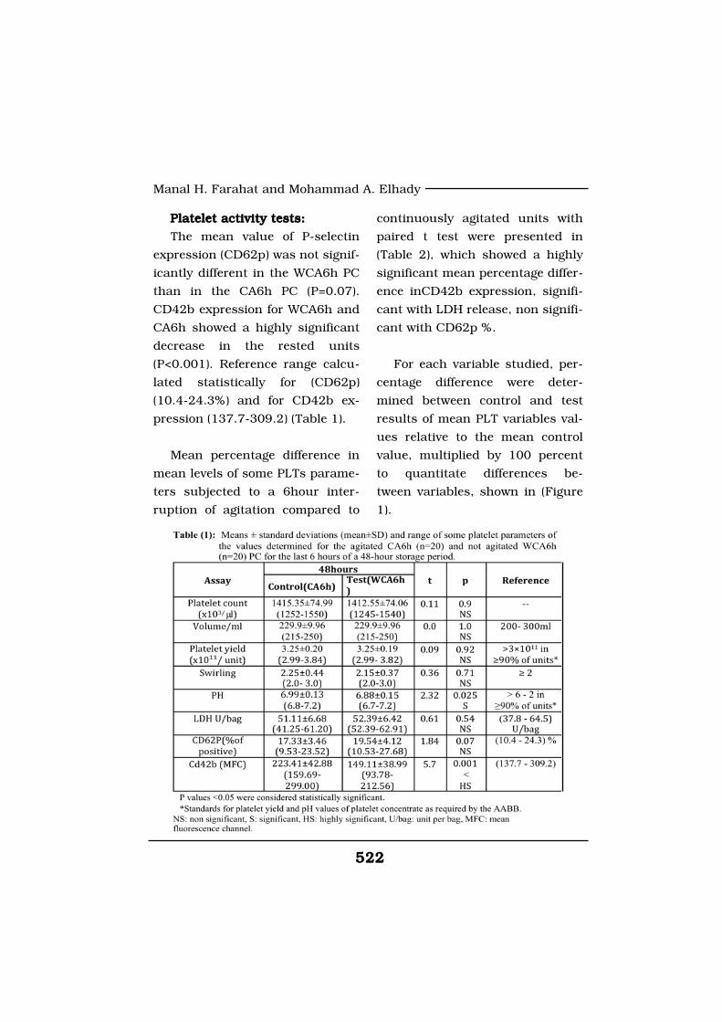
522522
Manal H. Farahat and Mohammad A. Elhady
Platelet activity tests:Platelet activity tests:
The mean value of P-selectin
expression (CD62p) was not signif-
icantly different in the WCA6h PC
than in the CA6h PC (P=0.07).
CD42b expression for WCA6h and
CA6h showed a highly significant
decrease in the rested units
(P<0.001). Reference range calcu-
lated statistically for (CD62p)
(10.4-24.3%) and for CD42b ex-
pression (137.7-309.2) (Table 1).
Mean percentage difference in
mean levels of some PLTs parame-
ters subjected to a 6hour inter-
ruption of agitation compared to
continuously agitated units with
paired t test were presented in
(Table 2), which showed a highly
significant mean percentage differ-
ence inCD42b expression, signifi-
cant with LDH release, non signifi-
cant with CD62p %.
For each variable studied, per-
centage difference were deter-
mined between control and test
results of mean PLT variables val-
ues relative to the mean control
value, multiplied by 100 percent
to quantitate differences be-
tween variables, shown in (Figure
1).
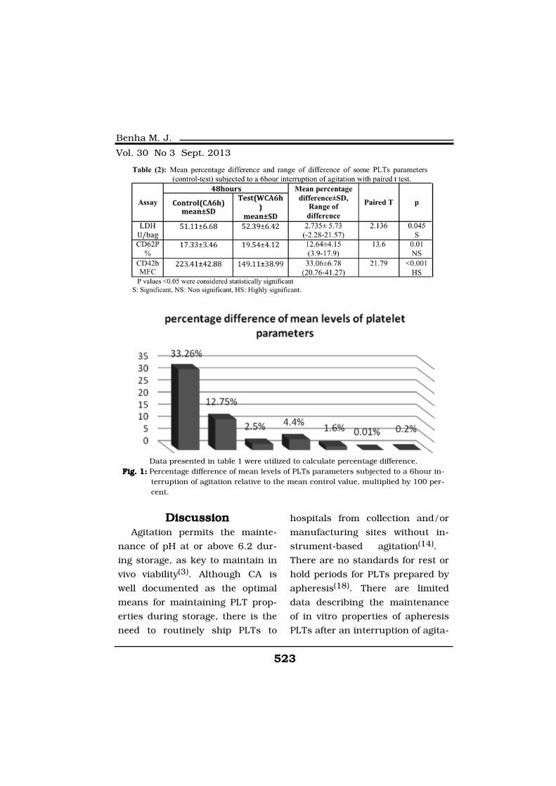
523
Benha M. J.
Vol. 30 No 3 Sept. 2013
DiscussionDiscussionAgitation permits the mainte-
nance of pH at or above 6.2 dur-
ing storage, as key to maintain in
vivo viability(3). Although CA is
well documented as the optimal
means for maintaining PLT prop-
erties during storage, there is the
need to routinely ship PLTs to
hospitals from collection and/or
manufacturing sites without in-
strument-based agitation(14).
There are no standards for rest or
hold periods for PLTs prepared by
apheresis(18). There are limited
data describing the maintenance
of in vitro properties of apheresis
PLTs after an interruption of agita-
Data presented in table 1 were utilized to calculate percentage difference.Fig. 1:ig. 1: Percentage difference of mean levels of PLTs parameters subjected to a 6hour in-
terruption of agitation relative to the mean control value, multiplied by 100 per-cent.
524524
Manal H. Farahat and Mohammad A. Elhady
tion during actual shipment of
PLTs(3). The primary goal to as-
sess the influence of interrupting
CA, which is the accepted stan-
dard for maximally maintaining
PLT properties during storage(11).
In this work we studied to what
extent 6 h of resting after 42 h of
storage from the time they were
collected, affect platelet quantity
(count , platelet content ”yield” per
bag), quality (swirling, pH), viabili-
ty (LDH release) and activity
(CD62P%, CD42b expression).
In the current study regarding
PLT count/units, ninety five per-
cent (1/20) units of apheresis-PC
met the desired quality control cri-
teria according to AABB standards
required for Apheresis platelets
>3x10¹¹ in ≥90% of units(12). PLT
yield per unit in the CA6h and
WCA6 PC were in the normal range.
So, Platelet count, PLT yield were
maintained in both PC (CA6h,
WCA6) units stored for 48 h.
Swirling was present within
score 2, 3 in all units studied
CA6h and WCA6h PC. No unit
had scored 1 swirling. Thus 6 h of
resting seemed not affect the
swirling properties of the PC.
Which was similar with finding of
Naghadeh H et al(4) who observed
no differences in the swirling score
between CA6h and WCA6h PC
prepared from platelet-rich plas-
ma. Results corresponded also
with the findings of Singh R et al(12), Bertolini F et al(19), and also
with Hunter S et al(20) that
showed that the maintenance of
grade 2 to 3 swirling essentially
guarantees the quality of PC.
According to AABB standards
required for Apheresis PLTs pH at
end of allowable storage >6 • 2 in
≥90% of units(13). We found that
pH of our all studied apheresis-PC
did not drop below 6.8 in the con-
trol PC or below 6.7 in PC that
were rested for 6 h throughout 48
h of storage. So, although our pH
value decreased but was main-
tained within acceptable range,
and mean pH percentage differ-
ence were decreased relative to
continuously agitated products by
1.6%. In previous study by Vassal-
lo R et al(5) with whole blood-
derived PLTs have demonstrated
that a contiguous 24-hour inter-
rupted agitation results in mainte-
nance of PC pH value of 6.5 or
525
Benha M. J.
Vol. 30 No 3 Sept. 2013
greater .Another study by Moroff
G et al(11) showed that Trima
apheresis PLT components that
were stored with (CA) and (WCA)
during two separate periods, im-
mediately after collection and be-
tween Day 2 and Day 3 of storage,
mean pH levels on Day 5of storage
were decreased in (WCA) units
with relative to (CA) products by
0.25. We could explain the differ-
ences between these results and
ours to the difference in prepara-
tion method of the platelets, the
period of stopping agitation and
time at which the study done dur-
ing the storage period.
LDH release measurements in
the two groups of our PC (CA6h
and WCA6h) indicating that stop-
ping agitation did not have major
effects on platelet viability, which
was agreed with finding of Nagha-
deh H et al(4) observed that 6 h of
resting did not have a deleterious
effect on pH, LDH release and ris-
tocetin-induced platelet aggrega-
tion (GPIb-related).
So, the mean difference in lev-
els of variable parameters in our
study regarding the platelet quan-
tity, quality and viability appear to
be small possibly without major
effect on PLTs viability.
Increased P-selectin (CD62P)
expression during storage has been
reported by several authors, where-
as GPIb (CD42b) has been shown
to decrease during storage(21).
Triulzi D el al(22), by using the
appearance of P selectin (CD62)
on the surface of platelets as an
index of activation, have suggest-
ed that more than one third of the
platelets in the concentrate ex-
press CD62 within 4 h of storage.
Lozano M et al(23) also found that
20 to 30% of platelets in platelet
concentrates expressed CD62 and
reported that CD62 expression
correlated poorly with percent re-
covery after transfusion.
The percent increase of platelet
activation between CA and WCA
units after 48h of storage for
CD62P% was 12.75% in our study
which was close to Moroff G et al(11) who found differences in
CD62-positive PLTs being less
than 20% for CA and WCA units
after 5 days of storage (Utilized
two periods without agitation, im-
mediately after collection for 7 to 8

526526
Manal H. Farahat and Mohammad A. Elhady
hours and a subsequent period for
24 hours between Day 2 and Day
3 of storage). The percentage dif-
ference for CD42b expression was
found 33.26 for our study. A
study by Sandgren P et al(9) found
that storage of PCs reduce expres-
sion of GP1bα on the surface of
platelets; this reduction is as-
sumed to decrease the adhesive
capacity. On the other hand,
Wang C et al(7) reported that acti-
vated platelets exhibit down regu-
lation of GPIb (CD42; the throm-
bin binding site), also suggested
that a 65% reduction in this ex-
pression of GPIb, has no effect on
platelet adhesion during flow con-
ditions also, Tynngård N(21) re-
ported a decline in the surface ex-
pression of GPIb with platelet
storage up to ten days, together
with impaired ability of the plate-
lets to be become activated by
thrombin as storage time in-
creased. The membrane content of
GPIb with storage has been re-
ported as a 35% loss.
So, in this study the baseline
platelet activity changes observed
was corresponding to the findings
of others, these small differences
appear to be only quantitative and
may reflect differences in PLT
product, heterogeneity of PC pre-
pared using the standard protocol,
manipulation during collection or
processing of the platelets and the
different periods of storage with-
out agitation.
In conclusion, From a clinical
point of view, the differences be-
tween CA and WCA of PLTs units,
for various in vitro variables meas-
ured after 48h of storage were lim-
ited, although some differences
were significant, these minimally
influenced differences of in vitro
properties relative to those of
matched continuously agitated
products would appear not to re-
flect any major changes that
might influence in vivo viability.
Of course more research is needed
to understand how agitation can
best be modified prior to transfusion,
and how exactly this interruption
of continuous agitation for 6 h, af-
ter 42 h of continuous agitation,
affects PC quality, quantity and vi-
ability needs more investigation.
Conflict of Interest:Conflict of Interest:
None of the authors has any
conflict of interest.
527
Benha M. J.
Vol. 30 No 3 Sept. 2013
AcknowledgementsAcknowledgementsThe authors thank members'
staff of blood bank center for tech-
nical help with collection and
preparation of the apheresis units.
ReferencesReferences1. Thielea T., Iugab C., Ja-1. Thielea T., Iugab C., Ja-
netzky S., Schwertz H., Gesellnetzky S., Schwertz H., Gesell
Salazar M., Fürll B., Völker U.,Salazar M., Fürll B., Völker U.,
Greinacher A. and Steil L.Greinacher A. and Steil L.
(2012):(2012): Early storage lesions in
apheresis platelets are induced by
the activation of the integrin
αIIbβ3 and focal adhesion signal-
ing pathways. JOURNAL OF
PROTEOMICS 76: 297-31.
2. Ahmed A., Leheta O. and2. Ahmed A., Leheta O. and
Younes S. (2010):Younes S. (2010): In vitro assess-
ment of platelet storage lesion in
leucoreduced random donor plate-
let concentrates. Blood Transfus.
8(1): 28-35.
3. Wagner S., Vassallo R.,3. Wagner S., Vassallo R.,
Skripchenko A., Einarson M.,Skripchenko A., Einarson M.,
Seetharaman S. and Moroff G.Seetharaman S. and Moroff G.
(2008): (2008): The influence of simulated
shipping conditions (24- or 30-hr
interruption of agitation) on the in
vitro properties of apheresis plate-
lets during 7-day storage. TRANS-
FUSION 48: 1072-1080.
4. Naghadeh H., Badlou B.,4. Naghadeh H., Badlou B.,
Ferizhandy A., MohammadrezaFerizhandy A., Mohammadreza
T. and Shahram V. (2013):T. and Shahram V. (2013): Six
hours of resting platelet concen-
trates stored at 22-24 Â˚C for 48
hours in permeable bags pre-
served pH, swirling and lactate de-
hydrogenase better and caused
less platelet activation. Blood
Transfus 11: 400-404.
5. Vassallo R., Wagner S., Ei-5. Vassallo R., Wagner S., Ei-
narson M., Nixon J., Ziegler D.narson M., Nixon J., Ziegler D.
and Moroff G. (2009): and Moroff G. (2009): Mainte-
nance of in vitro properties of leu-
koreduced whole blood–derived
pooled platelets after a 24-hour
interruption of agitation. TRANS-
FUSION 49: 2131-2135.
6. Shrivastava M. (2009):6. Shrivastava M. (2009): The
platelet storage lesion. Transfu-
sion and Apheresis Science 41:
105-113.
7. Wang Ch., Mody M., Herst7. Wang Ch., Mody M., Herst
R., Sher G. and Freedman J.R., Sher G. and Freedman J.
(1999):(1999): Flow cytometric analysis
of platelet function in stored plate-
let concentrates. Transfusion Sci-
ence 20: 129-139.
8. Kamath S., Blann D. and8. Kamath S., Blann D. and
Lip H. (2001):Lip H. (2001): Platelet activation:
528528
Manal H. Farahat and Mohammad A. Elhady
assessment and quantification.
European Heart Journal 22:
1561-1571.
9. Sandgren P., Hansson M.,9. Sandgren P., Hansson M.,
Gulliksson H. and Shanwell A.Gulliksson H. and Shanwell A.
(2007):(2007): Storage of buffy-coat-
derived platelets in additive solu-
tions at 4 °C and 22 °C: flow cy-
tometry analysis of platelet glyco-
protein expression. Vox Sanguinis
93: (1) 27-36.
10. Albanyan A., Harrison P.10. Albanyan A., Harrison P.
and Murphy M. (2009):and Murphy M. (2009): Markers
of platelet activation and apopto-
sis during storage of apheresis-
and buffy coat–derived platelet
concentrates for 7 days. TRANS-
FUSION 49: 108-117.
11. Moroff G., Kurtz J., See-11. Moroff G., Kurtz J., See-
tharaman S. and Wagner S.tharaman S. and Wagner S.
(2011): (2011): Storing apheresis platelets
without agitation with simulated
shipping conditions during two
separate periods: immediately af-
ter collection and subsequently
between Day 2 and Day3. TRANS-
FUSION 51: 636-642.
12. Singh R., Marwaha N.,12. Singh R., Marwaha N.,
Malhotra P. and Dash S. (2009):Malhotra P. and Dash S. (2009):
Quality assessment of platelet
concentrates prepared by platelet
rich plasma-platelet concentrate,
buffy coat poor-platelet concen-
trate (BC-PC) and apheresis-PC
methods. Asian J Transfus Sci. 3
(2): 86-94.
13. Stroncek D. and Rebulla13. Stroncek D. and Rebulla
P. (2007):P. (2007): Platelet transfusions.
Lancet 370: 427-438.
14. Moroff G., Kurtz J., See-14. Moroff G., Kurtz J., See-
tharaman S., Skripchenko A.,tharaman S., Skripchenko A.,
Awatefe H., Thompson-Awatefe H., Thompson-
Montgomery D., Myrup A. andMontgomery D., Myrup A. and
Wagner S. (2012):Wagner S. (2012): Comparative
in vitro evaluation of apheresis
platelets stored with 100% plasma
or 65% platelet additive solution
III/35% plasma and including pe-
riods without agitation under sim-
ulated shipping conditions.
TRANSFUSION 52: 834-843.
15. Amorini A., Tuttobene M.,15. Amorini A., Tuttobene M.,
[...] and Denti G. (2007):[...] and Denti G. (2007): Evalua-
tion of biochemical parameters in
platelet concentrates stored in
glucose solution. Blood transfu-
sion 5(1): 24-32.
16. Van der Meer F. and Piet-16. Van der Meer F. and Piet-
er F. (2011):er F. (2011): Platelet preservation:
agitation and containers. Transfus
529
Benha M. J.
Vol. 30 No 3 Sept. 2013
Apher Sci. 44: 297-304.
17. G. Singh (2007):17. G. Singh (2007): Determi-
nation of Cutoff Score for a Diag-
nostic Test. The Internet Journal
of Laboratory Medicine 2 (1). DOI:
10.5580/1aca
18. Skripchenko A., Myrup A.,18. Skripchenko A., Myrup A.,
Awatefe H., Thompson-MontgomeryAwatefe H., Thompson-Montgomery
D. and Wagner S. (2012):D. and Wagner S. (2012): A rest
period before agitation may improve
some in vitro apheresis platelet
parameters during storage.
TRANSFUSION 52: 1433-1438.
19. Bertolini F., Rebulla P.,19. Bertolini F., Rebulla P.,
Riccardi D., Cortellaro M., Rem-Riccardi D., Cortellaro M., Rem-
zi M.L. and Sirchia G. (1989):zi M.L. and Sirchia G. (1989):
Evaluation of platelet concentrates
prepared from buffy coats and
stored in a glucose free crystalloid
medium. Transfusion 29: 605-
609.
20. Hunter S., Nixon J. and20. Hunter S., Nixon J. and
Murphy S. (2001):Murphy S. (2001): The effect of
the interruption of agitation on
platelet quality during storage for
transfusion. TRANSFUSION 41:
809-814.
21. Tynngård N. (2009):21. Tynngård N. (2009): Prep-
aration, storage and quality con-
trol of platelet concentrates.
Transfus Apher Sci. 41(2): 97-104.
22. Triulzi D., Kickler T. and22. Triulzi D., Kickler T. and
Braine H. (1992):Braine H. (1992): Detection and
significance of alpha granule
membrane protein 140 expression
on platelets collected by aphere-
sis. Transfusion 32: 529-533.
23. Lozano M., Rivera J.,23. Lozano M., Rivera J.,
Gonzalez-Conejero R., MoraledaGonzalez-Conejero R., Moraleda
J. and Vincente V. (1997):J. and Vincente V. (1997): Loss
of high-affinity thrombin receptors
during platelet concentrate stor-
age impairs the reactivity of plate-
lets to thrombin.Transfusion 37:
368-375.

530530
Manal H. Farahat and Mohammad A. Elhady
Published byPublished by
Benha Faculty of MedicineBenha Faculty of Medicine
SOME PLATELET PROPERTIES OFSOME PLATELET PROPERTIES OFRESTING APHERESIS PLATELETRESTING APHERESIS PLATELETCONCENTRATE FOR THE LAST 6CONCENTRATE FOR THE LAST 6
HOURS OF A 48-HOURHOURS OF A 48-HOURSTORAGE PERIODSTORAGE PERIOD
Manal H. Farahat MD and Mohammad A. Elhady MDManal H. Farahat MD and Mohammad A. Elhady MD
BENHAMEDICALJOURNAL
REPRINT
Volume 30 Number 3Sept. 2013
531
Benha M. J.
Vol. 30 No 3 Sept. 2013
IntroductionIntroductionOpen heart surgery in cirrhotic
patients had relatively high risk of
morbidity and mortality when
compared to non cirrhotic patients
mainly Postoperative liver decom-
position, bleeding and high inci-
dence of postoperative wound in-
fection(1-5).
Child Pugh classification(6) (Ta-
ble 1) consists of certain clinical,
OUTCOME OF CORONARY ARTERY BYPASSOUTCOME OF CORONARY ARTERY BYPASSIN PATIENTS WITH CHILD-PUGH CLASSIN PATIENTS WITH CHILD-PUGH CLASS
A LIVER CIRRHOSISA LIVER CIRRHOSIS
Mohamed Ahmed El-Awady MD and Moataz Rezk MDMohamed Ahmed El-Awady MD and Moataz Rezk MDLecturer, Cardiothoracic Surgery Department,
Banha Faculty of Medicine, Banha University, Egypt.
AbstractAbstractObjectives:Objectives: Coronary artery bypass grafting in cirrhotic patients car-
ries high risk of morbidity and mortality. Most of these complicationsare related to hepato-renal failure, bleeding and postoperative woundinfection rather than cardiac problems.
Methods:Methods: Prospective study to evaluate elective CABG early postop-erative outcome of elective CABG in patients with Class A Child Pughliver cirrhosis.
Results:Results: From October 2007 to April 2011total 59 patients withClass A liver failure underwent elective CABG.37male, 22female.42hypertensives and 28diabetics. Mean1st 24hours chest tube drainagewas 853.80±56.10ml, minimal 130ml maximum 3500ml. 12 patients(20.3%) were re-explored for bleeding. Mean ventilation time was10.48±6.65 hours. Mean ICU stay was 59.52±13.91hours. Two patients(3.38%) died one patient due to hepato-renal failure (re explored 3times) while the 2nd patient died after delayed recovery due to cerebralhemorrhage. 20 patients (33.89%) had wound infection, two need de-bridement and rewiring. Mean hospital stay was 9.18±2.29days. Totalmorbidity was 49%. Total mortality was 3.38%.
Conclusion:Conclusion: Elective CABG can be tolerated satisfactorily in class AChild Pugh cirrhotic patients with high incidence of the postoperativecomplications specially bleeding and wound infection.
532532
Mohamed Ahmed El-Awady and Moataz Rezk
laboratory and radiological param-
eters to classify liver cirrhotic pa-
tients into A, B and C classes. Class
A has the best condition while
class C has the worst condition.
Model for End-Stage Liver Dis-
ease (MELD) score(7) are calculat-
ed in cirrhotic patients to help in
prediction of the excepted morbid-
ity and mortality. MELD score
ranging from 6 to 40, patients
with score of 6 are the best ill pa-
tients while patients of 40 are the
sickest one.
The expected morbidity and
mortality is much higher in pa-
tients with advanced Child Pugh
Classification and high MELD score
but there is no accurate predica-
tor of outcome of open heart sur-
gery in liver cirrhosis patients.
Some studies used Child Pugh
Classification others used Model
for End-Stage Liver Disease (MELD)
score to evaluate the outcome of
CABG in liver cirrhosis patients
but most of these studies of had
limited number of patients(4,5).
The number of cirrhotic patients
undergoing CABG is increasing in
Egypt as it has one of the highest
percentages of hepatitis C patients
in the world ranging from 10% to
13% of the population(8), most of
these patients are class A Child
Pugh classification so we had this
prospective study to evaluate the
outcome of CABG in A class Child
Pugh patients. All preoperative,
operative and postoperative details
are collected and analyzed.
Patients and MethodsPatients and MethodsFrom April 2008 to April 2011
a prospective study to evaluate the
outcome of elective CABG in class
A cirrhotic patients. All patients
had full clinical evaluation and
full laboratory evaluation includ-
ing complete blood picture, com-
plete liver function and complete
renal functions test. Enzyme-
linked immunosorbent assay (ELI-
SA) test was used in diagnose hep-
atitis markers. Abdominal ultra
sound is done for all patients to
evaluate the liver condition and
diagnosis portal Hyperion. No liver
biopsy was taken. Child Pugh
classification and MELD score are
calculated for all patients.
Inclusion criteria:Inclusion criteria:
1- Elective CABG.
2- Class A Child Pugh.
533
Benha M. J.
Vol. 30 No 3 Sept. 2013
3-Good left ventricular function
with ejection fraction above 35%.
Exclusion criteria are:Exclusion criteria are:
1- Class B and C Child Pugh
classification.
2- Emergency or urgent CABG.
3- Redo CABG.
4- Open heart surgeries rather
than CABG like valve surgery or
combined surgery like CABG+
valve surgery.
5- Patients with poor left ven-
tricular function with ejection
fraction below 35%.
6- Renal failure patients on reg-
ular dialysis.
All preoperative, operative and
postoperative data including 3
months follow up after discharge
home are collected and analyzed.
All patients continue on same
drugs they usually use until the
morning of the surgery except
anti-platelets, which are stopped
for 7 days before surgery.
After the patients are anesthe-
tizes, midline sternotomy is done.
Pedicled left internal mammary ar-
tery (LIMA) is harvested in all pa-
tients.
All operations were performed
utilizing conventional cardiopul-
monary bypass (CPB) giving cold
antigrade crystalloid cardioplegic
solution repeated every 30 min-
utes. CPB was conducted using a
membrane oxygenator and mild
hypothermia (35C).
Packed red blood cells were ad-
ministered when haematocrit was
less than 25%. Fresh frozen plas-
ma and platelets were adminis-
tered when platelet count was less
than 40000/ml or as a part of
control postoperative bleeding.
Postoperative complications were
classified as follow:
- Pulmonary: pneumonia, venti-
lator dependence greater than 48
hours, excessive pleural effusion
requiring an additional drainage.
- Infectious (superficial and
deep sternal wound infection).
- Bleeding complications (re-
exploration because of excessive
mediastinal bleeding or cardiac
tamponade requiring drainage).
- Renal complications (increase
in serum creatinine greater than
1.5mg/dl, oliguria (<0.5 ml/kg/
min) for more than 6 hours post-
operatively or any other indication
534534
Mohamed Ahmed El-Awady and Moataz Rezk
for dialysis).
- Other postoperative complica-
tions related to liver diseases,
such as encephalopathy, hyperbil-
irubinemia and gastrointestinal
bleeding as a result of varices
were also recorded.
Mortality is defined as death dur-
ing a hospitalization for surgery, re-
gardless of length of stay, or within
30 days from hospital discharge.
Values of continuous variables
were expressed as mean and stan-
dard deviation performed with
computerized statistical packages
(SPSS 18.0 software, SPSS, Chica-
go, IL, USA).
ResultsResultsTotal 59 patients were eligible
for the study. Main cause of liver
cirrhosis was hepatitis C (42 pa-
tients) and Hepatitis B (17patients)
with no Alcoholic cirrhosis.47
male and 12 female .28 patients
were diabetics while 42 were hy-
pertensives. Mean MELD score
was15.2±3.38 (minimal 8, maxi-
mum 23). Table 2 summarizes the
preoperative patient’s profile.
Mean number of grafts was
2.72±0.57. Mean cardiopulmonary
bypass time was 62.27±6.40 min-
utes (minimum45, maximum78).
Mean Cross-clamp time it was
41.53±5.85 (minimum 28 maxi-
mum 55min). No patients need in-
tra aortic balloon pump. Table 3
summarizes the operative data.
The postoperative mean chest
tube drainage was 853.80±56.10ml;
minimal 130ml maximum 3500
ml. Mean packed RBCS transfu-
sion was 2.18±1.68 units. Mean
fresh frozen plasma (FFP) Transfu-
sion was 3.64±2.48 units. Mean
platelets transfusion was 3.99±1.30
units. No postoperative myocardial
infarction diagnosed by ECG and
repeated cardiac enzyme. 12 pa-
tients were re-explored for bleed-
ing (20.3%), 8 of them had cardiac
tamponade. Mean ventilation time
was 10.48±6.6. Mean ICU stay was
59.52±13.91 hours. 20 patients
(33.89%) had wound infection, 15
of them had leg wound infection, 2
patients with mediastinitis needed
debridement and rewiring. one pa-
tient had left sided clotted hae-
mothorax diagnosed by CT chest
drained though left mini thoracot-
omy. 21 patients (35.59%) had left
pleural effusion, all treated medi-
535
Benha M. J.
Vol. 30 No 3 Sept. 2013
cally except 6 patents needed re-
peated pleural taping. Four pa-
tients readmitted due to medias-
tinitis, 2 of them had rewiring.
Mean Hospital stay was 9.18±2.29
days. Total morbidity was 49%.
Total mortality was 3.38%. (2 pa-
tients),1st was re-explored 3 times
for bleeding and died due to hepa-
to-renal failure, 2nd patient had
delayed recovery with right sided
hemiplegia .CT brain showed cere-
bral hemorrhage. Table 4 sum-
merizes the postoperative outcome.
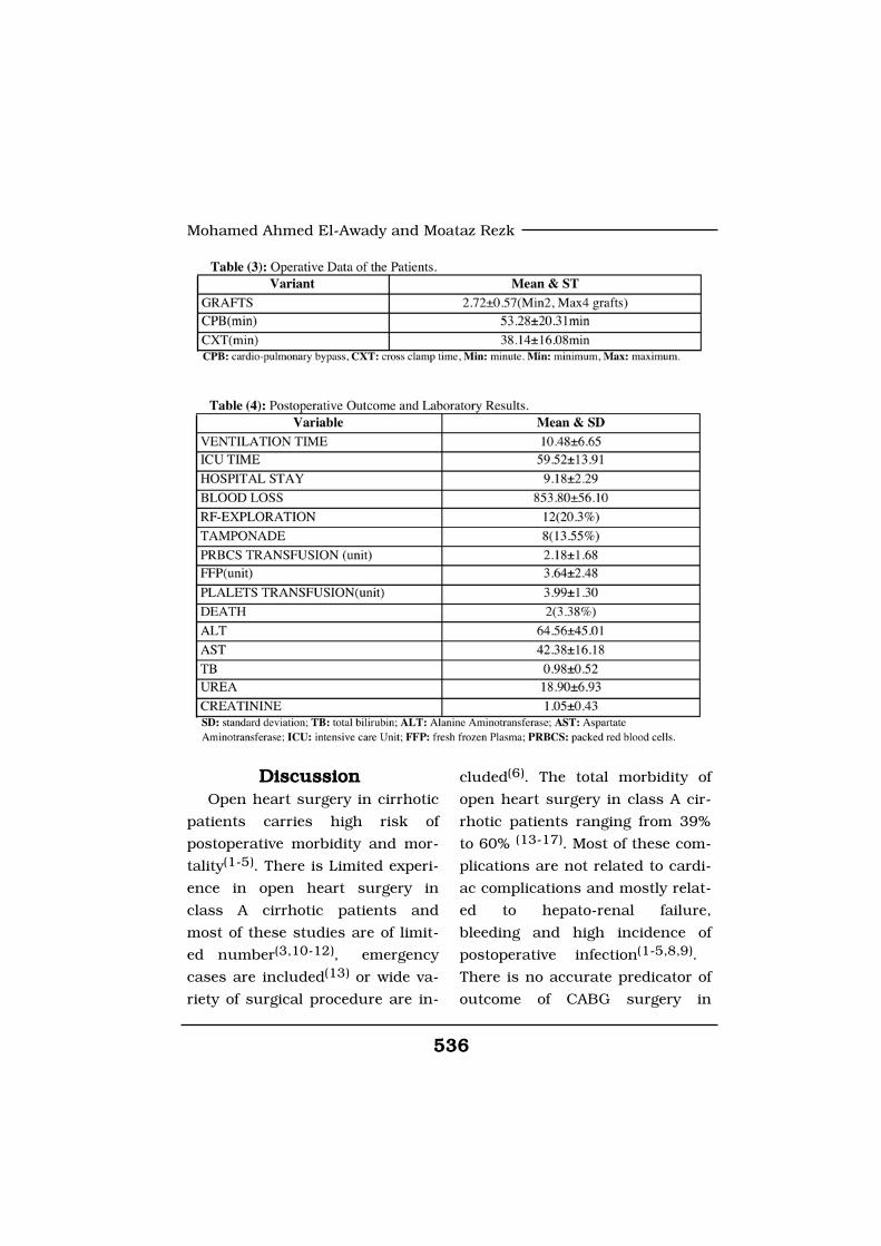
536536
Mohamed Ahmed El-Awady and Moataz Rezk
Discussion Discussion Open heart surgery in cirrhotic
patients carries high risk of
postoperative morbidity and mor-
tality(1-5). There is Limited experi-
ence in open heart surgery in
class A cirrhotic patients and
most of these studies are of limit-
ed number(3,10-12), emergency
cases are included(13) or wide va-
riety of surgical procedure are in-
cluded(6). The total morbidity of
open heart surgery in class A cir-
rhotic patients ranging from 39%
to 60% (13-17). Most of these com-
plications are not related to cardi-
ac complications and mostly relat-
ed to hepato-renal failure,
bleeding and high incidence of
postoperative infection(1-5,8,9).
There is no accurate predicator of
outcome of CABG surgery in
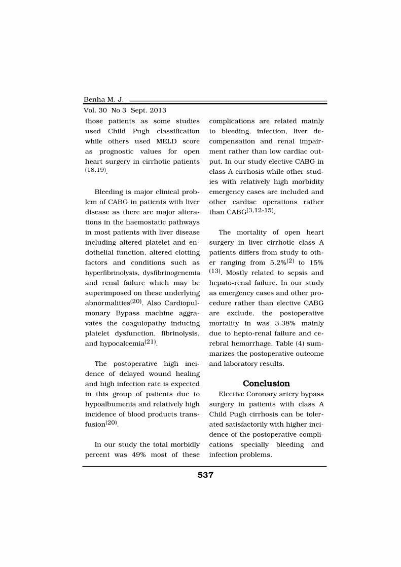
537
Benha M. J.
Vol. 30 No 3 Sept. 2013
those patients as some studies
used Child Pugh classification
while others used MELD score
as prognostic values for open
heart surgery in cirrhotic patients(18,19).
Bleeding is major clinical prob-
lem of CABG in patients with liver
disease as there are major altera-
tions in the haemostatic pathways
in most patients with liver disease
including altered platelet and en-
dothelial function, altered clotting
factors and conditions such as
hyperfibrinolysis, dysfibrinogenemia
and renal failure which may be
superimposed on these underlying
abnormalities(20). Also Cardiopul-
monary Bypass machine aggra-
vates the coagulopathy inducing
platelet dysfunction, fibrinolysis,
and hypocalcemia(21).
The postoperative high inci-
dence of delayed wound healing
and high infection rate is expected
in this group of patients due to
hypoalbumenia and relatively high
incidence of blood products trans-
fusion(20).
In our study the total morbidly
percent was 49% most of these
complications are related mainly
to bleeding, infection, liver de-
compensation and renal impair-
ment rather than low cardiac out-
put. In our study elective CABG in
class A cirrhosis while other stud-
ies with relatively high morbidity
emergency cases are included and
other cardiac operations rather
than CABG(3,12-15).
The mortality of open heart
surgery in liver cirrhotic class A
patients differs from study to oth-
er ranging from 5.2%(2) to 15%(13). Mostly related to sepsis and
hepato-renal failure. In our study
as emergency cases and other pro-
cedure rather than elective CABG
are exclude, the postoperative
mortality in was 3.38% mainly
due to hepto-renal failure and ce-
rebral hemorrhage. Table (4) sum-
marizes the postoperative outcome
and laboratory results.
ConclusionConclusionElective Coronary artery bypass
surgery in patients with class A
Child Pugh cirrhosis can be toler-
ated satisfactorily with higher inci-
dence of the postoperative compli-
cations specially bleeding and
infection problems.
538538
Mohamed Ahmed El-Awady and Moataz Rezk
ReferencesReferences1- Akira Marui, Takeshi Ki-1- Akira Marui, Takeshi Ki-
mura, Shiro Tanaka, et al.mura, Shiro Tanaka, et al.
(2011): (2011): Coronary Revasculariza-
tion in Patients with Liver Cirrho-
sis: Ann Thorac Surg; 91:1393-
1399.
2- Amit Modi, Hunaid A. Voh-2- Amit Modi, Hunaid A. Voh-
ra and Clifford W. Barlowra and Clifford W. Barlow
(2010):(2010): Do patients with liver cir-
rhosis undergoing cardiac surgery
have acceptable outcomes? Inter-
act CardioVasc Thorac Surg; 11:
630-634.
3- Akimasa Morisaki, Mitsu-3- Akimasa Morisaki, Mitsu-
haru Hosono, Yasuyuki Sasaki,haru Hosono, Yasuyuki Sasaki,
et al. (2010):et al. (2010): Risk factor analysis
in patients with liver cirrhosis un-
dergoing cardiovascular opera-
tions. Ann Thorac Surg; 89: 811-
817.
4- Nobuhiko Hayashida, Tak-4- Nobuhiko Hayashida, Tak-
ahiro Shoujima, Hideki Teshi-ahiro Shoujima, Hideki Teshi-
ma, et al. (2004):ma, et al. (2004): Clinical out-
come after cardiac operations in
patients with Cirrhosis: The An-
nals of thoracic surgery; 77(2):
500-505.
5- Yong An, Ying Bin Xiao 5- Yong An, Ying Bin Xiao
and Qian-Jin Zhong (2007):and Qian-Jin Zhong (2007):
Open-heart surgery in patients
with liver cirrhosis: Eur J Cardi-
othorac Surg; 31: 1094-1098.
6- Lucey M.R., Brown K.A.,6- Lucey M.R., Brown K.A.,
Everson G.T., et al. (1997):Everson G.T., et al. (1997): Mini-
mal criteria for placement of
adults on the liver transplant
waiting list: a report of a national
conference organized by the Amer-
ican Society of Transplant Physi-
cians and the American Associa-
tion for the Study of Liver
Diseases. Liver Transpl Surg. Nov;
3(6): 628-37.
7- Wiesner R., Edwards E.,7- Wiesner R., Edwards E.,
Freeman R., et al. (2003): Freeman R., et al. (2003): Model
for end-stage liver disease (MELD)
and allocation of donor livers. Gas-
troenterology vol. 124 (1) pp. 91.
8- Mohamed M.K. (2004): 8- Mohamed M.K. (2004): Ep-
idemiology of HCV in Egypt 2004:
The Afro-Arab Liver Journal; 3(2):
41-52.
9- John D. Klemperer, Wilson9- John D. Klemperer, Wilson
Ko, Karl H. Krieger, et al. (1998):Ko, Karl H. Krieger, et al. (1998):
Cardiac operations in patients
with cirrhosis Ann Thorac Surg;
65:85-87.
10- Philippe Bizouarn, An-10- Philippe Bizouarn, An-

539
Benha M. J.
Vol. 30 No 3 Sept. 2013
toine Ausseur, Pascal Des-toine Ausseur, Pascal Des-
seigne, et al. (1999):seigne, et al. (1999): Early and
late outcome after elective cardiac
surgery in patients with cirrhosis.
Ann Thorac Surg; 67: 1334-1338.
11- Takashi Murashita, Tat-11- Takashi Murashita, Tat-
suhiko Komiya, Nobushige Ta-suhiko Komiya, Nobushige Ta-
mura, et al. (2009):mura, et al. (2009): Preoperative
evaluation of patients with liver
cirrhosis undergoing open heart
surgery. Gen Thorac Cardiovasc
Surg. Jun; 57(6): 293-7.
12- Farzan Filsoufi, Salzberg12- Farzan Filsoufi, Salzberg
S.P., Rahmanian P.B., et al.S.P., Rahmanian P.B., et al.
(2007):(2007): Early and late outcome of
cardiac surgery in patients with
liver cirrhosis. Liver Transpl Jul;
13(7): 990-5.
13- Matthias Thielmann, Ach-13- Matthias Thielmann, Ach-
met Mechmet, Markus Neuhaus-met Mechmet, Markus Neuhaus-
er, et al. (2010):er, et al. (2010): Risk prediction
and outcomes in patients with liv-
er cirrhosis undergoing open-
heart surgery. Eur J Cardiothorac
Surg; 38: 592-599.
14- Cheng-Hsin Lin, Fang-14- Cheng-Hsin Lin, Fang-
Yue Lin, Shoei-Shen Wang, etYue Lin, Shoei-Shen Wang, et
al. (2005):al. (2005): cardiac surgery in pa-
tients with liver cirrhosis .The An-
nals of Thoracic Surgery; 79, 5,
1551-1554.
15- Mohsen Hammad, Asem15- Mohsen Hammad, Asem
Elfert, Hasan Elbateaet, et al.Elfert, Hasan Elbateaet, et al.
(2006):(2006): Clinical outcome after car-
diac operations using cardiopul-
monary bypass in patients with
liver dysfunction. Arab J Gas-
troenterol; 7(3): 98-101.
16- Yukiharu Sugimura, Ma-16- Yukiharu Sugimura, Ma-
saaki Toyama, Masanori Katoh,saaki Toyama, Masanori Katoh,
et al. (2012):et al. (2012): Analysis of open
heart surgery in patients with liver
cirrhosis. Asian Cardiovasc Tho-
rac; 20: 263-268.
17- Akira Marui, Takeshi Ki-17- Akira Marui, Takeshi Ki-
mura, Shiro Tanaka, et al.mura, Shiro Tanaka, et al.
(2011): (2011): Coronary Revasculariza-
tion in Patients with Liver Cirrho-
sis. Ann Thorac Surg; 91: 1393-
1399.
18- Suman A., Barnes D.S.,18- Suman A., Barnes D.S.,
Zein N.N., et al. (2004):Zein N.N., et al. (2004): Predict-
ing outcome after cardiac surgery
in patients with cirrhosis a com-
parison of Child-Pugh and MELD
scores. Clin Gastroenterol Hepa-
tol; 2:719-723.
19- Gorav Ailawadi, Damien19- Gorav Ailawadi, Damien
J. LaPar, Brian R. Swenson, etJ. LaPar, Brian R. Swenson, et
540540
Mohamed Ahmed El-Awady and Moataz Rezk
al. (2009):al. (2009): Model for end-stage liv-
er disease predicts mortality for
tricuspid valve surgery. Ann Tho-
rac Surg; 87: 1460-1468.
20- Tripodi A., Salerno F.,20- Tripodi A., Salerno F.,
Chantarangkul V., et al. (2005):Chantarangkul V., et al. (2005):
Evidence of normal thrombin gen-
eration in cirrhosis despite abnor-
mal conventional coagulation
tests. Hepatology; 41: 553-8.
21- Pollard R.J., Sidi A., Gib-21- Pollard R.J., Sidi A., Gib-
by G.L., et al. (1998):by G.L., et al. (1998): Aortic sten-
osis with end-stage liver disease:
prioritizing surgical and anesthet-
ic therapies. J Clin Anesth 1998;
10: 253-261.
541
Benha M. J.
Vol. 30 No 3 Sept. 2013
Published byPublished by
Benha Faculty of MedicineBenha Faculty of Medicine
OUTCOME OF CORONARY ARTERYOUTCOME OF CORONARY ARTERYBYPASS IN PATIENTS WITHBYPASS IN PATIENTS WITH
CHILD-PUGH CLASSCHILD-PUGH CLASSA LIVER CIRRHOSISA LIVER CIRRHOSIS
Mohamed Ahmed El-Awady MD and Moataz Rezk MDMohamed Ahmed El-Awady MD and Moataz Rezk MD
BENHAMEDICALJOURNAL
REPRINT
Volume 30 Number 3Sept. 2013
541
Benha M. J.
Vol. 30 No 3 Sept. 2013
NUTRITIONAL MANAGEMENT OF PREDIALYSISNUTRITIONAL MANAGEMENT OF PREDIALYSISRENAL FAILURE PATIENTS USINGRENAL FAILURE PATIENTS USING
GUM ARABICGUM ARABIC
Radwa Mohamed Abd El-Shakour B.Sc.*,Radwa Mohamed Abd El-Shakour B.Sc.*,Mousa Abduo Salem MD*Mousa Abduo Salem MD*
and Nabil Mohamed Abd El-Fattah Hassan MD**and Nabil Mohamed Abd El-Fattah Hassan MD***Department of Food Science and Technology, Faculty of Agriculture, Tanta University
** Department of Nephrology, Urology & Nephrology Center,
Mansoura University
AbstractBackground:Background: Adequate nutritional management is a mainstay in
treatment of predialysis renal failure. The current protocols have manydrawbacks. GA proved effective for those patients in previous studies.
Aim of work: The present study aimed to investigate the effect of GAon biochemical and clinical parameters in predialysis patients and theirneed to dialysis.
Materials and Methods: Materials and Methods: The study comprised two groups: Group I(Treatment group): included 22 patients and received Arabic gum in ad-dition to the conventional therapeutic intervention. Group II (Controlgroup): included 19 patients who received conventional therapeutic in-tervention. In the treatment group, patients received a daily dose of 100gm of Arabic gum dissolved in water or natural juice equally dividedinto morning and evening dose of 50 gm. All patients were subjected tocareful history taking, thorough clinical examination and laboratory in-vestigations including renal function, serum electrolytes and blood pic-ture. All participants were followed monthly for 4 months.
Results:Results: In the study follow up, treatment with gum Arabic resultedin progressive significant improvement in renal function (creatinineclearance, serum creatinine and serum urea). There were also signifi-cant decline in serum Ca, P, Na and K levels in the treatment group. Inaddition, treatment group had better blood pressure control and none ofpatients needed dialysis in the study period in comparison with 7 pa-tients in the control group. Uremic fetor disappeared in 18 patients of
542
Radwa Mohamed Abd El-Shakour, et al....
IntroductionDiet potentially plays a major
role in the progression and com-
plications of predialysis CKD.
Moderate protein consumption
along with a diet low in sodium
might slow kidney disease pro-
gression. Increasing vegetable pro-
tein intake might decrease serum
phosphorus, uremic toxins, and
kidney damage.(1)
A properly implemented dietary
treatment for patients with chron-
ic renal failure (CRF) can correct
several metabolic and endocrine
disturbances and delay initiation
of dialysis, but concerns exist
about the risk of malnutrition and
protein depletion.(2) Also, the
practical implementation of pro-
tein restriction through dietary in-
tervention has been hindered on
multiple levels, including patient
nonadherence and and lack of
health care resources.(3) Further-
more, larger scale studies demon-
strated lack of effectiveness of pro-
tein restriction strategy.(4)
Consequently, more kidney
protective strategies are needed to
reduce the burden of complete
kidney failure from chronic kidney
disease (CKD).(4)
Gum arabic (GA) is a branched-
chain, complex polysaccharide, ei-
ther neutral or slightly acidic.
Pharmacologically, GA has been
claimed to act as an anti-oxidant,
and to protect against experimen-
tal hepatic-, renal- and cardiac
toxicities in rats. It has also been
claimed to alleviate the adverse ef-
fects of chronic renal failure in
humans.(5) However, clinical data
on its efficacy in human studies
are scarce.
Aim of StudyThe present study aim to inves-
the treatment group while no improvement occurred in the controlgroup.
Conclusions: Conclusions: Nutritional supplementation with Gum Arabic in pre-dialysis CRF patients results in general improvement of laboratory andclinical parameters with subsequent delay in disease progression andneed of dialysis.
Keywords:Keywords: Chronic renal failure, gum Arabic, renal nutrition.
543
Benha M. J.
Vol. 30 No 3 Sept. 2013
tigate the efficacy of gum Arabic in
predialysis chronic renal failure
patients.
Materials and MethodsThe present study is an inter-
ventional longitudinal case control
study. It was conducted at Man-
soura Nephrology Center, Man-
soura, Egypt after obtaining the
required permissions. Informed
consent was obtained from all par-
ticipants.
Patients were selected to partic-
ipate in the study had chronic re-
nal failure in the predialysis stag-
es with no known allergy or
contraindication to Arabic gum or
any of its ingredients.
The study comprised two
groups: Group I (Treatment
group): included 22 patients and
received Arabic gum in addition to
the conventional therapeutic inter-
vention. Group II (Control group):
included 19 patients who received
conventional therapeutic interven-
tion.
In the treatment group, pa-
tients received a daily dose of 50
gm of Arabic gum dissolved in wa-
ter or natural juice equally divided
into morning and evening dose of
25 gm. The used sample was sub-
jected toxicological, biochemical
and microbiological assessment in
the National Nutrition Institute
Laboratories and food science and
Technology department, faculty of
Agriculture, Tanta university.
Both groups received low protein
diet.
All patients were subjected to
careful history taking, thorough
clinical examination with particu-
lar notion to uremic fetor which
was diagnosed if the patient had a
urine-odor breath. Laboratory in-
vestigations included creatinine
clearance, blood urea, serum crea-
tinine, serum phosphorous, cal-
cium, potassium and sodium,
fasting blood sugar, blood bicarbo-
nate, hemoglobin, hematocrit val-
ue and platelets count. All partici-
pants were followed monthly for 4
months.
Data obtained from the present
study were computed using SPSS
versions 17 under the platform of
Microsoft Windows XP, Profession-
al Edition. Continuous data were
expressed in the form of mean ±
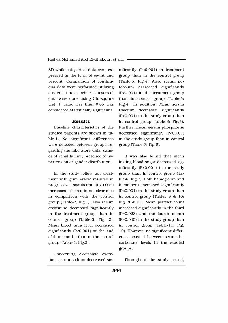
544
Radwa Mohamed Abd El-Shakour, et al....
SD while categorical data were ex-
pressed in the form of count and
percent. Comparison of continu-
ous data were performed utilizing
student t test, while categorical
data were done using Chi-square
test. P value less than 0.05 was
considered statistically significant.
ResultsBaseline characteristics of the
studied patients are shown in ta-
ble-1. No significant differences
were detected between groups re-
garding the laboratory data, caus-
es of renal failure, presence of hy-
pertension or gender distribution.
In the study follow up, treat-
ment with gum Arabic resulted in
progressive significant (P<0.002)
increases of creatinine clearance
in comparison with the control
group (Table-2; Fig.1). Also serum
creatinine decreased significantly
in the treatment group than in
control group (Table-3; Fig. 2).
Mean blood urea level decreased
significantly (P<0.001) at the end
of four months than in the control
group (Table-4; Fig.3).
Concerning electrolyte excre-
tion, serum sodium decreased sig-
nificantly (P<0.001) in treatment
group than in the control group
(Table-5; Fig.4). Also, serum po-
tassium decreased significantly
(P<0.001) in the treatment group
than in control group (Table-5;
Fig.4). In addition, Mean serum
Calcium decreased significantly
(P<0.001) in the study group than
in control group (Table-6; Fig.5).
Further, mean serum phosphorus
decreased significantly (P<0.001)
in the study group than in control
group (Table-7; Fig.6).
It was also found that mean
fasting blood sugar decreased sig-
nificantly (P<0.001) in the study
group than in control group (Ta-
ble-8; Fig.7). Both hemoglobin and
hematocrit increased significantly
(P<0.001) in the study group than
in control group (Tables 9 & 10;
Fig. 8 & 9). Mean platelet count
increased significantly in the third
(P=0.023) and the fourth month
(P=0.045) in the study group than
in control group (Table-11; Fig.
10). However, no significant differ-
ences existed between serum bi-
carbonate levels in the studied
groups.
Throughout the study period,
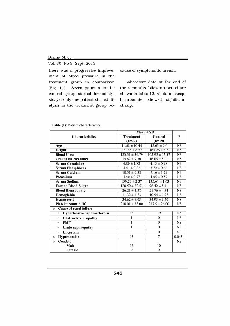
545
Benha M. J.
Vol. 30 No 3 Sept. 2013
there was a progressive improve-
ment of blood pressure in the
treatment group in comparison
(Fig. 11). Seven patients in the
control group started hemodialy-
sis, yet only one patient started di-
alysis in the treatment group be-
cause of symptomatic uremia.
Laboratory data at the end of
the 4 months follow up period are
shown in table-12. All data (except
bicarbonate) showed significant
change.
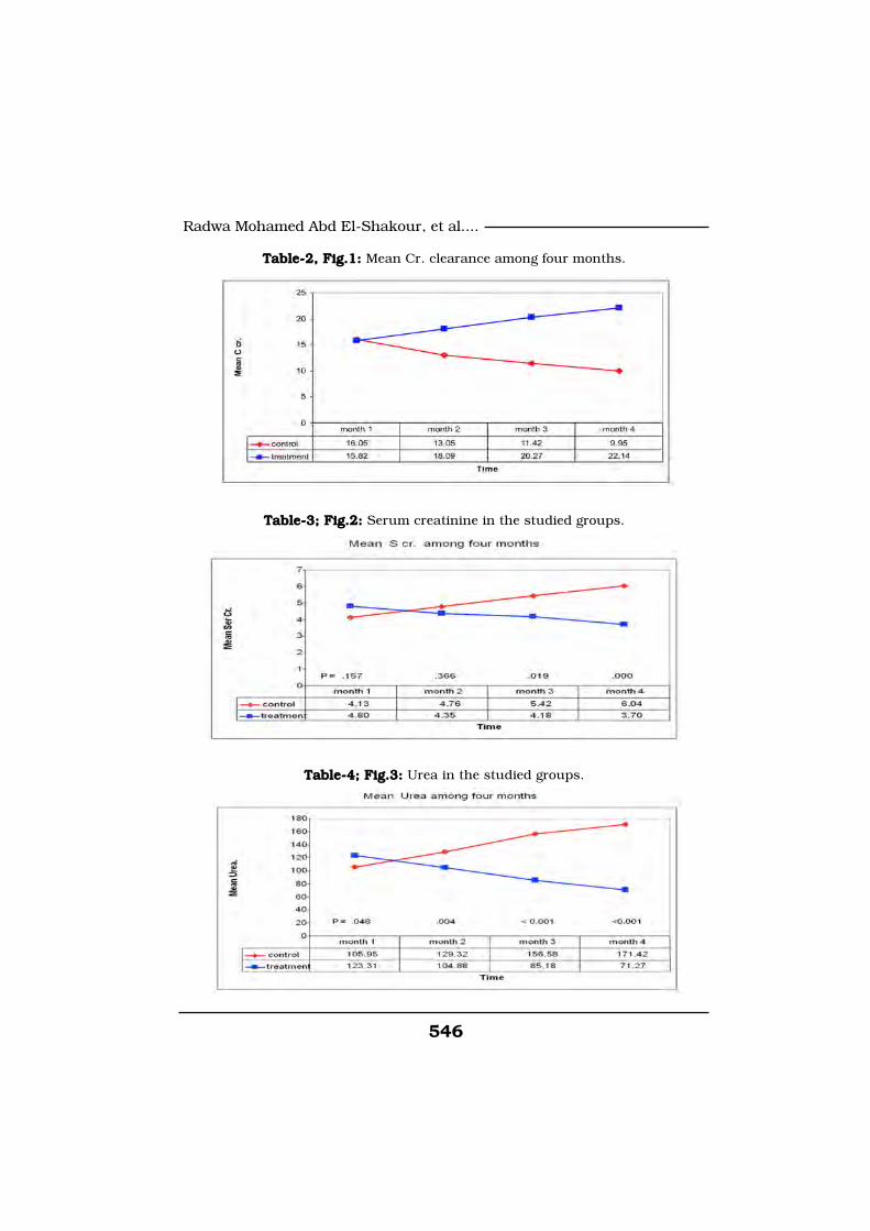
546
Radwa Mohamed Abd El-Shakour, et al....
Table-2, Fig.1: Table-2, Fig.1: Mean Cr. clearance among four months.
Table-3; Fig.2: Table-3; Fig.2: Serum creatinine in the studied groups.
Table-4; Fig.3: Table-4; Fig.3: Urea in the studied groups.

547
Benha M. J.
Vol. 30 No 3 Sept. 2013
Table-5; Fig.4: Table-5; Fig.4: Serum sodium in the studied groups.
Table-6; Fig.5: Table-6; Fig.5: Serum potassium in the studied groups.
Table-7; Fig.6: Table-7; Fig.6: Serum calcium in the studied groups.
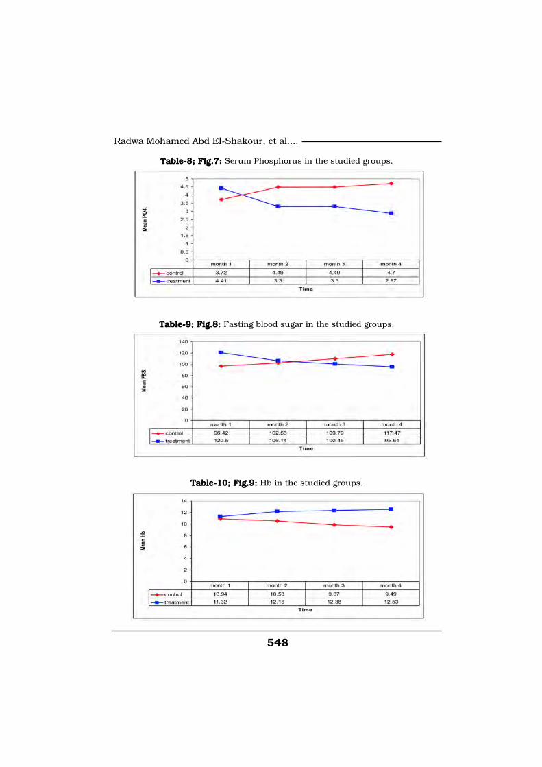
548
Radwa Mohamed Abd El-Shakour, et al....
Table-8; Fig.7: Table-8; Fig.7: Serum Phosphorus in the studied groups.
Table-9; Fig.8: Table-9; Fig.8: Fasting blood sugar in the studied groups.
Table-10; Fig.9: Table-10; Fig.9: Hb in the studied groups.
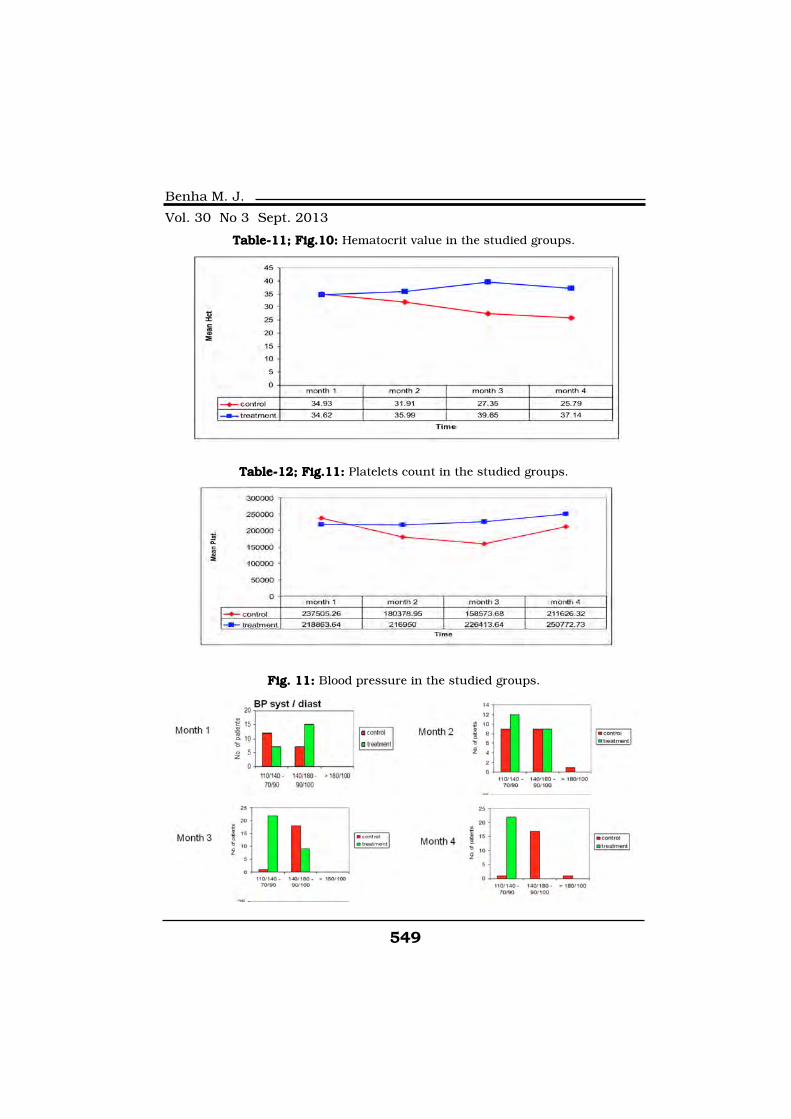
549
Benha M. J.
Vol. 30 No 3 Sept. 2013
Table-11; Fig.10: Table-11; Fig.10: Hematocrit value in the studied groups.
Table-12; Fig.11: Table-12; Fig.11: Platelets count in the studied groups.
Fig. 11: Fig. 11: Blood pressure in the studied groups.
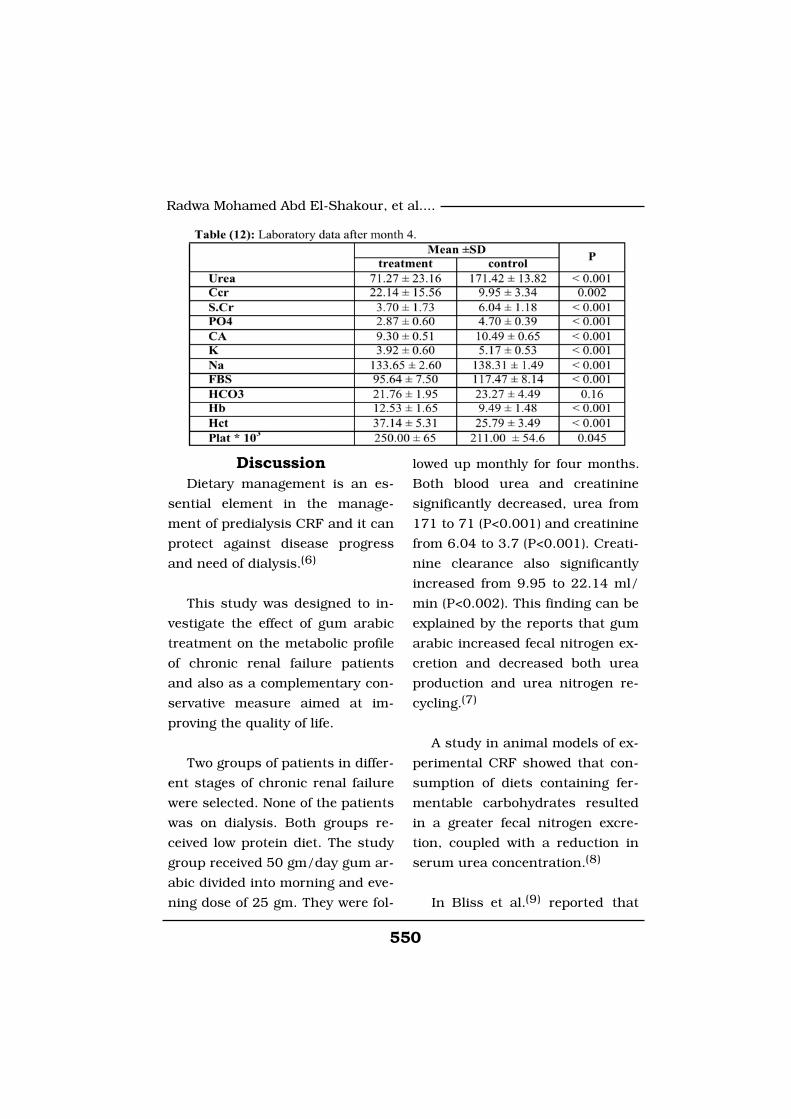
550
Radwa Mohamed Abd El-Shakour, et al....
DiscussionDietary management is an es-
sential element in the manage-
ment of predialysis CRF and it can
protect against disease progress
and need of dialysis.(6)
This study was designed to in-
vestigate the effect of gum arabic
treatment on the metabolic profile
of chronic renal failure patients
and also as a complementary con-
servative measure aimed at im-
proving the quality of life.
Two groups of patients in differ-
ent stages of chronic renal failure
were selected. None of the patients
was on dialysis. Both groups re-
ceived low protein diet. The study
group received 50 gm/day gum ar-
abic divided into morning and eve-
ning dose of 25 gm. They were fol-
lowed up monthly for four months.
Both blood urea and creatinine
significantly decreased, urea from
171 to 71 (P<0.001) and creatinine
from 6.04 to 3.7 (P<0.001). Creati-
nine clearance also significantly
increased from 9.95 to 22.14 ml/
min (P<0.002). This finding can be
explained by the reports that gum
arabic increased fecal nitrogen ex-
cretion and decreased both urea
production and urea nitrogen re-
cycling.(7)
A study in animal models of ex-
perimental CRF showed that con-
sumption of diets containing fer-
mentable carbohydrates resulted
in a greater fecal nitrogen excre-
tion, coupled with a reduction in
serum urea concentration.(8)
In Bliss et al.(9) reported that
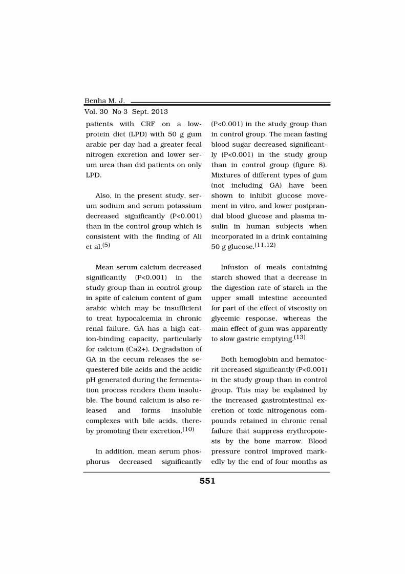
551
Benha M. J.
Vol. 30 No 3 Sept. 2013
patients with CRF on a low-
protein diet (LPD) with 50 g gum
arabic per day had a greater fecal
nitrogen excretion and lower ser-
um urea than did patients on only
LPD.
Also, in the present study, ser-
um sodium and serum potassium
decreased significantly (P<0.001)
than in the control group which is
consistent with the finding of Ali
et al.(5)
Mean serum calcium decreased
significantly (P<0.001) in the
study group than in control group
in spite of calcium content of gum
arabic which may be insufficient
to treat hypocalcemia in chronic
renal failure. GA has a high cat-
ion-binding capacity, particularly
for calcium (Ca2+). Degradation of
GA in the cecum releases the se-
questered bile acids and the acidic
pH generated during the fermenta-
tion process renders them insolu-
ble. The bound calcium is also re-
leased and forms insoluble
complexes with bile acids, there-
by promoting their excretion.(10)
In addition, mean serum phos-
phorus decreased significantly
(P<0.001) in the study group than
in control group. The mean fasting
blood sugar decreased significant-
ly (P<0.001) in the study group
than in control group (figure 8).
Mixtures of different types of gum
(not including GA) have been
shown to inhibit glucose move-
ment in vitro, and lower postpran-
dial blood glucose and plasma in-
sulin in human subjects when
incorporated in a drink containing
50 g glucose.(11,12)
Infusion of meals containing
starch showed that a decrease in
the digestion rate of starch in the
upper small intestine accounted
for part of the effect of viscosity on
glycemic response, whereas the
main effect of gum was apparently
to slow gastric emptying.(13)
Both hemoglobin and hematoc-
rit increased significantly (P<0.001)
in the study group than in control
group. This may be explained by
the increased gastrointestinal ex-
cretion of toxic nitrogenous com-
pounds retained in chronic renal
failure that suppress erythropoie-
sis by the bone marrow. Blood
pressure control improved mark-
edly by the end of four months as
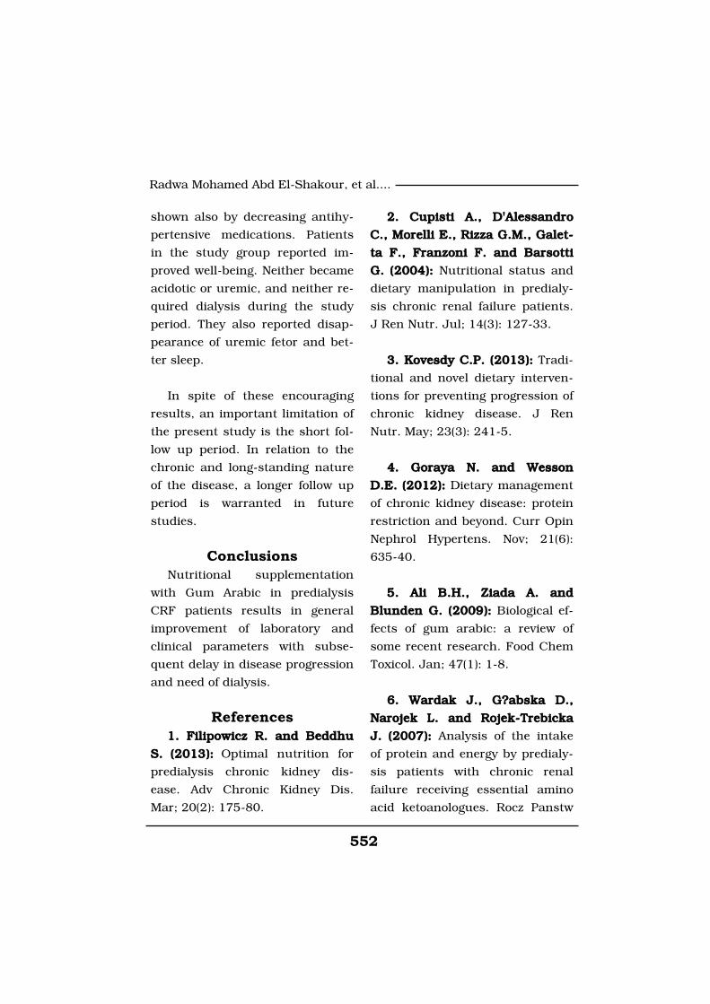
552
Radwa Mohamed Abd El-Shakour, et al....
shown also by decreasing antihy-
pertensive medications. Patients
in the study group reported im-
proved well-being. Neither became
acidotic or uremic, and neither re-
quired dialysis during the study
period. They also reported disap-
pearance of uremic fetor and bet-
ter sleep.
In spite of these encouraging
results, an important limitation of
the present study is the short fol-
low up period. In relation to the
chronic and long-standing nature
of the disease, a longer follow up
period is warranted in future
studies.
ConclusionsNutritional supplementation
with Gum Arabic in predialysis
CRF patients results in general
improvement of laboratory and
clinical parameters with subse-
quent delay in disease progression
and need of dialysis.
References1. Filipowicz R. and Beddhu1. Filipowicz R. and Beddhu
S. (2013): S. (2013): Optimal nutrition for
predialysis chronic kidney dis-
ease. Adv Chronic Kidney Dis.
Mar; 20(2): 175-80.
2. Cupisti A., D'Alessandro2. Cupisti A., D'Alessandro
C., Morelli E., Rizza G.M., Galet-C., Morelli E., Rizza G.M., Galet-
ta F., Franzoni F. and Barsottita F., Franzoni F. and Barsotti
G. (2004):G. (2004): Nutritional status and
dietary manipulation in predialy-
sis chronic renal failure patients.
J Ren Nutr. Jul; 14(3): 127-33.
3. Kovesdy C.P. (2013):3. Kovesdy C.P. (2013): Tradi-
tional and novel dietary interven-
tions for preventing progression of
chronic kidney disease. J Ren
Nutr. May; 23(3): 241-5.
4. Goraya N. and Wesson4. Goraya N. and Wesson
D.E. (2012):D.E. (2012): Dietary management
of chronic kidney disease: protein
restriction and beyond. Curr Opin
Nephrol Hypertens. Nov; 21(6):
635-40.
5. Ali B.H., Ziada A. and5. Ali B.H., Ziada A. and
Blunden G. (2009):Blunden G. (2009): Biological ef-
fects of gum arabic: a review of
some recent research. Food Chem
Toxicol. Jan; 47(1): 1-8.
6. Wardak J., G?abska D.,6. Wardak J., G?abska D.,
Narojek L. and Rojek-TrebickaNarojek L. and Rojek-Trebicka
J. (2007):J. (2007): Analysis of the intake
of protein and energy by predialy-
sis patients with chronic renal
failure receiving essential amino
acid ketoanologues. Rocz Panstw
553
Benha M. J.
Vol. 30 No 3 Sept. 2013
Zakl Hig.; 58(1): 153-8.
7. Assimon S.A. and Stein7. Assimon S.A. and Stein
T.P. (1994):T.P. (1994): Digestible fiber (gum
arabic), nitrogen excretion and
urea recycling in rats. Nutrition.
Nov-Dec; 10(6): 544-50.
8. Younes H., Alphonse J.C.,8. Younes H., Alphonse J.C.,
Behr S.R., Demigné C. and Ré-Behr S.R., Demigné C. and Ré-
mésy C. (1999):mésy C. (1999): Role of ferment-
able carbohydrate supplements
with a low-protein diet in the
course of chronic renal failure: ex-
perimental bases. Am J Kidney
Dis. Apr; 33(4): 633-46.
9. Bliss D.Z., Stein T.P.,9. Bliss D.Z., Stein T.P.,
Schleifer C.R. and Settle R.G.Schleifer C.R. and Settle R.G.
(1996):(1996): Supplementation with
G.A. fiber increases fecal nitrogen
excretion and lowers serum urea
nitrogen concentration in chronic
renal failure patients consuming a
low-protein diet. Am. J. Clin. Nutr.
63, 392-398.
10. Moundras C., Behr S.R.,10. Moundras C., Behr S.R.,
Demigné C., Mazur A. and Ré-Demigné C., Mazur A. and Ré-
mésy C. (1994):mésy C. (1994): Fermentable pol-
ysaccharides that enhance fecal
bile acid excretion lower plasma
cholesterol and apolipoprotein E-
rich HDL in rats. J. Nutr. 124,
2179-2188.
11. Edwards C.A., Blackburn11. Edwards C.A., Blackburn
N.A., Craigen L., Davison P.,N.A., Craigen L., Davison P.,
Tomlin J., Sugden K. and John-Tomlin J., Sugden K. and John-
son I.T. (1987):son I.T. (1987): Viscosity of food
gums determined in vitro related
to their hypoglycemic actions. Am.
J. Clin. Nutr. 46, 72-77.
12. Torsdottir I., Alpsten M.,12. Torsdottir I., Alpsten M.,
Andersson H. and Einarsson S.Andersson H. and Einarsson S.
(1989):(1989): Dietary guar gum effects
on postprandial blood glucose, in-
sulin and hydroxyproline in hu-
mans. J. Nutr. 119, 1925-1931.

554
Radwa Mohamed Abd El-Shakour, et al....
NUTRITIONAL MANAGEMENT OFNUTRITIONAL MANAGEMENT OFPREDIALYSIS RENAL FAILUREPREDIALYSIS RENAL FAILUREPATIENTS USING GUM ARABICPATIENTS USING GUM ARABIC
Radwa Mohamed Abd El-Shakour B.Sc.,Radwa Mohamed Abd El-Shakour B.Sc.,Mousa Abduo Salem MDMousa Abduo Salem MD
and Nabil Mohamed Abd El-Fattah Hassan MDand Nabil Mohamed Abd El-Fattah Hassan MD
BENHAMEDICALJOURNAL
REPRINT
Published byPublished by
Benha Faculty of MedicineBenha Faculty of Medicine Volume 30 Number 3Sept. 2013
555
Benha M. J.
Vol. 30 No 3 Sept. 2013
DIFFERENTIAL EXPRESSION OF P16DIFFERENTIAL EXPRESSION OF P16INK4aINK4aPROTEIN AND S100A4 PROTEINPROTEIN AND S100A4 PROTEIN
IN GASTRIC CARCINOMAIN GASTRIC CARCINOMA
Ghada A. Abd El-Fattah MD, Mohebat H. Gouda MDGhada A. Abd El-Fattah MD, Mohebat H. Gouda MDand Adel Z. El-Saediy MDand Adel Z. El-Saediy MD
Pathology Department, Faculty of Medicine, Benha University
AbstractAbstractPurpose:Purpose: Gastric cancer (GC) is an extremely common disease world-
wide. The p16 INK4a protein, is a tumor suppressor protein that inhibitsCDK4 and CDK6, which phosphorylate the RB protein. S100A4 isknown to be involved in cancer cell motility by virtue of its ability to ac-tivate non-muscle myosin. The present study aims at investigating roleof p16 INK4a, and S100A4 in progression of gastric carcinoma by analyz-ing p16 INK4a and S100A4 protein expression in gastric carcinomas andcorrelating their expression with clinicopathological findings.
Patients and Methods:Patients and Methods: Forty cases including 30 non-consecutiveretrospective selected gastric carcinomas (4 GI, 16 GII, 10 GIII), and 10cases of normal gastric tissue at resection margins of peptic ulcers,were taken as control. Cases were collected in the period 2009-2012,selected from files of pathology department, Faculty of Medicine- BenhaUniversity and Egyptian National Cancer Institute (NCI). Follow up ofthe selected cases was recorded for 18 months. Correlations betweenS100A4 and p16 INK4a immunoreactivity and clinicopathological charac-teristics were evaluated.
Results:Results: showed significant inverse correlation between p16 INK4a ex-pression and grade of carcinoma (P<0.05) as well as high significantcorrelation with type of gastric carcinoma (P<0.01). S100A4 was posi-tively correlated with tumor grade, lymph node metastasis, distant me-tastasis and tumor-node-metastasis (TNM) staging (P<0.05). S100A4 ex-pression was also significantly correlated with poor patient survival. Theworst survival was correlated to cases with low p16INK4a/high S100A4expression (P<0.01).
Conclusion: Conclusion: The immunohistochemical expression of both p16INK4a
556556
Ghada A. Abd El-Fattah, et al....
IntroductionIntroductionGastric cancer is the fourth
most common cancer in the world
with relative frequency of 7.8% of
all cancers. It is the second lead-
ing cause of cancer-related mor-
tality worldwide, accounting for
11.3% of cancer deaths (de Martel
C et al., 2012). In Egypt, cancer
stomach is in the eleventh rank
constituting 2.1% of all cancers
with male to female ratio 1.55 and
median age of 53 year (El-
Bolkainy et al., 2013). However,
the worldwide incidence of gastric
cancer has declined rapidly over
the recent few decades. Part of the
decline may be due to the recogni-
tion of certain risk factors such as
H. pylori and other dietary and en-
vironmental risks (Hannah, et al;
2012).
The current staging classifica-
tions of gastric carcinomas do not
produce accurate predictions of
patient outcomes. Molecular bio-
markers may account for this di-
versity and several prognostic fac-
tors have been identified (Fareed,
et al; 2009). However, none of
these methods have been proven
to be robust enough to be incorpo-
rated into routine practice.
A critical point in the cell cycle
is the G1/S transition checkpoint
which is frequently altered in tu-
mors. This is controlled by cyclins
and cyclin-dependent kinases
(CDK), which complex to induce
the progression of cells into the S-
phase by phosphorylating retino-
blastoma protein. INK4 compris-
ing p16INK4a, p15INK4B,
p18INK4C, and p19INK4D are fami-
ly of CDK inhibitors and binds
specifically to CDK4 and CDK6,
thereby preventing kinase activi-
ties (Hanan, et al; 2009).
P16, a 156-amino acid protein,
is encoded by the INK4A
(CDKN2A, MTS1) gene located on
chromosome 9p21. It exerts its
function by competing with cyclin
and S100A4 in gastric carcinoma is associated significantly with tumorgrade. Expression of S100A4 is significantly associated with lymph nodeand distant metastases, and poor prognosis. Estimation of both mark-ers can be used in planning the therapy and patient’s follow up.
Key words: Key words: p16INK4a, S100A4, tumor suppressor gene (TSG), gas-tric carcinoma (GC), immunohistochemistry (IHC).
557
Benha M. J.
Vol. 30 No 3 Sept. 2013
D in binding to CDK4 preventing
the activation of this kinase and
inhibiting its productive interac-
tion. Thus, p16 protein can specif-
ically associate with CDK4 and
disrupt the formation of active ki-
nase complexes preventing transi-
tion of cells from G1 to S phase
(Osanai, et al; 2011). Therefore,
inactivation of p16INK4a leads to
activation of cyclin/CDK complex-
es, resulting in cell cycle progres-
sion. Alterations of p16INK4a gene
are known to occur in many pri-
mary tumors (Hannah, et al;
2012).
S100A4 protein is the well-
studied member of S100 family. It
is localized in the nucleus, cyto-
plasm, and extracellular space
and possesses a wide range of bio-
logical functions. It appears to
take part in the homeostasis of
growth with apparent involvement
in growth factor signal transduc-
tion and apoptotic cell death
(House et al., 2011). Overexpres-
sion of the S100 calcium-binding
protein A4(S100A4) is involved in
epithelial-to-mesenchymal transi-
tion, oncogenic transformation,
angiogenesis, cytoskeletal integrity
and cancer metastasis. (Cabezo´n
et al., 2007 & Stein, et al, 2011
and Zhao, et al, 2013). S100A4 is
also named as metastasis-
associated Mts1 and fibroblast-
specific protein 1 (FSP1) because
of its close association with cancer
metastasis and constitutive ex-
pression in fibroblasts (Malashke-
vich et al., 2008). Therefore,
S100A4 protein expression is as-
sociated with patient outcome in a
number of tumor types (House et
al., 2011). The present study aims
at investigating role of p16 INK4a,
and S100A4 in progression of gas-
tric carcinoma by analyzing p16
INK4a and S100A4 protein expres-
sion in gastric carcinomas and
correlating their expression with
clinicopathological findings.
Materials and MethodsMaterials and MethodsClinical investigations and tis-
sue samples: The present study
was based on 40 cases including
ten cases of normal gastric tissue
at resection margins of peptic ul-
cer (non-neoplastic lesion) were
taken as control and 30 non-
consecutive retrospective selected
gastric carcinomas. Carcinoma
cases included 18 cases of adeno-
carcinoma, 6 cases of mucinous
carcinoma and 6 cases of signet
558558
Ghada A. Abd El-Fattah, et al....
ring carcinoma. Cases were col-
lected in the period 2009-2012.
They were selected from files of
pathology department, Faculty of
Medicine- Benha University and
Egyptian National Cancer Insti-
tute (NCI). Cases were selected ac-
cording the availability of clinical
and follow-up data for at least 18
months. Only patients with pri-
mary gastric cancer who had not
undergone previous irradiation or
chemotherapeutic treatment were
included in the study.
Staging was carried out accord-
ing to the TNM Classification Sys-
tem (Rosai, 2011).
Formalin-fixed, paraffin-embedded
gastric tissues were used. Three
sections of 4 micron thickness
were obtained from each case.
One section was H & E stained for
diagnosis and grading. Two expe-
rienced pathologists blindly and
independently confirmed the his-
tological diagnosis of each gastric
lesion and agreed on the grading.
Other sections were mounted on
positively- charged slides, immu-
nohistochemically stained with
antibodies against S100A4 and
p16 INK4a using the Ultra Vision
Detection System (Anti-polyvalent,
HRP/DAB, ready-to-use, Lab Vi-
sion corporation).
Immunohistochemical staining:
tissue sections mounted on posi-
tively- charged slides, were heated
at 60°C for 30 minutes then depa-
raffinized and rehydrated through
a series of xylene and alcohol be-
fore staining. After antigen retriev-
al with microwave treatment in
10mM citrate buffer (Neo-Markers,
Cat. # AP-9003), pH 6.0, endoge-
nous peroxidase was blocked with
3% hydrogen peroxide for 20 min-
utes. Sections were washed 3
times with cold 0.01 M phosphate
buffered saline (PBS). After block-
ing with 10% normal rabbit ser-
um, sections were incubated with
polyclonal antibody against
p16INK4a (clone 16P07 Neomark-
ers; dilution 1:100) and pre-
diluted ready to use S100A4 (Neo-
marker, LAB VISION, USA). Both
were incubated overnight at 4° C.
The prepared DAB-substrate-
chromogen solution was applied
and incubated for 5-15 minutes
until color intensity has been
reached. Lastly, sections were
counterstained with Mayer's Hem-
atoxylin. Samples of endometrial
559
Benha M. J.
Vol. 30 No 3 Sept. 2013
adenocarcinomas known to over-
express p16 INK4a were used as
positive controls. A tissue section
of breast cancer was used as posi-
tive control for S100A4. Negative
controls were performed by omit-
ting the primary antibody step.
Interpretation and evaluation of
p16INK4a immunohistochemical
staining: The stains were inter-
preted as positive if there was dis-
tinct diffuse cytoplasmic or nucle-
ar reactivity in the neoplastic
cells, greater than any back-
ground staining in the matched
non-neoplastic gastric tissue, in
more than 20% of the cells within
the different cases (Chen, et al,
2013).
Interpretation and evaluation of
S100A4 immunohistochemical
staining: S100A4 was stained yel-
low-brown in the cytoplasm and
nucleus. The degree of immunos-
taining based on the staining in-
tensity and percentage of positive
cells. The intensity grading scale
was according to the following cri-
teria: 0 (no staining), 1 (weak
staining, light yellow), 2 (moderate
staining yellow-brown) and 3
(strong staining, brown). Moderate
and strong staining indicated tu-
mors with high S100A4 expres-
sion, while no and weak staining
indicated low S100A4 expression
(Li, et al, 2013).
Statistical analysis: Statistical
analysis was performed using the
SPSS (version 16.0 for windows)
software package according to
Sperman's correlation coefficient.
Correlation between several vari-
ables was computed using Fish-
er's exact test. P value less than
0.05 (<0.05) was considered signif-
icant and <0.01 was highly signifi-
cant.
ResultsResultsA-A- Immunohistochemical re-Immunohistochemical re-
sults of p16sults of p16INK4aINK4a staining": staining":
All 10 cases (100%) of normal
gastric tissue showed positive cy-
toplasmic expression of p16INK4a,
while among 30 cases of gastric
carcinoma, 13 cases (43.3%) were
positive and 17 cases (56.7%)
were negative. This difference be-
tween expression of p16INK4a in
normal and malignant neoplastic
gastric tissue was statistically
highly significant (P<0.01) (Fig 1).
As regarding the type of cancer,
560560
Ghada A. Abd El-Fattah, et al....
11 cases (61.1%) of adenocarcino-
ma were positive for p16INK4a,
while all cases (100%) of signet
ring carcinoma showed negative
expression. Out of 6 mucinous
carcinomas, only 2 cases (33.3%)
were positive (P<0.01).
In relation to the tumor grade,
all cases (100%) of gastric carcino-
ma grade 1 showed positive
p16INK4a expression. Among cas-
es of grade II, 7 out of 16 cases
(43.75%) were positive, and 9 cas-
es (56.25%) were negative. Among
the 10 cases of gastric carcinoma
GIII, only 2 cases (20%) were posi-
tive for p16INK4a expression and 8
cases (80%) were negative and this
relationship is statistically highly
significant (P<0.05) (Fig 2).
All stage I cases (100%) showed
positive p16INK4a expression,
while only one case (12.5%) of
stage II was positive. Six cases
(75%) of stage III and 2 cases
(20%) of stage IV were positive for
p16INK4a expression, this rela-
tionship is statistically insignifi-
cant. (P>0.05).
- Concerning to state of lymph
node metastasis, 4 out of the 10
lymph node negative cases (40%)
showed positive p16INK4a expres-
sion, while positivity was seen in 9
of the 20 lymph node positive cas-
es (45%). This relationship was
statistically insignificant (P>0.05).
Insignificant correlation was
also detected between p16INK4a
expression and distant metastasis
(P>0.05). Eleven (55%) out of 20
metastasis free cases were positive
for p16INK4a expression and 2
(20%) out of 10 cases with distant
metastasis were positive.
Correlation with patient survi-
val was also statistically insignifi-
cant (P>0.05). Half of the cases of
living patients (50%) showed posi-
tive p16INK4a expression and 5
dead cases were positive (35.7%).
(Table 1).
(B) Immunohistochemical re-(B) Immunohistochemical re-
sults of S100A4 staining:sults of S100A4 staining:
All of the 10 cases of normal
gastric tissue (100%) showed low
cytoplasmic expression of S100A4,
while 23 cases (76.7%) of gastric
carcinoma showed high expres-
sion. This difference was highly
statistically significant (P<0.01)
(Fig 1).
561
Benha M. J.
Vol. 30 No 3 Sept. 2013
As regarding the type of carci-
noma, 12 cases (66.7%) of adeno-
carcinoma high S100A4 expres-
sion, while all cases (100%) of
signet ring carcinoma showed low
expression. Among 6 cases of mu-
cinous carcinoma only 1 case
(16.7%) was reported with low ex-
pression of S100A4 (P<0.05).
In relation to the tumor grade,
the expression of S100A4 was in-
creased together with loss of diffe-
rentiation. Only one case (25%) of
grade 1 showed high expression
while 12 cases of G2 (75%) and all
cases of G3 (100%) showed high
expression, and this relationship
is statistically highly significant
(P<0.05) (Fig 3).
As regarding the stage of gas-
tric carcinoma, one case (25%) of
stage I showed high expression. In
stage II and III, high S100A4 ex-
pression was shown in 6 cases
(75%). All cases (100%) of stage IV
cases showed high S100A4 ex-
pression. This relationship is sta-
tistically highly significant.
(P<0.05).
- Concerning the state of lymph
node, S100A4 expression is mark-
edly among cases with positive
L.N. metastases. All of these cas-
es, (100%) showed high expres-
sion. In the 10 lymph node nega-
tive cases (70%) showed low
S100A4 expression. This relation-
ship was statistically highly signif-
icant (P<0.01).
- All cases (100%) of those with
distant metastasis showed high
expression. In absence of distant
metastasis, 13 cases (65%)
showed high expression. A statis-
tically significant correlation was
found between S100A4 expression
and occurrence of distant metas-
tasis (P<0.05).
Regarding patient survival, the
expression of S100A4 was related
to poor prognosis. All of dead cas-
es (100%) showed high expres-
sion, while 9 cases (56.25%) of liv-
ing patients showed high
expression. This was a statistically
highly significant relationship
(P<0.01). (Table 2).
(C) Correlation between(C) Correlation between
p16p16INK4aINK4a & S100A4 expression & S100A4 expression
in gastric carcinoma:in gastric carcinoma:
Carcinoma cases were classi-
fied into 4 groups according to the

562562
Ghada A. Abd El-Fattah, et al....
expression of both p16INK4a &
S100A4. The worst prognosis was
correlated to the group of Low
p16INK4a/high S100A4. This cor-
relation was highly statistically
significant (p<0.01). The group of
high p16INK4a/low S100A4
showed good prognosis and this
correlation was also statistically
significant (p<0.05) (Table 3).
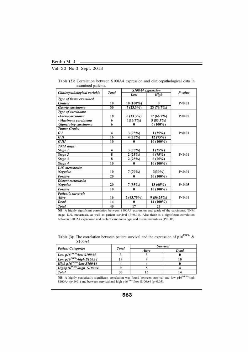
563
Benha M. J.
Vol. 30 No 3 Sept. 2013
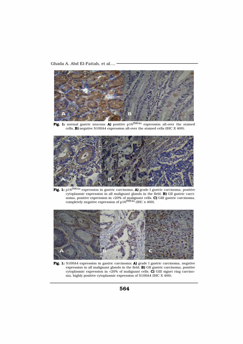
564564
Ghada A. Abd El-Fattah, et al....
Fig. 1: Fig. 1: normal gastric mucosa: A) A) positive p16INK4a expression all-over the stainedcells, B)B) negative S100A4 expression all-over the stained cells (IHC X 400).
Fig. 1: Fig. 1: p16INK4a expression in gastric carcinoma, A)A) grade I gastric carcinoma, positivecytoplasmic expression in all malignant glands in the field. B)B) GII gastric carci-noma, positive expression in <20% of malignant cells. C)C) GIII gastric carcinoma,completely negative expression of p16INK4a (IHC x 400).
Fig. 1: Fig. 1: S100A4 expression in gastric carcinoma: A) A) grade I gastric carcinoma, negativeexpression in all malignant glands in the field. B) B) GII gastric carcinoma, positivecytoplasmic expression in <20% of malignant cells. C) ) GIII signet ring carcino-ma, highly positive cytoplasmic expression of S100A4 (IHC X 400).
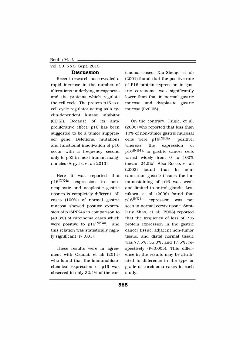
565
Benha M. J.
Vol. 30 No 3 Sept. 2013
DiscussionDiscussionRecent research has revealed a
rapid increase in the number of
alterations underlying oncogenesis
and the proteins which regulate
the cell cycle. The protein p16 is a
cell cycle regulator acting as a cy-
clin-dependent kinase inhibitor
(CDKI). Because of its anti-
proliferative effect, p16 has been
suggested to be a tumor suppres-
sor gene. Deletions, mutations
and functional inactivation of p16
occur with a frequency second
only to p53 in most human malig-
nancies (Argyris, et al; 2013).
Here it was reported that
p16INK4a expression in non-
neoplastic and neoplastic gastric
tissues is completely different. All
cases (100%) of normal gastric
mucosa showed positive expres-
sion of p16INK4a in comparison to
(43.3%) of carcinoma cases which
were positive to p16INK4a, and
this relation was statistically high-
ly significant (P<0.01).
These results were in agree-
ment with Osanai, et al; (2011)
who found that the immunohisto-
chemical expression of p16 was
observed in only 32.4% of the car-
cinoma cases. Xiu-Sheng, et al;
(2001) found that the positive rate
of P16 protein expression in gas-
tric carcinoma was significantly
lower than that in normal gastric
mucosa and dysplastic gastric
mucosa (P<0.05).
On the contrary, Tsujie, et al;
(2000) who reported that less than
10% of non-tumor gastric mucosal
cells were p16INK4a positive,
whereas the expression of
p16INK4a in gastric cancer cells
varied widely from 0 to 100%
(mean, 24.5%). Also Rocco, et al;
(2002) found that in non-
cancerous gastric tissues the im-
munostaining of p16 was weak
and limited to antral glands. Les-
nikova, et al; (2009) found that
p16INK4a expression was not
seen in normal cervix tissue. Simi-
larly Zhao, et al; (2003) reported
that the frequency of loss of P16
protein expression in the gastric
cancer tissue, adjacent non-tumor
tissue, and distal normal tissue
was 77.5%, 55.0%, and 17.5%, re-
spectively (P<0.005). This differ-
ence in the results may be attrib-
uted to difference in the type or
grade of carcinoma cases in each
study.

566566
Ghada A. Abd El-Fattah, et al....
In the current study, a signifi-
cant inverse correlation (p<0.05)
between p16INK4a expression and
grade of gastric carcinoma cases
was found, all cases of grade I,
(43.75%) of grade II, and (20%) of
grade III gastric carcinoma cases
showed positive p16INK4a expres-
sion. These results were parallel to
results reported by Tsujie, et al;
(2000) who reported a clinico-
pathologic survey indicated that a
low or no expression of p16INK4a
was associated with poorly differ-
entiated carcinoma (p=0.0133).
Rocco, et al; (2002) found that the
intensity of immunostaining was
inversely related to the grade of
differentiation of these tumors.
The loss of expression of
p16INK4a in high grades of carci-
nomas means loss of its anti-
proliferative activity. This may be
an important factor in uncon-
trolled gastric cell proliferation
and progression of the tumor to-
wards high grades.
On the contrary to the current
results Osanai, et al; (2011) re-
ported that There was no statisti-
cally significant relationship be-
tween the immunohistochemical
expression of p16 and the degree
of histological differentiation of tu-
mor, when analyzed the relation to
immunoreactivity score (p=0.81).
Also, Xiu-Sheng, et al; (2001) re-
ported that the positive rate of P16
protein expression in mucoid car-
cinoma 10.00% was significantly
lower than that in poorly differen-
tiated carcinoma 51.22%, undif-
ferentiated carcinoma 57.69% and
signet ring cell carcinoma 62.50%
(P<0.05). The key differences be-
tween this study and similar pre-
viously reported IHC studies are
firstly, the number of cases ana-
lyzed; secondly the absence of p16
in the lowest grade of differentia-
tion may reflect clonal expansion
of the cells with a more aggressive
phenotype.
As regarding type of carcinoma,
positive p16INK4a expression was
reported in 61.1% of adenocarcin-
omas, 33.3% of mucoid carcino-
mas, while all cases of signet ring
carcinoma were negative. This was
a highly statistically significant
correlation (p<0.01). These results
could be explained by the behav-
ior of different types of gastric car-
cinomas; 66.7% of mucinous car-
cinomas which are considered a
less differentiated carcinomas and
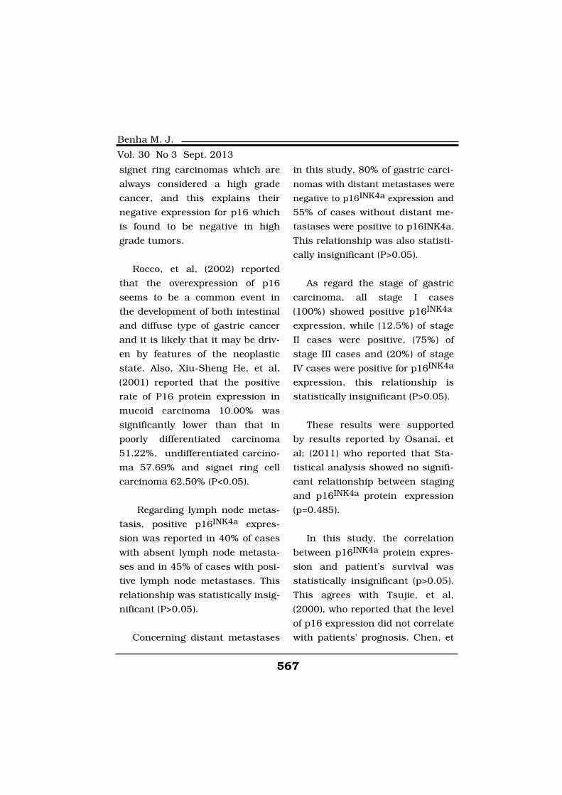
567
Benha M. J.
Vol. 30 No 3 Sept. 2013
signet ring carcinomas which are
always considered a high grade
cancer, and this explains their
negative expression for p16 which
is found to be negative in high
grade tumors.
Rocco, et al, (2002) reported
that the overexpression of p16
seems to be a common event in
the development of both intestinal
and diffuse type of gastric cancer
and it is likely that it may be driv-
en by features of the neoplastic
state. Also, Xiu-Sheng He, et al,
(2001) reported that the positive
rate of P16 protein expression in
mucoid carcinoma 10.00% was
significantly lower than that in
poorly differentiated carcinoma
51.22%, undifferentiated carcino-
ma 57.69% and signet ring cell
carcinoma 62.50% (P<0.05).
Regarding lymph node metas-
tasis, positive p16INK4a expres-
sion was reported in 40% of cases
with absent lymph node metasta-
ses and in 45% of cases with posi-
tive lymph node metastases. This
relationship was statistically insig-
nificant (P>0.05).
Concerning distant metastases
in this study, 80% of gastric carci-
nomas with distant metastases were
negative to p16INK4a expression and
55% of cases without distant me-
tastases were positive to p16INK4a.
This relationship was also statisti-
cally insignificant (P>0.05).
As regard the stage of gastric
carcinoma, all stage I cases
(100%) showed positive p16INK4a
expression, while (12.5%) of stage
II cases were positive, (75%) of
stage III cases and (20%) of stage
IV cases were positive for p16INK4a
expression, this relationship is
statistically insignificant (P>0.05).
These results were supported
by results reported by Osanai, et
al; (2011) who reported that Sta-
tistical analysis showed no signifi-
cant relationship between staging
and p16INK4a protein expression
(p=0.485).
In this study, the correlation
between p16INK4a protein expres-
sion and patient’s survival was
statistically insignificant (p>0.05).
This agrees with Tsujie, et al,
(2000), who reported that the level
of p16 expression did not correlate
with patients' prognosis. Chen, et
568568
Ghada A. Abd El-Fattah, et al....
al, (2013) also did not identify a
correlation between p16 levels and
patient survival.
Different studies showed that
S100A4 plays a role in tumor
growth, motility and invasion sug-
gesting that it is directly linked to
the progression of human carcino-
ma as in colorectal carcinoma
(Boye et al., 2010) and in prostatic
carcinoma (Yong-Wook et al.,
2010). In this study, it was report-
ed that all cases (100%) of non-
neoplastic gastric tissue showed
low S100A4 expression while
(76.7%) of gastric carcinoma cases
showed high expression of
S100A4 and this difference was
statistically significant (P<0.01).
This agrees with Yonemura, et
al, (2000) who reported that
S100A4 expression was detected
in 51 (55%) of 92 primary gastric
cancers and Li, et al, (2013) who
reported that 53 (62.35%) of gas-
tric carcinoma cases exhibited
S100A4 overexpression, in which
immunostaining was observed in
the cytoplasm or the nucleus of
the tumor cells.
In relation to the tumor grade,
the expression of S100A4 was in-
creased together with loss of diffe-
rentiation. (25%) of grade 1
showed high expression, (75%) of
G2 and all cases of G3 (100%)
showed high expression, and this
relationship is statistically highly
significant (P<0.05). This agreed
with Yonemura , et al, (2000) who
reported a strong relationship be-
tween S100A4 expression and his-
tological differentiation of gastric
adenocarcinomas. In their meta-
analysis on colorectal carcinoma,
Liu, et al, (2013) detected a higher
S100A4 expression with poor dif-
ferentiation. This could be ex-
plained by the highly variable fea-
ture of S100A4 expression which
might indicate the influence(s) of
cell cycle regulators, and especial-
ly epigenetic factor(s) in the tran-
scription of this gene.
Concerning the state of lymph
node metastasis, we found that all
cases with positive L.N. showed
high S100A4 expression. In the 10
lymph node negative cases, 7 cas-
es (70%) showed low expression.
This relationship was statistically
highly significant (P<0.01).
Also in this study we reported

569
Benha M. J.
Vol. 30 No 3 Sept. 2013
strong S100A4 expression in
100% of cases with distant metas-
tasis. In absence of distant metas-
tasis, (65%) showed low S100A4
expression. A statistically signifi-
cant correlation was found be-
tween S100A4 expression and oc-
currence of distant metastasis
(P<0.05).
In this work, it was found that
the S100A4 expression is in-
creased in relation to the stage of
gastric carcinoma. (75%) of stage I
showed low expression while
(100%) of stage IV cases showed
high expression. This relationship
is statistically highly significant.
(P<0.01).
Comparing to other studies, Li,
et al, (2013) reported that S100A4
overexpression was closely asso-
ciated with the gastric LN metas-
tasis (P=0.000) and distant metas-
tasis (P=0.024). Zhao, et al, (2013)
found that gastric S100A4 was
positively correlated with lymph
node metastasis and tumor-node-
metastasis (TNM) staging (P<0.05).
The same were the results of
Wang, et al, (2010) who reported
that gastric Expression of S100A4
in gastric cancer is associated sig-
nificantly with lymph node and
distant metastases
In the meta-analysis on color-
ectal cancer done by Liu, et al,
(2013), the results suggested a
significant association between
high S100A4 expression and ad-
vanced TNM stage, as well as the
presence of lymph node metasta-
sis. Pooled data also suggested an
evident trend towards higher
S100A4 expression with poor dif-
ferentiation and distant metasta-
ses.
As regarding endometrioid car-
cinoma, Xie, et al, (2007) reported
that S100A4 expression was sig-
nificantly higher in stage III and
IV tumors compared with stage I.
These results could be ex-
plained by the function of
S100A4. It stimulates cell motility,
invasion, angiogenesis and partici-
pates in the regulation of cell
death. Invasion and motility is
probably promoted through induc-
tion of epithelial to mesenchymal
transition EMT. Cell motility, inva-
sion, and angiogenesis all contrib-
ute to stimulation of metastasis.
S100A4 enhances the turnover of
570570
Ghada A. Abd El-Fattah, et al....
myosin IIA filaments at the lead-
ing edge of migrating cells, result-
ing in increased motility, which
could contribute to an increased
metastatic capacity of cancer
cells, Then S100A4-stimulated
plasmin activation may also con-
tribute to the observed activation
of MMP-2 and MMP- 13 which
help tumor cells to travel through
the surrounding stroma (Boye and
Mælandsmo, 2010).
In this study, it was found that
the expression of S100A4 was re-
lated to poor prognosis. All of dead
cases (100%) showed high expres-
sion, while (43.75%) of living pa-
tients showed low expression. This
was a statistically highly signifi-
cant relationship (P<0.01).
In their studies on gastric car-
cinoma, Zhao, et al, (2013) &
Wang, et al, (2010) reported
S100A4 as a marker for poor prog-
nosis. Also Yonemura, et al,
(2000) reported that patients with
S100A4-positive tumors survived
significantly poorer than did those
with S100A4-negative tumors
On other studies Liu, et al,
(2013) detected that more than
twelve studies investigated the re-
lation between S100A4 and pa-
tient survival and practically dem-
onstrated a significant association
between S100A4 overexpression
and worse prognosis in CRC pa-
tients. Stein, et al (2011) study
was carried upon both colorectal
and gastric cancer and they re-
ported that a high S100A4 expres-
sion correlates with aggressive tu-
mor growth and poor prognosis in
colorectal cancer. Overexpression
of S100A4 is also related to ag-
gressiveness and metastasis in
gastric cancer. This correlation to
poor prognosis could be explained
by the strong correlation between
high S100A4 levels and advanced
tumor stage.
On the contrary, in their study,
Pedersen, et al (2002) did not find
any association between the ex-
pression levels of S100A4 and
clinical outcome. But they thought
that this conflict occurred because
they investigated snap-frozen, ace-
tone-fixed tumor biopsies, while
others examined archival formal-
in-fixed, paraffin-embedded speci-
mens. Such differences in preser-
vation and fixation could possibly
affect the results.
571
Benha M. J.
Vol. 30 No 3 Sept. 2013
The classification of patients
into 4 groups according to expres-
sion of both markers reveals that
the worst prognosis was related to
the group of low p16INK4a/ high
S100A4 expression and this was
highly statistically significant
(p<0.01). This could be explained
by the relation between advanced
tumor stage and high S100a4 lev-
els plus the relation between the
negative p16INK4a and loss of tu-
mor differentiation.
In conclusion, this study sug-
gests that p16INK4a is related to
the differentiation of the gastric
carcinoma while S100A4 upregu-
lation is positively associated with
the growth, invasion, metastasis
and differentiation of gastric carci-
nomas. p16INK4a and S100A4
may be useful markers to predict
progression of gastric carcinoma,
while S100A4 may be a promising
marker for detection of progres-
sion, aggressive behavior and
prognosis of gastric carcinomas.
Estimation of both markers could
be valuable in planning the thera-
py and patient’s follow up.
ReferencesReferences• Argyris P.P., Kademani D.,• Argyris P.P., Kademani D.,
Pambuccian S.E., et al. (2013):Pambuccian S.E., et al. (2013):
Comparison between p16 INK4A
immunohistochemistry and human
papillomavirus polymerase chain
reaction assay in oral papillary
squamous cell carcinoma. J Oral
Maxillofac Surg.; 71(10): 1676-82.
• Boye K. and Mælandsmo G.• Boye K. and Mælandsmo G.
(2010):(2010): Mechanisms involved in
tumor progression. Am J of Path-
ol, Vol. 176, No. 2.
• Boye K., Nesland J.M.,• Boye K., Nesland J.M.,
Sandstad B., et al. (2010): Sandstad B., et al. (2010): Nu-
clear S100A4 is a novel prognostic
marker in colorectal cancer. Eur J
Cancer.; 46(16): 2919-25.
• Cabezo´n T., Celis J.E.,• Cabezo´n T., Celis J.E.,
Skibshoj I., et al. (2007): Skibshoj I., et al. (2007): Expres-
sion of S100A4 by a variety of cell
types present in the tumor micro-
environment of human breast can-
cer. Int J Cancer, 121: 1433-1444.
• Chen Y., Chu H. and Lewis• Chen Y., Chu H. and Lewis
B.C. (2013): B.C. (2013): p16 Stimulates
CDC42-Dependent Migration of
Hepatocellular Carcinoma Cells.
PLoS One, 8(7).
• de Martel C., Ferlay J.,• de Martel C., Ferlay J.,
Franceschi S., et al. (2012): Franceschi S., et al. (2012): Glo-
572572
Ghada A. Abd El-Fattah, et al....
bal burden of cancers attributable
to infections in 2008: a review and
synthetic analysis. Lancet Oncol;
13(6): 607.
• EL-Bolkainy M., Akram M.,• EL-Bolkainy M., Akram M.,
Gouda I., EL-Bolkainy T. andGouda I., EL-Bolkainy T. and
Badawy O. (2013):Badawy O. (2013): Pathology of
Cancer, 4thed, gastrointestinal
cancers; 200:204.
• Fareed K.R., Kaye P., Soom-• Fareed K.R., Kaye P., Soom-
ro I.N., et al. (2009): ro I.N., et al. (2009): Biomarkers
of response to therapy in oesopha-
go-gastric cancer. Gut.; 58: 127-143.
• Hanan M. Abd El-Moneim,• Hanan M. Abd El-Moneim,
and Dalia M. Abd El-Rehimand Dalia M. Abd El-Rehim
(2009):(2009): Immunohistochemical and
Molecular Study of p16
INK4aExpression in Pituitary
Adenoma. Journal of the Egyptian
Nat. Cancer Inst., Vol. 21, No. 4:
351-360.
• Hannah H. and Wong P.C.• Hannah H. and Wong P.C.
(2012):(2012): Immunohistochemical fea-
tures of the gastrointestinal tract
tumors. Journal of Gastrointesti-
nal Oncology. Vol 3, No 3.
• House R.P., Maria, et al.• House R.P., Maria, et al.
(2011):(2011): Two Functional S100A4
Monomers Are Necessary for Reg-
ulating Nonmuscle Myosin-IIA and
HCT116 Cell Invasion. Biochemis-
try; 50(32): 6920-6932.
• Lesnikova I., Lidang M. and• Lesnikova I., Lidang M. and
Hamilton S. (2009):Hamilton S. (2009): p16 as a di-
agnostic marker of cervical neo-
plasia: a tissue microarray study
of 796 archival specimens. Diag-
nostic Pathology, 4: 22.
• Li H., Liu Z., Xu C., et al.• Li H., Liu Z., Xu C., et al.
(2013):(2013): Overexpression of S100A4
is closely associated with the pro-
gression and prognosis of gastric
cancer in young patients. Oncol
Lett.; 5(5): 1485-1490.
• Liu Y., Tang W., Wang J., et• Liu Y., Tang W., Wang J., et
al. (2013):al. (2013): Clinicopathological
and prognostic significance of
S100A4 overexpression in colorec-
tal cancer: a meta-analysis. Diag-
nostic Pathology, 8: 181.
• Malashkevich V.N., Varney• Malashkevich V.N., Varney
K.M., Garrett S.C., et al. (2008):K.M., Garrett S.C., et al. (2008):
Structure of Ca2_-bound S100A4
and its interaction with peptides
derived from non-muscle myosin-
IIA. Biochemistry, 47: 5111-5126.
• Osanai M.H., Edelweiss• Osanai M.H., Edelweiss
M.I., Meurer L. (2011): M.I., Meurer L. (2011): Protein
573
Benha M. J.
Vol. 30 No 3 Sept. 2013
p16 INK4aimmunohistochemical
expression in adenocarcinoma of
the esophagus. ABCD, arq. bras.
cir. dig. vol. 24 no. 4.
• Pedersen K.B., Nesland• Pedersen K.B., Nesland
J.M., Fodstad Ø., et al. (2002):J.M., Fodstad Ø., et al. (2002):
Expression of S100A4, E-
Cadherin, α- and β- Catenin in
breast cancer biopsies. Br. J. Can-
cer, 87: 1281-1286.
• Rocco A., Schandl L., Nar-• Rocco A., Schandl L., Nar-
done G., et al. (2002):done G., et al. (2002): Loss of
expression of tumor suppressor
p16(INK4) protein in human pri-
mary gastric cancer is related to
the grade of differentiation. Dig
Dis.; 20(1): 102-5.
• Rosai J. (2011):• Rosai J. (2011): Gastrointes-
tinal tract. In Rosai and Acker-
man's Surgical Pathology, 10th ed.
Mosby. P: 627-635.
• Stein U., Burock S., Herr-• Stein U., Burock S., Herr-
mann P., et al. (2011): mann P., et al. (2011): Diagnos-
tic and prognostic value of metas-
tasis inducer S100A4 transcripts
in plasma of colon, rectal, and
gastric cancer patients. J Mol
Diagn. 13(2): 189-98.
• Tsujie M., Yamamoto H., To-• Tsujie M., Yamamoto H., To-
mita N., et al. (2000): mita N., et al. (2000): Expression
of tumor suppressor gene p16
(INK4) products in primary gastric
cancer. Oncology; 58(2): 126-36.
• Wang Y.Y., Ye Z.Y., Zhao• Wang Y.Y., Ye Z.Y., Zhao
Z.S., et al. (2010):Z.S., et al. (2010): High-level ex-
pression of S100A4 correlates
with lymph node metastasis and
poor prognosis in patients with
gastric cancer. Annals of surgical
oncology 17:1, 89-97.
• Xie R., Loose D.S., Shipley• Xie R., Loose D.S., Shipley
G.L., et al. (2007): G.L., et al. (2007): Hypomethyla-
tion-induced expression of S100A4
in endometrial carcinoma. Modern
Pathology, (20): 1045-1054.
• Xiu-Sheng He, Qi Su and• Xiu-Sheng He, Qi Su and
Zhu-Chu Chen (2001): Zhu-Chu Chen (2001): Expres-
sion, deleton and mutation of p16
gene in human gastric cancer.
World J Gastroenterol; 7(4): 515-
521.
• Yonemura Y., Endou Y., Ki-• Yonemura Y., Endou Y., Ki-
mura K., et al. (2000): mura K., et al. (2000): Inverse
expression of S100A4 and E-
Cadherin is associated with meta-
static potential in gastric cancer.
Clin Cancer Res., (6): 4234.
• Yong-Wook K., In Ho C.,• Yong-Wook K., In Ho C.,
574574
Ghada A. Abd El-Fattah, et al....
Kyung Do K., et al. (2010):Kyung Do K., et al. (2010): Sig-
nificance of S100A2 and S100A4
Expression in the Progression of
Prostate Adenocarcinoma. Korean
J Urol.; 51(7): 456-62.
• Zhao G.H., Li T.C., Shi L.H.,• Zhao G.H., Li T.C., Shi L.H.,
et al. (2003):et al. (2003): Relationship be-
tween inactivation of p16 gene
and gastric carcinoma. World J
Gastroenterol; 9(5): 905-9.
• Zhao Y., Zhang T. and Wang• Zhao Y., Zhang T. and Wang
Q. (2013):Q. (2013): S100 calcium-binding
protein A4 is a novel independent
prognostic factor for the poor
prognosis of gastric carcinomas.
Oncol Rep 30(1): 111-8.
575
Benha M. J.
Vol. 30 No 3 Sept. 2013
Published byPublished by
Benha Faculty of MedicineBenha Faculty of Medicine
DIFFERENTIAL EXPRESSION OF P16DIFFERENTIAL EXPRESSION OF P16INK4aINK4aPROTEIN AND S100A4 PROTEINPROTEIN AND S100A4 PROTEIN
IN GASTRIC CARCINOMAIN GASTRIC CARCINOMA
Ghada A. Abd El-Fattah MD, Mohebat H. Gouda MDGhada A. Abd El-Fattah MD, Mohebat H. Gouda MDand Adel Z. El-Saediy MDand Adel Z. El-Saediy MD
BENHAMEDICALJOURNAL
REPRINT
Volume 30 Number 3Sept. 2013
BENHA
MEDICAL
JOURNAL
VOLUME 30 NO. 3
Sept. 2013
LAPAROSCOPIC ASSISTED VAGINAL HYSTERECTOMY (LAVH)VERSUS HAND ASSISTED LAPAROSCOPIC HYSTERECTOMY(HALH) IN GYNECOLOGICAL TUMOURSSheiref Kotb MD, Nazem Shams MD, Ashraf Khater MD and Mo-hamed El-Metwally M.Sc
PREVALENCE OF OCCUPATIONAL BURNOUT AMONG MANSOU-RA UNIVERSITY HOSPITALS’ RESIDENTS AND ASSISTANT LEC-TURERSAhmed A. Albadry M.Sc, Ahmed N. Sleem MD, Nadia A. Montas-ser MD and EL-Sayed A. El-Naggar MD
GLYPICAN-3 EXPRESSION IN HEPATOCELLULAR CARCINOMAIN RELATION TO THE GRADE OF DIFFERENTIATIONEman Tawfik Enan MD, Amira Kamal El-Hawary MD, Dina AbdEl-Aziz El-Tantawy MD, Nagwa Mokhtar Helal MD and Maha Mo-hamed Abo-Hashem MD
EFFECTS OF TAMOXIFEN ON LIPID PROFILES IN POST-MENOPAUSAL BREAST CANCER PATIENTSFatma M.F. Akl MD and Ibrahim A. Abdel Aal MD
EFFECTS OF INSULIN AND VITAMIN E ON THE EXPRESSION OFGLIAL FIBRILLARY ACIDIC PROTEIN AND OXIDATIVE STRESSIN THE CEREBELLUM OF DIABETIC RATSAdel A. Bondok Ph.D, Adel A. Elhawary Ph.D, Mohamed I. AbdoPh.D, Rania N. Kamal Ph.D and Hany M. Sonpol M.Sc
WNT/β-CATENIN SIGNALING PATHWAY IN BREAST CARCINO-GENESIS IN RATSAmina Ahmad Baiomy Ph.D, Hoda Ahmad Nada Ph.D, LamiaaArafa Ph.D, Maha Amin Ph.D and Lamiaa El-Abbasy M.Sc.
BELOW 2 YEARS CHILDREN CAREGIVERS’ KNOWLEDGE, ATTI-TUDE AND BELIEFS TOWARDS IMMUNIZATIONNadia Abd El-Hamed Montasser MD, Randah Mohamad HelalMD, Noha El-Adawi MD, Eman Mostafa, Fatma Abd El-Rahman,Maged Saad and Soha Hamza
THE ROLE OF MR DIFFUSION IN DIFFERENTIATION OF MALIG-NANT AND BENIGN HEPATIC FOCAL LESIONSMahmoud Abd El-Latif El-Shewail MD, Galal El Hawary MD, AdelEl-Badrawy MD, Hatem El-Alfy MD
DOPAMINE TRANSPORTER 3'UTR VNTR GENOTYPE AND WIS-CONSIN CARD SORTING TEST IN CHILDREN WITH ADHDAMONG EGYPTIAN POPULATIONMohamed Elwasify MD, Zeinab Gomaa MD, Elsayed ElNaggarMD, Farha Elshenawy MD and Vishwaiit Nimgaonkar MD
1
2
3
4
5
6
7
8
9
9
21
33
41
53
75
93
107
129
CONTENTS

EFFECT OF ORAL ANTICOAGULANT (WARFARIN SODIUM -COUMADIN) ON ARTERIAL BLOOD PRESSURE (EXPERIMENTALSTUDY)Ahmed A. El-Gendy Ph.D and Ghayaty E.A.D. MD
BIOLOGICAL AND SEROLOGICAL STUDIES IN SCHOOL CHIL-DREN TO EVALUATE WIDAL TEST AS A SINGLE DIAGNOSTICONE IN TYPHOID FEVERSoheir A. Abd El-Samie MD, Ibrahim M. Rageh MD, MohammedE. Metwally Ph.D and Fatma A. M. Mohammed B.Sc
ACUTE APPENDICITIS IN THE OVER-FIFTY AGE GROUPHasan I. Fadeel MD, Mohan Patro MD, Farag M. Mikael MD, Oth-man issa Mohamed Abou Bakr MD and Yousef Thabet Ali MD
PHYSICAL INTIMATE PARTNER VIOLENCE: PREVALENCE, PRE-DICTORS, AND HEALTH IMPACTNesrine Saad Farrag M.Sc, Nadia Abd El-Hamed Montasser MD,Farida Abdel Wahab El-Sayed MD and Ghada El-Khawaga MD
INDUCTION OF CANCER STEM CELL IN HEPATOCELLULAR CA-RINOMA: THIOACETAMIDE MODELHuda El-Tahry Ph.D, Omar Gabr Ph.D, Farha El-Chennawi Ph.D,Dalia Saleh Ph.D and Amira Othman M.Sc
EVALUATION OF DIFFERENT SCORING SYSTEMS PREDICTINGNONSENTINEL LYMPH NODE STATUS IN BREAST CANCER PA-TIENTSMagdy B. E-Moghazy MD, Ashraf M. Shoma MD, Abd El-AzeemEl-Ganash MD, Maha M. Abu Hashem MD and Ahmed MoatamedMD
ANEMIA, PHYSICAL PERFORMANCE RELATIONSHIP AMONGELDERY PATIENTS ATTENDING GERIATRIC OUTPATIENT CLIN-ICS IN MANSOURA CITYFatma Magdi Ibrahim M.Sc, Soad Hassan Abd El Hamid MD andFarida Abdel-Wahab Ph.D
EFFECT OF ANTICOAGULANT (WARFARIN) AND L-CARNITINEON HAEMOSTATIC FUNCTION AND OXIDATIVE STRESS INSTREPTOZOTOCIN-INDUCED DIABETIC RATSAhmed A. El-Gendy Ph.D and Amr M. Abbas Ph.D
OUTCOME OF BAHA SURGERY USING THE INFERIORLY BASEDPARTIAL THICKNESS SKIN FLAP Mahmoud El-Sayed Ali, MBBCh, MSc, FRCS, MD
EVALUATION OF THE ROLE OF LAPAROSCOPY IN THE MAN-AGEMENT OF BLUNT ABDOMINAL TRAUMAAbu-Sheashaa M.S. MRCS, Dawood I. MD, El-Sedek M. MD, Na-shat Noaman MD and Ahmed Negm MD
IMMUNOHISTOCHEMICAL PROFILING OF B-CELL LYMPHOMAINTERMEDIATE BETWEEN DIFFUSE LARGE B-CELL LYMPHO-MA AND CLASSICAL HODGKIN LYMPHOMANirmeen Megahed M.Sc, Azmy Abdelhameed MD, Asmaa GadoMD, Maha Amin MD, Naoko Asano MD, Seiichi Kato MD, Kat-suyoshi Takata MD, Aki Mitsuda MD and Shigeo Nakamura MD
10
11
12
13
14
15
16
17
18
19
20
139
151
161
171
185
197
213
235
265
281
297
EFFECT OF ERYTHROPOIETIN AND STEM CELLS ON ANIMALMODEL OF CHRONIC NEPHROPATHYMohammed E. Sarhan MD, Hanaa G. El-Serougy MD, MohammedA. Sobh MD, Abdel Aziz M. Hussein MD and Mohammed E. Sala-ma M.Sc
EFFECT OF ISS-ODN-INDUCED TLR9 STIMULATION ON EXPER-IMENTALLY INDUCED AIRWAY INFLAMMATION IN MICEMona M. El-Haroun M.Sc., Ali M. Yousef MD and Tarek M. Ibra-him Ph.D
PROGNOSTIC AND PREDICTIVE VALUES OF KI67 PROLIFERA-TIVE INDEX IN DIFFUSE LARGE B -CELL LYMPHOMASeham El-Sayed Abdel-Khalek MD and Azza Abdel-Aziz MD
PROTECTIVE EFFECT OF QUERCETIN ON LIVER DAMAGE INSTREPTOZOTOCIN-INDUCED DIABETIC RATSOla A. El Gohary MD and Abeer A. Shoman MD
PROTECTIVE EFFECT OF QUERCETIN AGAINST INDOMETHA-CIN INDUCED GASTRIC ULCER IN RATSMona A. Said MD and Naglaa Y. Nafeh MD
CAUDAL BUPIVACAINE, KETAMINE AND THEIR COMBINATIONFOR PEDIATRIC POSTOPERATIVE ANALGESIAGehan A. Tarbeeh MD
EFFECT OF INTRAPERITONEAL INSTILLATION OF LORNOXI-CAM, AND BUPIVACAINE COMBINATION ON PATIENTS OUT-COME AFTER LAPAROSCOPIC CHOLECYSTECTOMYGhada El-Rahmawy MD, Hosam Ghazy MD and Amr Moawad MD
COLOR DOPPLER ECHOCARDIOGRAPHIC CHANGES 2 YEARSAFTER 2D PLANNED RADIATION THERAPY FOR LEFT BREASTCANCER, ITS RELATION TO CARDIAC BIOMARKERSMona M. Halim MD and Shaheer K. George MD
METRONOMIC LOW DOSE CARBOPLATIN AFTER RADIOTHERA-PY IN STAGE III TESTICULAR SEMINOMAMona M. Halim MD and Nazzem Shams MD
COMPARISON OF NERVE STIMULATOR VERSUS ULTRASOUNDGUIDED BRACHIAL PLEXUS BLOCK FOR UPPER EXTREMITYSURGERYAly E. Rashad MD, Salwa M. Sabry MD, Ahmed G. Sadek MD, Ol-fat M. Ismail MD and Amer Abd A. Attia MD
STUDY OF CAUSES OF LOW BACK PAIN AND ITS PREVALENCEIN REPRESENTATIVE COHORT OF EGYPTIAN POPULATIONEl-Boghdady I. MD, El-Kady B. MD, Onsy N. MD and Seif El-DeinS. M.Sc
VINORELBINE AND 5 FLUORO-URACIL / FOLINIC ACID VERSUSDOCETAXEL AS FIRST LINE TREATMENT FOR PATIENTS WITHMETASTATIC BREAST CANCERI. Abdel Halim MD, E. El-Sherbini MD and N. Haddad MD
21
22
23
24
25
26
27
28
29
30
31
32
307
329
347
363
379
393
405
417
429
441
455
467
EFFECT OF PRESSURE VERSUS VOLUME CONTROLLED VENTI-LATION ON RESPIRATORY MECHANICS, HEMODYNAMICS ANDINTRA-ABDOMINAL PRESSURE DURING ABDOMINOPLASTYMedhat Mikhail Messeha MD, Tarek Abdel Aziz Ibrahim Ph.D,Walaa Safaa Eldin Elkharboutly MD, Ashraf Mohamed WahbaWafa MD and Saleh Ibrahim Elawady MD
COMPARISON OF THE EFFECTS OF LORNOXICAM TO PARA-CETAMOL WHEN ADDED TO LIDOCAINE FOR INTRAVENOUSREGIONAL ANESTHESIA IN PATIENTS UNDERGOING HANDAND FOREARM SURGERYNasr Mahmoud Abdallah Sief El-Nasr MD, Mohamed MohamedAbd Elhaq MD and Ahmed Ragab Abd Elhakeim MD
SOME PLATELET PROPERTIES OF RESTING APHERESIS PLATE-LET CONCENTRATE FOR THE LAST 6 HOURS OF A 48-HOURSTORAGE PERIODManal H. Farahat MD and Mohammad A. Elhady MD
OUTCOME OF CORONARY ARTERY BYPASS IN PATIENTS WITHCHILD-PUGH CLASS A LIVER CIRRHOSISMohamed Ahmed El-Awady MD and Moataz Rezk MD
NUTRITIONAL MANAGEMENT OF PREDIALYSIS RENAL FAILUREPATIENTS USING GUM ARABICRadwa Mohamed Abd El-Shakour B.Sc., Mousa Abduo Salem MDand Nabil Mohamed Abd El-Fattah Hassan MD
DIFFERENTIAL EXPRESSION OF P16 INK4aPROTEIN ANDS100A4 PROTEIN IN GASTRIC CARCINOMAGhada A. Abd El-Fattah MD, Mohebat H. Gouda MD and Adel Z.El-Saediy MD
33
34
35
36
37
38
481
501
515
531
541
555