Lack of Galectin-3 Disturbs Mesenteric Lymph NodeHomeostasis and B Cell Niches in the Course ofSchistosoma mansoni InfectionFelipe L. Oliveira1*, Camila Brand1, Adelzon A. Paula1, Katia D. Arcanjo1, Daniel K. Hsu2, Fu-Tong Liu2,
Christina M. Takiya3, Radovan Borojevic1, Roger Chammas4,5, Marcia C. El-Cheikh1*
1 Laboratorio de Proliferacao e Diferenciacao Celular, Instituto de Ciencias Biomedicas, Universidade Federal do Rio de Janeiro, Rio de Janeiro, Brazil, 2 Department of
Dermatology, School of Medicine, University of California Davis, Sacramento, California, United States of America, 3 Laboratorio de Patologia Celular – Instituto de Ciencias
Biomedicas – Universidade Federal do Rio de Janeiro, Rio de Janeiro, Brazil, 4 Laboratorio de Oncologia Experimental, Departamento de Radiologia e Oncologia,
Faculdade de Medicina, Universidade de Sao Paulo, Sao Paulo, Brazil, 5 Instituto do Cancer do Estado de Sao Paulo, Sao Paulo, Brazil
Abstract
Galectin-3 is a b-galactoside-binding protein that has been shown to regulate pathophysiological processes, includingcellular activation, differentiation and apoptosis. Recently, we showed that galectin-3 acts as a potent inhibitor of B celldifferentiation into plasma cells. Here, we have investigated whether galectin-3 interferes with the lymphoid organization ofB cell compartments in mesenteric lymph nodes (MLNs) during chronic schistosomiasis, using WT and galectin-3-/- mice.Schistosoma mansoni synthesizes GalNAcb1-4(Fuca1-3)GlcNAc(Lac-DiNAc) structures (N-acetylgalactosamine b1-4 N-acetylglucosamine), which are known to interact with galectin-3 and elicit an intense humoral response. Antigens derivedfrom the eggs and adult worms are continuously drained to MLNs and induce a polyclonal B cell activation. In the presentwork, we observed that chronically-infected galectin-3-/- mice exhibited a significant reduced amount of macrophages andB lymphocytes followed by drastic histological changes in B lymphocyte and plasma cell niches in the MLNs. The lack ofgalectin-3 favored an increase in the lymphoid follicle number, but made follicular cells more susceptible to apoptoticstimuli. There were an excessive quantity of apoptotic bodies, higher number of annexin V+/PI- cells, and reduced clearanceof follicular apoptotic cells in the course of schistosomiasis. Here, we observed that galectin-3 was expressed in non-lymphoid follicular cells and its absence was associated with severe damage to tissue architecture. Thus, we convey newinformation on the role of galectin-3 in regulation of histological events associated with B lymphocyte and plasma cellniches, apoptosis, phagocytosis and cell cycle properties in the MLNs of mice challenged with S.mansoni.
Citation: Oliveira FL, Brand C, Paula AA, Arcanjo KD, Hsu DK, et al. (2011) Lack of Galectin-3 Disturbs Mesenteric Lymph Node Homeostasis and B Cell Niches inthe Course of Schistosoma mansoni Infection. PLoS ONE 6(5): e19216. doi:10.1371/journal.pone.0019216
Editor: David Joseph Diemert, The George Washington University Medical Center, United States of America
Received October 26, 2010; Accepted March 30, 2011; Published May 6, 2011
This is an open-access article, free of all copyright, and may be freely reproduced, distributed, transmitted, modified, built upon, or otherwise used by anyone forany lawful purpose. The work is made available under the Creative Commons CC0 public domain dedication.
Funding: This work was supported by Conselho Nacional de Pesquisa e Desenvolvimento (CNPq - 472553/2008-9); Fundacoes de Amparo a Pesquisa do Estadodo Rio de Janeiro e de Sao Paulo (FAPERJ and FAPESP) and Associacao Paul Ehrlich de Biologia Celular Aplicada a Medicina (APABCAM). These funders had no rolein study design, data collection and analysis, decision to publish, or preparation of the manuscript.
Competing Interests: The authors have declared that no competing interests exist.
* E-mail: [email protected] (FLdO); [email protected] (MCEC)
Introduction
Schistosomiasis is a helminth disease that affects more than 200
million people predominantly in developing countries [1].
Schistosoma mansoni infection is a long lasting inflammatory reaction
characterized by the presence of adult worms living in the
mesenteric venous system, depositing their eggs in small
submucosal veins of the intestine. Some of these eggs are washed
through the portal blood flow into the liver, where they cause
granulomatous inflammatory reactions [2]. A typical Th2 response
is well defined in the acute phase and the evolution towards the
chronic phase is associated with a down-regulation of several
aspects of the immune response to parasites [3]. Egg and worm
antigens are continuously drained to mesenteric lymph nodes
(MLNs), where they induce an intense polyclonal B cell activation
and germinal center reaction in the lymphoid follicles (LFs),
concomitant with development of splenomegaly [2,4,5].
Lymph nodes have a well-defined lymphoid architecture: a
cortical region consisting mostly of B lymphocytes, macrophages
and follicular dendritic cells (FDCs) densely packed and organized
into LFs; a paracortical region (deep cortex) presenting predom-
inantly T lymphocytes, scarce B lymphocytes and dendritic cells;
and a medullary region formed by macrophages and plasma cells
organized in cellular cords, besides sinuses that conduct the lymph,
cells and secreted immunoglobulin to the venous blood system [6].
This structural organization contributes to B cell activation and
proliferation (B220 or CD45RA+ cells) into LFs, establishing
germinal centers and/or inducing the B cell differentiation into
plasmablasts (CD138+) and Blimp-1+ immunoglobulin secreting
plasma cells [7–9]. Part of these activated B cells undergo
apoptosis and are eliminated by macrophages or resident
immature dendritic cells [10–12].
During schistosomiasis, both eggs and the adult worms
synthesize GalNAcb1-4(Fuca1-3)GlcNAc(Lac-DiNAc) structures
(N-acetylgalactosamine b1-4 N-acetylglucosamine) that interact
with galectin-3. The latter is a conserved b-galactoside-binding
protein expressed by macrophages that can elicit an intra-hepatic
granulomatous reaction and a vigorous humoral immune response
PLoS ONE | www.plosone.org 1 May 2011 | Volume 6 | Issue 5 | e19216

[13,14]. This lectin regulates cell-cell and cell-extracellular matrix
interactions, cell signaling, inflammatory responses and biological
events, such as cellular activation, migration, differentiation,
apoptosis and tumor metastasis [15]. Moreover, galectin-3 acts
as a powerful pro-inflammatory molecule to myeloid cells by
inducing chemotaxis of monocytes and phagocytosis by macro-
phages [16,17]. It also controls T cell activation, proliferation and
death [18,19], modulates carbohydrate-dependent thymocyte
interactions in thymic microenvironments [20], and inhibits
conventional/B2 and peritoneal/B1 lymphocytes differentiation
into plasma cells [21–23].
Recently, we showed that one of the hallmarks of S.mansoni-
infected galectin-32/2 mice is disturbed plasmacytogenesis
involving the spleen, bone marrow and MLNs [22]. The increase
of plasma cells in the MLNs associated with the continuous arrival
of mesenteric antigens could disturb the tissue organization of the
lymphoid compartments. Thus, in this work, we investigated the
possible interference of galectin-3 in the organization of MLNs in
the course of chronic murine schistosomiasis. It was observed that
in infected galectin-32/2 mice there was significant histological
disorganization in the B and plasma cell niches, which correlated
with abnormal cell survival rate and inadequate clearance. We
propose that galectin-3 contributes to the maintenance of MLN
architecture and drives immune responses by regulating B cell
differentiation during S. mansoni infection.
Materials and Methods
Mice and Schistosoma mansoni infectionInbred C57/bl6 wild type (WT) and galectin-32/2 mice
(backcrossed to C57/bl6 for 10 generations) [24] matched by
age and sex were obtained from a colony bred at the Federal
University of Rio de Janeiro (Brazil). All mice procedures were
performed in accordance with institutional guidelines (protocol
number DAHEICB 009, Federal University of Rio de Janeiro).
Uninfected mice were used as controls. Thirty day-old mice were
infected by percutaneous penetration of 40 S. mansoni cercariae
(BH strain, Oswaldo Cruz Institute, Rio de Janeiro, Brazil). Mice
were euthanized using a carbon dioxide chamber 90–95 days after
infection, corresponding to the chronic phase of the disease [25].
Cell suspensions and flow cytometryCell suspensions from MLNs of infected WT and galectin-32/2
mice were obtained ex vivo by standard mechanical procedures and
washed twice with Phosphate Buffer Solution (PBS), pH 7.2,
containing 3% Fetal Bovine Serum (FBS), quantified and their
concentration adjusted to 16106 cells/mL for flow cytometry
analysis. The cells were incubated with Fc blocker (Clone 2.4G2)
for 10 min before adding the following monoclonal antibodies:
FITC anti-B220, anti-Mac 1 and anti-CD4; PE anti-CD19, anti-
CD8 and anti-CD-138; PE Cy5.5 anti-Mac1, anti-Gr-1 and anti-
B220 (BD Bioscience, USA). The samples were assayed in a flow
cytometer (FACSCalibur, BD Bioscience, USA) and the resulting
data analyzed using the CellQuest and WinMDI 2.9 software
packages. DNA-content was measured by propidium iodide
labeling using Vindelov solution [26].
Phenotype of lymph nodal cellsLymph node cells were characterized according to phenotypic
markers, as follows: monocytes (CD192 B2202 Mac-1+ Gr-1+/low
CD42 CD82 CD1382), macrophages (CD192 B2202 Mac-1+
Gr-12 CD42 CD82 CD1382), granulocytes (CD192 B2202
Mac-1+ Gr-1+/high CD42 CD82 CD1382), CD4+ T cells (CD192
B2202 Mac-12 Gr-12 CD4+ CD82 CD1382), CD8+ T cells
(CD192 B2202 Mac-12 Gr-12 CD42 CD8+ CD1382), plasma-
cytoid dendritic cells (CD192 B220+ Mac-12 Gr-1+ CD4+ CD82
CD1382), B2/conventional B cells (CD19+ B220+/high Mac-12
Gr-12 CD42 CD82 CD1382), B1 cells (CD19+ B220+/low Mac-
1+ Gr-12 CD42 CD82 CD1382) and plasma cells (CD192
B2202/low Mac-12 Gr-12 CD42 CD82 CD138+).
Histological preparationsFor histological analyses, WT and galectin-32/2 mice were
sacrificed during the chronic phase of schistosomiasis infection (5
animals per group). Mesenteric lymph nodes were removed, cut
into 0.5 mm-thick slices, washed in cold saline and fixed in 10%
buffered formalin fixative. After 12 h of fixation, specimens were
dehydrated in alcohol and embedded in paraffin. Sections of 5 mm
were obtained and stained with hematoxylin & eosin (H&E).
Uninfected WT and galectin-32/2 mice were used as control
groups.
Quantification of lymphoid folliclesLFs were characterized as well-defined rounded clusters
containing lymphocyte-like cells and they were quantified per
microscopic field using the Image J software (original magnifica-
tion, 25X). For each experiment, sectioned samples were obtained
from mesenteric lymph nodes of five WT and galectin-32/2 mice,
both uninfected and infected with S. mansoni.
ImmunohistochemistryParaffin-embedded sections were de-waxed and hydrated. After
inhibition of endogenous peroxidase, sections were incubated for
1 h with 0.01 M PBS containing 5% BSA, 4% skim milk, 0.1%
Triton X-100 (Sigma Aldrich, USA), 0.05% Tween-20, and 10%
normal goat serum and incubation with the following purified
antibodies: anti-gal-3 (clone M3/38; American Type Culture
Collection, Manassas, VA, USA, at 1:10 in PBS, 3% BSA and 1%
normal goat serum), anti-B220, anti-CD138 and Blimp-1 (Santa
Cruz Biotechnology, USA) overnight at 4uC in a humid chamber.
Antibodies were detected with a biotinylated anti-rat IgG (BA-
4001, Vector Laboratories, Burlingame, CA, USA) and developed
with avidin-peroxidase (1:50 in PBS) (Sigma Aldrich, USA), using
diaminobenzidyne as the chromogen. Sections were counter-
stained with Harris’ hematoxylin. Bright-field pictures were
acquired using an Evolution MP 5.0 RTV Color camera (Media
Cybernetics, Canada). As negative controls, sections of WT and
knockout mice tissue were incubated with non-immune rat serum
instead of anti-galectin-3 antibody.
Apoptosis and Phagocytosis assaysMLNs from WT and galectin-32/2 mice were dissociated and
the cells were cultured in RPMI 1640 medium supplemented with
10% SFB in 12-well plates (Corning, USA) for 2 h at 37uC and
5% CO2 atmosphere. The non-adherent cells were collected and
induced to apoptosis by heating at 43uC for 60 minutes [27].
Subsequently, the apoptotic and dead cells were marked with
annexin V-FITC and propidium iodide (PI), and quantified by
flow cytometry. Adherent cells were obtained and maintained at
37uC. Apoptotic-induced non-adherent cells were co-cultured with
these adherent cells (ratio of 4 non-adherent to 1 adherent cells)
during 24 h and 72 h days, at 37uC and 5% CO2 atmosphere.
The floating cells were washed out and the resting cells were
stained by the May-Grunwald-Giemsa method [28]. Adherent-
phagocytic cells were identified by the formation of translucent
vacuoles and phagosomes inside the cytoplasm and differentiated
from the adherent non-phagocytic cells by the absence of them.
Gal-3 Regulates B Cell Niches
PLoS ONE | www.plosone.org 2 May 2011 | Volume 6 | Issue 5 | e19216
Results represent a mean of three independent experiments
performed using MLNs from infected WT and galectin-32/2
mice. Images were captured using a QColor-3 camera (Olympus,
Japan) and analyzed with the Q-Capture software.
Immunofluorescence to MOMA-2 markerDirect immunofluorescence staining of MLNs was carried out
after de-waxing and rehydration of sections. Auto-fluorescence
and charge affinity were inhibited by 0.06% potassium perman-
ganate and 50 mM ammonium chloride. Triton 0.3% - BSA 5%
was used to block possible non-specific binding before incubation
with the Alexa 488-conjugated anti-MOMA-2 monoclonal
antibody (Serotec, USA) overnight at 4uC in a humid chamber.
Sections were counterstained with DAPI and visualized using an
Olympus IX81 confocal microscope (Olympus, Japan). Images
were acquired using the Cell M software (Olympus, Japan).
Statistical AnalysisThe statistical tests were accomplished using the Tukey’s
multiple comparison test (t-test); significance threshold was fixed
at p#0.05.
Results
Galectin-3 has been reported as a modulatory molecule that
regulates B cell differentiation into plasma cells [21–23]. First,
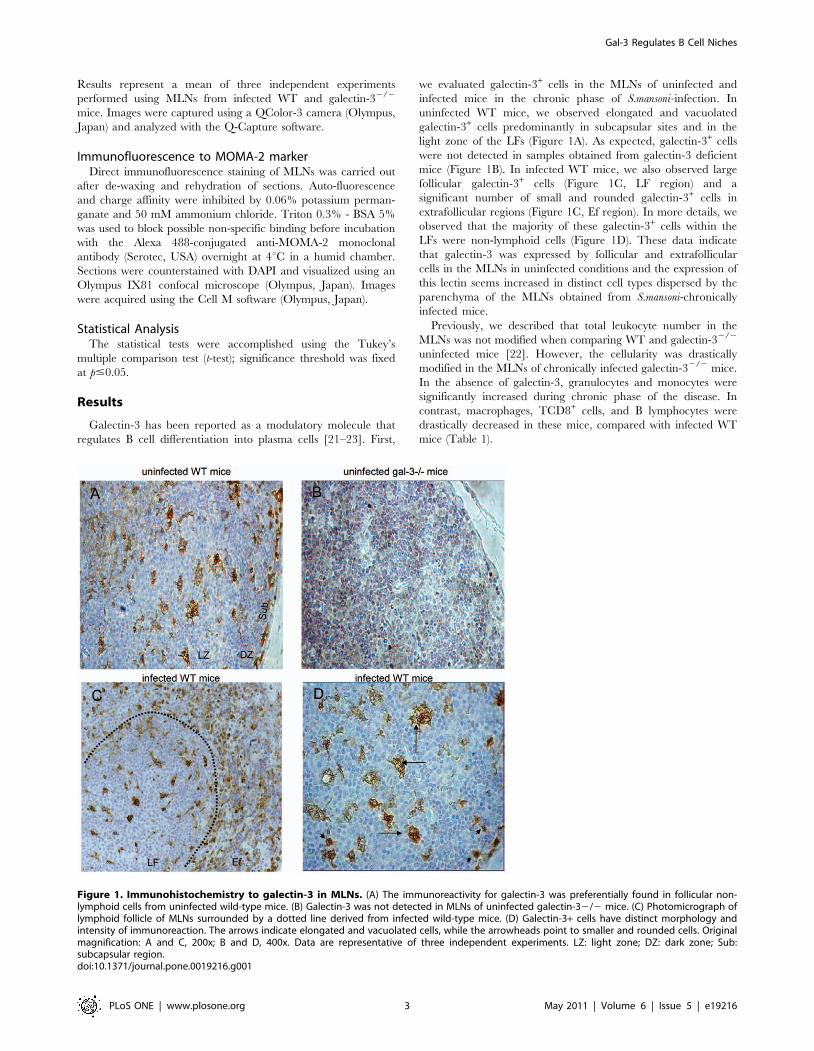
we evaluated galectin-3+ cells in the MLNs of uninfected and
infected mice in the chronic phase of S.mansoni-infection. In
uninfected WT mice, we observed elongated and vacuolated
galectin-3+ cells predominantly in subcapsular sites and in the
light zone of the LFs (Figure 1A). As expected, galectin-3+ cells
were not detected in samples obtained from galectin-3 deficient
mice (Figure 1B). In infected WT mice, we also observed large
follicular galectin-3+ cells (Figure 1C, LF region) and a
significant number of small and rounded galectin-3+ cells in
extrafollicular regions (Figure 1C, Ef region). In more details, we
observed that the majority of these galectin-3+ cells within the
LFs were non-lymphoid cells (Figure 1D). These data indicate
that galectin-3 was expressed by follicular and extrafollicular
cells in the MLNs in uninfected conditions and the expression of
this lectin seems increased in distinct cell types dispersed by the
parenchyma of the MLNs obtained from S.mansoni-chronically
infected mice.
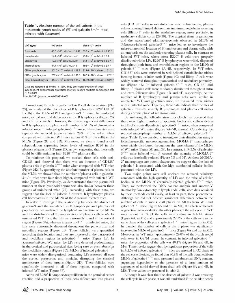
Previously, we described that total leukocyte number in the
MLNs was not modified when comparing WT and galectin-32/2
uninfected mice [22]. However, the cellularity was drastically
modified in the MLNs of chronically infected galectin-32/2 mice.
In the absence of galectin-3, granulocytes and monocytes were
significantly increased during chronic phase of the disease. In
contrast, macrophages, TCD8+ cells, and B lymphocytes were
drastically decreased in these mice, compared with infected WT
mice (Table 1).
Figure 1. Immunohistochemistry to galectin-3 in MLNs. (A) The immunoreactivity for galectin-3 was preferentially found in follicular non-lymphoid cells from uninfected wild-type mice. (B) Galectin-3 was not detected in MLNs of uninfected galectin-32/2 mice. (C) Photomicrograph oflymphoid follicle of MLNs surrounded by a dotted line derived from infected wild-type mice. (D) Galectin-3+ cells have distinct morphology andintensity of immunoreaction. The arrows indicate elongated and vacuolated cells, while the arrowheads point to smaller and rounded cells. Originalmagnification: A and C, 200x; B and D, 400x. Data are representative of three independent experiments. LZ: light zone; DZ: dark zone; Sub:subcapsular region.doi:10.1371/journal.pone.0019216.g001
Gal-3 Regulates B Cell Niches
PLoS ONE | www.plosone.org 3 May 2011 | Volume 6 | Issue 5 | e19216
Considering the role of galectin-3 in B cell differentiation [21–
23], we analyzed the phenotype of B lymphocytes (B220+ CD19+
B cells) in the MLNs of WT and galectin-32/2 mice. In uninfected
mice, we did not find differences in the B lymphocytes (Figure 2A
and 2B, respectively). However, there were significant differences
in B lymphocyte and plasma cell number in MLNs of chronically-
infected mice. In infected galectin-32/2 mice, B lymphocytes were
significantly reduced (approximately 29% of the cells), when
compared with infected WT mice (about 47% of the cells) in the
MLNs (Figure 2C and 2D). Moreover, we clearly found a cell
subpopulation expressing lower levels of surface B220 in the
absence of galectin-3 (Figure 2D, arrow), suggesting that these cells
could be differentiating into B2202/low plasma cells.
To reinforce this proposal, we marked these cells with anti-
CD138 and observed that there was an increase of CD138+
plasma cells in galectin-32/2 mice when compared with WT mice
(Figure 2E). By quantifying the absolute number of plasma cells in
the MLNs, we showed that the number of plasma cells in galectin-
32/2 mice were four times higher, compared with infected WT
mice (Figure 2F). Previously, we demonstrated that the plasma cell
number in these lymphoid organs was also similar between these
groups of uninfected mice [22]. According with these data, we
suggest that the lack of galectin-3 disturbs B lymphocyte-plasma
cell homeostasis in the MLNs of the S.mansoni-infected mice.
In order to investigate the relationship between the absence of
galectin-3 and the imbalance in B lymphocyte and plasma cell
populations, we analyzed the lymphoid architecture of the MLNs
and the distribution of B lymphocytes and plasma cells in situ. In
uninfected WT mice, the LFs were normally found in the cortical
region (Figure 3A), whereas in galectin-32/2 uninfected mice, the
LFs were abnormally dispersed throughout the paracortical and
medullary regions (Figure 3B). These follicles were quantified
according their location and they are increased in the paracortical
and medullary regions in galectin-32/2 mice (Figure 3E). In
S.mansoni-infected WT mice, the LFs were detected predominantly
in the cortical and paracortical sites, being rare or even absent in
the medullary region (Figure 3C). MLNs of infected galectin-32/2
mice were widely disorganized, containing LFs scattered all over
the cortex, paracortex and medulla, disrupting the classical
architecture of these organs (Figure 3D). These follicles were
significantly increased in all of these regions, compared with
infected WT mice (Figure 3F).
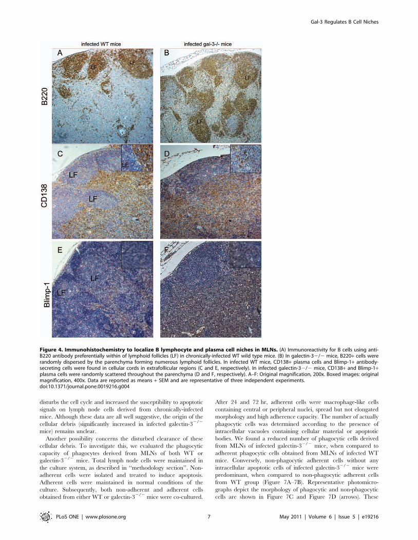
Activated B220+ B lymphocytes proliferate in the germinal center
reaction and a proportion of these cells differentiate into plasma
cells (CD138+ cells) in extrafollicular sites. Subsequently, plasma
cells expressing Blimp-1 differentiate into immunoglobulin secreting
cells (Blimp-1+ cells) in the medullary region, more precisely, in
medullary cellular cords [29,30]. The atypical tissue organization
and the exacerbated plasmacytogenesis observed in MLNs of
Schistosoma-infected galectin-32/2 mice led us to investigate the
micro-anatomical location of B lymphocytes and plasma cells, with
an emphasis on the antibody-secreting plasma cells. In contrast to
infected WT mice, where most B220+ B cells were properly
distributed within LFs, B220+ B lymphocytes were widely dispersed
throughout both intra and extrafollicular regions in the MLNs of
galectin-32/2 mice (Figure 4A–4B, respectively). In WT mice,
CD138+ cells were enriched in well-defined extrafollicular niches
forming intense cellular cords (Figure 4C) and Blimp-1+ cells were
widely scattered throughout paracortical and medullary parenchy-
ma (Figure 4E). In infected galectin-32/2 mice, CD138+ and
Blimp-1+ plasma cells were randomly distributed throughout intra
and extra-follicular sites (Figure 4D and 4F, respectively). As the
number of B lymphocytes and plasma cells were similar in
uninfected WT and galectin-3 mice, we evaluated these niches
only in infected mice. Together, these data indicate that the lack of
galectin-3 disturbs severely B lymphocyte and plasma cell niches
during chronic phase of schistosomiasis.
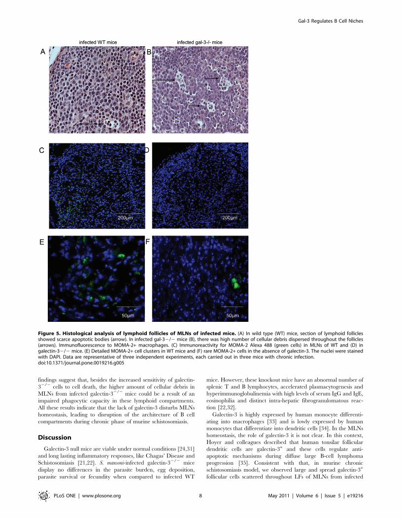
By analyzing the follicular structures closely, we observed that
there were higher numbers of apoptotic bodies and cellular debris
in LFs of chronically-infected galectin-32/2 mice, when compared
with infected WT mice (Figure 5A–5B, arrows). Considering the
reduced macrophage number in MLNs of infected galectin-32/2
mice (Table 1), we decided to investigate their distribution using a
follicular-specific macrophage-marker. MOMA-2+ macrophages
were widely distributed throughout the parenchyma of the MLNs
of WT mice (Figute 5C and 5E). In contrast, in MLNs of galectin-
32/2 mice infected with S. mansoni, the quantity of MOMA-2+
cells was drastically reduced (Figure 5D and 5F). As these MOMA-
2+ macrophages are potent phagocytes, we suggest that the lack of
galectin-3 is associated with the high number of cellular bodies
presented within the LFs.
Two major points were still unclear: the reduced cellularity
compared with the high quantity of LFs and the raise of cellular
bodies in the MLNs of chronically-infected galectin-32/2 mice.
Then, we performed the DNA content analysis and annexin-V
staining by flow cytometry in lymph nodal cells, since data obtained
by these methods could clarify, at least in part, these phenomena.
Although we did not observe significant differences between the
number of cells in sub-G1/G0 phases on MLNs from WT and
galectin-32/2 mice (Figure 6A and 6B, in M1), the effects of the lack
of galectin-3 were evident in the other phases of the cell cycle. In WT
mice, about 57.7% of the cells were cycling in G1/G0 stages
(Figure 6A, in M2) and approximately 22.7% of the cells were in the
same phase of the cell cycle in galectin-32/2 mice (Figure 6B, in M2).
In parallel, the number of cells in the S phase was significantly
increased in MLNs of galectin-32/2 mice (Figure 6A and 6B, in M3).
Moreover, in WT mice, approximately 24.8% of the lymph nodal
cells were in G2/M phase. In contrast, in infected galectin-32/2
mice, the proportion of the cells was 40.1% (Figure 6A and 6B, in
M4). These results suggest that the significant proportion of the cells
in MLNs of infected galectin-32/2 mice are arrested in G2 phase of
the cell cycle. Besides, we found that 30.8% of the cells obtained from
MLNs of galectin-32/2 mice presented an abnormal DNA content,
suggesting hyperploidy or the presence of a large amount of
aggregates of nuclei derived from dead cells (Figure 6A and 6B, in
M5). These values are presented in table 2.
Although it was clear that the absence of galectin-3 was arresting
the cell cycle in G2 phase, it was still undefined if the cellular debris
Table 1. Absolute number of the cell subsets in themesenteric lymph nodes of WT and galectin-32/2 miceinfected with S.mansoni.
Cell types WT mice Gal-32/2 mice
Total cells 86.66106 cells/mL611.42 63.26106 cells/mL66.35 *
Granulocytes 19.16105 cells/mL64.7 25.86105 cells/mL67.3
Monocytes 12.86105 cells/mL62.9 34.56105 cells/mL63.6 *
Macrophages 44.46105 cells/mL64.8 19.96105 cells/mL62.9 *
CD4+ lymphocytes 201.26105 cells/mL618.1 219.66105 cells/mL621.2
CD8+ lymphocytes 262.46105 cells/mL631.3 167.56105 cells/mL627.2 *
Total B lymphocytes 343.76105 cells/mL621.2 161.96105 cells/mL618.9 *
Data are reported as means 6 SEM, They are representative of threeindependent experiments, Statistical analysis: Tukey’s multiple comparison test(*, P,0.05).doi:10.1371/journal.pone.0019216.t001
Gal-3 Regulates B Cell Niches
PLoS ONE | www.plosone.org 4 May 2011 | Volume 6 | Issue 5 | e19216
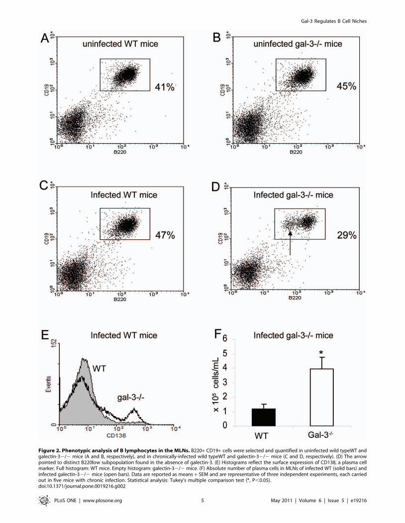
Figure 2. Phenotypic analysis of B lymphocytes in the MLNs. B220+ CD19+ cells were selected and quantified in uninfected wild typeWT andgalectin-32/2 mice (A and B, respectively), and in chronically-infected wild typeWT and galectin-32/2 mice (C and D, respectively). (D) The arrowpointed to distinct B220low subpopulation found in the absence of galectin-3. (E) Histograms reflect the surface expression of CD138, a plasma cellmarker. Full histogram: WT mice. Empty histogram: galectin-32/2 mice. (F) Absolute number of plasma cells in MLNs of infected WT (solid bars) andinfected galectin-32/2 mice (open bars). Data are reported as means + SEM and are representative of three independent experiments, each carriedout in five mice with chronic infection. Statistical analysis: Tukey’s multiple comparison test (*, P,0.05).doi:10.1371/journal.pone.0019216.g002
Gal-3 Regulates B Cell Niches
PLoS ONE | www.plosone.org 5 May 2011 | Volume 6 | Issue 5 | e19216

observed was consequence of this disturbed cell cycle. Thus, we used
annexin-V and propidium iodide staining to quantify possible
apoptotic and dead cells and compared samples of WT and
galectin-32/2 mice, both chronically-infected with S.mansoni. We
observed that approximately 19.9% of the cells were annexin V+/
PIneg in the MLNs of WT mice (Figure 6C, in R2), whereas about
30.9% of the cells were annexin V+/PIneg in the MLNs of galectin-
32/2 mice (Figure 6D, in R2). These data suggest that the lack of
galectin-3 promotes a significant increase in the number of cells
undergoing apoptosis. We did not find differences in the number of
annexin V+/PI+ dead cells when compared WT (5.5% of the cells)
and galectin-32/2 mice (4.9% of the cells) (Figure 6C and 6D, in
R3). Perhaps, galectin-3 has an anti-apoptotic role in lymph node
cells in the course of chronic phase of schistosomiasis.
In order to investigate the possible anti-apoptotic role of
galectin-3, we provided apoptotic stimuli by means of raising the
temperature in MLNs cells from infected WT and galectin-32/2
mice. The cells of MLNs of both WT and galectin-32/2 mice were
induced to apoptosis maintained in a culture system at 43uCduring 1 hour. After this time, these cells were stained with
annexin-V and propidium iodide (PI). We observed that cells from
MLNs of infected galectin-32/2 mice were more susceptible to
apoptosis, when compared to their WT counterpart (Figure 6E).
Taken together, these data indicate that the lack of galectin-3
Figure 3. Histological analysis of MLNs of WTwild-type and galectin-32/2 mice. Midsagittal section of MLN showing lymphoid folliclespreferentially within the cortex and scarcely in paracortex in uninfected and infected WT mice (A and C, respectively). Histological section from MLNsof uninfected and infected galectin-32/2 mice exhibiting lymphoid follicles randomly scattered throughout the cortex, paracortex and medulla (Band D, respectively). The samples were stained with hematoxilin and eosin. Lymphoid follicles were quantified by microscopic field in uninfected (E)and infected mice (F), with magnification of 25x. The solid bars indicate the WT mice and the open bars represent galectin-32/2 mice. Data arereported as means + SEM and are representative of three independent experiments. Statistical analysis: Tukey’s multiple comparison test (*, P,0.05).A–D, original magnification: 200x.doi:10.1371/journal.pone.0019216.g003
Gal-3 Regulates B Cell Niches
PLoS ONE | www.plosone.org 6 May 2011 | Volume 6 | Issue 5 | e19216
disturbs the cell cycle and increased the susceptibility to apoptotic
signals on lymph node cells derived from chronically-infected
mice. Although these data are all well suggestive, the origin of the
cellular debris (significantly increased in infected galectin-32/2
mice) remains unclear.
Another possibility concerns the disturbed clearance of these
cellular debris. To investigate this, we evaluated the phagocytic
capacity of phagocytes derived from MLNs of both WT or
galectin-32/2 mice. Total lymph node cells were maintained in
the culture system, as described in ‘‘methodology section’’. Non-
adherent cells were isolated and treated to induce apoptosis.
Adherent cells were maintained in normal conditions of the
culture. Subsequently, both non-adherent and adherent cells
obtained from either WT or galectin-32/2 mice were co-cultured.
After 24 and 72 hr, adherent cells were macrophage-like cells
containing central or peripheral nuclei, spread but not elongated
morphology and high adherence capacity. The number of actually
phagocytic cells was determined according to the presence of
intracellular vacuoles containing cellular material or apoptotic
bodies. We found a reduced number of phagocytic cells derived
from MLNs of infected galectin-32/2 mice, when compared to
adherent phagocytic cells obtained from MLNs of infected WT
mice. Conversely, non-phagocytic adherent cells without any
intracellular apoptotic cells of infected galectin-32/2 mice were
predominant, when compared to non-phagocytic adherent cells
from WT group (Figure 7A–7B). Representative photomicro-
graphs depict the morphology of phagocytic and non-phagocytic
cells are shown in Figure 7C and Figure 7D (arrows). These
Figure 4. Immunohistochemistry to localize B lymphocyte and plasma cell niches in MLNs. (A) Immunoreactivity for B cells using anti-B220 antibody preferentially within of lymphoid follicles (LF) in chronically-infected WT wild type mice. (B) In galectin-32/2 mice, B220+ cells wererandomly dispersed by the parenchyma forming numerous lymphoid follicles. In infected WT mice, CD138+ plasma cells and Blimp-1+ antibody-secreting cells were found in cellular cords in extrafollicular regions (C and E, respectively). In infected galectin-32/2 mice, CD138+ and Blimp-1+plasma cells were randomly scattered throughout the parenchyma (D and F, respectively). A–F: Original magnification, 200x. Boxed images: originalmagnification, 400x. Data are reported as means + SEM and are representative of three independent experiments.doi:10.1371/journal.pone.0019216.g004
Gal-3 Regulates B Cell Niches
PLoS ONE | www.plosone.org 7 May 2011 | Volume 6 | Issue 5 | e19216
findings suggest that, besides the increased sensitivity of galectin-
32/2 cells to cell death, the higher amount of cellular debris in
MLNs from infected galectin-32/2 mice could be a result of an
impaired phagocytic capacity in these lymphoid compartments.
All these results indicate that the lack of galectin-3 disturbs MLNs
homeostasis, leading to disruption of the architecture of B cell
compartments during chronic phase of murine schistosomiasis.
Discussion
Galectin-3 null mice are viable under normal conditions [24,31]
and long lasting inflammatory responses, like Chagas’ Disease and
Schistosomiasis [21,22]. S. mansoni-infected galectin-32/2 mice
display no differences in the parasite burden, egg deposition,
parasite survival or fecundity when compared to infected WT
mice. However, these knockout mice have an abnormal number of
splenic T and B lymphocytes, accelerated plasmacytogenesis and
hyperimmunoglobulinemia with high levels of serum IgG and IgE,
eosinophilia and distinct intra-hepatic fibrogranulomatous reac-
tion [22,32].
Galectin-3 is highly expressed by human monocyte differenti-
ating into macrophages [33] and is lowly expressed by human
monocytes that differentiate into dendritic cells [34]. In the MLNs
homeostasis, the role of galectin-3 it is not clear. In this context,
Hoyer and colleagues described that human tonsilar follicular
dendritic cells are galectin-3+ and these cells regulate anti-
apoptotic mechanisms during diffuse large B-cell lymphoma
progression [35]. Consistent with that, in murine chronic
schistosomiasis model, we observed large and spread galectin-3+
follicular cells scattered throughout LFs of MLNs from infected
Figure 5. Histological analysis of lymphoid follicles of MLNs of infected mice. (A) In wild type (WT) mice, section of lymphoid folliclesshowed scarce apoptotic bodies (arrow). In infected gal-32/2 mice (B), there was high number of cellular debris dispersed throughout the follicles(arrows). Immunofluorescence to MOMA-2+ macrophages. (C) Immunoreactivity for MOMA-2 Alexa 488 (green cells) in MLNs of WT and (D) ingalectin-32/2 mice. (E) Detailed MOMA-2+ cell clusters in WT mice and (F) rare MOMA-2+ cells in the absence of galectin-3. The nuclei were stainedwith DAPI. Data are representative of three independent experiments, each carried out in three mice with chronic infection.doi:10.1371/journal.pone.0019216.g005
Gal-3 Regulates B Cell Niches
PLoS ONE | www.plosone.org 8 May 2011 | Volume 6 | Issue 5 | e19216

WT mice, while the bulk of rounded lymphocyte-like cells were
galectin-3-negative.
MLNs continuously draining the major part of tissues involved
by schistosomiasis. In the course of the chronic phase, there is
progressive hyperplasia and the lymphoid organization is main-
tained [25]. In this work, we showed that the basic structural
aspects of the MLNs of galectin-32/2 mice were softly disturbed
independently of the infection. However, the course of the chronic
schistosomiasis significantly amplified these histological disorders
and the MLNs of S. mansoni-infected galectin-32/2 mice contained
macrophage and B2 lymphocyte niches severely modified. Our
results are not sufficient to prove whether galectin-3 controls these
microenvironments, although it has been described that resident
macrophages are responsible for phagocytosis of apoptotic cells
[17] and constitutively these cells control the distinct steps of
trafficking and differentiation of these B cells [36]. Since S.mansoni
Figure 6. Cell cycle analysis and apoptosis index in MLNs of WT and galectin-32/2 infected mice. Histograms represent the stages of cellcycle in WT (A) and Gal-32/2 mice (B) infected with S.mansoni. In both graphs, the phases were described as bellow: M1 - sub G1/G0; M2 – G1/G0; M3– S phase; M4 – G2/M and M5 – hyperploid cells. (C–D) Quantification of Annexin-V+/Propidium iodide (PI) 2 apoptotic cells (gated in R2 region) andAnnexin-V+/PI+ dead cells (gated in R3 region), in WT (C) and Gal-32/2 mice (D). (E) Quantification of apoptotic cells induced by high temperature.Solid bars represent WT mice and open bars indicate Gal-32/2 mice. Data are reported as means 6 SEM and are representative of three independentexperiments. Statistical analysis: Tukey’s multiple comparison test (*, P,0.05). A–B, original magnification, 400x.doi:10.1371/journal.pone.0019216.g006
Table 2. Relative number of cells during cell cycle events inMLNs of mice chronically-infected with Schistosoma mansoni.
Region WT mice Gal-32/2 mice Cell cycle stage
M1 0.27% 0.60% Fragmented DNA
M2 57.71% 22.72% G1/G0 phases *
M3 9.03% 3.00% S phase
M4 24.79% 40.13% G2/M phases *
M5 9.94% 30.85% Hyperploid *
Data are reported as means 6 SEM, They are representative of threeindependent experiments, Statistical analysis: Tukey’s multiple comparison test(*, P,0.05).doi:10.1371/journal.pone.0019216.t002
Gal-3 Regulates B Cell Niches
PLoS ONE | www.plosone.org 9 May 2011 | Volume 6 | Issue 5 | e19216

chronically-infected galectin-32/2 mice had delayed monocyte-
macrophage differentiation [22], we propose that the disorgani-
zation on B lymphocyte and plasma cell niches is narrowly
associated with this eminent macrophage dysfunction. In infected
WT mice, B lymphocytes and plasma cells are normally
distributed throughout follicular and extrafollicular sites, respec-
tively. However, in infected galectin-32/2 mice, these organiza-
tional scenarios are widely modified, where B220+ B cells, CD138+
plasma cells and Blimp-1+ antibody-secreting cells are abnormally
misplaced throughout the cortex, paracortex and medullar
regions.
It is known that strict mechanisms regulate B cell decision
between follicular and extrafollicular areas, where B lymphocytes
rapidly differentiate into antibody-secreting cells [37]. Although
some light has been shed on this subject, it remains unclear how
galectin-3 regulates B cell differentiation into plasma cells. In this
context, it was shown that galectin-3 inhibits Blimp-1 expression in
different experimental models, interfering with terminal differen-
tiation of B lymphocytes in antibody-secreting plasma cells
[21,23]. The increase of Blimp-1+ cells in the absence of
galectin-3 endorses the hypothesis that galectin-3 is a potential
regulator of Blimp-1 expression.
The macrophage dysfunction can also be associated with the
higher rate of cell death and reduced phagocytosis levels due to the
absence of galectin-3, where we did observe a significant histological
disorder in the distribution of MOMA-2+ macrophages. These cells
were described as typical tissue macrophages predominantly
detected in subcapsular sinus, follicles (tingible body macrophages)
and throughout paracortical and medullary regions [38]. By
definition, tingible body macrophages are large phagocytic cells
containing many apoptotic cells in distinct states of degradation
[39]. In this work, we demonstrated that the number of total and
MOMA-2+ macrophages are both decreased and these macro-
phages have reduced phagocytic capacity to engulf apoptotic cells in
the MLNs from infected galectin-32/2 mice.
In accordance, Miyake and colleagues showed that injected
dead cell bodies were rapidly engulfed by macrophages in the
splenic marginal zone, indicating a critical role of macrophages in
quickly removing apoptotic residues [40]. During schistosomiasis,
soluble eggs antigens (SEA) favor the activation-induced cell death
of follicular B and T lymphocytes [41]. Here, we did find an
elevated number of cellular debris inside of LFs in the MLNs of
chronically-infected galectin-32/2 mice. Thus, we propose that, at
least in part, galectin-3 plays a regulatory role in anti-apoptotic
events and/or phagocytosis of dead cells during schistosomiasis.
DNA content analysis findings corroborate the cellularity
results. We found that the total cell number was significantly
reduced in the MLNs of infected galectin-32/2 mice. Analyzing
the cell cycle data, we did note that cells arrested in G1 stage were
numerically reduced, while in G2 stage, the cellularity was
increased. Possibly, the absence of galectin-3 downregulates
mitotic cycles and favors the generation of cells more susceptible
to apoptosis. Paradoxically, LFs in the MLNs of infected galectin-
32/2 mice were more numerous, but the majority presented
suggestive lower cellular density and apoptotic bodies accumulated
inside them.
Figure 7. Quantification of phagocytic and non-phagocytic cells of MLNs of WT and galectin-32/2 infected mice. Non-adherent lymphnodal cells were collected, induced to apoptosis by high temperature and co-cultured with adherent cells to be engulffed. Measurement ofphagocytic and non-phagocytic cells after 24 h (A) and 72 h (B) of co-culture procedures. The solid bars indicate the wild-type mice and the openbars represent the gal-32/2 mice. Phagocytic cells had a translucent vacuole and phagosomes (C, arrow), while non-phagocytic cells were identifiedby the absence of phagossomes and clear citoplasm (D, arrow). Data are reported as means 6 SEM and are representative of three independentexperiments using cells derived from chronically-infected mice. Statistical analysis: Tukey’s multiple comparison test (*, P,0.05).doi:10.1371/journal.pone.0019216.g007
Gal-3 Regulates B Cell Niches
PLoS ONE | www.plosone.org 10 May 2011 | Volume 6 | Issue 5 | e19216

In conclusion, we provide clues on the role of galectin-3 in
driving histological changes in MLNs of mice infected with
S.mansoni. We suggest that the tissue disorganization observed in
the absence of galectin-3 is, at least partially, responsible for an
abnormal immune regulation and changes in cell number and
activities, including survival, apoptosis, phagocytosis, and differ-
entiation. Nevertheless, tissue damages and/or lost of appropriate
cell interactions and constrains in lymphoid tissue might
contribute to some aspects of immune response against to
helminths, as well as tumorigenesis and progression of lympho-
proliferative diseases, such as leukemia and lymphomas.
Acknowledgments
The authors are grateful to PhD student Antonio Palumbo, Programa de
Pos-Graduacao em Ciencias Morfologicas ICB/UFRJ - Rio de Janeiro, by
his support in the immunofluorescence analysis in confocal microscope.
Author Contributions
Conceived and designed the experiments: FLO RC MCE-C. Performed
the experiments: FLO CB AAP. Analyzed the data: FLO CB AAP KDA
DKH F-TL CMT RB RC MCE-C. Contributed reagents/materials/
analysis tools: CMT RB RC MCE-C. Wrote the paper: FLO F-TL RC
MCE-C.
References
1. Hotez PJ, Brindley PJ, Bethony JM, King CH, Pearce EJ, et al. (2008) Helminth
infections: the great neglected tropical diseases. J Clin Invest 118(4): 1311–21.
Review.2. El-Cheikh MC, Dutra HS, Minoprio P, Borojevic R (1994) Increase of B-
lymphocyte number and activity during experimental murine schistosomiasismansoni. Braz J Med Biol Res 27: 1605–1617.
3. Grzych JM, Pearce E, Cheever A, Caulada ZA, Caspar P, et al. (1991) Egg
deposition is the major stimulus for the production of Th2 cytokines in murineschistosomiasis mansoni. J Immunol 146: 1322–1327.
4. Borojevic R (1992) Experimental murine schistosomiasis mansoni: establishmentof the chronic phase of the disease. Mem Inst Oswaldo Cruz 87: 171–174.
Review.5. Lenzi HL, Oliveira DN, Pelajo-Machado M, Borojevic R, Lenzi JA (1996)
Coelom-associated lymphomyeloid tissue (milky spots): site of lymphoid and
myelomonocytic cell generation. Braz J Med Biol Res 29: 19–24.6. Randolph GJ, Angeli V, Swartz MA (2005) Dendritic-cell trafficking to lymph
nodes through lymphatic vessels. Nat Rev Immunol 8: 617–28. Review.7. MacLennan IC (1994) Germinal centers. Annu Rev Immunol 12: 117–139.
Review.
8. Angelin-Duclos C, Cattoretti G, Lin KI, Calame K (2000) Commitment of Blymphocytes to a plasma cell fate is associated with Blimp-1 expression in vivo.
J Immunol 165: 5462–5471.9. Benson MJ, Erickson LD, Gleeson MW, Noelle RJ (2007) Affinity of antigen
encounter and other early B-cell signals determine B-cell fate. Curr Opin
Immunol 19: 275–80. Review.10. Willard-Mack CL (2006) Normal structure, function, and histology of lymph
nodes. Toxicol Pathol 34: 409–424.11. Hanayama R, Tanaka M, Miwa K, Shinohara A, Iwamatsu A, et al. (2006)
Identification of a factor that links apoptotic cells to phagocytes. Nature 417:182–187.
12. Nakamura M, Yagi H, Kayaba S, Ishii T, Gotoh T, et al. (1996) Death of
germinal center B cells without DNA fragmentation. Eur J Immunol 26:1211–1216.
13. van den Berg TK, Honing H, Franke N, van Remoortere A, Schiphorst WECM,et al. (2004) LacdiNAc-glycans constitute a parasite pattern for galectin-3-
mediated immune recognition. J Immunol 173: 1902–1907.
14. Nyame AK, Lewis FA, Doughty BL, Correa-Oliveira R, Cummings RD (2003)Immunity to schistosomiasis: glycans are potential antigenic targets for immune
intervention. Exp Parasitol 104: 1–13.15. Yang RY, Rabinovich GA, Liu FT (2008) Galectins: structure, function and
therapeutic potential. Expert Rev Mol Med 13: 1–24.16. Sano H, Hsu DK, Yu L, Apgar JR, Kuwabara I, et al. (2000) Human galectin-3
is a novel chemoattractant for monocytes and macrophages. J Immunol 165:
2156–2164.17. Sano H, Hsu DK, Apgar JR, Yu L, Sharma BB, et al. (2003) Critical role of
galectin-3 in phagocytosis by macrophages. J Clin Invest 112: 389–397.18. Yang RY, Hsu DK, Liu FT (1996) Expression of galectin-3 modulates T-cell
growth and apoptosis. Proc Natl Acad Sci U S A 93: 6737–6742.
19. Joo HG, Goedegebuure PS, Sadanaga N, Nagoshi M, von Bernstorff W, et al.(2001) Expression and function of galectin-3, a beta-galactoside-binding protein
in activated T lymphocytes. J Leukoc Biol 69: 555–64.20. Villa-Verde DM, Silva-Monteiro E, Jasiulionis MG, Farias-De-Oliveira DA,
Brentani RR, et al. (2002) Galectin-3 modulates carbohydrate-dependentthymocyte interactions with the thymic microenvironment. Eur J Immunol 32:
1434–1444.
21. Acosta-Rodrıguez EV, Montes CL, Motran CC, Zuniga EI, Liu FT, et al. (2004)Galectin-3 mediates IL-4-induced survival and differentiation of B cells:
functional cross-talk and implications during Trypanosoma cruzi infection.J Immunol 172: 493–502.
22. Oliveira FL, Frazao P, Chammas R, Hsu DK, Liu FT, et al. (2007) Kinetics of
mobilization and differentiation of lymphohematopoietic cells during experi-
mental murine schistosomiasis in galectin-3 -/- mice. J Leukoc Biol 82: 300–310.23. Oliveira FL, Chammas R, Ricon L, Fermino ML, Bernardes ES, et al. (2009)
Galectin-3 regulates peritoneal B1-cell differentiation into plasma cells.Glycobiology 19: 1248–1258.
24. Hsu DK, Yang RY, Pan Z, Yu L, Salomon DR, et al. (2000) Targeted
disruption of the galectin-3 gene results in attenuated peritoneal inflammatoryresponses. Am J Pathol 156: 1073–1083.
25. El-Cheikh MC, Bonomo AC, Rossi MI, Pinho MF, Borojevic R (1998)Experimental murine schistosomiasis mansoni: modulation of the B-1 lympho-
cyte distribution and phenotype expression. Immunobiology 199: 51–62.26. Vindeløv LL, Hansen HH, Christensen IJ, Spang-Thomsen M, Hirsch FR, et al.
(1980) Clonal heterogeneity of small-cell anaplastic carcinoma of the lung
demonstrated by flow-cytometric DNA analysis. Cancer Res 40: 4295–4300.27. Montalvao F, Almeida GM, Silva EM, Borges VM, Vasconcellos R, et al. (2010)
Apoptotic lymphocytes treated with IgG from Trypanosoma cruzi infectionincrease TNF-a secretion and reduce parasite replication in macrophages.
Eur J Immunol 40: 417–425.
28. El-Cheikh MC, Borojevic R (1990) Extramedullar proliferation of eosinophilgranulocytes in chronic Schistosomiasis mansoni is mediated by a factor secreted
by inflammatory macrophages, Infect. Immun 58: 816–821.29. Mueller SN, Germain RN (2009) Stromal cell contributions to the homeostasis
and functionality of the immune system. Nat Rev Immunol 9: 618–629.
30. Dumic J, Dabelic S, Flogel M (2006) Galectin-3: an open-ended story. BiochimBiophys Acta 1760: 616–635. Review.
31. Colnot C, Ripoche MA, Milon G, Montagutelli X, Crocker PR, et al. (1998)Maintenance of granulocyte numbers during acute peritonitis is defective in
galectin-3-null mutant mice. Immunology 94: 290–296.32. Breuilh L, Vanhoutte F, Fontaine J, van Stijn CM, Tillie-Leblond I, et al. (2007)
Galectin-3 modulates immune and inflammatory responses during helminthic
infection: impact of galectin-3 deficiency on the functions of dendritic cells.Infect Immun 75: 5148–5157.
33. Liu FT, Hsu DK, Zuberi RI, Kuwabara I, Chi EY, et al. (1995) Expression andfunction of galectin-3, a beta-galactoside-binding lectin, in human monocytes
and macrophages. Am J Pathol 147: 1016–1028.
34. Dietz AB, Bulur PA, Knutson GJ, Matasic R, Vuk-Pavlovic S (2000) Maturationof human monocyte-derived dendritic cells studied by microarray hybridization.
Biochem Biophys Res Commun 275: 731–738.35. Hoyer KK, Pang M, Gui D, Shintaku IP, Kuwabara I, et al. (2004) An Anti-
Apoptotic Role for Galectin-3 in Diffuse Large B-Cell Lymphomas.Amer J Pathology 164: 893–902.
36. Karlsson MCI, Guinamard R, Bolland S, Sankala M, Steinman RM, et al.
(2003) Macrophages control the retention and trafficking of B lymphocytes in thesplenic marginal zone. J Exp Med 198: 333–340.
37. Martin F, Kearney JF (2002) Marginal-zone B cells. Nat Rev Immunol 5:323–335. Review.
38. Kraal G, Rep M, Janse M (1987) Macrophages in T and B cell compartments
and other tissue macrophages recognized by monoclonal antibody MOMA-2.An immunohistochemical study. Scand J Immunol 26: 653–661.
39. Smith JP, Burton GF, Tew JG, Szakal AK (1998) Tingible body macrophages inregulation of germinal center reactions. Dev Immunol 6: 285–294.
40. Miyake Y, Asano K, Kaise H, Uemura M, Nakayama M, et al. (2007) Criticalrole of macrophages in the marginal zone in the suppression of immune
responses to apoptotic cell-associated antigens. J Clin Invest 117: 2268–2278.
41. Lundy SK, Lerman SP, Boros DL (2001) Soluble egg antigen-stimulated Thelper lymphocyte apoptosis and evidence for cell death mediated by FasL(+) T
and B cells during murine Schistosoma mansoni infection. Infect Immun 69:271–80.
Gal-3 Regulates B Cell Niches
PLoS ONE | www.plosone.org 11 May 2011 | Volume 6 | Issue 5 | e19216