


EFFECT OF COUROUPTIA GUIANENSIS ON
N-DIETHYLNITROSAMINE INDUCED OXIDATIVE STRESS
IN WISTAR RATS
Dissertation Submitted to
THE TAMIL NADU Dr. M.G.R. MEDICAL UNIVERSITY
Chennai-32
In Partial fulfillment for the award of degree of
MASTER OF PHARMACY
IN
PHARMACOLOGY
SUBMITTED BY
Reg.No. 26103097
Under the guidance of
Mr. V. RAJESH., M. PHARM
DEPARTMENT OF PHARMACOLOGY
J.K.K.NATTRAJA COLLEGE OF PHARMACY
KOMARAPALAYAM - 638 183,TAMILNADU.
MAY-2012

Certificate
EVALUATION CERTIFICATE
This is to certify that the dissertation work entitled “Effect of courouptia
guianensis on N-diethylnitrosamine induced oxidative stres in wistar rats”
submitted by the student bearing Reg. No. 26103097 to “The Tamil Nadu Dr.
M.G.R. Medical University”, Chennai, in partial fulfillment for the award of degree
of MASTER OF PHARMACY in PHARMACOLOGY was evaluated by us
during the examination held on……………………….
Internal Examiner External Examiner
CERTIFICATE
This is to certify that the work embodied in this dissertation “Effect of
courouptia guianensis on N-diethylnitrosamine induced oxidative stres in
wistar rats”, submitted to “The Tamil Nadu Dr. M.G.R. Medical University”,
Chennai, was carried out by Ms. K.V. KAVITHA [Reg.No. 26103097], in the
Partial fulfillment of award of degree of MASTER OF PHARMACY in
Pharmacology under direct supervision of Mr. V.
RAJESH, M. Pharm, H.O.D of Pharmacology, J.K.K. Nattraja College of
Pharmacy, Komarapalayam, during the academic year 2011-2012.
PLACE: Komarapalayam Dr. P. PERUMAL, M. Pharm., Ph. D., A.I.C.,
DATE: Principal,
J.K.K.Nattraja College of Pharmacy,
Komarapalayam – 638 183,
Tamil Nadu.
CERTIFICATE
This is to certify that the work embodied in this dissertation, “Effect of
courouptia guianensis on N-diethylnitrosamine induced oxidative stres in
wistar rats” submitted to “The Tamil Nadu Dr.M.G.R.Medical University”,
Chennai, was carried out by K. V.KAVITHA [Reg. No. 26103097], for the Partial
fulfillment of degree of MASTER OF PHARMACY in Department of
Pharmacology under my guidance and direct supervision, J.K.K. Nattraja College of
Pharmacy, Komarapalayam, during the academic year 2011-2012.
PLACE: Komarapalayam V.RAJESH, M. Pharm,
DATE: H.O.D of pharmacology,
J.K.K.Nattraja college ofPharmacy,Komarapalayam – 638183,Tamil Nadu.
DECLARATION
The work presented in this dissertation entitled “Effect of courouptia
guianensis on N-diethylnitrosamine induced oxidative stres in wistar rats”, was
carried out by me, under the direct supervision V.RAJESH, M. Pharm, H.O.D of
pharmacology., J.K.K. Nattraja College of Pharmacy, Komarapalayam.
I further declare that, this work is original and has not been submitted in part
or full for the award of any other degree or diploma in any other university and the
thesis is ready for evaluation.
PLACE : Komarapalayam K.V.KAVITHA,
DATE : Reg.No: 26103097.

Acknowledgement

ACKNOWLEDGEMENT
At the outset, I am thankful to my PARENTS, HUSBAND and God for
blessing me with great strength and courage to complete my dissertation. Behind
every success there are lots of efforts, but efforts are fruitful due to helping hands
making the passage smoother. So, I am thankful to all those hands and people who
made my work grand success.
I am proud to dedicate my humblest regards and deep sense of gratitude and
heartfelt thanks to late Thiru. J.K.K. NATARAJAH CHETTIAR, founder of our
college. I wish to express my sincere thanks to our most respectful correspondent
Tmt. N. SENDAMARAAI and our beloved Director Mr. S. OMM
SHARRAVANA, B.Com, LLB., and Executive director Mr. S. OM
SINGARAVEL, B.E.,M.S., for enabling us to do the project work.
I take this opportunity with pride and immense pleasure expressing my deep
sense of gratitude to our respectable and beloved guide Mr. RAJESH. V,
M.Pharm., Assistant professor and head of Department of Pharmacology, J.K.K.
Nattraja College of Pharmacy, whose active guidance, innovative ideas, constant
inspiration, untiring efforts help, encouragement and continuous supervision has
made the presentation of dissertation a grand and glaring success to complete this
research work successfully.
I express my heartful thanks to our respectable and beloved principal, Mr. P.
PERUMAL, M.Pharm., Ph.D., A.I.C., Principal, J.K.K. Nattraja College of
Pharmacy, Komarapalayam. For his indispensable support which enabled us to
complete this task vast success.
My glorious acknowledgement to Dr. K. SENGODAN, M.B.B.S.,
administrative officer for encouraging us in a kind and generous manner to complete
this work.
My sincere thanks to Mrs. M. Sudha, M. Pharm., Assistant Professor, Mr.
P. Ashok Kumar, Ph. D, Professor and Mrs. R. Krishnaveni, M. Pharm, Asst.
professor, Department of Pharmacology for their valuable suggestions during my
project.
My sincere thanks to Mr. V. Sekar, M. Pharm., Head & Professor, Mr. S.
Jayaseelan, M. Pharm., Asst. Professor, Mr. Boopathy, M. Pharm., Assistant
Professor, Mr. Senthilraja, M. Pharm. Asst. Professor, Department of
Pharmaceutical Analysis for their valuable suggestions.
I expresses my sincere thanks to Mr.R.sampath kumar, M.Pharm., ph.D.,
Head & Professor of the department, Mrs. S. Bhama, M. Pharm., asst. professor,
Mr. Jaganathan, M. Pharm., Lecturer, Mr. R. Kanagasabai, B. Pharm., M.Tech.,
Asst. Professor, Department of Pharmaceutics, for their valuable help during my
project.
I express my sincere thanks to Dr. P. Sivakumar, M.Pharm., Ph.D.,
Professor, Mr. M. Vijayabaskaran, M.Pharm., Asst. Professor,
Mrs. Vaijayanthimala, M.Pharm, Assistant Professor,
Mrs. K. Mahalakshmi, M.Pharm. Lecturer, Department of Pharmaceutical
Chemistry, for their valuable suggestion and inspiration.
My sincere thanks to Dr. S.Sureshkumar, M.Pharm., Ph.D., Head &
Professor of the Department of Pharmacognosy and Mr. M. K. Senthilkumar,
M.Pharm., Asst. Professor, Department of Pharmacognosy for their valuable
suggestions.
I express my sincere thanks to Mr. N. Venkateswara Murthy, M. Pharm.,
Asst Professor & Head, Mr. P. Siva Kumar, M. Pharm., Lecturer, Mr. Rajarajan,
M. Pharm., Lecturer. Ms. S. Thangamani, M.Pharm., Lecturer, Department of
Pharmacy practice for their valuable suggestions.
My sincere thanks to Mr. N. Kadhiravel , M.C.A., for his help during the
project. I am delighted to Mrs. V. Gandhimathi, M.A., M.L.I.S., Librarian., Mrs.
S. Jayakla, B.A., Asst., for providing necessary facilities from Library at the time of
Work. I extend my thanks to Mr. S. Venkatesan, Storekeeper, Mr. Manikandan,
computer lab Assistant, and Mr. Rama Subramanyam G.N, our lab assistant for
their help during the project.
I am thankful to all my classmates, friends, and juniors.
I pay tribute to my lovable parents, and my husband
Mr. T.R.Chandrakaladharan for their inspiration and moral support that helped
me to complete this work successfully.
It is very difficult task to acknowledge the services to thank all those gentle
people. So I would like to thank all those people who have helped me directly or
indirectly to complete this project work successfully.
K.V.KAVITHA

Dedicated to
Almighty
My Beloved Parents
And
My Husband

Contents
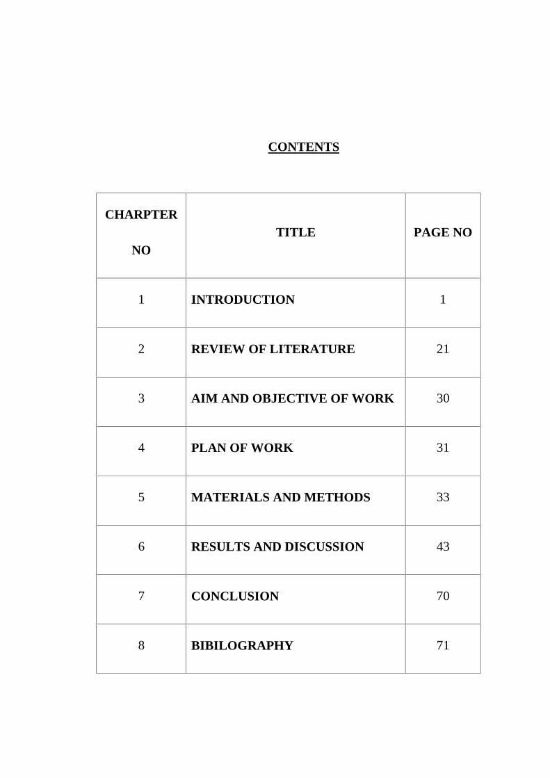
CONTENTS
CHARPTER
NOTITLE PAGE NO
1 INTRODUCTION 1
2 REVIEW OF LITERATURE 21
3 AIM AND OBJECTIVE OF WORK 30
4 PLAN OF WORK 31
5 MATERIALS AND METHODS 33
6 RESULTS AND DISCUSSION 43
7 CONCLUSION 70
8 BIBILOGRAPHY 71
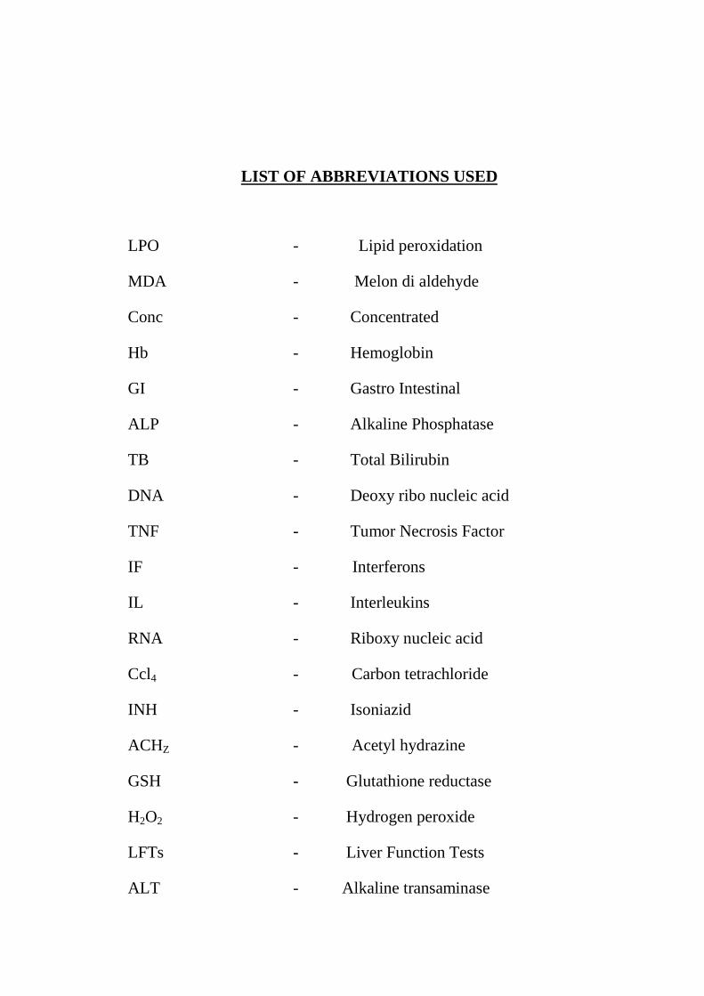
LIST OF ABBREVIATIONS USED
LPO - Lipid peroxidation
MDA - Melon di aldehyde
Conc - Concentrated
Hb - Hemoglobin
GI - Gastro Intestinal
ALP - Alkaline Phosphatase
TB - Total Bilirubin
DNA - Deoxy ribo nucleic acid
TNF - Tumor Necrosis Factor
IF - Interferons
IL - Interleukins
RNA - Riboxy nucleic acid
Ccl4 - Carbon tetrachloride
INH - Isoniazid
ACHZ - Acetyl hydrazine
GSH - Glutathione reductase
H2O2 - Hydrogen peroxide
LFTs - Liver Function Tests
ALT - Alkaline transaminase
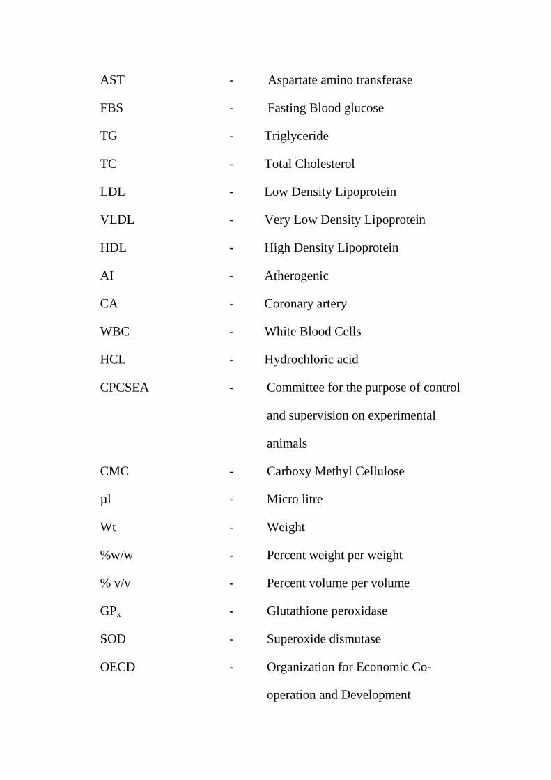
AST - Aspartate amino transferase
FBS - Fasting Blood glucose
TG - Triglyceride
TC - Total Cholesterol
LDL - Low Density Lipoprotein
VLDL - Very Low Density Lipoprotein
HDL - High Density Lipoprotein
AI - Atherogenic
CA - Coronary artery
WBC - White Blood Cells
HCL - Hydrochloric acid
CPCSEA - Committee for the purpose of control
and supervision on experimental
animals
CMC - Carboxy Methyl Cellulose
µl - Micro litre
Wt - Weight
%w/w - Percent weight per weight
% v/v - Percent volume per volume
GPx - Glutathione peroxidase
SOD - Superoxide dismutase
OECD - Organization for Economic Co-
operation and Development
IU/L - International Units per Litre
g/dl - gram per deci litre
mg/dl - milli gram per deci litre
gms - grams
mg/kg - milli gram per kilo gram
nmol - nano mole
U/mg - Units per milli gram
% - Percentage
Kg - Kilogram
IP - Intra Peritoneal
SC - Subcutaneous
LCAT - Lecithin cholesterol acyl transferase
LDL-c - low density lipoprotein-cholesterol
HDL-c - High density lipoprotein-cholesterol
LPL - Lipoprotein lipase
LRP - LDL-receptor related protein
GPO - Glycerol-3-phosphate oxidase
EDTA - Ethylene Diamine Tetra Acetic acid
CVD - Cardio Vascular Disease
CAD - Coronary Artery Disease
CAT - Catalase
Chapter I
Introduction
INTRODUCTION
INTRODUCTION TO HERBAL MEDICINE
Nature is enriched with pharmacologically active molecules which have been
used for the treatment of various incurable diseases (kokate et al., 2000; Ravi et al.,
2009; Trease and Evans, 1983). Herbal medicines are recommended for different
kind of biological activity for health care needs (Najiah et al., 2011; Nithya and
Baskar, 2011). The basic source of knowledge of modern medicine is plants. Herbal
medicine also called herbology or botanical medicine. Products obtained from plant
source are used for the treatment in wide variety of forms without any chemical
modification.
About 75 to 80% of world population is using herbal medicine for primary
health care because of less side effects, good compatability with human body and
good cultural acceptability (Karim et al., 2011; Premanath et al.,2011; Kapoor
and Saraf, 2011). The trend of using herbal medicines has increased enormously.
Herbal medicines, derived from scientific heritage and ancient civilization.
Renewable sources of raw materials are used for making herbal drugs by eco
friendly process and they are used for certain diseases where no modern medicine is
available. All parts of plants contain various medicinal properties (Mukherjee et al
2002). The plant extracts and its active constituents are screened for various
pharmacological activities. Herbalism has a long tradition of use and it contains
wide variety of chemical compounds used to treat many diseases.
Auyrvedha is a holistic traditional health care system in which human body,
mind and soul are taken into consideration for treatment. There is a world wide
belief that herbal medicines are safer and less damaging to human body than modern
medicines (Kraft et al 2007). The use of herbal medicine is enormously increased
as science began to take a closer look at herbal remedies. As malnutrition and
poverty is overruling in India, plant derived products will reduce the cost of health
care. So herbal medicines are widely acceptable among people and India has a rich
history of using herbal medicines for various treatments. India is enriched with
variety of flora due to different climatic conditions. Among 500,000 species of
plants on earth, about 5000 of them have been studied by modern science for
medicinal purpose.
Herbal formulations now serve as a basis of drug discovery initially it was
dispensed in the form of crude drugs such as tinctures, powders, teas, juices and
other formulations. All plant parts and its extracts have been used in herbal medicine
over the centuries. The world health organization recently defined traditional
medicine including herbal drugs that, it is a synthesis of generations of therapeutic
experience of practicing physicians of indigenous system of medicine. Traditional
preparations include medicinal plants, organic matter and minerals where herbal
drugs constitute medicinal plants primarly used for health therapy. Recently modern
medicines are derived from plant source with some modification to improve the
activity. The parts of medicinal plants should be standardized on the basis of their
major compounds and subjected to limited safety studies in animal before
marketing.
History of herbal medicine
Herbal medicine is an oldest form of health care system used by all cultures
throughout history. In 20th century much of the scientific medicine was derived from
herbal lore of native peoples. Researchers found that people in different parts of the
world used herbs as medicine for their health care. In the early 19th century, active
ingredients from the plants were extracted and modified by the scientist and later
chemists made their own version of plant compounds.
The first written herbal record was in 2800 BC in china and western herbal
medicine dated back to ancient Greece. Hippocrates wrote first herbal medicine in
Greek. A classification system of herbal remedies and illness was developed by an
herbal practitioner, Galen in 200 AD. Europeans used herbs as medicine in 15th
century. Chinese emperor Shen Nong wrote an authoritative treatise on herbs and
that is using still today. In 1941, pharmacist and medicine act is passed which
gave rights to pharmacists to dispense herbal medicines. The british herbal
medicine association was also formed and published british herbal pharmacopeia
(www.herbal/supplement/resource.com).
Recently World Health Organization estimated that more than 80% of world
population is using herbal medicine to treat diseases. In 1989 World health assembly
adopted a resolution about the importance of herbal medicines in individuals and
communities. WHO developed guidelines for herbal medicine assessment and it was
ratified by 6th international conference of Drug and Regulatory Authorities held at
Ottawa in same year. WHO guidelines include quality assessment, safety
assessment, stability and toxicology studies. All scientific generated data projected
herbal medicine in a proper perspective and sustained in a global market.
Evidence and importance scientific of herbal medicine
Several clinical trails are done on herbs in recent years, many of them have
shown that herb is a safe and effective alternative to modern medicines. One recent
study compared the quality of clinical trails of phytomedicine to matched trails with
modern medicines and concluded that method and reporting quality of western
clinical trails of phytomedicines was on superior to modern medicine(Nartley et al
2007). While evaluating any clinical study, it is important to consider quality and
design of the study and factors include nature of medicine investigated, goal of the
study and how they were measured, dose, length of the treatment. Many
pharmaceutical companies are now conducting researches on plant material
which is collected from rain forest and other places for therapeutic purpose
(Spinella et al 2002).
Treatment with herbal medicine is holistic. The approach involves
“balancing the body's vital energy” with a belief that it can treat any diseases.
Almost 25% of conventional medicines are based on plant origins, example: aspirin,
quinine, digitalis etc. Pharmacologist, botanist and microbiologist are searching new
herbal medicines in different parts of the world for health care needs. Drug
discovery from plants begins with botanists, ethnobotanist, ethnopharmacologists or
plant ecologists who will collect and identifies the plant. Molecular biology plays an
important role in medicinal plant drug discovery for determination of appropriate
screening assays towards relevent molecular targets. Herbal medicines must be
prepared according to good manufacturing practices. The specific active constituents
in a herb works to treat diseases. The identification of active principles and
evaluation of extracts should ensure safety and effective pharmacological activity
(Prakash et al 1998).
India has well recorded and well practiced knowledge of traditional herbal
medicine. The regulatory agency should take a preventive measure against the
misuse of herbal medicines as was done by US-FDA by banning dietary supplement
cholestin. Recently drugs are applied to standardization procedures to elucidate
analytical marker compounds. For the entry of herbal medicine into the developed
countries, the basic requirements include well documented traditional use, single
plant medicines, safety, stability, standardization based on activity, plant medicines
should be free from pesticides, heavy metals and pharmacological activity studies in
animals. All these data are the supportive measures for the herbal medicine and it
has gained much more importance in the field of medicine (Kamboj et al 2000).
Oxidative stress
Oxidative stress is a general term which is used to describe the steady state
level of oxidative damage in a cell, tissue or organ caused by the reactive oxygen
species. This damage can affect a entire organism or a specific molecule. It is a
imbalance between the generations of oxygen derived radicals and the organism's
antioxidant potential (devasagayam et al 1995). Through the production of
peroxides and free radicals, toxic effects are produced as a result of disturbances in
the normal redox state of tissues that damage all components of the cell, including
proteins, lipids and DNA. Reactive oxygen species and oxidative stress in liver cells
plays an important role in liver diseases. Some reactive oxidative species can even
act as a messangers through a redox signaling phenomenon.
Free Radicals and Reactive Oxygen
A radical (often, but unnecessarily called a free radical) is an atom or group
of atoms that have one or more unpaired electrons. Radicals can have positive,
negative or neutral charge. They are formed as necessary intermediates in a variety
of normal biochemical reactions, but when generated in excess or not appropriately
controlled, radicals can wreak havoc on a broad range of macromolecules. A
prominent feature of radicals is that they have extremely high chemical reactivity,
which explains not only their normal biological activities, but how they inflict
damage on cells.
Oxygen Radicals
There are many types of radicals, but those of most concern in biological
systems are derived from oxygen, and known collectively as reactive oxygen
species. Oxygen has two unpaired electrons in seperate orbitals in its outer shell.
This electronic structure makes oxygen especially susceptible to radical formation.
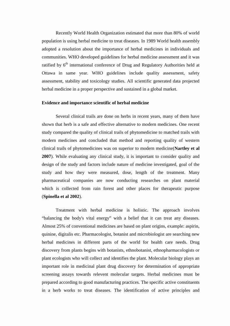
Sequential reduction of molecular oxygen (equivalent to sequential addition of
electrons) leads to formation of a group of reactive oxygen species:
superoxide anion
peroxide (hydrogen peroxide)
hydroxyl radical
The structure of these radicals is shown in the figure below, along with the
notation used to denote them. Note the difference between hydroxyl radical and
hydroxyl ion, which is not a radical.
Another radical derived from oxygen is singlet oxygen, designated as 1O2.
This is an excited form of oxygen in which one of the electrons jumps to a superior
orbital following absorption of energy.
Formation of Reactive Oxygen Species
Oxygen-derived radicals are generated constantly as part of normal aerobic
life. They are formed in mitochondria as oxygen is reduced along the electron
transport chain. Reactive oxygen species are also formed as necessary intermediates
in a variety of enzyme reactions. Examples of situations in which oxygen radicals
are overproduced in cells include:
White blood cells such as neutrophils specialize in producing oxygen
radicals, which are used in host defense to kill invading pathogens.
Cells exposed to abnormal environments such as hypoxia or hyperoxia
generate abundant and often damaging reactive oxygen species. A number of
drugs have oxidizing effects on cells and lead to production of oxygen
radicals.
Ionizing radiation is well known to generate oxygen radicals within
biological systems. Interestingly, the damaging effects of radiation are higher
in well oxygenated tissues than in tissues deficient in oxygen.
Biological Effects of Reactive Oxygen
It is best not to think of oxygen radicals as "bad". They are generated in a
number of reactions essential to life and, as mentioned above, phagocytic cells
generate radicals to kill invading pathogens. There is also a large body evidence
indicating that oxygen radicals are involved in intercellular and intracellular
signalling. For example, addition of superoxide or hydrogen peroxide to a variety of
cultured cells leads to an increased rate of DNA replication and cell proliferation - in
other words, these radicals function as mitogens.
Despite their beneficial activities, reactive oxygen species clearly can be
toxic to cells. By definition, radicals possess an unpaired electron, which makes
them highly reactive and thereby able to damage all macromolecules, including
lipids, proteins and nucleic acids.
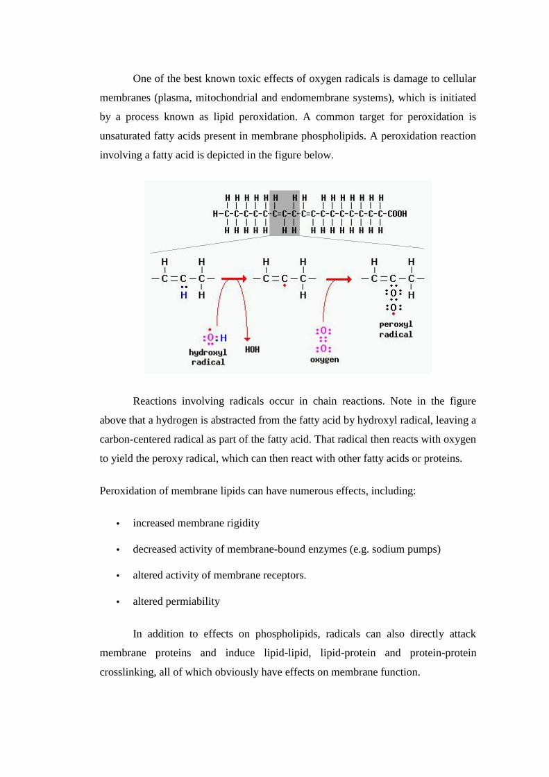
One of the best known toxic effects of oxygen radicals is damage to cellular
membranes (plasma, mitochondrial and endomembrane systems), which is initiated
by a process known as lipid peroxidation. A common target for peroxidation is
unsaturated fatty acids present in membrane phospholipids. A peroxidation reaction
involving a fatty acid is depicted in the figure below.
Reactions involving radicals occur in chain reactions. Note in the figure
above that a hydrogen is abstracted from the fatty acid by hydroxyl radical, leaving a
carbon-centered radical as part of the fatty acid. That radical then reacts with oxygen
to yield the peroxy radical, which can then react with other fatty acids or proteins.
Peroxidation of membrane lipids can have numerous effects, including:
increased membrane rigidity
decreased activity of membrane-bound enzymes (e.g. sodium pumps)
altered activity of membrane receptors.
altered permiability
In addition to effects on phospholipids, radicals can also directly attack
membrane proteins and induce lipid-lipid, lipid-protein and protein-protein
crosslinking, all of which obviously have effects on membrane function.
Sources of free radicals
• Internal source
• External source
• Physiological factors
Internal sources
Some internal sources are mitochondria, phagocytes, xanthine oxidase,
reactions involving iron and other transition metals, arachidonate pathways,
peroxisomes, ischaemia, exercise and inflammation. These include enzymatic
reactions involved in respiratory chain, in prostaglandin synthesis, in cytochrome p
450 system and in phagocytosis.
External sources
They are environmental pollutant, cigarette smoke, radiations, certain drugs,
anesthetics, pesticides, industrial solvents and ozone. These can be non enzymatic
reactions free radicals can also emerged from ionizing radiations.
Physiological factors
Disease status and mental conditions like stress and emotions can also form
free radical.
Types of free radicals
• Superoxide radical
• Hydroperoxyl radical
• Hydrogen peroxide
• Triplet oxygen
• Active oxygen
Superoxide radical
It can oxidize sulphur, ascorbic acid and it can able to reduce metal ions and
Cytochrome C. It can act as both oxidant and reactant. A reaction leads to the
formation of hydrogen peroxide and oxygen is catalysed by superoxide dismutase.
Hydroperoxy radical
Formed by transfer of a proton to a oxygen atom. It is also called as
perhydroxyl radical which is a protonated form of superoxide .
Hydrogen peroxide
It will act as a substrate in oxidation reaction involving synthesis of organic
molecule. It is produced by univalent reduction of superoxide produces hydrogen
peroxide and the effects are breaking up of DNA resulting in single strand breaks the
formation of DNA protein cross link.
Triplet oxygen
Ions and elements are reacted with triplet oxygen to form oxides. It will form
active peroxide radicals and it will undergo auto oxidation of unsaturated fattyacids.
Singlet oxygen
These are formed from hydrogen peroxide molecule. On decomposition it
produces superoxide and hydroxyl radicals. It is not a free radical but it arises from
some radical reactions.
Damages caused by free radicals
Inactivation of free radicals cause damage to all cellular macromolecules
such as proteins, carbohydrates, lipids and nucleic acid and causes various diseases.
Oxidative damage to proteins and DNA
Oxidative destruction on protein results in site specific aminoacid
modification, fragmentation of peptide chain, aggregation of cross linked reaction
products, altered electrical charges and increased susceptibility to proteolysis.
Oxidative attack on DNA results in base degradation, single strand breakage and
cross link to proteins.
Free radical and diseases
Diseases like diabetes, hypertension, cancer, artherosclerosis,
ischemia/reperfusion, inflammatory diseases(rheumatoid arthritis, pancreatitis and
inflammatory bowel diseases), neurological diseases are caused by free radicals.
Free radicals are not harmful always. To destroy invading pathogenic microbes
which causes diseases, white blood cells release free radicals, thus sometime it is
useful in the human body. Free radicals causes progressive adverse changes like
aging pigments are stored in the subsarcolmal region of the muscle fibres which
results in aging.
Mechanisms for Protection Against Radicals
Life on Earth evolved in the presence of oxygen, and necessarily adapted by
evolution of a large battery of antioxidant systems. Some of these antioxidant
molecules are present in all lifeforms examined, from bacteria to mammals,
indicating their appearance early in the history of life.
Many antioxidants work by transiently becoming radicals themselves. These
molecules are usually part of a larger network of cooperating antioxidants that end
up regenerating the original antioxidant. For example, vitamin E becomes a radical,
but is regenerated through the activity of the antioxidants vitamin C and glutathione.
Enzymatic Antioxidants
Three groups of enzymes play significant roles in protecting cells from
oxidant stress:
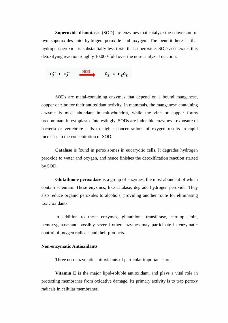
Superoxide dismutases (SOD) are enzymes that catalyze the conversion of
two superoxides into hydrogen peroxide and oxygen. The benefit here is that
hydrogen peroxide is substantially less toxic that superoxide. SOD accelerates this
detoxifying reaction roughly 10,000-fold over the non-catalyzed reaction.
SODs are metal-containing enzymes that depend on a bound manganese,
copper or zinc for their antioxidant activity. In mammals, the manganese-containing
enzyme is most abundant in mitochondria, while the zinc or copper forms
predominant in cytoplasm. Interestingly, SODs are inducible enzymes - exposure of
bacteria or vertebrate cells to higher concentrations of oxygen results in rapid
increases in the concentration of SOD.
Catalase is found in peroxisomes in eucaryotic cells. It degrades hydrogen
peroxide to water and oxygen, and hence finishes the detoxification reaction started
by SOD.
Glutathione peroxidase is a group of enzymes, the most abundant of which
contain selenium. These enyzmes, like catalase, degrade hydrogen peroxide. They
also reduce organic peroxides to alcohols, providing another route for eliminating
toxic oxidants.
In addition to these enzymes, glutathione transferase, ceruloplasmin,
hemoxygenase and possibly several other enzymes may participate in enzymatic
control of oxygen radicals and their products.
Non-enzymatic Antioxidants
Three non-enzymatic antioxidants of particular importance are:
Vitamin E is the major lipid-soluble antioxidant, and plays a vital role in
protecting membranes from oxidative damage. Its primary activity is to trap peroxy
radicals in cellular membranes.
Vitamin C or ascorbic acid is a water-soluble antioxidant that can reduce
radicals from a variety of sources. It also appears to participate in recycling vitamin
E radicals. Interestingly, vitamin C also functions as a pro-oxidant under certain
circumstances.
Glutathione may well be the most important intracellular defense against
damage by reactive oxygen species. It is a tripeptide (glutamyl-cysteinyl-glycine).
The cysteine provides an exposed free sulphydryl group (SH) that is very reactive,
providing an abundant target for radical attack. Reaction with radicals oxidizes
glutathione, but the reduced form is regenerated in a redox cycle involving
glutathione reductase and the electron acceptor NADPH.
In addition to these "big three", there are numerous small molecules that
function as antioxidants. Examples include bilrubin, uric acid, flavonoids and
carotenoids.
Reactive oxygen species
It includes oxygen radicals and several non radical oxidizing agents like
hypochlorous acid, hydrogen peroxide, ozone etc. Reactive oxygen species have the
tendency to donate oxygen to other species and it is responsible for the harmful
effects of oxygen. They are highly reactive and unstable. Oxidative damage results
in many diseases due to the presence of wide variety of oxygen free radicals and
reactive species in the human body and food.
Reactive oxygen species include
• Hydroxyl radicals (-OH)
• Superoxide anions (O2-).
• Hydrogen peroxides ( H2O2)
• Organic peroxides (R-OOH)
• Nitric oxide
• Singlet oxygen
• Peroxynitrite
Oxidative stress and its effects
Simply oxidative stress is a damage made to a cell through oxidative process.
Cells produce energy as a result of breathing, because of this activity highly reactive
molecules called free radicals are formed. Oxidation is a normal process, but
disturbances in that process such as attraction of free radical to a another molecule in
the body results in toxic effects. The reactive oxygen species such as peroxides and
free radicals are created from the metabolism of oxygen and they are generated by
endogenous and exogenous process.
Figure: 1 Oxidants contained within cigarette smoke
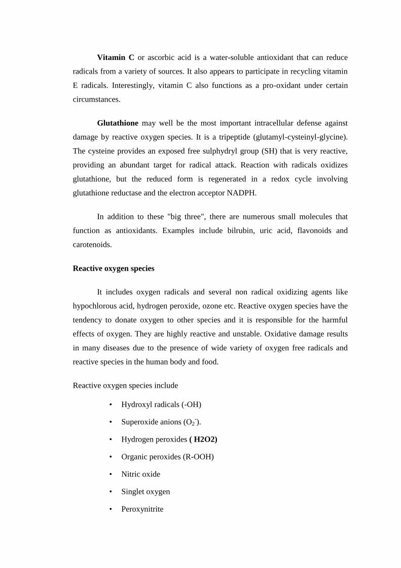
During oxidative cellular mechanism, hydrogen peroxide is produced that
comes from breakdown of reactive oxygen species, the superoxide anion radicals
(O2-). Superoxide is broken down into hydrogen peroxide and oxygen.Superoxide
cause damage to the cells that produces mutations in the superoxide dismutase
enzyme which leads to alanine transaminases (ALS), chracterised by loss of
motornuerons in brainstem and spinalcord causes apoptosis through oxidative stress.
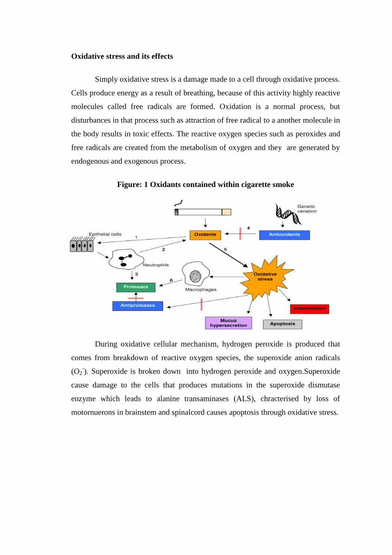
The complex network of antioxidant enzymes and metabolites joined
together to prevent oxidative damage to cellular components such as DNA, lipids
and proteins.
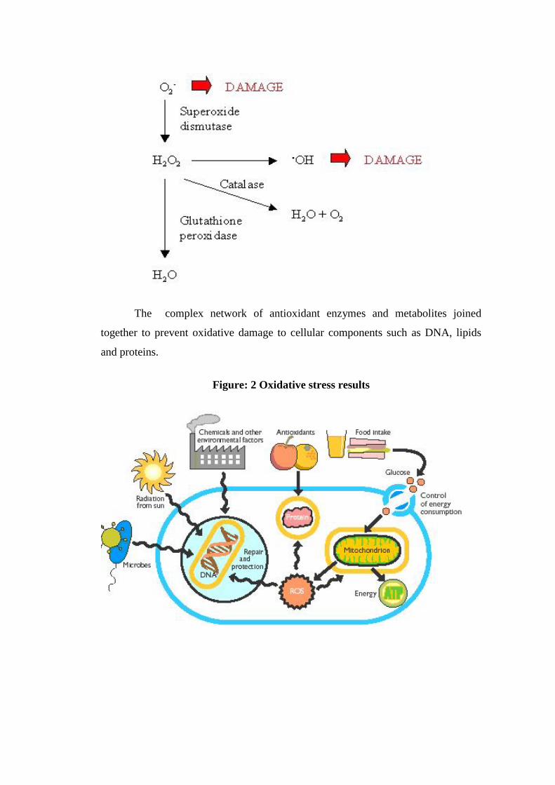
Figure: 2 Oxidative stress results
Oxidative stress and disease
Science has discovered that oxidative stress may cause more than seventy
diseases. Oxidative stress is a common mechanism for the initiation and
development of hepatic damage which leads to various liver disorders. Oxidative
stress has major role in cardiovascular diseases. Low density lipoprotein oxidation
trigger artherogenesis process which results in artherosclerosis and finally
cardiovascular diseases. However antioxidant enzymes protects DNA from
oxidative damage which cause cancer. So demand is great for the development of
antioxidant agents. Diseases may vary depending on the toxins and stress in the
body.
Some of the diseases caused by oxidative stress are:
• Cancer
• Lung disease
• Heart disease
• Arthritis
• Diabetis
• Fibromyalgia
• Autoimmune diseases
• Neurodegenerative diseases like parkinsonism and alzhemier's
• Eye diseases like macular degeneration
Antioxidant therapy has gained more important in the treatment of these
diseases. Oxidative stress has an impact on body's aging process also. The decrease
in melatonin levels seen with age correlates with an increase in neurogenerative
disorders such as Parkinson’s disease, Alzheimer’s disease, Huntington’s disease
and stroke, all disorders involve oxidative stress. In general, the production of
Reactive oxygen species (ROS) increases with aging and is related with DNA
damage to the tissues (www.preventive /health/guide.com).
Antioxidants
An antioxidant is a molecule which is capable of inhibiting the oxidation of
other molecule. While oxidation reaction it transfer electrons or hydrogen atom from
a substance to an oxidizing agent. Chain reactions are formed by the free radicals
produced during oxidation reaction and it causes damage to the cell. By removing
free radical intermediates, antioxidants inhibit oxidation reaction. Generally
antioxidant system remove or prevent the reactive species, before they damage the
cell components. The function of antioxidant is not to remove the entire oxidants but
to keep at optimum level (Docampo et al 1995).
The interaction between different antioxidants with various metabolites and
enzyme is having synergistic and interdependent effect on one another. The action
of antioxidant is based upon the function of other members of antioxidant system.
The protection provided by one antioxidant depend opon its concentration, its
reaction towards particular reactive oxygen species and status of antioxidant with
which it reacts. Some compounds produce antioxidant by chelating transition metal
and preventing the formation of free radical.
Classification of antioxidants
• Natural antioxidants
• Synthetic antioxidants
Natural antioxidants
They are differ in their physical and chemical properties and composition,
mechanism of action and their site of action. They are classified into following
categories
Antioxidant enzymes
The antioxidant enzyme such as superoxide dismutase(SOD), catalase(CAT),
glutathione peroxidase(GPx), glutathione reductase and glutathione transferase has
an important role in destroying free radicals.
Superoxide dismutase (SOD) first reduces (adds an electron to) the radical
superoxide (O2-) to form hydrogen peroxide (H2O2) and oxygen (O2).
2O2- + 2H --SOD--> H2O2 + O2
Catalase and GPx then work simultaneously with the protein glutathione to
reduce hydrogen peroxide and ultimately produce water (H2O).
2H2O2 --CAT--> H2O + O2
H2O2 + 2glutathione --GPx--> oxidized glutathione + 2H2O
(The oxidized glutathione is then reduced by another antioxidant enzyme --
glutathione reductase.)
Other enzyme act as secondary antioxidants to protect the cell from further damage.
Low molecular weight antioxidants
Through free radical scavenging property, it will delay or inhibit cellular
damage. Two types of low molecular weight antioxidants are
• Lipid soluble antioxidants
• Water soluble antioxidants
Lipid soluble antioxidants
Carotenoids, tocopherol, quinones, bilirubin and polyphenols will come to
this category. It will act against lipid peroxy radical as highly effective scavengers.
Lipid peroxy radical are formed as a result of free radical chain reaction of lipid
peroxidation within lipoprotein.
Water soluble antioxidants
They are ascorbic acid, uric acid and polyphenols. It cannot act on the lipid
moiety oflow density lipoprotein. It will support lipophillic antioxidants and
regenerate them.
Synthetic antioxidants
They are approved by Food and Drug Administration. They are synthetic
chemicals .Eg:
Butylated hydroxyl anisole (BHA), Butylated hydroxyl toluene (BHT),
Tertiary butylated hydroxyl quinine (TVHQ).
Mechanism of action of antioxidants
It act by
• Scavenging initiating radicals eg:Action of superoxide dismutase in
lipid phase to trap superoxide free radicals
• Reduction of concentration of reactive oxygen species, eg: Glutathione
• Chain breaking reaction. eg:Action of α-tocopherol in lipid phase to trap
free radical
• By chelating transition meal catalyst eg: Action of group of compound
by sequestering transition metals.
Regulation of antioxidant enzymes
The regulation of antioxidant enzymes mainly depends on the oxidant status
of the cell, as it form the first line of defence against free radical. Enzyme
modulating action of various hormones like growth hormone,prolactin and
melatonin are also involved in their regulation.Melatonsin is a derivative of
aminoacid tryptophan, protect membrane lipids and nuclear DNA from oxidative
damage. It has the ability to stimulate various antioxidant enzymes.
It will directly neutralize several reactive oxygen species including hydrogen
peroxide, either by stimulating gene expression for the enzymes or by potentiating
their activity.The reduction in enzyme activity was may also be due to reduction in
their biosynthesis or due to their excessive utilization in trapping generated free
radicles.It was also noticed that severe damage in liver decrease antioxidant defense
in liver.Liver injury produce intracellular stress results in lipid peroxidation of
membrane alomg with alteration of structural and functional characteristics of
membrane results in altered function of antioxidant enzymes (Halliwell et al 1999).
Diethylnitrosamine
It is an N-nitroso alkyl compound which is a potent hepatotoxin and
hepatocarcinogen, after repeated administration in experimental animals it causes
tumors. Nitrosamines are compounds formed by the combination of amines and
nitrates or nitrites (Sivanesam karthikeyan et al 2010). Many studies have recently
shown that in gastric juice of human stomach, nitrosamines can be formed by a
process called endogenous nitrosation. In many vegetables nitrates will be found, the
bacteria in the mouth chemically reduce nitate to nitrite which can form nitrosating
agents. In acidic environment of the stomach amines containing food that react with
these nitrosating agents to form nitrosamines.
It can be seen in variety of products to which humans may exposed such as
soyabean, cheese, salted and dried fish, cured meat, tobacco smoke, alcoholic
bevareges and ground water. It can also seen in environment and can synthesis
endogenously. Its exposure is dangerous to human population. Its metabolic
activation is responsible for toxic effects, which results in release of highly reactive
intermediates results in hepatocellular damage (Kannampalli pradeep et al 2007).
Oxidative stress has a major role in diethylnitrosaamine induced hepatotoxicity.
Many studies reported that continuous intrahepatic necroinflammatory changes were
seen during liver damage induced by diethylnitrosamine.
Diethylnitrosamine undergo metabolic activation by cytochrome P 450
enzymes to form reactive electrophiles results in oxidative stress which further leads
to cytotoxicity, mutagenicity and carcinogenicity. In the liver through an alkylated
mechanism, diethylnitrosamine is hydroxylated by cytochrome P450 isoenzymes to
become bioactive. Ethylation of the bases occurred as a result of reaction of
bioactivated diethylnitrosamine with DNA. The ethyl DNA adducts interrupt base
pairing results in mutation and activation of proto-oncogenes. Due to the generation
of reactive oxygen species; it will initiate peroxidative damage to the cell and
diethylnitrosamine will change antioxidant defense system in tissues.When the
concentration of reactive oxygen species generated exceeds cell'ss antioxidant
capability, oxidative damage to tissues or cells occurs. Diethylnitrosamine induced
liver damage by enhancing monocytes / monocytes activation and eventual
hepatocyte DNA damage. From the bioactivation of Diethylnitrosamine
intermediate reactive compounds are originated , with important cell constituents it
form covalent bonds, thus inducing mutation, cancer and necrosis.
The hepatocellular damage was observed histologically (thirty days after
Diethylnitrosamine administration) with elevated levels of serum alkaline
phosphatase, bilirubin,total protein, albumin, globulin and a simul;taneous fall in
levels of marker enzymes in liver tissue. Oxidative stress of liver was confirmed by
elevated levels of lipid peroxidation (LPO) as the membrane lipids are more
susceptible to reactive oxygen species and decrease in enzymic and non-enzymic
antioxidant activities.
Novel compounds are developing with antioxidant and hepatoprotective
activity to treat or prevent cellular damage. Plant based medicines with good
hepatoprotective activity are available and that can be used without any side effects.
Many studies have showed that natural antioxidants will support the endogenous
antioxidants defenses from reactive oxygen species ravage and by neutralizing free
radicals it will restore optimum balance.
Chapter II
Literature Review
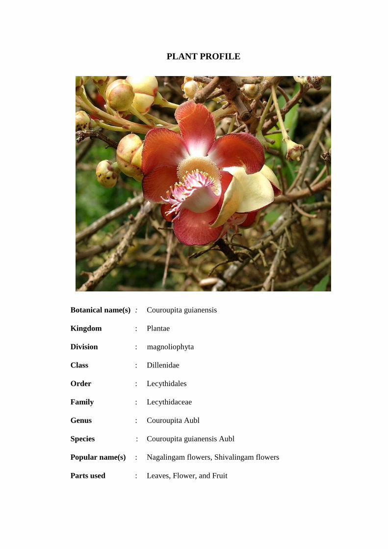
PLANT PROFILE
Botanical name(s) : Couroupita guianensis
Kingdom : Plantae
Division : magnoliophyta
Class : Dillenidae
Order : Lecythidales
Family : Lecythidaceae
Genus : Couroupita Aubl
Species : Couroupita guianensis Aubl
Popular name(s) : Nagalingam flowers, Shivalingam flowers
Parts used : Leaves, Flower, and Fruit
VERNACULAR NAMES
Hindi : Nagalinga flower
Tamil : Nagalingam flower
Telugu : Shivalinga flower or Nagamalli flower
or Mallikarjuna flower
Kannada : Lingada mara
Marathi : Shivalingam flower
Bengali : Kaman gola
Common name
Cannon ball tree
ORGIN, DISTRIBUTION AND MORPHOLOGY
Cannon ball tree is native to rain forest of the guiana’s in north eastern south
America. Its a large deciduous topical tree. it possesses a dense, often narrow crown
with leaves, clustered at the tip of branches. Leaves, upto 6"long are oval, oblong or
broadly lance shaped with serrate margin. It flowers in racemes. The amazingly
complex, yellow, reddish and pink flower of cannon ball tree are heavenly scented-a
cross between a fine expensive perfume and a wonderful flower scent. These are
3" to 5" waxy pink and dark red flowers growing directly on the bark of the trunk.
Flowers have six petals about 5cm and 2 inches long. They are large orange red,
strongly perfumed. They are sterile, zygomorphic, and they have thick tangled
extrusions that grow on a turnk. Flowering month is march to September.
The tree bears directly on the trunk and main branches, large globose woody
fruits. They will be hanging in clusters, like balls on a string. The fruit contains
small seeds in a white, unpleasant smelling white jelly, which are exposed when the
upper half of the fruit goes off like a cover. The long dangling fruity branches give
the tree an unkempt appearance. It is pollinated by bats and they are very important
for the survival of numerous species of plants. Although a plant of moist soils, it
grows well under dry conditions..
CHEMICAL CONSTITUENTS
Flowers yield an aliphatic hydrocarbon, stigmasterol, alkaloids, phenolic,
flavanoids, active principles like isatin and idirubin.It contains flavanoids-2'4'-
dihydoxy-6-methoxy-3'5'-dimethyl chalcone,7-hydroxy-5-methoxy-6, 8-dimethyl
flavone and the phenolic acid 4-hydroxy benzoic acid.
MEDICINAL USES
It is used as
• Antibiotic
• Antifungal
• Antiseptic
• Analgesic
• To cure colds
Juice from leaves is used for
• Inflammation
• Fever
• Alopecia
• Skin diseases
• Malaria
Fruit is used to
• Disinfect wounds
• Treat stomachache
• Cold
• malaria
• Toothache
Flower is used as
• Perfume
• Anti microbial
LITERATURE REVIEW
Mariana M.G. Pinheiro et al. ,(2010) studied antinociceptive activity of crude
ethanol extract and its fractions in three analgesic models(acetic acid-induced
contortions, tail flick, and hot plate) from couroupita guianensis leaves. To
elucidate mechanism of action from fractions, animals were pretreated (30 min)
with atropine (muscarinic receptor antagonist,1mg/kg sc), mecamylamine
(nicotinic receptor antagonist 2mg/kg sc), naloxone (opioid receptor antagonist
2mg/kg sc). Results showed all fractions produce antinociceptive activity in the
tail flick model.Crude ethanol extracts and its fractions significantly inhibited
number of contortions induced by acetic acid. Most prominent effect was
observed in crude ethanol extract.
Sanjay Prahalad Umachigi et al., (2009) evaluated antimicrobial, wound
healing and antioxidant potential of Couroupita guianensis in rats. Ethanolic
extract of whole plant Couroupita guianensis for the treatment of dermal
wounds in rats was studied on excision and incision wound models. HPTLC of
total extract was recorded for the purpose of standardization. Various parameters
of wound incision, epithelization period , scar area , tensile strength and
hydroxyproline measurements along with wound contraction were used to
evaluate the effect of Couroupita guianensis on wound healing. The results
obtained showed that Couroupita guianensis accelerate the wound healing
process by decrease in surface area of the wound and increase in tensile strength.
Antimicrobial activity was studied against gram positive and gram negative
bacteria compared to Erythromycin and Tetracycline. Moderate activity was
observed against all organisms.
Ana Martinez et al., (2011) has done protective effect against oxygen reactive
species and skin fibroblast stimulation of Couroupita guianensis leaf extracts.
Hydroalcoholic leaf extracts of Couroupita guianensis was examined for
antioxidant activity, phytochemical and total phenolic composition , stimulation
of skin fibroblast proliferation and UV absorption. The radical scavenging
capacity , reducing power and protection against joint oxidation of linoleic acid
and β-carotene bleaching oxidation in emulsion were used to evaluate the
antioxidant activity. Result of the study strongly indicated that invitro
antioxidant activity, which may be due to the presence of high total phenolic
content. It also suggest that hydroalcoholic leaf extract of Couroupita guianensis
have promising skin care properties.
V. Rajamanickam et al., (2009) studied flower extracts of couroupita
guianensis for invitro anthelmintic activity. Chloroform,acetone and ethanol
extacts of flower of couroupita guianensis showed anthelmintic activity at a
concentration of 50mg/ml and100mg/ml against adult earth worm pheritima
posthuma.Activity was found to be increased according to the dose and
compared with standard drug piperazine citrate.
M.R.Khan et al., (2008) evaluated antibiotic acitivity of Couroupita guianensis.
The result showed that methanolic extract of leaves, flowers, fruits ,stem and
root bark has antibacterial and antifungal activity than aqueous and chloroform
extract. It was found that klebsiella pneumonia and staphylococcus areus were
the most susceptible bacteria and candida albicans and aspergillus fumigates
were the susceptible fungi. Flowers , leaves, fruit pulp showed maximum
antibacterial effect but bark of the plant showed least activity.
D.Pradhan et al., (2008) has performed the immunomodulatory acitivity of the
methanolic extract of Couroupita guianensis flower in rats. Successive
methanolic extract of flowers of Couroupita guianensis showed significant
immunostimulant activity on both the specific and non-specific immune
mechanism. The results are encouraging enough to pursue bioactivity guided
fractionation of this extract and structural elucidation of the active
phytoconstituents.
Dr.Shaijesh Wankhede et al.,(2009) evaluated the anxiolytic effect of
methanolic root extract of couroupita guianansis in mouse.The effects of
extracts on spontaneous activity and neuromuscular co-ordination were
assessed.The result revealed that methanolic root extract showed good anxiolytic
activity.
Sivanesan Karthikeyan et al.,(2006) has showed that the Silymarin modulates
the oxidant-antioxidant imbalance during diethylnitrosamine induced oxidative
stress in rats. Diethylnitrosamine induced hepatocellular damage was indicated
by the elevated levels of serum aspartate transaminase , serum alanine
transaminase and lipid peroxidation, and also the decrease in the levels of
superoxide dismutase, catalase , glutathione peroxidase glutathione redutase and
glutathione-s-transferase in the liver tissues. The results showed that the
posttreatment with silymarin orally for 30 days exhibits a good
hepatoprotective and antioxidant potential against diethylnitrosamine induced
hepatocellular damage in rats.
Kannampalli Pradeep et al.,(2010) evaluated the protective effect of Cassia
fistula on diethylnitrosamine hepatocellular damage and oxidative stress in
ethanol pretreated rats. The result suggest that oral administration of ethanolic
leaf extract of Cassia fistula for 30 days to ethanol + diethlynitrosamine treated
rats showed good hepatoprotective and antioxidant potential when compared to
standard hepatoprotective drug ,silymarin.
Anupam Bishayee et al.,(2009) evaluated resveratrol suppresses oxidative
stress and inflammatory response in diethylnitrosamine-intiated rat
hepatocarcinogenesis. They provide the evidence that attenuation of oxidative
stress and suppression of inflammatory response mediated by Nrf2 (hepatic
nuclear factor E2-related factor) showed chemopreventive effects against
chemically-induced hepatic tumorigenesis in rats.
Malgorzata Kujawska et al., (2010) investigated cloudy apple juice protect
against chemically induced oxidative stress in rats. The cloudy apple juice
exhibited very distinct protective effect on hepatic antioxidant enzymes. Results
showed that protective action of apples phytochemicals by preventing damages
of essential cellular macromolecules in the conditions of chemically induced
oxidative stress in rats.
R . Gayathri et al., (2009) evaluated ursolic acid attenuates oxidative stress
mediated hepatocellular carcinoma induction by diethylnitrosamine in male
Wistar rats. Antioxidant status was assessed by alterations in level of lipid
peroxides and protein carbonyls. Oral administration ursolic acid 20mg/kg b.w
for 6 weeks decreased the levels of lipid peroxides and protein carbonyls. The
result showed the effectiveness of ursolic acid in reducing oxidative stress
mediated changes in rats liver.
Sabry M Shaarawy et al.,(2009) investigated protective effects of garlic and
silymarin on diethylnitrosamine induced rats hepatotoxicity. Diethylnitrosamine
increased oxidative stress, although administration of garlic or silymarin
significantly reduced liver toxicity,combined administration was more effective
in preventing hepatotoxicity. Hence they proved that garlic and silymarin have
synergistic effect.
Ramanathan Sambath Kumar et al.,(2007) evaluated antioxidant defense
system in wistar albino rats assessed by the methanol extract of Bauhinia
recemosa against diethylnitrosamine induced hepatocarcinogenesis.
Diethylnitrosamine treated rats, significantly elevated levels of serum enzymes,
bilirubin, and decreased levels of uric acid and protein were observed. Result
suggest that methanol extract of Bauhinia racemosa produced a protective effect
by decreasing the level of serum enzymes, bilirubin, and increased protein and
uric acid levels, it exert chemopreventive effect by suppressing nodule
development and increasing the level of antioxidant.
Perumal Subramanian et al.,(2003) evaluated S-Allylcysteine inhibits
circulatory lipid peroxidation and promotes antioxidants in dietylnitrosamine-
induced carcinogenesis. Result showed that rats treated with S-Allylcysteine
showed inhibition of tumor incidence and lipid peroxidation with simultaneous
elevation in antioxidants. Antioxidants level was enhanced by reducing the
formation of free radicals.
Prasanna Galhana et al., (2009) evaluated anti hepatocarcinogenic ayurvedic
herbal remedy reduces the extent of diethylnitrosamine induced oxidative
stress in rats. Results showed that treatment with decoction prepared from a
mixture of nigella sativa seeds, hemidesmus indicus roots and smilax glabra
rhizome-6gm/kg/day, for a period of ten weeks provide protection against
diethylnitrosamine mediated changes in oxidative stress and produce anti
hepatocarcinogenic effect.
Thamilarasan Manivasagam et al.,(2005) studied the chemopreventive effect
of diallyl disulphide on N-nitrosodiethylamine induced hepatocarcinogenesis. In
N-nitrosodiethylamine treated rats,the levels of thiobarbituric acid substances
and activities of superoxide dismutase and catalase were decreased whereas
reduced glutathione and glutathione peroxidase were increased.Oral
administration of diallyl disulphide(60mg/kg bodywt) produce chemopreventive
effect by modulating the oxidant-antioxidant status of living system.
Nermin A.H. Sadik et al.,(2008) studied the efficacy of dietary supplementation
with blue berries on diethylnitrosamine-initiated hepatocarcinogenesis in male
wistar rats.Results suggested that blue berries caused decreased in elevated
serum levels of α-fetoprotein, homocysteine, glutathione, deoxyribonucleic acid,
ribonucleic acid, and activity of glutathione reductase in liver and
histopathological damage was minimized in that group. It was documented that
blue berries was a chemopreventive natural supplement for liver cancer.
Mohamed. M. Sayed-ahmed et al.,(2010) evaluated thymoquinone attenuates
diethylnitrosamine induction of hepatic carcinogenesis through antioxidant
signaling. Results showed that thymoquinone supplementation prevents the
development of diethylnitrosamine induced liver cancer by decreasing oxidative
stress and preserving both the activity and mRNA expression of antioxidant
enzymes.
Chapter III
Aim and Objective
AIM AND OBJECTIVE OF STUDY
Diethylnitrosamine is an N-nitrosoalkyl compound, categorized as a potent
hepatotoxin and hepatocarcinogen in experimental animals (Jose et al., 1998). The
main cause for concern is that diethylnitrosamine is found in a wide variety of foods
like cheese, soyabean, smoked, salted and dried fish, cured meat and alcoholic
beverages (Liao et al., 2001). Metabolism of certain therapeutic drugs is also
reported to produce diethylnitrosamine (Akintonwa, 1985). It is also found in
tobacco smoke at a concentration ranging from 1 to 2ng/cigarette and in baby bottle
nipples at a level of 10 ppb (IARC, 1972). Diethylnitrosamine is reported to
undergo metabolic activation by cytochrome P450 enzymes to form reactive
electrophiles which cause oxidative stress leading to cytotoxicity, mutagenicity and
carcinogenicity (Archer, 1989). The detection of diethylnitrosamine in commonly
consumed food products makes the human population vulnerable to its exposure.
As oxidative stress plays a central role in diethylnitrosamine induced
hepatotoxicity, the use of antioxidants would offer better protection to counteract
liver damage (Vitaglione et al., 2004). This constraint underscores the need for the
development of novel potent antioxidant property. Since modern medicines have
little to offer for alleviation of oxidative stress in hepatic diseases, plant based
preparations are employed in treatment of liver disorders. Number of medicinal
plants have shown antioxidant and hepatoprotective activity due to the presence of
active constituents. From the literature survey, it was found that the flower of C
ouroupita guianensis is rich in flavonoids, phenolic compounds and alkaloids. It was
also noticed that Couroupita guianensis is used as antimicrobial, antiseptic,
analgesic, anti-inflammatory, antipyretic, antimalarial etc. This widespread use of
Couroupita guianensis in traditional medicine promoted me to evaluate the
antioxidant status of flower extract in chemically induced hepatic injury in albino
rats. Since oxidative stress plays a main role in hepatic injury which further leads to
hepatocarcinogenesis, N-diethylnitrosamine which cause oxidative injury and
hepatocarcinogenesis, is selected for the study.

Chapter IV
Plan of Work
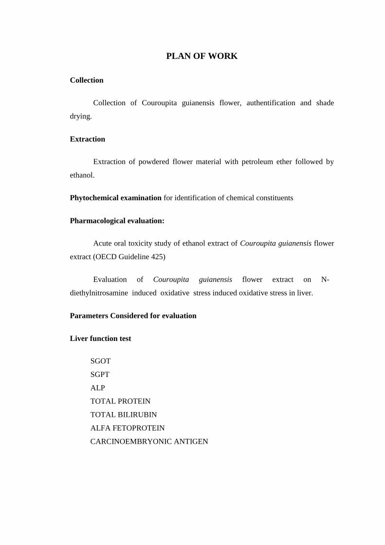
PLAN OF WORK
Collection
Collection of Couroupita guianensis flower, authentification and shade
drying.
Extraction
Extraction of powdered flower material with petroleum ether followed by
ethanol.
Phytochemical examination for identification of chemical constituents
Pharmacological evaluation:
Acute oral toxicity study of ethanol extract of Couroupita guianensis flower
extract (OECD Guideline 425)
Evaluation of Couroupita guianensis flower extract on N-
diethylnitrosamine induced oxidative stress induced oxidative stress in liver.
Parameters Considered for evaluation
Liver function test
SGOT
SGPT
ALP
TOTAL PROTEIN
TOTAL BILIRUBIN
ALFA FETOPROTEIN
CARCINOEMBRYONIC ANTIGEN
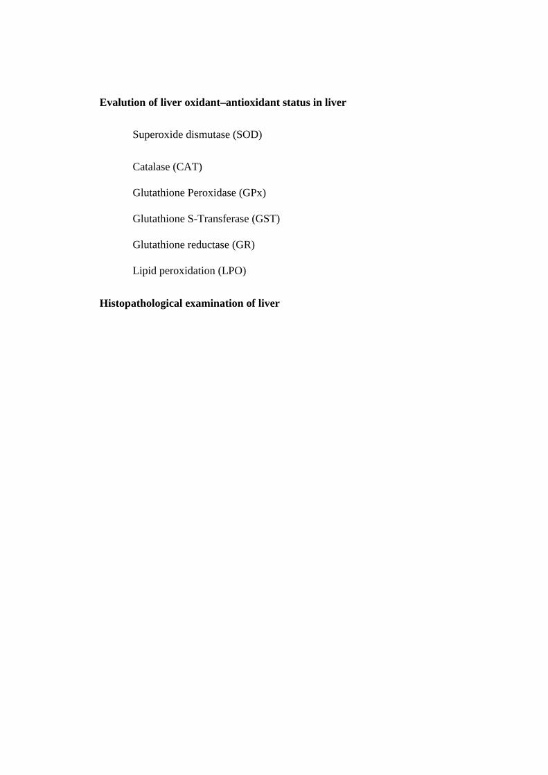
Evalution of liver oxidant–antioxidant status in liver
Superoxide dismutase (SOD)
Catalase (CAT)
Glutathione Peroxidase (GPx)
Glutathione S-Transferase (GST)
Glutathione reductase (GR)
Lipid peroxidation (LPO)
Histopathological examination of liver
Chapter V
Materials and Method
MATERIALS AND METHODS
Chemicals:
Diethylnitrosamine is purchased from sigma Aldrich chemical company, (St.
Louis, MO, USA) petroleum ether and ethanol was purchased from Nice chemicals
Pvt Ltd. Silymarin was obtained as gratis from Himalaya drug company, bengaluru,
india. All other chemicals used were of analytical grade and were purchased locally.
PLANT MATERIAL
The flowers of couroupita guianensis were collected from the botanical
garden, J. K. K. Nattraja college of pharmacy, Komarapalayam. The flowers were
taxonomically identified, confirmed and authenticated by the botanical survey of
India, souther circle, Tamilnadu agricultural university, Coimbatore with
authentication number BSI/SRC/5/23/2011-12/TECH-883. The voucher specimen
was retained in our laboratory for further reference.
EXTRACTION
The collected flowers were shade dried completely. The dried material was
then coarsely powdered and was sieved (sieve # 40) to get uniform coarse powder.
The dried coarse powder was defatted with petroleum ether (60 - 80°c ) in a
soxhlet extractor in order to remove fatty substances, which may interfere with the
isolation of chemical constituents. The defatted marc was dried and it was subjected
to extraction with ethanol (95%) in a soxhlet apparatus for 72 hours.The solvent was
then distilled off and the extract obtained was concentrated to dryness under reduced
pressure and percentage yield was calculated.
PHYTOCHEMICAL SCREENING
The extract obtained was subjected to Priliminary Phytochemical screening
(Khandelwal and Kokate, 1995).
Test for alkaloids:
Small of extract was dissolved in 10 ml of 0.1N dilute hydrochloric acid and
filtered. The filtrate was used to test the presence of alkaloids.
Mayer’s test
Filtrate was treated with Mayer’s reagent. Formation of yellow cream
precipitate indicates the presence of alkaloids.
Dragendroff’s test
Filtrate was treated with Dragendroff’s reagent. Formation of red coloured
precipitate indicates the presence of alkaloids.
Hager’s test
Filtrate was treated with Hager’s reagent. Formation of yellow coloured
precipitate indicates the presence of alkaloids.
Wagner’s Test
Filtrate was treated with wagner’s reagent. Formation of brown (or) reddish
brown precipitate indicates the presence of alkaloids.(Rosenthalar, 1930)
Detection of Phytosterols and Triterpenoids :
0.5 gm of extract was treated with 10ml of chloroform and filtered. The
filtrate was used to test the presence of phytosterols and Triterpenoids.
Libermann’s Test
To 2 ml filtrate in hot alcohol, few drops of acetic anhydride were added.
Formation of brown precipitate indicates the presence of sterols.
Libermann’s Burchard Test
100 mg of extract was treated with 2 ml of chloroform and filtered. To the
filtrate few drops of acetic anhydride was added, boiled and cooled. Concentrated
H2So4 was added through the sides of the test tube. Formation of brown ring at the
junction indicates the presence of steroidal saponins.
Salkowski Test
To the test extract solution few drops of Concentrated H2So4 was added,
shaken and allowed to stand, lower layer turns red indicates the presence of sterols.
(Peach and Tracey, 1957)
Detection of Flavoniods :
Shinoda Test
To 100 mg of extract, few fragments of magnesium metal were added in a
test tube, followed by drop wise addition of concentrated hydrochloric acid.
Formation of magenta colour indicates the presence of Flavonoids.
Alkaline Reagent Test
To 100 mg of extract, few drops of sodium hydroxide solution were added in
a test tube. Formation of intense yellow colour that becomes colourless on addition
of few drops of dilute hydrochloric acid indicates the presence of flavanoids.
(Shellard, 1957)
Detection of Saponins :
Foam test
The extract was diluted with 20 ml of distilled water and it was shaken in a
graduated cylinder for 15 minutes. A 1cm layer of foam indicates the presence of
Saponins.
Detection of Proteins and Amino acids:
100 mg of extract was taken in 10 ml of water and filtered. The filtrate was
used to test the presence of protein and amino acids.
Millon’s Test
2 ml of filtrate was treated with 2 ml of millon’s reagent in a Test tube and
heated in a water bath for 5 minutes, cooled and few drops of NaNo2 were added.
Formation of white precipitate, which turns to red upon heating, indicates the
presence of proteins and amino acids.
Ninhydrin Test
2 ml of filtrate, 0.25% ninhydrin reagent was added in a test tube and boiled
for 2 minutes. Formation of blue colour indicates the presence of amino acids.
Biuret Test
2 ml of filtrate was treated with 2 ml of 10% sodium hydroxide in a test and
heated for 10 minutes. A drop of 7% copper sulphate solution was added in the
above mixture. Formation of purplish violent indicates the presence of proteins.
Detection of Fixed oils and Fats:
Oily Spot Test
One drop of extract was placed on filter paper and the solvent was
evaporated. An oily stain of filter paper indicates the presence of fixed oil.
(Rosenthalar, 1930)
Detection of Phenolics and Tannins:
100 mg of extract was boiled with 1ml of distilled water and filtered. The
filtrate was used for the test.
Ferric chloride Test
To 2 ml of filtrate, 2ml of 1% ferric chloride was added in a test tube.
Formation of bluish black colour indicates the presence of phenolic nucleus.
Lead acetate Test
To 2 ml of filtrate, few drops of lead acetate solution were added in a test
tube. Formation of yellow precipitate indicates the presence of tannins.
Detection of Carbohydrate:
500 mg of extract was dissolved in 5ml of distilled water and filtered. The
filtrate was used to test the presence of carbohydrates.
Molisch test
To one ml of filtrate, two drops of Molisch reagent was added in a test tube
and 2 ml of concentrated H2So4 was added carefully along the side of the test tube.
Formation of violet ring at the junction indicates the presence of carbohydrates.
Fehling’s test
To one ml of filtrate, 4 ml of fehling’s reagent was added in a test tube and
heated for 10 minutes in a water bath. Formation of red precipitate indicates the
presence of reducing sugar.
Benedict’s test
Filtrate was treated with Benedict’s reagent and heated on water bath.
Formation of orange red precipitate indicates the presence of reducing sugars.
Detection of Glycosides:
0.5 gm of extract was hydrolyzed with 20 ml of 0.1N dilute hydrochloric
acid and filtered. The filtrate was used to test the presence of glycosides.
Modified Borntrager’s test
1 ml of filtrate 2 ml of 1% ferric chloride solution was added in a test tube
and heated for 10 minutes in boiling water bath. The mixture was cooled and shaken
with equal volume of benzene. The benzene layer was separated and treated with
half its volume of ammonia solution. Formation of rose pink or cherry colour in the
ammonical layer indicates the presence of anthranol glycoside.
Legal’s test
To 1 ml of filtrate, 3 ml of sodium nitroprusside in pyridine and methanolic
alkali (KOH) was added in a test tube. Formation of pink to blood red colour
indicates the presence of cardiac glycoside.
Keller Killiani Test
Small portion from the extract was shaken with 1ml of Glacial acetic acid
containing trace of ferric chloride. 1 ml of concentrated H2So4 was added carefully
by the sides of the test tube. A blue colour in the acetic acid layer and red colour at
the junction of two liquids indicate the presence of glycosides. (Rosenthalar, 1930).
PHARMACOLOGICAL SCREENING
Acute oral toxicity study of Couroupita guianensis flower extract
Animals
Swiss albino mice of female sex weighing 20-25gms were used for the study.
The animals were obtained from Agricultural University, Mannuthy, Thrissur, kerala
(328/99/CPCSEA) and were housed in polypropylene cages. The animals were
maintained under standard laboratory conditions (250 + 20C; 12hr light and dark
cycle). The animals were fed with standard diet and water ad libitum. Ethical
clearance (for handling of animals and the procedures used in study) was obtained
from the Institutional Animal Ethical Committee (887/ac/05/CPCSEA) before
performing the study on animals. The proposal number is 31MP15JUN11
Acute oral toxicity study
Acute oral toxicity study of Couroupita guianensis flower extract was carried
out as per OECD guideline 425 (Up and Down procedure). The test procedure
minimizes the number of animals required to estimate the acute oral toxicity. The
test allows the observation of signs of toxicity and can also be used to identify
chemicals that are likely to have low toxicity.
Animals were fasted (food but not water was with held overnight) prior to
dosing. The fasted body weight of each animal was determined and the dose was
calculated according to the body weight.
Limit test at 2000mg/kg
The extract was administered in the dose of 2000mg/kg body weight orally
to one animal. If the first test animal survives, then four other animals were dosed
sequentially; therefore, a total of five animals were tested. Animals were observed
individually at least once during the first 30 minutes after dosing, periodically
during the first 24 hours (with special attention given during the first 4 hour), and
daily thereafter, for a total of 14 days. After the experimental period, the animals
were weighed and humanely killed and their vital organs including heart, lungs,
liver, kidneys, spleen, adrenals, sex organs and brain were grossly examined
(OECD Guidance; 2000)
Effect of Couroupita guianensis flower extract on N-diethylnitrosamine
induced hepatic damage in wistar rats.
ANIMALS:
Experiments were carried out according to the guidelines of CPCSEA
(Committee for the Purpose of Control and Supervision of Experiment on Animals,
New Delhi, India. The protocol of experiments were approved by Institutional
Animal Ethics Committee (IAEC) (887/ac/CPCSEA), J.K.K. Nattraja college of
pharmacy, Komarapalayam, Nammakal district.
Male wistar rats weighing about 100 -200gms, were obtained from
agricultural university, mannuthy, Thrissur. The animals were maintained in animal
house under standard environmental condition (250±20c) and 12hr/12hr light and
dark cycle. Animals were fed with standard pellet diet (Hindustan Lever Ltd,
mumbai, india) and water ad.libtum. The experimental protocol was approved by the
Institutional Animal Ethics Committee (IAEC) and experiments were conducted
according to the CPCSEA, India guidelines on the use and care of experimental
animals.
PROCEDURE
Total 30 animals were used for this study and it was divided into 5 groups of
6 animals each.
Group I :Rats served as controls received normal saline 1ml/kg (i.p.) on day 0 and
carboxymethylcellulose ( 2ml/kg, orally) for 30 days.
Group II : Rats were administered with a single dose of Diethylnitrosamine (200
mg/kg b.w , i.p.) in saline on day 0 and 0.5% w/v carboxymethylcellulose 2ml/kg
(orally) from day 1 to 30.
Group III : Rats were administered with diethylnitrosamine (200 mg/kg b.w.,i.p) in
saline on day 0 followed by extract (200 mg/kg.,p.o ) in carboxymethylcellulose
from day 1 to 30.
Group IV : Rats were administered with diethylnitrosamine (200 mg/kg b.w.,i.p) in
saline on day 0 followed by extract (400 mg/kg.,i.p) in carboxymethylcellulose from
day 1 to 30.
Group V : Rats were administered with diethylnitrosamine (200mg/kg.,i.p) in saline
on day 0 followed by silymarin (50 mg/kg b.w.,p.o.) in carboxymethylcellulose from
day 1 to 30.
At the end of experimental period, blood sample was collected from retro-
orbital plexus under anaesthesia and serum was seperated by centrifugation, which
was subjected to biochemical analysis.
Animals were sacrificed by cervical decapitation and the liver was excised,
washed in ice cold saline and blotted to dryness. A 1% homogenate of the liver
tissue was prepared in Tris-Hcl buffer (0.1M; PH 7.4), centrifuged at 1000 rpm for
10 minutes at 4°c to remove the cell debris. The clear supernatant is used for further
biochemical assays (Pradeep et al., 2007).
ASSESSMENT OF HEPATOPROTECTIVE ACTIVITY
Morphological parameters
Biochemical parameters
BIOCHEMICAL PARAMETERS:
Serum glutamate oxaloacetate transaminase (SGOT), Serum glutamate
pyruvate transaminase (SGPT), Alkaline phosphatase(ALP), Albumin (ALB),
Globulin (GLO), Total protein (T.PRO), Total bilirubin (T.B), Direct bilirubin
(D.B), Indirect bilirubin .(I.B), Alpha fetoprotein (AFP), Carcino embryogenic
antigen (CEA) were analysed in serum. Superoxide dismutase (SOD), Catalase
(CAT), Glutathione peroxidase (GPX), Glutathione-S- transferase (GST),
Glutathione reductase (GR), Lipid peroxidation (LPO), Vitamin C, Vitamin E were
analysed in liver tissue.
Histopathology: Immediately after blood collection the animals were sacrificed and
the liver was collected and fixed in 10% neutral formalin. The tissues were then
embedded in molten paraffin wax and were ultra sectioned (5-6μm thickness),
stained with hematoxylin and eosin and were examined under light microscope for
histopathological changes (Amit Khatri et al., 2009).
Satistical analysis
Results were expressed as mean ± standard error of mean(SEM). The results
were analysed for statistical significance by one way ANOVA followed by dunnett’s
test (Graphpad Software Inc,La Jolla, CA. Trial version ). The criterion for statistical
significance was set at p < 0.05.
Chapter VI
Results and Discussion
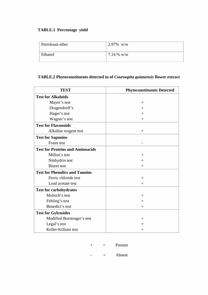
TABLE.1 Percentage yield
TABLE.2 Phytoconstituents detected in of Couroupita guianensis flower extract
TEST Phytoconstituents Detected
Test for AlkaloidsMayer’s testDragendroff’sHager’s testWagner’s test
++++
Test for FlavonoidsAlkaline reagent test +
Test for SaponinsFoam test -
Test for Proteins and AminoacidsMillon’s testNinhydrin testBiuret test
+++
Test for Phenolics and TanninsFerric chloride testLead acetate test
++
Test for carbohydratesMolisch’s testFehling’s testBenedict’s test
+++
Test for GylcosidesModified Borntrager’s testLegal’s testKeller-Killiani test
+++
+ = Present
- = Absent
Petroleum ether 2.97% w/w
Ethanol 7.14 % w/w
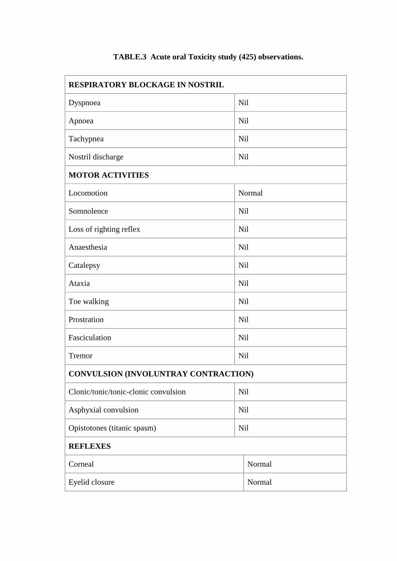
TABLE.3 Acute oral Toxicity study (425) observations.
RESPIRATORY BLOCKAGE IN NOSTRIL
Dyspnoea Nil
Apnoea Nil
Tachypnea Nil
Nostril discharge Nil
MOTOR ACTIVITIES
Locomotion Normal
Somnolence Nil
Loss of righting reflex Nil
Anaesthesia Nil
Catalepsy Nil
Ataxia Nil
Toe walking Nil
Prostration Nil
Fasciculation Nil
Tremor Nil
CONVULSION (INVOLUNTRAY CONTRACTION)
Clonic/tonic/tonic-clonic convulsion Nil
Asphyxial convulsion Nil
Opistotones (titanic spasm) Nil
REFLEXES
Corneal Normal
Eyelid closure Normal
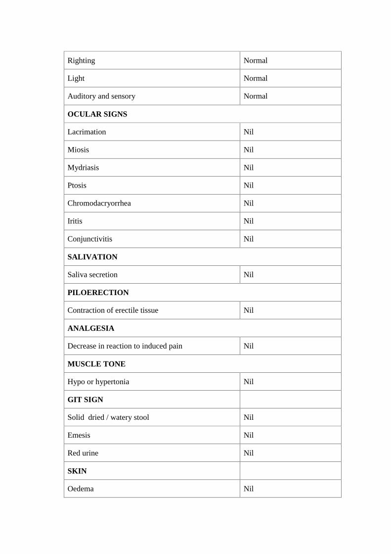
Righting Normal
Light Normal
Auditory and sensory Normal
OCULAR SIGNS
Lacrimation Nil
Miosis Nil
Mydriasis Nil
Ptosis Nil
Chromodacryorrhea Nil
Iritis Nil
Conjunctivitis Nil
SALIVATION
Saliva secretion Nil
PILOERECTION
Contraction of erectile tissue Nil
ANALGESIA
Decrease in reaction to induced pain Nil
MUSCLE TONE
Hypo or hypertonia Nil
GIT SIGN
Solid dried / watery stool Nil
Emesis Nil
Red urine Nil
SKIN
Oedema Nil
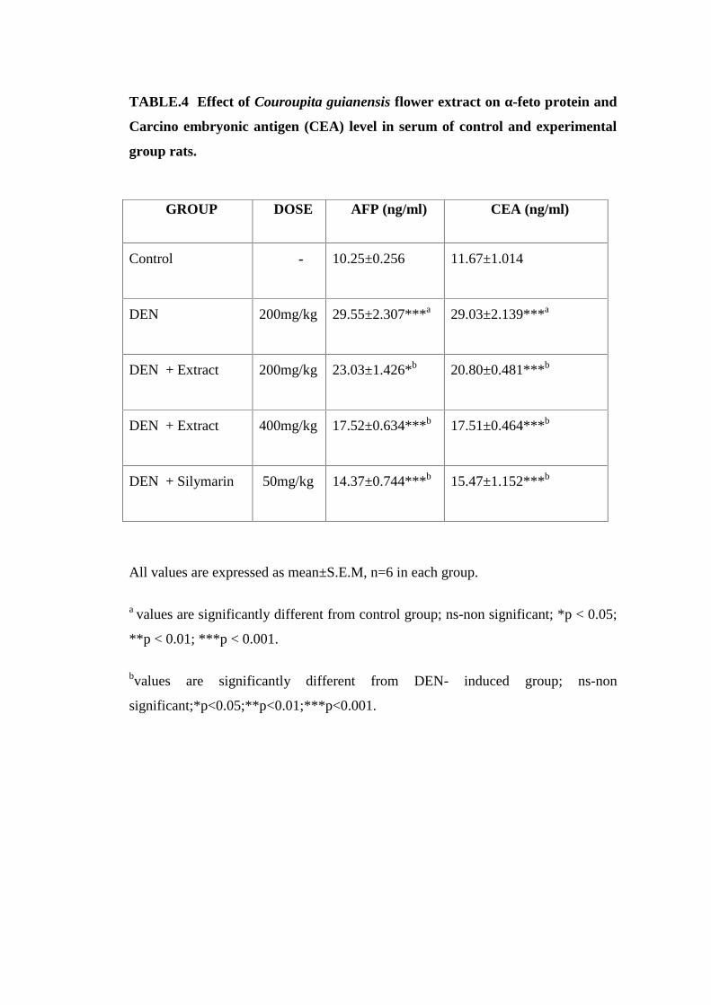
TABLE.4 Effect of Couroupita guianensis flower extract on α-feto protein and
Carcino embryonic antigen (CEA) level in serum of control and experimental
group rats.
All values are expressed as mean±S.E.M, n=6 in each group.
a values are significantly different from control group; ns-non significant; *p < 0.05;
**p < 0.01; ***p < 0.001.
bvalues are significantly different from DEN- induced group; ns-non
significant;*p<0.05;**p<0.01;***p<0.001.
GROUP DOSE AFP (ng/ml) CEA (ng/ml)
Control - 10.25±0.256 11.67±1.014
DEN 200mg/kg 29.55±2.307***a 29.03±2.139***a
DEN + Extract 200mg/kg 23.03±1.426*b 20.80±0.481***b
DEN + Extract 400mg/kg 17.52±0.634***b 17.51±0.464***b
DEN + Silymarin 50mg/kg 14.37±0.744***b 15.47±1.152***b
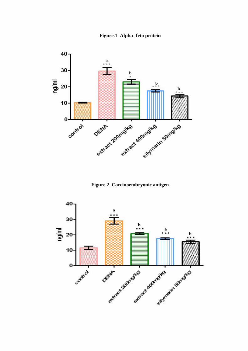
Figure.1 Alpha- feto protein
Figure.2 Carcinoembryonic antigen
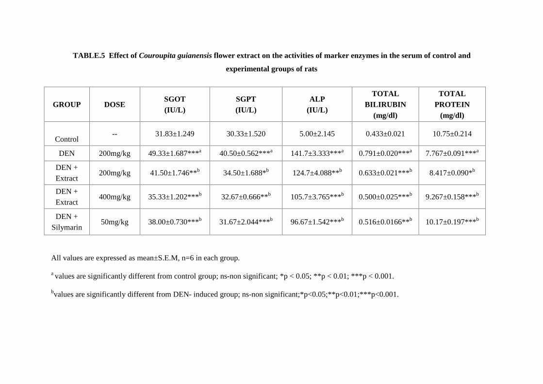
TABLE.5 Effect of Couroupita guianensis flower extract on the activities of marker enzymes in the serum of control and
experimental groups of rats
GROUP DOSESGOT(IU/L)
SGPT(IU/L)
ALP(IU/L)
TOTALBILIRUBIN
(mg/dl)
TOTALPROTEIN
(mg/dl)
Control-- 31.83±1.249 30.33±1.520 5.00±2.145 0.433±0.021 10.75±0.214
DEN 200mg/kg 49.33±1.687***a 40.50±0.562***a 141.7±3.333***a 0.791±0.020***a 7.767±0.091***a
DEN +
Extract200mg/kg 41.50±1.746**b 34.50±1.688*b 124.7±4.088**b 0.633±0.021***b 8.417±0.090*b
DEN +
Extract400mg/kg 35.33±1.202***b 32.67±0.666**b 105.7±3.765***b 0.500±0.025***b 9.267±0.158***b
DEN +
Silymarin50mg/kg 38.00±0.730***b 31.67±2.044***b 96.67±1.542***b 0.516±0.0166**b 10.17±0.197***b
All values are expressed as mean±S.E.M, n=6 in each group.
a values are significantly different from control group; ns-non significant; *p < 0.05; **p < 0.01; ***p < 0.001.
bvalues are significantly different from DEN- induced group; ns-non significant;*p<0.05;**p<0.01;***p<0.001.
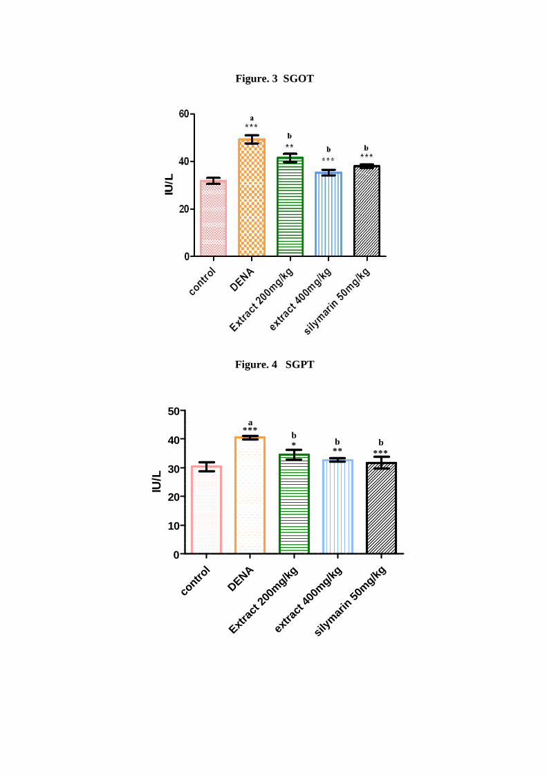
Figure. 3 SGOT
Figure. 4 SGPT
contro
l
DENA
Extrac
t 200
mg/kg
extra
ct 40
0mg/kg
silym
arin 50
mg/kg0
10
20
30
40
50***
a
*b
**b
***b
IU/L
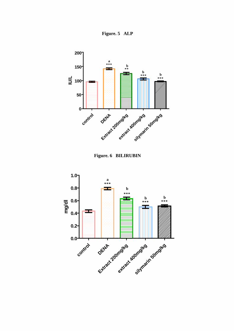
Figure. 5 ALP
contro
l
DENA
Extrac
t 200
mg/kg
extra
ct 40
0mg/kg
silym
arin 50
mg/kg0
50
100
150
200
***a
**b
***b
***b
IU/L
Figure. 6 BILIRUBIN
contro
l
DENA
Extrac
t 200
mg/kg
extra
ct 40
0mg/kg
silym
arin 50
mg/kg0.0
0.2
0.4
0.6
0.8
1.0***
a
***b
***b
***b
mg/
dl
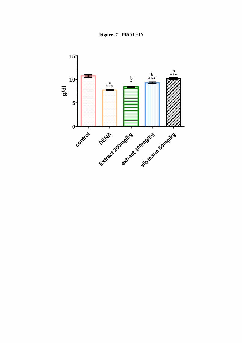
Figure. 7 PROTEIN
contro
l
DENA
Extrac
t 200
mg/kg
extra
ct 40
0mg/kg
silym
arin 50
mg/kg0
5
10
15
***a *
b ***b ***
b
g/dl
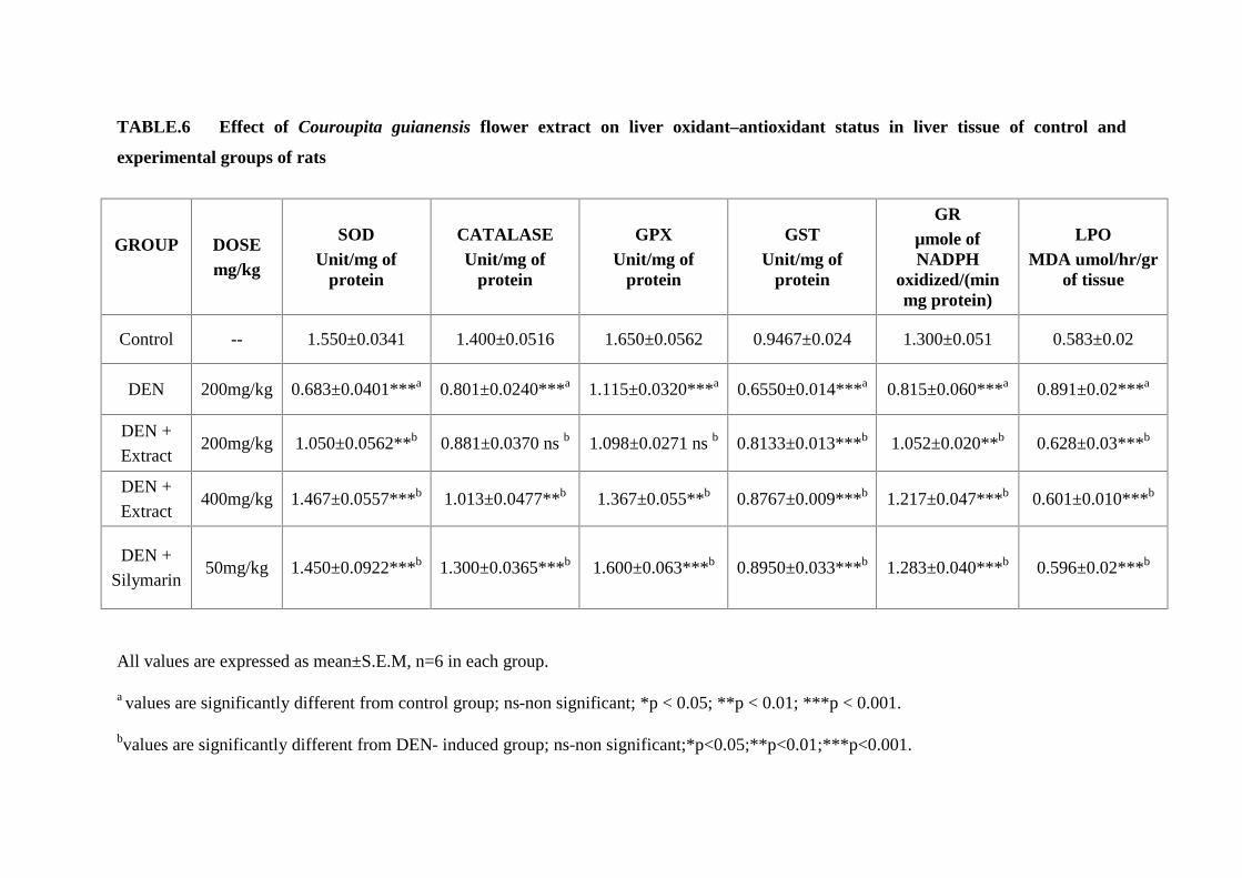
TABLE.6 Effect of Couroupita guianensis flower extract on liver oxidant–antioxidant status in liver tissue of control and
experimental groups of rats
GROUP DOSEmg/kg
SODUnit/mg of
protein
CATALASEUnit/mg of
protein
GPXUnit/mg of
protein
GSTUnit/mg of
protein
GRμmole ofNADPH
oxidized/(minmg protein)
LPOMDA umol/hr/gr
of tissue
Control -- 1.550±0.0341 1.400±0.0516 1.650±0.0562 0.9467±0.024 1.300±0.051 0.583±0.02
DEN 200mg/kg 0.683±0.0401***a 0.801±0.0240***a 1.115±0.0320***a 0.6550±0.014***a 0.815±0.060***a 0.891±0.02***a
DEN +
Extract200mg/kg 1.050±0.0562**b 0.881±0.0370 ns b 1.098±0.0271 ns b 0.8133±0.013***b 1.052±0.020**b 0.628±0.03***b
DEN +
Extract400mg/kg 1.467±0.0557***b 1.013±0.0477**b 1.367±0.055**b 0.8767±0.009***b 1.217±0.047***b 0.601±0.010***b
DEN +
Silymarin50mg/kg 1.450±0.0922***b 1.300±0.0365***b 1.600±0.063***b 0.8950±0.033***b 1.283±0.040***b 0.596±0.02***b
All values are expressed as mean±S.E.M, n=6 in each group.
a values are significantly different from control group; ns-non significant; *p < 0.05; **p < 0.01; ***p < 0.001.
bvalues are significantly different from DEN- induced group; ns-non significant;*p<0.05;**p<0.01;***p<0.001.
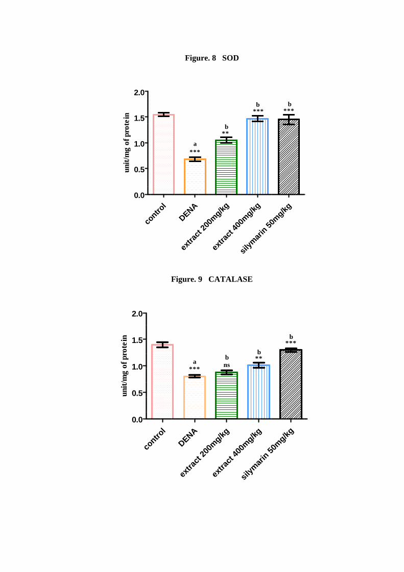
Figure. 8 SOD
contro
l
DENA
extra
ct 20
0mg/kg
extra
ct 40
0mg/kg
silym
arin 50
mg/kg0.0
0.5
1.0
1.5
2.0
***a
**b
***b
***b
unit
/mg
of p
rote
in
Figure. 9 CATALASE
contro
l
DENA
extra
ct 20
0mg/kg
extra
ct 40
0mg/kg
silym
arin 50
mg/kg0.0
0.5
1.0
1.5
2.0
***a ns
b **b
***b
unit
/mg
of p
rote
in

Figure. 10 GPX
Figure. 11 GST
contro
lDENA
extra
ct 20
0mg/kg
extra
ct 40
0mg/kg
silym
arin 50
mg/kg0.0
0.5
1.0
1.5
***a ***
b***
b ***b
m
oles
of C
DN
B u
tili
zed/
min
/mg
prot
ein
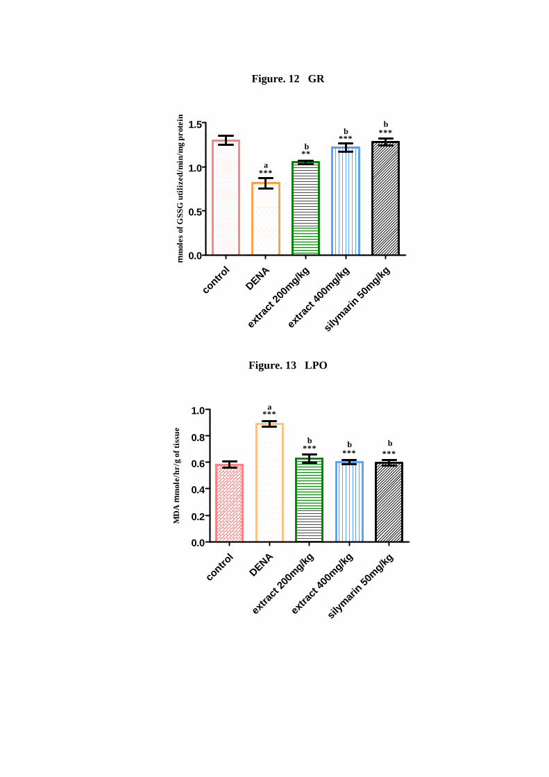
Figure. 12 GR
contro
l
DENA
extra
ct 20
0mg/kg
extra
ct 40
0mg/kg
silym
arin 50
mg/kg0.0
0.5
1.0
1.5
***a
**b
***b ***
b
m
oles
of G
SSG
uti
lize
d/m
in/m
g pr
otei
n
Figure. 13 LPO
contro
lDENA
extra
ct 20
0mg/kg
extra
ct 40
0mg/kg
silym
arin 50
mg/kg0.0
0.2
0.4
0.6
0.8
1.0 ***a
***b
***b
***b
MD
A
mol
e/hr
/g o
f tis
sue
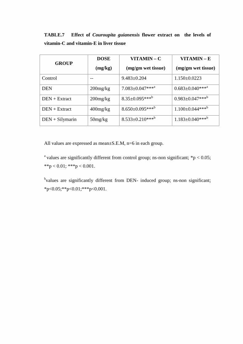
TABLE.7 Effect of Couroupita guianensis flower extract on the levels of
vitamin-C and vitamin-E in liver tissue
GROUPDOSE
(mg/kg)
VITAMIN – C
(mg/gm wet tissue)
VITAMIN – E
(mg/gm wet tissue)
Control -- 9.483±0.204 1.150±0.0223
DEN 200mg/kg 7.083±0.047***a 0.683±0.040***a
DEN + Extract 200mg/kg 8.35±0.095***b 0.983±0.047***b
DEN + Extract 400mg/kg 8.650±0.095***b 1.100±0.044***b
DEN + Silymarin 50mg/kg 8.533±0.210***b 1.183±0.040***b
All values are expressed as mean±S.E.M, n=6 in each group.
a values are significantly different from control group; ns-non significant; *p < 0.05;
**p < 0.01; ***p < 0.001.
bvalues are significantly different from DEN- induced group; ns-non significant;
*p<0.05;**p<0.01;***p<0.001.
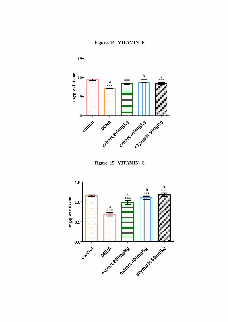
Figure. 14 VITAMIN- E
contro
l
DENA
extra
ct 20
0mg/kg
extra
ct 40
0mg/kg
silym
arin 50
mg/kg0
5
10
15
***
***ab
***b
***b
mg/
g w
et t
issu
e
Figure. 15 VITAMIN- C
contro
l
DENA
extra
ct 20
0mg/kg
extra
ct 40
0mg/kg
silym
arin 50
mg/kg0.0
0.5
1.0
1.5
***a
***b ***
b ***b
mg/
g w
et t
issu
e
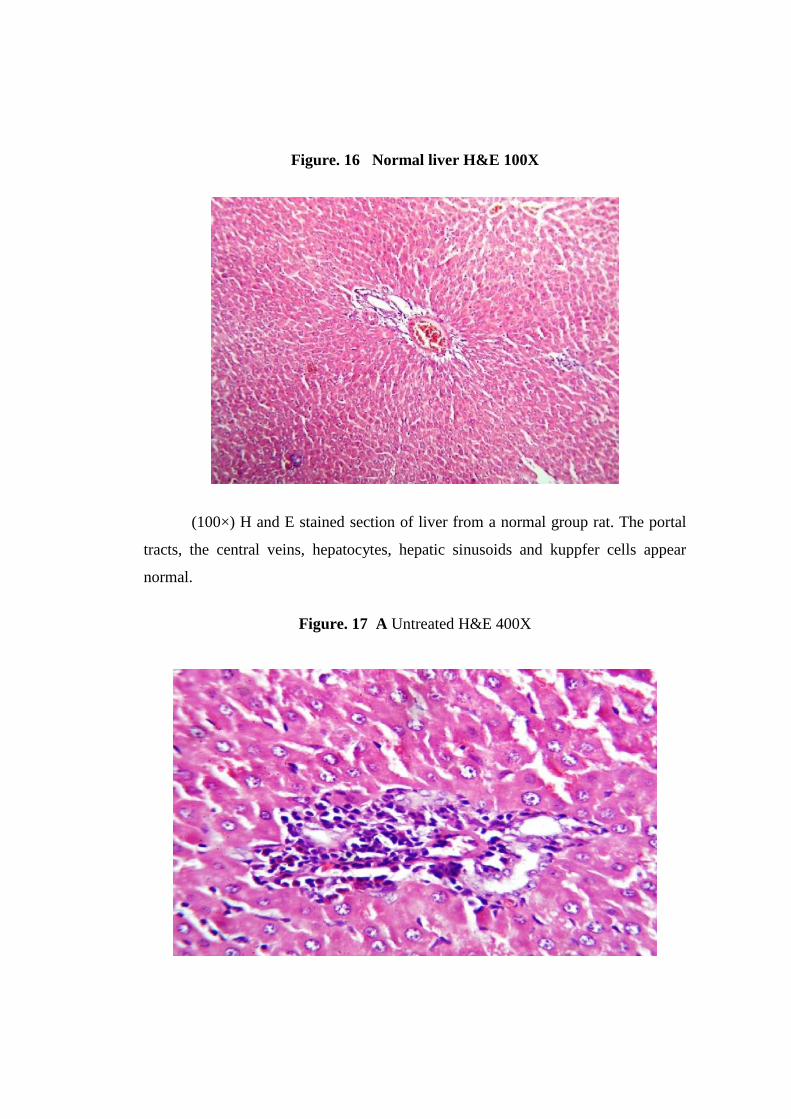
Figure. 16 Normal liver H&E 100X
(100×) H and E stained section of liver from a normal group rat. The portal
tracts, the central veins, hepatocytes, hepatic sinusoids and kuppfer cells appear
normal.
Figure. 17 A Untreated H&E 400X
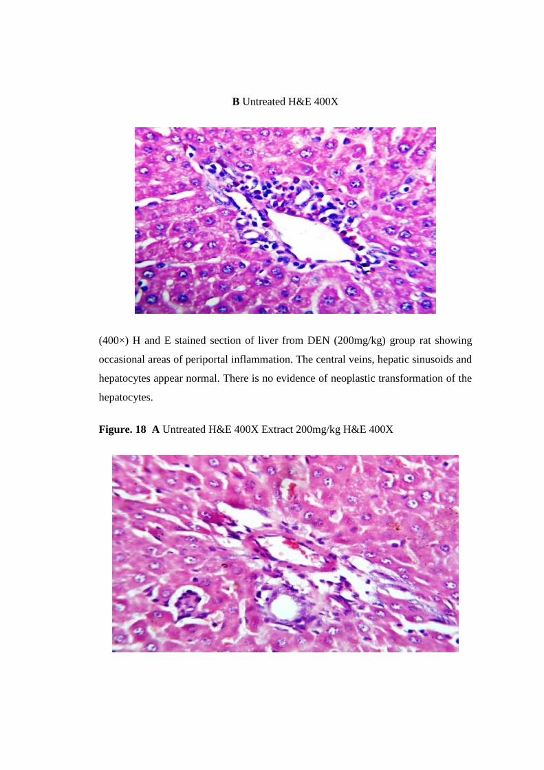
B Untreated H&E 400X
(400×) H and E stained section of liver from DEN (200mg/kg) group rat showing
occasional areas of periportal inflammation. The central veins, hepatic sinusoids and
hepatocytes appear normal. There is no evidence of neoplastic transformation of the
hepatocytes.
Figure. 18 A Untreated H&E 400X Extract 200mg/kg H&E 400X
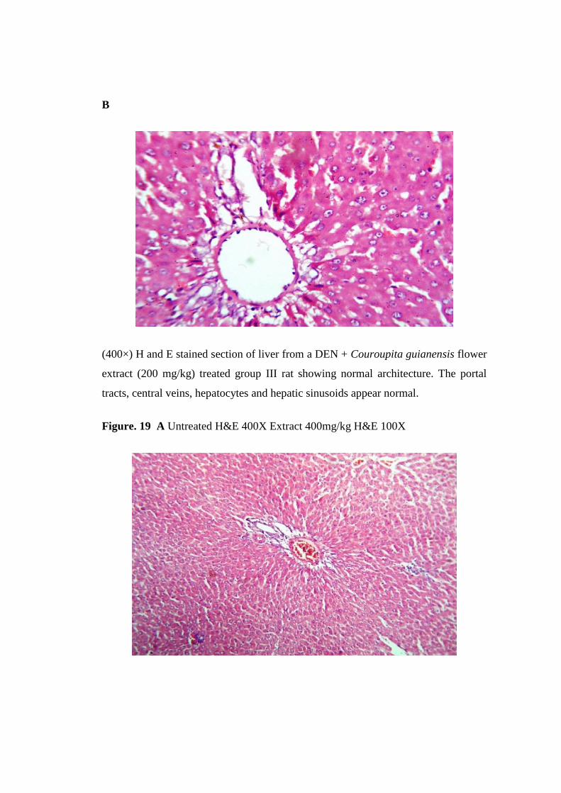
B
(400×) H and E stained section of liver from a DEN + Couroupita guianensis flower
extract (200 mg/kg) treated group III rat showing normal architecture. The portal
tracts, central veins, hepatocytes and hepatic sinusoids appear normal.
Figure. 19 A Untreated H&E 400X Extract 400mg/kg H&E 100X
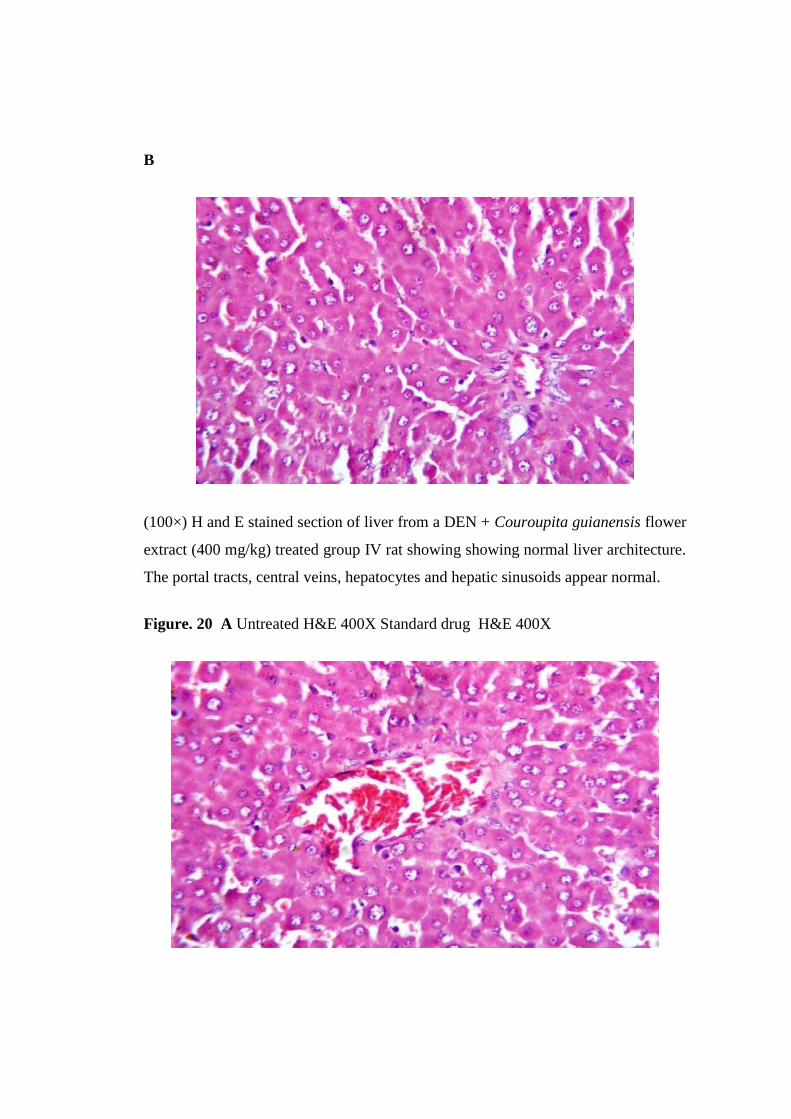
B
(100×) H and E stained section of liver from a DEN + Couroupita guianensis flower
extract (400 mg/kg) treated group IV rat showing showing normal liver architecture.
The portal tracts, central veins, hepatocytes and hepatic sinusoids appear normal.
Figure. 20 A Untreated H&E 400X Standard drug H&E 400X
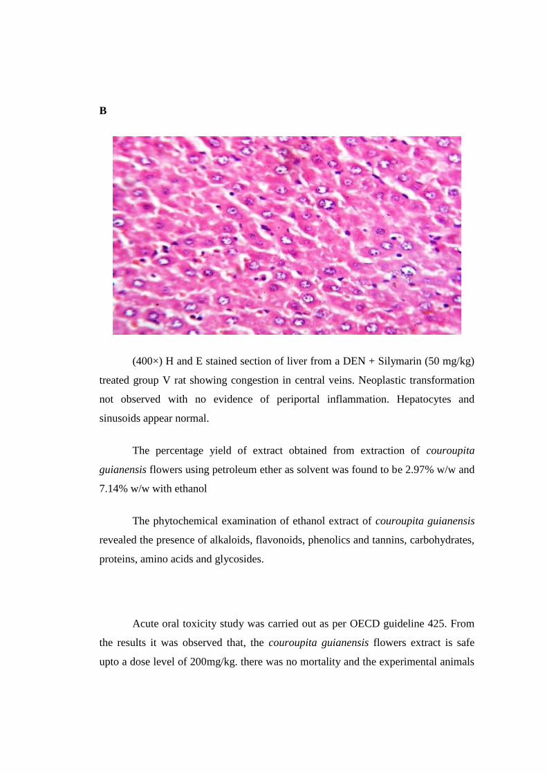
B
(400×) H and E stained section of liver from a DEN + Silymarin (50 mg/kg)
treated group V rat showing congestion in central veins. Neoplastic transformation
not observed with no evidence of periportal inflammation. Hepatocytes and
sinusoids appear normal.
The percentage yield of extract obtained from extraction of couroupita
guianensis flowers using petroleum ether as solvent was found to be 2.97% w/w and
7.14% w/w with ethanol
The phytochemical examination of ethanol extract of couroupita guianensis
revealed the presence of alkaloids, flavonoids, phenolics and tannins, carbohydrates,
proteins, amino acids and glycosides.
Acute oral toxicity study was carried out as per OECD guideline 425. From
the results it was observed that, the couroupita guianensis flowers extract is safe
upto a dose level of 200mg/kg. there was no mortality and the experimental animals
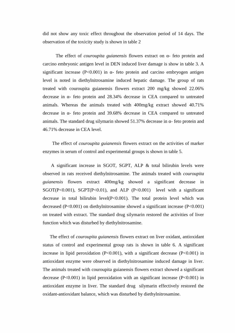
did not show any toxic effect throughout the observation period of 14 days. The
observation of the toxicity study is shown in table 2
The effect of couroupita guianensis flowers extract on α- feto protein and
carcino embryonic antigen level in DEN induced liver damage is show in table 3. A
significant increase (P<0.001) in α- feto protein and carcino embryogen antigen
level is noted in diethylnitrosamine induced hepatic damage. The group of rats
treated with couroupita guianensis flowers extract 200 mg/kg showed 22.06%
decrease in α- feto protein and 28.34% decrease in CEA compared to untreated
animals. Whereas the animals treated with 400mg/kg extract showed 40.71%
decrease in α- feto protein and 39.68% decrease in CEA compared to untreated
animals. The standard drug silymarin showed 51.37% decrease in α- feto protein and
46.71% decrease in CEA level.
The effect of couroupita guianensis flowers extract on the activities of marker
enzymes in serum of control and experimental groups is shown in table 5.
A significant increase in SGOT, SGPT, ALP & total bilirubin levels were
observed in rats received diethylnitrosamine. The animals treated with couroupita
guianensis flowers extract 400mg/kg showed a significant decrease in
SGOT(P<0.001), SGPT(P<0.01), and ALP (P<0.001) level with a significant
decrease in total bilirubin level(P<0.001). The total protein level which was
decreased (P<0.001) on diethylnitrosamine showed a significant increase (P<0.001)
on treated with extract. The standard drug silymarin restored the activities of liver
function which was disturbed by diethylnitrosamine.
The effect of couroupita guianensis flowers extract on liver oxidant, antioxidant
status of control and experimental group rats is shown in table 6. A significant
increase in lipid peroxidation (P<0.001), with a significant decrease (P<0.001) in
antioxidant enzyme were observed in diethylnitrosamine induced damage in liver.
The animals treated with couroupita guianensis flowers extract showed a significant
decrease (P<0.001) in lipid peroxidation with an significant increase (P<0.001) in
antioxidant enzyme in liver. The standard drug silymarin effectively restored the
oxidant-antioxidant balance, which was disturbed by diethylnitrosamine.
A significant decrease (P<0.001) in vitamin-C and vitamin-E was noted in
liver of diethylnitrosamine control animals. Whereas the animal treated with extract
and silymarin showed a significant increase (P<0.001) in antioxidants vitamin C and
vitamin E.
DISCUSSION
Oxidative stress is a common mechanism contributing to initiation and
progression of hepatic damage in a variety of liver disorders. Oxidative stress is
associated with damage to a wide range of macromolecular species including lipids,
proteins, and nucleic acids thereby producing major interrelated derangements of
cellular metabolism including peroxidation of lipids (Sun, 1990). Reactive oxygen
species (ROS) formed from endogenous (or) exogenous sources are highly reactive,
toxic and mutagenic (Halliwell, 1994). Diethylnitrosamine, one of the most
important environmental carcinogen, has been suggested to cause the generation of
reactive oxygen species(ROS) resulting in oxidative stress and cellular injury
(Bartsch et al., 1989). As liver is the main site for diethylnitrosamine metabolism,
the production of ROS in liver may be responsible for its carcinogenic effects
(Bansal et al., 2005) . The involvement of oxidative stress in diethylnitrosamine
induced hepatotoxicity and carcinogenicity underscores the need for development of
novel compound with potent antioxidant activity. In this study, diethylnitrosamine
administration to rats leads to a marked elevation in levels of serum SGOT, SGPT,
ALP and total bilirubin levels, which is indicative of hepatocellular damage. The
increase in SGOT, SGPT and ALP levels might be due to the possible release from
cytoplasm, into the blood circulation rapidly after rupture of plasma membrane and
cellular damage. High levels of SGOT indicates liver damage, such as that caused by
viral hepatitis as well as cardiac infraction and muscle injury. SGPT catalyses the
conversion of alanine to pyruvate and glutamate and is release in similar manner.
Therefore SGPT is more specific to the liver and is thus a better parameter for
detecting liver injury (Rao and Mishra, 1997). These enzymes are the most
sensitive markers employed in the diagnosis of hepatic damage because they are in
cytoplasmic location and hence released into the circulation after cellular damage
(Wroblewski, 1959; Sallie et al., 1991). Treatment with Couroupita guianensis
flower extract significantly reduced the activities of the enzymes in DEN treated
rats. This indicates that couroupita guianensis flower extract tends to prevent liver
damage by maintaining integrity of the plasma memberane, thereby suppressing the
leakage of enzyme through membranes, exhibiting hepatoprotective activity.
Elevated ALP level may indicate cholestasis (partial or full blockade of bile
ducts). Since bile ducts bring bile from the liver to gall bladder and intestine,
inflammation/damage in liver cause spillage of ALP in blood stream. ALP levels
typically rise to several times the normal level following the bile obstruction or intra
hepatic cholestasis. Causes of elevated ALP also include bilary cirrhosis, fatty liver
and liver tumor (Quinn and Johnston, 1997). Significant reduction in ALP levels in
Couroupita guianensis flower extract treated group indicates the protective effect of
Couroupita guianensis flower against diethylnitrosamine induced hepatic injury.
Bilirubin is a yellow pigment produced when heme is catabolised.
Hepatocytes render bilirubin water soluble and therefore easily excretable by
conjugating it with glucuronic acid prior to secreting it into bile by active transport.
Hyperbilirubinemia may result from the production of more bilirubin than the liver
can process or obstruction of excretory ducts of the liver (Tolulope Olalepe et al.,
2010). Serum bilirubin is considered as one of the true test of liver function since it
reflects the ability of the liver to take up and process bilirubin into bile. Elevated
levels may indicate several illness. High levels of total bilirubin in
diethylnitrosamine induced stress may be due to its toxic effect. The significant
reduction in the level of total bilirubin in the serum of Couroupita guianensis flower
extract treated rats suggest the hepatoprotective potential of Couroupita guianensis
flowers.
Elevated of serum alpha fetoprotein (ALP) levels has been reported in several
diseases including hepatocellular carcinoma. AFP is a serum protein similar in size,
structure and aminoacid composition to serum albumin, but it is detectable only in
minute amounts in the serum of normal adults. Elevated serum concentrations of this
protein can be achieved in the adult by exposure to hepatotoxic agents. It is a 72KDa
α1globulin with an uncertain biological function, is synthesized during embryogenic
life by foetal yolk sac, liver and intestinal tract. AFP has high specificity for
hepatocarcinoma (Abelev, 1971; Liu et al., 2006). Its serum concentration can be
used to confirm hepatocarcinoma and for the diagnosis of tumor response to therapy.
More than 90% of patients with hepatic cancer have increased serum AFP levels
(Jahan et al., 2011). A level above 500ng/ml of AFP in human adults can be
indicative of hepatocellular carcinoma, germ cell tumors, and metastatic cancers of
liver.
Carcinoembryonic antigen (CEA) is a type of protein molecule found in many
different cells of the body, but typically associated with certain tumors and the
developing fetus. The most frequent cancer which cause an increase in CEA is
cancer of colon and rectum. Other include cancers of pancreas, stomach, breast,
lung, and certain type of thyroid and ovarian conditions. Benign conditions which
can elevate CEA are smoking, infections, inflammatory bowel disease, pancreatitis,
cirrhosis of liver. Levels of CEA increase with an increase in tumour size. Serum
values of more that 5ng/ml indicate metastasis (Jahan et al., 2011).
In the present study a significant increase in levels of AFP and CEA was
observed in diethylnitrosamine induced hepatic injury. Treatment with Couroupita
guianensis flower extract showed a significant decrease in AFP and CEA levels
which indicates a positive prognosis. The decrease level on Couroupita guianensis
flower treatment prevents the neoplastic growth and reduces hepatic disorder
Oxidative damage in a cell or tissue occurs when the concentration of reactive
oxygen species (O2., H2O2 and OH.) generated exceeds the antioxidant capability of
the cells (Sies, 1991). The status of lipid peroxidation as well as altered levels of
certain endogenous radical scavengers is taken as direct evidence for oxidative stress
( Khan, 2006). Free radical scavenging enzymes like SOD and catalase protect the
biological system from oxidative stress. The SOD dismutates superoxide radicals
(O2.-) into hydrogen peroxide (H 2O2) and O2 (Fridovich, 1986). Catalase further
detoxifies H2O2 into H2O and O2 (Murray et al., 2003). Thus SOD, catalase and
glutathione peroxidase act mutually and constitute the enzymatic antioxidative
defense mechanism against reactive oxygen species (Bhattacharjee and Sil, 2006).
The decrease in the activities of these enzymes in the present study could be
attributed to the excessive utilization of these enzymes in inactivating free radicals
generated during the metabolism of diethylnitrosamine. This is further substantiated
by an elevation in the levels of lipid peroxidation. Lipid peroxidation (LPO) has
been postulated as being the destructive process in liver injury due to
diethylnitrosamine. The excessive ROS generated during diethylnitrosamine
metabolism rapidly reacts with lipid membrane. This initiates lipid peroxidation
chain reaction, which leads to formation of several toxic by products such as
malondialdehyde and 4-hydroxynonenal which can attach cellular targets including
DNA, inducing mutagenicity and carcinogencity (Zwart et al., 1999).
Administration of diethylnitrosamine has been reported to generate lipid
peroxidation in general (Hietnen, 1987). The results of the present study are in
accordance with these reports. Restoration in the levels of lipid peroxidation by
Couroupita guianensis flower extract could be related to its ability to scavenge
reactive oxygen species, thus preventing further damage to membrane lipids.
Excessive liver damage and oxidative stress caused by diethylnitrosamine,
depleted the levels of non-enzymatic antioxidants like vitamin C and vitamin E.
Non-enzymatic antioxidants like vitamin-C and E act synergistically to scavenge the
free radicals formed in the biological system. GSH acts synergistically with vitamin-
E in inhibiting oxidative stress and acts against lipid peroxidation (Chaudiere,
1994). Vitamin-C also scavenges and detoxifies free radicals in combination with
vitamin-E and GSH (George,2003). It plays a vital role by regenerating the reduced
form of vitamin-E and preventing the formation of free radicals (Das, 1994). The
decreased levels of these antioxidant vitamins observed during diethylnitrosamine
administration might be due to the excessive utilization of these vitamins in
scavenging the free radicals formed during the metabolism of diethylnitrosamine.
Extract treated animals showed a significant increase in vitamin C and vitamin E
levels. This shows Couroupita guianensis flower extract maintains the level of
antioxidant vitamins, thereby protecting the cells from further oxidative stress. A
significant decrease in activities of GST and GR was observed in diethylnitrosamine
treated rats, which may be due to decreased expression of antioxidants during
hepatocellular damage. The observations are in accordance with the reports of
kweon et al 2003 who demonstrated that diethylnitrosamine induced hepatocellular
injury was escorted by a substantial fall in glutathione peroxidase and GST activity,
which improved on administration of antioxidants. In the present study, treatment
with Couroupita guianensis flower extract maintains the activity of GR and GST in
liver. This is indicative of the potent antioxidant activity possessed by Couroupita
guianensis flower. From the results of invivo antioxidant study, it was observed that
Couroupita guianensis flower is effective in scavenging the free radicals released
during the metabolism of diethylnitrosamine.
Chapter VII
Conclusion
CONCLUSION
In conclusion, the present investigation shows that the Couroupita
guianensis flower extract exhibits excellent hepatoprotective property by restoring
the hepatic marker enzymes and by stabilizing and increasing all the components of
antioxidant defense system during diethylnitrosamine induced oxidative stress in
rats. I suggest that the natural antioxidants and scavenging agents in Couroupita
guianensis flower extract might be effective as hepatoprotectors and thus, may have
some obvious therapeutic implications. Therefore, it seems logical to infer that
Couroupita guianensis flowers, because of its antioxidant property, might be
capable of protecting the hepatic tissue from diethylnitrosamine-induced injury.
The ethanol extract of Couroupita guianensis flower extract is found to be
rich in flavonoids, phenolics and tannins in our preliminary photochemical
screening. Since flavonoids, phenolics and tannins are powerful antioxidants, their
presence in Couroupita guianensis flower extract flower extract might be
responsible for antioxidant property, which might be involved in the
hepatoprotective activity.

Chapter VIII
Bibliography
BIBLIOGRAPHY
1. Abeler GI. Alpha-fetoprotein in association with malignant tumors. Adv.
Cancer res. 1971; 14:295-397.
2. Amer, J., Ghoti, H., Rachmilewitz, E., Koren, A., Levin, C. and Fibach, E.
(January 2006). "Red blood cells, platelets and polymorphonuclear
neutrophils of patients with sickle cell disease exhibit oxidative stress that
can be ameliorated by antioxidants". British Journal of Haematology ,january
2006, 132 (1): 108–113.
3. Amit khatri, Arun Garg, Shyam S, Agrawal. Evaluation of hepatoprotective
activity of aerial parts of Tephrosia purpurea L and stem bark of Tecomella
undulate. J.Ethnopharmacol 2009; 25:1645-1646.
4. Ana Martínez, Enma Conde, Andrés Moure, Herminia Domínguez & Ramón
J. Estévez . Protective effect against oxygen reactive species and skin
fibroblast stimulation of Couroupita guianensis leaf extracts. Natural
Product Research, 2012; Vol 26: 314-322.
5. Anupam Bishayee, Kendra F. Barnes, Deepak Bhatia, Altaf S. Darvesh, and
Richard T. Carroll Resveratrol Suppresses Oxidative Stress and
Inflammatory Response in Diethylnitrosamine-Initiated Rat
Hepatocarcinogenesis. Cancer Prev Research, 2010; 753-763
6. Article source:html.www.herbalplace.com/alternative medicine/herbal –
medicine.html.
7. Article source:www.herbal-supplement-resource.com.
8. Bagchi K and Puri S “Pathways of oxidative damage”. Eastern
Mediterranean Health Journal, 1998, 4(2), 350-360.
9. Bansal AK, Bansal M, Soni G, Bhatnagar D. protective role of vitamine E
pre- treatmen N-nitrosodiethylamine induced oxidative stress in rat liver.
Chem. Boil. Interact 2005; 156: 101-111.
10. Bartsch H, Hietanen E, malaveille C. Carcinogenic nitrosamines; free radical
aspects of their action. Free radic. Biol. Med. 1989; 7:637-644.
11. Ben- Erik van Wyk, Medicinal plants of the world ; Time editions, 2009,
page no. 8.
12. Carcinoembryogenic antigen (CEA) [ home page on the internet] No date
(cited 2012 feb24).Available from: http://www. Medicine net.
Com/carcinoembryonic_antigen/article.htm.
13. Chaudiere J. some chemical and Biochemical constrains of oxidative stress
in living cells. In: Rice-Evans CA, Burdon RH(Eds). Free Radical damage
and its control. Elsevier science, Amsterdam, pp.25-66.
14. Cutler RG, Antioxidants, aging and longevity, In: Free Radicals in Biology,
Pryor, WA, Vol 6 Ed, Academic Press, Orlando. FL, 1984, 371-428.
15. D.Pradhan,P.K.Panda,G.Tripathy.Evaluation of immunomodulatory activityof the methanolic extract of couroupita guianensis flowers in rats.Indianjournel of natural products and resources,2009,vol.8(1):37-42.
16. Das S. Vitamin E in the genesis and prevention of cancer. A review Aeta
oncol 1994; 33: 615-619.
17. Devasagayam, TPA; Tilac JC, Boloor KK, Sane Ketaki S, Ghaskadbi Saroj
S, Lele RD "Free Radicals and Antioxidants in Human Health: Current
Status and Future Prospects". Journal of Association of Physicians of India,
october 2004, 52: 796.
18. Docampo, R. "Antioxidant mechanisms". In J. Marr and M. Müller, (Eds.).
Biochemistry and Molecular Biology of Parasites. London: Academic Press,
1995, pp. 147–160.
19. Dr.Shaijesh Wankhede,Manoj Gambhire,Archana Juvekar.Anxiolytic effectof methanolic root extract of couroupita guianensis in mouse models ofanxiety.Pharmacology online 3,2009.
20. Evans MD, Cooke MS . "Factors contributing to the outcome of oxidative
damage to nucleic acids". Bioessays,may 2004, 26 (5): 533–42.
21. Fridovich I. superoxide dismutases. Adv. Enzymol. 1986; 58:61-97.
22. Gayathri R, D Kalpana Deepa Priya, GR Gunassekaran, Dhanapal
Sakthisekara. Ursolic acid attenuates oxidative stress-mediated
Hepatocellular Carcinoma Induction by Diethylnitrosamine in Male Wistar
Rats, Asian Pacific Journal of Cancer Prevention, 2009; Vol 10, 933-938.
23. George J. ascorbic acid concentrations in diemethylnitrosamine, induced
hepatic fibrosis in rats. Clin. Chim. Aeta 2003; 335:39-47.
24. Gratus C, Wilson S, Greenfield SM, Damery SL, Warmington SA, Grieve R,
Steven NM, Routledge P. The use of herbal medicines by people with
cancer: a qualitative study. Complement Altern Med. 2009 May 14;9:14.
25. Halliwell B, free radical, antioxidant and human disease: curiosity, cause or
consequence? Lancet 1994; 344:721-724.
26. Halliwell, B.” Antioxidant defense mechanisms”: From the beginning to the
end. Free Radical Research, 1999, 31:261–272.
27. Hasan SS, Ahmed SI, Bukhari NI, Loon WC. Use of complementary and
alternative medicine among patients with chronic diseases at outpatient
clinics. Complement Ther Clin Pract. 2009 Aug;15(3):152-7.
28. Hietnen E, Ahotupa M, Bartsch H. Lipid peroxidation and chemically
induced cancer in rats fed lipid-rich diet, in: Lapis K, Kekharst S(Eds),
carcinogenesis and tumor progression, 1987, vol 4. Akademiaikiado,
Budapest, pp.9-16.
29. Jahan MS, Vani G, Shyamaladevi C.S. Anti-carcinogenic effect of solanum
trilobatum in diethylnitrosamine induced and Phenobarbital promoted
hepatocarcinogenesis in rats. Asian J. Biochem 2011: 1-8.
30. Kampooj VP, Herbal medicine- current science,January 2000, volume. 78,
NO.1- 10 .
31. Kannampalli Pradeep, Chandrasekaran Victor Raj Mohan, Kupannan
Gobianand And Sivanesan Karthikeyan. Protective effect of Cassia
fistula Linn. on diethylnitrosamine induced hepatocellular damage and
oxidative stress in ethanol pretreated rats. Biological Research, 2010;
43: 113-125,
32. Kannampalli Pradeep, Chandrasekaran Victor Raj Mohan, Sivanesan
Karthikeyan,Kuppanan Gobianand, “Silymarin modulates the oxidant–
antioxidant imbalance during diethylnitrosamine induced oxidative stress in
rats” European journal of Pharmacology,2007, 560 : 110–116.
33. Khan M.R,Kihara M,Omoloso A.D.Evaluation of antibiotic activity ofcouroupita guianensis.Journel of herbs,speices and medicinal plants,2008,vol10(3):95-108.
34. Khan SM. Protective effect of black tea extract on the levels of lipid
peroxidation and antioxidant enzymes in liver of mice with pesticide induced
liver injury. Cell Biochem. Funct 2006; 24:327-332.
35. Khandelwal KR AND Kokate CK. Practical pharmacognosy, 4th edition,
vallabh prakashan, new delhi; 1995:pp.110.
36. Koehn, F. E. and Carter, G. T . , The evolving role of natural products in
drug discovery. Nature Rev. Drug Discov . , 2005, 4, 206-220.
37. Kokate, Gokhale SB . Pharmacognosy, Pune, Nirali Prakashan. 16th ED .
2001; Page No: 134, 173, 435.
38. Kraft K. Complementary/Alternative Medicine in the context of prevention
of disease and maintenance of health. Prev Med. 2009 May 22.
39. Kweon S, Park KA, Choi H. chemopreventive effect of garlic powder diet in
diethylnitrosamine-induced rat hepatocarcinogenesis. Life sci 2003; 73:
2515-2526.
40. Liu JG, Zhao HJ, Liu YJ, Wang XL. Effect of selenium-enriched malt on
hepatocarcinogenesis, paraneoplastic syndrome and the hormones regulating
blood glucose in rats treated with diethylnitrosamine. Life sci 2006; 78(20):
2315-2321.
41. Małgorzata Kujawska, Ewa Ignatowicz, Małgorzata Ewertowska, Jarosław
Markowski and Jadwiga Jodynis-Liebert. Cloudy apple juice protects against
chemical-induced oxidative stress in rat, European Journal Of Nutrition,
2010; Vol 50: 53-60.
42. Mariana M.G. Pinheiro, Sidnei O. Bessa, Catharina E. Fingolob, Ricardo M.
Kusterb, Maria Eline Matheusa, Fábio S. Menezesc, Patrícia Dias Fernandesa,
Antinociceptive activity of fractions from Couroupita guianensisAubl.
Leaves. Journal of ethnopharmacology 2010; vol.127: 407-413.
43. Mohammed M.Sayed-Ahmed,Abdulazz M-Aleisa,Salim S.Al-Rejaie,abdulaziz A.Al-yahya,Othman A.Al-Shabanah,MahmoudN.Nagi.Thymoquinone attenuates diethylnitrosamine induction of hepaticcarcinogenesis through antioxidant signaling.Oxidative medicine and cellularlongevity,2010,vol 3,4:254-261.
44. Moquin B, Blackman MR, Mitty E, Flores S. Complementary and alternative
medicine (CAM). Geriatr Nurs. 2009 May-Jun;30(3):196-203. Review.
45. Mukherjee,P.K., Extract values, Quality control of herbal drugs, 2002: 1st
edition: 189-191.
46. Murray RK, granner DK, Mayes PA, Rodwell VW. Harper’s illustrated
Biochemistry 2003, 26th. The McGraw- Hill companies Inc.
47. Nartey, L., Huwiler-Muentener, K., Shang, A., Liewald, K., Jueni, P., Egger,
M. (2007). Matched-pair study showed higher quality of placebo-controlled
trials in Western phytotherapy than conventional medicine. Journal of
Clinical Epidemiology, 60(8), 787-794 *
48. Nermin.A.H. Sadik, Shohda A,Maraghy E.L, Manal. F. Ismail.Diethylnitrosamine induced hepatocarcinogenesis in rats:Possiblechemoprevention by blueberries. African journel of biochemistryresearch,2008,vol 2(3):81-87.
49. OECD, guidance document on acute oral toxicity 425.environmental health
and safety monograph series on testing assessment 2000; No.24.
50. Peach T and Tracey MV. Modern methods in plant analysis. Springer verlog.
Berlin 1957; pp.387.
51. Perumal Subramanian,Sundaresan.S.S.Allyl-cysteine inhibits circulatorylipid peroxidation and promotes antioxidants in N-nitrosodiethylamine-induced carcinogenesis.2003,vol 55(1):37-42.
52. Pradeep k, victorraj mohan c, Gobi anand k, karthikeyan s, silymarin
modulates the oxidant. Antioxidant imbalance during diethylnitrosamine
induced oxidative stress in rats. European journal of pharmacology 2007;
560: 110-116.
53. Prakash, v., Indian medicinal plants : current status-1, Ethnobotany, 1998,
10, 112-121.
54. Prasanna Galhena,Ira Thabrew,Mayuri G Tammitiyagodage,Vasanthi AHanna Rachel.Ayurvedic herbal remedy reduces the extent ofdiethylnitrosamine induced oxidative stress in rats.2009,vol 5(7):19-27.
55. Quinn PG, Johnston DE, detection of chronic liver disease: Costs and
denefits. Gastroenterologist 1997; 5: 58-77.
56. Rajamanickam V, A. Rajasekaran, S. Darlin quine, M. Jesupillai, R. Sabitha:
Anthelmintic activity of the flower extract of Couroupita guianensis. The
Internet Journal of Alternative Medicine, 2009; Vol 8: 1.
57. Rama Krishna G, Bala raghavendran H, Vinodh kumar R, devaki
T.Suppression of N-nitrosodiethylamine induced hepatocarcinogenesis by
silymarin in rats. Chemico-Biological Interaction 2006;161:104-114.
58. Ramanathan Sampath kumar,Sunderam. R.S, Kanagasabi.R., Vijaya.M.,Perumal.B.P., Guptha M.,Sivakumar.P,.Effect of Bauhinia racemosa ondiethylnitrosamine induced hepatocarcinogenis in rats.Amj Chin Med2007,35(1):103-14.
59. Rao KS, Mishra SH. Anti-inflammatory and hepatoprotective activities of
sida rhambifolia Linn. Indian J pharmacol 1997; 29:110-116.
60. Rattan SI ,. "Theories of biological aging: genes, proteins, and free radicals".
Free Radic. Res. ,2006, 40 (12): 1230–8.
61. Rosenthalar K. chemical investigation of plants. Bell G and sons, London
1930;23-29.
62. Sabry M Shaarawy, Amany A Tohamy, Saad M. Elgendy, Zakaria Y Abd
Elmageed, Abeer Bahnasy, Maha S Mohamed, Emad Kandil, Khalid
Matrougui. Protective Effects of Garlic and Silymarin on NDEA-Induced
Rats Hepatotoxicity. Int J Biol Sci 2009; 5(6):549-557.
63. Sallie R, Tredger JM, Willam R. Drug and the liver. Biopharm. Drug dispos
1991; 12: 251-259.
64. Sanjay. Prahalad Umachigi; Jayaveera K. N; Ashok kumar C. K; G. S.
Kumar Antimicrobial, Wound Healing and Antioxidant potential of
Couroupita guianensis in rats , Pharmacologyonline, 2007; Vol 3: 269-281.
65. Shellard EJ. Practical plant chemistry for pharmacy studies. Pitman medical
publishing co.ltd; London 1957;34-80.
66. Shivkumar, Free radicals and antioxidants: Food and human system,
advances in applied science research, 2011, 2, (1): 121-135.
67. Sies H. oxidative stress: oxidants and antioxidants 1991. Academic press,
san diego, California.
68. Sies, Helmut , "Oxidative stress: Oxidants and antioxidants". Experimental
physiology, 1997, 82 (2): 291–5.
69. Sivanesan Karthikeyan, Kannampalli Pradeep, Chandrasekaran Victor Raj
Mohan, Kuppanan Gobianand. Silymarin modulates the oxidant–antioxidant
imbalance during diethylnitrosamine induced oxidative stress in rats,
European Journal of Pharmacology, 2007: 110–116 .
70. Sivanesan karthikeyan.,” Protective effect of Cassia fistula Linn. on
diethylnitrosamine induced hepatocellular damage and oxidative stress in
ethanol pretreated rats”.journel of biological research vol. 43, 2010: 113-125.
71. Spelman, K. Philosophy in Phytopharmacology: Ockham's Razor versus
synergy. Journel of Herbal Pharmacotherapy,2005, 5(2), 31-47.
72. Spinella, M. The importance of pharmacological synergy in psychoactive
herbal medicines. Alternative Medicine Review,2002, 7(2), 130-137.
73. Sun Y, Free radicals, antioxidant enzyme, and carcinogenesis, free radic.
Boil. Med. 1990; 8: 583-599.
74. Thamilarasan manivasagam, Perumal Subramanian, ganapathy suthakar,
musthafa Mohamed Essa. The chemopreventive effect of diallyl disulphide
on N-nitrosodiethylamine induced hepatocarcinogenesis. Journal of applied
biomedicine, 2005;4: 187-191.
75. Tolulope olaleye M, Afolabi C, Akinmoladun, Adebayo A Ogunboye,
Afolabi A Akindahunsi. Antioxidant activity and hepatoprotective activity of
leaf extract of boerhaavia diffusa Linn. Against acetaminophen induced liver
damage in rats. Food chem. Toxicol 2010; 48: 2200-2205.
76. Tortora, Grabowski. Principles of anatomy and physiology, 10th edition page
no: 950-965.
77. Wilson S, Greenfield SM, Damery SL, Warmington SA, Grieve R, Steven
NM, Routledge P. The use of herbal medicines by people with cancer: a
qualitative study. Complement Altern Med. 2009 May 14;9:14.
78. Wroblewski F. the clinical significance of transaminase activities of serum.
Am. J. med 1959; 27:911-923.
79. Zwart LD, Meerman JN, Commandeur JM. Biomarkers of free radical
damage applications in experimental animals and humans. Free Radic.
Boil.med 1999; 26:202-226.


![[IJCT V3I4P9] Authors:Ms. Sivashankari .A, Mrs. Kavitha .S.K](https://static.cupdf.com/doc/110x72/58a5dd5d1a28abd14d8b55f5/ijct-v3i4p9-authorsms-sivashankari-a-mrs-kavitha-sk.jpg)

