Introduction to the diagnosis and management of common opportunistic
infections (Ols)
Module 4 Sub module OIs
Opportunistic Infections
Pneumocystis carinii pneumonia (PCP)
Penicilliosis Recurrent pneumonia Cryptococcus Toxoplasmosis Oesophageal
candidasis Mycobacterium Avium
Complex (MAC) Cytomegalovirus (CMV)
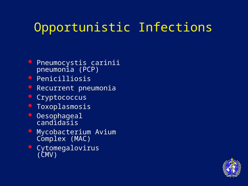
Natural course & common clinical manifestations
1000 900 800 700 600 500 400 300 200 100
50 <50
0
PCP Cryptococcal meningitis PPE
CD4 COUNT
0 3 6 9 1 2 3 4 5 6 7 8 9 10 Months Years
TB
Oral candida OHL
HZV
CMV MAC
TB
TB
TB
Cryptosporidial diarrhea
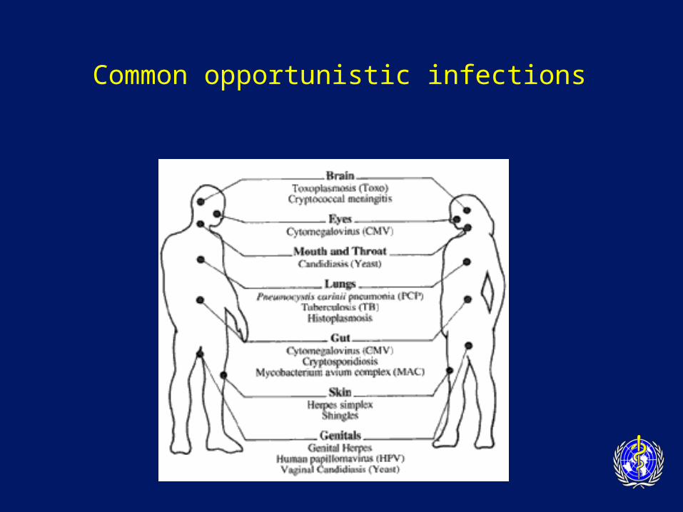
Common opportunistic infections
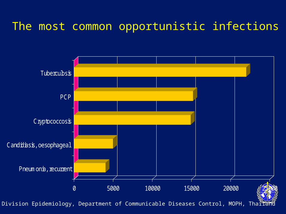
The most common opportunistic infections
0 5000 10000 15000 20000 25000
Pneumonia, recurrent
Candidiasis, oesophageal
Cryptococcosis
PCP
Tuberculosis
Division Epidemiology, Department of Communicable Diseases Control, MOPH, Thailand
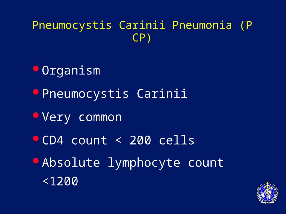
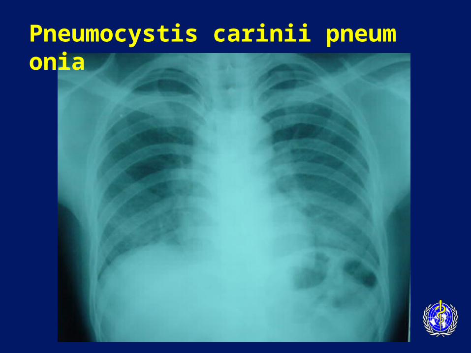
Pneumocystis Carinii Pneumonia (PCP)
Organism
Pneumocystis Carinii
Very common
CD4 count < 200 cells
Absolute lymphocyte count <1200
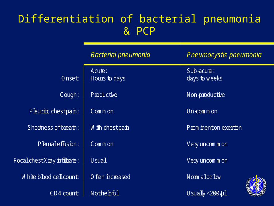
Differentiation of bacterial pneumonia & PCP
Bacterial pneumonia Pneumocystis pneumonia
Onset: Acute: Hours to days
Sub-acute: days to weeks
Cough: Productive Non-productive
Pleuritic chest pain: Common Un-common
Shortness of breath: With chest pain Prominent on exertion
Pleural effusion: Common Very uncommon
Focal chest Xray infiltrate: Usual Very uncommon
White blood cell count: Often increased Normal or low
CD4 count: Not helpful Usually <200/µl
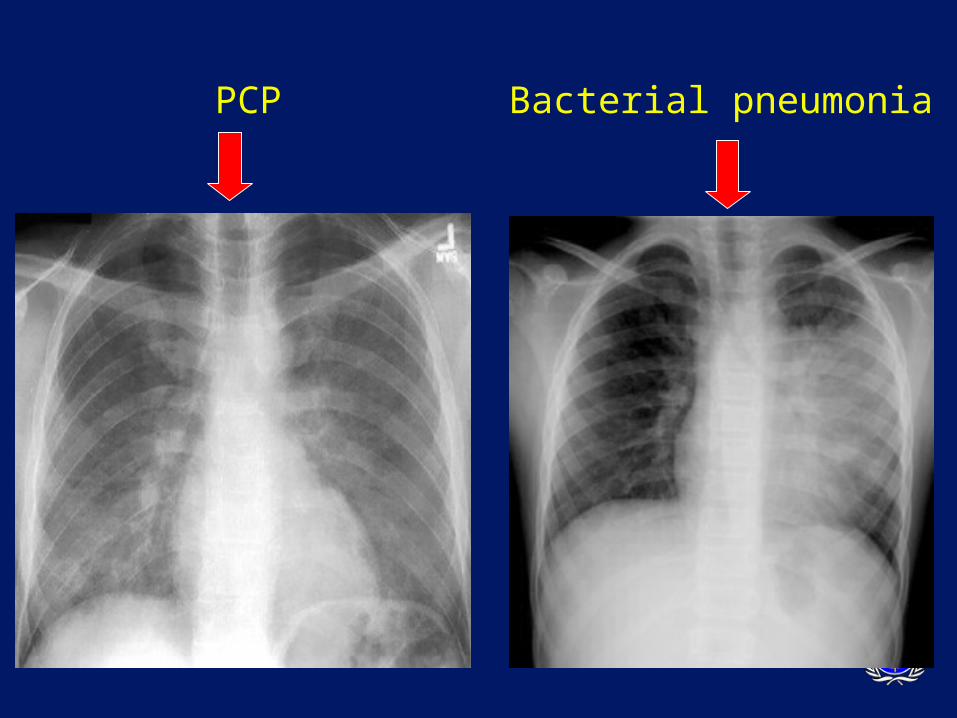
PCP Bacterial pneumonia
Pneumocystis carinii pneumonia
PCP
Diagnosis
– Frequently clinical
– Typical symptoms
– Response to treatment
– Microscopic demonstration of
P. carinii in lung secretions/tissue
– Culture unavailable
PCP
Diagnosis
– special methods to obtain specimens are necessary
• Induced sputum/B.A.L./Biopsy DDX:
– MTB, bacterial pneumonia, fungal pneumonia, lymphoma, KS
PCP
Treatment
–Trimethoprim-Sulfamethoxazole
–drug of choice (iv 15 mg/kg/day or oral 2 DS tablets tid)
–3 weeks recommended
–Allergy to TMP-SMX
–Corticosteroids if severely hypoxic
PCP
Alternative treatment for allergic patients
(all for 21 days)
• pentamidine
• dapsone + trimethoprim
• clindamycin + primaquine
• atovaquone– less effective
PCP
Prognosis:– 100% fatal untreated– Level of hypoxaemia best predicts outco
me
Secondary Prophylaxis– co-trimoxazole 1-2 tabs daily– Dapsone 100 mg daily – aerosilized pentamidine 300 mg monthly
Penicilliosis
Organism:
Penicillium marneffei
Endemic area:
– SE Asia (Northern Thailand, Southern China, Vietnam, Indonesia, Hong Kong)
– 3rd most common OI in Northern Thailand
CD4 count < 100 cells
Penicilliosis
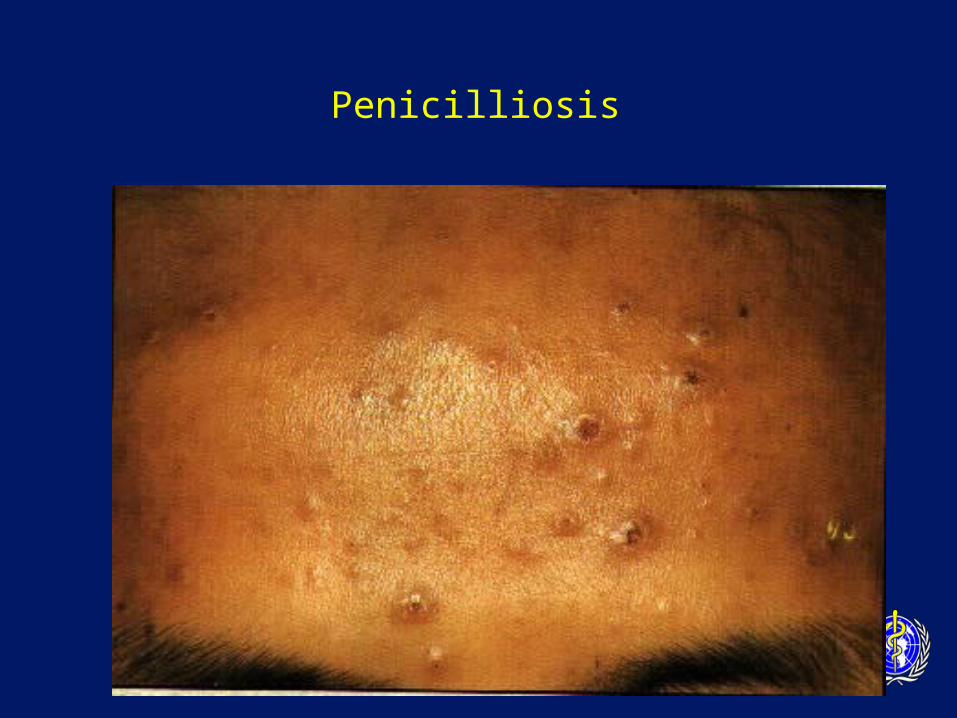
Clinical symptoms:
– Fever (99%)
– papulo-necrotic skin lesions (71%)
– weight loss (76%)
– anaemia (77%)
– lymphadenopathy (58%)
– hepatomegaly (51%)
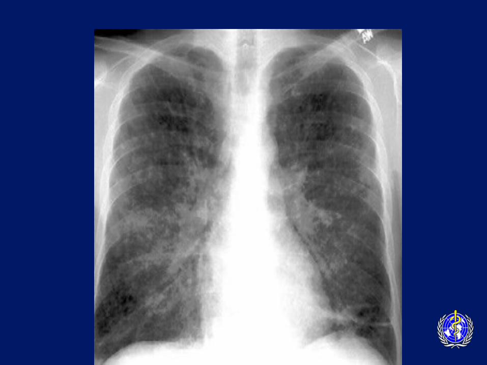
– productive cough
– lung disease
Penicilliosis
Diagnosis
– Presumptive:microscopy on smear– Definitive: culture– DDx:
• other disseminated mycobacterial or fungal disease
Penicilliosis
Penicilliosis
Penicilliosis
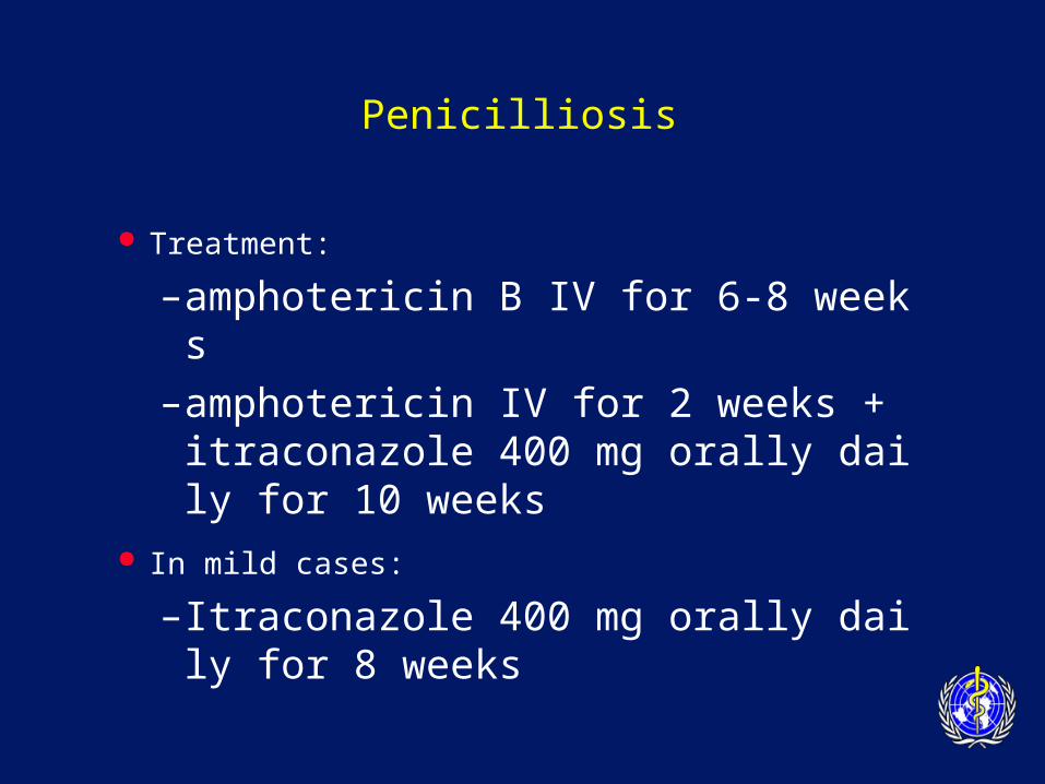
Treatment:
–amphotericin B IV for 6-8 weeks
–amphotericin IV for 2 weeks + itraconazole 400 mg orally daily for 10 weeks
In mild cases:
– Itraconazole 400 mg orally daily for 8 weeks
Penicilliosis
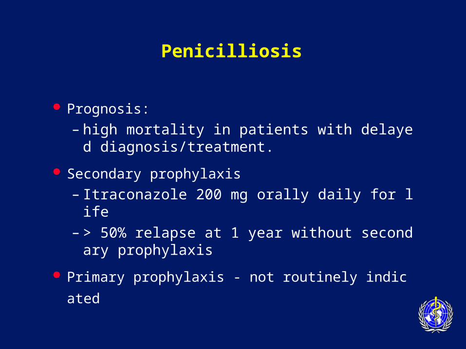
Prognosis:
– high mortality in patients with delayed diagnosis/treatment.
Secondary prophylaxis
– Itraconazole 200 mg orally daily for life– > 50% relapse at 1 year without secondary pr
ophylaxis
Primary prophylaxis - not routinely indicated
Recurrent Pneumonia
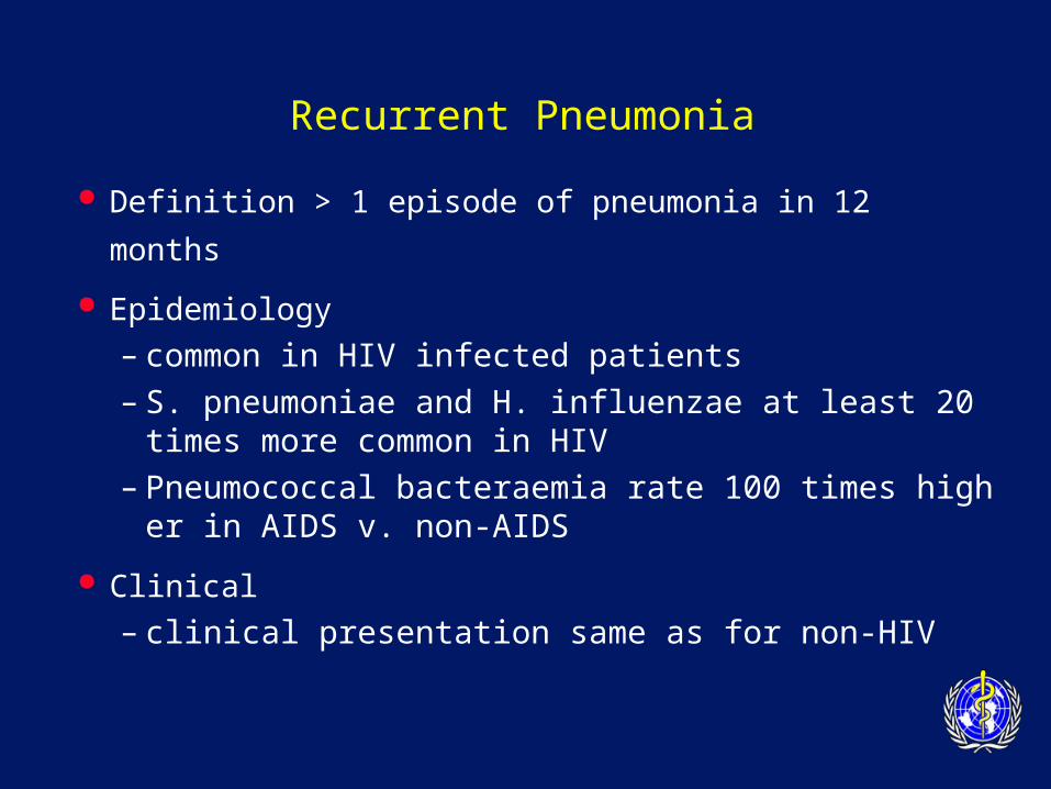
Definition > 1 episode of pneumonia in 12 months
Epidemiology
– common in HIV infected patients
– S. pneumoniae and H. influenzae at least 20 times more common in HIV
– Pneumococcal bacteraemia rate 100 times higher in AIDS v. non-AIDS
Clinical
– clinical presentation same as for non-HIV
Recurrent Pneumonia
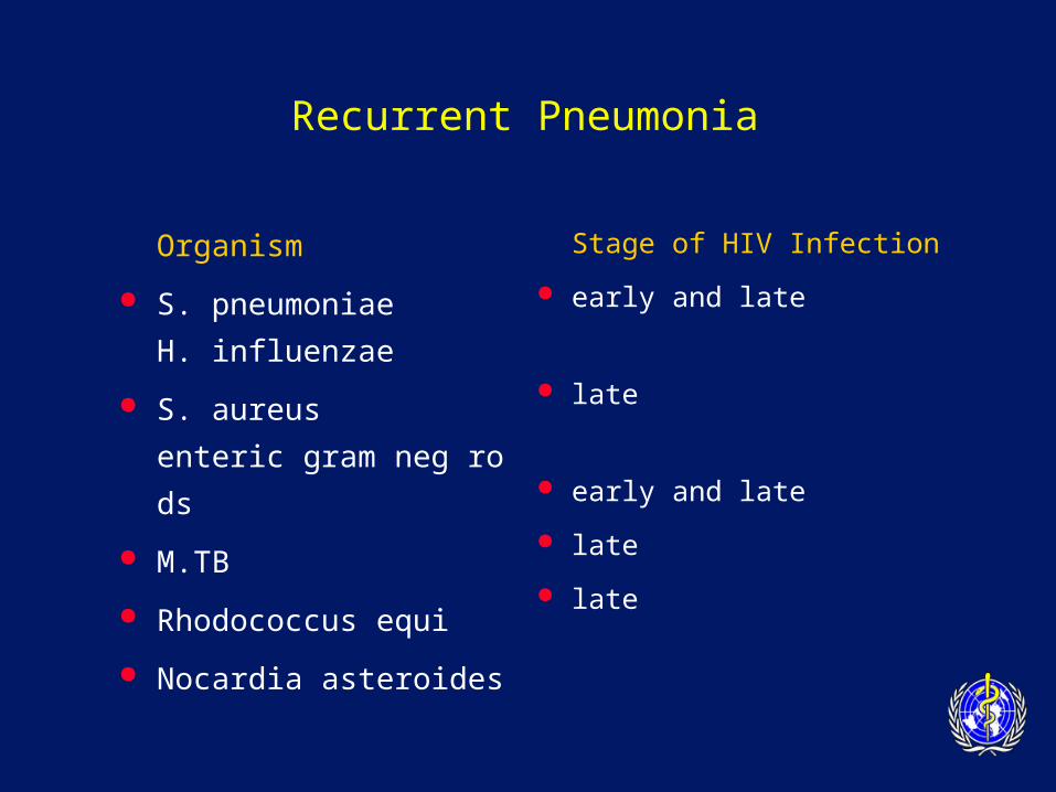
Organism
S. pneumoniae
H. influenzae
S. aureus
enteric gram neg rods
M.TB
Rhodococcus equi
Nocardia asteroides
Stage of HIV Infection
early and late
late
early and late
late
late
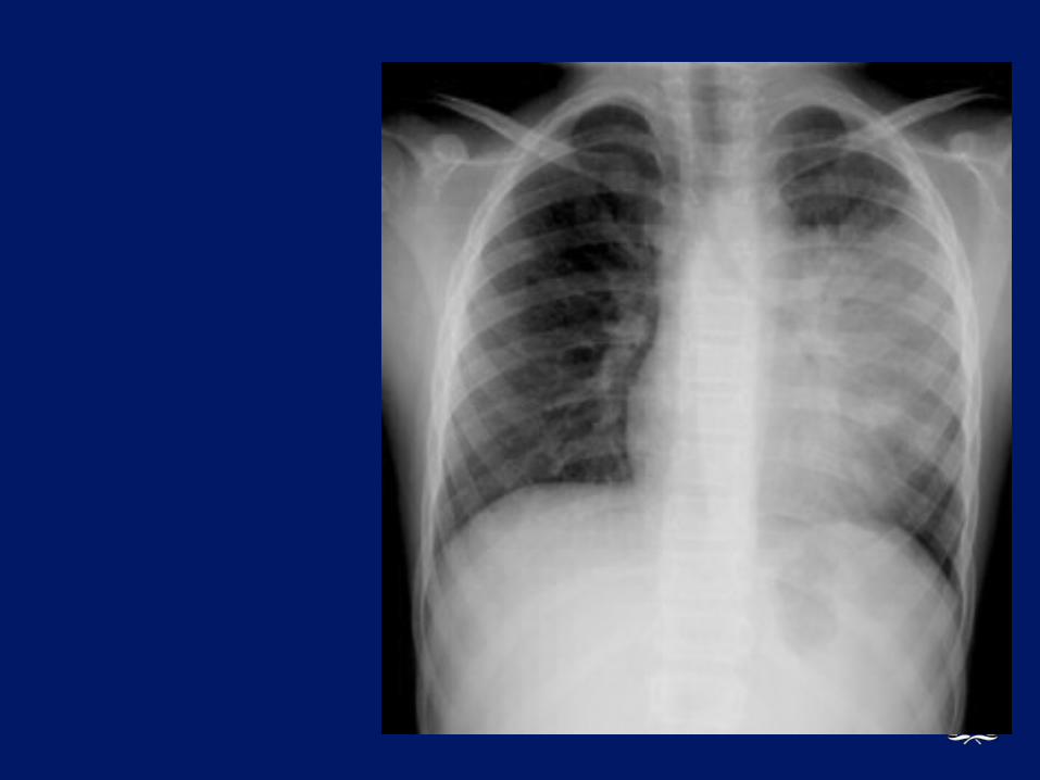
Recurrent Pneumonia
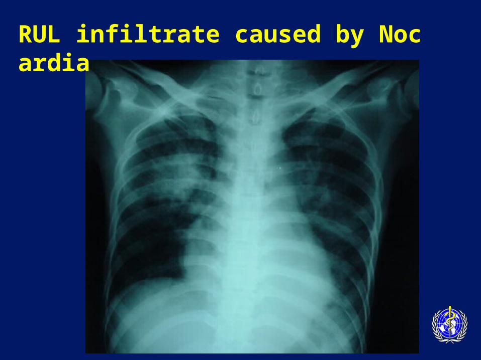
RUL infiltrate caused by Nocardia
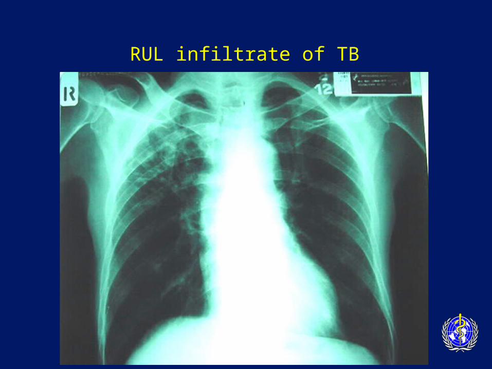
RUL infiltrate of TB
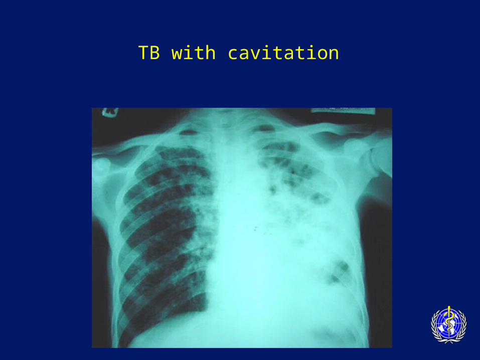
TB with cavitation
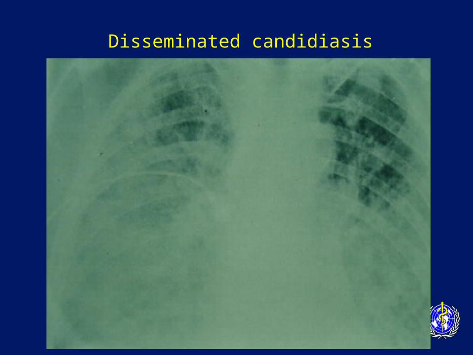
Disseminated candidiasis
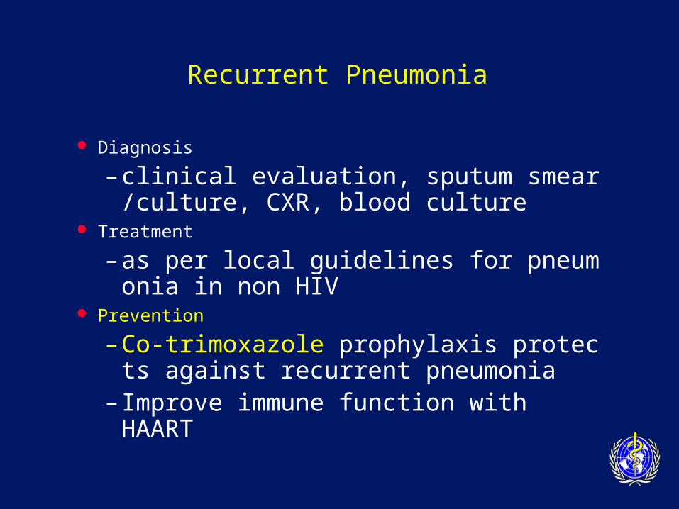
Recurrent Pneumonia
Diagnosis
– clinical evaluation, sputum smear/culture, CXR, blood culture
Treatment
– as per local guidelines for pneumonia in non HIV
Prevention
– Co-trimoxazole prophylaxis protects against recurrent pneumonia
– Improve immune function with HAART
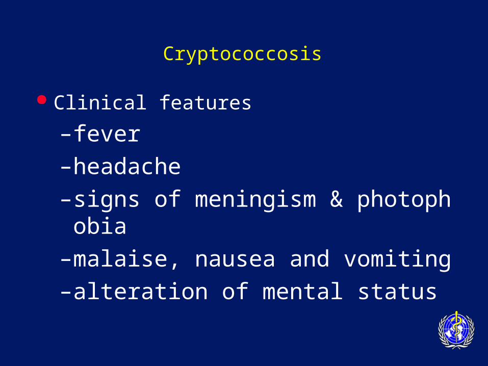
Cryptococcosis
Clinical features
–fever
–headache
–signs of meningism & photophobia
–malaise, nausea and vomiting
–alteration of mental status
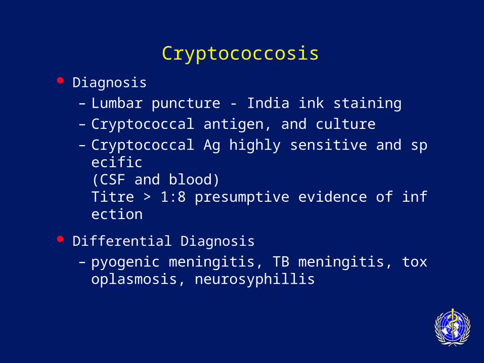
Cryptococcosis
Diagnosis
– Lumbar puncture - India ink staining– Cryptococcal antigen, and culture– Cryptococcal Ag highly sensitive and specific
(CSF and blood)Titre > 1:8 presumptive evidence of infection
Differential Diagnosis
– pyogenic meningitis, TB meningitis, toxoplasmosis, neurosyphillis
Encapsulated yeast of Cryptococcus neoformans in CSF India ink preparation
Cryptococcosis
Cryptococcosis
Cryptococcosis
Treatment of Cryptococcal Meningitis
– Induction phase• amphotericin B iv daily for 14 days• consider adding 5-flucytosine (5-FC)
– Consolidation phase• fluconazole 400 mg po daily for 8 week
Cryptococcosis
Prognosis
– mortality rates as high as 30% despite therapy
Secondary Prophylaxis
– fluconazole 200-400 mg daily
– itraconazole 100-200 mg po bid (less effective than fluconazole)
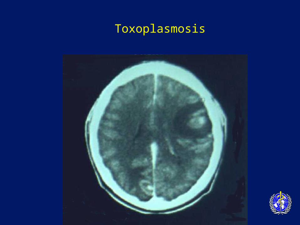
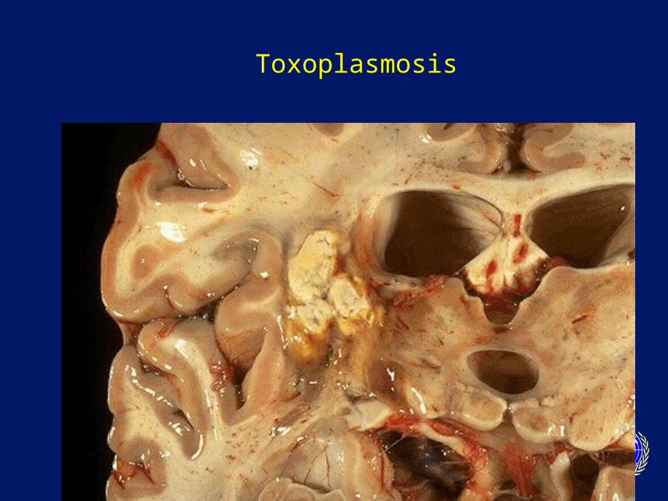
Toxoplasmosis
Organism: Toxoplasma gondii
Epidemiology:
– Cats the definitive hosts
– Ingestion of faecally contaminated material
– Ingestion of undercooked meat
CD4 count < 100
Toxoplasmosis
Clinical Features:
– encephalitis the most common manifestation (90%)• fever (70%), headaches (60%), focal neurological signs,
reduced consciousness (40%), seizures (30%)
• Constellation of fever, headache, and neurological deficit is classic
– chorio-retinitis
– pneumonitis
– disseminated disease
Toxoplasmosis
Diagnosis
– positive serology with typical syndrome
– suggestive CT/MRI scan:• multiple, bilateral cerebral lesions; hypodense with
ring enhancement
– Differential diagnosis
– CNS lymphoma, tuberculoma, fungal abscess, cryptococcosis, PML
Toxoplasmosis
Toxoplasmosis
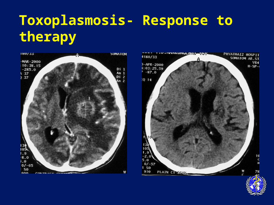
Toxoplasmosis- Response to therapy
Toxoplasmosis
Treatment
– Empirical therapy reasonable as trial, at least for 2 weeks
– Pyrimethamine plus folinic acid plus either sulfadiazine or clindamycin
– 6 weeks therapy at least, or until 3 weeks after complete scan resolution
– Corticosteroids for raised intracranial pressure
Toxoplasmosis
Secondary Prophylaxis
– Essential because latent (cyst) phase cannot be erdicated
– Pyrimethamine plus folinic acid plus sulfadiazine (or clindamycin)
– relapse occurs in 20-30% of patients despite maintenance therapy
– Improve immunity with HAART
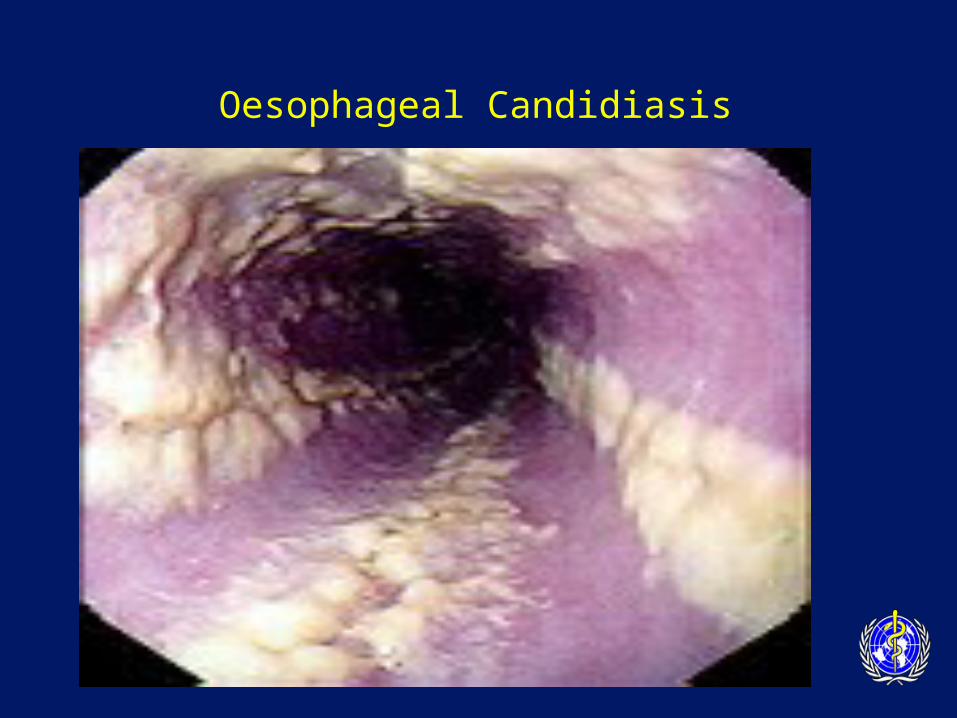
Oesophageal Candidiasis
Organism: Candida yeast
CD4 count < 200
Clinical symptoms
– dysphagia, retrosternal pain
– oral thrush in 50-90%
– endoscopy • ulceration • plaques
Oesophageal Candidiasis
Oesophogeal Candidiasis
Diagnosis
– oral thrush and dysphagia sufficient
– consider endoscopy if• symptoms without oral thrush
• failure of empirical antifungal therapy
– Treatment
– Fluconazole 200-400 mg /day until resolved
– Long term suppressive therapy if recurrent
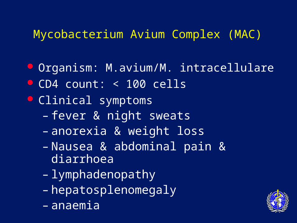
Mycobacterium Avium Complex (MAC)
Organism: M.avium/M. intracellulare CD4 count: < 100 cells Clinical symptoms
– fever & night sweats – anorexia & weight loss – Nausea & abdominal pain & diarrhoea– lymphadenopathy– hepatosplenomegaly– anaemia
MAC
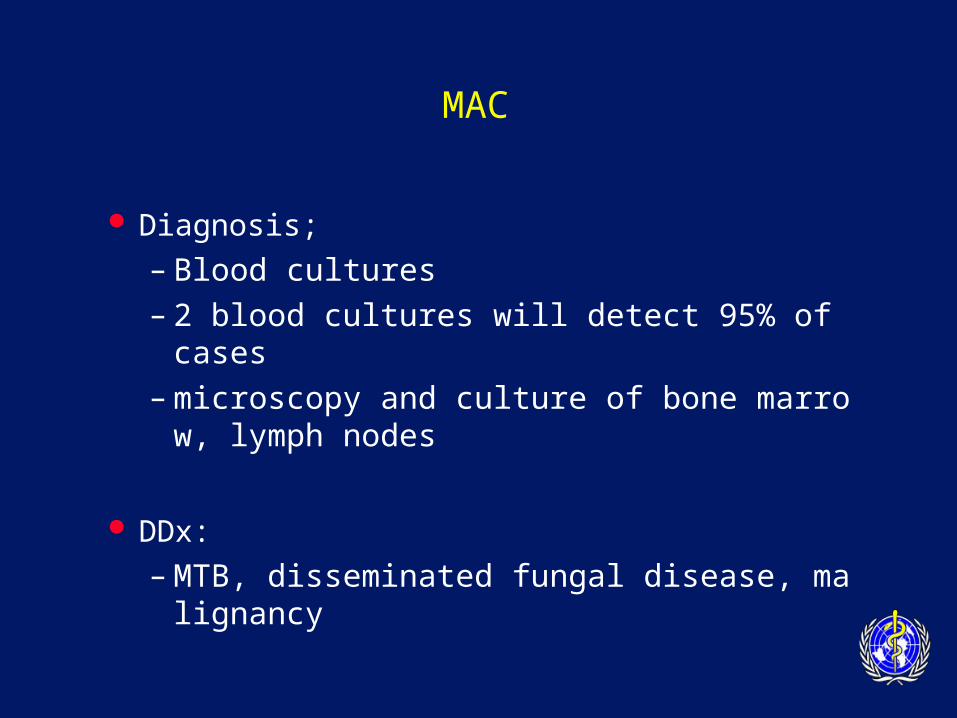
Diagnosis;
– Blood cultures
– 2 blood cultures will detect 95% of cases
– microscopy and culture of bone marrow, lymph nodes
DDx:
– MTB, disseminated fungal disease, malignancy
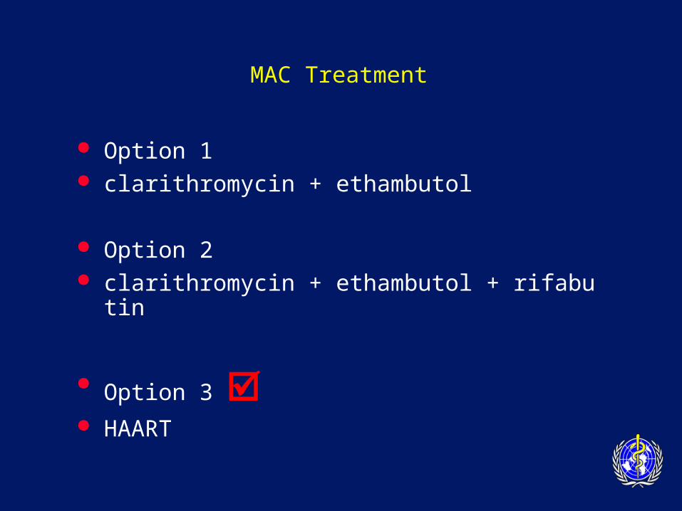
MAC Treatment
Option 1 clarithromycin + ethambutol
Option 2 clarithromycin + ethambutol + rifabutin
Option 3 HAART
MAC
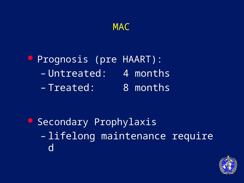
Prognosis (pre HAART):
– Untreated: 4 months
– Treated: 8 months
Secondary Prophylaxis
– lifelong maintenance required
CMV Disease
Epidemiology:
– a worldwide human herpes virus
– 3 periods of transmission• perinatal, chidhood, reproductive years
– in LDC’s, > 90% of children infected by 2 yo
CD4 < 50
emerging pathogen in SE Asia?
CMV Retinitis
Clinical:
– field defects
– floaters
– blurred vision
– rapid deterioration in vision
Diagnosis:
– typical fundoscopic appearance in a seropositive patient

CMV Retinitis
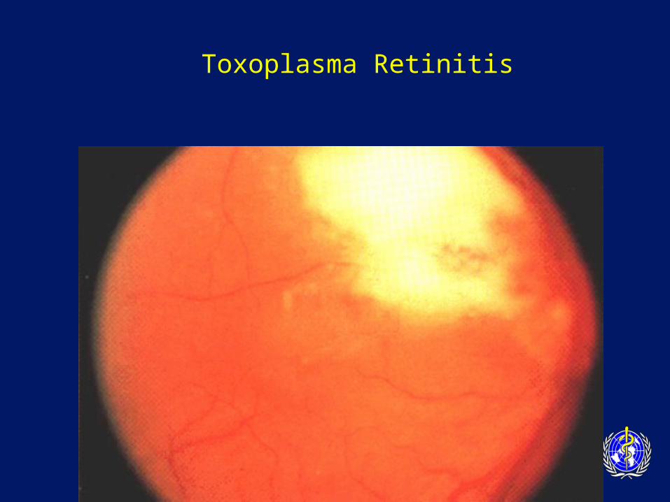
Toxoplasma Retinitis
Managing CMV retinitis
Treatment
– expensive and toxic
– maintenance therapy essential
– ganciclovir/foscarnet
– IVI or intra-vitreal
– HAART
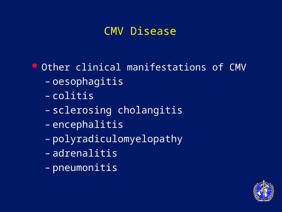
CMV Disease
Other clinical manifestations of CMV
– oesophagitis
– colitis
– sclerosing cholangitis
– encephalitis
– polyradiculomyelopathy
– adrenalitis
– pneumonitis
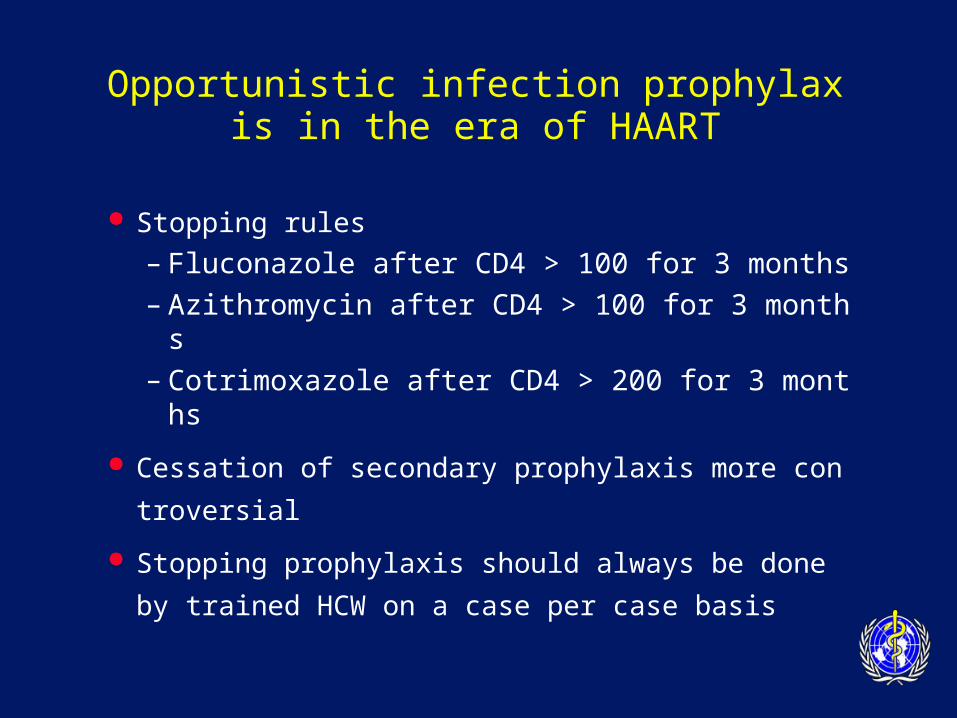
Opportunistic infection prophylaxis in the era of HAART
Stopping rules
– Fluconazole after CD4 > 100 for 3 months
– Azithromycin after CD4 > 100 for 3 months
– Cotrimoxazole after CD4 > 200 for 3 months
Cessation of secondary prophylaxis more contr
oversial
Stopping prophylaxis should always be done by
trained HCW on a case per case basis

Opportunistic Infections Key Points
Very uncommon in those on successful AR
V
Predictable according to CD4 count
Prevention better than cure
Secondary ‘maintenance’ therapy required
Educate patients