Independent Recruitment of an O-Methyltransferase forSyringyl Lignin Biosynthesis in Selaginella moellendorffii W
Jing-Ke Weng,a,1 Takuya Akiyama,b,2 John Ralph,c and Clint Chapplea,3
a Department of Biochemistry, Purdue University, West Lafayette, Indiana 47907b U.S. Dairy Forage Research Center, U.S. Department of Agriculture–Agricultural Research Service, Madison, Wisconsin 53706c Department of Biochemistry and Department of Energy Great Lakes Bioenergy Research Center, University of Wisconsin,
Madison, Wisconsin 53726
Syringyl lignin, an important component of the secondary cell wall, has traditionally been considered to be a hallmark of
angiosperms because ferns and gymnosperms in general lack lignin of this type. Interestingly, syringyl lignin was also
detected in Selaginella, a genus that represents an extant lineage of the most basal of the vascular plants, the lycophytes.
In angiosperms, syringyl lignin biosynthesis requires the activity of ferulate 5-hydroxylase (F5H), a cytochrome P450-
dependent monooxygenase, and caffeic acid/5-hydroxyferulic acid O-methyltransferase (COMT). Together, these two
enzymes divert metabolic flux from the biosynthesis of guaiacyl lignin, a lignin type common to all vascular plants, toward
syringyl lignin. Selaginella has independently evolved an alternative lignin biosynthetic pathway in which syringyl subunits
are directly derived from the precursors of p-hydroxyphenyl lignin, through the action of a dual specificity phenylpropanoid
meta-hydroxylase, Sm F5H. Here, we report the characterization of an O-methyltransferase from Selaginella moellendorffii,
COMT, the coding sequence of which is clustered together with F5H at the adjacent genomic locus. COMT is a bifunctional
phenylpropanoid O-methyltransferase that can methylate phenylpropanoid meta-hydroxyls at both the 3- and 5-position
and function in concert with F5H in syringyl lignin biosynthesis in S. moellendorffii. Phylogenetic analysis reveals that Sm
COMT, like F5H, evolved independently from its angiosperm counterparts.
INTRODUCTION
Paleobotanical and stratigraphic data suggest that the earliest
tracheophytes arose during the Late Silurian and Devonian
periods (;420 to ;360 million years ago) (Kenrick and Crane,
1997). This group of plants distinguished themselves from prim-
itive bryophytes by the development of vascular tissue that was
capable of transporting fluids throughout the plant body. It is
thought that the evolution of vasculature in plants involved the
recruitment of the plant phenylpropanoid metabolic pathway to
synthesize and deposit the heterogeneous aromatic polymer
lignin in the xylem cell wall (Boyce et al., 2004; Weng and
Chapple, 2010). Lignin physically reinforces the plant cell wall
and provides xylem cells with the strength to withstand the
tension generated during transpiration, and by stiffening the cell
walls of supportive tissues such as fibers, lignin provides plants
with the structural support to stand upright. The innovation of
lignified vascular tissue marked a significant step of early land
plants toward their ultimate adaptation to the terrestrial environ-
ment, which consequently facilitated their dominance of the
Earth’s flora during the Carboniferous period (;360 to ;300
million years ago) (Friedman and Cook, 2000).
Lignin found in angiosperms is generally comprised of three
major types of aromatic units, p-hydroxyphenyl (H), guaiacyl (G),
and syringyl (H) units, which derive, following polymerization,
from three p-hydroxycinnamyl alcohols: p-coumaryl alcohol,
coniferyl alcohol, and sinapyl alcohol, also known as the mono-
lignols (Boerjan et al., 2003). Studies on the monolignol biosyn-
thetic pathway over the past two decades have revealed that
lignin monomer synthesis requires, among other enzymes, a
suite of cytochrome P450-dependent monooxygenases (P450s)
and S-adenosyl-L-methionine (SAM)-dependent O-methyltrans-
ferases (OMTs) (Boerjan et al., 2003). Whereas three P450s,
cinnamic acid 4-hydroxylase (C4H), p-coumaroyl shikimic acid
39-hydroxylase, and ferulic acid 5-hydroxylase (F5H) catalyze the
aromatic hydroxylations para andmeta to the sidechain, caffeoyl-
CoA O-methyltransferase and caffeic acid O-methyltransferase
(COMT) subsequently methylate the freemeta-hydroxyls gener-
ated by C39H and F5H, resulting in a set of intermediates with
their ring hydroxylation/methoxylation status characteristic of H,
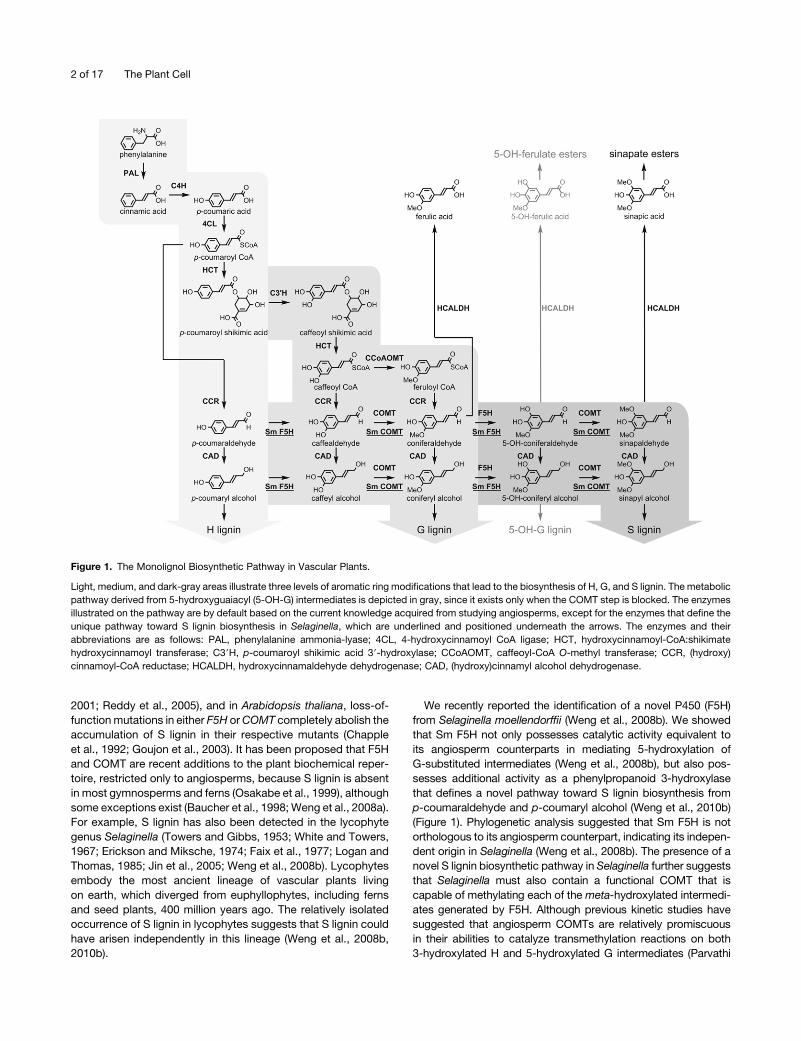
G, or S units in lignin (Figure 1).
Among the ring modification enzymes mentioned above, F5H
and COMT constitute a branch of the phenylpropanoid pathway
that is only required for the biosynthesis of S lignin. Down-
regulation of F5H or COMT in transgenic alfalfa (Medicago sativa)
has been shown to result in a reduction of S lignin (Guo et al.,
1 Current address: Howard Hughes Medical Institute, Jack H. SkirballCenter for Chemical Biology and Proteomics, Salk Institute for BiologicalStudies, La Jolla, CA 92037.2 Current address: Wood Chemistry Laboratory, Department of Bio-material Sciences, University of Tokyo, Bunkyo-ku, Tokyo 113-8657,Japan.3 Address correspondence to [email protected] author responsible for distribution of materials integral to thefindings presented in this article in accordance with the policy describedin the Instructions for Authors (www.plantcell.org) is: Clint Chapple([email protected]).WOnline version contains Web-only data.www.plantcell.org/cgi/doi/10.1105/tpc.110.081547
This article is a Plant Cell Advance Online Publication. The date of its first appearance online is the official date of publication. The article has been
edited and the authors have corrected proofs, but minor changes could be made before the final version is published. Posting this version online
reduces the time to publication by several weeks.
The Plant Cell Preview, www.aspb.org ã 2011 American Society of Plant Biologists. All rights reserved. 1 of 17
2001; Reddy et al., 2005), and in Arabidopsis thaliana, loss-of-
function mutations in either F5H orCOMT completely abolish the
accumulation of S lignin in their respective mutants (Chapple
et al., 1992; Goujon et al., 2003). It has been proposed that F5H
and COMT are recent additions to the plant biochemical reper-
toire, restricted only to angiosperms, because S lignin is absent
in most gymnosperms and ferns (Osakabe et al., 1999), although
some exceptions exist (Baucher et al., 1998; Weng et al., 2008a).
For example, S lignin has also been detected in the lycophyte
genus Selaginella (Towers and Gibbs, 1953; White and Towers,
1967; Erickson and Miksche, 1974; Faix et al., 1977; Logan and
Thomas, 1985; Jin et al., 2005; Weng et al., 2008b). Lycophytes
embody the most ancient lineage of vascular plants living
on earth, which diverged from euphyllophytes, including ferns
and seed plants, 400 million years ago. The relatively isolated
occurrence of S lignin in lycophytes suggests that S lignin could
have arisen independently in this lineage (Weng et al., 2008b,
2010b).
We recently reported the identification of a novel P450 (F5H)
from Selaginella moellendorffii (Weng et al., 2008b). We showed
that Sm F5H not only possesses catalytic activity equivalent to
its angiosperm counterparts in mediating 5-hydroxylation of
G-substituted intermediates (Weng et al., 2008b), but also pos-
sesses additional activity as a phenylpropanoid 3-hydroxylase
that defines a novel pathway toward S lignin biosynthesis from
p-coumaraldehyde and p-coumaryl alcohol (Weng et al., 2010b)
(Figure 1). Phylogenetic analysis suggested that Sm F5H is not
orthologous to its angiosperm counterpart, indicating its indepen-
dent origin in Selaginella (Weng et al., 2008b). The presence of a
novel S lignin biosynthetic pathway in Selaginella further suggests
that Selaginella must also contain a functional COMT that is
capable of methylating each of the meta-hydroxylated intermedi-
ates generated by F5H. Although previous kinetic studies have
suggested that angiosperm COMTs are relatively promiscuous
in their abilities to catalyze transmethylation reactions on both
3-hydroxylated H and 5-hydroxylated G intermediates (Parvathi
Figure 1. The Monolignol Biosynthetic Pathway in Vascular Plants.
Light, medium, and dark-gray areas illustrate three levels of aromatic ring modifications that lead to the biosynthesis of H, G, and S lignin. The metabolic
pathway derived from 5-hydroxyguaiacyl (5-OH-G) intermediates is depicted in gray, since it exists only when the COMT step is blocked. The enzymes
illustrated on the pathway are by default based on the current knowledge acquired from studying angiosperms, except for the enzymes that define the
unique pathway toward S lignin biosynthesis in Selaginella, which are underlined and positioned underneath the arrows. The enzymes and their
abbreviations are as follows: PAL, phenylalanine ammonia-lyase; 4CL, 4-hydroxycinnamoyl CoA ligase; HCT, hydroxycinnamoyl-CoA:shikimate
hydroxycinnamoyl transferase; C39H, p-coumaroyl shikimic acid 39-hydroxylase; CCoAOMT, caffeoyl-CoA O-methyl transferase; CCR, (hydroxy)
cinnamoyl-CoA reductase; HCALDH, hydroxycinnamaldehyde dehydrogenase; CAD, (hydroxy)cinnamyl alcohol dehydrogenase.
2 of 17 The Plant Cell

et al., 2001; Zubieta et al., 2002), data fromolder literature showed
thatOMTextracted fromgymnospermspeciesgenerally have little
activity toward 5-hydroxyferulic acid compared with caffeic acid,
suggesting that the specific function of COMT for S lignin biosyn-
thesis is probably not fundamental to all tracheophytesbut rather a
newly evolved feature in angiosperms (Kuroda, 1983).
Here, we report the characterization of a series of OMTs from
S. moellendorffii. We provide both in vivo and in vitro evidence
to show that Selaginella contains a functional COMT that is
capable of mediating specific methylation reactions on meta-
hydroxylated lignin biosynthetic intermediates. Similarly to the
case of Sm F5H, the COMT in Selaginella also appears to have
evolved independently of its angiosperm counterparts. Taken
together, our data suggest that independent occurrences of S
lignin in phylogenetically divergent angiosperm and lycophyte
lineages are due to independent recruitment of biosynthetic
enzymes and the pathways defined by them.
RESULTS
Cell Wall Structure in the Stem of S. moellendorffii
Selaginella possesses a protostele with xylem cells surrounded
by a cortical cylinder (Figures 2A and 2D), an anatomy distinct
from the eustele typically found in angiosperms. Using theMaule
histochemical staining method, we previously observed that S
lignin is predominantly deposited in the cortex, rather than the
xylem, in Selaginella (Weng et al., 2008b). To gain more insight
into the cell wall secondary thickening and lignification process in
these two different tissue types in S. moellendorffii stem, we first
investigated the stem cell wall structure by scanning electron
microscopy. Sections from both young shoot (;1 cm from the
apex) and mature stem (;1 cm from the base) were prepared
and examined. In the xylem of both young and mature stem,
secondary cell walls with scalariform thickenings were observed
in most of the vessel element cells (Figures 2B and 2E). Although
many xylem vessel elements from the young shoot were found to
contain living contents and were probably not yet mature (Figure
2B), all xylem vessel elements in the mature stem were fully
differentiated and had lost their cell contents (Figure 2E). Most of
the cortical cells in the young shoot were living cells with obvious
cell contents (Figure 2C). The presence of a thick secondary wall
could be observed in cortical cells (Figure 2C), reminiscent of the
sclerified interfascicular fiber cells found in angiosperms. Like
the xylem vessel elements, the cortical cells also die at maturity
(Figure 2F).
Tissue-Specific Lignin Analysis in S. moellendorffii Stem
It has been shown previously using histochemical staining
methods and derivatization followed by reductive cleavage
(DFRC) lignin analysis that the Selaginella stem cortex contains
lignin derived from H, G, and S units, while xylem contains G
lignin with only a trace of S lignin (Weng et al., 2008b, 2010b). To
further delineate the composition of the whole lignin material and
begin to obtain some structural insights regarding the interunit
linkage distributions of the lignins in the two different tissue types
in Selaginella stem, we took advantage of the unique protostelic
structure of Selaginella stem and separated, with the aid of
microscopy examination, enough xylem and cortical tissue for
NMR analysis. Changes in the S:G:H distribution in the lignins are
most readily visualized from the aromatic region of NMR spectra,
particularly the two-dimensional 13C–1H correlation (HSQC)
spectra correlating protons with their attached carbons (Figure
3A). The Selaginellawhole stem lignin is S-rich (S:G:H = 76:22:2).
Unlike in angiosperms, however, there is also a substantial H
component; this is also seen in species such as kenaf (Hibiscus
cannabinus; Kim et al., 2008). There are also some unrecogniz-
able components in the aromatic region of the spectrum, com-
ponents that appear to be removed by more exhaustive solvent
Figure 2. Cell Wall Structure of S. moellendorffii Examined by Scanning Electron Microscopy.
Global view of cross sections of Selaginella young (A) and old (D) stems showing a protostelic arrangement with xylem surrounded by cortex. Higher
magnification photographs illustrate xylem ([B] and [E]) and cortical cells ([C] and [F]) from young ([B] and [C]) and old ([E] and [F]) regions of the stem.
Bar = 100 mm in (A) and (D) and 10 mm in (B), (C), (E), and (F).
Independent Occurrence of COMT 3 of 17
extraction during cell wall isolation. The p-hydroxybenzoates
found to acylate some lignins, including those of poplar (Populus
sp), are not present. The most striking observations come from
examination of spectra from the separated xylem-enriched
material versus the residual cortex. Xylem contains essentially
pure G lignin, with only;5% S units in this isolated fraction. The
cortex spectrummore closely resembles thewholematerial, with
a high syringyl level and similar S:G ratio. The H-level was not
measured due to overlap with other contours but again appears
to be associated with only the cortex fraction.
The sidechain region in the NMR spectra peripherally reflects
the changes in the S:G:H distribution and is rich in detail
Figure 3. Tissue-Specific Lignin Analysis in S. moellendorffii.
(A) Aromatic regions of two-dimensional HSQC NMR spectra of acetylated cellulolytic enzyme lignins revealing compositional aspects. An isolated
Bjorkman lignin from poplar was analyzed in parallel for comparison because it has a similar S:G level as that of the Selaginella lignin samples but has
p-hydroxybenzoates acylating the g-positions of some lignin sidechains. S:G:H levels are from uncorrected volume integrals of the analogous S2/6,
G2, and H2/6 correlations (with the G2 integral being logically doubled); PB levels are uncorrected integrals and are expressed as a percentage of the
total S+G+H level. WT, wild type.
(B) Sidechain regions of two-dimensional HSQC NMR spectra of acetylated cellulolytic enzyme lignins reflecting the resulting structural differences (i.e.,
the distribution of bonding patterns between the units that result from radical coupling reactions from the differing monomer supplies). Some effects are
pronounced; for example, 5-coupling can only occur in H or G units; consequently, units B and D are more pronounced in the G-rich xylem and are
relatively minor in the S-rich cortex. Resinols (C) are generally more prevalent in S-rich lignins; their virtual absence in the cortex lignin is most unusual
(see Supplemental Figure 1 online). The relatively newly discovered spirodienones S (Ralph et al., 2006; Zhang et al., 2006) are typically more prevalent
in S-rich lignins; they are readily seen in the S-rich cortex lignin.
4 of 17 The Plant Cell

regarding the types and distribution of interunit bonding patterns
present in the lignin fraction (Figure 3B; see Supplemental Figure
1 online). The xylem lignin spectrum is typical of a G-rich lignin
with residual polysaccharides (Ralph et al., 1999). It contains
evidence for themajor b-ether unitsA, resinolsC, and, due to the
availability of the 5-position for radical coupling in guaiacyl units,
phenylcoumaran B and dibenzodioxocin D units. The cortex
spectrum, as is typical for S-rich lignins, is particularly rich in
b-ether unitsA, has only low levels of 5-linked (B andD) units, and
readily shows the spirodienone units S (from b-1-coupling reac-
tions) that have only recently been authenticated and are most
prevalently associated with S-rich lignins (Zhang et al., 2006).
Identification of S. moellendorffii COMT Candidate Genes
AngiospermCOMTs belong to the SAM-dependentmethyltrans-
ferase superfamily, which all use SAMas themethyl group donor,
yielding S-adenosyl-L-homocysteine and the methylated deriv-
ative of the substrate as products (Zubieta et al., 2001). We
hypothesized that the COMT fromSelaginellamay also belong to
this family and could be identified based on sequence similarity.
We performed a BLAST search using Arabidopsis COMT (At
COMT) as the probe against the S. moellendorffii genome. Four
putative Selaginella COMT candidate genes were identified,
which showed amino acid sequence identity with At COMT
ranging from 37 to 51% (Table 1). To our surprise, oneSelaginella
COMT candidate (51% identical to At COMT) was found directly
adjacent to Sm F5H in the genome. Its expression is driven in the
opposite direction from a promoter within the same intergenic
region (Figure 4). We cloned all four Selaginella COMT candidate
genes for further functional analysis.
Complementation of the Arabidopsis COMT-Deficient
Mutant by Sm COMT
To test the function of Selaginella COMT candidate genes in
planta, we transformed them into an Arabidopsis COMT-deficient
mutant, omt1-2, under the control of the Arabidopsis C4H pro-
moter and examined the plants for phenotypic complementation.
Arabidopsis omt1-2 completely lacks S units in its lignin and
accumulates 5-hydroxyferulate esters, which substitute for a large
proportion of the sinapate esters normally found in rosette leaves
(Figures 1 and 4) (Weng et al., 2010a). These observations are
consistent with previous reports of another knockout allele of
Arabidopsis omt1 (Goujon et al., 2003).
The leafmethanolic extracts frommultiple T1 transformants for
each Selaginella COMT candidate construct were analyzed by
HPLC. Three of the candidates failed to complement the leaf
hydroxycinnamate ester phenotype of omt1-2 (see Supplemen-
tal Figure 2 online), but the Selaginella COMT candidate that is
adjacent to Sm F5H almost entirely alleviated the accumulation
of 5-hydroxyferulate esters and restored normal sinapoyl malate
and sinapoyl glucose accumulation in omt1-2 leaves (Figure 5A).
More rigorous analysis at the T2 generation further confirmed
that the leaf sinapoyl malate content was brought back to wild-
type levels in all the independent transgenic lines examined
(Figure 5B). This datum suggests that this Selaginella COMT
candidate can take the place of At COMT in sinapate ester
biosynthesis in vivo and was thus designated as Sm COMT.
To test whether Sm COMT can complement the lignin pheno-
type of omt1-2, we first examined the lignin composition of
omt1-2/At C4H:Sm COMT transgenic plants using the Maule
histochemical staining reagent. Whereas omt1-2 stem sections
stain yellow in both the sclerenchyma and the xylem, a reaction
that indicates the absence of S lignin, omt1-2/AtC4H:SmCOMT
transgenic plants exhibit a staining pattern similar to thewild type
with red staining localized in the sclerified parenchyma cells,
suggesting S lignin accumulation in these cells (Figure 5C). To
definitively determine whether the positive Maule staining ob-
served in the Sm COMT transgenic plants is due to the presence
of S lignin units, we analyzed the cell wall samples from these
plants by DFRC lignin analysis, a method specific for b-O-4–
linked lignin units (Lu andRalph, 1998). The results showed that S
lignin biosynthesis was restored in the transgenic plants (Figure
5D); all the independent transgenic lines exhibited a lignin Smole
percentage similar to that of the wild type (Table 2).
The unique structure of the Selaginella genome locus contain-
ing Sm COMT and Sm F5H suggests that the expression of
these two genes may be coregulated by common cis-regulatory
elements located in the same promoter region. To test the hy-
pothesis that these cis-regulatory elements related to S lignin
biosynthesis in Selaginella could be conserved in all vascular
plants and are recognizable by the lignification-associated tran-
scription factors in angiosperms, we transformed a Selaginella
genomic fragment harboring Sm COMT, Sm F5H, and their
39-downstream regions into the Arabidopsis double mutant of
omt1-2 and the F5H-null fah1-2 and looked for complementa-
tion. However, none of the T1 transgenic plants exhibited the
complemented lignin phenotype (see Supplemental Figure 3
online), suggesting that the cis-regulatory elements in the pro-
moter region of Sm COMT and Sm F5H are probably not
conserved between Selaginella and Arabidopsis.
Table 1. Amino Acid Sequence Percentage Identity among At COMT
and Selaginella COMT Candidates
Sm
COMT
Sm
COMT-Like1
Sm
COMT-Like2
Sm
COMT-Like3
At COMT 51 40 43 37
Sm COMT 40 55 36
Sm COMT-like1 39 29
Sm COMT-like2 36
Figure 4. The Arrangement of COMT and F5H in the S. moellendorffii
Genome.
Sm COMT and Sm F5H are clustered and oriented in an opposite
direction from each other.
Independent Occurrence of COMT 5 of 17

Figure 5. Complementation of the Arabidopsis COMT-Null Mutant by Sm COMT.
(A) HPLC profiles of 3-week-old Arabidopsis leaf extracts. 5-OH-FG, 5-hydroxyferuloyl glucose; SG, sinapoyl glucose; 5-OH-FM, 5-hydroxyferuloyl
malate; SM, sinapoyl malate.
(B) Restoration of leaf sinapoyl malate production in four independent lines of omt1-2/At C4H:Sm COMT transgenic plants quantified by HPLC. Error
bars represent 1 SD of triplicate samples. WT, wild type.
(C) Maule staining of 6-week-old Arabidopsis inflorescence stem sections. Bars = 200 mm.
(D) Gas chromatograms of the DFRC lignin analysis monomer products from cell wall samples prepared from 3-month-old Arabidopsis inflorescence
stems. G/S, guaiacyl/syringyl lignin derivative; c/t, cis/trans; IS, internal standard.
6 of 17 The Plant Cell

Enzyme Kinetic Analysis of Sm COMT
To assess the substrate specificity of Sm COMT, we expressed
N-terminal hexahistidine-tagged Sm COMT in Escherichia coli,
purified the tagged protein using nickel affinity chromatography,
and performed kinetic assays using a range ofmeta-hydroxylated
phenylpropanoid pathway intermediates, including caffeic acid,
caffealdehyde, caffeyl alcohol, 5-hydroxyferulic acid, 5-hydroxy-
coniferaldehyde, and 5-hydroxyconiferyl alcohol. At COMT was
also expressed, purified, and assayed in parallel for comparison.
The kinetic constants were inferred from these assays and sum-
marized in Table 3 (see Supplemental Figures 4 and 5 online.
At COMT shows kinetic properties comparable to those pre-
viously reported for M. sativa COMT (Ms COMT) (Parvathi et al.,
2001). Caffealdehyde and 5-hydroxyconiferaldehyde are the
preferred substrates for At COMT, with their Km values at the
lowest among all the tested substrates. Caffeyl alcohol and
5-hydroxyconiferyl alcohol are less preferable substrates, with
their Vmax/Km values slightly lower than those for the aldehydes.
Caffeic acid and 5-hydroxyferulic acid are poor substrates for At
COMT, with their Vmax/Km values at hundreds of times lower than
those of their corresponding aldehydes and alcohols.
As they are for At COMT, caffeic acid and 5-hydroxyferulic acid
are poor substrates for Sm COMT, but with Vmax/Km values even
lower than those values for At COMT. At the aldehyde and
alcohol level, Sm COMT shows kinetic constants in a range
generally comparable to those of At COMT, although Sm COMT
exhibits higher catalytic efficiency with alcohols as substrates,
exhibiting both lower Km values and higher Vmax values. These
results indicate that Sm COMT has a catalytic capacity compa-
rable to those of its angiosperm counterparts in mediating
methylation reactions on 3-hydroxylated or 5-hydroxylated lignin
biosynthetic precursors at the aldehyde and alcohol levels.
Interestingly, we also observed that for caffeyl alcohol and
5-hydroxyconiferyl alcohol assays, Sm COMT may exhibit sub-
strate inhibition toward the two alcohols, a phenomenon thatwas
not seen with other substrates or when At COMT was assayed
with any substrate (see Supplemental Figures 4 and 5 online).
To test whether Sm COMT can also methylate hydroxycinna-
moyl CoA esters, we performed enzyme assays using 5-hydroxy-
feruloyl CoA, a potential methyl acceptor, with a concentration
series ranging from 100 to 1 mM. However, no methyltransferase
activity could be detected in these assays, suggesting that
hydroxycinnamoyl CoA esters are unlikely to be relevant sub-
strates for Sm COMT in vivo.
To further characterize COMT in Selaginella, we extracted total
soluble protein from S. moellendorffii whole-plant tissue and
assayed its activity toward a range of potential COMT substrates.
Unfortunately, these compounds were degraded by an unknown
enzymatic activity present in the crude extract, possibly polyphe-
nol oxidases or diooxygenases, which are known to metabolize
catechol-substituted phenolic substrates (Prescott and John,
1996;Marusek et al., 2006). To solve this problem,we fractionated
the Selaginella crude protein extract by size exclusion followed by
ion exchange chromatography. This procedure yielded a fraction
in which the COMT activity was free from the confounding
activities described above, facilitating detailed kinetic analysis.
The kinetic constants measured using this Selaginella enzyme
fraction against caffeic acid, caffealdehyde, caffeyl alcohol,
5-hydroxyferulic acid, 5-hydroxyconiferaldehyde, and 5-hydroxy-
coniferyl alcohol are comparable to the values determined using
recombinant Sm COMT (see Supplemental Table 1 online), con-
sistent with the hypothesis that the protein we have characterized
in recombinant form is the dominant COMT in Selaginella.
Tissue-Specific Expression of Sm COMT
To evaluate the tissue specificity of Sm COMT expression in
Selaginella, we first conducted a quantitative RT-PCR (qRT-PCR)
experiment using RNA extracted from various tissues, including
microphyll, strobilus, stem, rhizome, root, and bulbil. Across the
six tissue types, Sm COMT is expressed at the highest level in
the stem tissue, consistent with its role in lignin biosynthesis (Fig-
ure 6A). We also examined the mRNA abundance of Sm F5H
in parallel, which revealed a similar tissue transcript distribution
pattern to that of Sm COMT (Figure 6A), consistent with the
hypothesis that these two genes are coregulated.
To further investigate the expression pattern of Sm COMT
in stem, we preformed in situ hybridization experiments on
Table 2. S Lignin Mole Percentage of Columbia Wild Type, omt1-2,
and omt1-2/At C4H:Sm COMT T2 Transgenic Plants Quantified by
DFRC Lignin Analysis
Genotype Lignin Mol % S
Wild type 24 6 6
omt1-2 N.D.
omt1-2/At C4H:Sm COMT (1) 16 6 5
omt1-2/At C4H:Sm COMT (2) 20 6 5
omt1-2/At C4H:Sm COMT (3) 22 6 4
omt1-2/At C4H:Sm COMT (4) 24 6 5
N.D., not detectable; 6 represents 1 SD for biological triplicates.
Table 3. Kinetic Properties of Recombinant Sm COMT and At COMT toward meta-Hydroxylated Phenylpropanoid Intermediates
At COMT Sm COMT
Substrate Km (mM) Vmax (nkat·mg�1) Vmax/Km Km (mM) Vmax (nkat·mg�1) Vmax/Km
Caffeic acid 8.05 3 102 16.3 2.02 3 10�2 5.55 3 103 0.422 7.60 3 10�5
Caffealdehyde 0.914 4.88 5.34 12.35 1.68 0.136
Caffeyl alcohol 7.70 11.5 1.49 1.17 4.45 3.80
5-OH-ferulic acid 2.25 3 102 14.5 6.44 3 10�2 4.24 3 103 0.951 2.24 3 10�4
5-OH-coniferaldehyde 2.74 23.0 8.39 6.97 6.21 0.891
5-OH-coniferyl alcohol 4.91 21.4 4.36 0.784 4.80 6.12
Independent Occurrence of COMT 7 of 17

Selaginella stem cross sections. While the antisense probe gave
hybridization signals in the cortical cells where S lignin is pre-
dominantly deposited (Figure 6B), the sense control probe did
not give any hybridization signal (Figure 6C). Notably, the hy-
bridization signal was also observed in the phloem cells sur-
rounding the xylem. The transcript localization pattern of Sm
COMT in stem observed in this study is almost identical to that of
Sm F5H as previously reported (Weng et al., 2008b), which
provides further evidence to support the contention that these
two genes may be coregulated.
Phylogenetic Analysis of Sm COMT
To infer the phylogeny of Sm COMT within the plant OMT family,
we performed Bayesian phylogenetic analysis using Sm COMT,
angiospermCOMTs, angiospermOMTswith known functions that
can methylate various types of phenylpropanoids, and some
OMTs from different land plant lineages with unknown functions
(Figure 7; see Supplemental Data Set 1 online). Although all the
angiospermCOMTs are clustered together into a clade, sister to a
group containing chalcone OMT from alfalfa and catechol OMT
from tobacco (Nicotiana tabacum), Sm COMT falls into another
clade only distantly related to the angiosperm COMT clade. The
OMTs related toSmCOMT in this clade include flavone/isoflavone
7-OMTs from barley (Hordeum vulgare) and alfalfa, eugenol OMT
from sweet basil (Ocimum basilicum), two unknown OMTs from
Sitka spruce (Picea sitchensis) and Arabidopsis, as well as Sm
COMT-like2 and Sm COMT-like3. An unknown OMT from Phys-
comitrella patens, being the only COMT homolog found in its
genome, clustered together with Sm COMT-like1, which could
represent the ancestral form of this enzyme family that is highly
diversified in higher plants. The observations suggest that COMT
may have been recruited for S lignin biosynthesis independently in
angiosperms and Selaginella.
Sequence Analysis and Modeling of Sm COMT
To gain more insight into the evolution and structure-function
relationships within the COMT family, we compared Sm COMT
with other related proteins in a multiple sequence alignment
(Figure 8). This analysis showed that the three catalytic residues,
His-269, Glu-297, andGlu-329, previously identified inMsCOMT
were highly conserved in SmCOMT (Zubieta et al., 2002), except
that the E297 in Ms COMT was substituted with an Asp residue.
The residues for SAMbinding are also highly conserved between
Sm COMT and other OMTs. Whereas angiosperm COMT ortho-
logs are almost entirely conserved in the residues that constitute
the methyl acceptor binding pocket, Clarkia breweri IEOMT, a
derived OMT paralogous to COMT, shows many nonconserva-
tive substitutions at these residues, consistent with its distinct
activity as a phenylpropanoid para-hydroxyl O-methyltransfer-
ase (Wang and Pichersky, 1998). Although Sm COMT is catalyt-
ically equivalent to angiosperm COMTs, major nonconservative
substitutions (Leu-136, Ala-162, His-166, and Phe-172 in Ms
COMT substituted to Phe-126, Glu-152, Gly-156, and Val-162 in
Sm COMT) were found in the corresponding substrate binding
residues in Sm COMT, which further supports the notion that the
Figure 6. Expression Pattern of COMT in S. moellendorffii.
(A) qRT-PCR analysis of transcript abundance of Sm COMT and Sm F5H
in various tissue types. Error bars represent 1 SD of biological triplicates.
(B) and (C) In situ hybridization of Sm COMT mRNAs in Selaginella
transverse sections using antisense (B) and sense (C) Sm COMT probes.
Bars = 200 mm.
8 of 17 The Plant Cell

substrate specificity of Sm COMT arose independently from that
of angiosperm COMTs.
We then generated a SmCOMT structural model based on the
Ms COMT crystal structure (Zubieta et al., 2002) and compared
the two structures in a spatial manner (Figure 9). The Sm COMT
model shares a similar tertiary structurewithMsCOMT, including
the N-terminal domain responsible for dimerization in Ms COMT
(Figure 9A), suggesting that SmCOMT is likely to exist as dimer in
solution like other previously reported plant OMTs (Gang et al.,
2002; Zubieta et al., 2002). The Sm COMT model contains a
methyl acceptor binding pocket similar to that of Ms COMT in
overall geometry (Figure 9B). Interestingly, the four nonconser-
vative substitutions in the SmCOMTactive sitementioned above
form two pairs of spatially contacting residues (F126-V162 and
E152-G156), each of which exchange two residues of similar
character in reverse orientation, likely resulting in similar spatial
filling (Figure 9B). This observation further suggests that, at the
molecular level, angiosperms and Selaginella have adopted
independent evolutionary solutions toward convergent COMT
enzymatic activity geared for lignin biosynthesis.
Characterization of Mutated At COMT and Sm COMT
From the modeling study, we noticed that the position of a pair
of residues, His-166 and Ala-162, in Ms COMT in the vicinity of
the para-hydroxyl of the phenylpropanoid substrate, may be
Figure 7. Bayesian Phylogenetic Tree of Sm COMT, Angiosperm COMTs, and Related OMTs from Various Plant Lineages.
Bayesian posterior probabilities are indicated at each node. The scale measures evolutionary distance in substitutions per amino acid. The taxonomy
information of the sequences is indicated by the symbols at the right of the gene names (filled square, dicot; open square, monocot; filled triangle,
gymnosperm; filled circle, lycophyte; open triangle, bryophyte). CAOMT, catechol O-methyltransferase; CHOMT, chalcone O-methyltransferase;
EOMT, eugenol O-methyltransferase; FOMT, flavonoid 7-O-methyltransferase; IEOMT, (iso)eugenol O-methyltransferase; IOMT, isoflavone
O-methyltransferase.
Independent Occurrence of COMT 9 of 17

occupied byGlu-152 andGly-156 in SmCOMT (Figure 9B). It has
been previously suggested that His-166 in Ms COMT could
hydrogen bond with the para-hydroxyl moiety of the substrate,
contributing to the proper substrate positioning (Zubieta et al.,
2002). However, the replacement of this residue with a Glu
residue in Sm COMT raises the possibility that His-166 in Ms
COMT and/or Glu-152 in SmCOMTmay serve as a general base
that could deprotonate the substrate para-hydroxyl group to
further facilitate the transfer of a methyl group to the meta-
hydroxyl position. To test whether these two residues are
essential for catalysis, we first generated At COMT-H164A
(corresponding to His-166 in Ms COMT), Sm COMT-E152A
andSmCOMT-E152Qmutants using site-directedmutagenesis,
expressed the mutant proteins in E. coli, and analyzed their
kinetic properties against caffeyl alcohol. We found that all three
mutants retained OMT catalytic activity, with the catalytic effi-
ciency of At COMT-H164A and Sm COMT-E152Q slightly com-
promised (Table 4). These results disproved our initial hypothesis
and indicated that His-164 in At COMT andGlu-152 in SmCOMT
are dispensable for catalysis.
The sequence alignment and modeling study suggested four
active site residues differ between an angiospermCOMTandSm
COMT. To test whether these residues are interchangeable
between an angiosperm COMT and Sm COMT, we generated
Figure 8. Sequence Alignment Analysis of Sm COMT, Together with Two Angiosperm COMTs, and Related OMTs.
Based on the crystal structure of Ms COMT, the residues involved in different aspects of the enzymatic process are highlighted in color. Yellow, catalytic
residues; pink, SAM binding residues; blue, substrate binding residues at the active site. Asterisks are placed on top of every tenth site in the alignment.
At, Arabidopsis thaliana; Cb, Clarkia breweri; Ms, Medicago sativa; Sb, Sorghum bicolor; Sm, Selaginella moellendorffii; Pa, Picea abies; Pp,
Physcomitrella patens.
10 of 17 The Plant Cell

mutated versions of At COMT carrying A160E/H164G and
L134F/F170V substitutions, such that the mutated residues
correspond to those in SmCOMT. Similarly reciprocally mutated
versions of Sm COMT, Sm COMT-E152A/G156H and Sm
COMT-F126L/V162F, were also generated. The kinetic analysis
of these mutants toward caffeyl alcohol revealed that the Sm
COMT mutants substituted with the At COMT residues are
almost unaffected in kinetic constants. On the contrary, replac-
ing At COMT with the corresponding residues from Sm COMT
drastically reduced the catalytic efficiency of the enzyme, which
could be due to the destabilizing effect of these substitutions in
the At COMT sequence context (Table 4).
DISCUSSION
S Lignin Biosynthesis in Angiosperms and Selaginella
Evolved through Convergent Evolution
Our understanding of the lignin biosynthetic pathway has been
primarily based on decades of research in economically impor-
tant crop species and trees, as well as the model plant Arabi-
dopsis, all of which are flowering plants (Boerjan et al., 2003; Li
et al., 2008). The identification of a bifunctional COMT from
Selaginella described in this study, in addition to the previous
discovery of the dual meta-hydroxylase F5H from the same
species (Weng et al., 2008b, 2010b), indicate thatSelaginella has
adopted a biochemical pathway distinct from that in angio-
sperms to synthesize sinapyl alcohol. In contrast with angio-
sperms, where S lignin biosynthesis is entirely dependent on the
availability of G-substituted intermediates, coniferaldehyde and
coniferyl alcohol, Selaginella appears to direct H-substituted
intermediates, p-coumaraldehyde and p-coumaryl alcohol, to-
ward S lignin biosynthesis via the bifunctional F5H and COMT
(Figure 1).
Enzymes that catalyze the same reaction in different orga-
nisms are usually encoded by orthologous genes; however, there
are cases where the converged enzymatic activities were de-
rived independently (Galperin et al., 1998). For example, fructose
1,6-bisphosphate aldolase in yeast (Saccharomyces cerevisiae)
and rabbit (Oryctolagus cuniculus) muscle are capable of cata-
lyzing the same reaction but are only distantly related (Warburg
and Christian, 1943; Cooper et al., 1996). In plant secondary
metabolism, limonene synthase was found to have arisen inde-
pendently in angiosperms and gymnosperms (Bohlmann et al.,
1998), and of particular relevance to this study, eugenol OMT
evolved independently inC. breweri and sweet basil (Gang et al.,
2002) (Figure 7). Our phylogenetic analysis suggests that Sm
COMT belongs to a plant OMT clade divergent from the clade
that contains angiosperm COMTs (Figure 7). The fact that many
nonconservative substitutions exist in the substrate binding site
Figure 9. Molecular Modeling of Sm COMT.
(A) Tertiary structure of the Sm COMT model (red) superimposed on Ms COMT (blue). The ligands of the template Ms COMT structure, S-adenosyl-L-
homocysteine and 5-OH coniferaldehyde, are shown as sticks (green, carbon; red, oxygen; blue, nitrogen; yellow, sulfur).
(B) Putative SmCOMT active site (red) overlaid with Ms COMT active site. Nonconservative differences in those potential substrate binding residues are
emphasized in text.
Table 4. Kinetic Analysis of Wild-Type and Various Mutant Forms of
Sm COMT and At COMT toward Caffeyl Alcohol
Protein Km (mM) Vmax (nkat·mg�1) Vmax/Km
At COMT wild type 7.70 11.5 1.49
At COMT-H164A 15.4 6.71 0.436
At COMT-A160E/H164G 23.0 0.493 2.14 3 10�2
At COMT-L134F/F170V 27.2 1.44 5.29 3 10�2
Sm COMT wild type 1.17 4.45 3.80
Sm COMT-E152A 1.42 7.10 5.00
Sm COMT-E152Q 2.49 3.30 1.33
Sm COMT-E152A/G156H 1.49 3.77 2.53
Sm COMT-F126L/V162F 1.09 5.97 5.48
Independent Occurrence of COMT 11 of 17

of SmCOMT compared with its angiosperm counterparts further
supports the independent origins of lignin biosynthetic COMT
activities in Selaginella and angiosperms. Unfortunately, gene
knockout technology is not currently available in Selaginella, so it
is not possible to conduct the loss-of-function experiment to
determinewhether anyOMTs in addition to SmCOMT function in
S lignin synthesis in Selaginella. Specifically, we cannot exclude
the possibility that other OMTs, widely diverged from angio-
sperm COMTs, escaped notice during our bioinformatic analysis
of the Selaginella genome. Nevertheless, Sm COMT, but not the
other threeSelaginellaOMT homologs, is a functional COMT and
is a good candidate for the one that functions in S lignin
biosynthesis in Selaginella.
Interestingly, the in vitro enzyme assays revealed that Sm
COMT is not entirely identical to its angiosperm counterparts in
terms of kinetic properties. Whereas we and others have shown
that angiosperm COMTs exhibit higher catalytic efficiency to-
ward meta-hydroxylated phenylpropanoid aldehydes than alco-
hols (Parvathi et al., 2001; Zubieta et al., 2002), alcohols are
superior substrates for Sm COMT, which could be related to the
different roles these enzymes have in S lignin biosynthesis,
the angiosperm enzymes acting only once in the pathway, and
the Selaginella enzyme acting twice. Furthermore, Sm COMT
may exhibit substrate inhibition toward caffeyl alcohol and
5-hydroxyconiferyl alcohol, a property not observed in At
COMT or in the previously characterized Ms COMT (Parvathi
et al., 2001); however, the importance of this phenomenon has
yet to be investigated.
The Genes for S Lignin Biosynthesis in S. moellendorffii
Are Clustered
The clustering of genes, frequently in operons, is a common
feature of prokaryotic genomes (Salgado et al., 2000). By contrast,
gene clustering that can produce polycistronic mRNA is generally
absent in the genomes of higher eukaryotes (Blumenthal, 1998).
There are occasional cases where functionally related genes are
arranged closely at a certain genomic locus in higher eukaryotes
and are transcriptionally coregulated. For example, the Arabidop-
sis genome contains an operon-like gene cluster composed of
four metabolic genes that are involved in triterpene thalianol
biosynthesis (Field and Osbourn, 2008). In maize (Zea mays), five
genes required for the biosynthesis of the phytoalexin 2,4-
dihydroxy-1,4-benzoxazin-3-one are clustered on chromosome
4 (Frey et al., 1997). Although such operon-like gene clusters do
not generate polycistronic mRNA, the individual genes in these
clusters are coordinately regulated. In Selaginella, the two genes
involved in S lignin biosynthesis, F5H and COMT, are arranged at
the same genomic locus in a gene cluster. The two genes share a
common upstream region and may be under the control of
common cis-regulatory elements. Consistent with this model,
the two genes show similar tissue- and cell-specific expression
patterns. It is noteworthy that such regulation coordination ap-
pears not to exist for angiosperm F5H and COMT. In Arabidopsis,
b-glucuronidase expression driven by the COMT promoter is
strongly targeted to xylem and is also present in interfascicular
fibers and mature phloem in stem (Goujon et al., 2003), whereas
b-glucuronidase expression in Arabidopsis stems driven by the
F5H promoter together with its 39 downstream regulatory region
(Ruegger et al., 1999) localizes primarily in the interfascicular fiber
cells (J. Humphreys and C. Chapple, unpublished data). An
alternative explanation for the clustering may come from the field
of population genetics: The origin of these operon-like gene
clusters could be initially driven by the greater likelihood of fixation
of multiple semidominant or dominant traits in populations if they
are linked (Barton, 2000).
The discovery of a gene cluster for S lignin biosynthesis in the
Selaginella genome raises the question of whether similar gene
clustering is common in theSelaginella genome. Indeed,we have
identified another gene cluster that contains homologs of C4H
and chalcone isomerase, at least if their apparent homology is
indicative of their function. These data suggest that there may be
opportunities for elucidating gene function in Selaginella based
upon clustering of genes of known function with unknown genes
that may function in the same pathway.
Involvement of Different Lignin Biosynthetic Pathways in
Selaginella Stem Cortex and Xylem
In angiosperms, it has been shownby qualitative UVmicroscopy,
histochemical staining, and cell type–specific lignin analysis that
lignin monomer distribution varies in different cell types in vas-
cular tissue (Musha and Goring, 1975; Saka and Goring, 1988;
Chapple et al., 1992; Nakashima et al., 2008). In general, lignin in
xylem cells is dominated by G units, relative to interfascicular
fiber cells where both G and S lignin units are present. Such
observations have been attributed to the cell-specific expression
of lignin biosynthetic genes and consequently the cell-specific
recruitment of different branches of the lignin biosynthetic path-
way toward different monomers (Meyer et al., 1998; Chen et al.,
2000). The unique protostelic structure of Selaginella stem has
allowed us to separate the two lignified cell types, xylem and
cortex, with relative ease. We showed that Selaginella xylem, like
angiosperm xylem, also contains lignin composed of almost
entirely G units, whereas cortical cells, analogous to angio-
sperms’ interfascicular fiber cells, contain lignin with a high S/G
ratio. Such a lignin distribution pattern is consistent with the in
situ localization of Sm F5H and Sm COMT transcripts in the
cortex, suggesting that the S lignin biosynthetic pathway medi-
ated by Sm F5H and Sm COMT is primarily active in the cortex.
Interestingly, we found that H lignin units are present only in
the cortex where high levels of S lignin are deposited but are
absent in xylemwhere S lignin exists only in trace amounts. This
observation suggests that a pool of H-substituted intermedi-
ates may be available in the cortex where it can be readily
diverted toward S monolignol biosynthesis by the combined
activities of Sm F5H and SmCOMT (Figure 1). Although SmF5H
and Sm COMT can catalyze the hydroxylation reaction and
O-methylation on both of the two meta-positions of lignin bio-
synthetic precursors, the G-rich lignin in xylem suggests that
an additional independent phenylpropanoid 3-hydroxylation and
3-O-methylation pathway for G lignin biosynthesis, independent
of at least Sm F5H and possibly Sm COMT, exists in the xylem.
Presumably, such reactions could be catalyzed by C39H and
caffeoyl-CoA O-methyltransferase in a similar fashion as in an-
giosperms, as putative orthologs of these genes are present in
12 of 17 The Plant Cell

the Selaginella genome; however, their exact biochemical func-
tions in lignin biosynthesis are still to be elucidated in Selaginella.
The Chemical Characteristics of G and S Lignin May Impact
Plant Physiology
We showed that Selaginella xylem, similar to angiosperm xylem,
is rich in G lignin, which further supports the importance of G
lignin in water transport (Donaldson, 2001). The structural char-
acteristics of the G-rich lignin extracted from Selaginella xylem
revealed in this study, together with previous studies (Ralph
et al., 2004), indicate that G monomers can form C–C linkages
through the 5-position of the aromatic ring, such as in structures
B and D shown in Figure 3B, resulting in a more compact and
hydrophobic lignin polymer. These properties of G lignin prob-
ably imbue the plant cell wall with properties more suitable for
water transport than does S lignin. The fact that the transgenic
F5H-overexpressing Arabidopsis, with a lignin composed of
essentially no G units, shows a modest collapsed xylem pheno-
type further implicates the unique role of G lignin in water
transport (Weng et al., 2010a).
Although S lignin has arisen independently in Selaginella and
angiosperms, it is deposited mainly in anatomically analogous
tissues, such as cortical and interfascicular fiber cells, but not in
xylem cells. Convergent evolution at not only the chemical level
but also at the cell-specific distribution level implies that S lignin in
Selaginella and angiosperms provides similar selective advan-
tages ineach lineage. For example, theability to synthesizeS lignin
might confer better mechanical strength to the vascular tissue in
angiosperms (Li et al., 2001). It has also been suggested that the
enhanced mechanical support provided by S lignin could be
important in the maintenance of structural integrity during drought
stress (Micco and Aronne, 2007). Interestingly, the occurrences of
vessel elements in Selaginella, angiosperms, and the S lignin–
containing gymnosperm species under the order of Gnetales are
thought to be derived via convergent evolution as well (Duerden,
1934; Logan and Thomas, 1985; Carlquist, 1996). The indepen-
dent occurrence of S lignin in the Selaginella cortex and angio-
sperm fiber cells might also have provided the overall surrounding
physical strength that allows the development of large water-
conducting vessel elements in xylem.
METHODS
Plant Materials
Selaginella moellendorffii was obtained from Plant Delights Nursery and
grown in a local greenhouse under 50% shade cloth.Arabidopsis thaliana
was grown under a 16-h-light/8-h-dark photoperiod at 100mE·m22·s21 at
228C. The Arabidopsis COMT T-DNA insertion mutant omt1-2 was
ordered from the ABRC under accession number CS25167 (Alonso
et al., 2003).
Scanning Electron Microscopy
Selaginella stems were cut into ;3-mm lengths and mounted in a vice
cryo holder using cryo-mount adhesive. Samples were plunged into liquid
nitrogen and then transferred under vacuum to the cryo prechamber.
They were fractured, sublimated for a total of 25 min at 2858C, and then
coated with platinum for 120 s prior to moving to the main chamber
cryostage. The samples were imaged with an FEI NOVA nanoSEM
FESEM operating at 3 or 5 kV, aperture 6, spot size 3, and 4- to 16-mm
working distance, and were held at 21208C during imaging.
DFRC Lignin Analysis
Cell wall samples were prepared as previously described by grinding
plant stem tissue to a fine powder in liquid nitrogen followed by extraction
in neutral phosphate buffer, 80% ethanol, and acetone (Meyer et al.,
1998). The DFRC lignin analysis was performed essentially as previously
reported (Lu and Ralph, 1998). Briefly, cell wall samples were digested in
acetyl bromide/acetic acid solution containing 4,49-ethylidenebisphenol
as an internal standard. The reactions were dried down using N2 gas,
dissolved in dioxane/acetic acid/water (5/4/1, v/v/v), subjected to reduc-
tive cleavage with Zn dust, purified with C-18 SPE columns (SUPELCO),
and acetylated with pyridine/acetic anhydride (2/3, v/v). The lignin deriv-
atives were quantified by gas chromatography with flame ionization
detection using response factors relative to the internal standard of 1.30
for coniferyl alcohol diacetate and 1.44 for sinapyl alcohol diacetate.
NMR Lignin Analysis
For NMR lignin analysis, cell wall samples were preground using a Retsch
MM301 shaker mill for 3 min at 30 Hz and extracted sequentially with
water, 80% methanol, acetone, chloroform-acetone (1/1, v/v), and ace-
tone again. The obtained isolated cell walls were ball-milled for 1.0 h for
cortex and for 0.5 h for xylem (in 20 min on/10 min off cycles) using a
Retsch PM100 ball mill running at 600 rpmwith zirconium dioxide vessels
(50 mL) containing ZrO2 ball bearings (103 10 mm). The ball milled walls
were then digested at 308C with crude cellulases (Cellulysin; Calbio-
chem), 30 mg/g of sample, in pH 5.0 acetate buffer for 2 d to generate
cellulolytic enzyme lignin (CEL) (Chang et al., 1975). The CEL fraction was
then completely dissolved in DMSO/N-methylimidazole (2/1, v/v) (Lu and
Ralph, 2003). Following acetic anhydride addition, acetylated enzyme
lignins were obtained for NMR spectroscopy.
NMR spectra of acetylated CEL samples in CDCl3 were acquired at 300
K on a 750-MHz (DMX-750) Bruker Biospin instrument equipped with a
sensitive cryogenically cooled 5-mm TXI 1H/13C/15N gradient probe with
inverse geometry. The central solvent peak was used as an internal
(dC 77.0, dH 7.26 ppm). All processing and integration calculations were
conducted using Bruker Biospin’s TopSpin v. 2.1 software. An adiabatic
HSQC experiment (hsqcetgpsisp) was chosen for its superior phasing
and peak shapes as well as uniform reduced J dependence excitation
(Kupce and Freeman, 2007). The following parameters were used: 16
transient spectral increments were acquired from 10 to 0 ppm in F2 (1H)
using 3002 data points for an acquisition time of 200 ms, an interscan
delay of 1 s (for a total scan recycle time of 1.2 s), 170 to 0 ppm in F1 (13C)
using 512 increments (F1 acquisition time: 8 ms), with a total acquisition
time of 7 h. 13C Decoupling during acquisition was performed by GARP
composite pulses from the high-power output decoupling channel.
Processing to a final matrix of 2k 3 1k points used typical matched
Gaussian apodization in F2 (line broadening = 20.15, Gaussian broad-
ening factor = 0.001) and a squared cosine-bell in F1. Lignin assignments
were via comparison with previously assigned spectra (Lu and Ralph,
2003; Ralph et al., 2006).
Cloning of Sm COMT and Sm COMT-Like Genes
The Sm COMT cDNA corresponding to the open reading frame (ORF)
region was cloned by RT-PCR using a gene-specific primer pair, cc1812-
cc1813, and A-T cloned into pGEM T-Easy vector (Promega) to generate
pCC0941. The ORF regions of Sm COMT-like1 cDNA and Sm COMT-
like2 cDNA were RT-PCR amplified using the gene-specific primer pair
Independent Occurrence of COMT 13 of 17

cc2139-cc2140 and cc2141-cc2142, respectively. Sm COMT-like3 could
not be amplified by RT-PCR, using RNA extracted from Selaginella whole
plant, indicating it may not be normally expressed. Therefore, the genomic
DNA corresponding to the ORF region of Sm COMT-like3 was PCR
amplified with the gene-specific primer pair cc2143-cc2144. The resulting
PCR products of the three Sm COMT-like genes were recombined with
pCC1155, a Gateway entry vector modified from pDONR 221 (Invitrogen),
to generate entry clone pCC1275, pCC1276, and pCC1277, respectively.
Detailed information for primers used in this research is summarized in
Supplemental Table 2 online.
Transgenic Arabidopsis
To generate the At C4H:Sm COMT construct, pCC0966, the Sm COMT
ORF, was PCR amplified from template pCC0941 using primer pair
cc1842-cc1841. The amplicon was digested with MfeI and ligated into
EcoRI-digested pCC0964. To generate the base binary vector pCC0964
for expressing Sm COMT in planta under the control of the Arabidopsis
C4H promoter, the 2977-bp Arabidopsis C4H promoter was SalI and
EcoRI released from pCC0916 (Weng et al., 2008b) and ligated into SalI-
and EcoRI-digested pCAMBIA1390 (CAMBIA). To generate At C4H:Sm
COMT-like1, At C4H:Sm COMT-like2, and At C4H:Sm COMT-like3 con-
structs, pCC1275, pCC1276, and pCC1277 were recombined with
pCC0996, a modified version of pCC0916 with a Gateway cassette and
Basta selection marker in planta, to form destination constructs desig-
nated as pCC1302, pCC1303, and pCC1304 respectively. To generate a
binary vector containing the Sm F5H-Sm COMT genomic locus, a 9050-
bp Selaginella genomic fragment, containing Sm F5H, Sm COMT, and
their respective 39-downstream regions, was PCR amplified from the
genome using the primer pair cc1931-cc1932. The amplicon was di-
gested with NotI and ligated into NotI-digested pCC1122, a Gateway
entry vector modified from pDONR 221 (Invitrogen), to form the entry
clone pCC1127. pCC1127was subsequently recombinedwith pCC1136,
a promoterless Gateway binary vector modified from the backbone of
pBI101.2, to generate the destination construct pCC1150. Constructs
were introduced into Agrobacterium tumefaciens C58 pGV3850 by
electroporation and then transformed into omt1-2 using the floral dip
method (Weigel and Glazebrook, 2002).
Leaf-Soluble Phenylpropanoid Analysis
Three-week-old Arabidopsis rosette leaves were extracted with 50%
methanol and analyzed by reverse-phase HPLC. Leaf extracts were
separated on a Microsorb-MV C18 column (Ranin Instruments) using a
gradient from1.5%acetic acid to 35%acetonitrile in 1.5%acetic acid at a
flow rate of 1 mL·min21.
Histochemistry
Maule’s staining of lignin in Arabidopsis was conducted as described
(Chapple et al., 1992). Briefly, hand sections of Arabidopsis stem were
fixed in 4% glutaraldehyde, rinsed in water, and treated for 10 min with
0.5%KMnO4. Sections were then rinsedwith water, treated for 5min with
10% HCl, rinsed in water, mounted in concentrated NH4OH, and exam-
ined by dark-field microscopy.
qRT-PCR
Total RNAwas extracted fromvarious tissue types ofSelaginella using the
RNeasy plant mini kit (Qiagen). RT reactions were performed using
ImProm-II reverse transcriptase (Promega) for each tissue type following
the protocol provided by the manufacturer. The resulting cDNA was
treated with RNase and used as template for real-time PCR. Quantitative
real-time PCR using SYBR Green was performed on the StepOne Real-
Time PCR system (Applied Biosystems) using the DDCT method with the
default cycling program. Primer pair cc2616-cc2617 was used for Sm
F5H and cc2618-cc2619 was used for Sm COMT, whereas cc2620-
cc2621 was used for Sm ACTIN as internal standard. All the primer pairs
have an amplification efficiency of higher than 90%.
In Situ Hybridization
To examine the localization of SmCOMTmRNA in S. moellendorffii stem,
8-mm sections of paraffin-embedded S. moellendorffii stem were sub-
jected to in situ hybridization as previously described (Vielle-Calzada
et al., 1999). To generate SmCOMT antisense or sense probes, pCC0941
was linearized with NcoI or NdeI and transcribed from the SP6 promoter
or the T7 promoter, respectively, using the SP6/T7 transcription kit
(Roche Applied Science).
COMT Expression, Purification, and Mutagenesis
To generate N-terminally 63His tagged SmCOMT and At COMT, the ORF
ofSmCOMTor AtCOMTwasPCRamplified from template pCC0941or an
At COMT cDNA clone 154J19T7 (Zhang et al., 1997) using primer pair
cc1822-cc1823 or cc2316-2317. The PCR products were digested with
NheI and HindIII and ligated into XbaI and HindIII digested pET28a(+)
(Novagen) to generate pCC0956 [Sm COMT-pET28a(+)] and pCC1484 [At
COMT-pET28a(+)], respectively.
Various Sm COMT and At COMT mutants were generated using the
QuikChange site-directed mutagenesis kit (Stratagene) following the
procedure instructed in the manual. The Sm COMT-E152A, G156H
mutant was generated using primer cc2321; the Sm COMT-F126L,
V162F mutant was generated using primer pair cc2322-cc2323; the Sm
COMT-E152Q mutant was generated using primer cc2324; the Sm
COMT-E152A mutant was generated using primer cc2381; the At
COMT-A160E/H164G mutant was generated using primer cc2318;
the At COMT- L134F/F170V mutant was generated using primer pair
cc2319-cc2320; and the At COMT-H164A mutant was generated using
primer cc2380.
Constructs containing Sm COMT, At COMT, and their mutants were
transformed into Escherichia coli strain BL21 (DE3). Transformed E. coli
were grown at 378C in Luria-Bertani medium containing 100 mg/mL
ampicillin until OD600 reached 0.6. After induction with 0.4 mM isopropyl
b-D-1-thiogalactopyranoside, the cultures were grown at 258C for
another 10 h. The E. coli cells were pelleted by centrifugation and
resuspended in lysis buffer (50 mM NaCl and 20 mM Tris-HCl, pH 8.0), in
which lysozyme and DNaseI were added to lyse the cells for half an hour
at room temperature. The lysis solution was frozen overnight at 2808C
before centrifugation to retrieve the supernatant. The supernatant was
passed through a HiTrap chelating HP column (GE Healthcare) fitted on a
FPLC system (GE Healthcare) and washed with 10 bed volume of rinsing
buffer (50 mM NaCl, 20 mM Tris-HCl, pH 8.0, and 20 mM imidazole), and
the His-tagged protein was eluted using elution buffer (50 mM NaCl, 20
mM Tris-HCl, pH 8.0, and 250 mM imidazole). The fractions containing
target protein were pooled, desalted over a Zeba Spin Desalting Column
(Thermo Fisher Scientific), and stored at 2808C. The protein concentra-
tion was determined using the Bradford assay (Bio-Rad). The protein was
diluted to an appropriate concentration for kinetic assays in COMT assay
buffer (100 mM Tris-HCl, pH 7.5, 0.2 mM MgCl2, and 20% glycerol).
Preparation and Fractionation of Total Soluble Protein from
S. moellendorffii
S. moellendorffiiwhole-plant tissue (10 g fresh weight) was ground to fine
powder with mortar and pestle under liquid nitrogen, to which 30 mL
extraction buffer (50 mM sodium PIPES, pH 7.0, containing 20% glycerol
and 4mMEDTA) was added. Themixture was incubated at 48C for 15min
14 of 17 The Plant Cell

with gentle stirring, filtered through Miracloth (Calbiochem), and centri-
fuged at 12,000g to obtain clear supernatant. A sample of the total soluble
protein extract was desalted using a PD-10 desalting column (GE
Healthcare). The total protein extract was first fractionated using gel
exclusion chromatography with a Superdex 200 HR26/60 column (Phar-
macia Biosystems). The fractions containing specific COMT activity
against 5-hydroxyconiferaldehyde were collected, dialyzed, and further
fractionated by ion exchange chromatography with a Resource Q column
(GE Healthcare). The fractions containing specific COMT activity against
5-hydroxyconiferaldehyde were pooled and used for kinetic assays.
Enzyme Assays
Kinetic assays were performed in COMT assay buffer (100 mM Tris-
HCl, pH 7.5, 0.2 mMMgCl2, and 20% glycerol) in the presence of 100 mM
SAM and a series of concentrations of phenylpropanoid substrates.
The samples were incubated at 308C for 20 min after addition of the
enzyme, terminated by adding glacial acetic acid, extracted with ethyl
acetate, dried in vacuo, redissolved in 50% methanol, and analyzed
by HPLC, except for the 5-hydroxyferuloyl CoA assays, which were
analyzed by HPLC directly after being terminated by acetic acid ad-
dition. The kinetic constants, such as Km, Vmax, and Ki, were inferred
using the nonlinear regression function integrated in the GraphPad
Prism software.
Molecular Modeling of Sm COMT Structure
The Sm COMT model was built by SWISS-MODEL (Arnold et al., 2006)
using the Ms COMT structure (PDB ID: 1KYW) as a template. The Sm
COMT model was overlaid with the Ms COMT structure and displayed
using PyMOL 1.0 (Delano Scientific).
Phylogenetic Analysis
The amino acid alignment was created using the EXPRESSO 3D-coffee
function by default settings under the T-COFFEE multiple sequence
alignment server (Armougom et al., 2006), and the Bayesian phylogenetic
tree was built using MRBAYES 3.1.1 (Huelsenbeck and Ronquist, 2001).
The analysis invoked a comparable model (aamodelpr = mixed, nset = 6,
rates = invgamma). The Markov chain Monte Carlo (MCMC) analysis was
allowed to run for 1,000,000 generations with a sampling frequency of
every 1000th generation. The alignment used for phylogenetic tree
construction can be found in Supplemental Data Set 1 online.
Accession Numbers
TheS.moellendorffii genes identified and characterized in this study have
been deposited into GenBank under the following accession numbers:
Sm COMT (GQ166949), Sm COMT-like3 (GQ166950), Sm COMT-like3
(GQ166951), and Sm COMT-like3 (GQ166952). The accession numbers
for the sequences used in Figure 8 and Figure 9 are as follows: At COMT
(NP_200227), At COMT-like1 (NP_173534), At COMT-like2 (NP_173535),
At COMT-like3 (NP_173536), At COMT-like4 (NP_849693), At COMT-
like5 (NP_174579), At COMT-like6 (NP_974004), At COMT-like7 (NP_
974076), At COMT-like8 (NP_177805), At COMT-like9 (NP_177876), At
COMT-like10 (NP_177877), At COMT-like11 (NP_190882), At COMT-
like12 (NP_198533), At COMT-like13 (NP_200192), Cb IEOMT (AAC01533),
Cr COMT (AAK20170), Hv FOMT (CAA54616), Lp COMT (AAD10253), Ms
CHOMT (P93324), Ms COMT (AAB46623), Ms IOMT (AAY18582),
Nt CAOMT (CAA50561), Ob EOMT (Q93WU3), Pa OMT (CAI30878),
Pd COMT (Q43609), Pp OMT (XP_001762717), Ps OMT (ABK24146), Pt
COMT (AAF63200), Sb COMT (AAL57301), Sh COMT (2119166A), So
COMT (O82054), Ze COMT (Q43239), and Zm COMT (Q06509).
Supplemental Data
The following materials are available in the online version of this article.
Supplemental Figure 1. Sidechain Regions of 2D HSQC NMR
Spectra of Acetylated Lignins.
Supplemental Figure 2. HPLC Chromatograms of 3-Week-Old Ro-
sette Leaf Extract from Arabidopsis Columbia Wild Type, Arabidopsis
omt1, omt1-2/At C4H:Sm COMT-like1, omt1-2/At C4H:Sm COMT-
like2, and omt1-2/At C4H:Sm COMT-like3 Transgenic Plants.
Supplemental Figure 3. Characterization of the Arabidopsis omt1-2
fah1-2 Double Mutant Transformed with a 9050-bp Selaginella
Genomic Fragment Containing COMT, F5H, and Their cis-Regulatory
Region.
Supplemental Figure 4. Mechalis-Menten Plots of the Kinetic Assays
of Sm COMT and At COMT against Caffeic Acid, 5-Hydroxyferulic
Acid, Caffealdehyde, and 5-Hydroxyconiferaldehyde.
Supplemental Figure 5. Kinetic Assays of Sm COMT and At COMT
against Caffeyl Alcohol and 5-Hydroxyconiferyl Alcohol.
Supplemental Table 1. Kinetic Properties of Fractionated Selaginella
Soluble Protein Prep toward meta-Hydroxylated Phenylpropanoid
Intermediates.
Supplemental Table 2. Primers Used in This Study.
Supplemental Data Set 1. Amino Acid Sequence Alignment in Fasta
Format Used for the Phylogenetic Analysis Presented in Figure 7.
ACKNOWLEDGMENTS
We thank J.A. Banks for providing S. moellendorffii plant materials,
D. Sherman for the technical assistance with the scanning electron
microscopy, F. Lu and R. Dixon for providing chemicals for enzyme
assays, and G.V. Louie and J.P. Noel for insightful discussion. This work
is supported by the National Science Foundation (Grant IOB-0450289).
Partial funding to J.R. was via the Department of Energy Office of
Science (Grant DE-AI02-06ER64299) and the Department of Energy
Great Lakes Bioenergy Research Center (Department of Energy Office
of Science BER DE-FC02-07ER64494).
Received November 22, 2010; revised June 9, 2011; accepted June 22,
2011; published July 8, 2011.
REFERENCES
Alonso, J.M., et al. (2003). Genome-wide insertional mutagenesis of
Arabidopsis thaliana. Science 301: 653–657.
Armougom, F., Moretti, S., Poirot, O., Audic, S., Dumas, P., Schaeli,
B., Keduas, V., and Notredame, C. (2006). Expresso: Automatic
incorporation of structural information in multiple sequence align-
ments using 3D-Coffee. Nucleic Acids Res. 34(Web Server issue):
W604-W608.
Arnold, K., Bordoli, L., Kopp, J., and Schwede, T. (2006). The SWISS-
MODEL workspace: A web-based environment for protein structure
homology modelling. Bioinformatics 22: 195–201.
Barton, N.H. (2000). Genetic hitchhiking. Philos. Trans. R. Soc. Lond. B
Biol. Sci. 355: 1553–1562.
Baucher, M., Monties, B., Van Montagu, M., and Boerjan, W. (1998).
Biosynthesis and genetic engineering of lignin. Crit. Rev. Plant Sci. 17:
125–197.
Independent Occurrence of COMT 15 of 17
Blumenthal, T. (1998). Gene clusters and polycistronic transcription in
eukaryotes. Bioessays 20: 480–487.
Boerjan, W., Ralph, J., and Baucher, M. (2003). Lignin biosynthesis.
Annu. Rev. Plant Biol. 54: 519–546.
Bohlmann, J., Meyer-Gauen, G., and Croteau, R. (1998). Plant terpe-
noid synthases: Molecular biology and phylogenetic analysis. Proc.
Natl. Acad. Sci. USA 95: 4126–4133.
Boyce, C.K., Zwieniecki, M.A., Cody, G.D., Jacobsen, C., Wirick, S.,
Knoll, A.H., and Holbrook, N.M. (2004). Evolution of xylem lignifica-
tion and hydrogel transport regulation. Proc. Natl. Acad. Sci. USA
101: 17555–17558.
Carlquist, S. (1996). Wood, bark, and stem anatomy of gnetales: A
summary. Int. J. Plant Sci. 157: S58–S76.
Chang, H.M., Cowling, E.B., Brown, W., Adler, E., and Miksche, G.
(1975). Comparative studies on cellulolytic enzyme lignin and milled
wood lignin of sweetgum and spruce. Holzforschung 29: 153–159.
Chapple, C.C., Vogt, T., Ellis, B.E., and Somerville, C.R. (1992). An
Arabidopsis mutant defective in the general phenylpropanoid path-
way. Plant Cell 4: 1413–1424.
Chen, C., Meyermans, H., Burggraeve, B., De Rycke, R.M., Inoue, K.,
De Vleesschauwer, V., Steenackers, M., Van Montagu, M.C.,
Engler, G.J., and Boerjan, W.A. (2000). Cell-specific and conditional
expression of caffeoyl-coenzyme A-3-O-methyltransferase in poplar.
Plant Physiol. 123: 853–867.
Cooper, S.J., Leonard, G.A., McSweeney, S.M., Thompson, A.W.,
Naismith, J.H., Qamar, S., Plater, A., Berry, A., and Hunter, W.N.
(1996). The crystal structure of a class II fructose-1,6-bisphosphate
aldolase shows a novel binuclear metal-binding active site embedded
in a familiar fold. Structure 4: 1303–1315.
Donaldson, L.A. (2001). Lignification and lignin topochemistry—An
ultrastructural view. Phytochemistry 57: 859–873.
Duerden, H. (1934). On the occurrence of vessels in selaginella. Ann.
Bot. (Lond.) 48: 459–465.
Erickson, M., and Miksche, G.E. (1974). Characterization of pterido-
phyta lignins by oxidative-degradation. Holzforschung 28: 157–159.
Faix, O., Gyzas, E., and Schweers, W. (1977). Comparative investiga-
tions on different fern lignins. Holzforschung 31: 137–144.
Field, B., and Osbourn, A.E. (2008). Metabolic diversification—Inde-
pendent assembly of operon-like gene clusters in different plants.
Science 320: 543–547.
Frey, M., Chomet, P., Glawischnig, E., Stettner, C., Grun, S.,
Winklmair, A., Eisenreich, W., Bacher, A., Meeley, R.B., Briggs,
S.P., Simcox, K., and Gierl, A. (1997). Analysis of a chemical plant
defense mechanism in grasses. Science 277: 696–699.
Friedman, W.E., and Cook, M.E. (2000). The origin and early evolution of
tracheids in vascular plants: Integration of palaeobotanical and neo-
botanical data. Philos. Trans. R. Soc. Lond. B Biol. Sci. 355: 857–868.
Galperin, M.Y., Walker, D.R., and Koonin, E.V. (1998). Analogous
enzymes: Independent inventions in enzyme evolution. Genome Res.
8: 779–790.
Gang, D.R., Lavid, N., Zubieta, C., Chen, F., Beuerle, T., Lewinsohn,
E., Noel, J.P., and Pichersky, E. (2002). Characterization of phenyl-
propene O-methyltransferases from sweet basil: Facile change
of substrate specificity and convergent evolution within a plant
O-methyltransferase family. Plant Cell 14: 505–519.
Goujon, T., Sibout, R., Pollet, B., Maba, B., Nussaume, L., Bechtold,
N., Lu, F., Ralph, J., Mila, I., Barriere, Y., Lapierre, C., and Jouanin,
L. (2003). A new Arabidopsis thaliana mutant deficient in the expres-
sion of O-methyltransferase impacts lignins and sinapoyl esters. Plant
Mol. Biol. 51: 973–989.
Guo, D., Chen, F., Inoue, K., Blount, J.W., and Dixon, R.A. (2001).
Downregulation of caffeic acid 3-O-methyltransferase and caffeoyl
CoA 3-O-methyltransferase in transgenic alfalfa. impacts on lignin
structure and implications for the biosynthesis of G and S lignin. Plant
Cell 13: 73–88.
Huelsenbeck, J.P., and Ronquist, F. (2001). MRBAYES: Bayesian
inference of phylogenetic trees. Bioinformatics 17: 754–755.
Jin, Z.F., Matsumoto, Y., Tange, T., Akiyama, T., Higuchi, M., Ishii,
T., and Iiyama, K. (2005). Proof of the presence of guaiacyl-syringyl
lignin in Selaginella tamariscina. J. Wood Sci. 51: 424–426.
Kenrick, P., and Crane, P.R. (1997). The origin and early evolution of
plants on land. Nature 389: 33–39.
Kim, H., Ralph, J., and Akiyama, T. (2008). Solution-state 2D NMR of
ball-milled plant cell wall gels in DMSO-d6. Bioenerg. Res. 1: 56–66.
Kupce, E., and Freeman, R. (2007). Compensated adiabatic inversion
pulses: Broadband INEPT and HSQC. J. Magn. Reson. 187: 258–265.
Kuroda, H. (1983). Comparative studies on O-methyltransferases in-
volved in lignin biosynthesis. Wood Res. 69: 91–135.
Li, L., Cheng, X.F., Leshkevich, J., Umezawa, T., Harding, S.A., and
Chiang, V.L. (2001). The last step of syringyl monolignol biosynthesis
in angiosperms is regulated by a novel gene encoding sinapyl alcohol
dehydrogenase. Plant Cell 13: 1567–1586.
Li, X., Weng, J.K., and Chapple, C. (2008). Improvement of biomass
through lignin modification. Plant J. 54: 569–581.
Logan, K.J., and Thomas, B.A. (1985). Distribution of lignin derivatives
in plants. New Phytol. 99: 571–585.
Lu, F., and Ralph, J. (1998). The DFRC method for lignin analysis. 2.
Monomers from isolated lignins. J. Agric. Food Chem. 46: 547–552.
Lu, F., and Ralph, J. (2003). Non-degradative dissolution and acetyla-
tion of ball-milled plant cell walls: High-resolution solution-state NMR.
Plant J. 35: 535–544.
Marusek, C.M., Trobaugh, N.M., Flurkey, W.H., and Inlow, J.K.
(2006). Comparative analysis of polyphenol oxidase from plant and
fungal species. J. Inorg. Biochem. 100: 108–123.
Meyer, K., Shirley, A.M., Cusumano, J.C., Bell-Lelong, D.A., and
Chapple, C. (1998). Lignin monomer composition is determined by
the expression of a cytochrome P450-dependent monooxygenase in
Arabidopsis. Proc. Natl. Acad. Sci. USA 95: 6619–6623.
Micco, V.D., and Aronne, G. (2007). Anatomical features, monomer
lignin composition and accumulation of phenolics in 1-year-old
branches of the Mediterranean Cistus ladanifer L. Bot. J. Linn. Soc.
155: 361–371.
Musha, Y., and Goring, D.A.I. (1975). Distribution of syringyl and
guaiacyl moieties in hardwoods as indicated by ultraviolet micros-
copy. Wood Sci. Technol. 9: 45–58.
Nakashima, J., Chen, F., Jackson, L., Shadle, G., and Dixon, R.A.
(2008). Multi-site genetic modification of monolignol biosynthesis in
alfalfa (Medicago sativa): Effects on lignin composition in specific cell
types. New Phytol. 179: 738–750.
Osakabe, K., Tsao, C.C., Li, L., Popko, J.L., Umezawa, T., Carraway,
D.T., Smeltzer, R.H., Joshi, C.P., and Chiang, V.L. (1999). Coniferyl
aldehyde 5-hydroxylation and methylation direct syringyl lignin bio-
synthesis in angiosperms. Proc. Natl. Acad. Sci. USA 96: 8955–8960.
Parvathi, K., Chen, F., Guo, D., Blount, J.W., and Dixon, R.A. (2001).
Substrate preferences of O-methyltransferases in alfalfa suggest new
pathways for 3-O-methylation of monolignols. Plant J. 25: 193–202.
Prescott, A.G., and John, P. (1996). DIOXYGENASES: Molecular
structure and role in plant metabolism. Annu. Rev. Plant Physiol.
Plant Mol. Biol. 47: 245–271.
Ralph, J., Akiyama, T., Kim, H., Lu, F., Schatz, P.F., Marita, J.M.,
Ralph, S.A., Reddy, M.S., Chen, F., and Dixon, R.A. (2006). Effects
of coumarate 3-hydroxylase down-regulation on lignin structure. J.
Biol. Chem. 281: 8843–8853.
Ralph, J., Kim, H., Peng, J., and Lu, F. (1999). Arylpropane-1,3-diols in
lignins from normal and CAD-deficient pines. Org. Lett. 1: 323–326.
Ralph, J., Lundquist, K., Brunow, G., Lu, F., Kim, H., Schatz, P.F.,
16 of 17 The Plant Cell

Marita, J.M., Hatfield, R.D., Ralph, S.A., Christensen, J.H., and
Boerjan, W. (2004). Lignins: Natural polymers from oxidative coupling
of 4-hydroxyphenyl- propanoids. Phytochem. Rev. 3: 29–60.
Reddy, M.S., Chen, F., Shadle, G., Jackson, L., Aljoe, H., and Dixon,
R.A. (2005). Targeted down-regulation of cytochrome P450 enzymes
for forage quality improvement in alfalfa (Medicago sativa L:). Proc.
Natl. Acad. Sci. USA 102: 16573–16578.
Ruegger, M., Meyer, K., Cusumano, J.C., and Chapple, C. (1999).
Regulation of ferulate-5-hydroxylase expression in Arabidopsis in the
context of sinapate ester biosynthesis. Plant Physiol. 119: 101–110.
Saka, S., and Goring, D.A.I. (1988). The distribution of lignin in
white birch wood as determined by bromination with Tem-Edxa.
Holzforschung 42: 149–153.
Salgado, H., Moreno-Hagelsieb, G., Smith, T.F., and Collado-Vides,
J. (2000). Operons in Escherichia coli: Genomic analyses and predic-
tions. Proc. Natl. Acad. Sci. USA 97: 6652–6657.
Towers, G.H.N., and Gibbs, R.D. (1953). Lignin chemistry and the
taxonomy of higher plants. Nature 172: 25–26.
Vielle-Calzada, J.P., Thomas, J., Spillane, C., Coluccio, A., Hoeppner,
M.A., and Grossniklaus, U. (1999). Maintenance of genomic imprinting
at the Arabidopsis medea locus requires zygotic DDM1 activity. Genes
Dev. 13: 2971–2982.
Wang, J., and Pichersky, E. (1998). Characterization of S-adenosyl-L-
methionine:(iso)eugenol O-methyltransferase involved in floral scent
production in Clarkia breweri. Arch. Biochem. Biophys. 349: 153–160.
Warburg, O., and Christian, W. (1943). Isolation and cristilisation of the
fermentative enzyme zymohexase. Biochem. Z. 314: 149–176.
Weigel, D., and Glazebrook, J. (2002). Arabidopsis: A Laboratory Man-
ual. (Cold Spring Harbor, NY: Cold Spring Harbor Laboratory Press).
Weng, J.K., Akiyama, T., Bonawitz, N.D., Li, X., Ralph, J., and
Chapple, C. (2010b). Convergent evolution of syringyl lignin biosyn-
thesis via distinct pathways in the lycophyte Selaginella and flowering
plants. Plant Cell 22: 1033–1045.
Weng, J.K., Banks, J.A., and Chapple, C. (2008a). Parallels in lignin
biosynthesis: A study in Selaginella moellendorffii reveals conver-
gence across 400 million years of evolution. Commun. Integr. Biol. 1:
20–22.
Weng, J.K., and Chapple, C. (2010). The origin and evolution of lignin
biosynthesis. New Phytol. 187: 273–285.
Weng, J.K., Li, X., Stout, J., and Chapple, C. (2008b). Independent
origins of syringyl lignin in vascular plants. Proc. Natl. Acad. Sci. USA
105: 7887–7892.
Weng, J.K., Mo, H., and Chapple, C. (2010a). Over-expression of F5H
in COMT-deficient Arabidopsis leads to enrichment of an unusual
lignin and disruption of pollen wall formation. Plant J. 64: 898–911.
White, E., and Towers, G.H.N. (1967). Comparative biochemistry of
lycopods. Phytochemistry 6: 663–667.
Zhang, H., Wang, J., and Goodman, H.M. (1997). An Arabidopsis gene
encoding a putative 14-3-3-interacting protein, caffeic acid/5-hydroxy-
ferulic acid O-methyltransferase. Biochim. Biophys. Acta 1353: 199–202.
Zhang, L.M., Gellerstedt, G., Ralph, J., and Lu, F.C. (2006). NMR
studies on the occurrence of spirodienone structures in lignins. J.
Wood Chem. Technol. 26: 65–79.
Zubieta, C., He, X.Z., Dixon, R.A., and Noel, J.P. (2001). Structures of
two natural product methyltransferases reveal the basis for substrate
specificity in plant O-methyltransferases. Nat. Struct. Biol. 8: 271–279.
Zubieta, C., Kota, P., Ferrer, J.L., Dixon, R.A., and Noel, J.P. (2002).
Structural basis for the modulation of lignin monomer methylation by
caffeic acid/5-hydroxyferulic acid 3/5-O-methyltransferase. Plant Cell
14: 1265–1277.
Independent Occurrence of COMT 17 of 17

DOI 10.1105/tpc.110.081547; originally published online July 8, 2011;Plant Cell
Jing-Ke Weng, Takuya Akiyama, John Ralph and Clint ChappleSelaginella moellendorffii
-Methyltransferase for Syringyl Lignin Biosynthesis in OIndependent Recruitment of an
This information is current as of March 30, 2018
Supplemental Data /content/suppl/2011/06/29/tpc.110.081547.DC1.html
Permissions https://www.copyright.com/ccc/openurl.do?sid=pd_hw1532298X&issn=1532298X&WT.mc_id=pd_hw1532298X
eTOCs http://www.plantcell.org/cgi/alerts/ctmain
Sign up for eTOCs at:
CiteTrack Alerts http://www.plantcell.org/cgi/alerts/ctmain
Sign up for CiteTrack Alerts at:
Subscription Information http://www.aspb.org/publications/subscriptions.cfm
is available at:Plant Physiology and The Plant CellSubscription Information for
ADVANCING THE SCIENCE OF PLANT BIOLOGY © American Society of Plant Biologists