Histology LaboratoriesMolecules to Systems
2003Compiled by
James D. Jamieson, MD/PhDThomas L. Lentz, MD
No part of this image collection may be distributed outside of the Yale University Intranet.
Alberts, B. et al. Molecular Biology of the Cell. 4th Edition, Garland Science, New York, 2002.Gartner, L. P. and Hiatt, J. L. Color Atlas of Histology, Williams & Wilkins,
Baltimore, 1994.Kerr, J. B. Atlas of Functional Histology. Mosby, London, 1999.Kessel, R. G. and Kardon, R. H. Tissues and Organs: a text-atlas of scanning
electron microscopy. W. H. Freeman, San Francisco, 1979.Lentz, T. L. Cell Fine Structure. W. B. Saunders, Philadelphia, 1971.Lodish, H. et al. Molecular Cell Biology. W. H. Freeman, New York, 2000.Mizoguti, H. Color Slide Atlas of Histology. Nihon Shashin Shinbunsha, Tokyo.Young, B. and Heath, J. W. Wheater’s Functional Histology. Churchill
Livingstone, Edinburgh, 2000.Micrographs taken by George Palade, Marilyn Farquhar, James D. Jamieson,
Nicolai Simionescu, Maya Simionescu, David Castle, Thomas L. Lentz.
Web Resourceshttp://info.med.yale.edu/webpath/webpath.htmCushing Library Educational Software/Cell Biology/Several Histology Resources
AcknowledgementsSources of Micrographs, Diagrams and Figures

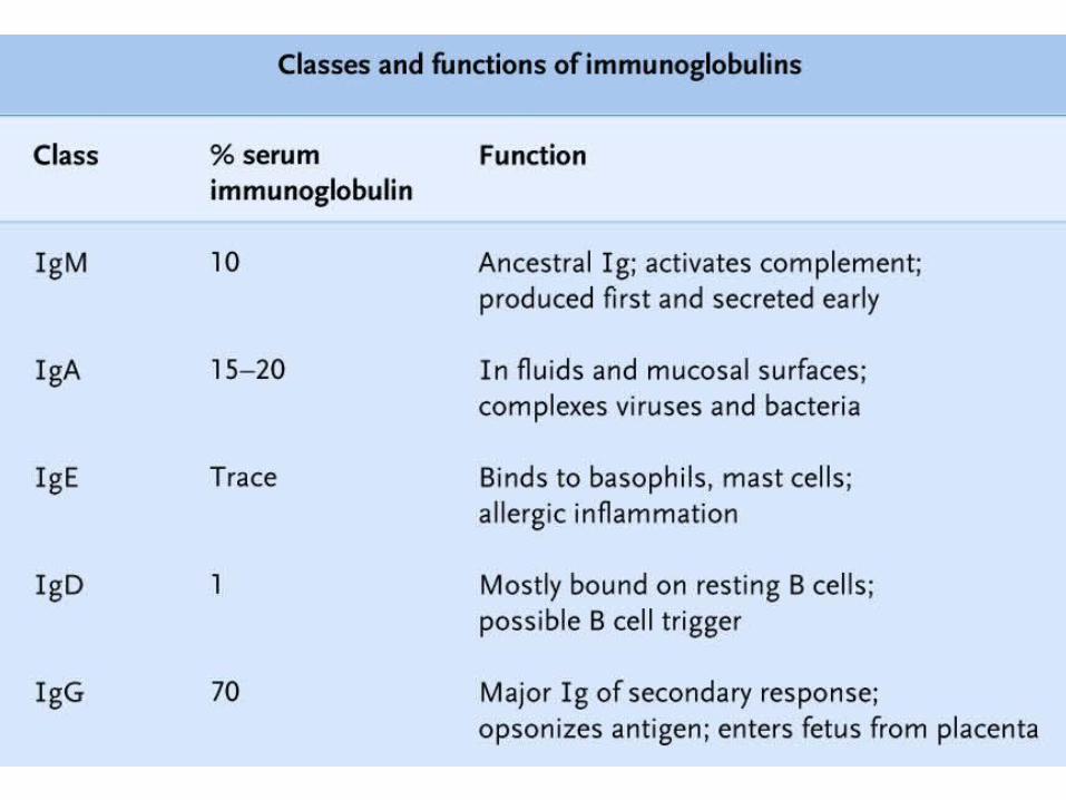
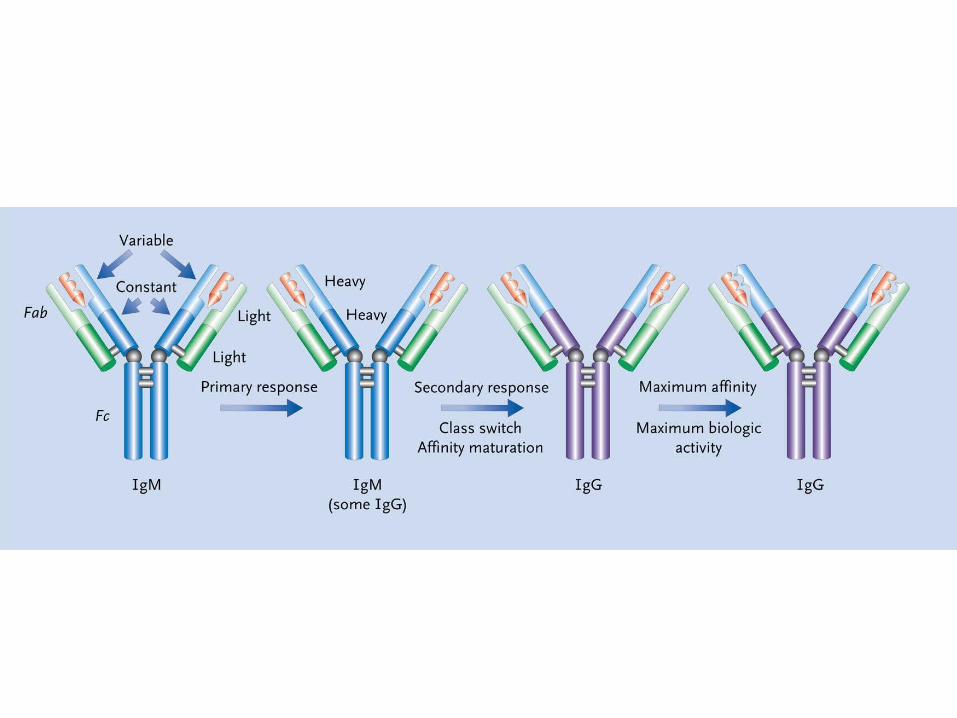
Lymphoid System Laboratory


Plasma Cell

Peyer’s Patches (Lymphoid Follicles) in Small Gut
Germinal Center (GC)
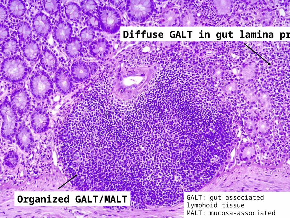
Organized GALT/MALT
Diffuse GALT in gut lamina propria
GALT: gut-associated lymphoid tissueMALT: mucosa-associated lymphoid tissue

Peyer’s Patch
Lymphoid follicleGerminal center

Lymph Node
Cortex
Medulla
Secondary follicles
Subcapsular sinus
Efferent lymphatics
Hilus Medullary cords
Afferent lymphatic

Medulla
Cortex
Germinal centers
Lymph Node

Subcapsular sinus
Afferent lymphatic with valve (V)

Secondary Follicle
Germinal center: B cell proliferation and maturation
Mantle of resting and memory B cells
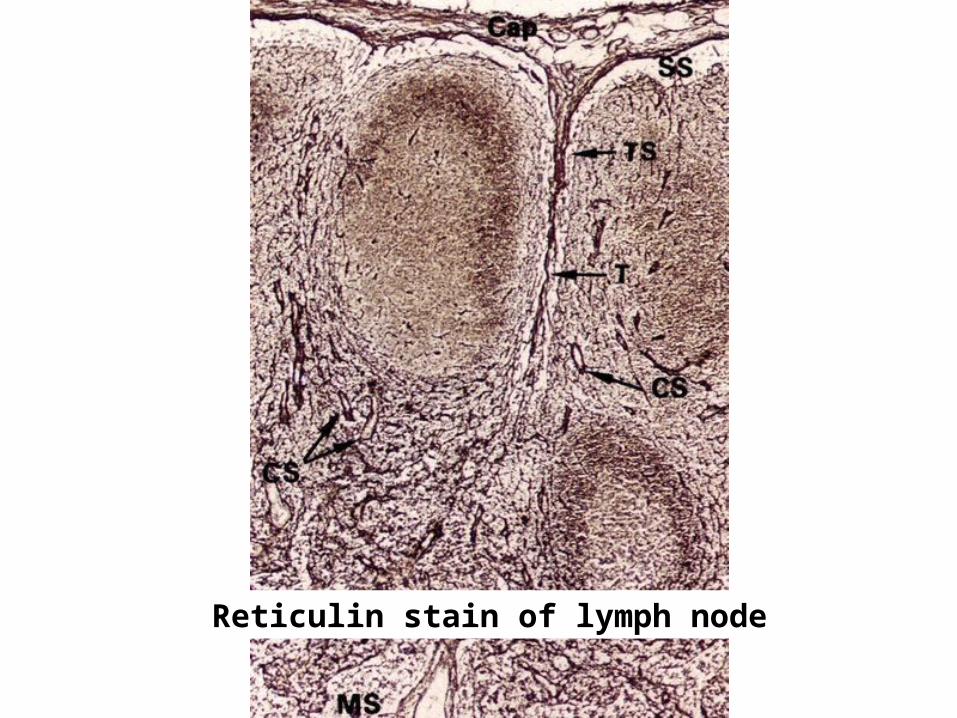
Reticulin stain of lymph node

Palatine tonsil


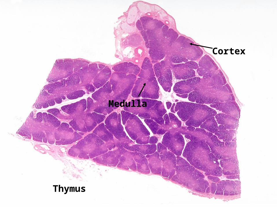
Thymus
Cortex
Medulla

Thymus
Cortex (developing T cells)
Medulla

Thymic medulla with Hassall’s corpuscles
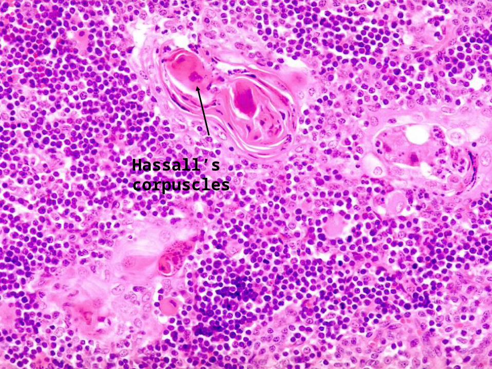
Hassall’s corpuscles
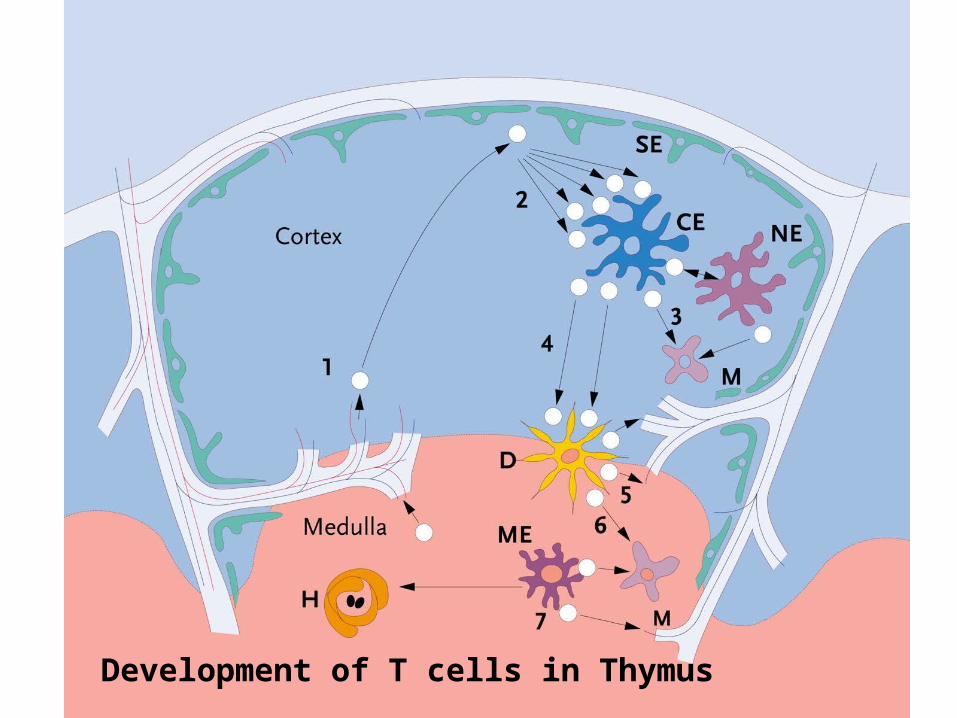
Development of T cells in Thymus

Spleen

Spleen
Red pulp
White pulp
PALS
Trabeular artery
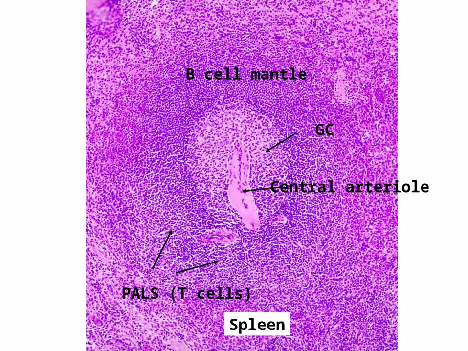
B cell mantle
PALS (T cells)
GC
Central arteriole
Spleen

Spleen Red Pulp
Endothelial cells
Venous sinus
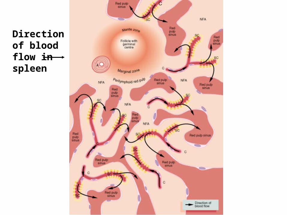
Direction of blood flow in spleen
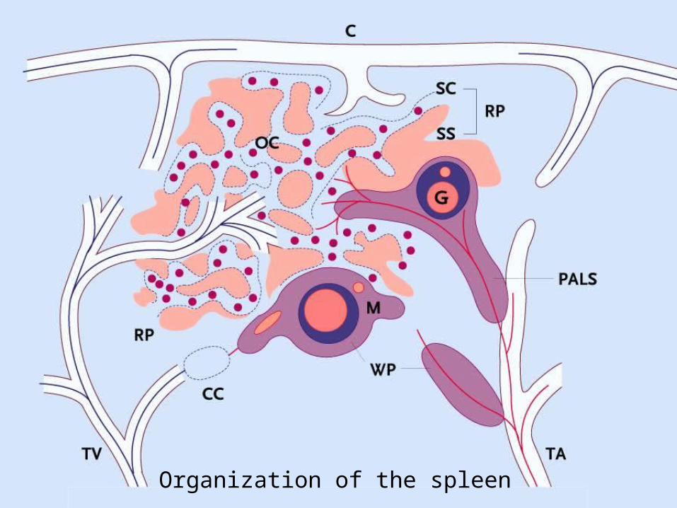
Organization of the spleen

Some Pathology

Benign Reactive Lymph Node

Benign Reactive Lymph NodeWhich cells are responding?

Adenocarcinoma Metastases to Lymph NodeHow did the tumor cells get to this location?

Adenocarcinoma Metastases to Lymph NodeHow did the tumor cells get to this location?