Functional Manipulations ofAcetylcholinesterase Splice VariantsHighlight Alternative Splicing Contributionsto Murine Neocortical Development
Amir Dori1,2, Jonathan Cohen1, William F. Silverman3, Yaakov
Pollack4 and Hermona Soreq2
1Department of Neurosurgery, Soroka University Medical
Center, Beer-Sheva, Israel, 2Life Sciences Institute, The
Hebrew University of Jerusalem, Jerusalem, Israel,3Department of Morphology, Zlotowski Center for
Neuroscience, Faculty of Health Sciences, Ben-Gurion
University of the Negev, Beer-Sheva, Israel and 4Department of
Microbiology and Immunology, Faculty of Health Sciences,
Ben-Gurion University of the Negev, Beer-Sheva, Israel
Proliferation and differentiation of mammalian central nervoussystem progenitor cells involve concertedly controlled transcrip-tional and alternative splicing modulations. Searching for thedevelopmental implications of this programming, we manipulatedspecific acetylcholinesterase (AChE) splice variants in the embry-onic mouse brain. In wild type mice, ‘synaptic’ AChE-S appearedin migrating neurons, whereas the C-terminus cleaved off thestress-induced AChE-R variant associated with migratory radialglial fibers. Antisense suppression of AChE-R reduced neuronalmigration, allowing increased proliferation of progenitor cells. Incontrast, transgenic overexpression of AChE-R was ineffective,whereas transgenic excess of enzymatically active AChE-S orinactive AChE-Sin suppressed progenitors proliferation alone orboth proliferation and neuronal migration, respectively. Our findingsattribute to alternative splicing events an interactive major role inneocortical development.
Keywords: alternative splicing, neurogenesis, neuronal migration, radialglia, readthrough acetylcholinesterase
Introduction
Neocortex development involves generation of projection
neurons in the ventricular zone (VZ) in response to as yet
incompletely understood cues, from a progenitor neuroepithe-
lium that is mostly composed of radial glial cells (Tamamaki
et al., 2001; Malatesta et al., 2003). These progenitor cells
undergo interkinetic nuclear migration, whereby DNA replica-
tion occurs when the nucleus is in the basal (outer) portion of
the VZ, and division adjacent to the ventricular surface give rise
to two daughter cells (Fig. 1A) (Boulder Committee, 1970).
Neurogenesis in mice commences on embryonic day (E) 11,
with production of the first post-mitotic neurons and their exit
from the VZ, while their sister cells re-enter the cell cycle and
continue to proliferate (Takahashi et al., 1995). Clonally related
proliferating cells coalesce in small, gap junction-coupled
clusters that cycle synchronously (Bittman et al., 1997), giving
rise to a clonally related output of neurons (Cai et al., 1997). As
neurogenesis proceeds, proliferation slows under the influence
of cell cycle-related proteins (Delalle et al., 1999), and cell
divisions primarily yield post-mitotic neurons, while the pro-
portion of daughter cells that continue to proliferate is greatly
decreased (Takahashi et al., 1996).
Following mitosis, the neural progeny reach the developing
cortex, i.e. the cortical plate (CP), either by somatic trans-
location, whereby a neuronal cell body migrates within a
pial-contacting radial process (Nadarajah et al., 2001) or by
locomotion, whereby newborn neurons develop elongated
leading and trailing processes that are oriented in a radial
direction, and migrate along radial glial fibers (Rakic, 1972).
Their adherence to nearby radial glial fibers activates multiple
intercellular events, such as cell--cell recognition and trans-
membrane signal transduction, that facilitate their movement
along the fibers (Rakic et al., 1994). Migrating neurons regulate
andmaintain the function of radial glia as migratory guides while
radial glia regulate migration along their processes (Anton et al.,
1997). Migrating neurons first pass through a subventricular
zone (SVZ) occupied by a secondary proliferative population of
cells adjacent to the VZ, and then through an intermediate zone
(IZ). They enter the CP and reach its most superficial portion,
adjacent to a marginal zone (MZ), where radial glial fibers
arborize before terminating at the pial surface (Fig. 1A,B)
(Gadisseux et al., 1989). Neurons are therefore guided by radially
directed processes of their founder cells, reaching their appro-
priate destination in clonally related clusters (Noctor et al.,
2001). They are positioned in the CP in an ‘inside-out’ sequence,
with newly arriving cells settling superficial to those that arrived
earlier (Rakic, 1974). Laminar fate, representing an aspect of
neuronal phenotype, was shown to be specified by gene ex-
pression in the progenitor cells, accompanied by progressive
restriction of multipotency (Frantz and McConnell, 1996).
Proliferation, differentiation and programmed gene expres-
sion in the developing nervous system may all be subject to
modulation by stress. Embryonic stress (e.g. ischemia--hypoxia)
attenuates neuronal migration to the cerebral neocortex, re-
sulting in morphological changes that are often accompanied by
postnatal behavioral deficits (Tashima et al., 2001). Similarly,
prenatal maternal stress impairs development of the offspring,
reducing, for example, learning and behavioral performance
(Kofman, 2002), and increasing the incidence of brain malfor-
mation and reduced head circumference (Mulder et al., 2002).
Improved understanding of the molecular mechanisms under-
lying the effects of stress on brain development is therefore of
considerable importance.
A notable cascade common to both brain development and
stress responses involves alternative splicing of pre-mRNA tran-
scripts, e.g. glutamic acid decarboxylase (Kuppers et al., 2000), G
protein isoforms (Morishita et al., 1999), the transcriptional
repressor ATF3 (Hashimoto et al., 2002) or potassium channels
(Xie and Black, 2001). However, the relationships between these
splicing modifications and the physiological changes occurring
during development and under stress remained obscure.
One way to explore this question is to directly manipulate the
embryonic expression levels and/or properties of specific vari-
ant mRNA transcripts of a neuronally-expressed gene that is
subject to transcription and splicing changes under both de-
velopment and stress, and observe the outcome with respect to
subsequent developmental events. The acetylcholinesterase
gene (ACHE) emerges as an appropriate example for such
Cerebral Cortex V 15 N 4 � Oxford University Press 2004; all rights reserved
Cerebral Cortex April 2005;15:419--430
doi:10.1093/cercor/bhh145
Advance Access publication August 5, 2004
by guest on January 13, 2016http://cercor.oxfordjournals.org/
Dow
nloaded from
a study. It is known, for example, that ACHE gene expression
undergoes major changes during development and that its AChE
protein product, reported previously to regulate cell prolifera-
tion and neurite outgrowth, iswidely considered a sensitive early
marker of histochemical differentiation (Layer and Willbold,
1995). Thus, in the adult brain, acute stress induces overproduc-
tion of the relatively rare soluble ‘readthrough’ AChE variant
AChE-R by alternative splicing of the AChE pre-mRNA (Fig. 1C,D)
(Kaufer et al., 1998). AChE overproduction acts in the short term
to reduce available acetylcholine (ACh) and attenuate choliner-
gic neurotransmission (Soreq and Seidman, 2001), but subse-
quent accumulation may last weeks after exposure (Meshorer
et al., 2002) and may induce vulnerability to head injury
(Shohami et al., 2000). It is plausible, therefore, that changes in
ACHE gene expression are involved in both development- and
stress-related responses of the mammalian brain.
To examine the involvement of alternative splicing in cortical
development, we subjected mouse embryos to antisense oligo-
nucleotide suppression or to transgenic overexpression of
specific AChE splice variants, and quantified the effects on
cortical development. Here, we report that both ‘synaptic’ AChE
(AChE-S) and AChE-R mRNA are expressed by progenitor cells
in the VZ and undifferentiated cells in the CP. However, while
the membrane-associated AChE-S was detected in migrating
neurons, embryonic brain AChE-R undergoes C-terminal cleav-
age, similar to the modification characterizing the AChE-R
isoform found in blood (Grisaru et al., 2001), and appeared in
radial glial fibers. Transgenic manipulations of AChE variants,
moreover, induced changes in progenitor cell proliferation as
well as neuronal migration, suggesting physiological and path-
ophysiological roles for alternative splicing of AChE in cortical
development.
Materials and Methods
AnimalsCD1 and FVB/N mice were used for antisense and transgenic experi-
ments respectively. Vaginal plugs on post-mating morning designated
E0. Pregnant dams were anesthetized by intra-muscular injection of
a ketamin and xylazine mixture (50 and 10 mg/kg body wt, respect-
ively). Embryos were removed and dissected in cold phosphate buffered
saline (PBS). Heads (E11--15) and brains (E16--17) were immersed in 4%
paraformaldehyde in PBS (48 h, 4�C), embedded in paraffin and
sectioned at 4 lm in the coronal plane. Animal care followed in-
stitutional guidelines according to NIH published guidelines.
BrdU and Oligonucleotides InjectionsPregnant dams were injected intraperitoneally (i.p.) with bromo-
deoxyuridine (BrdU; 50 mg/kg in 7 mM NaOH--saline solution; Sigma,
St Louis, MO). Post-injection time points at 1, 2, 4, 12 and 48 h served to
detect labeled nuclei at S-phase, S+G2 (with a few mitotic cells in the
VZ), S+G2+M, G1 and post-mitotic cells in the VZ, or a cohort of neurons
‘born’ on E14, that migrated and reached the CP, respectively. EN101,
a 20-mer antisense-oligodeoxynucleotide was previously shown to
primarily suppress AChE-R mRNA (Cohen et al., 2002; Meshorer et al.,
2002) is targeted to exon-2 of mouse AChE mRNA. Its three 39-terminal
nucleotides 59-CTGCAATATTTTCTTGC*A*C*C-39 (stars) were 2-O-
methylated for nuclease protection. Inversely oriented oligodeoxy-
nucleotides (INV101) with the same sequence as the antisense, but
oriented from 39 to 59 served as control. Oligonucleotides were
dissolved in saline and injected i.p. three successive times at 12 h
intervals, initiated 12 h following BrdU injection on E14 (40 or 100 lg/kgper injection). Animals were sacrificed 48 h after BrdU injection.
ImmunohistochemistrySections were deparaffinized, microwave-treated (750 W, 15 min) in
0.01 M citric buffer, pH 6.0, and blocked (30 min) in 5% normal goat,
rabbit or horse serum in PBS with 0.5% Tween-20 (PBST) for AChE
Readthrough Peptide (ARP), AChE Synaptic Peptide (ASP) or AChE
N-terminus (N-trm), and nestin, respectively (Fig. 1D). Immunoreactions
(90 min, room temperature) were with rabbit anti-ARP (Sternfeld et al.,
2000), goat anti-ASP [Santa Cruz Biotechnology, Santa Cruz, CA; AChE
(C-16)] or goat anti-AChE [Santa Cruz N-terminal AChE (N-19)], 1:100 in
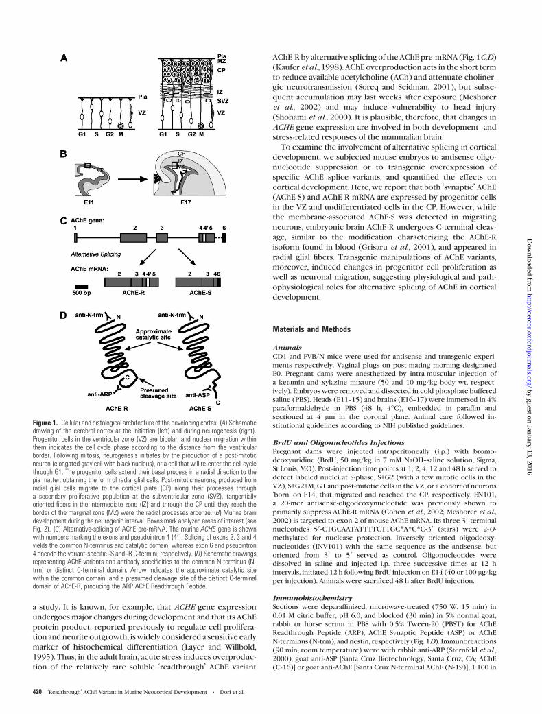
Figure 1. Cellular and histological architecture of the developing cortex. (A) Schematicdrawing of the cerebral cortex at the initiation (left) and during neurogenesis (right).Progenitor cells in the ventricular zone (VZ) are bipolar, and nuclear migration withinthem indicates the cell cycle phase according to the distance from the ventricularborder. Following mitosis, neurogenesis initiates by the production of a post-mitoticneuron (elongated gray cell with black nucleus), or a cell that will re-enter the cell cyclethrough G1. The progenitor cells extend their basal process in a radial direction to thepia matter, obtaining the form of radial glial cells. Post-mitotic neurons, produced fromradial glial cells migrate to the cortical plate (CP) along their processes througha secondary proliferative population at the subventricular zone (SVZ), tangentiallyoriented fibers in the intermediate zone (IZ) and through the CP until they reach theborder of the marginal zone (MZ) were the radial processes arborize. (B) Murine braindevelopment during the neurogenic interval. Boxes mark analyzed areas of interest (seeFig. 2). (C) Alternative-splicing of AChE pre-mRNA. The murine ACHE gene is shownwith numbers marking the exons and pseudointron 4 (49). Splicing of exons 2, 3 and 4yields the common N-terminus and catalytic domain, whereas exon 6 and pseuointron4 encode the variant-specific -S and -R C-termini, respectively. (D) Schematic drawingsrepresenting AChE variants and antibody specificities to the common N-terminus (N-trm) or distinct C-terminal domain. Arrow indicates the approximate catalytic sitewithin the common domain, and a presumed cleavage site of the distinct C-terminaldomain of AChE-R, producing the ARP AChE Readthrough Peptide.
420 ‘Readthrough’ AChE Variant in Murine Neocortical Development d Dori et al.
by guest on January 13, 2016http://cercor.oxfordjournals.org/
Dow
nloaded from

PBST containing 2.5% serum. Immunoreactivity for ARP was eliminated
by incubation of the antiserum with synthetic ARP (Sternfeld et al.,
2000) at a molar ratio of 1:5, attesting to specificity of the antiserum (not
shown). TUJ1 antibody (Lee et al., 1990) (generously provided by
Dr A. Frankfurter) and mouse anti-nestin (Developmental Studies
Hybridoma Bank, University of Iowa, Iowa City, IA) were 1:500 in PBST.
Secondary IgG were biotin-conjugated goat anti-rabbit for ARP, donkey
anti-goat for ASP or N-trm, and horse anti-mouse for nestin detection
(Vector), 1:200 in PBST containing 2.5% serum (1 h). TUJ1 detection
involved goat anti-mouse Cy3-conjugated IgG (Jackson Immuno-
research Laboratories, West Grove, PA), 1:200 in PBST. Biotinylated
antibodies were incubated with avidin-bound peroxidase complex
(ABC Elite, Vector Laboratories) for 1 h, rinsed with 0.05 M Tris, pH
7.6, and reacted for 90 s with 0.05% diaminobenzidine (Sigma) and
0.006% H2O2 in 0.05 M Tris, pH 7.6, with 0.05% nickel ammonium
sulfate. Selected sections were counterstained with Gill-2 hematoxylin
(Sigma).
Immunochemistry for the nuclear antigen Ki67 was used to monitor
cell proliferation. Ki67, previously used to label dividing cells in the
human embryonic VZ (Weissman et al., 2003), is expressed by pro-
liferating cells during late G1, S, M and G2 phases of the cell cycle
(Gerdes et al., 1984; Scholzen and Gerdes, 2000), and is often used to
evaluate the proliferative fraction of solid tumors (Scholzen and Gerdes,
2000). The utility of Ki67 as a proliferative marker that is comparable to
BrdU labeling was previously tested for neurogenesis in the adult
dentate gyrus of the hippocampus, where its expression mimicked that
of BrdU when examined soon after exogenous BrdU administration.
Experimental increases in the number of mitotic cells by ischemia, or
their reductions by radiation produced parallel changes in BrdU and
Ki-67 labeling (Kee et al., 2002). Ki67 staining increases during S-phase,
reaches a peak during metaphase (du Manoir et al., 1991) and decreases
during ana- and telophase (Starborg et al., 1996). Quantification of Ki67
expression was compiled by measuring the mean sum of pixel values in
a 50 3 100 lm rectangle at the apical portion of the VZ, positioned
100 lm lateral to the dorsomedial to medial cortical border, similar to
that done for detection of AChE.
BrdU LabelingSections were treated with 100 lg/ml deoxyribonuclease in PBST
(30 min), incubated with mouse anti-BrdU (Becton-Dickinson, Missis-
sauga, Ontario, Canada; 1:100 in PBST, 2 h), followed by anti-mouse Cy2-
conjugated IgG (Jackson; 1:50 in PBST) or biotinylated goat anti-
mouse IgG. Processing was as described above.
In Situ HybridizationPreviously detailed probes and procedure (Meshorer et al., 2002) were
modified as follows. Cy5-conjugated streptavidin and Cy3-conjugated
anti-digoxygeninwere employed for detection of biotin- and digoxygenin-
labeled probes, respectively [1:200 in Tris-buffered saline with 0.1%
Tween-20 (TBST); Jackson]. In situ hybridization was combined with
TUJ1-immunofluorescence as detailed above, or with BrdU-immuno-
fluorescence applying fast-red reaction with alkaline-phosphatase
(AP)-conjugated streptavidin (Zymed Laboratories, San Francisco, CA;
1:25 in TBST, 1 h), followed by BrdU-immunofluorescence with Cy2-
conjugated anti-mouse IgG.
Confocal MicroscopyImages of 1-lm-thick sections were captured by excitation at 488, 543,
633 and 488 nm of Cy2, Cy3, Cy5 and Fast-Red, respectively. Emission
was measured with band-passes of 505--545 or 560--615 nm or long-
passes of 650 and 560 nm, respectively. The microscope’s detector and
amplifier were calibrated by referring to sections expected to have the
highest signal as 100% (E11 for ontogeny experiments, INV101 for
antisense experiments). The focus was adjusted to the point of maximal
intensity, and the detector and amplifier were adjusted to obtain the
optimal image. For subsequent sections, the focus was adjusted but the
same amplifier and detector values weremaintained to reach the narrow
depth of maximal signal intensity.
Regions of AnalysesSectors of analysis were 200 lm wide and distant 100 lm from the
medial edge of the lateral ventricle, within the posterior-medial portion
of the future somatosensory area. Digitized images were analyzed in
a ‘blind’ manner. BrdU-immunostaining was considered positive if nuclei
were darkly stained or at least three puncta were discerned.
Image AnalysesAt least three embryonic brains from at least three different litters were
analyzed for each group. Averaged cell counts were obtained by averaging
values from three or four non-consecutive sections from each brain, for
all analyzed brains in each group. Confocal signal was converted to
grayscale for intensity measurements of pixel values (Scion Image, Scion
Corporation, Frederick, MD). Analysis of variance (ANOVA; Statistica
software, StatSoft, Tulsa, OK) was used to compare multiple groups and
a one-tailed t-test (Microsoft Excel) was used to compare two groups.
ImmunoblotsCerebral homogenates yielding soluble AChE from E17 control and
transgenic embryos were processed as described (Birikh et al., 2003).
Immunodetection was with rabbit anti-ARP (1:250), goat anti-ASP
(1:500) and goat anti-N-trm (1:500).
Catalytic ActivityAcetylthiocholine hydrolysis was measured spectrophotometrically as
described (Kaufer et al., 1998). Iso-OMPA (tetraisopropylpyrophosphor-
amide) was used to block butyrylcholinesterase activity (5 3 10–5 M).
Results
Pre-AChE mRNA Splicing Shift at the Ventricular Zone
ACHE gene expression was first studied by in situ hybridization
in the VZ during the neurogenic interval (Fig. 1B). At the onset
of neurogenesis (E11), a time of intense progenitor cell pro-
liferation, cytoplasmic AChE-R and AChE-S mRNA (Fig. 1C) were
co-localized in most VZ cells (Fig. 2A). Expression was most
intense in the apical portion of the VZ, close to the ventricular
lumen. With the advance of neurogenesis, e.g. at E13, cyto-
plasmic expression of the AChE isoforms became pronounced
in clusters of adjoining cells in the basal portion of the VZ
(Fig. 2A,C). These AChE-expressing clusters, which included
from two to >20 cells, coalesced at various points along their
common borders. By E15, AChE-expressing cell clusters were
smaller, and by E17 they were limited to very small clusters or
single cells (Fig. 2A). The subcellular distribution of AChE
mRNA had also changed, so that intense signals were observed
primarily in the basal pole of labeled cells (Fig. 2B). Densito-
metric measurements of AChE expression demonstrated a grad-
ual reduction in labeling intensity throughout the VZ during
neurogenesis. Expression areas of AChE splice variants exhibi-
ted a parallel reduction (Fig. 2C), implying decreasing numbers
of expressing cells. Both AChE-R and AChE-S mRNA declined in
the VZ. However, some of the cell clusters at E13 maintained
the intense level of AChE-R expression (Fig. 2A, open arrow). A
statistically significant reduction in AChE-S expression, as de-
termined for both intensity and signal area, was observed from
E11 to E13, while reduction of AChE-R was delayed until E15,
apparently reflecting a transient dominance of AChE-R over
AChE-S at E13 (Fig. 2C).
Proliferating Cells but not Terminally DifferentiatedNeurons Display Splicing Shift at the Ventricular Zone
A 2 h pulse of BrdU was used to distinguish between S+G2+Mversus G1 or post-mitotic nuclei in order to determine the
proliferative profile of AChE expressing cells in the VZ. During
such a short pulse, BrdU is continuously available for incorpor-
ation into nuclei in the S-phase, while the earliest of these nuclei
advance through G2 and initiate mitosis (Takahashi et al., 1992).
Cerebral Cortex April 2005, V 15 N 4 421
by guest on January 13, 2016http://cercor.oxfordjournals.org/
Dow
nloaded from

Combined with in situ hybridization, anti-BrdU immuno-
fluorescence demonstrated that the AChE mRNA-labeled
clusters included both BrdU positive and negative nuclei,
suggesting that the clusters comprise both S+G2+M and G1 or
post-mitotic cells (Fig. 3A). The majority of intensely AChE-
expressing cells were located at the basal portion of the VZ, i.e.
in the S-phase zone. Adjacent to the ventricular lumen, i.e. in the
G2+M zone, AChE expression was relatively sparse, with cells in
mitosis expressing the transcripts at their basal pole (Fig. 3A,
arrows and insets).
BrdU-negative cells in the VZ represent either proliferative
cells re-entering the cell cycle through G1 or post-mitotic cells
in the process of migration out of the VZ. To differentiate
between these two possibilities, in situ hybridization for
AChE-R mRNA was combined with immunofluorescence for
beta-III tubulin (TUJ1), an early marker of neurons (Geisert and
Frankfurter, 1989) (Fig. 3B). The AChE-R mRNA positive
clusters were TUJ1 negative, suggesting that AChE-expressing
cells that were refractory to the 2 h BrdU pulse were pro-
liferative cells, i.e. cells at G1 phase. Nevertheless, not all of the
non-AChE expressing cells were labeled by TUJ1, indicating the
existence of another or intermediate cell population.
AChE Gene Expression in Migrating Neurons
AChE expression patterns in the IZ were examined to assess the
potential involvement of the protein and its splice variants in
neuronal migration from the VZ to the cortical plate. Both AChE-
R and AChE-S mRNA were observed either as individual IZ cells
or as clusters, with reduced labeling compared with the CP or
VZ (Fig. 4A,B). These cells were radially oriented, suggesting that
theyweremigrating fromtheVZ to theCP. Immunofluorescence
Figure 3. AChE-S and AChE-R expressing cell clusters in ventricular zone include cellsin all phases of the cell cycle but not post-mitotic neurons. (A) AChE-R and AChE-Sgene expression (red) in cell clusters, one of each is marked by a white border line.BrdU immunofluorescence (green) in the ventricular zone (VZ) at E14 following a 2 hBrdU pulse injection. Gaps of non-expressing cells are marked by asterisks. AChE-expressing clusters (arrowheads) include BrdU-positive and negative nuclei. Basalpolar AChE expression is observed in cells near mitosis (arrows, inset). (B) AChE-Rlabeled clusters in the VZ are TUJ1-negative. TUJ1-positive cells in the VZ at E15(arrowheads) are located between the AChE-R clusters. Note the horizontal orientationof TUJ1 positive cells in the subventricular zone (SVZ), which are apparentlytangentially migrating neurons.
Figure 2. Transient splicing shift from AChE-S to AChE-R in ventricular zone cellclusters (A) AChE-R (blue) and AChE-S (red) mRNA signals during cortical development.Confocal images of double-labeling in situ hybridization. AChE-R mRNA signal intensitywas color coded, with yellow to blue color-gradient representing decreasing signal-intensity (right column, color bar). Intensely expressing cells or clusters (arrows), non-expressing cells (arrowheads) and AChE-R dominance over AChE-S expression (openarrow) are marked. (B) An enlargement of arrow-marked AChE expressing cells at E17in (A). (C) AChE-R and AChE-S signal intensities and distribution areas (mean±SD) andratios in the ventricular zone (VZ). Note transient peak of R/S ratio.
422 ‘Readthrough’ AChE Variant in Murine Neocortical Development d Dori et al.
by guest on January 13, 2016http://cercor.oxfordjournals.org/
Dow
nloaded from

with the TUJ1 antibody intensely labeled horizontally oriented
cells in the IZ that were apparently involved in tangential
migration (Fig. 4B,C). Indeed, combined AChE-R mRNA/TUJ1
labeling demonstrated that the AChE-R expressing cells in the IZ
wereTUJ1-negative (Fig. 4B,C).With further development, AChE
expression in the CP appeared in clusters of intensely labeled
cells, surrounded bymoderately expressing cells (Fig. 4A,B). The
intensely labeled cells were more prominent in the superficial
portion compared with the deep CP. This suggested that the
younger, newly arriving cells in the CP expressed more AChE
transcripts than earlier arriving cells that had already undergone
some differentiation. Subsequently, at E17, the intense signals in
the superficial cell layer of the CP became significantly higher
than those of the deep CP portion. Combined AChE-R mRNA/
TUJ1 immunofluorescence demonstrated a complementary
pattern, similar to that observed in the VZ (Fig. 4B,C). The
distribution of TUJ1 exhibited increasing density of labeled cells
from the superficial to deeper portions of the CP, implying again
that AChE was intensely expressed by the relatively undifferen-
tiated cells, and declined as the number of differentiated cells
increased. Although TUJ1 expression was essentially detected in
the deeper portion of theCP, a fewof these cellswere seen at the
superficial portion adjacent to theMZ (Fig. 4C).We assume these
cells to be young neurons that were about to be displaced by
incoming newly arriving migrating neurons or possibly cells in
transition fromAChE toTUJ1-expressing cells. In addition to this,
some horizontally oriented TUJ1 cells were detected in the MZ,
possibly Cajal-Retzius cells.
Distinct Localization Patterns of AChE Splice Variantsduring Cortical Development
AChE splice variants are identical in most of their sequence,
differing, primarily in their C-termini (30 residues of AChE-R
peptide, ARP and 39 residues of AChE-S peptide, ASP; Fig. 1D).
ARP, ASP and the common N- terminus all demonstrated
cytoplasmic immunostaining patterns in VZ cells, similar to
that of AChE mRNA, with a gradual decrease in intensity and
in the number of expressing cells, as well as reduced clustering
of intensely labeled cells (Fig. 5). Moreover, at E15, ARP-
immunoreactive cell processes were observed ascending from
the VZ (Fig. 5, arrows) and extending radially through the total
thickness of the cortical wall to terminate at the pial surface.
This pattern, which is characteristic of radial glia cells
(Gadisseux et al., 1989), was most readily observed in the
medial neocortex. There, fibers could be clearly traced into the
marginal zone (MZ), where they arborized before terminating
at the pial surface (Fig. 6A).
Developing Brain AChE-R is C-terminally Cleaved
The immunoreactivity of radial glia to antibodies targeted at the
C-terminus of AChE-R, but not to its N-terminus (Fig. 5),
suggested cleavage of AChE-R to separate the C-terminal
domain that includes ARP, from the core AChE-R protein (Fig.
1D). This is consistent with AChE found in blood (Grisaru et al.,
2001). Consistent with this observation, soluble proteins ex-
tracted from E17 cerebral cortex demonstrated an intense ARP
immunoreactive band of 18 kDa in addition to a 65 kDa band
that appears to be intact AChE (Fig. 6B). In contrast to this,
antibodies directed against the N-terminal domain (N-trm)
common to all AChE variants (Fig. 1D) revealed several slowly
migrating bands (Fig. 6B). These included a lightly labeled band
that paralleled the 65kD band shown with anti-ARP and
representing the non-cleaved AChE-R, while the most intense
band was of ~55 kDa, reflecting the core AChE-R domain
following removal of the C-terminus. Negligible immuno-
reactivity was observed to antibodies directed against ASP
Figure 4. AChE-R mRNA expression in the embryonic cortical plate precedes neuronal differentiation. (A) AChE-R (blue) and AChE-S (red) mRNAs at E15 and E17. Note non-homogeneous distribution of intense signals in cell clusters (arrowheads) in the superficial portion of the cortical plate (CP), and deep situated lightly stained cells (asterisk).Intermediate zone (IZ) clusters are seen at E15 (arrows). (B) The corresponding brain regions. (C) AChE-R mRNA (red) and immunolabeled TUJ1 (green) in the cortical wall at E15and E17. Note TUJ1-negative AChE-R-positive cell clusters (arrows) and TUJ1-positive AChE-R-negative cells (arrowheads) in the superficial and deep cortical plate, respectively.TUJ1-positive AChE-R-negative horizontally orientated cells, likely migrating tangentially, are seen in the IZ.
Cerebral Cortex April 2005, V 15 N 4 423
by guest on January 13, 2016http://cercor.oxfordjournals.org/
Dow
nloaded from

(Fig. 6B), suggesting that ASP remained attached to AChE-S,
rendering it insoluble and therefore not extractable by this
procedure. These results indicate that AChE-R, but not AChE-S,
is subject to cleavage of its C-terminal domain in the brain and
that the vast majority of AChE-R in the developing cortex
undergoes C-terminal cleavage.
Anti-ARP, which was originally raised against a glutathione S-
transferase--human ARP fusion protein, was not immunoreactive
against synthetic ASP (not shown), attesting to its specificity, yet
displayed clear immunoreactivity to synthetic murine ARP (Fig.
6B). The immunoreactivity of these two distinct amino acid
sequences to the same antiserum suggested evolutionary con-
servation of ARP structural epitopes, despite the disparity in
sequence.
Anti-ARP Labels Migration-associated Glial Processes
With the thickening of the cortical wall, ARP-labeled fibers in
the IZ became arched (from medial to lateral), and resumed
a radial alignment, orthogonal to the pial surface as they entered
the CP (Fig. 6D-2,6). Immunohistochemistry for nestin, a marker
of radial glia (Lendahl et al., 1990), exhibited a similar pattern of
fibers in adjacent sections (Fig. 6D-1,5). This alignment is typical
of the morphology of radial glia (Gadisseux et al., 1989; Misson
et al., 1988). In contrast to the pronounced staining by anti-ASP
and anti-N-trm antisera, ARP labeling was faint in the cytoplasm
of migrating cells in the IZ (Fig. 6D-2,6).
Figure 6. Cleaved AChE-R C-terminus in migratory glial fibers and AChE-S in migratingcells perikarya. (A) E15, AChE Readthrough Peptide (ARP)-immunostained fibersascend from the ventricular zone (VZ, arrowheads), reaching the medial cortex marginalzone (MZ) where they branch and terminate at the pia matter. (B) Immunoblot analysis.Anti-ARP labels syntheticmurine ARP (mARP), 18 and 65 kDa bands from E17 cerebrum.Antibodies to N-terminus (N-trm) label 65 and 55 kDa proteins, whereas anti-AChESynaptic Peptide (ASP) shows negligible immunoreactivity. (C) Coronal section schemeof migratory pathways within the analyzed regions in the cortex (CTX), striatum (STR)and lateral cortical stream (LCS) at E17. (D) Immunohistochemical staining. (1--4)Cortical wall. (5--9) Intermediate zone, enlarged from the boxed regions in 1--4. (1, 5, 9,13, 17) Nestin. Note labeled radial glial processes (arrowheads), and dense bundle ofglial fibers in the LCS. (2, 6, 10, 14, 18) ARP. Note cells at the VZ/subventricular zone(SVZ), radial glial processes (arrowheads), LCS and STR fibers and lightly labeledsuperficial cortical plate (CP) cells. (3, 4, 7, 8, 11, 12, 15, 16, 19, 20) ASP and N-trm.Note cells at the VZ/SVZ, clusters of radially oriented cells in the intermediate zone (IZ,arrows), prominent staining in the CP superficial cell layer, and in perikarya of cells withinthe LCS area, oriented parallel to migratory direction (arrowheads).
Figure 5. Developmental decreases in AChE immunolabeling. Photomicrographsshow AChE Readthrough Peptide (ARP), AChE Synaptic Peptide (ASP) and N-terminus(N-trm) labeling in the ventricular zone (VZ). E13 cell clusters and E17 single cells aremarked by arrowheads. E15 ARP-immunostained radial fibers ascending from the VZinto the overlying subventricular zone (SVZ) are shown (arrows).
424 ‘Readthrough’ AChE Variant in Murine Neocortical Development d Dori et al.
by guest on January 13, 2016http://cercor.oxfordjournals.org/
Dow
nloaded from

Within the CP, intense immunoreactivity of ASP and the N-
terminus was observed in the cells at the superficial cell layer,
i.e. newly arriving CP cells, similar to the pattern of AChE gene
expression (Fig. 6D-3,4). In contrast, ARP immunoreactivity in
this cell layer was sparse, compared with its intensity in the VZ
(Fig. 6D-2).
To examine whether ARP immunoreactivity is apparent in
other migration-associated glial processes, immunoreactivity
was examined in the lateral cortical stream (LCS) and in the
striatum (Fig. 6C). The dense glial bundles of the LCS were
strongly positive for both nestin and ARP (Fig. 6D-9,13 and
10,14, respectively), extending ventrolaterally from the lateral
edge of the VZ between the neocortex and the striatum. Both
nestin and ARP demonstrated ramification of this bundle into
fibers that assume an orthogonal orientation to the pial surface
as they penetrate the neocortex. In contrast, ASP and the
common N-terminus peptide were not detected in the LCS fiber
bundle or in its ramifications, but were labeled in the cytoplasm
of migrating cells in the region of the LCS (arrowheads in
Fig. 6D-15,16 and insets in 19,20, respectively). In the striatum,
both nestin and ARP labeled glial fiber processes extended from
the VZ area to the differentiating part of the striatum, which
were not immunolabeled by either the ASP or the common N-
trm antibodies (Fig. 6D-17--20).
Antisense Suppression of AChE-R mRNA AttenuatesNeuronal Migration
The distinct patterns of ARP and ASP immunostaining we
observed suggested that these two peptides and/or their
corresponding proteins may play distinct roles during neuronal
migration. To challenge this hypothesis, we labeled a cohort of
migrating cells with BrdU prior to their terminal mitosis in the
VZ (Fig. 7A). To reduce AChE-R during neuronal migration, we
employed mouse EN101, an antisense oligonucleotide capable
of inducing selective destruction of mouse neuronal AChE-R
mRNA (Cohen et al., 2002). Fluorescent double-labeling in situ
hybridization was performed to quantify AChE mRNA variants in
the cortical wall following EN101 injection during neuronal
migration. Cell density within the VZ as well as its thickness
were similar in control and EN101-treated brains. A reduction in
labeling was observed, however, which could not be attributed
to reduction in the number of AChE expressing cells. Signal
intensity for AChE-R mRNA, measured and compared in uniform
100350lmsquare samples in the apical portionof theVZ (121±12 cells; Fig. 7B) exhibited a 34% reduction following EN101
compared with control treatment with the inversely oriented
oligonucleotide sequence, INV101, both at 100 lg/kg (n = 10,
P < 0.05). In contrast, AChE-S mRNA labeling was reduced by
only 7% (n = 10), which was not statistically significant (Fig. 7B).
The effect of AChE-R mRNA reduction on cell migration was
evaluated by counting the number of BrdU immunoreactive
nuclei present in the dorsomedial CP 48 h after BrdU injection
(Fig. 7C). EN101 treatment significantly reduced the number of
BrdU immunoreactive cells in the CP by 26% compared with the
INV101 treatment (n = 13; ANOVA, P < 0.01; Fig. 7D). Lower
concentrations of EN101 or INV101 (40 lg/kg) failed to elicit
Figure 7. Antisense oligonucleotide suppression of AChE-R production attenuates cellarrival to the cortical plate. (A) Photomicrographs (top) and schematic drawings(bottom) of BrdU incorporation by progenitor cells into nuclei during S-phase, theirsubsequent inter-kinetic movement in the ventricular zone (VZ) and migration towardthe cortical plate. At E14, 12 h after BrdU injection, labeled nuclei have all moved awayfrom the ventricular surface, indicating that they have become post-mitotic. By 48 hpost-injection, many BrdU-immunostained nuclei have reached the cortical plate (CP).Note that the majority of labeled nuclei in the CP are darkly labeled, whereas those in theVZ/subventricular zone (SVZ) become lightly labeled. Arrows point to mitotic BrdU-labeled cells, arrowheads point to proliferating cells in the SVZ. (B) AChE-R and AChE-SmRNA following EN101 or control INV101 treatment. Histograms show signal intensityin 100 3 50 lm square sectors at the inner portion of the VZ (box). (C) BrdU-immunoreactive nuclei in the cortical plate. Coronal 200 lm cortical sectors, followingINV101 or EN101 treatment. (D) BrdU-positive cell counts. Attenuated migrationdisplayed dose dependence (40 versus 100 lg/kg) and sequence specificity (EN101versus INV101). Note that the total number of immunoreactive cells in the CPþIZremained unchanged.
Cerebral Cortex April 2005, V 15 N 4 425
by guest on January 13, 2016http://cercor.oxfordjournals.org/
Dow
nloaded from

discernible effects on cell migration to the CP (Fig. 7D),
indicating dose-dependence. Consistent with this, significantly
more BrdU-immunoreactive cells were detected in the IZ under
EN101 treatment (at 100 lg/kg) compared with INV101 or low
dose EN101 (n = 13) (ANOVA, P < 0.05) (Fig. 7C,D). The total
number of BrdU-immunoreactive cells in the CP and IZ was
similar between all treated groups, demonstrating that post-
mitotic cell survival was unchanged (Fig. 7D). Therefore, the
EN101-mediated reduction of neuronal migration reflected
attenuated progression of cells from the IZ to the CP.
AChE-R mRNA Destruction Increases Proliferation inthe Ventricular Zone
The gradual reduction of AChE gene expression during neuro-
genesis, in parallel with the restriction of proliferation in the
VZ, suggested an involvement of AChE and the AChE-associated
migration process with proliferation of progenitor cells (Fig. 8A).
The effect of AChE-R on proliferation in the VZ was examined by
EN101 treatment in animals treated with BrdU 48 h prior to
sacrifice. Immunocytochemical labeling of the Ki67 nuclear
antigen appeared in most of the cells at the M-phase region in
theVZof themouse developing neocortex at E16. Fewcells in the
basal portion of the VZ were intensely stained, though, the
majority of labeled cells in that region exhibited light, punctate
nuclear staining (Fig. 8B). Compared with INV101, EN101 treat-
ment significantly increasedKi67expression in theVZ (Fig. 8C,D),
suggesting increased re-entry into the cell cycle vs exiting the cell
cycle following reduction of AChE-R expression by EN101.
Both Catalytic and Non-catalytic AChE Activities AffectProliferation and Migration
VZ neuronal progenitors express both AChE-S and AChE-R. The
observedeffect of EN101described above, therefore, impliedone
of two possibilities: (i) AChE-R alone reduces proliferation; or (ii)
AChhydrolysis, common toAChE-R andAChE-S is responsible. To
distinguish between these possibilities, we injected BrdU into
E14 transgenic mice overexpressing (i) the membrane adhering
AChE-S (TgS, Beeri et al., 1995); (ii) soluble AChE-R (TgR,
Sternfeld et al., 2000); or (iii) an enzymatically inactive form of
AChE-S (TgSin, Sternfeld et al., 1998) (Fig. 9A). The animals were
sacrificed 48 h later. Reverse transcriptase--polymerase chain
reaction was employed to confirm the expression of each of the
AChE variants, and acetylthiocholine hydrolysis measurements
confirmed increased catalytic activity in brain homogenates from
the TgR andTgS but not TgSin strains (Fig. 9B). ARP expression in
embryonic brains from the three transgenic and control groups,
appeared similar by immunoblot analysis (not shown), suggesting
that high AChE-R levels are maintained during cortical develop-
ment. Arrival of cells in the CP from both TgR or TgS over-
expressing embryos was unchanged relative to parent strain
controls. However, in TgSin embryos, the average number of
BrdU immunoreactive cells in the CP was significantly reduced
compared with control mice as well as those of TgS or TgR mice
(Fig. 9B), suggesting a role for ACh in neuronal migration.
The effect of overexpressing AChE splice variants on pro-
genitor cells proliferation was examined in the transgenic
mouse strains by Ki67 labeling and density quantification in
the VZ. Ki67 expression was reduced in both TgS and TgSin, but
not in TgR embryos compared with controls (Fig. 9B). This
suggests that the non- hydrolytic activity of AChE-S acts to
reduce proliferation in the VZ, whereas its hydrolytic activity
has a role in promoting neuronal migration to the CP.
Discussion
Using the regulation of AChE pre-mRNA processing as a case
study, we explored the involvement of alternative splicing
modulations in shaping the developing brain. We found tran-
sient changes in AChE pre-mRNA processing during murine
cortical development. Using transgenic and antisense manipu-
lations of these variants, we have further demonstrated causal
involvement of such changes in progenitor proliferation and the
shift toward neuronal migration and differentiation which
together shape the mammalian cortical plate.
While many neuronal mRNAs are subject to alternative
splicing modulations during brain development (e.g. the cla-
thrin assembly protein 3 (AP-3) (Ishihara-Sugano and Nakae,
1997), the protein tyrosine phosphatases PTP-SL and PTPBR7
Figure 8. Antisense treatment increases proliferation in the ventricular zone andreduces cell arrival to the cortical plate. (A) Proliferating cells in the ventricular zone(VZ) can continue proliferating or migrate to the cortical plate (CP), possibly sendingsignals (broken arrow) that modulate proliferation. (B) Ki67-immunostaining (E16coronal section counterstained with hematoxylin). Adjacent to the ventricular border,where mitosis occurs, note the intense nuclear labeling of cells in metaphase (largearrowhead), anaphase (small arrowhead) and G2 or beginning of G1 (long arrow).Some basal VZ cells were intensely stained (open arrows), but most were lightlylabeled with punctated nuclear staining (small arrow). (C) Ki67-immunostainingfollowing INV101 or EN101 treatments. 100 3 50 lm sectors at the inner VZ (box)served for density measurements. (D) Ki67-immunoreactivity density in the VZfollowing EN101 treatment. *P\ 0.05. INV101 served as control.
426 ‘Readthrough’ AChE Variant in Murine Neocortical Development d Dori et al.
by guest on January 13, 2016http://cercor.oxfordjournals.org/
Dow
nloaded from

(Van Den Maagdenberg et al., 1999) and G protein isoforms
(Morishita et al., 1999), the information accumulated on AChE’s
splice variants and their putative roles in neuronal development
and functioning provides added value to this particular example.
First, the alternative splicing shift in AChE pre-mRNA process-
ing occurred in proliferating progenitors prior to their neuronal
commitment, marking a checkpoint between proliferation and
migration. Secondly, our analyses pointed at four distinct
functions for AChE in cortical development: (i) ACh hydrolysis,
common to AChE-S and AChE-R; (ii) non-catalytic structural
features of the core domain, also common to both variants;
(iii) migration-supportive properties of ARP, the cleavable C-
terminus of AChE-R; and (iv) adherent capacities of ASP, the
corresponding uncleaved C-terminus of AChE-S, which joins
AChE-S tetramers to a proline-rich membrane anchor (PRiMA)
structural subunit (Perrier et al., 2002) but also drives AChE-S to
the cell nucleus (Perry et al., 2002). In the following, we discuss
the implications of each of these roles for cortical development.
Concerted Effects on Progenitor Migration andProliferation
During murine brain development, alternative splicing modula-
tion yields a relative dominance of AChE-R, which we found to
be a pre-protein to its cleavable C-terminus ARP, compatible
with its cleavage under stress in the mouse and human blood
(Grisaru et al., 2001; Cohen et al., 2003; Pick et al., 2004). In the
developing cortex, ARP interacts with migration-supportive
radial glia, unlike the core AChE domain and the uncleaved
variant AChE-S, which persist in migrating and differentiating
neurons. Moreover, antisense suppression of AChE-R produc-
tion attenuated neuronal migration to the CP, suggesting causal
involvement of the splice shift in this process. In addition, the
antisense treatment increased neuronal progenitor prolifera-
tion. This could have reflected a proliferation-inhibitory effect
of AChE-R itself or of EN101-resistant AChE-S in the attenuated
progenitors. To distinguish between these possibilities, pro-
genitor proliferation and neuronal migration were compared in
transgenic mice overexpressing distinct AChE variants. AChE-R
excess had no effect, whereas both AChE-S and its genetically
inactivated mutant AChE-Sin suppressed proliferation, and
AChE-Sin further suppressed migration. These findings sug-
gested a non-catalytic, proliferation-inhibiting effect for AChE-S,
possibly acting through AChE-R or in an AChE-R-dependent
manner. Thus, reduction of AChE-R following EN101 treatment
abolished the capacity of AChE-S to attenuate proliferation.
Additionally, suppression of neuronal migration by AChE-Sin is
compatible with the assumption that ACh hydrolysis is pivotal
for neuronal migration, supporting the view of ACh as a regu-
lator of neuronal migration (Lauder and Schambra, 1999).
Role in Glial Cell Differentiation
The dynamic changes that take place in the VZ during cortical
development include increased cell cycle length (Takahashi
et al., 1995), reduction of symmetric mitotic divisions (Chenn
and McConnell, 1995), restriction in layer specification
(McConnell and Kaznowski, 1991) and change in radial glia
phenotype (Hartfuss et al., 2001). During brain development,
AChE expression in the VZ decreased at the end of neuro-
genesis, when radial glia transform to astrocytes (Hartfuss et al.,
2001). AChE involvement in cell proliferation was previously
proposed in several brain and hematopoietic cell types (Karpel
et al., 1996; Sharma et al., 2001; Perry et al., 2002). Its growth-
regulatory role in hematopoietic progenitors (Paoletti et al.,
1992; Lev Lehman et al., 1997) was more recently attributed to
ARP, the cleavable C-terminus of AChE-R (Grisaru et al., 2001).
Antisense suppression of AChE-R production enhanced pro-
liferation of cultured osteoblastoma cells as well (Grisaru et al.,
1999), suggesting a wide cell type specificity to this effect.
In the adult brain, AChE levels are very low in all types of
normal adult glia, but increase in astrocytic tumors, with a shift
in alternative splicing favoring AChE-R production in more
aggressive tumors (Perry et al., 2002), resembling nestin eleva-
tion (Sugawara et al., 2002). Taken together with the present
work, this suggests a causal interrelationship between AChE
alternative splicing and glial cell de-differentiation.
Figure 9. AChE splice-variant manipulations show involvement with both proliferationand migration. (A) Schematic representation of cell and cell-membrane binding andinteraction with acetylcholine (ACh) for AChE variants. Transgenic (Tg) strains over-expressing variant AChE are: TgS, overexpressing the membrane associated AChhydrolysing AChE-S; TgR, overexpressing the soluble ACh hydrolysing AChE-R; andTgSin, overexpressing the membrane-associated AChE-S which has been mutated bysequence insertion encoding seven amino acids to the catalytic site to abolish its AChhydrolytic activity. (B) Ellman’s reaction, cell arrival to the cortical plate (CP) and Ki67-immunoreactivity in the ventricular zone (VZ) at E16 following BrdU injection at E14.*P\ 0.05 when compared with control, TgR or TgS; **P\ 0.01 when comparedwith control or TgR.
Cerebral Cortex April 2005, V 15 N 4 427
by guest on January 13, 2016http://cercor.oxfordjournals.org/
Dow
nloaded from

Clustering of AChE-expressing Cells
Following the initiation of neurogenesis, AChE was detectable
in clusters of VZ proliferating cells which included all phases of
the cell cycle, though were sparsely detected during mitosis,
and did not include post-mitotic neurons. This resembles pre-
viously shown cell clusters, thought to dynamically couple by
gap junctions during all phases of the cell cycle except M, and
contain radial glial cells but not migrating or post-mitotic
neurons (Bittman et al., 1997). Cell clustering during cortical
development likely reflects assembled clonally related dividing
cells (Cai et al., 1997) and includes cell clusters expressing
choline acetyltransferase (ChAT), the rate-limiting enzyme in
ACh synthesis (Schambra et al., 1989). That ACh stimulates
cortical precursor cell proliferation in vitro through muscarinic
receptor activation (Ma et al., 2000) may suggest that AChE,
expressed in such clusters, functions by hydrolyzing ACh and
terminating its activity as a morphogenic cue. That TgSin
embryos display reduced progenitors proliferation may suggest
additional mechanisms that are not dependent on ACh hydro-
lysis. Alternatively, or in addition, AChE-Sin incorporation into
progenitors’ membranesmight have limited the incorporation of
enzymatically active AChE to these sites, creating a cholinergic
imbalance.
ARP May Exert an Independent Migratory Effect
By E13, the neuroepithelium exhibits the radial glial phenotype
(Malatesta et al., 2003; Tamamaki et al., 2001), stretching fibers
to the pia matter. ARP was detected throughout the full length
of these cells, whereas the perikaryons of migrating cells in the
IZ and arriving cells in the CP were positive for both ASP and
the N-terminal core of AChE. Cleaved ARP was detected in the
mouse serum following forced swim stress, where its presence
accompanies blood cell progenitor proliferation (Grisaru et al.,
2001) and in humans following lipopolysaccharide (LPS)
exposure, concomitant with the psychological impact of such
exposure (Cohen et al., 2003). Migrating cells in the IZ express
AChE-R mRNA but not ARP, suggesting secretion of this soluble
peptide. Conversely, radial glial fibers are decorated for ARP but
not the common N-terminus, and protein blot analysis demon-
strated that the C-terminus of AChE-R, including ARP, is de-
tached from the larger, N-terminal portion of AChE. Combined
with the antisense and transgenic manipulations, these findings
support the notion that ARP participates in the neural migration
role of radial glia within the developing cortex.
Radial Migration of Intermediate Zone Neurons
AChE’s involvement in cell migration was proposed previously
based on its expression in migrating sensory rat dorsal thalamic
neurons (Schlaggar et al., 1993). Furthermore, an AChE-coated
substrate induced migration and clustering of cultured spinal
motoneurons (Bataille et al., 1998), suggesting an extracellular
effect of AChE on cell migration and cell--cell interaction. In our
study, transient in vivo antisense reduction of AChE-R reduced
cell arrival to the CP, with cells remaining on their way, in the IZ.
In contrast, constitutive overexpression of AChE-R in transgenic
mice did not elicit an increase in cell arrival to the CP. Also, ARP
levels were similar in control, TgR, TgS and TgSin embryos,
suggesting robust control over ARP in brain development, with
increased but limited production of AChE-R, in turn suggesting
that its transgenic overexpression did not contribute to neuronal
migration at that phase. Nevertheless, AChE-Sin overexpression
exhibited a reduction in cell arrival to the CP, suggesting that the
hydrolytic activity of AChE-S promotes neuronal migration.
Nevertheless, ChAT is expressed primarily in tangentially ori-
ented cells in the IZ (Schambra et al., 1989), suggesting that
these cells do not migrate radially, and indicating that ACh
hydrolysis may indirectly influence radial migration.
Cortical Plate Differentiation
Transient AChE expression was previously described in young
post-mitotic neurons in a superficial layer of the chick neuro-
epithelium (Layer et al., 1988), which later comes to cover the
entire surface of the embryonic chicken brain. Furthermore,
shortly after chick neurons initiate AChE expression, they
extend long projecting neurites (Layer, 1991) and establish
distant connections (Weikert et al., 1990). Murine ChAT
immunoreactivity was reported in the early arriving cells of
the margin between the IZ and CP (Schambra et al., 1989),
suggesting that ACh may possibly induce AChE expression. In
our study, AChE-R and AChE-S mRNAs were both expressed in
clusters of newly arriving, i.e. undifferentiated, neurons. ASP and
the common N-terminus exhibited similar immunoreactivity to
that of both AChE-S and AChE-R transcripts in the CP, whereas
ARP was located along radial glia. AChE-R secreted from
differentiating cells at the CP may hence regulate cell migration.
Conversely, the effect of AChE-S on neurite extension was
attributed to the adhesion properties of its neuroligin-like core
domain (Andres et al., 1997; Grifman et al., 1998; Sternfeld et al.,
1998), independently of its catalytic activity (for review see
Soreq and Seidman, 2001). The neuroligin family of brain-
specific mammalian AChE-homologues (Ichtchenko et al.,
1996), is of particular importance to brain development, espe-
cially in excitatory synapses (Ichtchenko et al., 1996; Song et al.,
1999). In PC12 cells, antisense suppression of AChE-R restricted
differentiation and neurite extension in a manner restorable by
transfected neuroligin-1 (Grifman et al., 1998). This suggested
redundant properties for AChE and neuroligins, possibly
through binding to neurexin Ib, and provided a possible mech-
anism for AChE’s involvement in neuronal differentiation and
network formation in the cortical plate. Mutated neuroligin
increases the risk of autism (Jamain et al., 2003), likely through
impaired interaction with b-neurexins, neuronal surface pro-
teins (Ullrich et al., 1995) involved with neuronal differentia-
tion, axogenesis and neural network formation (Dean et al.,
2003). That overexpressed AChE-S suppresses neurexin Ibproduction in embryonic motoneurons of TgS mice (Andres
et al., 1997), thus highlights the putative importance of the
alternative splicing shift for brain development.
Prenatal Stress and AChE Malexpression
In the adult brain, stress and blockade of AChE enhance ACh
release, with balance retrieved by AChE-R overproduction
(Kaufer et al., 1998). Our findings suggest that both ACh release
and AChE-R excess may interfere with cortical development.
This provides a tentative explanation to the effects shown for
acute, transient or chronic embryonic stress as well as anti-
AChE intoxication in later forming years. Even defects that are
morphologically non-apparent may result with aberrant micro-
structures, as may be the case with TgS mice which are subject
to early neurodegeneration. No structural or cortical lamination
abnormalities were observed in these mice; nevertheless, they
display progressive accumulation of pathologic, curled neuronal
428 ‘Readthrough’ AChE Variant in Murine Neocortical Development d Dori et al.
by guest on January 13, 2016http://cercor.oxfordjournals.org/
Dow
nloaded from

processes in the somatosensory cortex, whereas transgenic
excess of AChE-R attenuates this appearance (Sternfeld et al.,
2000). The developmental construction of the mammalian
cortical plate thus reflects a well-concerted balance of alter-
native splicing shifts which may be perturbed under environ-
mental exposure to anticholinesterases (e.g. common agricultural
insecticides) or traumatic experiences.
Notes
The authors are grateful to Dr A. Frankfurter for anti-TUJ1 antibodies.
This study was supported by US Army Medical Research and Material
Command Grant DAMD 17-99-1-9547, Israel Science Fund Grant 618/
02 (to H.S.). A.D. was a Post-Doctoral Fellow of the National Institute for
Psychobiology in Israel (Fellowship in Memory of Mrs Leah M. Smith),
at the Hebrew University.
Address correspondence to Hermona Soreq, Department of Biological
Chemistry, Institute of Life Sciences, The Edmond J. Safra Campus, The
Hebrew University of Jerusalem, Givat Ram, Jerusalem 91904, Israel.
Email: [email protected].
References
Andres C, Beeri R, Friedman A, Lev Lehman E, Henis S, Timberg R, Shani
M, Soreq H (1997) Acetylcholinesterase-transgenic mice display
embryonic modulations in spinal cord choline acetyltransferase and
neurexin Ibeta gene expression followed by late-onset neuromotor
deterioration. Proc Natl Acad Sci USA 94:8173--8178.
Anton ES, Marchionni MA, Lee KF, Rakic P (1997) Role of GGF/
neuregulin signaling in interactions between migrating neurons
and radial glia in the developing cerebral cortex. Development
124:3501--3510.
Bataille S, Portalier P, Coulon P, Ternaux JP (1998) Influence of
acetylcholinesterase on embryonic spinal rat motoneurones growth
in culture: a quantitative morphometric study. Eur J Neurosci
10:560--572.
Beeri R, Andres C, Lev Lehman E, Timberg R, Huberman T, Shani M,
Soreq H (1995) Transgenic expression of human acetylcholinester-
ase induces progressive cognitive deterioration in mice. Curr Biol
5:1063--1071.
Birikh KR, Sklan EH, Shoham S, Soreq H (2003) Interaction of ‘read-
through’ acetylcholinesterase with RACK1 and PKCbeta II correlates
with intensified fear-induced conflict behavior. Proc Natl Acad Sci
USA 100:283--288.
Bittman K, Owens DF, Kriegstein AR, LoTurco JJ (1997) Cell coupling
and uncoupling in the ventricular zone of developing neocortex. J
Neurosci 17:7037--7044.
Boulder Committee (1970) Embryonic vertebrate central nervous
system: revised terminology. The Boulder Committee. Anat Rec
166:257--261.
Cai L, Hayes NL, Nowakowski RS (1997) Synchrony of clonal cell
proliferation and contiguity of clonally related cells: production of
mosaicism in the ventricular zone of developing mouse neocortex. J
Neurosci 17:2088--2100.
Chenn A, McConnell SK (1995) Cleavage orientation and the asymmet-
ric inheritance of Notch1 immunoreactivity in mammalian neuro-
genesis. Cell 82:631--641.
Cohen O, Erb C, Ginzberg D, Pollak Y, Seidman S, Shoham S, Yirmiya R,
Soreq H (2002) Neuronal overexpression of ‘readthrough’ acetyl-
cholinesterase is associated with antisense-suppressible behavioral
impairments. Mol Psychiatry 7:874--885.
Cohen O, Reichenberg A, Perry C, Ginzberg D, Pollmacher T, Soreq H,
Yirmiya R (2003) Endotoxin-induced changes in humanworking and
declarative memory associate with cleavage of plasma ‘readthrough’
acetylcholinesterase. J Mol Neurosci 21:199--212.
Dean C, Scholl FG, Choih J, DeMaria S, Berger J, Isacoff E, Scheiffele P
(2003) Neurexin mediates the assembly of presynaptic terminals.
Nat Neurosci 6:708--716.
Delalle I, Takahashi T, Nowakowski RS, Tsai LH, Caviness VS (1999)
Cyclin E-p27 opposition and regulation of the G1 phase of the cell
cycle in the murine neocortical PVE: a quantitative analysis of mRNA
in situ hybridization. Cereb Cortex 9:824--832.
du Manoir S, Guillaud P, Camus E, Seigneurin D, Brugal G (1991) Ki-67
labeling in postmitotic cells defines different Ki-67 pathways within
the 2c compartment. Cytometry 12:455--463.
Frantz GD, McConnell SK (1996) Restriction of late cerebral cortical
progenitors to an upper-layer fate. Neuron 17:55--61.
Gadisseux JF, Evrard P, Misson JP, Caviness VS (1989) Dynamic structure
of the radial glial fiber system of the developingmurine cerebral wall.
An immunocytochemical analysis. Brain Res Dev Brain Res 50:55--67.
Geisert EE Jr, Frankfurter A (1989) The neuronal response to injury as
visualized by immunostaining of class III beta-tubulin in the rat.
Neurosci Lett 102:137--141.
Gerdes J, Lemke H, Baisch H, Wacker HH, Schwab U, Stein H (1984) Cell
cycle analysis of a cell proliferation-associated human nuclear
antigen defined by the monoclonal antibody Ki-67. J Immunol
133:1710--1715.
GrifmanM, GalyamN, Seidman S, Soreq H (1998) Functional redundancy
of acetylcholinesterase and neuroligin in mammalian neuritogenesis.
Proc Natl Acad Sci USA 95:13935--13940.
Grisaru D, Lev Lehman E, Shapira M, Chaikin E, Lessing JB, Eldor A,
Eckstein F, Soreq H (1999) Human osteogenesis involves differen-
tiation-dependent increases in the morphogenically active 39 alter-
native splicing variant of acetylcholinesterase. Mol Cell Biol 19:
788--795.
Grisaru D, Deutsch V, Shapira M, Pick M, Sternfeld M, Melamed-Book N,
Kaufer D, Galyam N, Gait MJ, Owen D, Lessing JB, Eldor A, Soreq H
(2001) ARP, a peptide derived from the stress-associated acetylcho-
linesterase variant, has hematopoietic growth promoting activities.
Mol Med 7:93--105.
Hartfuss E, Galli R, Heins N, Gotz M (2001) Characterization of CNS
precursor subtypes and radial glia. Dev Biol 229:15--30.
Hashimoto Y, Zhang C, Kawauchi J, Imoto I, Adachi MT, Inazawa J,
Amagasa T, Hai T, Kitajima S (2002) An alternatively spliced isoform
of transcriptional repressor ATF3 and its induction by stress stimuli.
Nucleic Acids Res 30:2398--2406.
Ichtchenko K, Nguyen T, Sudhof TC (1996) Structures, alternative
splicing, and neurexin binding of multiple neuroligins. J Biol Chem
271:2676--2682.
Ishihara-Sugano M, Nakae H (1997) Developmentally regulated mRNA
splicing of clathrin assembly protein 3 (AP-3). Brain Res Mol Brain
Res 52:290--298.
Jamain S, Quach H, Betancur C, Rastam M, Colineaux C, Gillberg IC,
Soderstrom H, Giros B, Leboyer M, Gillberg C, Bourgeron T (2003)
Mutations of the X-linked genes encoding neuroligins NLGN3 and
NLGN4 are associated with autism. Nat Genet 34:27--29.
Karpel R, Sternfeld M, Ginzberg D, Guhl E, Graessmann A, Soreq H
(1996) Overexpression of alternative human acetylcholinesterase
forms modulates process extensions in cultured glioma cells. J
Neurochem 66:114--123.
Kaufer D, Friedman A, Seidman S, Soreq H (1998) Acute stress facilitates
long-lasting changes in cholinergic gene expression. Nature
393:373--377.
Kee N, Sivalingam S, Boonstra R, Wojtowicz JM (2002) The utility of
Ki-67 and BrdU as proliferative markers of adult neurogenesis.
J Neurosci Methods 115:97--105.
Kofman O (2002) The role of prenatal stress in the etiology of
developmental behavioural disorders. Neurosci Biobehav Rev
26:457--470.
Kuppers E, Sabolek M, Anders U, Pilgrim C, Beyer C (2000) Develop-
mental regulation of glutamic acid decarboxylase mRNA expression
and splicing in the rat striatum by dopamine. Brain Res Mol Brain Res
81:19--28.
Lauder JM, Schambra UB (1999) Morphogenetic roles of acetylcholine.
Environ Health Perspect 107:65--69.
Layer PG (1991) Cholinesterases during development of the avian
nervous system. Cell Mol Neurobiol 11:7--33.
Layer PG, Rommel S, Bulthoff H, Hengstenberg R (1988) Independent
spatial waves of biochemical differentiation along the surface of
chicken brain as revealed by the sequential expression of acetyl-
cholinesterase. Cell Tissue Res 251:587--595.
Cerebral Cortex April 2005, V 15 N 4 429
by guest on January 13, 2016http://cercor.oxfordjournals.org/
Dow
nloaded from

Layer PG, Willbold E (1995) Novel functions of cholinesterases in
development, physiology and disease. Prog Histochem Cytochem
29:1--99.
Lee MK, Tuttle JB, Rebhun LI, Cleveland DW, Frankfurter A (1990) The
expression and posttranslational modification of a neuron-specific
beta-tubulin isotype during chick embryogenesis. Cell Motil Cyto-
skeleton 17:118--132.
Lendahl U, Zimmerman LB, McKay RD (1990) CNS stem cells express
a new class of intermediate filament protein. Cell 60:585--595.
Lev Lehman E, Deutsch V, Eldor A, Soreq H (1997) Immature human
megakaryocytes produce nuclear-associated acetylcholinesterase.
Blood 89:3644--3653.
Ma W, Maric D, Li BS, Hu Q, Andreadis JD, Grant GM, Liu QY, Shaffer KM,
Chang YH, Zhang L, Pancrazio JJ, Pant HC, Stenger DA, Barker JL
(2000) Acetylcholine stimulates cortical precursor cell proliferation
in vitro via muscarinic receptor activation and MAP kinase phos-
phorylation. Eur J Neurosci 12:1227--1240.
Malatesta P, Hack MA, Hartfuss E, Kettenmann H, Klinkert W, Kirchhoff
F, Gotz M (2003) Neuronal or glial progeny: regional differences in
radial glia fate. Neuron 37:751--764.
McConnell SK, Kaznowski CE (1991) Cell cycle dependence of laminar
determination in developing neocortex. Science 254:282--285.
Meshorer E, Erb C, Gazit R, Pavlovsky L, Kaufer D, Friedman A, Glick D,
Ben-Arie N, Soreq H (2002) Alternative splicing and neuritic mRNA
translocation under long-term neuronal hypersensitivity. Science
295:508--512.
Misson JP, Edwards MA, Yamamoto M, Caviness VS, Jr. (1988) Mitotic
cycling of radial glial cells of the fetal murine cerebral wall:
a combined autoradiographic and immunohistochemical study.
Brain-Res 466:183--190.
Morishita R, Shinohara H, Ueda H, Kato K, Asano T (1999) High
expression of the gamma5 isoform of G protein in neuro-
epithelial cells and its replacement of the gamma2 isoform during
neuronal differentiation in the rat brain. J Neurochem 73:
2369--2374.
Mulder EJ, Robles de Medina PG, Huizink AC, Van den Bergh BR,
Buitelaar JK, Visser GH (2002) Prenatal maternal stress: effects on
pregnancy and the (unborn) child. Early Hum Dev 70:3--14.
Nadarajah B, Brunstrom JE, Grutzendler J, Wong RO, Pearlman AL (2001)
Two modes of radial migration in early development of the cerebral
cortex. Nat Neurosci 4:143--150.
Noctor SC, Flint AC, Weissman TA, Dammerman RS, Kriegstein AR
(2001) Neurons derived from radial glial cells establish radial units in
neocortex. Nature 409:714--720.
Paoletti F, Mocali A, Vannucchi AM (1992) Acetylcholinesterase in
murine erythroleukemia (Friend) cells: evidence for megakaryocyte-
like expression and potential growth-regulatory role of enzyme
activity. Blood 79:2873--2879.
Perrier AL, Massoulie J, Krejci E (2002) PRiMA: the membrane anchor of
acetylcholinesterase in the brain. Neuron 33:275--285.
Perry C, Sklan EH, Birikh K, Shapira M, Trejo L, Eldor A, Soreq H (2002)
Complex regulation of acetylcholinesterase gene expression in
human brain tumors. Oncogene 21:8428--8441.
Pick M, Flores-Flores C, Grisaru D, Shochat S, Deutsch V, Soreq H (2004)
Blood cells-specific acetylcholinesterase splice variations under
changing stimuli. Int J Dev Neurosci (in press).
Rakic P (1972) Mode of cell migration to the superficial layers of fetal
monkey neocortex. J-Comp-Neurol 145:61--83.
Rakic P (1974) Neurons in rhesus monkey visual cortex: systematic
relation between time of origin and eventual disposition. Science
183:425--427.
Rakic P, Cameron RS, Komuro H (1994) Recognition, adhesion, trans-
membrane signaling and cell motility in guided neuronal migration.
Curr Opin Neurobiol 4:63--69.
Schambra UB, Sulik KK, Petrusz P, Lauder JM (1989) Ontogeny of
cholinergic neurons in the mouse forebrain. J-Comp-Neurol
288:101--122.
Schlaggar BL, De Carlos JA, O’Leary DD (1993) Acetylcholinesterase as
an early marker of the differentiation of dorsal thalamus in
embryonic rats. Brain Res Dev Brain Res 75:19--30.
Scholzen T, Gerdes J (2000) The Ki-67 protein: from the known and the
unknown. J Cell Physiol 182:311--322.
Sharma KV, Koenigsberger C, Brimijoin S, Bigbee JW (2001) Direct
evidence for an adhesive function in the noncholinergic role of
acetylcholinesterase in neurite outgrowth. J Neurosci Res 63:165--175.
Shohami E, Kaufer D, Chen Y, Seidman S, Cohen O, Ginzberg D,
Melamed-Book N, Yirmiya R, Soreq H (2000) Antisense prevention
of neuronal damages following head injury in mice. J Mol Med
78:228--236.
Song JY, Ichtchenko K, Sudhof TC, Brose N (1999) Neuroligin 1 is
a postsynaptic cell-adhesion molecule of excitatory synapses. Proc
Natl Acad Sci USA 96:1100--1105.
Soreq H, Seidman S (2001) Acetylcholinesterase — new roles for an old
actor. Nat Rev Neurosci 2:294--302.
Starborg M, Gell K, Brundell E, Hoog C (1996) The murine Ki-67 cell
proliferation antigen accumulates in the nucleolar and heterochro-
matic regions of interphase cells and at the periphery of the mitotic
chromosomes in a process essential for cell cycle progression. J Cell
Sci 109:143--153.
Sternfeld M, Ming G, Song H, Sela K, Timberg R, Poo M, Soreq H (1998)
Acetylcholinesterase enhances neurite growth and synapse devel-
opment through alternative contributions of its hydrolytic capacity,
core protein, and variable C termini. J Neurosci 18:1240--1249.
Sternfeld M, Shoham S, Klein O, Flores-Flores C, Evron T, Idelson GH,
Kitsberg D, Patrick JW, Soreq H (2000) Excess ‘read-through’
acetylcholinesterase attenuates but the ‘synaptic’ variant intensifies
neurodeterioration correlates. Proc Natl Acad Sci USA 97:8647--8652.
Sugawara K-i, Kurihara H, Negishi M, Saito N, Nakazato Y, Sasaki T,
Takeuchi T (2002) Nestin as a marker for proliferative endothelium
in gliomas. Lab Invest 82:345--351.
Takahashi T, Nowakowski RS, Caviness VS Jr (1992) BUdR as an S-phase
marker for quantitative studies of cytokinetic behaviour in the
murine cerebral ventricular zone. J Neurocytol 21:185--197.
Takahashi T, Nowakowski RS, Caviness VS Jr (1995) The cell cycle of the
pseudostratified ventricular epithelium of the embryonic murine
cerebral wall. J Neurosci 15:6046--6057.
Takahashi T, Nowakowski RS, Caviness VS Jr (1996) The leaving or Q
fraction of the murine cerebral proliferative epithelium: a general
model of neocortical neuronogenesis. J Neurosci 16:6183--6196.
Tamamaki N, Nakamura K, Okamoto K, Kaneko T (2001) Radial glia is
a progenitor of neocortical neurons in the developing cerebral
cortex. Neurosci Res 41:51--60.
Tashima L, Nakata M, Anno K, Sugino N, Kato H (2001) Prenatal
influence of ischemia--hypoxia-induced intrauterine growth retar-
dation on brain development and behavioral activity in rats. Biol
Neonate 80:81--87.
Ullrich B, Ushkaryov YA, Sudhof TC (1995) Cartography of neurexins:
more than 1000 isoforms generated by alternative splicing and
expressed in distinct subsets of neurons. Neuron 14:497--507.
Van Den Maagdenberg AM, Bachner D, Schepens JT, Peters W, Fransen
JA, Wieringa B, Hendriks WJ (1999) The mouse Ptprr gene encodes
two protein tyrosine phosphatases, PTP-SL and PTPBR7, that display
distinct patterns of expression during neural development. Eur J
Neurosci 11:3832--3844.
Weikert T, Rathjen FG, Layer PG (1990) Developmental maps of
acetylcholinesterase and G4-antigen of the early chicken brain:
long-distance tracts originate from AChE-producing cell bodies. J
Neurobiol 21:482--498.
Weissman T, Noctor SC, Clinton BK, Honig LS, Kriegstein AR (2003)
Neurogenic radial glial cells in reptile, rodent and human: from
mitosis to migration. Cereb Cortex 13:550--559.
Xie J, Black DL (2001) A CaMK IV responsive RNA element mediates
depolarization-induced alternative splicing of ion channels. Nature
410:936--939.
430 ‘Readthrough’ AChE Variant in Murine Neocortical Development d Dori et al.
by guest on January 13, 2016http://cercor.oxfordjournals.org/
Dow
nloaded from