Formononetin ameliorates DSS-induced ulcerative colitis in mice through induction of Nrf2 in colons
Qian Yang1,2#, Gang Chen2#, Yang Yang2, Xueting Cai2, Zhonghua Pang2, Chunping Hu2, Shuangquan Zhang1*, Peng Cao2*
1. Jiangsu Province Key Laboratory for Molecular and Medical Biotechnology, Life Sciences College, Nanjing Normal University,
Nanjing 210046, China
2. Laboratory of Cellular and Molecular Biology, Jiangsu Province Institute of Traditional Chinese Medicine, Nanjing 210028,
Jiangsu, China
Abstract: Isoflavone formononetin (FN) is a main active component of red clover (Trifolium pratense L.), a medicinal plant
possessing antitumorigenic and antioxidant properties. In the present study, we aimed to examine the effect of FN on dextran
sulfate sodium (DSS)-induced ulcerative colitis (UC) in mice. The results showed that FN (25, 50 mg/kg) markedly attenuated
the loss of body weight, the disease activity index (DAI), shortening of colon length and tissue injury induced by DSS treatment.
In addition, the levels of tumor necrosis factor-α (TNF-α), interleukin-6 (IL-6), and cyclooxygenase-2 (COX-2) were also significantly
reduced in FN treatment group compared with the DSS group. Moreover, several representative oxidative stress parameters
in colorectum, including superoxide dismutase (SOD), methane dicarboxylic aldehyde (MDA), myeloperoxidase (MPO) and
8-oxoguanine, were markedly ameliorated. In this study, we also found that the expression of Nrf2 was increased by FN treatment.
However, symptoms of UC were not ameliorated in Nrf2 knockout mice. Taken together, FN could prevent the development
of UC through activating of Nrf2 axis, and the protective effect was Nrf2 dependent. Our results demonstrated that FN
might be a potential therapeutic agent in the treatment of UC.
Keywords: Nrf2, Ulcerative colitis, Isoflavone formononetin, Flammatory cytokines, Oxidative stress
CLC number: R965 Document code: A Article ID: 1003–1057(2016)3–178–11
Received: 2015-10-23, Revised: 2015-12-04, Accepted: 2016-01-11.
Foundation items: National Natural Science Foundation of China
(Grant No. 81274150, 81573680 and 81470179). #These authors contributed equally to this work. *Corresponding author. Tel./Fax: +86-25-85891053,
Tel./Fax: +86-25-85608666,
E-mail: [email protected], [email protected]
http://dx.doi.org/10.5246/jcps.2016.03.021
1. Introduction
As an aidiopathic, chronic inflammatory illness,
inflammatory bowel disease (IBD) includes two main
clinicopathological subtypes, principally ulcerative
colitis (UC) and the chronic relapsing inflammatory
disorder Crohn’s disease (CD)[1]. UC begins in the
rectum, proximally spreads in a continuous fashion and
frequently involves the periappendiceal region, and it
is characterized by acute pain, vomiting, weight loss,
diffuse mucosal inflammation, diarrhea and bloody
stool symptoms[2–4]. In contrast, CD is related to any
part of the gastrointestinal tract–most commonly the
terminal ileum or the perianal region[3]. The population
of UC patients has increased in Asia as well as in
Western countries, and this is associated with an
increased risk for colorectal cancer. Currently, a lot of
experiments have been conducted to study UC, however
the common molecular mechanisms provoking UC have
not been entirely explained. Accumulated studies have
suggested that the major UC-triggering factors are
immunization, apoptosis, heredity, physical environment
and infection. Among them, intestinal microbiota is
considered to be a significant factor in their etiology[5].
Clinical studies have shown the relationship between
quiescent IBD and peripheral polyneuropathy and
indicated the existence of axonal and demyelinating
polyneuropathy in the IBD patients[6]. Currently, some
specific drugs, including mesalamine agents, steroids,
thiopurines, and antibodies against tumor necrosis factor
alpha (TNF-α), are the mainstay of therapeutics for UC.
In the last several years, agents like budesonide-Multi-
Matrix System (MMX), adalimumab, golimumab, and
178 Journal of Chinese Pharmaceutical Sciences http://www.jcps.ac.cn
Copyright © 2016 Journal of Chinese Pharmaceutical Sciences, School of Pharmaceutical Sciences, Peking University http://www.jcps.ac.cn
www.jcps.ac.cn

179 Yang, Q. et al. / J. Chin. Pharm. Sci. 2016, 25 (3), 178–188
vedolizumab have also been used as a novel therapy,
provided new treatment options for UC patients [2].
Recent investigations have presented that the available
treatment therapies for UC include nutritional therapy,
leukocyte extract technique, hyperbaric oxygen therapy
and Chinese herb therapy, especially traditional Chinese
medicine with precise effect, lower recurrent rate, and
lower rate of adverse reactions[7].
In general, host defense against microbial antigen is
driven by immune system, eventually which is followed
by immune imbalance. Histologically, UC displays the
superficial inflammatory changes limited to mucosa
and submucosa with cryptitis and crypt abscesses. At
present, the mechanism against UC is bound up with
anti-inflammation, antibiosis and antioxidant. Oxidative
stress has been related to some clinical features in
IBD, such as tissue injury and fibrosis, and also to the
UC-associated colorectal cancer[8]. Similarly, a significant
increase in inflammation and DNA damage are also
associated with UC induced by dextran sulfate sodium
(DSS)[9]. Nuclear factor-erythroid 2-related factor-2 (Nrf2)
is a key transcription factor playing a central role in
cellular defense against oxidative and electrophilic
insults by induction of antioxidative and phase-2
detoxifying enzymes as well as related stress-response
proteins. Recent studies have demonstrated that Nrf2 is
also involved in attenuation of inflammation-associated
pathogenesis, as well as suppression of pro-inflammatory
signaling pathways[10,11]. Many studies have shown that
the expression of Nrf2 is decreased in DSS-induced
chronic colitis model[12,13]. Recently, the chemopreventive
effect of dietary digitoflavone on colitis-associated colon
tumorigenesis has been determined, which is correlated
with a induction of Nrf2 signaling pathway[14]. Therefore,
Nrf2 would be a key target for treatment of UC.
Isoflavone formononetin (FN) is a major isoflavonoid
constituent extracted from the inflorescence and branches
of red clover (Trifolium pratense L.). Red clover has
a long history of medicinal use in traditional Chinese
medicine, and it possesses diverse pharmacological
benefits such as anti-inflammation, anticancer, and
anti-oxidation activities[15]. Studies have proved that
FN significantly suppresses arachidonic acid release in
HT-29 human colon cancer cells[16]. In addition, FN
plays an important role in lung cancer suppression,
breast cancer prevention, prostate cancer prevention and
osteoperosis reduction[17,18]. However, the data of the
effect of FN on colitis remain insufficient. In this study,
we investigated the effect of FN on UC induced by DSS.
Furthermore, we assessed the relationship between FN
and Nrf2 on curative effect of DSS-induced acute and
chronic UC model. Our results provide evidence
for the broader use of FN as a chemotherapeutic agent
against UC.
2. Materials and methods
2.1. Chemicals and reagents
FN was purchased from Zelang Medical Technology
(Nanjing, China). DSS with a molecular mass between
36–50 kDa was obtained from Sigma-Aldrich chemical
Co. (St. Louis, MO, USA). Primary antibodies (COX-2,
8-oxoguanine, Nrf2, and γ-GCSc) were provided by
Santa Cruz Biotechnology (CA, USA). Immunohisto-
chemistry kits, such as SOD, MDA, and MPO (m)
ELISA Kit were obtained from Boster, China. ELISA
kits (TNF-α, and IL-10) were supplied from AMEKO,
China.
2.2. Animal model of colitis and treatment
Pathogen-free 8-week-old C57BL/6 male mice (Nrf2+/+)
and Nrf2 knockout (Nrf2–/–) mice were allowed to adapt
to the laboratory for 1 week prior to the experiment.
All animal experiments were performed in accordance
with the NIH Guidelines for the Care and Use of
Laboratory Animals. Acute colitis was induced by oral
administration of 3% DSS in autoclaved drinking water
for 7 d. In two FN groups, the mice were received the
dosage of 25 mg/kg and 50 mg/kg three days before
and during DSS treatment via oral gavage per day, a
total of 10 d. Three days later, the mice in acute colitis
experiment groups were sacrificed. In contrast, chronic
colitis in Nrf2+/+ and Nrf2–/– mice were induced by
Copyright © 2016 Journal of Chinese Pharmaceutical Sciences, School of Pharmaceutical Sciences, Peking University http://www.jcps.ac.cn
www.jcps.ac.cn
180 Yang, Q. et al. / J. Chin. Pharm. Sci. 2016, 25 (3), 178–188
three cycles of 2% DSS in drinking water for 5 d,
followed by normal water for 5 d[12]. In FN groups, the
mice were given 25 mg/kg and 50 mg/kg for 5 d before
and during DSS treatment via oral gavage per day. Mice
in vehicle group and DSS group were given the same
volume of water.
2.3. Colitis assessment
During the period of all experiments, daily routine
clinical evaluations, including body weight, stool characters
and hematochezia, were monitored every day. The evalua-
tion of disease activity index (DAI) was performed using
Murano’s methods as previously described, which was
calculated by combining scores of weight loss, stool
consistency and rectal bleeding divided by 3 [12].
Animals were sacrificed by cervical dislocation under
anesthesia. The colorectum was removed and the length
was determined. The distal colorectum was fixed in
4% paraformaldehyde overnight, embedded in paraffin
and stained with Hematoxylin and Eeosin (H&E)
and Periodic acid Schiff (PAS) according to standard
protocols.
2.4. Enzyme-linked immunosorbent assay
The concentration of inflammatory cytokines in
colorectal tissue, such as TNF-α and IL-10, was
quantified using enzyme-linked immunosorbent assay
(ELISA) kits. The levels of superoxide dismutase (SOD)
and MDA were used as indicators of oxidation resistance
and oxidative stress, respectively[12,19]. Myeloperoxidase
(MPO), which is identified as a specific marker for
leukocyte infiltration that can produce HOCl to resist
bacteria by catalyzing the conversion of H2O2 and
Cl–. In this study, colorectum from each group was
homogenized with tissue lysis buffer to extract total
protein at 4 °C. The homogenate was centrifuged at
12 000 g at 4 °C for 15 min. Protein sample concentration
was determined by BCA Protein Assay Kit. Ultimately,
the concentration of TNF-α, IL-10, SOD and MPO
in colorectal tissue was quantified according to the
manufacturer’s instructions.
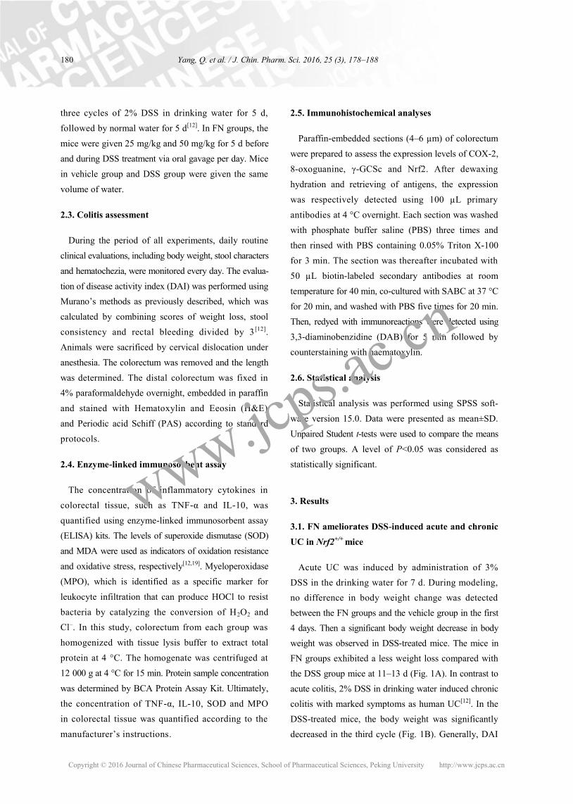
2.5. Immunohistochemical analyses
Paraffin-embedded sections (4–6 µm) of colorectum
were prepared to assess the expression levels of COX-2,
8-oxoguanine, γ-GCSc and Nrf2. After dewaxing
hydration and retrieving of antigens, the expression
was respectively detected using 100 µL primary
antibodies at 4 °C overnight. Each section was washed
with phosphate buffer saline (PBS) three times and
then rinsed with PBS containing 0.05% Triton X-100
for 3 min. The section was thereafter incubated with
50 µL biotin-labeled secondary antibodies at room
temperature for 40 min, co-cultured with SABC at 37 °C
for 20 min, and washed with PBS five times for 20 min.
Then, redyed with immunoreactions were detected using
3,3-diaminobenzidine (DAB) for 5 min followed by
counterstaining with haematoxylin.
2.6. Statistical analysis
Statistical analysis was performed using SPSS soft-
ware version 15.0. Data were presented as mean±SD.
Unpaired Student t-tests were used to compare the means
of two groups. A level of P<0.05 was considered as
statistically significant.
3. Results
3.1. FN ameliorates DSS-induced acute and chronic
UC in Nrf2+/+ mice
Acute UC was induced by administration of 3%
DSS in the drinking water for 7 d. During modeling,
no difference in body weight change was detected
between the FN groups and the vehicle group in the first
4 days. Then a significant body weight decrease in body
weight was observed in DSS-treated mice. The mice in
FN groups exhibited a less weight loss compared with
the DSS group mice at 11–13 d (Fig. 1A). In contrast to
acute colitis, 2% DSS in drinking water induced chronic
colitis with marked symptoms as human UC[12]. In the
DSS-treated mice, the body weight was significantly
decreased in the third cycle (Fig. 1B). Generally, DAI
Copyright © 2016 Journal of Chinese Pharmaceutical Sciences, School of Pharmaceutical Sciences, Peking University http://www.jcps.ac.cn
www.jcps.ac.cn

181 Yang, Q. et al. / J. Chin. Pharm. Sci. 2016, 25 (3), 178–188
exhibited features of loose feces, hematochezia and
body weight reduction, and it was used to evaluate
inflammation severity in colitis mice. The score of DAI
was significantly increased after DSS intake, whereas
it was markedly attenuated in FN-treated group. Colon
shortening is another index to reflect the disease severity
of colorectal inflammation. Figures 1A and B show that
a significant shortening of colorectum was observed in
DSS group compared with vehicle group and FN groups.
H&E-stained colorectum sections showed that the DSS
group exhibited distortion of crypts, loss of goblet cells,
severe epithelial injury and inflammatory cell infiltration in
mucosa and submucosa. However, FN groups exhibited
obvious protection of the colon crypt structures and
less severe histologic inflammation. The result of PAS
showed that goblet cells were significantly reduced in
DSS-treated group, and they were obviously increased
in FN groups (Fig. 2). These results suggested that
FN was capable of preventing DSS-induced colitis
in a dose-dependent manner.
Figure 1. Improvement role of FN against DSS-induced acute and chronic colitis in Nrf2+/+ mice. (A) Body weight changes after 3% DSS induction of colitis, DAI, intestines photograph and statistics of colorectum length of each group. (B) Body weight changes, DAI, intestine photograph and statistics of colorectum length in each group of 2% DSS induced chronic coliti s.
130
120
110
100
90
80
70 1 2 3 4 5 6 7 8 9 10 11 12 13 14
t (d)
Inti
al
bo
dy w
eig
ht
(%)
Vehicle DSS DSS + FN-1 DSS + FN-2
8
6
4
2
0 1 2 3 4 5 6 7 8 9 10 11 12 13 14
t (d)
DA
I
Vehicle DSS DSS + FN-1 DSS + FN-2
Vehicle
DSS
DSS + FN-1
DSS + FN-2
100
80
60
40
20
0 DSS – + + + FN (mg/kg) 0 0 25 50
Colo
rect
al
length
(m
m) ***
***
*** ***
** **
*** *** *
* *
(A) 120
110
100
90
80 1 3 5 7 9 11 13 15 17 19 21 23 25 27 29 31 33 35 37
t (d)
Inti
al
bo
dy w
eig
ht
(%)
Vehicle DSS DSS + FN-1 DSS + FN-2
4
3
2
1
0 1 3 5 7 9 11 13 15 17 19 21 23 25 27 29 31 33 35 37
t (d)
DA
I
Vehicle DSS DSS + FN-1 DSS + FN-2
Vehicle
DSS
DSS + FN-1
DSS + FN-2
100
80
60
40
20
0 DSS – + + + FN (mg/kg) 0 0 25 50
Colo
rect
al
length
(m
m) ***
*
***
**
*
(B)
***
*
*
Copyright © 2016 Journal of Chinese Pharmaceutical Sciences, School of Pharmaceutical Sciences, Peking University http://www.jcps.ac.cn
www.jcps.ac.cn

182 Yang, Q. et al. / J. Chin. Pharm. Sci. 2016, 25 (3), 178–188
3.2. FN regulates cytokine expression levels in
colorectum of Nrf2+/+ mice with DSS-induced acute
and chronic colitis
TNF-α, IL-10, and COX-2 are known to play a pivotal
role in the inflammation of colitis. Compared with the
vehicle group, the levels of TNF-α and COX-2 were
significantly increased in the colorectum of DSS group.
However, the administration of FN-2 (50 mg/kg/day)
significantly suppressed the accumulation of TNF-α and
COX-2 in the colonic tissues of mice with DSS-induced
acute and chronic colitis, but there was no significant
difference between FN-1 (25 mg/kg/day) and DSS
group in chronic colitis model. Additionally, the
anti-inflammatory cytokine IL-10 was decreased to
some extent after DSS induction, but IL-10 level was
significantly improved in the FN-2-treated mice. While
the level of IL-10 showed no significant difference
between FN-1 and DSS group in chronic colitis model
(Fig. 3). Taken together, our data suggested that FN
exerted an anti-inflammatory action in DSS-induced
acute and chronic colitis.
3.3. Effects of FN on oxidative stress in colorectum of
Nrf2+/+ mice with DSS-induced acute and chronic colitis
Oxidative stress parameters, such as SOD, MDA,
MPO and 8-oxoguanine, were determined in the present
work. The results showed that SOD activity was decreased
in DSS group, but it was dramatically increased in FN-2
group. The concentration of MDA, MPO and 8-oxoguanine
was significantly increased in DSS group, and FN-2 could
significantly inhibit the increase of these oxidative
stress parameters. However, no difference between
FN-1 and DSS group was found in the level of SOD
and MDA (Fig. 4). These results indicated that FN
possessed anti-oxidant properties and could prevent
DNA damage to some extent.
Figure 2. Protection of FN against intestinal lesion during acute and chronic colitis induced by DSS in Nrf2+/+ mice. (A) Representative H&E-stained and PAS colorectum sections (magnification ×400) in acute colitis mice. (B) H&E-stained and PAS colorectum sections in chron-ic colitis mice.
Vehicle DSS DSS + FN-1 DSS + FN-2 (A)
PA
S
H
&E
Acute
co
liti
s
Vehicle DSS DSS + FN-1 DSS + FN-2 (B)
PA
S
H
&E
Chro
nic
co
liti
s
Copyright © 2016 Journal of Chinese Pharmaceutical Sciences, School of Pharmaceutical Sciences, Peking University http://www.jcps.ac.cn
www.jcps.ac.cn

183 Yang, Q. et al. / J. Chin. Pharm. Sci. 2016, 25 (3), 178–188
3.4. FN can improve Nrf2 expression and protect
DSS-induced colitis in Nrf2+/+ mice
Nrf2 and γ-GCSc play a key role in antioxidant
mechanism. Based on expression level of the inflamma-
tory cytokines and antioxidants, Nrf2 and γ-GCSc
were also measured[19]. The protein levels of Nrf2
and γ-GCSc were increased in DSS-treated group
and could be reversed by the administration of FN,
suggesting that FN could increase Nrf2 expression and
reduce the oxidative stress (Fig. 5).
3.5. Effect of FN on DSS-induced chronic colitis in
Nrf2–/– mice
Nrf2–/– mice were used to verify the modulation of Nrf2 in
DSS-induced chronic colitis. The expression level of Nrf2 in
Nrf2+/+ and Nrf2–/– mice was determined and presented in
Figure 6. Figure 7 shows that no significant change in
weight, DAI, the length of colorectum and histopathology
was observed in Nrf2–/– mice treated by 2% DSS and FN.
These data suggested that FN had no protective effect
against DSS induced chronic colitis in Nrf2–/– mice.
Figure 3. Effects of FN on the production of inflammatory cytokines in colorectums of Nrf2+/+ mice with DSS-induced acute and chronic ulcerative colitis. (A) TNF-α, IL-10 in colorectums of acute ulcerative colitis model were determined by ELISA, and levels of COX-2 were examined by immunohistochemistry from colon sections (magnification ×100). (B) The concentration of TNF-α, IL-10 and the expression level of COX-2 in chronic ulcertive colitis model. *P<0.05; **P<0.01; ***P<0.001, versus DSS-treated group. Data are presented as mean SD of 7 mice in each group.
25
20
15
10
5
0 DSS – + + + FN (mg/kg) 0 0 25 50
Colo
nic
TN
F-α
(p
g/m
g p
rote
in)
***
* (A)
40
30
20
10
0 DSS – + + + FN (mg/kg) 0 0 25 50
Co
lon
ic I
L-1
0 (
pg/m
g p
rote
in)
*
60
40
20
0 DSS – + + + FN (mg/kg) 0 0 25 50
Colo
nic
TN
F-α
(p
g/m
g p
rote
in) ***
150
100
50
0 DSS – + + + FN (mg/kg) 0 0 25 50
Co
lon
ic I
L-1
0 (
pg/m
g p
rote
in)
***
**
(B)
Vehicle DSS
CO
X-2
DSS + FN-1 DSS + FN-2
Vehicle DSS
CO
X-2
DSS + FN-1 DSS + FN-2
Copyright © 2016 Journal of Chinese Pharmaceutical Sciences, School of Pharmaceutical Sciences, Peking University http://www.jcps.ac.cn
www.jcps.ac.cn

184 Yang, Q. et al. / J. Chin. Pharm. Sci. 2016, 25 (3), 178–188
Figure 5. The effects of FN on Nrf2 and γ-GCSc expression in colorectums of Nrf2+/+ mice with DSS-induced acute and chronic ulcerative colitis. (A) Expression of Nrf2 and γ-GCSc in colorectums of acute ulcerative colitis. (B) Levels of Nrf2 and γ-GCSc in colorectums of chronic ulcerative colitis.
40
30
20
10
0
Figure 4. Protection of FN against oxidative stress in colorectums during acute and chronic ulcerative colitis induced by DSS in Nrf2+/+ mice. (A) SOD activity, MDA MPO content and 8-oxoguanine levels (magnification ×100) in colorectal tissue of acute ulcerative colitis. (B) SOD activity, MDA and MPO content, and 8-oxoguanine levels in colorectal tissue of chronic ulcerative colitis. *P<0.05; **P<0.01; ***P<0.001, versus DSS-treated group. Data are presented as mean SD of 7 mice in each group.
DSS – + + + FN (mg/kg) 0 0 25 50
SO
D (
U/m
g p
rote
in)
***
(A)
DSS – + + + FN (mg/kg) 0 0 25 50
MD
A (
nm
ol/
mg p
rote
in) *
15
10
5
0
DSS – + + + FN (mg/kg) 0 0 25 50
MP
O (
pg/m
g p
rote
in)
* 18000
12000
6000
0
*
Vehicle DSS DSS + FN-1 DSS + FN-2
8-O
xo
guanin
e
DSS – + + + FN (mg/kg) 0 0 25 50
SO
D (
U/m
g p
rote
in)
* (B)
DSS – + + + FN (mg/kg) 0 0 25 50
MD
A (
nm
ol/
mg p
rote
in)
*
50
40
30
20
10
0
DSS – + + + FN (mg/kg) 0 0 25 50
MP
O (
pg/m
g p
rote
in)
**
20000
15000
10000
5000
0
*
Vehicle DSS DSS + FN-1 DSS + FN-2
8-O
xo
guan
ine
60
40
20
0
Acute
co
liti
s
Vehicle DSS DSS + FN-1 DSS + FN-2 (A)
γ-G
CS
c
Nrf
2
Ch
ronic
co
liti
s
Vehicle DSS DSS + FN-1 DSS + FN-2 (B)
γ-G
CS
c
Nrf
2
Copyright © 2016 Journal of Chinese Pharmaceutical Sciences, School of Pharmaceutical Sciences, Peking University http://www.jcps.ac.cn
www.jcps.ac.cn

185 Yang, Q. et al. / J. Chin. Pharm. Sci. 2016, 25 (3), 178–188
Figure 6. The expression levels of Nrf2 in Nrf2+/+ and Nrf2–/– mice. (A) The levels of Nrf2 exermined by Immunohistochemistry (magnification ×100). (B) The Nrf2 mRNA expression levels were determined by real-time PCR.
Figure 7. No improvement of FN against DSS-induced chronic colitis in Nrf2–/– mice. (A) Body weight changes after DSS induction of colitis. (B) DAI. (C) Statistics of colorectum length. (D) Representative H&E-stained and PAS-stained colorectum sections (magnification ×400).
4. Discussion
UC is associated with genetic and environment leading
to impairment of the intestinal mucosal barrier. Previous
studies have reported that IL-10–/–, TRAF6IEC-KO and
TMF–/– mice are susceptible to colitis[21,22]. Moreover,
attenuation of the Bin1 (Bridging integrator 1) gene
can limit UC pathogenicity in the mouse by supporting
mucosal barrier function and protecting integrity of the
lymphoid follicle, offering a novel strategy to treat UC
and possibly limiting risks of colorectal cancer[23]. At
present, an increasing number of studies are focusing on
the relationship between UC and gut microorganism[22,24].
The balanceable state would be destroyed as these
invasive antigens are introduced into the organism,
then the immune cells are activated and cytokines are
produced. When the anti-inflammation mechanisms are
not able to resolve the acute mocosal inflammation, pro-
inflammation responds to organism, showing up-regulation
of pro-inflammation cytokines, and inflammatory cellular
infiltration. More seriously, chronic inflammation may
cause tissue destruction such as fibrosis, abscess, fistula
and even cancer, which are driven by mucosal cytokine
infiltration[25]. UC is generally treated with anti-inflammatory
Nrf2+/+ Nrf2–/– (A)
Nrf+/+ Nrf–/–
(B) 5
4
3
2
1
0
Nrf
2 m
RN
A e
xpre
ssio
n
in c
olo
rect
um
Vehicle DSS DSS + FN-1 DSS + FN-2 (D)
PA
S
H
&E
Nrf
2–/–
DSS – + + + FN (mg/kg) 0 0 25 50
Co
lore
ctal
len
gth
(m
m)
(C) 100
80
60
40
20
0
Nrf2–/–
NS NS
120
110
100
90
80
70 1 3 5 7 9 11 13 15 17 19 21 23 25 27 29 31 33 35 37
t (d)
Inti
al
bo
dy w
eig
ht
(%)
Vehicle DSS DSS + FN-1 DSS + FN-2
(A) Nrf2–/–
NS NS
6
4
2
0
DA
I
1 3 5 7 9 11 13 15 17 19 21 23 25 27 29 31 33 35 37 t (d)
Vehicle DSS DSS + FN-1 DSS + FN-2
Nrf2–/–
NS NS
(B)
Copyright © 2016 Journal of Chinese Pharmaceutical Sciences, School of Pharmaceutical Sciences, Peking University http://www.jcps.ac.cn
www.jcps.ac.cn

186 Yang, Q. et al. / J. Chin. Pharm. Sci. 2016, 25 (3), 178–188
or immunosuppressive drugs. In recent years, more and
more studies have been conducted to develop effective
treatment for colitis. A new study has found that glucagon-
like peptide-2 (GLP-2) microspheres, a proper candidate
for the treatment of UC, is resistant to degradation and
can decrease the severity of DSS-induced UC[26].
Enoxaparin, a widely used antithrombotic agent, has been
reported to possess anti-inflammatory properties, and
it can significantly reduce the inflammatory pathology
associated with DSS-induced colitis in mice therefore
representing a novel therapeutic option for the manage-
ment of UC[27]. However, current therapies can only
reduce the inflammatory process of IBD, and improved
treatment modalities for the complex IBD are still
evolving. Therefore, haematopoietic stem cells (HSC)
and mesenchymal stromal cells (MSC) therapies are
both being investigated for the IBD treatment [28].
Although clinical drugs for the treatment of colitis have
been reported, most of these treatments often have
various side effects. Consequently, many patients turn
to alternative therapies, such as traditional plant-based
remedies. Geraniol has been proved as a potential
therapeutic agent for IBD by inhibiting pro-inflammatory
cytokines and NF-κB signaling[29].
In this study, we investigated the anti-inflammation
mechanism of FN in the vivo model of acute and
chronic colitis induced by DSS in C57BL/6 mice. After
treated with DSS, the mice exhibited clinical symptoms
and pathalogic change corresponding to those of human
UC, including loss of body weight, diarrhoea, bloody
faeces, mucosal ulceration and shortening of colon.
Two different concentrations of FN along with DSS
were used to evaluate its therapeutic effect on UC. The
results demonstrated that FN significantly suppressed
DSS-induced colitis in a dose-dependent manner by
decreasing DAI scores, weight loss and colonic
shortening. H&E and PAS findings were consistent with
DAI data. As shown in Figure 2, the colons of mice
treated with DSS presented more severe inflammatory
cell infiltration, mucosal erosion, and both distortion
and loss of crypts. In contrast, administration of FN
reduced all these above mentioned inflammatory changes,
suggesting that FN could ameliorate the inflammatory
condition induced by DSS. In order to further explore
the therapeutic mechanism of FN, the production of
pro-inflammatory cytokines (TNF-α, COX-2) and
anti-inflammatory (IL-10) were measured in colonic
tissue[30]. ELISA and immunohistochemical results
confirmed the up-regulation of pro-inflammatory
cytokines TNF-α and COX-2, and down-regulation of
IL-10 in DSS induced group. In contrast, a distinct
decrease in concentrations of TNF-α and COX-2 and
an increase in IL-10 concentration were observed
when FN was administered along with DSS.
UC is associated with oxidative stress. SOD, MDA
and MPO are important markers in oxidative stress.
Previous reports have proved a significant drop in SOD
activity and an obvious increase in the concentrations
of MDA and MPO in DSS-induced chronic colitis,
which were consistent with our experimental results.
However, increased MDA level and MPO activity
as well as reduced SOD activity in colonic tissue of
DSS-colitis group were significantly improved after oral
FN administration. Besides, the level of 8-oxoguanine,
a DNA damage marker, was significantly increased in
colorectum of DSS model, and it was set to normal in
DSS along with FN. Therefore, our results obviously
showed that FN could exert an anti-oxidative effect by
improvement of SOD activity and reducing peroxidation
of polyunsaturated fatty acids and DNA damage.
Nrf2 is a key transcription factor that protects cells
against oxidative stress by producing phase-2 detoxifying
enzymes and anti-oxidative enzymes[8,31,32]. Previous
studies have shown that Nrf2–/– mice are sensitive to
DSS-induced colitis, displaying significant increase in
incidence, colonic crypt loss, inflammatory cell infiltration
and so on[33]. Above all, inflammatory cytokines, such as
IL-1β, IL-6, TNF-α, iNOS and COX-2, are increased
in Nrf2–/– mice. Nrf2 signaling is also involved in
inflammation associated pathogenesis, such as gastritis,
colitis, rheumatoid arthritis and atherosclerosis. In our
study, DSS-induced chronic colitis in Nrf2–/– showed
no significant distinction compared with those of FN
treated group. However, the expression Nrf2 in mice
Copyright © 2016 Journal of Chinese Pharmaceutical Sciences, School of Pharmaceutical Sciences, Peking University http://www.jcps.ac.cn
www.jcps.ac.cn

187 Yang, Q. et al. / J. Chin. Pharm. Sci. 2016, 25 (3), 178–188
without genetic defect was significantly improved with
FN treatment. Above results suggested that the doses
of 50 mg/kg FN was superior to 25 mg/kg, and the two
doses could ameliorate DSS-induced acute and chronic
ulcerative colitis in mice.
Based on the findings of this study, we confirmed
that FN exhibited anti-inflammatory and anti-oxidant in
vivo by improving Nrf2 expression, activating anti-oxiditive
and anti-inflammatory gene expression. In the process
of colitis treatment, FN might be an effective candidate
for the treatment of UC.
Acknowledgements
This work was supported by the National Natural
Science Foundation of China (Grant No. 81274150,
81573680 and 81470179) and Jiangsu Province’s
Outstanding Leader Program of Traditional Chinese
Medicine.
References
[1] Trallori, G; Palli, D.; Saieva, C.; Bardazzi, G.; Bonanomi,
A.G.; d'Albasio,G.; Galli, M.; Vannozzi, G.; Milla, M.;
Tarantino, O.; Renai, F.; Messori, A.; Amorosi, A.;
Pacini, F.; Morettini, A. Scand. J. Gastroenterol. 1996,
31, 892–899.
[2] Grinspan, A.; Kornbluth, A. Curr. Gastroenterol. Rep.
2015, 17, 454.
[3] Khor, B.; Gardet, A.; Xavier, R.J. Nature. 2011, 474,
307–317.
[4] Ma, W.; Tang, S.; Cao, M.; Ge, Z.; Li, R.; Yu, S. J. Chin.
Pharm. Sci. 2014, 23, 610– 616.
[5] Ohkusa, T.; Koido, S. J. Infect. Chemother. 2015, 21,
761–768.
[6] Cetinkaya, Z.A.; Cetinkaya, Y.; Gencer, M.; Sezikli, M.;
Tireli, H.; Kurdaş, O.Ö.; Uluç, K.; Us, O.; Tanrıdağ, T.
Gut Liver. 2011, 5, 57–60.
[7] Sato, K.; Nito, N.;Yoshikawa, S.; Ando, T.; Karino, M.;
Ito, C.; Ikawa, R.; Ishiguro, H.; Takamatsu, M.;
Kobayashi, M.; Tsutazawa, T.; Oi, F.; Miyamori, T. Stud.
Health. Technol. Inform. 2015, 216, 325–328.
[8] Pereira, C.; Grácio, D.; Teixeira, J.P.; Magro, F. Inflamm.
Bowel. Dis. 2015, 21, 2403–2417.
[9] Parmar, A.R.; Trivedi, P.P.; Jena, G.B. Reprod. Toxicol.
2014 , 49, 171–184.
[10] Li, W.; Khor T.O.; Xu, C; Shen, G.; Jeong, W.S.; Yu, S.;
Kong, A.N. Biochem. Pharmacol. 2008, 76, 1485–1489.
[11] Cho, H.Y.; Kleeberger, S.R. Toxicol. Appl. Pharmacol.
2010, 244, 43–56.
[12] Chen, G.; Yang, Y.; Liu, M.; Teng, Z.; Ye, J.; Xu, Y.;
Cai, X.; Cheng, X.; Yang, J.; Hu, C.; Wang, M.; Cao, P.
J. Ethnopharmacol. 2015, 166, 146–156.
[13] Sánchez-Fidalgo, S.; Villegas, I.; Rosillo, M.Á.;
Aparicio-Soto, M.; de la Lastra, C.A. Mol. Nutr. Food.
Res. 2014, 59, 284–292.
[14] Yang, Y.; Cai, X.; Yang, J.; Sun, X.; Hu, C.; Yan, Z.;
Xu, X.; Lu, W.; Wang, X.; Cao, P. Mol. Cancer.
2014, 6, 13–48.
[15] Kathy Ka-Wai, A.Y.; Pui-Ching, L.; Joshua Ka-Shun, K.
Oncol. Rep. 2012, 28, 2188–2194.
[16] Jun, M.; Hong, J.; Jeong, W.S.; Ho, C.T. Mol. Nutr.
Food. Res. 2005, 49, 1154–1159.
[17] Yang, Y.; Zhao, Y.; Ai, X.; Cheng, B.; Lu, S. Int. J. Clin.
Exp. Pathol. 2014, 7, 8453–8461.
[18] Zhou, R.; Xu, L.; Ye, M.; Liao, M.; Du, H.; Chen, H.
Horm. Metab. Res. 2014, 46, 753–760.
[19] Shu, D.Z.; Wan, X.H.; Liu, H.R.; Yang, J.Q.; Zhou, Q.X.
J. Chin. Pharm. Sci. 2006, 15, 182–187
[20] Li, G.; Lee, M.J.; Liu, A.B.; Yang, Z.; Lin, Y.; Shih,
W.J.; Yang, C.S. Free Radic. Biol. Med. 2012, 52,
1151–1158.
[21] Bel, S.; Elkis, Y.; Elifantz, H.; Koren, O.; Ben-Hamo,
R.; Lerer-Goldshtein, T.; Rahimi, R.; Ben Horin, S.;
Nyska, A.; Shpungin, S.; Nir, U. Proc. Natl. Acad. Sci.
USA. 2014, 111, 4964–4969.
[22] Vlantis, K.; Polykratis, A.; Welz, P.S.; van Loo, G.;
Pasparakis, M.; Wullaert, A. Gut. 2015, 308–323.
[23] Thomas, S.; Mercado, J.M.; DuHadaway, J.; DiGuilio, K.;
Mullin, J.M.; Prendergast, G.C. Dig. Dis. Sci. 2015, 1–10.
[24] Verma, N.; Verma, R.; Kumari, R.; Ranjha, R.; Paul, J.
Inflamm. Res. 2014, 63, 161–169.
[25] Khansari, N.; Shakiba, Y.; Mahmoudi, M. Recent. Pat.
Inflamm. Allergy. Drug Discov. 2009, 3, 73–80.
Copyright © 2016 Journal of Chinese Pharmaceutical Sciences, School of Pharmaceutical Sciences, Peking University http://www.jcps.ac.cn
www.jcps.ac.cn

188 Yang, Q. et al. / J. Chin. Pharm. Sci. 2016, 25 (3), 178–188
[26] Wu, J.; Qi, K.; Xu, Z.; Wan, J. J. Microencapsul. 2015,
32, 1–10.
[27] Lean, Q.Y.; Eri, R.D.; Randall-Demllo, S.; Sohal, S.S.;
Stewart, N.; Peterson, G.M.; Gueven, N.; Patel, R.P.
PLoS One. 2015, 10, e0134259. doi: 10.1371/journal
[28] Irhimeh, M.R; Cooney, J. Curr. Stem Cell. Res. Ther.
2016, 11, 72–77.
[29] Medicherla, K.; Sahu, B.D.; Kuncha, M.; Kumar, J.M.;
Sudhakar, G.; Sistla, R. Food Funct. 2015, 6, 2984–2995.
[30] Neurath, M.F. Nat. Rev. Immunol. 2014, 14, 329–342.
[31] Yang, S.; Yu, S. J. Chin. Pharm. Sci. 2014, 23, 837–843.
[32] Que, L.L.; Wang, H.X.; Cao, B.S.;Yang, B.S.; Wang, K.;
Yu, S.W. J. Chin. Pharm. Sci. 2011, 20, 5–19 .
[33] Khor, T.O.; Huang, M.T; Prawan, A.; Liu, Y.; Hao, X;
Yu, S.; Cheung, W.K.; Chan, J.Y.; Reddy, B.S.; Yang, C.S.;
Kong, A.N. Cancer Prev. Res. 2008, 1, 187–191.
刺芒柄花素通过诱导Nrf2表达缓解DSS诱导的小鼠溃疡性结肠炎
杨倩1,2#, 陈刚2#, 杨洋2, 蔡雪婷2, 庞中化2, 胡春萍2, 张双全1*, 曹鹏2*
1. 南京师范大学 生命科学学院 分子生物技术与医学实验室, 江苏 南京 210046
2. 江苏省中医药研究院 细胞与分子生物学实验室, 江苏 南京 210028
摘要: 异黄酮刺芒柄花素是从红车轴草中提取的一种主要的活性成分。红车轴草是一种具有抗炎和抗癌功能的药用
植物。在本研究中, 我们旨在研究刺芒柄花素对DSS诱导的急性和慢性小鼠溃疡性结肠炎的作用。研究结果发现刺芒柄
花素 (25, 50 mg/kg) 可以明显地减弱DSS引起的体重下降, 疾病活动指数评分, 肠度缩短和组织损伤。进一步研究发现, 在
给予刺芒柄花素的保护组中, 肿瘤坏死因子(TNF-α), 白介素-6(IL-6) 和环氧合酶(COX-2)的含量都会明显降低; 结肠组织
中具有代表性的氧化压力参数, 包括超氧化物歧化酶和髓过氧化物酶的活性, 以及丙二醛和8-羟基鸟嘌呤的含量明显得到
改善;同时, 研究中还发现刺芒柄花素保护组中Nrf2表达量明显升高, 但是Nrf2基因敲除小鼠慢性溃疡性结肠炎症状没有
得到缓解。综上所述, 我们推断刺芒柄花素可以通过激活Nrf2的表达保护溃疡性结肠炎的发生。结果证明刺芒柄花素
可能对治疗溃疡性结肠炎具有潜在的作用价值。
关键词: Nrf2; 溃疡性结肠炎; 异黄酮刺芒柄花素; 炎症因子; 氧化应激
Copyright © 2016 Journal of Chinese Pharmaceutical Sciences, School of Pharmaceutical Sciences, Peking University http://www.jcps.ac.cn
www.jcps.ac.cn