Eye – lecture plan • Layers of eye• Chambers of eye• Segments of eye• Retractile (Dioptric) Media of Eye
• Development ( embryology) of Eye• Histology of
– Corneo-scleral part– Uvea ( Iris, Ciliary body, choroid)– Retina – (layers, rods & cones)– Lens

outer: corneo – scleral
Middle: Urea with its choroid, ciliary body and Iris
Inner: Retina has two layers (outer pigment and inner neuronal)
Layers

Chambers of Eye

Segments of Eye
• Anterior segment– Cornea – Contents of anterior chamber– Contents of posterior chamber
• Posterior segment– Vitreous chamber– Retinal layers, posterior sclera– uvea

Retractile (Dioptric) Media of Eye
• 1. Cornea –
– anterior window of light
– chief refractive element (1.378)
• 2. Aqueous humor –
– watery fluid in anterior and posterior chambers
– minor role in retractile media
– important in nutrition of cornea & lens (avascular organs)
• 3. Lens
– transparent, crystalline,
– important refractive part after cornea,
– suspended by zonules of Zinn
• 4. Vitreous body
– transparent gel,
– 99% is water with water soluble proteins, Hyaluronic acid, glycoproteins,
– acts as shock absorber (protects retina & maintains shape of eyes)

Development of Eye

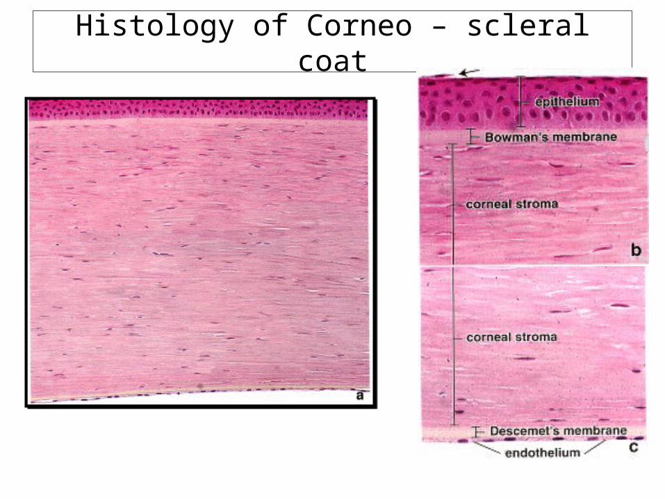
Histology of Corneo – scleral coat

Histology of Corneo – scleral coat
• Cornea – has five layers– Epithelium – nonkeratinized stratified squamous , basal layer
cells ( like skin) proliferate and replace the lost suface epithelial cells
– Bowman’s membrane – fibrillar, ends at limbus, main role in limiting the spread of infections
– Connective tissue stroma – 90% of corneal thickness, contains proteoglycans, So4 GAGs (Keratan and chodroitin So4)
– Decemet’s membrane – basal lamina of endothelial cells, made of meshwork of fibers and pores, regenerates after injury (unlike Bowman’s)
– Endothelium – single layer of squamous cells, all metabolic exchanges takes place here,
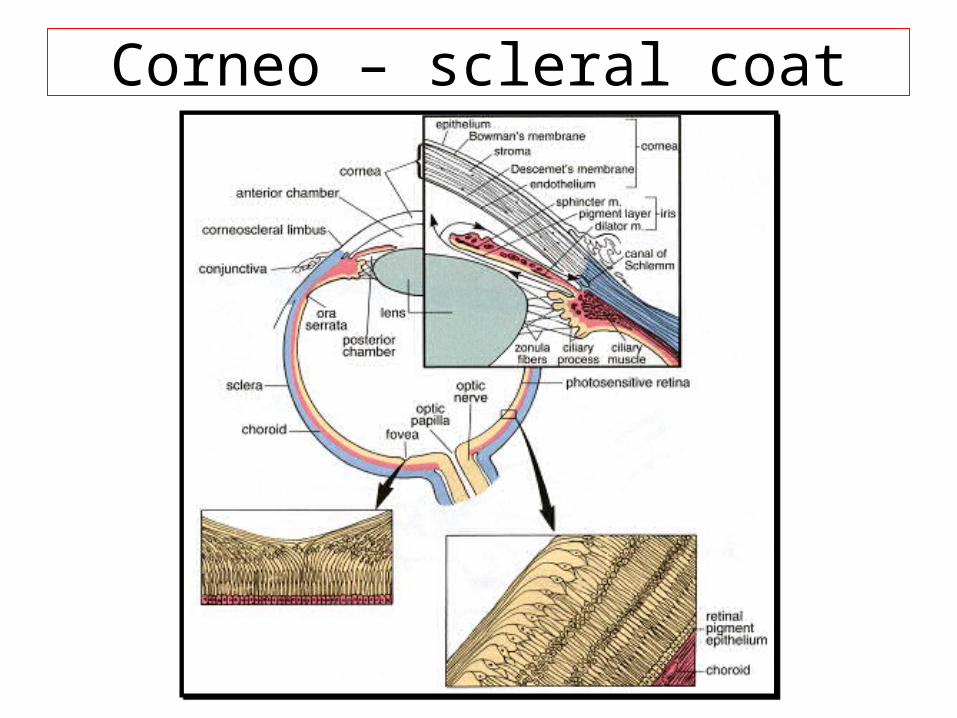
Corneo – scleral coat

Histology of Corneo – scleral coat
• Cornea– How cornea is transparent?– By precise regulation of water in stroma, if there is
endothelial damage corneal edema and corneal opacity
• Sclera – Dense connective tissue of flat collagen fibers and
meshwork of elastic fibers• Limbus
– transition zone – Has irido- corneal angle for drainage of aqueous
humor ( canal of schlemn)

Uvea - iris

Vascular coat (Uvea)
• Iris- most anterior part, forms diaphragm, pupil is central aperture, posterior pigment epithelium and myoepithelial layer next, two muscles (dilator and constrictor pupillae)
• Muscle of adaptation– Sphincter pupillae – circular band of SMC, parasympathetic
control ( CN III), causes reduced size of pupil in response to light– Dilator pupillae – radially oriented pigmented myoepithelial
cells, form anterior pigment epithelium, under sympathetic control (superior cervical ganglion), causes increased pupillary size in response to dim light
• Ciliary body – has ora serrata, anterior part is ciliary process, has ciliary muscle with three functional groups ( longitudinal – for drainage of aqueous, radial – flatten the lens for distant vision, circular- reduce tension on lens for near vision

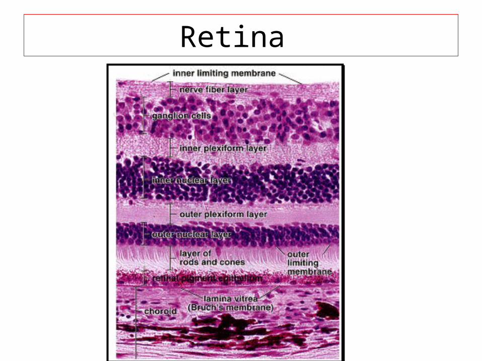
Retina
• Inner most layer• Consists of 10 layers of cells• Functionally two regions
– Non photosensitive (non visual) region– Photosensitive ( optic) region
• Optic disc or fovea centralis or blind spot in visual field – where optic nerve joins retina
• Retinal pigment epithelium – has important functions (absorb light and reduce glare, blood- retina barrier, Phagocytosis)
• Nuclei of Rods and cones form outer nuclear layer
Retina

Rods & Cones

Rods & Cones
• Rods – more in # (12 million), more sensitive to light, used in dim or night light), have maximum absorption at 496 nm of light ( black and white pictures)
• Cones – less in # (7million), three classes (L,M,S), less sensitive to light ( for day vision), have absorption at 420 (blue), 531(green) and 588 nm (red) of light, for color vision

Crystalline lens
Crystalline lens
• Transparent, avascular, biconvex, • Lens capsule – type IV collagen, • New lens fibers are produced through out the
life• Presbyopia – decreased elasticity and power
of accommodation with age• Cataract – loss of transparency, causes can
be infections, metabolic, hereditary, trauma, UV light