Progress in Lipid Research 44 (2005) 1–51
Progress inLipid Research
www.elsevier.com/locate/plipres
Review
Epoxide hydrolases: their roles and interactionswith lipid metabolism
John W. Newman, Christophe Morisseau, Bruce D. Hammock *
Department of Entomology, UCDavis Cancer Center, University of California, One Shields Avenue,
Davis, CA 95616, USA
Abstract
The epoxide hydrolases (EHs) are enzymes present in all living organisms, which transform epoxide con-
taining lipids by the addition of water. In plants and animals, many of these lipid substrates have potent
biologically activities, such as host defenses, control of development, regulation of inflammation and blood
pressure. Thus the EHs have important and diverse biological roles with profound effects on the physiolog-
ical state of the host organisms. Currently, seven distinct epoxide hydrolase sub-types are recognized in
higher organisms. These include the plant soluble EHs, the mammalian soluble epoxide hydrolase, the hep-
oxilin hydrolase, leukotriene A4 hydrolase, the microsomal epoxide hydrolase, and the insect juvenile hor-
mone epoxide hydrolase. While our understanding of these enzymes has progressed at different rates, herewe discuss the current state of knowledge for each of these enzymes, along with a distillation of our current
understanding of their endogenous roles. By reviewing the entire enzyme class together, both commonali-
ties and discrepancies in our understanding are highlighted and important directions for future research
pertaining to these enzymes are indicated.
� 2004 Elsevier Ltd. All rights reserved.
0163-7827/$ - see front matter � 2004 Elsevier Ltd. All rights reserved.
doi:10.1016/j.plipres.2004.10.001
* Corresponding author. Tel.: +1 530 752 7519; fax: +1 530 752 1537.
E-mail address: [email protected] (B.D. Hammock).
Nomenclature
ChEH cholesterol epoxide hydrolaseCOX cyclooxygenase, prostaglandin G/H synthaseDHET dihydroxy eicosatrienoic acidDHO dihydroxy octadecanoic acidDHOME dihydroxy octadecenoic acidEH epoxide hydrolaseEET epoxy eicosatrienoic acidEpOME epoxy octadecenoic acidFABP fatty acid binding proteinJH juvenile hormoneJHEH juvenile hormone epoxide hydrolaseHPETE hydroperoxy eicosatrienoic acidLDLR low density lipoprotein receptorLPL lipoprotein lipaseLTA4 leukotriene A4
LTB4 leukotriene B4
mEH microsomal epoxide hydrolaseNFjB nuclear factor kappa BPDK pyruvate dehydrogenase kinasePMN polymorphonuclear leukocytesPPARa peroxisome proliferator activated receptor alphasEH soluble epoxide hydrolaseTCPO 3,3,3-trichloropropene-1,2-oxideTHETA trihydroxy eicosatrieneoic acid
2 J.W. Newman et al. / Progress in Lipid Research 44 (2005) 1–51
Contents
1. Introduction . . . . . . . . . . . . . . . . . . . . . . . . . . . . . . . . . . . . . . . . . . . . . . . . . . . . . . . . . . . . . . . . . . . . 3
2. Soluble epoxide hydrolases . . . . . . . . . . . . . . . . . . . . . . . . . . . . . . . . . . . . . . . . . . . . . . . . . . . . . . . . . . 5
2.1. The plant sEHs . . . . . . . . . . . . . . . . . . . . . . . . . . . . . . . . . . . . . . . . . . . . . . . . . . . . . . . . . . . . . 5
2.1.1. Tissue distribution and sub-cellular localization . . . . . . . . . . . . . . . . . . . . . . . . . . . . . . . . . 5
2.1.2. Substrates . . . . . . . . . . . . . . . . . . . . . . . . . . . . . . . . . . . . . . . . . . . . . . . . . . . . . . . . . . . 6
2.1.3. Regulation . . . . . . . . . . . . . . . . . . . . . . . . . . . . . . . . . . . . . . . . . . . . . . . . . . . . . . . . . . . 7
2.1.4. Physiological role: cutin biosynthesis and host defense . . . . . . . . . . . . . . . . . . . . . . . . . . . . 7
2.2. The mammalian sEHs. . . . . . . . . . . . . . . . . . . . . . . . . . . . . . . . . . . . . . . . . . . . . . . . . . . . . . . . . 8
2.2.1. Tissue distribution and sub-cellular localization . . . . . . . . . . . . . . . . . . . . . . . . . . . . . . . . . 8
2.2.2. C-terminal domain substrates: epoxy fatty acids. . . . . . . . . . . . . . . . . . . . . . . . . . . . . . . . . 9
2.2.3. N-terminal domain substrates: lipid phosphates . . . . . . . . . . . . . . . . . . . . . . . . . . . . . . . . 11
2.2.4. Regulation . . . . . . . . . . . . . . . . . . . . . . . . . . . . . . . . . . . . . . . . . . . . . . . . . . . . . . . . . . 12
2.2.5. Physiological roles . . . . . . . . . . . . . . . . . . . . . . . . . . . . . . . . . . . . . . . . . . . . . . . . . . . . 13
J.W. Newman et al. / Progress in Lipid Research 44 (2005) 1–51 3
2.3. Hepoxilin epoxide hydrolase . . . . . . . . . . . . . . . . . . . . . . . . . . . . . . . . . . . . . . . . . . . . . . . . . . . 16
2.3.1. Tissue distribution and sub-cellular localization . . . . . . . . . . . . . . . . . . . . . . . . . . . . . . . . 17
2.3.2. Substrates . . . . . . . . . . . . . . . . . . . . . . . . . . . . . . . . . . . . . . . . . . . . . . . . . . . . . . . . . . 17
2.3.3. Regulation . . . . . . . . . . . . . . . . . . . . . . . . . . . . . . . . . . . . . . . . . . . . . . . . . . . . . . . . . . 17
2.3.4. Physiological roles . . . . . . . . . . . . . . . . . . . . . . . . . . . . . . . . . . . . . . . . . . . . . . . . . . . . 17
2.4. Leukotriene A4 hydrolase . . . . . . . . . . . . . . . . . . . . . . . . . . . . . . . . . . . . . . . . . . . . . . . . . . . . . 20
2.4.1. Tissue distribution and sub-cellular localization . . . . . . . . . . . . . . . . . . . . . . . . . . . . . . . . 20
2.4.2. Substrates . . . . . . . . . . . . . . . . . . . . . . . . . . . . . . . . . . . . . . . . . . . . . . . . . . . . . . . . . . 20
2.4.3. Regulation . . . . . . . . . . . . . . . . . . . . . . . . . . . . . . . . . . . . . . . . . . . . . . . . . . . . . . . . . . 21
2.4.4. Physiological role: inflammatory regulator . . . . . . . . . . . . . . . . . . . . . . . . . . . . . . . . . . . 22
3. Membrane associated epoxide hydrolases . . . . . . . . . . . . . . . . . . . . . . . . . . . . . . . . . . . . . . . . . . . . . . . 22
3.1. Microsomal epoxide hydrolase . . . . . . . . . . . . . . . . . . . . . . . . . . . . . . . . . . . . . . . . . . . . . . . . . . 22
3.1.1. Tissue distribution and sub-cellular localization . . . . . . . . . . . . . . . . . . . . . . . . . . . . . . . . 23
3.1.2. Substrates . . . . . . . . . . . . . . . . . . . . . . . . . . . . . . . . . . . . . . . . . . . . . . . . . . . . . . . . . . 24
3.1.3. Regulation . . . . . . . . . . . . . . . . . . . . . . . . . . . . . . . . . . . . . . . . . . . . . . . . . . . . . . . . . . 25
3.1.4. Physiological roles . . . . . . . . . . . . . . . . . . . . . . . . . . . . . . . . . . . . . . . . . . . . . . . . . . . . 26
3.2. Cholesterol epoxide hydrolase. . . . . . . . . . . . . . . . . . . . . . . . . . . . . . . . . . . . . . . . . . . . . . . . . . . 27
3.2.1. Tissue distribution and sub-cellular localization . . . . . . . . . . . . . . . . . . . . . . . . . . . . . . . . 27
3.2.2. Substrates . . . . . . . . . . . . . . . . . . . . . . . . . . . . . . . . . . . . . . . . . . . . . . . . . . . . . . . . . . 27
3.2.3. Regulation . . . . . . . . . . . . . . . . . . . . . . . . . . . . . . . . . . . . . . . . . . . . . . . . . . . . . . . . . . 27
3.2.4. Physiological roles . . . . . . . . . . . . . . . . . . . . . . . . . . . . . . . . . . . . . . . . . . . . . . . . . . . . 28
3.3. Juvenile hormone EH – the characterized insect EH . . . . . . . . . . . . . . . . . . . . . . . . . . . . . . . . . . 28
3.3.1. Tissue distribution and sub-cellular localization . . . . . . . . . . . . . . . . . . . . . . . . . . . . . . . . 29
3.3.2. Substrates . . . . . . . . . . . . . . . . . . . . . . . . . . . . . . . . . . . . . . . . . . . . . . . . . . . . . . . . . . 29
3.3.3. Regulation . . . . . . . . . . . . . . . . . . . . . . . . . . . . . . . . . . . . . . . . . . . . . . . . . . . . . . . . . . 29
3.3.4. Physiological roles . . . . . . . . . . . . . . . . . . . . . . . . . . . . . . . . . . . . . . . . . . . . . . . . . . . . 30
4. Conclusion . . . . . . . . . . . . . . . . . . . . . . . . . . . . . . . . . . . . . . . . . . . . . . . . . . . . . . . . . . . . . . . . . . . . 30
Acknowledgements . . . . . . . . . . . . . . . . . . . . . . . . . . . . . . . . . . . . . . . . . . . . . . . . . . . . . . . . . . . . . . 31
References. . . . . . . . . . . . . . . . . . . . . . . . . . . . . . . . . . . . . . . . . . . . . . . . . . . . . . . . . . . . . . . . . . . . . 31
1. Introduction
The oxidation of unsaturated lipids routinely yields epoxide-containing compounds, many ofwhich have important biological functions in a broad array of organisms. Both enzymatic [1–4]and autooxidative [5,6] routes of lipid epoxide synthesis have been reported. The chemical reac-tivity and resulting toxicity of epoxide containing chemicals can vary widely depending on chem-ical structure [7,8].
Multiple enzymes, including epoxide hydrolases (EHs), have evolved to transform epoxides intocompounds with decreased chemical reactivity, increased water solubility [9], and altered biolog-ical activity. EHs are ubiquitously found in nature. To date five EHs have been described in ver-tebrates: soluble EH (sEH), microsomal EH (mEH), cholesterol EH (ChEH), hepoxilin hydrolase

4 J.W. Newman et al. / Progress in Lipid Research 44 (2005) 1–51
and leukotriene A4 (LTA4) hydrolase [9–11]. Soluble EH orthologs are also found in plants, withroles in epoxy lipid metabolism [12–14], while the juvenile hormone EH (JHEH) is an epoxy-lipidmetabolizing enzyme in insects with homology to mEHs [15,16]. The sub-cellular localization andreported endogenous substrates of these EHs are shown in Table 1. Microbial EHs have been re-cently discussed [17] and will not be considered here.
While the soluble and microsomal EHs show structural characteristics suggesting derivationfrom a common ancestral gene, the LTA4 hydrolase is distinct [11,18]. Neither the ChEH northe hepoxilin hydrolase have been suitably characterized to evaluate structural relationship tothe other EHs [11]. However, the failure of the ChEH to form a covalent substrateintermediate suggests that it is structurally unrelated to the microsomal and soluble EHs[19]. While having different biochemical properties [20], the overlapping substrate specificityand sub-cellular localization of the sEH and the hepoxilin hydrolase suggest that these twoenzymes may serve complimentary roles. The unique nature and relative importance of thesetwo enzymes can still be debated as cytosolic hepoxilin EH-like activity is routinely reported[21].
If we consider the chemical reactivity of the various substrates, we can hypothesize two inde-pendent forces driving the evolution of these enzymes; cytoprotection vs. cellular signaling. Earlyinvestigations of these enzymes focused on their cytoprotective roles associated with toxicosis.While the mEH has a clear role in protecting cells from metabolically generated arene oxides[22–25], examples of cytoprotection mediated through other EHs are rare and generally irrelevantto environmental exposures [26,27]. The identification of endogenous substrates of these enzymes[4,28–30], and our growing understanding of their signaling functions is shedding light on thephysiological roles of various EHs.
This review will focus on the distribution, regulation, substrate/product profiles, and the endog-enous role of these enzymes within a greater context of lipid metabolism. The biochemical mech-anisms of action, as well as a more global description of substrates and inhibitors of these enzymeshave been reviewed elsewhere [11,31,32].
Table 1
Epoxide hydrolase localization and lipid substrates
Enzyme Sub-cellular localization Lipid substrates References
Plant soluble EH Cytosol; glyoxysomes Epoxy fatty; acids hydroxy,
epoxy fatty acids
[35,44]
Mammalian soluble EH Cytosol; peroxisomesa Epoxy fatty acids; fatty acid phosphates [78,81,113,115,117]
Hepoxilin EH Cytosol; platelet membranes Hydroxy, epoxy fatty acids [20,126]
LTA4 hydrolase Cytosol 5(6)-epoxyeicosa-poly-enoic acids [266]
Microsomal EH ER plasma memebrane Epoxy steroids; epoxy fatty acids [33,335,389]
Cholesterol EH ER Cholesterol epoxides [366,437]
Juvenile hormone EH ER Juvenile hormones; epoxy fatty acids [452,453]
a A low level tight association of the sEH with microsomes also occurs suggesting that some of this enzyme may be
localized to the endoplasmic reticulum (ER).

J.W. Newman et al. / Progress in Lipid Research 44 (2005) 1–51 5
2. Soluble epoxide hydrolases
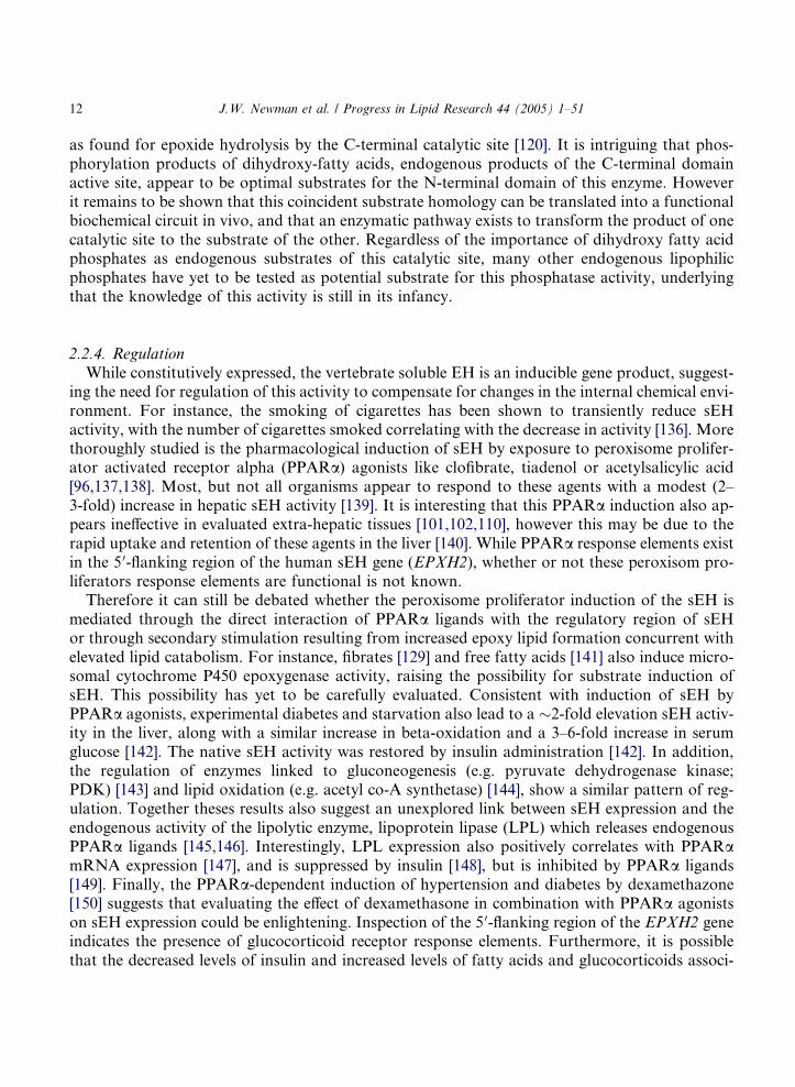
A number of EHs are found as soluble proteins within various cells. These include the ‘‘solubleEHs’’ from plants and animals, the hepoxilin hydrolase, and the zinc-metalloprotein leukotrieneA4 hydrolase. These enzymes are predominantly, but not completely localized in the cytosol. Eachof these enzymes is responsible for the hydrolysis of aliphatic epoxy fatty acids. With the excep-tion of the LTA4 hydrolase, the products of these reactions are the corresponding vicinal diols,when the starting material is a simple epoxy fatty acid (Fig. 1).
2.1. The plant sEHs
The sEHs isolated from plants are roughly 35 kDa a/ß-hydrolase fold enzymes, which can oc-cur as either monomeric or dimeric proteins [33]. These enzymes show structural homology to thebacterial haloalkane dehalogenase and the C-terminal domain of the mammalian sEH [34].
To date, sEHs have been reported from nine plants, soybean (Glycine max) [35], mouse earedcress (Arabidopsis thaliana) [36], potato (Solanum tuberosum) [37], common tobacco (Nicotiana
tabacum) [38], oilseed rape (Brassica napus) [39], pineapple (Ananas comosus) [40], spurge (Euphor-bia lagascae) [41], rice (Oryza sativa) [42], and rough lemon (Citrus jambhiri) [14]. To our knowl-edge, the rice, tobacco, and pineapple gene products have yet to be expressed. EH activity hasbeen also characterized in the particulate fractions of spinach (Spinacia olerecea) and apple(Malus pumila) [43], and the soluble fraction of the castor bean (Ricinus communis) [44], vetch (Vi-cia sativa) [12], maize (Zea mays), wheat (Triticum aedivum), celery (Apium graveolens), tobacco(N. tabacum) and soybean (Glycine max) [33]. It is evident that plants contain multiple EH iso-forms. At least three isoforms have been indicated in soybean, while unique constitutive and infec-tion-induced forms have been reported in tobacco [33].
2.1.1. Tissue distribution and sub-cellular localization
The plant soluble EHs have been isolated from or localized in germinated seeds, seedlings,roots, fruit, tubers, and leaves [14,33,35,37,40,41]. The tissue distribution is quite variable from
OH
O
HO
O
HO
O
OH
OHO
OH
HO
sEH LTA4 Hydrolase
Fig. 1. Both LTA4 hydrolase and the mammalian hepatic soluble EH can utilize leukotriene A4 as a substrate. While
the sEH produces a vicinal threo-diol from this substrate [121], LTA4 hydrolase yields LTB4 [323].
6 J.W. Newman et al. / Progress in Lipid Research 44 (2005) 1–51
plant to plant, and underlines the overall lack of knowledge of the plant EHs. As with the mam-malian soluble EHs, plant soluble EHs are found primarily in the cytosol, with a minor fractionbeing tightly associated with isolated microsomes [35]. In addition, subcellular fractionation ofcastor bean endosperm revealed a dual distribution of activity between the glyoxysomal andthe cytosolic fractions [44], reminiscent of the dual distribution between peroxisomes and cytosolfor the vertebrate orthologs described below.
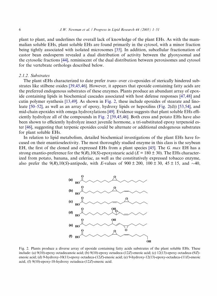
2.1.2. SubstratesThe plant sEHs characterized to date prefer trans- over cis-epoxides of sterically hindered sub-
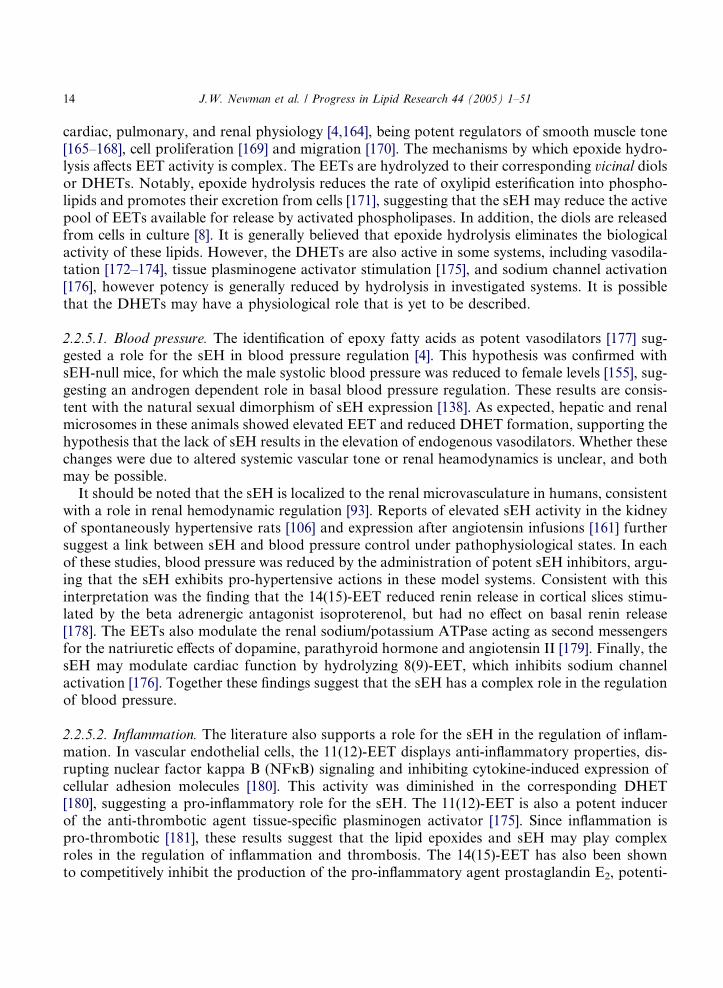
strates like stilbene oxides [39,45,46]. However, it appears that epoxide containing fatty acids arethe preferred endogenous substrates of these enzymes. Plants produce an abundant array of epox-ide containing lipids in biochemical cascades associated with host defense responses [47,48] andcutin polymer synthesis [13,49]. As shown in Fig. 2, these include epoxides of stearate and lino-leate [50–52], as well as an array of epoxy, hydroxy lipids or hepoxilins (Fig. 2(d)) [53,54], andmid-chain epoxides with omega hydroxylations [49]. Evidence suggests that plant soluble EHs effi-ciently hydrolyze all of the compounds in Fig. 2 [39,45,46]. Both cress and potato EHs have alsobeen shown to efficiently hydrolyze insect juvenile hormone, a tri-substituted epoxy terpenoid es-ter [46], suggesting that terpenic epoxides could be alternate or additional endogenous substratesfor plant soluble EHs.
In relation to lipid metabolism, detailed biochemical investigations of the plant EHs have fo-cused on their enantioselectivity. The most thoroughly studied enzyme in this class is the soybeanEH, the first of the cloned and expressed EHs from a plant species [45]. The G. max EH has astrong enantio-preference for the 9(R),10(S)-epoxystearic acid (E = 180 ± 30). The EHs character-ized from potato, banana, and celeriac, as well as the constitutively expressed tobacco enzyme,also prefer the 9(R),10(S)-antipode, with E-values of 900 ± 200, 100 ± 30, 45 ± 15, and �40,
HO
O O
HO
O O
HO
O O
HO
O
HO
O
HO
O O
OHO
OH
O
OH
(a)
(b)
(c)
(d)
(e)
(f)
Fig. 2. Plants produce a diverse array of epoxide containing fatty acids substrates of the plant soluble EHs. These
include: (a) 9(10)-epoxy octadecanoic acid; (b) 9(10)-epoxy octadeca-(12Z)-eneoic acid; (c) 12(13)-epoxy octadeca-(9Z)-
eneoic acid; (d) 9-hydroxy-10(11)-epoxy octadeca-(12Z)-eneoic acid; (e) 9-hydroxy-12(13)-epoxy-octadeca-(11E)-eneoic
acid, (f) 9(10)-epoxy-18-hydroxy octadeca-(12Z)-eneoic acid.
J.W. Newman et al. / Progress in Lipid Research 44 (2005) 1–51 7
respectively [33]. The wheat, maize, rice, and infection-induced tobacco enzymes show little to noenantioselectivity (1 6 E 6 4). It has also been demonstrated that that the soybean, potato, andtobacco EHs stereo convert (±)9,10-epoxystearic acid antipodes by attack at the (S)-carbon tothe corresponding threo-(R,R)-diol in >85% excess [33,35].
2.1.3. RegulationWhile plants contain constitutive soluble EHs, inducible isoforms of these enzymes have also
been reported [36,37,55]. For instance, the natural growth and differentiation of meristematic tis-sue is associated with increased EH transcription, in the potato leaf relative to the expanding andmature leaf [37]. Similarly, in the spurge (E. lagascae), a germination-specific EH has recently beenreported [55]. The transcription of these inducible enzymes can also be increased by exogenousexposure to hormones involved in germination, development, growth, fruit ripening, and host-defense [36,37]. In particular, responsiveness to the growth hormones auxin and ethylene[36,56] and the host-defense regulator methyl jasmonate [37,57] have been noted. It is of equalinterest to note that plant soluble EHs are not responsive to cytokinin, absisic acid, 6-benzyl-aminopurine, or gibberellin [36,37]. The interested reader is directed toward the following recentreviews for background on these hormones and their interactions [58–65].
In cress, the sEH transcript of the stems and leaves was weakly induced by drought stress, whileauxin (indole acetic acid) and auxin mimics (e.g. 2,4-dichlorophenoxy acetic acid and naphthaleneacetic acid) strongly induced this enzyme in pre-bolting young plants [36]. However, the EH activ-ity level in vetch seedlings was insensitive to auxin mimics [12]. In the soybean, the sEH mRNAisolated from both germinating seeds and constitutive expression in the plant body showed induc-tion by ethylene treatment [66]. In the potato, physical trauma of the leaf induced a sEH, as didexposure to exogenous methyl jasmonate [37]. Viral infection of the common tobacco has alsobeen reported to increase the expression of sEH in aerial bodies of the plant [33,38]. Each of theseexamples therefore suggests that in plants soluble EHs are expressed in response to stress.
2.1.4. Physiological role: cutin biosynthesis and host defenseThe substrate specificity and regulatory behavior of the plant soluble EHs argue for a primary
function of this enzyme in host defense and growth. The defensive functions of these enzymes canbe related to both passive (cutin biosynthesis) and active (anti-fungal chemical synthesis) roles.Cutin biosynthesis is also activated during initial plant growth and this may explain the associa-tion of heightened EH gene transcription during vegetative expansion.
Cutin is the waxy cuticle covering the aerial surfaces of plant providing a physical barrier topathogens while allowing gas exchange [67]. The 9(10)-epoxy 18-hydroxy and 9,10,18-trihydroxyoctadecanoic acids are common monomers of cutin poly esters in plants [13]. The enzymatichydration of the 18-hydroxy-epoxystearic acid has been demonstrated in apples (M. pumila)[43]. It is of interest that these cutin monomers themselves are also messengers in plant–pathogeninteractions that are released by fungal cutinases [68]. Consistent with an anti-fungal role, EH wasinduced in lemon leaves only after exposure to pathogenic fungus strains [14]. In addition, potatoleaves efficiently synthesize the linoleate derived triols 9(S),10(S),11(R)-trihydroxy-12(Z)-octadec-enoic acid and 9(S),12(S),13(S)-trihydroxy-10(E)-octadecenoic acid, which have potent anti-fungal properties [53]. The enzymatic production of such substances has also been observed ingarlic roots [54] and apple fruit [69]. Plants have also derived biosynthetic routes to prevent epoxy
8 J.W. Newman et al. / Progress in Lipid Research 44 (2005) 1–51
fatty acid hydrolysis by sEHs. In particular, the in vivo synthesis of the linoleate 9,10-epoxide, orvernolic acid, appears to occur from linolyl-phosphotidyl choline, and the product is moveddirectly into triglycerides [52,70]. This route of synthesis thereby avoids interaction with sEHs,allowing epoxide accumulation in these seeds that are released upon germination [55].
It has also been reported that the (±)12(13)- but not (±)9(10)-epoxide of linoleic acid is a potentcompetitive inhibitor of allene oxide cyclase [71,72], a critical enzyme in jasmonic acid synthesis.While the physiological relevance of this observation has not been fully evaluated, it is intriguingthat both allene oxide synthase [73] and at least one sEH [66] are ethylene inducible genes. There-fore, it is possible that the sEH also serves a role in regulating jasmonate signaling during periodsof host response to attack by pathogens or insects.
2.2. The mammalian sEHs
The mammalian soluble EHs are homodimers, of �62 kDa monomeric subunits [74] with iso-electric points between 5 and 6 [46,75]. Each monomer is comprised of two distinct structural do-mains, linked by a proline-rich peptide segment [34,76]. The epoxide hydrolase activity resides inthe �35-kDa C-terminal domain, which contains an a/ß-hydrolase fold structure homologous tothe bacterial haloalkane dehalogenase, the plant soluble EHs and the microsomal EH [34]. Theroughly 25-kDa N-terminal domain contains a distinct a/ß fold topology belonging to the halo-acid dehalogenase enzyme superfamily [34,76]. The N-terminal domain catalytic site is a func-tional phosphatase [77,78], with apparent specificity for fatty acid diol phosphates [78]. Inaddition, the N-terminal domain appears to serve a critical role in stabilization of the domain-swapped architecture of the dimer [76].
Soluble EH activity has been documented in all vertebrates investigated including teleost fish:rainbow trout (Salmo gairdneri), golden medaka (Oryzias latipes), fathead minnow (Pimphales
promelas), marine scup (Stenotomus chrysops) [27,79,80]; rodents: mouse (Mus mus), rat (Ratusnorwiegicus), rabbit (Oryctolagus cuniculus), guinea pig (Cavia porcellus), hamster (Mesocricetus
sp.) [81–84]; domestic pig (Sus domesticus) [85]; domestic horse (Equus caballus) [86]; and prima-tes: rhesus monkey (Macaca mulatto), baboon (Papio sp.), human (Homo sapiens) [74,87]. To ourknowledge, the sEH has been cloned and expressed from the human [74], mouse [88], rat [89], andpig [85]. Based on analyses of the transcript sequences of the sEH genes of various organisms theenzyme is highly conserved [34].
2.2.1. Tissue distribution and sub-cellular localization
The sEH is broadly distributed in vertebrate tissues [10,90]. In mammals, activity has been de-tected in the liver [91], kidney [92,93], lungs [94,95], heart, brain, spleen [96], adrenals [97], intes-tine, urinary bladder [97], vascular endothelium and smooth muscle [93,98], placenta [99], skin[100], mammary gland [101], testis [96,102] and leukocytes [103]. The specific activity of sEH ishighest in the liver, followed by the kidney, with lower levels in extra hepatic tissues [96,97].The expression of sEH has also been observed in striated muscle [104] and ovary [105]. Immuno-reactive proteins have also been reported in stomach, pancreas, prostate, tonsils, lymph nodes,and uterus [90]. While distribution of the sEH is diffuse in the liver [96], a more focal distributionis described for other tissues, and it appears to co-localize with cytochrome P450 2C9 in manytissues [90]. In the kidneys, the sEH appears concentrated in the renal cortex [106], and more spe-

J.W. Newman et al. / Progress in Lipid Research 44 (2005) 1–51 9
cifically to the renal microvasculature [93] and possibly proximal tubule [90]. Similarly, sEH ap-pears localized to vascular tissues in the lung [95]. The distribution of the sEH in glandular tissuesappears complex, being localized to the adrenal cortex and peripheral islet cells in the pancreas,but diffuse in the pituitary [90].
Historically, the sEH was referred to as the cytosolic EH based on the primary isolation ofcharacteristic activity in cytosolic cellular fractions [91,107]. However, sEH activity is also isolatedin microsomal fractions. Early studies reported ‘‘an integral microsomal protein which is not dis-sociated from the membrane by repeated washing, high ionic strength salt, or chaotropic agentsolutions, or by sonication’’ [108]. Later studies using both activity and immunological techniqueshave replicated this finding [106,109,110]. Therefore epoxide hydrolase activity observed in micro-somal preparations should not be assigned to a specific hydrolase without conducting appropriateinhibitor or immunoprecipitation experiments. Besides the apparent microsomal association, thesEH has also been shown to localize in peroxisomes, being isolated in the light mitochondrial frac-tion [111]. Approximately 60% of the total sEH activity was isolated in the cytosol, and inductionby clofibrate did not affect this distribution, while shifting cytosolic catalase activity from �4% to15–35% [112]. This dual compartmentalization on the sEH between the cytosol and peroxisomewas later supported by the identification of an impaired peroxisomal targeting sequence at the car-boxy terminal of the rat sEH [113], which is conserved in all cloned mammalian sEHs.
2.2.2. C-terminal domain substrates: epoxy fatty acidsThe catalytic site situated in the C-terminal domain of the sEH is responsible for its well defined
epoxide hydrolase activity [76,114]. As described for the plant sEHs, the vertebrate sEHs prefertrans- over cis-epoxides of sterically hindered substrates like stilbene oxides [81]. However, bothsaturated [115,116] and unsaturated [117] cis-epoxy fatty acids are excellent sEH substrates. Aswith plants, animals produce a broad array of epoxide containing aliphatic lipids, which haveroles in the regulation of vascular tone, inflammation and cell growth [4,118]. With respect tothe vertebrate soluble EH, the mono and diepoxides of unsaturated fatty acids have been the mostthoroughly studied. To date, hydroxy, epoxy lipids (i.e. hepoxilins) have not been evaluated assubstrates for this enzyme, however, considering the homology between the vertebrate and plantsEHs [33,46], these compounds are likely substrates.
As shown in Table 2, detailed biochemical evaluations have been reported with fatty acidmonoepoxides and either purified or recombinant EHs from rodents. The reported Km for epoxylipids with rodent sEHs range from �3 to 40 lM with maximum velocities ranging from notdetectable to 9 lmol product/min/mg of protein. From the compiled results in Table 2, it canbe seen that the sEH has a preference for epoxides distal to the carboxyl terminal and that ithydrolyzes 5,6-epoxy fatty acids poorly. Furthermore, sEH preferentially hydrolyzes the epoxye-icosatrienoic acid (EET) enantiomers that are the dominant endogenous products [119,120]. Theelimination of olefins by catalytic hydrogenation reduced hydrolysis rates of the arachidonate de-rived epoxides, as did methylation of the free acids [120]. The enzymatic addition of water to the11,12-EET antipodes and 14(S),15(R)-EET were not regioselective, while the 14(R),15(S)-EETwas selectively hydrated at C15 and both enantiomers of the 8,9-EET, but not its methyl ester,proceeded by hydrolysis at C9 [120]. Increasing the number of cis-olefins appears to increasethe efficiency and enantioselectivity of catalysis [33,119,120], however either the presence oftrans-olefins, conjugated olefins, or trans-epoxides appear to reduce the affinity of epoxy fatty

Table 2
Specific activity of rodent sEHs with various epoxy lipids
Substrate Absolute conformation Km (lM) Vmax (lmol/min/mg) Vmax/Km References
14,15-EETa 14(R),15(S) 4 9.03 2.3 [120]
(±) – 4.53 – [119]
14(S),15(R) 5 1.36 0.27 [120]
11,12-EETa 11(S),12(R) 4 3.02 0.76 [120]
(±) – 1.65 – [119]
11(R),12(S) 3 0.82 0.27 [120]
8,9-EETa 8(S),9(R) 5 3.10 0.62 [120]
(±) – 1.45 – [119]
8(R),9(S) 41 0.83 0.020 [120]
5,6-EET (±) – <0.1 – [119]
12,13-EpOME (±) 6.2 2.67 0.43 [8]
9,10-EpOME (±) 5.2 1.86 0.36 [8]
9,10-EpO (±) 11 3.5 0.31 [116]
14,15-LTA4d (±) 11 0.90 0.081 [125]
14,15-LTA4b (±) 48 1.5 0.031 [84]
11,12-LTA4b,c (±) 18 2.4 0.13 [84]
5,6-LTA4b (±) 25 2.1 0.084 [84]
5,6-LTA4 (±) 5 0.55 0.11 [265]
a Dominant endogenous antipodes.b Purified guinea pig liver sEH; other reported values are for purified mouse sEH.c 11(S),12(S)-trans-epoxy-(5Z,7E,9E,14Z)-eicosatetraenoic acid.d 14(S),15(S)-trans-epoxy-(5Z,8Z,10E,12E)-eicosatetraenoic acid.
10 J.W. Newman et al. / Progress in Lipid Research 44 (2005) 1–51
acids for the sEH. Regardless, the conjugated tetraeneoic fatty acid leukotriene A4 is a substratefor the sEH purified from mouse liver, which produces the corresponding 5,6-dihydroxy-7,9,11,14-eicosatetraenoic acid [121]. This 5,6-diol is the predominant metabolite formed byLTA4 hydrolysis in homogenates of kidney, heart, and brain [122]. The related 11,12- and14,15-trans-epoxy tetraenoic fatty acids have also been reported as endogenous products of plate-lets [123] and HL-60 cells [124]. The formation of 14,15-dihydroxy eicosatetraenoic acid has beenachieved in vitro using purified mouse soluble EH [125] and associated with pulmonary hepoxilinhydrolase activity [126].
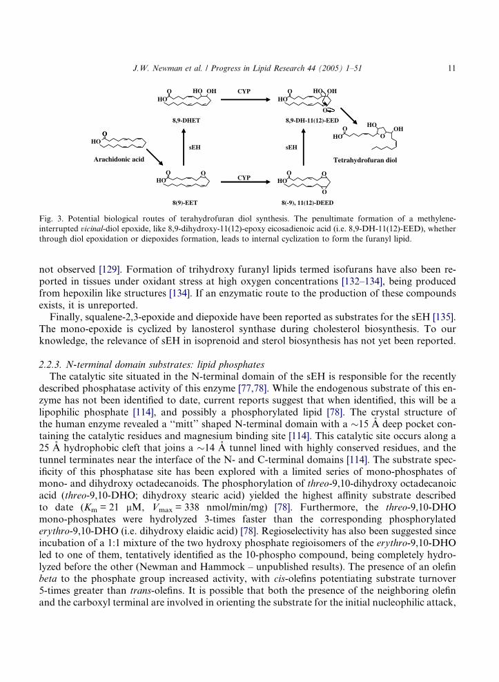
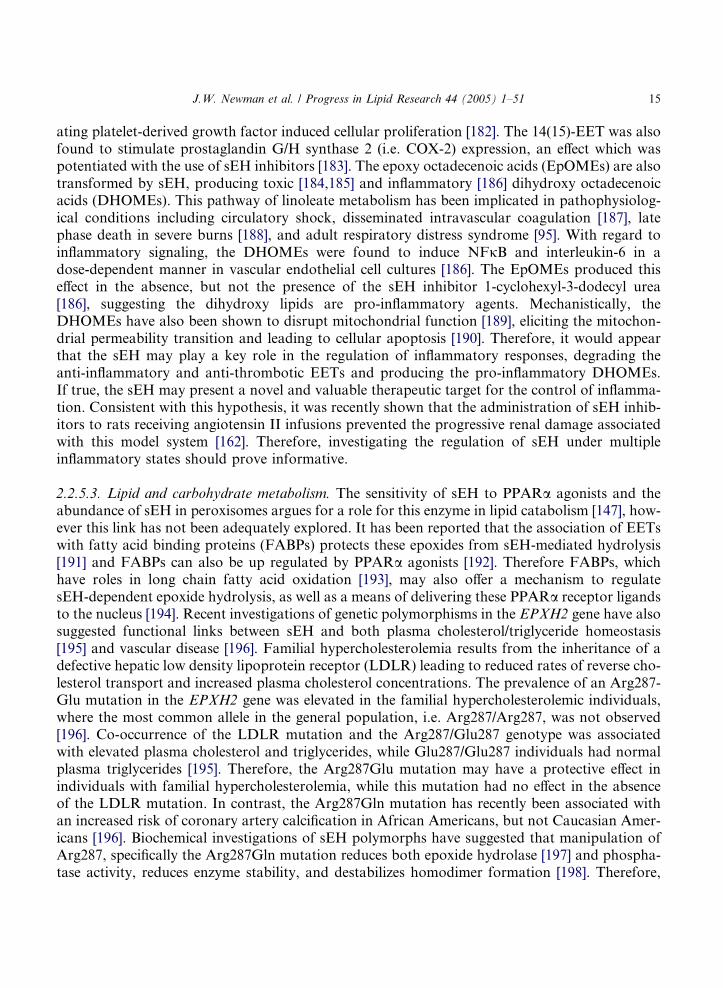
In addition to the monoepoxy fatty acids, diepoxy fatty acids have also been reported as sub-strates for the sEH [127,128]. At a concentration of 7.5 lg/ml (i.e. �1.2 M) affinity purified sEHtransformed 9(10),12(13)-diepoxy octadecanoic acid into the corresponding tetraols, while a20-fold dilution yielded only cyclization products containing dihydroxy tetrahydrofuran struc-tures, without tetraol formation [128]. In vitro assays suggest that the sEH is responsible forthe formation of these compounds in mammalian tissue homogenates [129], and these structureshave been reported as mitogenic endocrine disrupting components in corn husks [130,131]. Fig. 3displays two potential biosynthetic routes of tetrahydrofuran diol synthesis, both including anepoxide hydrolysis step. Regardless of the absolute route, tetrahydrofuran diols formation isdependent upon the oxidation of methylene interrupted olefins since larger cyclic products are
O
O
HOO
CYP
HOO O
HOO HO OH
HOO HO OH
HOO
O
HOOH
CYP HOO O
O
sEH sEH
8(9)-EET 8(-9), 11(12)-DEED
8,9-DHET 8,9-DH-11(12)-EED
Arachidonic acid Tetrahydrofuran diol
Fig. 3. Potential biological routes of terahydrofuran diol synthesis. The penultimate formation of a methylene-
interrupted vicinal-diol epoxide, like 8,9-dihydroxy-11(12)-epoxy eicosadienoic acid (i.e. 8,9-DH-11(12)-EED), whether
through diol epoxidation or diepoxides formation, leads to internal cyclization to form the furanyl lipid.
J.W. Newman et al. / Progress in Lipid Research 44 (2005) 1–51 11
not observed [129]. Formation of trihydroxy furanyl lipids termed isofurans have also been re-ported in tissues under oxidant stress at high oxygen concentrations [132–134], being producedfrom hepoxilin like structures [134]. If an enzymatic route to the production of these compoundsexists, it is unreported.
Finally, squalene-2,3-epoxide and diepoxide have been reported as substrates for the sEH [135].The mono-epoxide is cyclized by lanosterol synthase during cholesterol biosynthesis. To ourknowledge, the relevance of sEH in isoprenoid and sterol biosynthesis has not yet been reported.
2.2.3. N-terminal domain substrates: lipid phosphatesThe catalytic site situated in the N-terminal domain of the sEH is responsible for the recently
described phosphatase activity of this enzyme [77,78]. While the endogenous substrate of this en-zyme has not been identified to date, current reports suggest that when identified, this will be alipophilic phosphate [114], and possibly a phosphorylated lipid [78]. The crystal structure ofthe human enzyme revealed a ‘‘mitt’’ shaped N-terminal domain with a �15 A deep pocket con-taining the catalytic residues and magnesium binding site [114]. This catalytic site occurs along a25 A hydrophobic cleft that joins a �14 A tunnel lined with highly conserved residues, and thetunnel terminates near the interface of the N- and C-terminal domains [114]. The substrate spec-ificity of this phosphatase site has been explored with a limited series of mono-phosphates ofmono- and dihydroxy octadecanoids. The phosphorylation of threo-9,10-dihydroxy octadecanoicacid (threo-9,10-DHO; dihydroxy stearic acid) yielded the highest affinity substrate describedto date (Km = 21 lM, Vmax = 338 nmol/min/mg) [78]. Furthermore, the threo-9,10-DHOmono-phosphates were hydrolyzed 3-times faster than the corresponding phosphorylatederythro-9,10-DHO (i.e. dihydroxy elaidic acid) [78]. Regioselectivity has also been suggested sinceincubation of a 1:1 mixture of the two hydroxy phosphate regioisomers of the erythro-9,10-DHOled to one of them, tentatively identified as the 10-phospho compound, being completely hydro-lyzed before the other (Newman and Hammock – unpublished results). The presence of an olefinbeta to the phosphate group increased activity, with cis-olefins potentiating substrate turnover5-times greater than trans-olefins. It is possible that both the presence of the neighboring olefinand the carboxyl terminal are involved in orienting the substrate for the initial nucleophilic attack,
12 J.W. Newman et al. / Progress in Lipid Research 44 (2005) 1–51
as found for epoxide hydrolysis by the C-terminal catalytic site [120]. It is intriguing that phos-phorylation products of dihydroxy-fatty acids, endogenous products of the C-terminal domainactive site, appear to be optimal substrates for the N-terminal domain of this enzyme. Howeverit remains to be shown that this coincident substrate homology can be translated into a functionalbiochemical circuit in vivo, and that an enzymatic pathway exists to transform the product of onecatalytic site to the substrate of the other. Regardless of the importance of dihydroxy fatty acidphosphates as endogenous substrates of this catalytic site, many other endogenous lipophilicphosphates have yet to be tested as potential substrate for this phosphatase activity, underlyingthat the knowledge of this activity is still in its infancy.
2.2.4. Regulation
While constitutively expressed, the vertebrate soluble EH is an inducible gene product, suggest-ing the need for regulation of this activity to compensate for changes in the internal chemical envi-ronment. For instance, the smoking of cigarettes has been shown to transiently reduce sEHactivity, with the number of cigarettes smoked correlating with the decrease in activity [136]. Morethoroughly studied is the pharmacological induction of sEH by exposure to peroxisome prolifer-ator activated receptor alpha (PPARa) agonists like clofibrate, tiadenol or acetylsalicylic acid[96,137,138]. Most, but not all organisms appear to respond to these agents with a modest (2–3-fold) increase in hepatic sEH activity [139]. It is interesting that this PPARa induction also ap-pears ineffective in evaluated extra-hepatic tissues [101,102,110], however this may be due to therapid uptake and retention of these agents in the liver [140]. While PPARa response elements existin the 5 0-flanking region of the human sEH gene (EPXH2), whether or not these peroxisom pro-liferators response elements are functional is not known.
Therefore it can still be debated whether the peroxisome proliferator induction of the sEH ismediated through the direct interaction of PPARa ligands with the regulatory region of sEHor through secondary stimulation resulting from increased epoxy lipid formation concurrent withelevated lipid catabolism. For instance, fibrates [129] and free fatty acids [141] also induce micro-somal cytochrome P450 epoxygenase activity, raising the possibility for substrate induction ofsEH. This possibility has yet to be carefully evaluated. Consistent with induction of sEH byPPARa agonists, experimental diabetes and starvation also lead to a �2-fold elevation sEH activ-ity in the liver, along with a similar increase in beta-oxidation and a 3–6-fold increase in serumglucose [142]. The native sEH activity was restored by insulin administration [142]. In addition,the regulation of enzymes linked to gluconeogenesis (e.g. pyruvate dehydrogenase kinase;PDK) [143] and lipid oxidation (e.g. acetyl co-A synthetase) [144], show a similar pattern of reg-ulation. Together theses results also suggest an unexplored link between sEH expression and theendogenous activity of the lipolytic enzyme, lipoprotein lipase (LPL) which releases endogenousPPARa ligands [145,146]. Interestingly, LPL expression also positively correlates with PPARamRNA expression [147], and is suppressed by insulin [148], but is inhibited by PPARa ligands[149]. Finally, the PPARa-dependent induction of hypertension and diabetes by dexamethazone[150] suggests that evaluating the effect of dexamethasone in combination with PPARa agonistson sEH expression could be enlightening. Inspection of the 5 0-flanking region of the EPXH2 geneindicates the presence of glucocorticoid receptor response elements. Furthermore, it is possiblethat the decreased levels of insulin and increased levels of fatty acids and glucocorticoids associ-

J.W. Newman et al. / Progress in Lipid Research 44 (2005) 1–51 13
ated with starvation and diabetes may be the mechanism behind sEH induction in these physio-logical states, as hypothesized for PDK [143].
A number of studies have also indicated hormonal regulation of the sEH in mammals, withsEH activity being elevated in males vs. females for both mice and rats [138,151–153]. In mice,the sexual dimorphism of sEH activity was more pronounced in the male kidney (283%) vs. theliver (55%), when compared to females [138]. Castration decreased activity in both organs, whichwas restored by testosterone supplementation [138]. Consistent with these observations, sEH genetranscription was also found to be induced by androgens in a castration/testosterone supplemen-tation study of male rats [154]. In the later study, the drop in sEH occurred along with a set ofoxidative stress-related genes, which included thioredoxin, peroxiredoxin 5, superoxide dismutase2, glutathione peroxidase 1, microsomal glutathione-S-transferase, and glutathione reductase[154]. As in the castrated males, testosterone administration to females led to a more dramaticincrease in kidney sEH activity than that of liver, while having no effect on unaltered males[138]. On the other hand, ovariectomy resulted in a 30% increase in sEH activity in both the liverand kidney of female mice [138]. In contrast, estradiol administration reduced hepatic sEH activ-ity in males, while having no effect on intact females [153]. Interestingly, hypophysectomy (i.e.pituitary gland removal) lead to an increase in female hepatic sEH activity, while decreasing thisactivity in males [152] suggesting that these effects were due to the loss of gonadotropic hormones.Consistent with this supposition was the finding that growth hormone supplementation had noeffect on sEH activity [152]. Therefore, it would appear that systemic sEH expression is underthe control of the hypothalamic–pituitary–gonadal axis.
Developmental processes also regulate the levels of the sEH. Little is known about the impor-tance of sEH in development; however the viability of sEH knockout mice [155] suggests that thelack of the adult hepatic gene is not critical in fetal development. The earliest sEH activity doc-umented in vertebrate development was in the golden medaka, Oryzias latipes, a teleost fish, at 2days post fertilization [27] corresponding to the late blastula formation, before significant cellulardifferentiation. Activity associated with sEH has been reported as early as 14 weeks of gestation inman, appearing in multiple tissues [97] without noticeable changes in activity [97,156,157]. In malerats, hepatic sEH increased steadily post-partum until puberty [151], while this activity in the liverand lung of horses were unchanged between weaning and adulthood [86]. Age-dependent changesin sEH have also been reported in male C57/B6 mice, where activity increased until 15 monthsthen decreased by 59% at 30 months [158]. It is possible that these changes are directly relatedto androgen-dependent regulation of the sEH expression in the rodent, and may translate directlyto man, where reductions in androgen production also occur with age [159,160].
2.2.5. Physiological roles
While yet to be fully characterized, significant insights into the endogenous role of the sEH havebeen gained recently. These advances have resulted from considering the biological pathways reg-ulated and mediated by sEH substrates, the generation of sEH null mice [155], the use of meta-bolically stable sEH inhibitors [161,162], and the analysis of sEH polymorphs [163].
It is clear that the sEH plays a critical role in regulatory cascades influenced by epoxide-con-taining lipids. The best studied of the endogenous sEH substrates are the EETs, and a thoroughreview of the metabolism and biochemical function of these epoxy lipids has been recently pub-lished [4]. At the systemic level, the EETs have significant roles in the regulation of vascular,
14 J.W. Newman et al. / Progress in Lipid Research 44 (2005) 1–51
cardiac, pulmonary, and renal physiology [4,164], being potent regulators of smooth muscle tone[165–168], cell proliferation [169] and migration [170]. The mechanisms by which epoxide hydro-lysis affects EET activity is complex. The EETs are hydrolyzed to their corresponding vicinal diolsor DHETs. Notably, epoxide hydrolysis reduces the rate of oxylipid esterification into phospho-lipids and promotes their excretion from cells [171], suggesting that the sEH may reduce the activepool of EETs available for release by activated phospholipases. In addition, the diols are releasedfrom cells in culture [8]. It is generally believed that epoxide hydrolysis eliminates the biologicalactivity of these lipids. However, the DHETs are also active in some systems, including vasodila-tation [172–174], tissue plasminogene activator stimulation [175], and sodium channel activation[176], however potency is generally reduced by hydrolysis in investigated systems. It is possiblethat the DHETs may have a physiological role that is yet to be described.
2.2.5.1. Blood pressure. The identification of epoxy fatty acids as potent vasodilators [177] sug-gested a role for the sEH in blood pressure regulation [4]. This hypothesis was confirmed withsEH-null mice, for which the male systolic blood pressure was reduced to female levels [155], sug-gesting an androgen dependent role in basal blood pressure regulation. These results are consis-tent with the natural sexual dimorphism of sEH expression [138]. As expected, hepatic and renalmicrosomes in these animals showed elevated EET and reduced DHET formation, supporting thehypothesis that the lack of sEH results in the elevation of endogenous vasodilators. Whether thesechanges were due to altered systemic vascular tone or renal heamodynamics is unclear, and bothmay be possible.
It should be noted that the sEH is localized to the renal microvasculature in humans, consistentwith a role in renal hemodynamic regulation [93]. Reports of elevated sEH activity in the kidneyof spontaneously hypertensive rats [106] and expression after angiotensin infusions [161] furthersuggest a link between sEH and blood pressure control under pathophysiological states. In eachof these studies, blood pressure was reduced by the administration of potent sEH inhibitors, argu-ing that the sEH exhibits pro-hypertensive actions in these model systems. Consistent with thisinterpretation was the finding that the 14(15)-EET reduced renin release in cortical slices stimu-lated by the beta adrenergic antagonist isoproterenol, but had no effect on basal renin release[178]. The EETs also modulate the renal sodium/potassium ATPase acting as second messengersfor the natriuretic effects of dopamine, parathyroid hormone and angiotensin II [179]. Finally, thesEH may modulate cardiac function by hydrolyzing 8(9)-EET, which inhibits sodium channelactivation [176]. Together these findings suggest that the sEH has a complex role in the regulationof blood pressure.
2.2.5.2. Inflammation. The literature also supports a role for the sEH in the regulation of inflam-mation. In vascular endothelial cells, the 11(12)-EET displays anti-inflammatory properties, dis-rupting nuclear factor kappa B (NFjB) signaling and inhibiting cytokine-induced expression ofcellular adhesion molecules [180]. This activity was diminished in the corresponding DHET[180], suggesting a pro-inflammatory role for the sEH. The 11(12)-EET is also a potent inducerof the anti-thrombotic agent tissue-specific plasminogen activator [175]. Since inflammation ispro-thrombotic [181], these results suggest that the lipid epoxides and sEH may play complexroles in the regulation of inflammation and thrombosis. The 14(15)-EET has also been shownto competitively inhibit the production of the pro-inflammatory agent prostaglandin E2, potenti-
J.W. Newman et al. / Progress in Lipid Research 44 (2005) 1–51 15
ating platelet-derived growth factor induced cellular proliferation [182]. The 14(15)-EET was alsofound to stimulate prostaglandin G/H synthase 2 (i.e. COX-2) expression, an effect which waspotentiated with the use of sEH inhibitors [183]. The epoxy octadecenoic acids (EpOMEs) are alsotransformed by sEH, producing toxic [184,185] and inflammatory [186] dihydroxy octadecenoicacids (DHOMEs). This pathway of linoleate metabolism has been implicated in pathophysiolog-ical conditions including circulatory shock, disseminated intravascular coagulation [187], latephase death in severe burns [188], and adult respiratory distress syndrome [95]. With regard toinflammatory signaling, the DHOMEs were found to induce NFjB and interleukin-6 in adose-dependent manner in vascular endothelial cell cultures [186]. The EpOMEs produced thiseffect in the absence, but not the presence of the sEH inhibitor 1-cyclohexyl-3-dodecyl urea[186], suggesting the dihydroxy lipids are pro-inflammatory agents. Mechanistically, theDHOMEs have also been shown to disrupt mitochondrial function [189], eliciting the mitochon-drial permeability transition and leading to cellular apoptosis [190]. Therefore, it would appearthat the sEH may play a key role in the regulation of inflammatory responses, degrading theanti-inflammatory and anti-thrombotic EETs and producing the pro-inflammatory DHOMEs.If true, the sEH may present a novel and valuable therapeutic target for the control of inflamma-tion. Consistent with this hypothesis, it was recently shown that the administration of sEH inhib-itors to rats receiving angiotensin II infusions prevented the progressive renal damage associatedwith this model system [162]. Therefore, investigating the regulation of sEH under multipleinflammatory states should prove informative.
2.2.5.3. Lipid and carbohydrate metabolism. The sensitivity of sEH to PPARa agonists and theabundance of sEH in peroxisomes argues for a role for this enzyme in lipid catabolism [147], how-ever this link has not been adequately explored. It has been reported that the association of EETswith fatty acid binding proteins (FABPs) protects these epoxides from sEH-mediated hydrolysis[191] and FABPs can also be up regulated by PPARa agonists [192]. Therefore FABPs, whichhave roles in long chain fatty acid oxidation [193], may also offer a mechanism to regulatesEH-dependent epoxide hydrolysis, as well as a means of delivering these PPARa receptor ligandsto the nucleus [194]. Recent investigations of genetic polymorphisms in the EPXH2 gene have alsosuggested functional links between sEH and both plasma cholesterol/triglyceride homeostasis[195] and vascular disease [196]. Familial hypercholesterolemia results from the inheritance of adefective hepatic low density lipoprotein receptor (LDLR) leading to reduced rates of reverse cho-lesterol transport and increased plasma cholesterol concentrations. The prevalence of an Arg287-Glu mutation in the EPXH2 gene was elevated in the familial hypercholesterolemic individuals,where the most common allele in the general population, i.e. Arg287/Arg287, was not observed[196]. Co-occurrence of the LDLR mutation and the Arg287/Glu287 genotype was associatedwith elevated plasma cholesterol and triglycerides, while Glu287/Glu287 individuals had normalplasma triglycerides [195]. Therefore, the Arg287Glu mutation may have a protective effect inindividuals with familial hypercholesterolemia, while this mutation had no effect in the absenceof the LDLR mutation. In contrast, the Arg287Gln mutation has recently been associated withan increased risk of coronary artery calcification in African Americans, but not Caucasian Amer-icans [196]. Biochemical investigations of sEH polymorphs have suggested that manipulation ofArg287, specifically the Arg287Gln mutation reduces both epoxide hydrolase [197] and phospha-tase activity, reduces enzyme stability, and destabilizes homodimer formation [198]. Therefore,
16 J.W. Newman et al. / Progress in Lipid Research 44 (2005) 1–51
these studies of sEH polymorphisms suggest that the sEH may play a complex role in the homeo-static regulation of known risk factors of cardiovascular disease.
The fatty acid epoxygenase pathways have also been implicated in the hormonal regulation ofglucose and lipid metabolism [199], suggesting that the sEH may be important in these system aswell. In cultured pancreatic islet cells the 8(9)-, 11(12)-, and 14(15)-EET were found to stimulateglucagon release, but not effect insulin secretion [200]. The discovery of epoxygenases in this tissue[201] supports an autocrine role for these EETs in the pancreas. In hepatocytes, the EETs alsostimulate vasopressin-induced glycogenolysis [202]. Cortisol secretion by the adrenal gland wasalso stimulated by 14,15-EET [203], which would promote gluconeogenesis, decrease glucose uti-lization, and increase circulating fatty acids. Together, these reports suggest that the EETs arehyperglycemic/hypolipidemic factors, and by corollary, the sEH may play a hypoglycemic/hyper-lipidemic role in normal metabolism. If true, the induction of sEH by PPARa agonists may rep-resent a homeostatic response to these anti-hyperlipidemic agents.
2.2.5.4. Reproduction. The sEH may also play roles in gonadal tissues. In the testis, the sEH ispresent along with epoxide synthesizing enzymes, and roles in epididymal motility and sperm con-centration have been speculated [102]. In leutinized granulosa cells of the human ovary, nanomo-lar concentrations of the 14,15-EET have been reported to induce estrogen secretions [204]. In theporcine ovary, sEH expression was also seen to peak at estrus during the hours preceding ovula-tion, with elevated activity being observed in the cells of the granulosa vs. theca [85]. In addition, aunique gonadal sEH transcript, EPXH2B (NCBI Accession #: AY098585; Hennebold, J.D. andAdashi, E.Y.) has been identified in the mouse ovary, in which the first 44 amino acids of the ex-pressed protein would be altered, eliminating phosphatase activity.
2.2.5.5. Phosphatase. The recent discovery of a catalytically active phosphatase in the N-terminaldomain of the sEH raises new questions about the endogenous role of this enzyme. To date, stud-ies suggest that the substrate of this domain is hydrophobic, and possibly a lipid phosphate [78].As with other related phosphotransferases, a critical DXDX(T/V) catalytic motif is situated with-in 15 amino acids of the N-terminal [77]. Therefore the gonadal EPXH2B isoform should retainepoxide hydrolase but lack phosphatase activity. A thorough investigation of the substrate spec-ificity and inhibitor sensitivity of the phosphatase domain will inevitably enhance our understand-ing of the role of the sEH.
2.3. Hepoxilin epoxide hydrolase
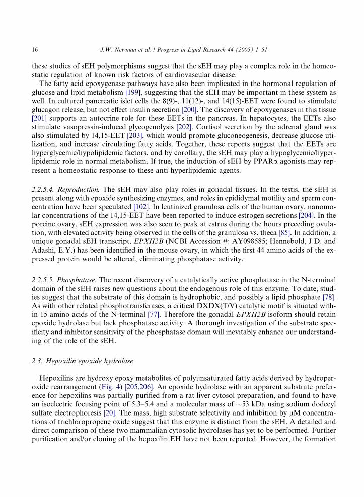
Hepoxilins are hydroxy epoxy metabolites of polyunsaturated fatty acids derived by hydroper-oxide rearrangement (Fig. 4) [205,206]. An epoxide hydrolase with an apparent substrate prefer-ence for hepoxilins was partially purified from a rat liver cytosol preparation, and found to havean isoelectric focusing point of 5.3–5.4 and a molecular mass of �53 kDa using sodium dodecylsulfate electrophoresis [20]. The mass, high substrate selectivity and inhibition by lM concentra-tions of trichloropropene oxide suggest that this enzyme is distinct from the sEH. A detailed anddirect comparison of these two mammalian cytosolic hydrolases has yet to be performed. Furtherpurification and/or cloning of the hepoxilin EH have not been reported. However, the formation

O
OH
(E)R R'
O
R'
OH
(Z)
R
Hepoxilin As
Hepoxilin Bs
Fig. 4. Basic structure of the hepoxilin sub-families. Numerical subscripts indicate the number of olefins in the molecule
such that those derived from arachidonic acid constitute the 3-series (A3, B3), while docosahexeneoic acid yields the 5-
series (A5, B5) [223].
J.W. Newman et al. / Progress in Lipid Research 44 (2005) 1–51 17
of trioxilins have been identified in various organisms including humans [126,207], rats [208], andthe barnacles Balanus amphitrite and Elminius modestus [209].
2.3.1. Tissue distribution and sub-cellular localizationSystematic evaluations of hepoxilin hydrolase activity distributions have not been performed to
date. However, this activity appears to be widely distributed in mammals, as indicated by the pres-ence or formation of trioxilins reported in liver [20], platelets [126,210], brain (homogenates, hip-pocampus and pineal gland) [211–214], rat aorta [215], skin [21,207,216], and pancreas [217,218].
2.3.2. SubstratesAs indicated above, the hepoxilin EH appears to have a high substrate specificity for the hep-
oxilins, as opposed to either leukotriene A4 or trans-stilbene oxide [20]. The hepoxilins are struc-turally classified into to groups as described in Fig. 4, the c-hydroxy epoxides separated by trans
olefins (i.e. hepoxilin As), and the alpha-hydroxy epoxides (i.e. hepoxilin Bs), while the total olefincount in the molecule are indicated by numerical subscripts (i.e. arachidonic acid derived hepox-ilins are A3s and B3s) [219]. These compounds are produced from lipid hydroperoxides either byautooxidative interaction with ferrous proteins [220] or enzymatically [221] by the action ofhydroperoxide isomerases acting on lipid hydroperoxides [206] as shown in Fig. 5. Hepoxilins pro-duced from the isomerization of 12-hydroperoxy eicosatrienoic acid (12-HPETE) and 15-HPETEhave been reported [54,206,210,222]. In addition, the corresponding hepoxilins and trioxilins fromdocosahexenoic acid are produced in rat tissues [223].
2.3.3. RegulationTo the best of our knowledge, no information exists on the regulation of the hepoxilin epoxide
hydrolase. Considering the implication of hepoxilins as regulators of numerous areas of physiol-ogy, this area is a deserving one for the focus of future research.
2.3.4. Physiological rolesThe hydrolysis of hepoxilins appears to play a vital role in mammals by rapidly transforming
these compounds to their corresponding trihydroxy metabolites, trioxilins. This action is, how-ever in competition with both glutathione conjugation [212,215,224,225] in various tissues and
HOO
12-LOX
HydroperoxideIsomerase
15-LOX
8-H,11(12)-EET
HOO
O
OH
10-H,11(12)-EET
HOO
OH
HO OH
HOO
HOO
HOO
O OH
HOO
HO O
HOO
HO OH OH
HOO
HO HO OH
15-H,11(12)-EET111-H,14(15)-EET
HydroperoxideIsomerase
15-HPETE
Arachidonic Acid
HOO
HOO
HOO
O
HO
HOO HO
HO OH
11,14,15-THET 11,12,15-THET8,11,12-THET 10,11,12-THET
EH EH EH EH
12-HPETE
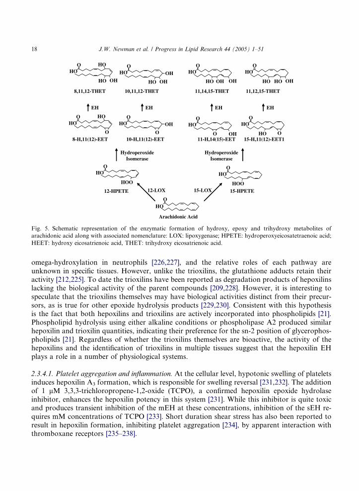
Fig. 5. Schematic representation of the enzymatic formation of hydroxy, epoxy and trihydroxy metabolites of
arachidonic acid along with associated nomenclature: LOX: lipoxygenase; HPETE: hydroperoxyeicosatetraenoic acid;
HEET: hydroxy eicosatrienoic acid, THET: trihydroxy eicosatrienoic acid.
18 J.W. Newman et al. / Progress in Lipid Research 44 (2005) 1–51
omega-hydroxylation in neutrophils [226,227], and the relative roles of each pathway areunknown in specific tissues. However, unlike the trioxilins, the glutathione adducts retain theiractivity [212,225]. To date the trioxilins have been reported as degradation products of hepoxilinslacking the biological activity of the parent compounds [209,228]. However, it is interesting tospeculate that the trioxilins themselves may have biological activities distinct from their precur-sors, as is true for other epoxide hydrolysis products [229,230]. Consistent with this hypothesisis the fact that both hepoxilins and trioxilins are actively incorporated into phospholipids [21].Phospholipid hydrolysis using either alkaline conditions or phospholipase A2 produced similarhepoxilin and trioxilin quantities, indicating their preference for the sn-2 position of glycerophos-pholipids [21]. Regardless of whether the trioxilins themselves are bioactive, the activity of thehepoxilins and the identification of trioxilins in multiple tissues suggest that the hepoxilin EHplays a role in a number of physiological systems.
2.3.4.1. Platelet aggregation and inflammation. At the cellular level, hypotonic swelling of plateletsinduces hepoxilin A3 formation, which is responsible for swelling reversal [231,232]. The additionof 1 lM 3,3,3-trichloropropene-1,2-oxide (TCPO), a confirmed hepoxilin epoxide hydrolaseinhibitor, enhances the hepoxilin potency in this system [231]. While this inhibitor is quite toxicand produces transient inhibition of the mEH at these concentrations, inhibition of the sEH re-quires mM concentrations of TCPO [233]. Short duration shear stress has also been reported toresult in hepoxilin formation, inhibiting platelet aggregation [234], by apparent interaction withthromboxane receptors [235–238].
J.W. Newman et al. / Progress in Lipid Research 44 (2005) 1–51 19
Like the sEH, the hepoxilin hydrolase appears to have a role in the regulation of inflammatoryevents. Neutrophils can synthesize hepoxilins, where they bind tightly and selectively to the intra-cellular face of neutrophil membranes [239,240] causing an initial rapid rise in intracellular calciumfollowed by a slow decline to a plateau [241,242]. This bimodal effect on calcium was caused by aninitial release of calcium from the endoplasmic reticulum, followed by a tight sequestration of thecation in the mitochondria [243], and is preceded by the receptor mediated activation of phospho-lipase C and A2 [244]. The hepoxilins also inhibit calciummobilization in neutrophils stimulated byvarious inflammatory agents including formyl-methionyl-leucyl-phenylalanine, platelet-activatingfactor and leukotriene B4 [245]. In addition, these compounds can elicit neutrophil shape change[246] and is a potent chemotactic agent [247] suggesting a role in neutrophil activation. Therefore,the identification of hepoxilins as endogenous products of neutrophils, their ability to modulate thefunction of these cells, their ability to enhance vascular permeability [248] and the elevated forma-tion of hepoxilins and trioxilins by skin under inflammatory insult [207,249] suggest a role for thehepoxilin epoxide hydrolase in the modulation of inflammatory responses.
2.3.4.2. Smooth muscle tone. The hepoxilins have been reported to have direct actions on smoothmuscle tone. Hepoxilin A3 sensitized both thoracic aorta and portal vein from rats to the contrac-tile effect of noradrenalin, more potently than the peptide-analog [250]. In addition, guinea pigtrachea contraction induced by the potent bronchoconstrictor neurokinin A was potentiated byhepoxilins and unaffected by trioxilins [228], suggesting that the hepoxilin epoxide hydrolase activ-ity is critical for resolving/balancing bronchiospastic conditions mediated through the hepoxilins.
2.3.4.3. Carbohydrate metabolism. On a systemic level, hepoxilins are involved in the regulation ofinsulin signaling, suggests that the hepoxilin epoxide hydrolase also plays a role in this criticalhomeostatic function. Early in the investigation of hepoxilin actions, these compounds were iden-tified as insulin secretogagues [251]. Consistent with this role, hepoxilins were found as metabolicproducts of pancreatic islets of Langerhans [217,218]. While the similarity between the effects ofleukotriene C4 and hepoxilin A3 on insulin secretion [252] suggests this function is mediatedthrough peptidyl-hepoxilins, the injection of arachidonic acid produced a large increase in theblood concentrations of thromboxane B2 and trioxilin A3 within 1 min [253]. Furthermore, themean concentration of this these products appeared greater in the diabetic rat than in the normalrat [253], suggesting an integral role for the hepoxilin epoxide hydrolase. Intra-arterial hepoxilinadministration induces insulin secretion in the fed, but not fasted rat [254]. The hepoxilin pathwayhas also been proposed to have a neuromodulatory role in the central nervous system [255,256]and are potentiators of neurite regeneration [257].
2.3.4.4. Summary and future perspectives. Therefore, the hepoxilin epoxide hydrolase activity invivo likely plays a modulatory role in inflammation, vascular physiology, systemic glucose metab-olism, neurological function, and possibly tissue repair post injury. While the hepoxilin hydrolaseappears to be a distinct enzyme, the substrate specificity of the sEH, and particularly the demon-strated ability of the plant sEHs to hydrolyze hepoxilins, suggests that this enzyme may also par-ticipate in this function. Therefore, the purification and cloning of the hepoxilin hydrolase will becritical to truly distinguish the physiological role of these two enzymes. It is also of some interest
20 J.W. Newman et al. / Progress in Lipid Research 44 (2005) 1–51
that the recently reported cyclopropyl hepoxilin analogs reported as novel thromboxane receptorantagonists with a host of interesting properties [235,237,258,259] may also be competitive inhib-itors of epoxide hydrolase activity [260].
2.4. Leukotriene A4 hydrolase
LeukotrieneA4hydrolase (LTA4hydrolase) is abifunctional zincmetaloprotease [261],whichdis-plays both epoxide hydrolase and aminopeptidase activities [262]. Interestingly, these two catalyticsites share a common carboxyl recognition site and binding of 5(S)-trans-5,6-oxidoeicosatetra-(7E,9E,11Z,14Z)-enoic acid, i.e. leukotrieneA4 (LTA4), inhibits peptidase activity [263]. LekotrieneA4 is synthesized from the 5 lipoxygenase product 5-HPETE.This relatively unstable epoxy lipid caneither be converted to peptidyl leukotrienes by leukotrieneC4 synthase [264], hydrolyzed by sEH to a5,6-dihydroxy metabolite [84,265], or converted to the 5(S),12(R)-dihydroxy eicosatetra-(6Z,8E,10E,14Z)-enoic acid metabolite leukotriene B4 (LTB4) by LTA4 hydrolase [266].
LTA4 hydrolases have been cloned from yeast (Saccharomyces cerevisiae) [267], frogs (Xenopuslaevis) [268], and mammals: mouse (Mus mus) [269,270], rat (Ratus norwiegicus) [271], human(homo sapien) [272,273]. Recently, a crystal structure of the human LTA4 hydrolase was obtainedand new insights into the catalytic mechanism of the enzyme have been elucidated [274–276].
2.4.1. Tissue distribution and sub-cellular localizationThe LTA4 hydrolase is a cytolsolic enzyme found both in heamopoietic [277,278] and paranch-
imal tissues [279]. The presence of LTA4 hydrolase activity has been documented in various or-gans and cell types using combinations of activity and histochemical detection. In the bloodstream LTA4 hydrolase occurs in neutrophils [278], macrophages [280], erythrocytes [279,281],and platelets [282], but not eosinophils, which release the peptidyl leukotriene LTC4 directly[283]. This enzyme is also found in the liver [279], lung [284], kidney [285], heart [270], adrenalcortex [270], gastro intestinal tract [286], spleen [270], skin [287,288], reproductive organs [289],cartilage [290], and brain [291]. Within these various organs, the enzyme has been localized to tis-sue-resident leukocytes [270,287,292], pulmonary [270,293], gastrointestinal [286], and corneal epi-thelium [294], skin epidermal and Langerhan cells [288], renal mesangial cells, all nephronsegments, and collecting tubules [270,295,296], vascular endothelium [279,281], vascular smoothmuscle [281], seminal vesicles [270], large luteal ovarian cells [289], and hepatocytes [270]. In addi-tion, the LTA4 hydrolase may also be found extracellularly, as demonstrated by its presence in cellfree bronchiolar alveolar lavage fluids [297], however this may simply reflect alveolar neutrophilinfiltration and lysis. Two unique LTA4 hydrolase mRNA splice variants have been reported thatare constitutively expressed in multiple tissues [281], however it is not known if each of these vari-ants are translated into a functional protein. It is of interest however that a related protein, ami-nopeptidase B, may also show weak LTA4 hydrolase activity [298,299], and that the LTA4
hydrolase isolated from pulmonary epithelium and neutrophils show a differential sensitivity topharmacological agents [300].
2.4.2. SubstratesAs the name suggests, LTA4 hydrolase displays a high degree of substrate specificity for LTA4.
The enzyme requires the presence of a free acid function and prefers a 7,9-trans-11,14-cis tetraene
J.W. Newman et al. / Progress in Lipid Research 44 (2005) 1–51 21
configuration in its substrates [301]. While the enzyme will transform the corresponding LTA3,containing a 7,9-trans-11-cis triene structure, to LTB3, it does so at �30-fold lower rate [302]or not at all [303]. LTA3 has also been described as a potent LTA4 hydrolase suicide substrate[304]. Similarly, LTA5 is a hydrolyzed at a 4-fold lower rate and acts as an inhibitor of LTA4
hydrolysis [305]. Substrate mediated inactivation studies using functional mutants resistant toinactivation suggest that substrate inactivation of LTA4 hydrolase is reliant on the substrate affin-ity for the catalytic site [306].
While a definitive description of the endogenous peptide substrate for the LTA4 hydrolase hasyet to be demonstrated, this protein metabolizes arginyl peptides with high efficiency and catalytictransformations are greatest with tripeptides [262]. In addition, opioid peptides including met5-enkephalin, leu5-enkephalin, dynorphin1-6, dynorphin1-7, and dynorphin1-8 have been describedas endogenous competitive inhibitors and substrates of the aminopeptidase site [307]. The cleav-age of N-terminal tyrosines from the enkephalins inactivated these analgesic peptides [307].
2.4.3. RegulationThe regulation of LTA4 hydrolase is achieved at transcriptional, post-translational, and func-
tional levels. In human polymorphonuclear leukocytes (PMNs), interleukin-4 and interleukin-13enhanced A23187-stimulated increased mRNA expression and protein synthesis of LTA4 hydro-lase, but not those of cPLA(2) or 5-LO [308]. In keratinocytes, LTA4 hydrolase protein expressionis down regulated by the anti-inflammatory agent cyclosporine A, but not 1,25-dihydroxyvitaminD3, all-trans retinoic acid, eicosatrienoic acid, dexamethasone, interferon-c or methotrexate[309,310]. In addition, LTA4 hydrolase expression is stimulated by human chorionic gonadotro-pin in leutial cells of the ovary during early pregnancy [289]. It is also of interest that in both fibro-blasts and esophageal epithelium, carcinogenic transformations lead to induction of LTA4
hydrolase gene expression [311,312]. Therefore the regulation of LTA4 hydrolase expression sug-gests the presence of specific transcriptional regulatory binding sites in the 5 0-flanking region ofthis gene.
Cloning of the LTA4 hydrolase 50-flanking region revealed the presence of several transcription-
factor consensus sequences, including a phorbol-ester-response element (AP2) and two xenobi-otic-response elements [273,313]. These findings are consistent with earlier studies investigatingthe effects of phorbol esters on LTB4 production indicating that LTA4 hydrolase is activatedby protein kinase C-dependent phosphorylation [314]. In fact, it has since been demonstrated thatbasal LTA4 hydrolase in vascular endothelium exists in an inactive, phosphorylated state [315].Phosphorylation at Ser415 is accomplished by protein phosphatase 1 in the presence, but not ab-sence, of an LTA4 hydrolase peptide substrate [315], suggesting dynamic regulation of LTB4 pro-duction by an intracellular kinase/phosphatase interaction. These findings suggest that thedepressed LTA4 hydrolase activity occurring in conjunction with stable protein levels in psoriaticskin lesions [316] may be a result of post-translational phosphorylaton of the LTA4 hydrolase.
The LTA4 hydrolase is inhibited by its substrates, a process which limits production of LTB4 inLTA4 synthase containing cells [277]. In the circulatory system and many tissues, this process isover come by leukocyte-resident cell interactions, where transcellular delivery of LTA4 from leu-kocytes allows the accelerated production of LTB4 [277]. It has also been noted that under con-ditions of essential fatty acid deficiencies, the production of a lipoxygenase metabolites result inthe inhibition of LTA4 hydrolase, decreasing basal LTB4 production below what would be

22 J.W. Newman et al. / Progress in Lipid Research 44 (2005) 1–51
expected from arachidonic acid depletion [317,318]. Whether this is due to the presence of aninhibitory substrate, or in fact an alteration in the phosphorylation state of the enzyme has notbeen clearly investigated.
Finally, the peptidase activity of LTA4 hydrolase is stimulated by chloride ions, and kineticanalysis of the results suggested the presence of an anion binding site [319]. This peptidase activityis in turn retarded by preincubation of the enzyme with LTA4, which could prolong the activity ofendogenous opioids during inflammatory episodes [320].
2.4.4. Physiological role: inflammatory regulatorThe current understanding of LTA4 hydrolase clearly indicates a pro-inflammatory role for this
enzyme [321–323]. The synthesis of LTB4 has been linked to the pathophysiology of variousinflammatory diseases of the skin [266,324], joints [325], bowels [325], lung [326], and kidney[327–329]. LTB4 is a potent chemokine which stimulates leukocyte degranulation [330], has leu-kotactic properties [331], and stimulates DNA synthesis, cell replication and IgG secretion[332]. Furthermore, LTA4 hydrolase-deficient mice are resistant to platelet-activating factor, sug-gesting that LTB4 is a mediator of systemic shock [322]. Mechanistically, it has been shown thatLTB4 can regulate leukocyte activation by modulating polyisoprenyl phosphate signaling. Specif-ically, LTB4 receptor stimulation activates phospholipase D and concurrently reduces presqualenediphosphate production, reducing this compounds blockade of leukocyte activation and superox-ide anion generation [333].
LTA4 hydrolase also plays a role in female reproduction. The sensitivity of LTA4 hydrolase tohuman chorionic gonadotropin, and the enhanced expression of this enzyme during corpus leut-eum formation suggest the involvement of LTB4 in luteal cells during early pregnancy [289].
A functional role for the peptidase activity of LTA4 hydrolase is still elusive. However, the abil-ity of this enzyme to inactivate enkephalins by cleavage of the terminal tyrosine residues is intrigu-ing [320]. The finding that inactivation of the LTA4 hydrolase by phosphorylation is accomplishedonly in the presence of a peptidase substrate [315] supports a role for the enkephalins in the res-olution of inflammation by preventing LTB4 production. The peptidase activity is in turn retardedby preincubation of the enzyme with LTA4, prolonging the activity of endogenous opioids duringinflammatory episodes [320]. The inactivation of these analgesic peptides during inflammatorystimulation provides a consistent role for both catalytic activities in the regulation of inflamma-tory events.
3. Membrane associated epoxide hydrolases
3.1. Microsomal epoxide hydrolase
Historically, the microsomal epoxide hydrolase was the first EH characterized and isolatedfrom mammalian liver [334–336]. The cDNA of the mEH has been isolated from several speciesincluding rat and human [337,338] and the corresponding enzymes have been expressed in differ-ent transgenic systems [339–342]. The mEH protein is made of 455-amino acid residues corre-sponding to a �50 kDa protein [343], with a strongly hydrophobic transmembrane anchor ofapproximately 20 residues at the N-terminal [344,345]. The C-terminal domain, which contains
J.W. Newman et al. / Progress in Lipid Research 44 (2005) 1–51 23
the catalytic residues, is homologous to a haloalkane dehalogenase, like the sEH [18,34]. Recently,a sEH from the fungus Aspergillus niger was found homologous to the mammalian mEH, butwithout the N-terminal anchor [346]. This fungal enzyme was recently crystallized [347]. In hu-mans, the mEH is the product of the EPXH1 gene on chromosome 1 [348]. Several single nucle-otide polymorphism sequences were identified in human [349] and have been found in associationwith the onset of several diseases and cancers [350–353].
3.1.1. Tissue distribution and sub-cellular localizationLike the sEH, the mEH has been found in nearly all mammalian tissues that have been evalu-
ated [10]. Early investigations by Oesch and collaborators reported the detection of mEH in 26different rat organs and tissues [354]. While mEH from animal livers has been primarily studied,mEH was also isolated from human adrenal glands [355], sinovial tissues [356], follicles isolatedfrom mouse ovaries [357], and in pulmonary bronchial epithelium [358]. Considering the wholeanimal, mEH activity is generally the highest in liver, with lower yet similar levels in testis, lungand heart [110]. However, the relative levels vary with environmental exposures, sex and age (see[10] and [359] for reviews). For instance, a 63-fold interindividual variation in mEH levels hasbeen reported in human livers [360].
It should be noted that in certain organs the mEH is localized within specific cell types, suchthat whole organ measurements do not necessarily reveal a localized high concentration of theenzyme. For example, while ubiquitously distributed in cerebral tissues, mEH is primarily local-ized in glial as opposed to neuronal cells [361], and has elevated activities in tissues which functionas blood- and cerebrospinal fluid-brain barriers [362]. In particular, the mEH activity in the cho-roid plexus approach or exceed those of the liver. It has been hypothesized that the choroid plexusmy serve both hormone generation and detoxification functions for the brain, in a fashion similarto that of the liver for the rest of the body [362]. Furthermore, mEH activity [363] and geneexpression [364] has been detected in human blood cells, especially in lymphocytes and monocytes,underlying the necessity to exsanguinate tissues before any mEH measurements. Finally, theexpression of mEH has been reported in numerous cancerous and primary cell lines (see [10]for review).
As the name implies, the mEH has been primarily isolated and characterized from microsomalpreparations [365]. As a precautionary note, EH activities in microsomes fractions should not beconfused with mEH, because prepared microsome fractions also contain the ChEHs [366] andsEH activity [106,109]. In addition, mEH activity has been found in the cytosol of neoplastic hu-man livers [367]. In liver, mEH is found on the smooth endoplasmic reticulum [368], but has alsobeen reported in association with the plasma membrane [369,370]. Interestingly, the topologicalorientation of mEH appears to be different in the ER, where the catalytic C-terminal domain facesthe cytosol [371], and in the plasma membrane where the C-terminal faces the extra-cellular med-ium [372]. Sequence analyses suggest that the association of the mEH with the membranes is dueto the presence of an N-terminal transmembrane anchor [344,345]. However, the removal of thisanchor does not result in a soluble protein [345], suggesting a strong hydrophobic interaction ofthis enzyme with the membrane. Furthermore, mEH was reported to be tightly associated withphospholipids [373,374], to be a subunit of a Na+-dependent bile acid transport system [369]and to represent a high affinity tamaoxifen binding site [375,376].
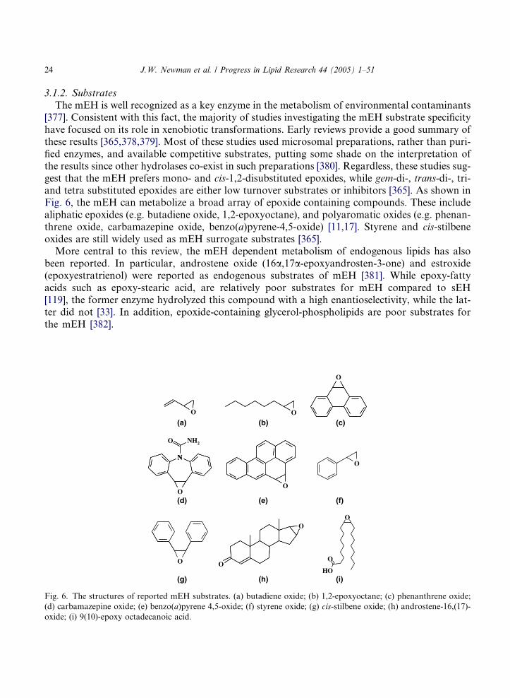
24 J.W. Newman et al. / Progress in Lipid Research 44 (2005) 1–51
3.1.2. Substrates
The mEH is well recognized as a key enzyme in the metabolism of environmental contaminants[377]. Consistent with this fact, the majority of studies investigating the mEH substrate specificityhave focused on its role in xenobiotic transformations. Early reviews provide a good summary ofthese results [365,378,379]. Most of these studies used microsomal preparations, rather than puri-fied enzymes, and available competitive substrates, putting some shade on the interpretation ofthe results since other hydrolases co-exist in such preparations [380]. Regardless, these studies sug-gest that the mEH prefers mono- and cis-1,2-disubstituted epoxides, while gem-di-, trans-di-, tri-and tetra substituted epoxides are either low turnover substrates or inhibitors [365]. As shown inFig. 6, the mEH can metabolize a broad array of epoxide containing compounds. These includealiphatic epoxides (e.g. butadiene oxide, 1,2-epoxyoctane), and polyaromatic oxides (e.g. phenan-threne oxide, carbamazepine oxide, benzo(a)pyrene-4,5-oxide) [11,17]. Styrene and cis-stilbeneoxides are still widely used as mEH surrogate substrates [365].
More central to this review, the mEH dependent metabolism of endogenous lipids has alsobeen reported. In particular, androstene oxide (16a,17a-epoxyandrosten-3-one) and estroxide(epoxyestratrienol) were reported as endogenous substrates of mEH [381]. While epoxy-fattyacids such as epoxy-stearic acid, are relatively poor substrates for mEH compared to sEH[119], the former enzyme hydrolyzed this compound with a high enantioselectivity, while the lat-ter did not [33]. In addition, epoxide-containing glycerol-phospholipids are poor substrates forthe mEH [382].
N
OO
O
O O
OO
O
HO
O
(a)O
(b)
O
(c)
(d) (e) (f)
(g) (h) (i)
N
NH2O
Fig. 6. The structures of reported mEH substrates. (a) butadiene oxide; (b) 1,2-epoxyoctane; (c) phenanthrene oxide;
(d) carbamazepine oxide; (e) benzo(a)pyrene 4,5-oxide; (f) styrene oxide; (g) cis-stilbene oxide; (h) androstene-16,(17)-
oxide; (i) 9(10)-epoxy octadecanoic acid.
J.W. Newman et al. / Progress in Lipid Research 44 (2005) 1–51 25
3.1.3. Regulation
The regulation of mEH can occur at the transcriptional, translational [11] or post-transla-tional level [383]. The induction of mEH has been well studied in animals, but the confidencewith which to extrapolate these results to humans is not known. In rodents, mEH can be in-duced by a variety of compounds that increase the rate of gene transcription [384]. The list ofknown inducers include phenobarbital, methylcholanthrene, polychlorinated biphenyls, trans-stilbene oxide [385,386], peroxisome proliferators [387], radiation [388], heavy metals [11],and certain steroids, including estroxide and its precursor estratetraenol [389]. Complicatingmatters, the effect of each inducer is variable upon age, sex, strain, and species [387,390].For instance in the rat, the induction of mEH by methadone is dependent on sexual hor-mones, in that castration results in methadone-dependent suppression of mEH, while ovarec-tomy yields methadone-dependent induction of this activity [391]. More recently, theassociation of mEH with various cytochrome P450s was shown to affect the rate of substratehydrolysis in vitro [383]. Of the tested P450s, CYP2C11 appeared to play the greatest role inthe association/activation of mEH.
Suppression of mEH activity has also been reported. Inducing a diabetic state using eitheralloxan or streptozotocin lead to a 71% reduction in hepatic mEH activity in rats [142]. Thisdepressed mEH activity was restored by insulin administration [142]. Recently, mEH activitywas also reported to increase in rat hepatocyte cultures following insulin exposure [392]. Aforced fast of 2–5 days also reduced mEH activity by �60% [142]. The expression of themEH gene was also severely downregulated in intra-epithelial lymphocytes from mice receivingtotal parenteral nutrition [393]. Suppression of mEH gene expression has also been reportedusing dexamethasone, gadolinium chloride, acriflavine and lipopolysaccharide [394–398], aswell as experimental traumatic injury [399]. Further glucocorticoid-dependent repression hasbeen directly attributed to interactions with the 5 0-flanking sequence of the EPXH1 gene[398]. Adrenylectomy in rats resulted in elevated mEH levels, which were reversed by dexa-methasone, but not deoxycorticosterone, supporting a role for the hypothalamus–pituitary–adrenal axis in mEH regulation [400]. In addition, hypophsectomy (i.e. pituitary gland removal)induced hepatic mEH activity in both males and females, and growth hormone supplementationreduced this activity below that of sham-operated animals [152]. These results suggested thatthe mEH in the liver is under suppressive control by the pituitary and that growth hor-mone may be the causal hormone involved in the sexually dimorphic expression of this enzyme[152].
Preliminary tests on human primary hepatocyte cultures indicate that the human mEH genemay only be modestly responsive to chemical exposures [401]. Furthermore, in humans the pres-ence of two single nucleotide polymorphisms in exons 3 and 4 [349,402] and several others in the5 0-flanking region [403] seems to effect the regulation of mEH gene transcription [11]. While thegenetic variants have a lower specific activity than the wild type, activity levels in human liverswere found to be independent of the polymorphism, indicating that the genetic variations onlymodestly impact the resulting mEH specific activity in vivo [23].
Developmentally, mEH gene expression has been reported to increase steadily in the liver ofman and correlate strongly with gestational age and protein expression and activity [404]. Expres-sion levels do not appear to correlate with either gestational age, activity, or immunoreactive pro-tein in other inspected tissues [404,405].
26 J.W. Newman et al. / Progress in Lipid Research 44 (2005) 1–51
3.1.4. Physiological roles
While well recognized as a critical enzyme in xenobiotic detoxification, the implication of themEH-dependent metabolism of endogenous lipid substrates is less well defined. Despite the factthat mEH null mice do not present an obvious phenotype without exposure to pro-carcinogens[406], there are several points indicating an endogenous role for this enzyme, beyond xenobioticmetabolism.
3.1.4.1. Cytoprotection. Epoxides are strained three-membered cyclic ethers, and when combinedwith electron withdrawing structures, can become highly reactive electrophilic mutagens, carcin-ogens or cytotoxins [365]. The conversion of epoxides to diols by the mEH generally results in lessmutagenic or carcinogenic compounds [334]. This detoxification role of mEH likely predominatesin the liver [10], and perhaps the choroids plexus of the brain [362], but mEH is also involved inthe extra-hepatic metabolism of these agents, such as pulmonary naphthalene metabolism [407].The protective role of mEH from xenobiotics was illustrated in the case of a man with a defect inmEH expression suffering from acute and severe phenytoin toxicity [408]. In addition, sorbinalhypersensitivity may also be related to a reduced mEH activity [409].
Interestingly, in the case of some polyaromatic compounds, such as benzo(a)pyrene 4,5-oxide,dihydrodiol formation can stabilize bay-region epoxides, increasing the mutagenic and carcino-genic potential of the product [410,411]. This pro-carcinogenic role of mEH was illustrated inmEH-null mice [406]. Furthermore, in human populations, mEH polymorphisms have been asso-ciated with the onset of numerous cancers [351–353,412–414] and the mEH, but not ChEH isupregulated in hyperplastic tissues [380]. In some populations, the role of mEH in xenobioticmetabolism may also be linked to the relationship between mEH polymorphism and emphysema[350,415,416] or Crohn�s disease [417].
3.1.4.2. Steroid metabolism. Numerous lines of evidence suggest that the mEH may play a role insteroid biosynthesis or metabolism. Epoxy-steroids are known endogenous compounds [418], themEH is found in steroidogenic tissues [110,357,381,419,420], mEH inhibitors interfere with testos-terone to estradiol conversion [421], potential relationships have been found between mEH poly-morphism and spontaneous abortion [422], preeclampsia [423] and polycystic ovary syndrome[424], the mEH has been identified as a subunit of an anti-estrogen binding site [375,376], andthe tested epoxy steroids are in fact hydrolyzed by mEH to their corresponding vicinal-diols[381,389]. In particular, the epoxides of estratetraenol and androstadienone, estroxide and adros-tene oxide, are good mEH substrates [381]. The endogenous roles of epoxy steroids are not wellknown, however these compounds may be toxic. For instance, estrogen epoxide has been hypoth-esized as a critical breast cancer initiation factor [425], whose formation is inhibited by tamoxifentreatment [426]. Therefore, mEH may be important in the cellular protection from steroid metab-olites, as it is in the metabolism of epoxidized xenobiotics [9].
3.1.4.3. Other. Beyond these direct roles in steroid metabolism and toxicant transformations, themEH may also have roles in bile acid transport and cellular responses to glucose metabolism. ThemEH has been described as mediating the Na+-dependent transport of bile acid into hepatocytes[369,427]. This role of mEH appears dependent on its expression on the surface of cells [428], andthe enzyme is apparently part of a multi-protein transport system [429]. However, the mechanism
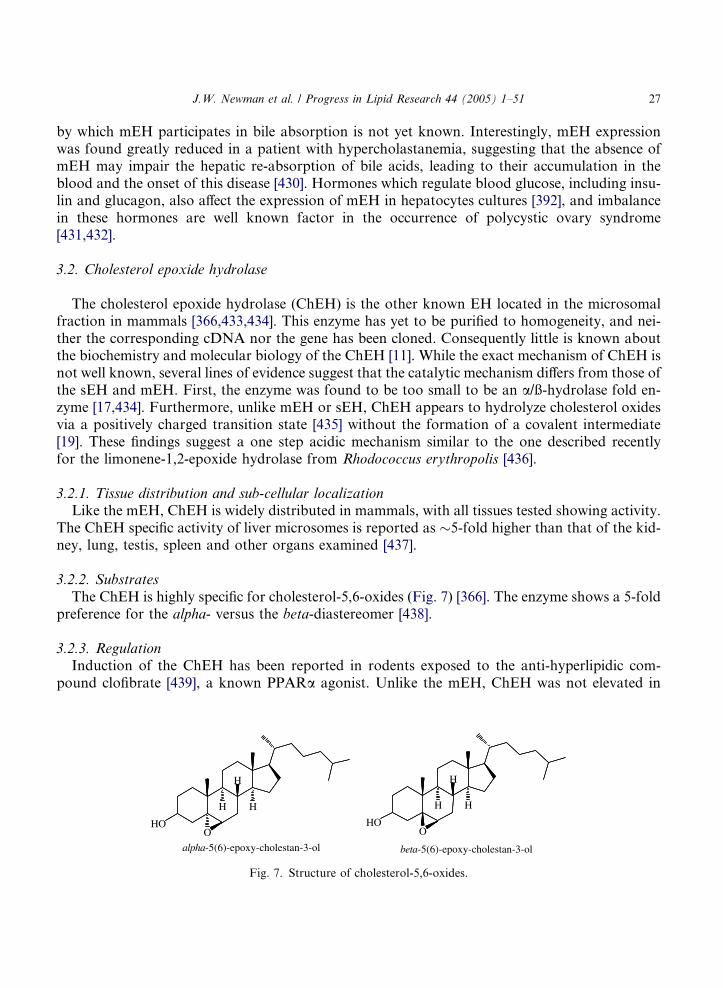
J.W. Newman et al. / Progress in Lipid Research 44 (2005) 1–51 27
by which mEH participates in bile absorption is not yet known. Interestingly, mEH expressionwas found greatly reduced in a patient with hypercholastanemia, suggesting that the absence ofmEH may impair the hepatic re-absorption of bile acids, leading to their accumulation in theblood and the onset of this disease [430]. Hormones which regulate blood glucose, including insu-lin and glucagon, also affect the expression of mEH in hepatocytes cultures [392], and imbalancein these hormones are well known factor in the occurrence of polycystic ovary syndrome[431,432].
3.2. Cholesterol epoxide hydrolase
The cholesterol epoxide hydrolase (ChEH) is the other known EH located in the microsomalfraction in mammals [366,433,434]. This enzyme has yet to be purified to homogeneity, and nei-ther the corresponding cDNA nor the gene has been cloned. Consequently little is known aboutthe biochemistry and molecular biology of the ChEH [11]. While the exact mechanism of ChEH isnot well known, several lines of evidence suggest that the catalytic mechanism differs from those ofthe sEH and mEH. First, the enzyme was found to be too small to be an a/ß-hydrolase fold en-zyme [17,434]. Furthermore, unlike mEH or sEH, ChEH appears to hydrolyze cholesterol oxidesvia a positively charged transition state [435] without the formation of a covalent intermediate[19]. These findings suggest a one step acidic mechanism similar to the one described recentlyfor the limonene-1,2-epoxide hydrolase from Rhodococcus erythropolis [436].
3.2.1. Tissue distribution and sub-cellular localizationLike the mEH, ChEH is widely distributed in mammals, with all tissues tested showing activity.
The ChEH specific activity of liver microsomes is reported as �5-fold higher than that of the kid-ney, lung, testis, spleen and other organs examined [437].
3.2.2. Substrates
The ChEH is highly specific for cholesterol-5,6-oxides (Fig. 7) [366]. The enzyme shows a 5-foldpreference for the alpha- versus the beta-diastereomer [438].
3.2.3. RegulationInduction of the ChEH has been reported in rodents exposed to the anti-hyperlipidic com-
pound clofibrate [439], a known PPARa agonist. Unlike the mEH, ChEH was not elevated in
HO
H
H
H
OHO
H
H
H
O
beta-5(6)-epoxy-cholestan-3-olalpha-5(6)-epoxy-cholestan-3-ol
Fig. 7. Structure of cholesterol-5,6-oxides.

28 J.W. Newman et al. / Progress in Lipid Research 44 (2005) 1–51
hyperplastic tissues [380]. Furthermore, ChEH is inhibited by its primary product, cholestanetriol[438], as well as ketocholestanols, and several cholesterol derivatives [435,440].
3.2.4. Physiological rolesWhile a definitive physiologic role of ChEH is not known, much is known about the biological
activity of the substrate and product of this enzyme. Cholesterol oxides and triols are naturallyoccurring components of human plasma, where the primarily occur as unesterified lipids [441].The epoxides appear to be formed through interactions with lipid hydroperoxides [442] as op-posed to a monooxygenase mediated process.
3.2.4.1. Cytoprotection vs vascular homeostasis. The weak mutagenicity of cholesterol oxides [443]suggests that the ChEH could play a role in protecting cells from these steroid toxicants. How-ever, the exceptional chemical stability of the cholesterol epoxide suggests that this mutagenic ef-fect is not through nucleophilic adduct formation. In addition, the corresponding cholestantriolsare themselves cytotoxic [444], and are associated with increased lipid peroxidation [445] and dis-ruption of actin microfilaments [446], therefore, the epoxide hydrolysis may actually represent anactivation event. Both cholesterol epoxide and triol have also been shown to alter various aspectsof vascular function [447]. These agents inhibit the production of the vasodilator prostacyclin[448,449], reduce platelet adhesion to endothelial mono-layers [449], and down regulate expressionof the LDLR gene [450]. Together, these reports suggest that the ChEH may play a critical role inthe regulation of vascular homeostasis.
3.2.4.2. Phospholipid biosynthesis. The cholestantriol has also been reported to activate cytidyltransferase, increasing phospholipids synthesis and altering phospholipid head group composition[451]. These changes in phospholipids may ultimately affect membrane properties and activity ofmembrane bound enzymes [451]. Therefore, the ChEH may be integrally involved in the regula-tion of phospholipid biosynthesis, and such a role is consistent with its induction by PPARaagonists.
3.3. Juvenile hormone EH – the characterized insect EH
The juvenile hormone epoxide hydrolase (JHEH) is an enzyme involved in the metabolic deg-radation of juvenile hormones (JHs), a series of structurally similar terpenoid esters containingterminal tri-substituted epoxides derived from farnesyl (Fig. 8). Regulation of this hormone gov-erns multiple aspects of insect growth and development. The degradation of these epoxy terpi-noids in insects has been well studied, and two hydrolytic pathways are known. The methylester is cleaved by a soluble esterase, JH esterase, and the tri-substituted epoxide is hydrolyzedby a microsomal enzyme, JHEH [452,453]. Each of these metabolic steps alters, if not eliminatesJH activity [452,453]. The relative role of epoxide hydration and ester hydrolysis in JH catabolismvary with species and insect life stage [454]. For instance, in Drosophile virilis, the esterase pathwayappears to dominate the regulation of the JH titer, however JHEH appears to assume this role inDrosophila melanogaster [455]. While the biological consequence of ester hydrolysis can be re-versed by methyltransferases, epoxide hydrolysis and further conjugation as phosphates[456,457] represent irreversible degradation.
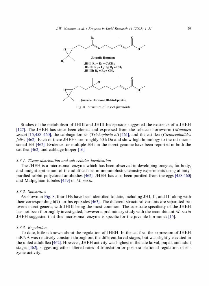
O
O
O
R2
O
O
O
O
R1
Juvenile Hormone III-bis-Epoxide
Juvenile Hormone
JH-I: R1 = R2 = C2CH5JH-II: R1 = C2H5; R2 = CH3JH-III: R1 = R2 = CH3
Fig. 8. Structure of insect juvenoids.
J.W. Newman et al. / Progress in Lipid Research 44 (2005) 1–51 29
Studies of the metabolism of JHIII and JHIII-bis-epoxide suggested the existence of a JHEH[127]. The JHEH has since been cloned and expressed from the tobacco hornworm (Manducasexta) [15,458–460], the cabbage looper (Trichoplusia ni) [461], and the cat flea (Ctenocephalidesfelis) [462]. Each of these JHEHs are roughly 50-kDa and show high homology to the rat micro-somal EH [462]. Evidence for multiple EHs in the insect genome have been reported in both thecat flea [462] and cabbage looper [16].
3.3.1. Tissue distribution and sub-cellular localization
The JHEH is a microsomal enzyme which has been observed in developing oocytes, fat body,and midgut epithelium of the adult cat flea in immunohistochemistry experiments using affinity-purified rabbit polyclonal antibodies [462]. JHEH has also been purified from the eggs [458,460]and Malpighian tubules [459] of M. sexta.
3.3.2. SubstratesAs shown in Fig. 8, four JHs have been identified to date, including JHI, II, and III along with
their corresponding 6(7)- or bis-epoxides [463]. The different structural variants are separated be-tween insect genera, with JHIII being the most common. The substrate specificity of the JHEHhas not been thoroughly investigated; however a preliminary study with the recombinantM. sextaJHEH suggested that this microsomal enzyme is specific for the juvenile hormones [15].
3.3.3. Regulation
To date, little is known about the regulation of JHEH. In the cat flea, the expression of JHEHmRNA was relatively constant throughout the different larval stages, but was slightly elevated inthe unfed adult flea [462]. However, JHEH activity was highest in the late larval, pupal, and adultstages [462], suggesting either altered rates of translation or post-translational regulation of en-zyme activity.
30 J.W. Newman et al. / Progress in Lipid Research 44 (2005) 1–51
3.3.4. Physiological roles
Although initially identified as a ‘‘factor’’ that keeps larval insects in the juvenile state, JHs and/or their metabolites have subsequently been shown to play critical roles in numerous insect lifeprocesses including development, metamorphosis, reproduction, diapause, migration, and metab-olism [452,464]. For instance, in metamorphosis the reduction in its titer initiates development[464], while the same decline appears to stimulate oviposition of fertilized eggs in the adult[455]. These diverse roles in the insect life cycle suggest that the biosynthesis, transport, and deg-radation of JH and/or its metabolites are carefully regulated. It has become evident that theJHEH is in fact an important enzyme in the regulation of this insect hormone, and thus influencessignificant portions of insect physiology. Potent selective inhibitors active in vivo have dramati-cally advanced the study of the physiological roles for the sEH in mammals [106] and the JH ester-ase in insects [453]. While attempts have been made to produce such compounds for the JHEH[465,466], potent and stable inhibitors are still needed.
4. Conclusion
The production of epoxide containing metabolites in biological organisms has led to the evo-lution of a diverse array of EHs. It is particularly interesting that many, if not all of these epox-idized metabolites are bioactive and serve as signaling molecules in their host organisms. Thus, theEHs appear to have evolved as critical regulators within complex signal transduction pathways.While our understanding of these enzymes has expanded greatly since their discovery, manyimportant questions remain to be answered, and doing so will allow a more refined interpretationof the importance and utility of various members of this enzyme class.
The bifunctional sEH and LTA4 hydrolase pose unique challenges. While epoxide hydrolysishas been well studied in these enzymes, the true roles of the sEH phosphatase and LTA4
hydrolase peptidase activities remains to be fully elucidated. Considering that each of theseenzymes appears to constitute a useful therapeutic target for the treatment of inflammatorydiseases, additional efforts are warranted to expand our understanding of these non-epoxidehydrolase functions. Continued investigations of sEH dependent metabolism in plant host de-fense are also warranted. In particular, direct evaluation of sEH as an endogenous regulatorof jasmonate signaling may allow a more detailed understanding of the control of stress re-sponses in these organisms. Similarly, efforts to elucidate the true importance of the mEHin steroid and glucose metabolism will undoubtedly expand our understanding of this enzymebeyond its function in xenobiotic detoxification. Furthermore, the JHEH is ripe for investiga-tions designed to fully elucidate the metabolic and catabolic mechanisms of juvenile hormonecontrol in insects. At present, the importance of the cholesterol epoxide hydrolase and hepox-ilin hydrolase are suggested by the activity of there substrates, however our understanding islimited. Efforts to clone and express these enzymes will greatly improve our ability to ask deci-sive questions about their physiological roles. Therefore, in the coming years, additional atten-tion focused on the EHs and their involvement in lipid metabolism will undoubtedly improveour understanding of an array of critical points of control in the physiological regulation ofboth plants and animals.

J.W. Newman et al. / Progress in Lipid Research 44 (2005) 1–51 31
Acknowledgements
The authors would like to thank Dr. R.M. Bostock and Dr. S.G. Kamita of the University ofCalifornia at Davis for their helpful input and review of the plant sEH and JHEH section of thismanuscript, respectively. This work was partially supported by NIEHS R37 ES02710, NIEHSSuperfund Basic Research Program P42 ES04699, NIEHS Center P30 ES05707, USDA Compet-itive Research Grant Program 2003-35302-13499, NIEHS Center for Children�s EnvironmentalHealth and Disease Prevention, 1 P01 ES11269, NIH/NHLBI R01 HL699-06A1, and NIH/NDDKD P30 DK35747.
References
[1] Ozawa T, Sugiyama S, Hayakawa M. Leukocytes biosynthesize leukotoxin (9,10-epoxy-12-octadecenoate) – a
novel cytotoxic linoleate epoxide. Adv Prostaglandin Thromboxane Leukot Res 1989;19:164–7.
[2] Richard DS, Applebaum SW, Sliter TJ, Baker FC, Schooley DA, Reuter CC, et al. Juvenile hormone bisepoxide
biosynthesis in vitro by the ring gland of Drosophila melanogaster: a putative juvenile hormone in the higher
Diptera. Proc Natl Acad Sci USA 1989;86:1421–5.
[3] Shimada T, Watanabe J, Inoue K, Guengerich FP, Gillam EM. Specificity of 17beta-oestradiol and
benzo[a]pyrene oxidation by polymorphic human cytochrome P4501B1 variants substituted at residues 48, 119
and 432. Xenobiotica 2001;31:163–76.
[4] Spector AA, Fang X, Snyder GD, Weintraub NL. Epoxyeicosatrienoic acids (EETs): metabolism and
biochemical function. Prog Lipid Res 2004;43:55–90.
[5] Sevanian A, Mead JF, Stein RA. Epoxides as products of lipid autoxidation in rat lungs. Lipids
1979;14:634–43.
[6] Gardner HW, Kleiman R. Degradation of linoleic acid hydroperoxides by a cysteine. FeCl3 catalyst as a model
for similar biochemical reactions. II. Specificity in formation of fatty acid epoxides. Biochim Biophys Acta
1981;665:113–24.
[7] Guengerich FP. Cytochrome P450 oxidations in the generation of reactive electrophiles: epoxidation and related
reactions. Arch Biochem Biophys 2003;409:59–71.
[8] Greene JF, Williamson KC, Newman JW, Morisseau C, Hammock BD. Metabolism of monoepoxides of methyl
linoleate: bioactivation and detoxification. Arch Biochem Biophys 2000;376:420–32.
[9] Seidegard J, Ekstrom G. The role of human glutathione transferases and epoxide hydrolases in the metabolism of
xenobiotics. Environ Health Perspect 1997;105(Suppl 4):791–9.
[10] Hammock BD, Storms DH, Grant DF. Epoxide hydrolases. In: Guengerich FP, editor. Comprehensive
toxicology, vol. 3. Oxford: Pergamon; 1997. p. 283–305.
[11] Fretland AJ, Omiecinski CJ. Epoxide hydrolases: biochemistry and molecular biology. Chem Biol Interact
2000;129:41–59.
[12] Pinot F, Bosch H, Salaun JP, Durst F, Mioskowski C, Hammock BD. Epoxide hydrolase activities in the
microsomes and the soluble fraction from Vicia Sativa seedlings. Plant Physiol Biochem 1997;35:103–10.
[13] Kolattukudy PE. Polyesters in higher plants. Adv Biochem Eng Biotechnol 2001;71:1–49.
[14] Gomi K, Yamamoto H, Akimitsu K. Epoxide hydrolase: a mRNA induced by the fungal pathogen Alternaria
alternata on rough lemon (Citrus jambhiri Lush). Plant Mol Biol 2003;53:189–99.
[15] Debernard S, Morisseau C, Severson TF, Feng L, Wojtasek H, Prestwich GD, et al. Expression and
characterization of the recombinant juvenile hormone epoxide hydrolase (JHEH) from Manduca sexta. Insect
Biochem Mol Biol 1998;28:409–19.
[16] VanHook Harris S, Marin Thompson D, Linderman RJ, Tomalski MD, Roe RM. Cloning and expression of a
novel juvenile hormone-metabolizing epoxide hydrolase during larval–pupal metamorphosis of the cabbage
looper, Trichoplusia ni. Insect Mol Biol 1999;8:85–96.
32 J.W. Newman et al. / Progress in Lipid Research 44 (2005) 1–51
[17] Arand M, Cronin A, Oesch F, Mowbray SL, Jones TA. The telltale structures of epoxide hydrolases. Drug Metab
Rev 2003;35:365–83.
[18] Arand M, Grant DF, Beetham JK, Friedberg T, Oesch F, Hammock BD. Sequence similarity of mammalian
epoxide hydrolases to the bacterial haloalkane dehalogenase and other related proteins. Implication for the
potential catalytic mechanism of enzymatic epoxide hydrolysis. FEBS Lett 1994;338:251–6.
[19] Muller F, Arand M, Frank H, Seidel A, Hinz W, Winkler L, et al. Visualization of a covalent intermediate
between microsomal epoxide hydrolase, but not cholesterol epoxide hydrolase, and their substrates. Eur J
Biochem 1997;245:490–6.
[20] Pace-Asciak CR, Lee WS. Purification of hepoxilin epoxide hydrolase from rat liver. J Biol Chem
1989;264:9310–3.
[21] Anton R, Camacho M, Puig L, Vila L. Hepoxilin B3 and its enzymatically formed derivative trioxilin B3 are
incorporated into phospholipids in psoriatic lesions. J Invest Dermatol 2002;118:139–46.
[22] Oesch F, Herrero ME, Hengstler JG, Lohmann M, Arand M. Metabolic detoxification: implications for
thresholds. Toxicol Pathol 2000;28:382–7.
[23] Omiecinski CJ, Hassett C, Hosagrahara V. Epoxide hydrolase – polymorphism and role in toxicology. Toxicol
Lett 2000;112-113:365–70.
[24] Morisseau C, Newman JW, Dowdy DL, Goodrow MH, Hammock BD. Inhibition of microsomal epoxide
hydrolases by ureas, amides, and amines. Chem Res Toxicol 2001;14:409–15.
[25] Gonzalez FJ. The use of gene knockout mice to unravel the mechanisms of toxicity and chemical carcinogenesis.
Toxicol Lett 2001;120:199–208.
[26] Marshall AD, Caldwell J. Influence of modulators of epoxide metabolism on the cytotoxicity of trans-anethole in
freshly isolated rat hepatocytes. Food Chem Toxicol 1992;30:467–73.
[27] Newman JW, Denton DL, Morisseau C, Koger CS, Wheelock CE, Hinton DE, et al. Evaluation of fish models
of soluble epoxide hydrolase inhibition. Environ Health Perspect 2001;109:61–6.
[28] Capdevila J, Chacos N, Falck JR, Manna S, Negro-Vilar A, Ojeda SR. Novel hypothalamic arachidonate
products stimulate somatostatin release from the median eminence. Endocrinology 1983;113:421–3.
[29] Toto R, Siddhanta A, Manna S, Pramanik B, Falck JR, Capdevila J. Arachidonic acid epoxygenase: detection of
epoxyeicosatrienoic acids in human urine. Biochimica et Biophysica Acta 1987;919:132–9.
[30] McGiff JC. Cytochrome P-450 metabolism of arachidonic acid. Annu Rev Pharmacol Toxicol 1991;31:339–69.
[31] Haeggstrom JZ. Structure, function, and regulation of leukotriene A4 hydrolase. Am J Respir Crit Care Med
2000;161:S25–31.
[32] Penning TD. Inhibitors of leukotriene A4 (LTA4) hydrolase as potential anti-inflammatory agents. Curr Pharm
Des 2001;7:163–79.
[33] Summerer S, Hanano A, Utsumi S, Arand M, Schuber F, Blee E. Stereochemical features of the hydrolysis of
9,10-epoxystearic acid catalysed by plant and mammalian epoxide hydrolases. Biochem J 2002;366:471–80.
[34] Beetham JK, Grant D, Arand M, Garbarino J, Kiyosue T, Pinot F, et al. Gene evolution of epoxide hydrolases
and recommended nomenclature. DNA Cell Biol 1995;14:61–71.
[35] Blee E, Schuber F. Occurrence of fatty acid epoxide hydrolases in soybean (Glycine max). Purification and
characterization of the soluble form. Biochem J 1992;282(Pt 3):711–4.
[36] Kiyosue T, Beetham JK, Pinot F, Hammock BD, Yamaguchi-Shinozaki K, Shinozaki K. Characterization of an
Arabidopsis cDNA for a soluble epoxide hydrolase gene that is inducible by auxin and water stress. Plant J
1994;6:259–69.
[37] Stapleton A, Beetham JK, Pinot F, Garbarino JE, Rockhold DR, Friedman M, et al. Cloning and expression of
soluble epoxide hydrolase from potato. Plant J 1994;6:251–8.
[38] Guo A, Durner J, Klessig DF. Characterization of a tobacco epoxide hydrolase gene induced during the
resistance response to TMV. Plant J 1998;15:647–56.
[39] Bellevik S, Zhang J, Meijer J. Brassica napus soluble epoxide hydrolase (BNSEH1). Eur J Biochem
2002;269:5295–302.
[40] Neuteboom LW, Kunimitsu WY, Christopher DA. Characterization and tissue-regulated expression of genes
involved in pineapple (Ananas comosus L.) root development. Plant Sci 2002;163:1021–35.
[41] Edqvist J, Farbos I. A germination-specific epoxide hydrolase from Euphorbia lagascae. Planta 2003;216:403–12.
J.W. Newman et al. / Progress in Lipid Research 44 (2005) 1–51 33
[42] Consortium TRCS. In-depth view of structure, activity, and evolution of rice chromosome 10. Science
2003;300:1566–9.
[43] Croteau R, Kolattukudy PE. Biosynthesis of hydroxyfatty acid polymers. Enzymatic hydration of 18-hydroxy-
cis-9,10-epoxystearic acid to threo 9,10,18-trihydroxystearic acid by a particulate preparation from apple (Malus
pumila). Arch Biochem Biophys 1975;170:73–81.
[44] Stark A, Houshmand H, Sandberg M, Meijer J. Characterization of the activity of fatty-acid epoxide hydrolase in
seeds of castor bean (Ricinus Communis L.) – presence of epoxide hydrolases in glyoxysomes and cytosol. Planta
1995;197:84–8.
[45] Blee E, Schuber F. Regio- and enantioselectivity of soybean fatty acid epoxide hydrolase. J Biol Chem
1992;267:11881–7.
[46] Morisseau C, Beetham JK, Pinot F, Debernard S, Newman JW, Hammock BD. Cress and potato soluble epoxide
hydrolases: purification, biochemical characterization, and comparison to mammalian enzymes. Arch Biochem
Biophys 2000;378:321–32.
[47] Blee E. Impact of phyto-oxylipins in plant defense. Trends Plant Sci 2002;7:315–22.
[48] Howe GA, Schilmiller AL. Oxylipin metabolism in response to stress. Curr Opin Plant Biol 2002;5:230–6.
[49] Lequeu J, Fauconnier ML, Chammai A, Bronner R, Blee E. Formation of plant cuticle: evidence for the
occurrence of the peroxygenase pathway. Plant J 2003;36:155–64.
[50] Blee E, Schuber F. Efficient epoxidation of unsaturated fatty acids by a hydroperoxide-dependent oxygenase. J
Biol Chem 1990;265:12887–94.
[51] Blee E, Schuber F. Stereochemistry of the epoxidation of fatty acids catalyzed by soybean peroxygenase. Biochem
Biophys Res Commun 1990;173:1354–60.
[52] Bafor M, Smith MA, Jonsson L, Stobart K, Stymne S. Biosynthesis of vernoleate (cis-12-epoxyoctadeca-cis-9-
enoate) in microsomal preparations from developing endosperm of Euphorbia lagascae. Arch Biochem Biophys
1993;303:145–51.
[53] Hamberg M. An epoxy alcohol synthase pathway in higher plants: biosynthesis of antifungal trihydroxy oxylipins
in leaves of potato. Lipids 1999;34:1131–42.
[54] Reynaud D, Ali M, Demin P, Pace-Asciak CR. Formation of 14,15-hepoxilins of the A (3) and B (3) series
through a 15-lipoxygenase and hydroperoxide isomerase present in garlic roots. J Biol Chem 1999;274:28213–8.
[55] Edqvist J, Farbos I. Characterization of germination-specific lipid transfer proteins from Euphorbia lagascae.
Planta 2002;215:41–50.
[56] Kotake T, Nakagawa N, Takeda K, Sakurai N. Auxin-induced elongation growth and expressions of cell wall-
bound exo- and endo-beta-glucanases in barley coleoptiles. Plant Cell Physiol 2000;41:1272–8.
[57] Kessler A, Halitschke R, Baldwin IT. Silencing the jasmonate cascade: induced plant defenses and insect
populations. Science 2004;305:665–8.
[58] Zazimalova E, Napier RM. Points of regulation for auxin action. Plant Cell Rep 2003;21:625–34.
[59] Stearns JC, Glick BR. Transgenic plants with altered ethylene biosynthesis or perception. Biotechnol Adv
2003;21:193–210.
[60] Cheong JJ, Choi YD. Methyl jasmonate as a vital substance in plants. Trends Genet 2003;19:409–13.
[61] Kakimoto T. Perception and signal transduction of cytokinins. Annu Rev Plant Biol 2003;54:605–27.
[62] Peng J, Harberd NP. The role of GA-mediated signalling in the control of seed germination. Curr Opin Plant Biol
2002;5:376–81.
[63] Ross JJ, O�Neill DP, Wolbang CM, Symons GM, Reid JB. Auxin–gibberellin interactions and their role in plant
growth. J Plant Growth Regul 2001;20:336–53.
[64] Gazzarrini S, McCourt P. Cross-talk in plant hormone signalling: what Arabidopsis mutants are telling us. Ann
Bot (Lond) 2003;91:605–12.
[65] Swarup R, Parry G, Graham N, Allen T, Bennett M. Auxin cross-talk: integration of signalling pathways to
control plant development. Plant Mol Biol 2002;49:411–26.
[66] Arahira M, Nong VH, Udaka K, Fukazawa C. Purification, molecular cloning and ethylene-inducible expression
of a soluble-type epoxide hydrolase from soybean (Glycine max [L.] Merr). Eur J Biochem 2000;267:2649–57.
[67] Heredia A. Biophysical and biochemical characteristics of cutin, a plant barrier biopolymer. Biochim Biophys
Acta 2003;1620:1–7.
34 J.W. Newman et al. / Progress in Lipid Research 44 (2005) 1–51
[68] Pinot F, Benveniste I, Salaun J-P, Loreau O, Noel J-P, Schreiber L, Durst F. Production in vitro by the
cytochrome P450 CYP94A1 of major C18 cutin monomers and potential messengers in plant–pathogen
interactions: enantioselectivity studies. Biochem J 1999;342:27–32.
[69] Beuerle T, Schwab W. Metabolic profile of linoleic acid in stored apples: formation of 13 (R)-hydroxy-9 (Z),11
(E)-octadecadienoic acid. Lipids 1999;34:375–80.
[70] Liu L, Hammond EG, Nikolau BJ. In vivo studies of the biosynthesis of vernolic acid in the seed of Vernonia
galamensis. Lipids 1998;33:1217–21.
[71] Hamberg M, Fahlstadius P. Allene oxide cyclase: a new enzyme in plant lipid metabolism. Arch Biochem Biophys
1990;276:518–26.
[72] Ziegler J, Hamberg M, Miersch O, Parthier B. Purification and characterization of allene oxide cyclase from dry
corn seeds. Plant Physiol 1997;114:565–73.
[73] Kubigsteltig I, Laudert D, Weiler EW. Structure and regulation of the Arabidopsis thaliana allene oxide synthase
gene. Planta 1999;208:463–71.
[74] Beetham JK, Tian T, Hammock BD. cDNA cloning and expression of a soluble epoxide hydrolase from human
liver. Arch Biochem Biophys 1993;305:197–201.
[75] Nourooz-Zadeh J, Winder BS, Dietze EC, Giometti CS, Tollaksen SL, Hammock BD. Biochemical
characterization of a variant form of cytosolic epoxide hydrolase induced by parental exposure to N-ethyl-N-
nitrosourea. Comp Biochem Physiol C 1992;103:207–14.
[76] Argiriadi MA, Morisseau C, Hammock BD, Christianson DW. Detoxification of environmental mutagens and
carcinogens: structure, mechanism, and evolution of liver epoxide hydrolase. Proc Natl Acad Sci USA
1999;96:10637–42.
[77] Cronin A, Mowbray S, Durk H, Homburg S, Fleming I, Fisslthaler B, et al. The N-terminal domain of
mammalian soluble epoxide hydrolase is a phosphatase. Proc Natl Acad Sci USA 2003;100:1552–7.
[78] Newman JW, Morisseau C, Harris TR, Hammock BD. The soluble epoxide hydrolase encoded by EPXH2 is a
bifunctional enzyme with novel lipid phosphate phosphatase activity. Proc Natl Acad Sci USA 2003;100:1558–63.
[79] Lauren DJ, Halarnkar PP, Hammcock BD, Hinton DE. Microsomal and cytosolic epoxide hydrolase and
glutathione S-transferase activities in the gill, liver, and kidney of the rainbow trout, Salmo gairdneri. Baseline
levels and optimization of assay conditions. Biochem Pharmacol 1989;38:881–7.
[80] Schlezinger JJ, Parker C, Zeldin DC, Stegeman JJ. Arachidonic acid metabolism in the marine fish Stenotomus
chrysops (Scup) and the effects of cytochrome P450 1A inducers. Arch Biochem Biophys 1998;353:265–75.
[81] Ota K, Hammock BD. Cytosolic and microsomal epoxide hydrolases: differential properties in mammalian liver.
Science 1980;207:1479–81.
[82] Waechter F, Merdes M, Bieri F, Staubli W, Bentley P. Purification and characterization of a soluble epoxide
hydrolase from rabbit liver. Eur J Biochem 1982;125:457–61.
[83] Meijer J, Lundqvist G, DePierre JW. Comparison of the sex and subcellular distributions, catalytic and
immunochemical reactivities of hepatic epoxide hydrolases in seven mammalian species. Eur J Biochem
1987;167:269–79.
[84] Miki I, Shimizu T, Seyama Y, Kitamura S, Yamaguchi K, Sano H, et al. Enzymic conversion of 11,12-
leukotriene A4 to 11,12-dihydroxy-5,14-cis-7,9-trans-eicosatetraen acid: purification of an epoxide hydrolase
from the guinea pig liver cytosol. J Biol Chem 1989;264:5799–805.
[85] Newman JW, Stok JE, Vidal JD, Corbin CJ, Huang Q, Hammock BD, et al. Cytochrome P450-dependent lipid
metabolism in pre-ovulatory follicles. Endocrinology 2004.
[86] Lakritz J, Winder BS, Noorouz-Zadeh J, Huang TL, Buckpitt AR, Hammock BD, et al. Hepatic and pulmonary
enzyme activities in horses. Am J Vet Res 2000;61:152–7.
[87] Pacifici GM, Lindberg B, Glaumann H, Rane A. Styrene oxide metabolism in rhesus monkey liver: enzyme
activities in subcellular fractions and in isolated hepatocytes. J Pharmacol Exp Ther 1983;226:869–75.
[88] Grant DF, Storms DH, Hammock BD. Molecular cloning and expression of murine liver soluble epoxide
hydrolase. J Biol Chem 1993;268:17628–33.
[89] Knehr M, Thomas H, Arand M, Gebel T, Zeller HD, Oesch F. Isolation and characterization of a cDNA
encoding rat liver cytosolic epoxide hydrolase and its functional expression in Escherichia coli. J Biol Chem
1993;268:17623–7.
J.W. Newman et al. / Progress in Lipid Research 44 (2005) 1–51 35
[90] Enayetallah AE, French RA, Thibodeau MS, Grant DF. Distribution of soluble epoxide hydrolase
and of cytochrome P450 2C8, 2C9, and 2J2 in human tissues. J Histochem Cytochem
2004;52:447–54.
[91] Wang P, Meijer J, Guengerich FP. Purification of human liver cytosolic epoxide hydrolase and comparison to the
microsomal enzyme. Biochemistry 1982;21:5769–76.
[92] Pichare MM, Gill SS. The regulation of cytosolic epoxide hydrolase in mice. Biochem Biophys Res Commun
1985;133:233–8.
[93] Yu Z, Davis BB, Morisseau C, Hammock BD, Olson JL, Kroetz DL, et al. Vascular localization of soluble
epoxide hydrolase in the human kidney. Am J Physiol Renal Physiol 2004;286:F720–6.
[94] Sevanian A, Stein RA, Mead JF. Lipid epoxide hydrolase in rat lung preparations. Biochim Biophys Acta
1980;614:489–500.
[95] Zheng J, Plopper CG, Lakritz J, Storms DH, Hammock BD. Leukotoxin-diol: a putative toxic mediator involved
in acute respiratory distress syndrome. Am J Respir Cell Mol Biol 2001;25:434–8.
[96] Oesch F, Schladt L, Hartmann R, Timms C, Worner W. Rat cytosolic epoxide hydrolase. Adv Exp Med Biol
1986;197:195–201.
[97] Pacifici GM, Temellini A, Giuliani L, Rane A, Thomas H, Oesch F. Cytosolic epoxide hydrolase in humans:
development and tissue distribution. Arch Toxicol 1988;62:254–7.
[98] VanRollins M, Kaduce TL, Knapp HR, Spector AA. 14,15-Epoxyeicosatrienoic acid metabolism in endothelial
cells. J Lipid Res 1993;34:1931–42.
[99] Wixtrom RN, Silva MH, Hammock BD. Cytosolic epoxide hydrolase in human placenta. Placenta
1988;9:559–63.
[100] Pham MA, Magdalou J, Totis M, Fournel-Gigleux S, Siest G, Hammock BD. Characterization of distinct forms
of cytochromes P-450, epoxide metabolizing enzymes and UDP-glucuronosyltransferases in rat skin. Biochem
Pharmacol 1989;38:2187–94.
[101] Silva MH, Wixtrom RN, Hammock BD. Epoxide-metabolizing enzymes in mammary gland and liver from
BALB/c mice and effects of inducers on enzyme activity. Cancer Res 1988;48:1390–7.
[102] Du Teaux SB, Newman JW, Morisseau C, Fairbairn EA, Jelks K, Hammock BD, et al. Epoxide hydrolases in
the rat epididymis: possible roles in xenobiotic and endogenous fatty acid metabolism. Toxicol Sci
2004;78:187–95.
[103] Draper AJ, Hammock BD. Soluble epoxide hydrolase in rat inflammatory cells is indistinguishable from soluble
epoxide hydrolase in rat liver. Toxicol Sci 1999;50:30–5.
[104] Johansson C, Stark A, Sandberg M, Ek B, Rask L, Meijer J. Tissue specific basal expression of soluble murine
epoxide hydrolase and effects of clofibrate on the mRNA levels in extrahepatic tissues and liver. Arch Toxicol
1995;70:61–3.
[105] Hennebold JD, Tanaka M, Saito J, Hanson BR, Adashi EY. Ovary-selective genes I: the generation and
characterization of an ovary-selective complementary deoxyribonucleic acid library. Endocrinology
2000;141:2725–34.
[106] Yu Z, Xu F, Huse LM, Morisseau C, Draper AJ, Newman JW, et al. Soluble epoxide hydrolase regulates
hydrolysis of vasoactive epoxyeicosatrienoic acids. Circ Res 2000;87:992–8.
[107] Hammock BD, Ratcliff M, Schooley DA. Hydration of an 18O epoxide by a cytosolic epoxide hydrolase from
mouse liver. Life Sci 1980;27:1635–41.
[108] Guenthner TM, Oesch F. Identification and characterization of a new epoxide hydrolase from mouse liver
microsomes. J Biol Chem 1983;258:15054–61.
[109] Moody DE, Hammock BD. Purification of microsomal epoxide hydrolase from liver of rhesus
monkey: partial separation of cis- and trans-stilbene oxide hydrolase. Arch Biochem Biophys 1987;258:
156–66.
[110] Waechter F, Bentley P, Bieri F, Muakkassah-Kelly S, Staubli W, Villermain M. Organ distribution of epoxide
hydrolases in cytosolic and microsomal fractions of normal and nafenopin-treated male DBA/2 mice. Biochem
Pharmacol 1988;37:3897–903.
[111] Waechter F, Bentley P, Bieri F, Staubli W, Volkl A, Fahimi HD. Epoxide hydrolase activity in isolated
peroxisomes of mouse liver. FEBS Lett 1983;158:225–8.
36 J.W. Newman et al. / Progress in Lipid Research 44 (2005) 1–51
[112] Eriksson AM, Zetterqvist MA, Lundgren B, Andersson K, Beije B, DePierre JW. Studies on the intracellular
distributions of soluble epoxide hydrolase and of catalase by digitonin-permeabilization of hepatocytes isolated
from control and clofibrate-treated mice. Eur J Biochem 1991;198:471–6.
[113] Arand M, Knehr M, Thomas H, Zeller HD, Oesch F. An impaired peroxisomal targeting sequence
leading to an unusual bicompartmental distribution of cytosolic epoxide hydrolase. FEBS Lett
1991;294:19–22.
[114] Gomez GA, Morisseau C, Hammock BD, Christianson DW. Structure of human epoxide hydrolase reveals
mechanistic inferences on bifunctional catalysis in epoxide and phosphate ester hydrolysis. Biochemistry
2004;43:4716–23.
[115] Gill SS, Hammock BD. Hydration of cis- and trans-epoxymethyl stearates by the cytosolic epoxide hydrase of
mouse liver. Biochem Biophys Res Commun 1979;89:965–71.
[116] Borhan B, Mebrahtu T, Nazarian S, Kurth MJ, Hammock BD. Improved radiolabeled substrates for soluble
epoxide hydrolase. Anal Biochem 1995;231:188–200.
[117] Chacos N, Capdevila J, Falck JR, Manna S, Martin-Wixtrom C, Gill SS, et al. The reaction of arachidonic acid
epoxides (epoxyeicosatrienoic acids) with cytosolic epoxide hydrolase. Arch Biochem Biophys 1983;233:639–48.
[118] Fleming I. Cytochrome p450 and vascular homeostasis. Circ Res 2001;89:753–62.
[119] Zeldin DC, Kobayashi J, Falck JR, Winder BS, Hammock BD, Snapper JR, et al. Regio- and enantiofacial
selectivity of epoxyeicosatrienoic acid hydration by cytosolic epoxide hydrolase. J Biol Chem 1993;268:6402–7.
[120] Zeldin DC, Wei S, Falck JR, Hammock BD, Snapper JR, Capdevila JH. Metabolism of epoxyeicosatrienoic acids
by cytosolic epoxide hydrolase: substrate structural determinants of asymmetric catalysis. Arch Biochem Biophys
1995;316:443–51.
[121] Haeggstrom J, Meijer J, Radmark O. Leukotriene A4. Enzymatic conversion into 5,6-dihydroxy-7,9,11,14-
eicosatetraenoic acid by mouse liver cytosolic epoxide hydrolase. J Biol Chem 1986;261:6332–7.
[122] Medina JF, Haeggstrom J, Kumlin M, Radmark O. Leukotriene A4: metabolism in different rat tissues. Biochim
Biophys Acta 1988;961:203–12.
[123] Westlund P, Palmblad J, Falck JR, Lumin S. Synthesis, structural identification and biological activity of 11,12-
dihydroxyeicosatetraenoic acids formed in human platelets. Biochim Biophys Acta 1991;1081:301–7.
[124] Lundberg U, Serhan CN, Samuelsson B. Appearance of an arachidonic acid 15-lipoxygenase pathway upon
differentiation of the human promyelocytic cell-line HL-60. FEBS Lett 1985;185:14–8.
[125] Wetterholm A, Haeggstrom J, Hamberg M, Meijer J, Radmark O. 14,15-Dihydroxy-5,8,10,12-eicosatetraenoic
acid. Enzymatic formation from 14,15-leukotriene A4. Eur J Biochem 1988;173:531–6.
[126] Pace-Asciak CR, Klein J, Speilberg SP. Epoxide hydratase assay in human platelets using hepoxilin A3 as a lipid
substrate. Biochim Biophys Acta 1986;875:406–9.
[127] Casas J, Harshman LG, Messeguer A, Kuwano E, Hammock BD. In vitro metabolism of juvenile hormone III
and juvenile hormone III bisepoxide by Drosophila melanogaster and mammalian cytosolic epoxide hydrolase.
Arch Biochem Biophys 1991;286:153–8.
[128] Halarnkar PP, Nourooz-Zadeh J, Kuwano E, Jones AD, Hammock BD. Formation of cyclic products from the
diepoxide of long-chain fatty esters by cytosolic epoxide hydrolase. Arch Biochem Biophys 1992;294:586–93.
[129] Moghaddam M, Motoba K, Borhan B, Pinot F, Hammock BD. Novel metabolic pathways for linoleic and
arachidonic acid metabolism. Biochim Biophys Acta 1996;1290:327–39.
[130] Markaverich B, Mani S, Alejandro MA, Mitchell A, Markaverich D, Brown T, et al. A novel endocrine-
disrupting agent in corn with mitogenic activity in human breast and prostatic cancer cells. Environ Health
Perspect 2002;110:169–77.
[131] Markaverich BM, Alejandro MA, Markaverich D, Zitzow L, Casajuna N, Camarao N, et al. Identification of an
endocrine disrupting agent from corn with mitogenic activity. Biochem Biophys Res Commun 2002;291:692–700.
[132] Fessel JP, Porter NA, Moore KP, Sheller JR, Roberts2nd LJ. Discovery of lipid peroxidation products formed in
vivo with a substituted tetrahydrofuran ring (isofurans) that are favored by increased oxygen tension. Proc Natl
Acad Sci USA 2002;99:16713–8.
[133] Fessel JP, Hulette C, Powell S, Roberts2nd LJ, Zhang J. Isofurans, but not F2-isoprostanes, are increased in the
substantia nigra of patients with Parkinson�s disease and with dementia with Lewy body disease. J Neurochem
2003;85:645–50.

J.W. Newman et al. / Progress in Lipid Research 44 (2005) 1–51 37
[134] Roberts II LJ, Fessel JP. The biochemistry of the isoprostane, neuroprostane, and isofuran pathways of lipid
peroxidation. Chem Phys Lipids 2004;128:173–86.
[135] Oesch F, Jerina DM, Daly JW. Substrate specificity of hepatic epoxide hydrase in microsomes and in a purified
preparation: evidence for homologous enzymes. Arch Biochem Biophys 1971;144:253–61.
[136] Petruzzelli S, Franchi M, Gronchi L, Janni A, Oesch F, Pacifici GM, et al. Cigarette smoke inhibits cytosolic but
not microsomal epoxide hydrolase of human lung. Hum Exp Toxicol 1992;11:99–103.
[137] Hammock BD, Ota K. Differential induction of cytosolic epoxide hydrolase, microsomal epoxide hydrolase, and
glutathione S-transferase activities. Toxicol Appl Pharmacol 1983;71:254–65.
[138] Pinot F, Grant DF, Spearow JL, Parker AG, Hammock BD. Differential regulation of soluble epoxide hydrolase
by clofibrate and sexual hormones in the liver and kidneys of mice. Biochem Pharmacol 1995;50:501–8.
[139] Oesch F, Hartmann R, Timms C, Strolin-Benedetti M, Dostert P, Worner W, et al. Time-dependence and
differential induction of rat and guinea pig peroxisomal beta-oxidation, palmitoyl-CoA hydrolase, cytosolic and
microsomal epoxide hydrolase after treatment with hypolipidemic drugs. J Cancer Res Clin Oncol
1988;114:341–6.
[140] Waddell WJ, Marlowe C, Rao MS, Reddy JK. In vivo distribution of a carcinogenic hepatic peroxisome
proliferator: whole-body autoradiography of [14C]ciprofibrate in the mouse. Carcinogenesis 1989;10:221–3.
[141] Viswanathan S, Hammock BD, Newman JW, Meerarani P, Toborek M, Hennig B. Involvement of CYP 2C9 in
Mediating the proinflammatory effects of linoleic acid in vascular endothelial cells. J Am Coll Nutr
2003;22:502–10.
[142] Thomas H, Schladt L, Knehr M, Oesch F. Effect of diabetes and starvation on the activity of rat liver epoxide
hydrolases, glutathione S-transferases and peroxisomal beta-oxidation. Biochem Pharmacol 1989;38:4291–7.
[143] Huang B, Wu P, Bowker-Kinley MM, Harris RA. Regulation of pyruvate dehydrogenase kinase expression by
peroxisome proliferator-activated receptor-alpha ligands, glucocorticoids, and insulin. Diabetes 2002;51:276–83.
[144] Sterchele PF, Sun H, Peterson RE, Vanden Heuvel JP. Regulation of peroxisome proliferator-activated receptor-
alpha mRNA in rat liver. Arch Biochem Biophys 1996;326:281–9.
[145] Ziouzenkova O, Asatryan L, Sahady D, Orasanu G, Perrey S, Cutak B, et al. Dual roles for lipolysis and
oxidation in peroxisome proliferation-activator receptor responses to electronegative low density lipoprotein. J
Biol Chem 2003;278:39874–81.
[146] Ziouzenkova O, Perrey S, Asatryan L, Hwang J, MacNaul KL, Moller DE, et al. Lipolysis of triglyceride-rich
lipoproteins generates PPAR ligands: evidence for an antiinflammatory role for lipoprotein lipase. Proc Natl
Acad Sci USA 2003;100:2730–5.
[147] Zhang J, Phillips DI, Wang C, Byrne CD. Human skeletal muscle PPARalpha expression correlates with fat
metabolism gene expression but not BMI or insulin sensitivity. Am J Physiol Endocrinol Metab
2004;286:E168–75.
[148] Pulinilkunnil T, Abrahani A, Varghese J, Chan N, Tang I, Ghosh S, et al. Evidence for rapid metabolic switching
through lipoprotein lipase occupation of endothelial-binding sites. J Mol Cell Cardiol 2003;35:1093–103.
[149] Carroll R, Severson DL. Peroxisome proliferator-activated receptor-alpha ligands inhibit cardiac lipoprotein
lipase activity. Am J Physiol Heart Circ Physiol 2001;281:H888–94.
[150] Bernal-Mizrachi C, Weng S, Feng C, Finck BN, Knutsen RH, Leone TC, et al. Dexamethasone induction of
hypertension and diabetes is PPAR-alpha dependent in LDL receptor-null mice. Nat Med 2003;9:1069–75.
[151] Denlinger CL, Vesell ES. Hormonal regulation of the developmental pattern of epoxide hydrolases. Studies in rat
liver. Biochem Pharmacol 1989;38:603–10.
[152] Inoue N, Fujiwara K, Iwata T, Imai K, Aimoto T. Involvement of pituitary hormone in the sex-related regulation
of hepatic epoxide hydrolase activity in mice. Biol Pharm Bull 1995;18:536–9.
[153] Inoue N, Yamada K, Imai K, Aimoto T. Sex hormone-related control of hepatic epoxide hydrolase activities in
mice. Biol Pharm Bull 1993;16:1004–7.
[154] Pang ST, Dillner K, Wu X, Pousette A, Norstedt G, Flores-Morales A. Gene expression profiling of androgen
deficiency predicts a pathway of prostate apoptosis that involves genes related to oxidative stress. Endocrinology
2002;143:4897–906.
[155] Sinal CJ, Miyata M, Tohkin M, Nagata K, Bend JR, Gonzalez FJ. Targeted disruption of soluble epoxide
hydrolase reveals a role in blood pressure regulation. J Biol Chem 2000;275:40504–10.

38 J.W. Newman et al. / Progress in Lipid Research 44 (2005) 1–51
[156] Pacifici GM, Colizzi C, Giuliani L, Rane A. Cytosolic epoxide hydrolase in fetal and adult human liver. Arch
Toxicol 1983;54:331–41.
[157] McCarver DG, Hines RN. The ontogeny of human drug-metabolizing enzymes: phase II conjugation enzymes
and regulatory mechanisms. J Pharmacol Exp Ther 2002;300:361–6.
[158] Kaur S, Gill SS. Age-related changes in the activities of epoxide hydrolases in different tissues of mice. Drug
Metab Dispos 1985;13:711–5.
[159] Kim YC. Hormonal replacement therapy and aging: Asian practical recommendations on testosterone
supplementation. Asian J Androl 2003;5:339–44.
[160] Leder BZ, Rohrer JL, Rubin SD, Gallo J, Longcope C. Effects of aromatase inhibition in elderly men with low or
borderline-low serum testosterone levels. J Clin Endocrinol Metab 2004;89:1174–80.
[161] Imig JD, Zhao X, Capdevila JH, Morisseau C, Hammock BD. Soluble epoxide hydrolase inhibition lowers
arterial blood pressure in angiotensin II hypertension. Hypertension 2002;39:690–4.
[162] Zhao X, Yamamoto T, Newman JW, Kim IH, Watanabe T, Hammock BD, et al. Soluble epoxide hydrolase
inhibition protects the kidney from hypertension-induced damage. J Am Soc Nephrol 2004;15:1244–53.
[163] Fornage M, Hinojos CA, Nurowska BW, Boerwinkle E, Hammock BD, Morisseau CH, et al. Polymorphism in
soluble epoxide hydrolase and blood pressure in spontaneously hypertensive rats. Hypertension 2002;40:485–90.
[164] Zhao X, Imig JD. Kidney CYP450 enzymes: biological actions beyond drug metabolism. Curr Drug Metab
2003;4:73–84.
[165] Campbell WB, Gebremedhin D, Pratt PF, Harder DR. Identification of epoxyeicosatrienoic acids as
endothelium-derived hyperpolarizing factors. Circ Res 1996;78:415–23.
[166] Gauthier KM, Deeter C, Krishna UM, Reddy YK, Bondlela M, Falck JR, et al. 14,15-Epoxyeicosa-5(Z)-enoic
acid: a selective epoxyeicosatrienoic acid antagonist that inhibits endothelium-dependent hyperpolarization and
relaxation in coronary arteries. Circ Res 2002;90:1028–36.
[167] Benoit C, Renaudon B, Salvail D, Rousseau E. EETs relax airway smooth muscle via an EpDHF effect: BK (Ca)
channel activation and hyperpolarization. Am J Physiol Lung Cell Mol Physiol 2001;280:L965–73.
[168] Zhu D, Bousamra II M, Zeldin DC, Falck JR, Townsley M, Harder DR, et al. Epoxyeicosatrienoic acids
constrict isolated pressurized rabbit pulmonary arteries. Am J Physiol Lung Cell Mol Physiol 2000;278:L335–43.
[169] Fleming I, Fisslthaler B, Michaelis UR, Kiss L, Popp R, Busse R. The coronary endothelium-derived
hyperpolarizing factor (EDHF) stimulates multiple signalling pathways and proliferation in vascular cells.
Pflugers Arch 2001;442:511–8.
[170] Sun J, Sui X, Bradbury JA, Zeldin DC, Conte MS, Liao JK. Inhibition of vascular smooth muscle cell migration
by cytochrome p450 epoxygenase-derived eicosanoids. Circ Res 2002;90:1020–7.
[171] Weintraub NL, Fang X, Kaduce TL, VanRollins M, Chatterjee P, Spector AA. Epoxide hydrolases regulate
epoxyeicosatrienoic acid incorporation into coronary endothelial phospholipids. Am J Physiol
1999;277:H2098–108.
[172] Fang X, Kaduce TL, Weintraub NL, VanRollins M, Spector AA. Functional implications of a newly
characterized pathway of 11,12-epoxyeicosatrienoic acid metabolism in arterial smooth muscle. Circ Res
1996;79:784–93.
[173] Oltman CL, Weintraub NL, VanRollins M, Dellsperger KC. Epoxyeicosatrienoic acids and dihydroxyeicosatri-
enoic acids are potent vasodilators in the canine coronary microcirculation. Circ Res 1998;83:932–9.
[174] Lu T, Katakam PV, VanRollins M, Weintraub NL, Spector AA, Lee HC. Dihydroxyeicosatrienoic acids are
potent activators of Ca (2+)-activated K (+) channels in isolated rat coronary arterial myocytes. J Physiol
2001;534:651–67.
[175] Node K, Ruan XL, Dai J, Yang SX, Graham L, Zeldin DC, et al. Activation of Galpha s mediates induction of
tissue-type plasminogen activator gene transcription by epoxyeicosatrienoic acids. J Biol Chem 2001;276:15983–9.
[176] Lee HC, Lu T, Weintraub NL, VanRollins M, Spector AA, Shibata EF. Effects of epoxyeicosatrienoic acids on
the cardiac sodium channels in isolated rat ventricular myocytes. J Physiol 1999;519(Pt 1):153–68.
[177] Carroll MA, Schwartzman M, Capdevila J, Falck JR, McGiff JC. Vasoactivity of arachidonic acid epoxides. Eur
J Pharmacol 1987;138:281–3.
[178] Henrich WL, Falck JR, Campbell WB. Inhibition of renin release by 14,15-epoxyeicosatrienoic acid in renal
cortical slices. Am J Physiol 1990;258:E269–74.
J.W. Newman et al. / Progress in Lipid Research 44 (2005) 1–51 39
[179] Maier KG, Roman RJ. Cytochrome P450 metabolites of arachidonic acid in the control of renal function. Curr
Opin Nephrol Hypertens 2001;10:81–7.
[180] Node K, Huo Y, Ruan X, Yang B, Spiecker M, Ley K, et al. Anti-inflammatory properties of cytochrome P450
epoxygenase-derived eicosanoids. Science 1999;285:1276–9.
[181] Esmon CT. Crosstalk between inflammation and thrombosis. Maturitas 2004;47:305–14.
[182] Fang X, Moore SA, Stoll LL, Rich G, Kaduce TL, Weintraub NL, et al. 14,15-Epoxyeicosatrienoic
acid inhibits prostaglandin E2 production in vascular smooth muscle cells. Am J Physiol
1998;275:H2113–21.
[183] Peri KG, Varma DR, Chemtob S. Stimulation of prostaglandin G/H synthase-2 expression by arachidonic acid
monoxygenase product, 14,15-epoxyeicosatrienoic acid. FEBS Lett 1997;416:269–72.
[184] Greene JF, Newman JW, Williamson KC, Hammock BD. Toxicity of epoxy fatty acids and related compounds
to cells expressing human soluble epoxide hydrolase. Chem Res Toxicol 2000;13:217–26.
[185] Moran JH, Weise R, Schnellmann RG, Freeman JP, Grant DF. Cytotoxicity of linoleic acid diols to renal
proximal tubular cells. Toxicol Appl Pharmacol 1997;146:53–9.
[186] Slim R, Hammock BD, Toborek M, Robertson LW, Newman JW, Morisseau CH, et al. The role of methyl-
linoleic acid epoxide and diol metabolites in the amplified toxicity of linoleic acid and polychlorinated biphenyls
to vascular endothelial cells. Toxicol Appl Pharmacol 2001;171:184–93.
[187] Hanaki Y, Kamiya H, Ohno M, Hayakawa M, Sugiyama S, Ozawa T. Leukotoxin, 9, 10-epoxy-12-
octadecenoate: a possible responsible factor in circulatory shock and disseminated intravascular coagulation.
Jpn J Med 1991;30:224–8.
[188] Kosaka K, Suzuki K, Hayakawa M, Sugiyama S, Ozawa T. Leukotoxin, a linoleate epoxide: its implication in the
late death of patients with extensive burns. Mol Cell Biochem 1994;139:141–8.
[189] Moran JH, Mon T, Hendrickson TL, Mitchell LA, Grant DF. Defining mechanisms of toxicity for linoleic acid
monoepoxides and diols in Sf-21 cells. Chem Res Toxicol 2001;14:431–7.
[190] Sisemore MF, Zheng J, Yang JC, Thompson DA, Plopper CG, Cortopassi GA, et al. Cellular characterization of
leukotoxin diol-induced mitochondrial dysfunction. Arch Biochem Biophys 2001;392:32–7.
[191] Widstrom RL, Norris AW, Van Der Veer J, Spector AA. Fatty acid-binding proteins inhibit hydration of
epoxyeicosatrienoic acids by soluble epoxide hydrolase. Biochemistry 2003;42:11762–7.
[192] Fujishiro K, Fukui Y, Sato O, Kawabe K, Seto K, Motojima K. Analysis of tissue-specific and PPARalpha-
dependent induction of FABP gene expression in the mouse liver by an in vivo DNA electroporation method.
Mol Cell Biochem 2002;239:165–72.
[193] Erol E, Kumar LS, Cline GW, Shulman GI, Kelly DP, Binas B. Liver fatty acid binding protein is required for
high rates of hepatic fatty acid oxidation but not for the action of PPARalpha in fasting mice. Faseb J
2004;18:347–9.
[194] Huang H, Starodub O, McIntosh A, Atshaves BP, Woldegiorgis G, Kier AB, et al. Liver fatty acid-binding
protein colocalizes with peroxisome proliferator activated receptor alpha and enhances ligand distribution to
nuclei of living cells. Biochemistry 2004;43:2484–500.
[195] Sato K, Emi M, Ezura Y, Fujita Y, Takada D, Ishigami T, et al. Soluble epoxide hydrolase variant (Glu287Arg)
modifies plasma total cholesterol and triglyceride phenotype in familial hypercholesterolemia: intrafamilial
association study in an eight-generation hyperlipidemic kindred. J Hum Genet 2004;49:29–34.
[196] Fornage M, Boerwinkle E, Doris PA, Jacobs D, Liu K, Wong ND. Polymorphism of the soluble epoxide
hydrolase is associated with coronary artery calcification in African–American subjects: The Coronary Artery
Risk Development in Young Adults (CARDIA) study. Circulation 2004;109:335–9.
[197] Przybyla-Zawislak BD, Srivastava PK, Vazquez-Matias J, Mohrenweiser HW, Maxwell JE, Hammock BD, et al.
Polymorphisms in human soluble epoxide hydrolase. Mol Pharmacol 2003;64:482–90.
[198] Srivastava PK, Sharma VK, Kalonia DS, Grant DF. Polymorphisms in human soluble epoxide hydrolase: effects
on enzyme activity, enzyme stability, and quaternary structure. Arch Biochem Biophys 2004;427:164–9.
[199] Sacerdoti D, Gatta A, McGiff JC. Role of cytochrome P450-dependent arachidonic acid metabolites in liver
physiology and pathophysiology. Prostaglandins Other Lipid Mediat 2003;72:51–71.
[200] Falck JR, Manna S, Moltz J, Chacos N, Capdevila J. Epoxyeicosatrienoic acids stimulate glucagon and insulin
release from isolated rat pancreatic islets. Biochem Biophys Res Commun 1983;114:743–9.
40 J.W. Newman et al. / Progress in Lipid Research 44 (2005) 1–51
[201] Zeldin DC, Foley J, Boyle JE, Moomaw CR, Tomer KB, Parker C, et al. Predominant expression of an
arachidonate epoxygenase in islets of Langerhans cells in human and rat pancreas. Endocrinology
1997;138:1338–46.
[202] Yoshida S, Hirai A, Tamura Y. Possible involvement of arachidonic acid metabolites of cytochrome P450
monooxygenase pathway in vasopressin-stimulated glycogenolysis in isolated rat hepatocytes. Arch Biochem
Biophys 1990;280:346–51.
[203] Nishimura M, Hirai A, Omura M, Tamura Y, Yoshida S. Arachidonic acid metabolites by cytochrome P-450
dependent monooxygenase pathway in bovine adrenal fasciculata cells. Prostaglandins 1989;38:413–30.
[204] Van Voorhis BJ, Dunn MS, Falck JR, Bhatt RK, VanRollins M, Snyder GD. Metabolism of arachidonic acid to
epoxyeicosatrienoic acids by human granulosa cells may mediate steroidogenesis. J Clin Endocrinol Metab
1993;76:1555–9.
[205] Pfister SL, Spitzbarth N, Zeldin DC, Lafite P, Mansuy D, Campbell WB. Rabbit aorta converts 15-HPETE
to trihydroxyeicosatrienoic acids: potential role of cytochrome P450. Arch Biochem Biophys
2003;420:142–52.
[206] Yu Z, Schneider C, Boeglin WE, Marnett LJ, Brash AR. The lipoxygenase gene ALOXE3 implicated in skin
differentiation encodes a hydroperoxide isomerase. Proc Natl Acad Sci USA 2003;100:9162–7.
[207] Anton R, Puig L, Esgleyes T, de Moragas JM, Vila L. Occurrence of hepoxilins and trioxilins in psoriatic lesions.
J Invest Dermatol 1998;110:303–10.
[208] Pace-Asciak CR, Lee SP, Martin JM. In vivo formation of hepoxilin A3 in the rat. Biochem Biophys Res
Commun 1987;147:881–4.
[209] Vogan CL, Maskrey BH, Taylor GW, Henry S, Pace-Asciak CR, Clare AS, et al. Hepoxilins and trioxilins in
barnacles: an analysis of their potential roles in egg hatching and larval settlement. J Exp Biol 2003;206:3219–26.
[210] Pace-Asciak CR. Formation of hepoxilin A4, B4 and the corresponding trioxilins from 12 (S)-hydroperoxy-
5,8,10,14,17-icosapentaenoic acid. Prostaglandins Leukot Med 1986;22:1–9.
[211] Pace-Asciak CR. Formation and metabolism of hepoxilin A3 by the rat brain. Biochem Biophys Res Commun
1988;151:493–8.
[212] Pace-Asciak CR, Laneuville O, Su WG, Corey EJ, Gurevich N, Wu P, et al. A glutathione conjugate of hepoxilin
A3: formation and action in the rat central nervous system. Proc Natl Acad Sci USA 1990;87:3037–41.
[213] Reynaud D, Delton I, Gharib A, Sarda N, Lagarde M, Pace-Asciak CR. Formation, metabolism, and action of
hepoxilin A3 in the rat pineal gland. J Neurochem 1994;62:126–33.
[214] Carlen PL, Gurevich N, Zhang L, Wu PH, Reynaud D, Pace-Asciak CR. Formation and electrophysiological
actions of the arachidonic acid metabolites, hepoxilins, at nanomolar concentrations in rat hippocampal slices.
Neuroscience 1994;58:493–502.
[215] Laneuville O, Corey EJ, Couture R, Pace-Asciak CR. Hepoxilin A3 (HxA3) is formed by the rat aorta and is
metabolized into HxA3-C, a glutathione conjugate. Biochim Biophys Acta 1991;1084:60–8.
[216] Anton R, Vila L. Stereoselective biosynthesis of hepoxilin B3 in human epidermis. J Invest Dermatol
2000;114:554–9.
[217] Pace-Asciak CR, Martin JM, Corey EJ, Su WG. Endogenous release of hepoxilin A3 from isolated perifused
pancreatic islets of Langerhans. Biochem Biophys Res Commun 1985;128:942–6.
[218] Pace-Asciak CR, Martin JM, Corey EJ. Hepoxilins, potential endogenous mediators of insulin release. Prog
Lipid Res 1986;25:625–8.
[219] Pace-Asciak CR, Reynaud D, Demin P, Nigam S. The hepoxilins. A review. Adv Exp Med Biol 1999;447:123–32.
[220] Derewlany LO, Pace-Asciak CR, Radde IC. Hepoxilin A, hydroxyepoxide metabolite of arachidonic acid,
stimulates transport of 45Ca across the guinea pig visceral yolk sac. Can J Physiol Pharmacol 1984;62:1466–9.
[221] Pace-Asciak CR, Reynaud D, Demin P. Enzymatic formation of hepoxilins A3 and B3. Biochem Biophys Res
Commun 1993;197:869–73.
[222] Moghaddam MF, Gerwick WH, Ballantine DL. Discovery of the mammalian insulin release modulator,
hepoxilin B3, from the tropical red algae Platysiphonia miniata and Cottoniella filamentosa. J Biol Chem
1990;265:6126–30.
[223] Reynaud D, Pace-Asciak CR. Docosahexaenoic acid causes accumulation of free arachidonic acid in rat pineal
gland and hippocampus to form hepoxilins from both substrates. Biochim Biophys Acta 1997;1346:305–16.
J.W. Newman et al. / Progress in Lipid Research 44 (2005) 1–51 41
[224] Pace-Asciak CR, Laneuville O, Chang M, Reddy CC, Su WG, Corey EJ. New products in the hepoxilin pathway:
isolation of 11-glutathionyl hepoxilin A3 through reaction of hepoxilin A3 with glutathione S-transferase.
Biochem Biophys Res Commun 1989;163:1230–4.
[225] Murphy RC, Zarini S. Glutathione adducts of oxyeicosanoids. Prostaglandins Other Lipid Mediat 2002;68–69:
471–82.
[226] Reynaud D, Rounova O, Demin PM, Pivnitsky KK, Pace-Asciak CR. Hepoxilin A3 is oxidized by human
neutrophils into its omega-hydroxy metabolite by an activity independent of LTB4 omega-hydroxylase. Biochim
Biophys Acta 1997;1348:287–98.
[227] Pace-Asciak CR, Reynaud D, Rounova O, Demin P, Pivnitsky KK. Hepoxilin A3 is metabolized into its omega-
hydroxy metabolite by human neutrophils. Adv Exp Med Biol 1999;469:535–8.
[228] Laneuville O, Couture R, Pace-Asciak CR. Neurokinin A-induced contraction of guinea-pig isolated trachea:
potentiation by hepoxilins. Br J Pharmacol 1992;107:808–12.
[229] Fang X, Kaduce TL, Weintraub NL, Spector AA. Cytochrome P450 metabolites of arachidonic acid: rapid
incorporation and hydration of 14,15-epoxyeicosatrienoic acid in arterial smooth muscle cells. Prostaglandins
Leukot Essent Fatty Acids 1997;57:367–71.
[230] Samuelsson B. Arachidonic acid metabolism: role in inflammation. Z Rheumatol 1991;50(Suppl 1):3–6.
[231] Margalit A, Sofer Y, Grossman S, Reynaud D, Pace-Asciak CR, Livne AA. Hepoxilin A3 is the endogenous
lipid mediator opposing hypotonic swelling of intact human platelets. Proc Natl Acad Sci USA 1993;90:
2589–92.
[232] Margalit A, Livne AA, Funder J, Granot Y. Initiation of RVD response in human platelets: mechanical-
biochemical transduction involves pertussis-toxin-sensitive G protein and phospholipase A2. J Membr Biol
1993;136:303–11.
[233] Guenthner TM. Selective inhibition and selective induction of multiple microsomal epoxide hydrolases. Biochem
Pharmacol 1986;35:839–45.
[234] Margalit A, Granot Y. Endogenous hepoxilin A3, produced under short duration of high shear-stress, inhibits
thrombin-induced aggregation in human platelets. Biochim Biophys Acta 1994;1190:173–6.
[235] Pace-Asciak CR, Reynaud D, Demin P, Aslam R, Sun A. A new family of thromboxane receptor antagonists
with secondary thromboxane synthase inhibition. J Pharmacol Exp Ther 2002;301:618–24.
[236] Reynaud D, Hinek A, Pace-Asciak CR. The hepoxilin analog PBT-3 inhibits heparin-activated platelet
aggregation evoked by ADP. FEBS Lett 2002;515:58–60.
[237] Qiao N, Reynaud D, Demin P, Halushka PV, Pace-Asciak CR. The thromboxane receptor antagonist PBT-3, a
hepoxilin stable analog, selectively antagonizes the TPalpha isoform in transfected COS-7 cells. J Pharmacol Exp
Ther 2003;307:1142–7.
[238] Reynaud D, Clark D, Qiao N, Rand ML, Pace-Asciak CR. The hepoxilin stable analogue, PBT-3, inhibits
primary, platelet-related hemostasis in whole blood measured in vitro with the PFA-100. Thromb Res
2003;112:245–8.
[239] Reynaud D, Demin P, Pace-Asciak CR. Hepoxilin binding in human neutrophils. Biochem Biophys Res
Commun 1995;207:191–4.
[240] Reynaud D, Demin P, Pace-Asciak CR. Hepoxilin A3-specific binding in human neutrophils. Biochem J
1996;313(Pt 2):537–41.
[241] Reynaud D, Demin PM, Sutherland M, Nigam S, Pace-Asciak CR. Hepoxilin signaling in intact human
neutrophils: biphasic elevation of intracellular calcium by unesterified hepoxilin A3. FEBS Lett 1999;446:236–8.
[242] Reynaud D, Pace-Asciak CR. 12-HETE and 12-HPETE potently stimulate intracellular release of calcium in
intact human neutrophils. Prostaglandins Leukot Essent Fatty Acids 1997;56:9–12.
[243] Mills L, Reynaud D, Pace-Asciak CR. Hepoxilin-evoked intracellular reorganization of calcium in human
neutrophils: a confocal microscopy study. Exp Cell Res 1997;230:337–41.
[244] Nigam S, Nodes S, Cichon G, Corey EJ, Pace-Asciak CR. Receptor-mediated action of hepoxilin A3 releases
diacylglycerol and arachidonic acid from human neutrophils. Biochem Biophys Res Commun 1990;171:944–8.
[245] Laneuville O, Reynaud D, Grinstein S, Nigam S, Pace-Asciak CR. Hepoxilin A3 inhibits the rise in free
intracellular calcium evoked by formyl-methionyl-leucyl-phenylalanine, platelet-activating factor and leukotriene
B4. Biochem J 1993;295(Pt 2):393–7.
42 J.W. Newman et al. / Progress in Lipid Research 44 (2005) 1–51
[246] Dho S, Grinstein S, Corey EJ, Su WG, Pace-Asciak CR. Hepoxilin A3 induces changes in cytosolic calcium,
intracellular pH and membrane potential in human neutrophils. Biochem J 1990;266:63–8.
[247] Sutherland M, Schewe T, Nigam S. Biological actions of the free acid of hepoxilin A3 on human neutrophils.
Biochem Pharmacol 2000;59:435–40.
[248] Laneuville O, Corey EJ, Couture R, Pace-Asciak CR. Hepoxilin A3 increases vascular permeability in the rat
skin. Eicosanoids 1991;4:95–7.
[249] Wang MM, Reynaud D, Pace-Asciak CR. In vivo stimulation of 12(S)-lipoxygenase in the rat skin by bradykinin
and platelet activating factor: formation of 12(S)-HETE and hepoxilins, and actions on vascular permeability.
Biochim Biophys Acta 1999;1436:354–62.
[250] Laneuville O, Couture R, Pace-Asciak CR. Hepoxilins sensitize blood vessels to noradrenaline – stereospecificity
of action. Br J Pharmacol 1992;105:297–304.
[251] Pace-Asciak CR, Martin JM. Hepoxilin, a new family of insulin secretagogues formed by intact rat pancreatic
islets. Prostaglandins Leukot Med 1984;16:173–80.
[252] Nathan MH, Pek SB. Lipoxygenase-generated icosanoids inhibit glucose-induced insulin release from rat islets.
Prostaglandins Leukot Essent Fatty Acids 1990;40:21–5.
[253] Pace-Asciak CR, Martin JM, Lee SP. Appearance of prostaglandins, thromboxane B2, and hepoxilin A3 in the
circulation of the normal and diabetic (BB) rat after arachidonic acid administration – correlation with plasma
insulin. Biochem Cell Biol 1988;66:901–9.
[254] Pace-Asciak CR, Demin PM, Estrada M, Liu G. Hepoxilins raise circulating insulin levels in vivo. FEBS Lett
1999;461:165–8.
[255] Carlen PL, Gurevich N, Wu PH, Su WG, Corey EJ, Pace-Asciak CR. Actions of arachidonic acid and hepoxilin
A3 on mammalian hippocampal CA1 neurons. Brain Res 1989;497:171–6.
[256] Pace-Asciak CR, Wong L, Corey EJ. Hepoxilin A3 blocks the release of norepinephrine from rat hippocampal
slices. Biochem Biophys Res Commun 1990;173:949–53.
[257] Amer RK, Pace-Asciak CR, Mills LR. A lipoxygenase product, hepoxilin A(3), enhances nerve growth factor-
dependent neurite regeneration post-axotomy in rat superior cervical ganglion neurons in vitro. Neuroscience
2003;116:935–46.
[258] Jankov RP, Luo X, Demin P, Aslam R, Hannam V, Tanswell AK, et al. Hepoxilin analogs inhibit bleomycin-
induced pulmonary fibrosis in the mouse. J Pharmacol Exp Ther 2002;301:435–40.
[259] Qiao N, Lam J, Reynaud D, Abdelhaleem M, Pace-Asciak CR. The hepoxilin analog PBT-3 induces apoptosis in
BCR-ABL-positive K562 leukemia cells. Anticancer Res 2003;23:3617–22.
[260] Prestwich GD, Lucarelli I, Park SK, Loury DN, Moody DE, Hammock BD. Cyclopropyl oxiranes: reversible
inhibitors of cytosolic and microsomal epoxide hydrolases. Arch Biochem Biophys 1985;237:361–72.
[261] Toh H, Minami M, Shimizu T. Molecular evolution and zinc ion binding motif of leukotriene A4 hydrolase.
Biochem Biophys Res Commun 1990;171:216–21.
[262] Orning L, Gierse JK, Fitzpatrick FA. The bifunctional enzyme leukotriene-A4 hydrolase is an arginine
aminopeptidase of high efficiency and specificity. J Biol Chem 1994;269:11269–73.
[263] Rudberg PC, Tholander F, Andberg M, Thunnissen MM, Haeggstrom JZ. Leukotriene A4 hydrolase:
identification of a common carboxylate recognition site for the epoxide hydrolase and aminopeptidase substrates.
J Biol Chem 2004.
[264] Bigby TD, Hodulik CR, Arden KC, Fu L. Molecular cloning of the human leukotriene C4 synthase gene and
assignment to chromosome 5q35. Mol Med 1996;2:637–46.
[265] Haeggstrom J, Wetterholm A, Hamberg M, Meijer J, Zipkin R, Radmark O. Enzymatic formation of 5,6-
dihydroxy-7,9,11,14-eicosatetraenoic acid: kinetics of the reaction and stereochemistry of the product. Biochim
Biophys Acta 1988;958:469–76.
[266] Iversen L, Kragballe K, Ziboh VA. Significance of leukotriene-A4 hydrolase in the pathogenesis of psoriasis. Skin
Pharmacol 1997;10:169–77.
[267] Kull F, Ohlson E, Lind B, Haeggstrom JZ. Saccharomyces cerevisiae leukotriene A4 hydrolase: formation of
leukotriene B4 and identification of catalytic residues. Biochemistry 2001;40:12695–703.
[268] Clamagirand C, Cadel S, Barre N, Cohen P. Evidence for a leukotriene A4 hydrolase in Xenopus laevis skin
exudate. FEBS Lett 1998;433:68–72.
J.W. Newman et al. / Progress in Lipid Research 44 (2005) 1–51 43
[269] Medina JF, Radmark O, Funk CD, Haeggstrom JZ. Molecular cloning and expression of mouse leukotriene A4
hydrolase cDNA. Biochem Biophys Res Commun 1991;176:1516–24.
[270] Habib GM, Cuevas AA, Barrios R, Lieberman MW. Mouse leukotriene A4 hydrolase is expressed at high levels
in intestinal crypt cells and splenic lymphocytes. Gene 1999;234:249–55.
[271] Makita N, Funk CD, Imai E, Hoover RL, Badr KF. Molecular cloning and functional expression of rat
leukotriene A4 hydrolase using the polymerase chain reaction. FEBS Lett 1992;299:273–7.
[272] Minami M, Minami Y, Ohno S, Suzuki K, Ohishi N, Shimizu T, et al. Molecular cloning and expression of
human leukotriene A4 hydrolase cDNA. Adv Prostaglandin Thromboxane Leukot Res 1989;19:478–83.
[273] Mancini JA, Evans JF. Cloning and characterization of the human leukotriene A4 hydrolase gene. Eur J Biochem
1995;231:65–71.
[274] Thunnissen MM, Nordlund P, Haeggstrom JZ. Crystal structure of human leukotriene A(4) hydrolase, a
bifunctional enzyme in inflammation. Nat Struct Biol 2001;8:131–5.
[275] Thunnissen MM, Andersson B, Samuelsson B, Wong CH, Haeggstrom JZ. Crystal structures of leukotriene A4
hydrolase in complex with captopril and two competitive tight-binding inhibitors. Faseb J 2002;16:1648–50.
[276] Andersson B, Kull F, Haeggstrom JZ, Thunnissen MM. Crystallization and X-ray diffraction data analysis of
leukotriene A4 hydrolase from Saccharomyces cerevisiae. Acta Crystallogr D Biol Crystallogr 2003;59:1093–5.
[277] McGee JE, Fitzpatrick FA. Erythrocyte-neutrophil interactions: formation of leukotriene B4 by transcellular
biosynthesis. Proc Natl Acad Sci USA 1986;83:1349–53.
[278] Odlander B, Jakobsson PJ, Rosen A, Claesson HE. Human B and T lymphocytes convert leukotriene A4 into
leukotriene B4. Biochem Biophys Res Commun 1988;153:203–8.
[279] Ohishi N, Minami M, Kobayashi J, Seyama Y, Hata J, Yotsumoto H, et al. Immunological quantitation and
immunohistochemical localization of leukotriene A4 hydrolase in guinea pig tissues. J Biol Chem
1990;265:7520–5.
[280] Skoog MT, Nichols JS, Wiseman JS. 5-lipoxygenase from rat PMN lysate. Prostaglandins 1986;31:561–76.
[281] Jendraschak E, Kaminski WE, Kiefl R, von Schacky C. The human leukotriene A4 hydrolase gene is expressed in
two alternatively spliced mRNA forms. Biochem J 1996;314(Pt 3):733–7.
[282] Tornhamre S, Sjolinder M, Lindberg A, Ericsson I, Nasman-Glaser B, Griffiths WJ, et al. Demonstration of
leukotriene-C4 synthase in platelets and species distribution of the enzyme activity. Eur J Biochem
1998;251:227–35.
[283] Palmantier R, Rocheleau H, Laviolette M, Mancini J, Borgeat P. Characteristics of leukotriene biosynthesis by
human granulocytes in presence of plasma. Biochim Biophys Acta 1998;1389:187–96.
[284] Manganaro F, Gaudette Y, Pombo-Gentile A, Singh K, Rakhit S. Purification and characterization of
leukotriene A4 epoxide hydrolase from dog lung. Prostaglandins 1988;36:859–74.
[285] Haeggstrom J, Radmark O, Fitzpatrick FA. Leukotriene A4-hydrolase activity in guinea pig and human liver.
Biochim Biophys Acta 1985;835:378–84.
[286] Higuchi K, Arakawa T, Matsumoto T, Shimizu T, Nagura H, Kobayashi K. Immunohistochemical localization
of cells that synthesize leukotriene B4 in human gastric mucosa. J Clin Gastroenterol 1992;14(Suppl 1):S64–7.
[287] Sola J, Godessart N, Vila L, Puig L, de Moragas JM. Epidermal cell-polymorphonuclear leukocyte cooperation
in the formation of leukotriene B4 by transcellular biosynthesis. J Invest Dermatol 1992;98:333–9.
[288] Spanbroek R, Stark HJ, Janssen-Timmen U, Kraft S, Hildner M, Andl T, et al. 5-Lipoxygenase expression in
Langerhans cells of normal human epidermis. Proc Natl Acad Sci USA 1998;95:663–8.
[289] Hattori N, Fujiwara H, Maeda M, Yoshioka S, Higuchi T, Mori T, et al. Human large luteal cells in the
menstrual cycle and early pregnancy express leukotriene A4 hydrolase. Mol Hum Reprod 1998;4:803–10.
[290] Amat M, Diaz C, Vila L. Leukotriene A4 hydrolase and leukotriene C4 synthase activities in human
chondrocytes: transcellular biosynthesis of Leukotrienes during granulocyte-chondrocyte interaction. Arthritis
Rheum 1998;41:1645–51.
[291] Deng YM, Xie QM, Chen JQ, Deng JF, Bian RL. Increase of LTB4 level and expression of LTA4-hydrolase
mRNA in lung tissue and cerebral cortex in asthmatic rats. Zhejiang Da Xue Xue Bao Yi Xue Ban 2003;32:296–9,
322.
[292] Maghni K, Robidoux C, Laporte J, Hallee A, Borgeat P, Sirois P. Purification of natural killer-like Kurloff cells
and arachidonic acid metabolism. Prostaglandins 1991;42:251–67.
44 J.W. Newman et al. / Progress in Lipid Research 44 (2005) 1–51
[293] Bigby TD, Lee DM, Meslier N, Gruenert DC. Leukotriene A4 hydrolase activity of human airway epithelial cells.
Biochem Biophys Res Commun 1989;164:1–7.
[294] Liminga M, Oliw EH. Studies of lipoxygenases in the epithelium of cultured bovine cornea using an air interface
model. Exp Eye Res 2000;71:57–67.
[295] Badr KF. Five-lipoxygenase products in glomerular immune injury. J Am Soc Nephrol 1992;3:907–15.
[296] Nakao A, Watanabe T, Ohishi N, Toda A, Asano K, Taniguchi S, et al. Ubiquitous localization of leukotriene
A4 hydrolase in the rat nephron. Kidney Int 1999;55:100–8.
[297] Munafo DA, Shindo K, Baker JR, Bigby TD. Leukotriene A4 hydrolase in human bronchoalveolar lavage fluid.
J Clin Invest 1994;93:1042–50.
[298] Foulon T, Cadel S, Cohen P. Aminopeptidase B (EC 3.4.11.6). Int J Biochem Cell Biol 1999;31:747–50.
[299] Fukasawa KM, Fukasawa K, Harada M, Hirose J, Izumi T, Shimizu T. Aminopeptidase B is structurally related
to leukotriene-A4 hydrolase but is not a bifunctional enzyme with epoxide hydrolase activity. Biochem J
1999;339(Pt 3):497–502.
[300] Baker JR, Kylstra TA, Bigby TD. Effects of metalloproteinase inhibitors on leukotriene A4 hydrolase in human
airway epithelial cells. Biochem Pharmacol 1995;50:905–12.
[301] Ohishi N, Izumi T, Minami M, Kitamura S, Seyama Y, Ohkawa S, et al. Leukotriene A4 hydrolase in the human
lung. Inactivation of the enzyme with leukotriene A4 isomers. J Biol Chem 1987;262:10200–5.
[302] Jakschik BA, Morrison AR, Sprecher H. Products derived from 5,8,11-eicosatrienoic acid by the 5-lipoxygenase-
leukotriene pathway. J Biol Chem 1983;258:12797–800.
[303] Sala A, Garcia M, Zarini S, Rossi JC, Folco G, Durand T. 14,15-Dehydroleukotriene A4: a specific substrate for
leukotriene C4 synthase. Biochem J 1997;328(Pt 1):225–9.
[304] Mancini JA, Waugh RJ, Thompson JA, Evans JF, Belley M, Zamboni R, et al. Structural characterization
of the covalent attachment of leukotriene A3 to leukotriene A4 hydrolase. Arch Biochem Biophys 1998;354:
117–24.
[305] Nathaniel DJ, Evans JF, Leblanc Y, Leveille C, Fitzsimmons BJ, Ford-Hutchinson AW. Leukotriene A5 is a
substrate and an inhibitor of rat and human neutrophil LTA4 hydrolase. Biochem Biophys Res Commun
1985;131:827–35.
[306] Mueller MJ, Andberg M, Haeggstrom JZ. Analysis of the molecular mechanism of substrate-mediated
inactivation of leukotriene A4 hydrolase. J Biol Chem 1998;273:11570–5.
[307] Griffin KJ, Gierse J, Krivi G, Fitzpatrick FA. Opioid peptides are substrates for the bifunctional enzyme LTA4
hydrolase/aminopeptidase. Prostaglandins 1992;44:251–7.
[308] Zaitsu M, Hamasaki Y, Matsuo M, Kukita A, Tsuji K, Miyazaki M, et al. New induction of leukotriene A (4)
hydrolase by interleukin-4 and interleukin-13 in human polymorphonuclear leukocytes. Blood 2000;96:601–9.
[309] Iversen L, Svendsen M, Kragballe K. Cyclosporin A down-regulates the LTA4 hydrolase level in human
keratinocyte cultures. Acta Derm Venereol 1996;76:424–8.
[310] Riddick CA, Ring WL, Baker JR, Hodulik CR, Bigby TD. Dexamethasone increases expression of 5-
lipoxygenase and its activating protein in human monocytes and THP-1 cells. Eur J Biochem 1997;246:112–8.
[311] Medina JF, Barrios C, Funk CD, Larsson O, Haeggstrom J, Radmark O. Human fibroblasts show expression of
the leukotriene-A4-hydrolase gene, which is increased after simian-virus-40 transformation. Eur J Biochem
1990;191:27–31.
[312] Chen X, Li N, Wang S, Wu N, Hong J, Jiao X, et al. Leukotriene A4 hydrolase in rat and human esophageal
adenocarcinomas and inhibitory effects of bestatin. J Natl Cancer Inst 2003;95:1053–61.
[313] Jendraschak E, Kaminski WE. Isolation of human promoter regions by Alu repeat consensus-based polymerase
chain reaction. Genomics 1998;50:53–60.
[314] McColl SR, Hurst NP, Betts WH, Cleland LG. Modulation of human neutrophil LTA hydrolase activity by
phorbol myristate acetate. Biochem Biophys Res Commun 1987;147:622–6.
[315] Rybina IV, Liu H, Gor Y, Feinmark SJ. Regulation of leukotriene A4 hydrolase activity in endothelial cells by
phosphorylation. J Biol Chem 1997;272:31865–71.
[316] Iversen L, Deleuran B, Hoberg AM, Kragballe K. LTA4 hydrolase in human skin: decreased activity, but normal
concentration in lesional psoriatic skin. Evidence for different LTA4 hydrolase activity in human lymphocytes
and human skin. Arch Dermatol Res 1996;288:217–24.
J.W. Newman et al. / Progress in Lipid Research 44 (2005) 1–51 45
[317] Stenson WF, Prescott SM, Sprecher H. Leukotriene B formation by neutrophils from essential fatty acid-deficient
rats. J Biol Chem 1984;259:11784–9.
[318] Cleland LG, James MJ, Proudman SM, Neumann MA, Gibson RA. Inhibition of human neutrophil leukotriene
B4 synthesis in essential fatty acid deficiency: role of leukotriene A hydrolase. Lipids 1994;29:151–5.
[319] Wetterholm A, Haeggstrom JZ. Leukotriene A4 hydrolase: an anion activated peptidase. Biochim Biophys Acta
1992;1123:275–81.
[320] Nissen JB, Iversen L, Kragballe K. Characterization of the aminopeptidase activity of epidermal leukotriene A4
hydrolase against the opioid dynorphin fragment 1–7. Br J Dermatol 1995;133:742–9.
[321] Claria J, Titos E, Jimenez W, Ros J, Gines P, Arroyo V, et al. Altered biosynthesis of leukotrienes and lipoxins
and host defense disorders in patients with cirrhosis and ascites. Gastroenterology 1998;115:147–56.
[322] Byrum RS, Goulet JL, Snouwaert JN, Griffiths RJ, Koller BH. Determination of the contribution of cysteinyl
leukotrienes and leukotriene B4 in acute inflammatory responses using 5-lipoxygenase- and leukotriene A4
hydrolase-deficient mice. J Immunol 1999;163:6810–9.
[323] Crooks SW, Stockley RA. Leukotriene B4. Int J Biochem Cell Biol 1998;30:173–8.
[324] Tsuji F, Miyake Y, Horiuchi M, Mita S. Involvement of leukotriene B4 in murine dermatitis models. Biochem
Pharmacol 1998;55:297–304.
[325] Tsuji F, Oki K, Fujisawa K, Okahara A, Horiuchi M, Mita S. Involvement of leukotriene B4 in arthritis models.
Life Sci 1999;64:PL51–6.
[326] Zaitsu M, Hamasaki Y, Matsuo M, Ichimaru T, Fujita I, Ishii E. Leukotriene synthesis is increased by
transcriptional up-regulation of 5-lipoxygenase, leukotriene A4 hydrolase, and leukotriene C4 synthase in
asthmatic children. J Asthma 2003;40:147–54.
[327] Menegatti E, Roccatello D, Fadden K, Piccoli G, De Rosa G, Sena LM, et al. Gene expression of 5-lipoxygenase
and LTA4 hydrolase in renal tissue of nephrotic syndrome patients. Clin Exp Immunol 1999;116:347–53.
[328] Nakao A, Nosaka K, Ohishi N, Noiri E, Suzuki T, Taniguchi S, et al. Long-term effects of LTB4 antagonist on
lipid induced renal injury. Kidney Int Suppl 1997;63:S236–8.
[329] Montero A, Uda S, Munger KA, Badr KF. LTA4 hydrolase expression during glomerular inflammation:
correlation of immunohistochemical localization with cytokine regulation. Adv Exp Med Biol 1999;469:
449–54.
[330] Kannan S. Amplification of extracellular nucleotide-induced leukocyte (s) degranulation by contingent autocrine
and paracrine mode of leukotriene-mediated chemokine receptor activation. Med Hypotheses 2002;59:261–5.
[331] Fitzpatrick F, Haeggstrom J, Granstrom E, Samuelsson B. Metabolism of leukotriene A4 by an enzyme in blood
plasma: a possible leukotactic mechanism. Proc Natl Acad Sci USA 1983;80:5425–9.
[332] Odlander B, Jakobsson PJ, Medina JF, Radmark O, Yamaoka KA, Rosen A, et al. Formation and effects of
leukotriene B4 in human lymphocytes. Int J Tissue React 1989;11:277–89.
[333] Levy BD, Fokin VV, Clark JM, WakelamMJ, Petasis NA, Serhan CN. Polyisoprenyl phosphate (PIPP) signaling
regulates phospholipase D activity: a �stop� signaling switch for aspirin-triggered lipoxin A4. Faseb J
1999;13:903–11.
[334] Oesch F. Mammalian epoxide hydrases: inducible enzymes catalysing the inactivation of carcinogenic and
cytotoxic metabolites derived from aromatic and olefinic compounds. Xenobiotica 1973;3:305–40.
[335] Oesch F. Purification and specificity of a human microsomal epoxide hydratase. Biochem J 1974;139:77–88.
[336] Oesch F. Microsomal epoxide hydrolase. In: Jakoby WB, editor. Enzymatic basis detoxication. New
York: Academic Press; 1980. p. 277–90.
[337] Falany CN, McQuiddy P, Kasper CB. Structure and organization of the microsomal xenobiotic epoxide
hydrolase gene. J Biol Chem 1987;262(12):5924–30.
[338] Jackson MR, Craft JA, Burchell B. Nucleotide and deduced amino acid sequence of human liver microsomal
epoxide hydrolase. Nucleic Acids Res 1987;15:7188.
[339] Jackson MR, Burchell B. Expression of human liver epoxide hydrolase in Saccharomyces pombe. Biochem J
1988;251:931–3.
[340] Skoda RC, Demierre A, McBride OW, Gonzalez FJ, Meyer UA. Human microsomal xenobiotic epoxide
hydrolase. Complementary DNA sequence, complementary DNA-directed expression in COS-1 cells, and
chromosomal localization. J Biol Chem 1988;263:1549–54.
46 J.W. Newman et al. / Progress in Lipid Research 44 (2005) 1–51
[341] Lacourciere GM, Vakharia VN, Tan CP, Morris DI, Edwards GH, Moos M, et al. Interaction of hepatic
microsomal epoxide hydrolase derived from a recombinant baculovirus expression system with a azarene oxide
and an aziridine substrate analogue. Biochemistry 1993;32:2610–6.
[342] Arand M, Muller F, Mecky A, Hinz W, Urban P, Pompon D, et al. Catalytic triad of microsomal epoxide
hydrolase: replacement of glu404 with asp leads to a strongly increased turnover rate. Biochem J 1999;337:37–43.
[343] Schladt L, Thomas H, Hartmann R, Oesch F. Human liver cytosolic epoxide hydrolases. Eur J Biochem
1988;176:715–23.
[344] Craft JA, Baird S, Lamont M, Burchell B. Membrane topology of epoxide hydrolase. Biochim Biophys Acta
1990;1046:32–9.
[345] Friedberg T, Lollmann B, Becker R, Holler R, Oesch F. The microsomal epoxide hydrolase has a single
membrane signal anchor sequence which is dispensable for the catalytic activity of this protein. Biochem J
1994;303(Pt 3):967–72.
[346] Arand M, Hemmer H, Durk H, Baratti J, Archelas A, Furstoss R, et al. Cloning and molecular characterization
of a soluble epoxide hydrolase from Aspergillus niger that is related to mammalian microsomal epoxide
hydrolase. Biochem J 1999;344:273–80.
[347] Zou J, Hallberg BM, Bergfors T, Oesch F, Arand M, Mowbray SL, et al. Structure of Aspergillus niger epoxide
hydrolase at 1.8 A resolution: implications for the structure and function of the mammalian microsomal class of
epoxide hydrolases. Structure Fold Des 2000;8:111–22.
[348] Hassett C, Robinson KB, Beck NB, Omiecinski CJ. The human microsomal epoxide hydrolase gene (EPHX1):
complete nucleotide sequence and structural characterization. Genomics 1994;23:433–42.
[349] Hassett C, Aicher L, Sidhu JS, Omiecinski CJ. Human microsomal epoxide hydrolase: genetic polymorphism and
functional expression in vitro of amino acid variants. Hum Mol Genet 1994;3:421–8.
[350] Smith CA, Harrison DJ. Association between polymorphism in gene for microsomal epoxide hydrolase and
susceptibility to emphysema. Lancet 1997;350:630–3.
[351] To-Figueras J, Gene M, Gomez-Catalan J, Pique E, Borrego N, Caballero M, et al. Microsomal epoxide
hydrolase and glutathione S-transferase polymorphisms in relation to laryngeal carcinoma risk. Cancer Lett
2002;187:95–101.
[352] Kiyohara C, Otsu A, Shirakawa T, Fukuda S, Hopkin JM. Genetic polymorphisms and lung cancer
susceptibility: a review. Lung Cancer 2002;37:241–56.
[353] Baxter SW, Choong DY, Campbell IG. Microsomal epoxide hydrolase polymorphism and susceptibility to
ovarian cancer. Cancer Lett 2002;177:75–81.
[354] Oesch F, Glatt H, Schmassmann H. The apparent ubiquity of epoxide hydratase in rat organs. Biochem
Pharmacol 1977;26:603–7.
[355] Papadopoulos D, Jornvall H, Rydstrom J, Depierre JW. Purification and initial characterization of microsomal
epoxide hydrolase from the human adrenal gland. Biochim Biophys Acta – Protein Struct Mol Enzymol
1994;1206:253–62.
[356] Backman JT, Siegle I, Zanger UM, Fritz P. Immunohistochemical detection of microsomal epoxide hydrolase in
human synovial tissue. Histochem J 1999;31:645–9.
[357] Cannady EA, Dyer CA, Christian PJ, Sipes IG, Hoyer PB. Expression and activity of microsomal epoxide
hydrolase in follicles isolated from mouse ovaries. Toxicol Sci 2002;68:24–31.
[358] Guenthner TM, Karnezis TA. Immunochemical characterization of human lung epoxide hydrolases. J Biochem
Toxicol 1986;1:67–81.
[359] Oesch F, Arand M. Induction of drug-metabolizing enzymes by short/intermediate-term exposure to peroxisome
proliferators: a synopsis. In: Moody ME, editor. Peroxisome proliferators: unique inducers of drug-metabolizing
enzymes. Boca Raton (FL): CRC Press, Inc; 1994. p. 161–73.
[360] Mertes I, Fleischmann R, Glatt HR, Oesch F. Interindividual variations in the activities of cytosolic and
microsomal epoxide hydrolase in human liver. Carcinogenesis 1985;6:219–23.
[361] Teissier E, Fennrich S, Strazielle N, Daval JL, Ray D, Schlosshauer B, et al. Drug metabolism in in
vitro organotypic and cellular models of mammalian central nervous system: activities of membrane-
bound epoxide hydrolase and NADPH-cytochrome P-450 (c) reductase. Neurotoxicology 1998;19:
347–55.
J.W. Newman et al. / Progress in Lipid Research 44 (2005) 1–51 47
[362] Ghersi-Egea JF, Leninger-Muller B, Suleman G, Siest G, Minn A. Localization of drug-metabolizing enzyme
activities to blood–brain interfaces and circumventricular organs. J Neurochem 1994;62:1089–96.
[363] Seidegard J, DePierre JW, Pero RW. Measurement and characterization of membrane-bound and soluble
epoxide hydrolase activities in resting mononuclear leukocytes from human blood. Cancer Res 1984;44:3654–60.
[364] Krovat BC, Tracy JH, Omiecinski CJ. Fingerprinting of cytochrome P450 and microsomal epoxide hydrolase
gene expression in human blood cells. Toxicol Sci 2000;55:352–60.
[365] Wixtrom RN, Hammock BD. Membrane-bound and soluble-fraction epoxide hydrolases: methodological
aspects. In: Zakim D, Vessey DA, editors. Biochemical pharmacology and toxicology, vol. 1. New York: John
Wiley and Sons, Inc; 1985. p. 1–93.
[366] Oesch F, Timms CW, Walker CH, Guenthner TM, Sparrow A, Watabe T, et al. Existence of multiple forms of
microsomal epoxide hydrolases with radically different substrate specificities. Carcinogenesis 1984;5:7–9.
[367] Gill SS, Ota K, Ruebner B, Hammock BD. Microsomal and cytosolic epoxide hydrolases in rhesus monkey liver,
and in normal and neoplastic human liver. Life Sci 1983;32:2693–700.
[368] Galteau MM, Antoine B, Reggio H. Epoxide hydrolase is a marker for the smooth endoplasmic reticulum in rat
liver. EMBO J 1985;4:2793–800.
[369] Von Dippe P, Amoui M, Alves C, Levy D. Na (+)-dependent bile acid transport by hepatocytes is mediated by a
protein similar to microsomal epoxide hydrolase. Am J Physiol 1993;264:G528–34.
[370] De Berardinis V, Moulis C, Maurice M, Beaune P, Pessayre D, Pompon D, et al. Human microsomal epoxide
hydrolase is the target of germander-induced autoantibodies on the surface of human hepatocytes. Mol
Pharmacol 2000;58:542–51.
[371] Holler R, Arand M, Mecky A, Oesch F, Friedberg T. The membrane anchor of microsomal epoxide hydrolase
from human, rat, and rabbit displays an unexpected membrane topology. Biochem Biophys Res Commun
1997;236:754–9.
[372] Zhu Q, von Dippe P, Xing W, Levy D. Membrane topology and cell surface targeting of microsomal epoxide
hydrolase. Evidence for multiple topological orientations. J Biol Chem 1999;274:27898–904.
[373] Bulleid NJ, Graham AB, Craft JA. Microsomal epoxide hydrolase of rat liver. Purification and characterization
of enzyme fractions with different chromatographic characteristics. Biochem J 1986;233:607–11.
[374] Griffin MJ. Regulation of rat liver epoxide hydrolase by tightly bound phosphoinositides. Proc Okla Acad Sci
1999;79:1–6.
[375] Mesange F, Sebbar M, Kedjouar B, Capdevielle J, Guillemot JC, Ferrara P, et al. Microsomal epoxide hydrolase
of rat liver is a subunit of the anti-oestrogen-binding site. Biochem J 1998;334(Pt 1):107–12.
[376] Mesange F, Sebbar M, Capdevielle J, Guillemot JC, Ferrara P, Bayard F, et al. Identification of two tamoxifen
target proteins by photolabeling with 4-(2-morpholinoethoxy)benzophenone. Bioconjug Chem 2002;13:766–72.
[377] Friedberg T, Becker R, Oesch F, Glatt H. Studies on the importance of microsomal epoxide hydrolase in the
detoxification of arene oxides using the heterologous expression of the enzyme in mammalian cells.
Carcinogenesis 1994;15:171–5.
[378] Oesch F, Jerina DM, Daly JW, Rice JM. Induction, activation and inhibition of epoxide hydrase: an anomalous
prevention of chlorobenzene-induced hepatotoxicity by an inhibitor of epoxide hydrase. Chem Biol Interact
1973;6:189–202.
[379] Lu AY, Miwa GT. Molecular properties and biological functions of microsomal epoxide hydrase. Annu Rev
Pharmacol Toxicol 1980;20:513–31.
[380] Batt AM, Siest G, Oesch F. Differential regulation of two microsomal epoxide hydrolases in hyperplastic nodules
from rat liver. Carcinogenesis 1984;5:1205–6.
[381] Vogel-Bindel U, Bentley P, Oesch F. Endogenous role of microsomal epoxide hydrolase. Ontogenesis, induction
inhibition, tissue distribution, immunological behaviour and purification of microsomal epoxide hydrolase with
16alpha, 17alpha-epoxyandrostene-3-one as substrate. Eur J Biochem 1982;126:425–31.
[382] Sevanian A, Stein RA, Mead JF. Metabolism of epoxidized phosphatidylcholine by phospholipase A2 and
epoxide hydrolase. Lipids 1981;16:781–9.
[383] Taura Ki K, Yamada H, Naito E, Ariyoshi N, Mori Ma MA, Oguri K. Activation of microsomal epoxide
hydrolase by interaction with cytochromes P450: kinetic analysis of the association and substrate-specific
activation of epoxide hydrolase function. Arch Biochem Biophys 2002;402:275–80.
48 J.W. Newman et al. / Progress in Lipid Research 44 (2005) 1–51
[384] Hardwick JP, Gonzalez FJ, Kasper CB. Transcriptional regulation of rat liver epoxide hydratase, NADPH-
Cytochrome P-450 oxidoreductase, and cytochrome P-450b genes by phenobarbital. J Biol Chem
1983;258:8081–5.
[385] Schilter B, Andersen MR, Acharya C, Omiecinski CJ. Activation of cytochrome P450 gene expression in the rat
brain by phenobarbital-like inducers. J Pharmacol Exp Ther 2000;294:916–22.
[386] DePierre JW, Seidegard J, Morgenstern R, Balk L, Meijer J, Astrom A, et al. Induction of cytosolic glutathione
transferase and microsomal epoxide hydrolase activities in extrahepatic organs of the rat by phenobarbital, 3-
methylcholanthrene and trans-stilbene oxide. Xenobiotica 1984;14:295–301.
[387] Grant DF, Moody DE, Beetham J, Storms DH, Moghaddam MF, Borhan B, et al. The response of soluble
epoxide hydrolase and other hydrolytic enzymes to peroxisome prolferators. In: Moody DE, editor.
Peroxisome proliferators: unique inducers of drug-metabolizing enzymes. Boca Raton (FL): CRC Press;
1994. p. 113–21.
[388] Nam SY, Cho CK, Kim SG. Correlation of increased mortality with the suppression of radiation-inducible
microsomal epoxide hydrolase and glutathione S-transferase gene expression by dexamethasone: effects on
vitamin C and E-induced radioprotection. Biochem Pharmacol 1998;56:1295–304.
[389] Fandrich F, Degiuli B, Vogel-Bindel U, Arand M, Oesch F. Induction of rat liver microsomal epoxide hydrolase
by its endogenous substrate 16alpha, 17alpha-epoxyestra-1,3,5-trien-3-ol. Xenobiotica 1995;25:239–44.
[390] Gontovnick LS, Bellward GD. Sex and age dependence of the selective induction of rat hepatic microsomal
epoxide hydratase following trans-stilbene oxide, l-alpha-acetylmethadol, or phenobarbital treatment. Biochem
Pharmacol 1980;29:3245–51.
[391] Gontovnick LS, Roelofs L, Bellward GD. The effects of gonadectomy on the hepatic activities of aryl
hydrocarbon hydroxylase, epoxide hydratase, and glutathione S-transferase in Wistar rats pretreated with oral
methadone. HCl. Can J Physiol Pharmacol 1979;57:286–90.
[392] Kim SK, Woodcroft KJ, Kim SG, Novak RF. Insulin and glucagon signaling in regulation of microsomal
epoxide hydrolase expression in primary cultured rat hepatocytes. Drug Metab Dispos 2003;31:1260–8.
[393] Wildhaber BE, Yang H, Tazuke Y, Teitelbaum DH. Gene alteration of intestinal intraepithelial lymphocytes with
administration of total parenteral nutrition. J Pediatr Surg 2003;38:840–3.
[394] Simmons DL, McQuiddy P, Kasper CB. Induction of the hepatic mixed-function oxidase system by synthetic
glucocorticoids. Transcriptional and post-transcriptional regulation. J Biol Chem 1987;262:326–32.
[395] Kim SG, Choi SH. Gadolinium chloride inhibition of rat hepatic microsomal epoxide hydrolase and glutathione
S-transferase gene expression. Drug Metab Dispos 1997;25:1416–23.
[396] Kim SG, Cho JY, Chung YS, Ahn ET, Lee KY, Han YB. Suppression of xenobiotic-metabolizing enzyme
expression in rats by acriflavine, a protein kinase C inhibitor. Effects on epoxide hydrolase, glutathione S-
transferases, and cytochromes p450. Drug Metab Dispos 1998;26:66–72.
[397] Choi SH, Kim SG. Lipopolysaccharide inhibition of rat hepatic microsomal epoxide hydrolase and glutathione S-
transferase gene expression irrespective of nuclear factor-kappaB activation. Biochem Pharmacol
1998;56:1427–36.
[398] Bell PA, Falany CN, McQuiddy P, Kasper CB. Glucocorticoid repression and basal regulation of the epoxide
hydrolase promoter. Arch Biochem Biophys 1990;279:363–9.
[399] Griffeth LK, Rosen GM, Rauchman EJ. Effects of model traumatic injury on hepatic drug metabolism in the rat.
VI. Major detoxification/toxification pathways. Drug Metab Dispos 1987;15:749–59.
[400] Horsfield BP, Reidy GF, Murray M. Studies on the developmental and adrenal regulation of cytosolic and
microsomal epoxide hydrolase activities in rat liver. Biochem Pharmacol 1992;44:815–8.
[401] Hassett C, Laurenzana EM, Sidhu JS, Omiecinski CJ. Effects of chemical inducers on human microsomal epoxide
hydrolase in primary hepatocyte cultures. Biochem Pharmacol 1998;55:1059–69.
[402] Gaedigk A, Spielberg SP, Grant DM. Characterization of the microsomal epoxide hydrolase gene in patients with
anticonvulsant adverse drug reactions. Pharmacogenetics 1994;4:142–53.
[403] Raaka S, Hassett C, Omiencinski CJ. Human microsomal epoxide hydrolase: 5 0-flanking region genetic
polymorphisms. Carcinogenesis 1998;19:387–93.
[404] Omiecinski CJ, Aicher L, Swenson L. Developmental expression of human microsomal epoxide hydrolase. J
Pharmacol Exp Ther 1994;269:417–23.
J.W. Newman et al. / Progress in Lipid Research 44 (2005) 1–51 49
[405] Fanucchi MV, Buckpitt AR, Murphy ME, Storms DH, Hammock BD, Plopper CG. Development of phase II
xenobiotic metabolizing enzymes in differentiating murine clara cells. Toxicol Appl Pharmacol 2000;168:253–67.
[406] Miyata M, Kudo G, Lee YH, Yang TJ, Gelboin HV, Fernandez-Salguero P, et al. Targeted disruption of the
microsomal epoxide hydrolase gene. Microsomal epoxide hydrolase is required for the carcinogenic activity of
7,12-dimethylbenz[a]anthracene. J Biol Chem 1999;274:23963–8.
[407] Zheng J, Cho M, Jones AD, Hammock BD. Evidence of quinone metabolites of naphthalene covalently bound to
sulfur nucleophiles of proteins of murine Clara cells after exposure to naphthalene. Chem Res Toxicol
1997;10:1008–14.
[408] Yoo JH, Kang DS, Chun WH, Lee WJ, Lee AK. Anticonvulsant hypersensitivity syndrome with an epoxide
hydrolase defect. Br J Dermatol 1999;140:181–3.
[409] Spielberg SP, Shear NH, Cannon M, Hutson NJ, Gunderson K. In-vitro assessment of a hypersensitivity
syndrome associated with sorbinil. Ann Intern Med 1991;114:720–4.
[410] Szeliga J, Dipple A. DNA adduct formation by polycyclic aromatic hydrocarbon dihydrodiol epoxides. Chem
Res Toxicol 1998;11:1–11.
[411] Szeliga J, Amin S. Quantitative reactions of anti 5,9-dimethylchrysene dihydrodiol epoxide with DNA and
deoxyribonucleotides. Chem Biol Interact 2000;128:159–72.
[412] Benhamou S, Reinikainen M, Bouchardy C, Dayer P, Hirvonen A. Association between lung cancer and
microsomal epoxide hydrolase genotypes. Cancer Res 1998;58:5291–3.
[413] Wang LD, Zheng S, Liu B, Zhou JX, Li YJ, Li JX. CYP1A1, GSTs and mEH polymorphisms and susceptibility
to esophageal carcinoma: study of population from a high-incidence area in north China. World J Gastroenterol
2003;9:1394–7.
[414] Gsur A, Zidek T, Schnattinger K, Feik E, Haidinger G, Hollaus P, et al. Association of microsomal epoxide
hydrolase polymorphisms and lung cancer risk. Br J Cancer 2003;89:702–6.
[415] Takeyabu K, Yamaguchi E, Suzuki I, Nishimura M, Hizawa N, Kamakami Y. Gene polymorphism for
microsomal epoxide hydrolase and susceptibility to emphysema in a Japanese population. Eur Respir J
2000;15:891–4.
[416] Budhi A, Hiyama K, Isobe T, Oshima Y, Hara H, Maeda H, et al. Genetic susceptibility for emphysematous
changes of the lung in Japanese. Int J Mol Med 2003;11:321–9.
[417] de Jong DJ, van der Logt EM, van Schaik A, Roelofs HM, Peters WH, Naber TH. Genetic polymorphisms in
biotransformation enzymes in Crohn�s disease: association with microsomal epoxide hydrolase. Gut
2003;52:547–51.
[418] Kadis B. Steroid epoxides in biologic systems: a review. J Steroid Biochem 1978;9:75–81.
[419] Hassett C, Turnblom SM, DeAngeles A, Omiecinski CJ. Rabbit microsomal epoxide hydrolase: isolation and
characterization of the xenobiotic metabolizing enzyme cDNA. Arch Biochem Biophys 1989;271:380–9.
[420] Papadopoulos D, Grondal S, Rydstrom J, DePierre JW. Levels of cytochrome P-450, steroidogenesis and
microsomal and cytosolic epoxide hydrolases in normal human adrenal tissue and corresponding tumors. Cancer
Biochem Biophys 1992;12:283–91.
[421] Hattori N, Fujiwara H, Maeda M, Fujii S, Ueda M. Epoxide hydrolase affects estrogen production in the human
ovary. Endocrinology 2000;141:3353–65.
[422] Wang X, Wang M, Niu T, Chen C, Xu X. Microsomal epoxide hydrolase polymorphism and risk of spontaneous
abortion. Epidemiology 1998;9:540–4.
[423] Laasanen J, Romppanen EL, Hiltunen M, Helisalmi S, Mannermaa A, Punnonen K, et al. Two exonic single
nucleotide polymorphisms in the microsomal epoxide hydrolase gene are jointly associated with preeclampsia.
Eur J Hum Genet 2002;10:569–73.
[424] Korhonen S, Romppanen EL, Hiltunen M, Helisalmi S, Punnonen K, Hippelainen M, et al. Two exonic single
nucleotide polymorphisms in the microsomal epoxide hydrolase gene are associated with polycystic ovary
syndrome. Fertil Steril 2003;79:1353–7.
[425] Yu FL. 17Beta-estradiol epoxidation as the molecular basis for breast cancer initiation and prevention. Asia Pac J
Clin Nutr 2002;11(Suppl 7):S460–6.
[426] Yu FL, Bender W. A proposed mechanism of tamoxifen in breast cancer prevention. Cancer Detect Prev
2002;26:370–5.
50 J.W. Newman et al. / Progress in Lipid Research 44 (2005) 1–51
[427] von Dippe P, Amoui M, Stellwagen RH, Levy D. The functional expression of sodium-dependent bile acid
transport in Madin–Darby canine kidney cells transfected with the cDNA for microsomal epoxide hydrolase. J
Biol Chem 1996;271:18176–80.
[428] von Dippe P, Zhu QS, Levy D. Cell surface expression and bile acid transport function of one topological form of
m-epoxide hydrolase. Biochem Biophys Res Commun 2003;309:804–9.
[429] Ananthanarayanan M, von Dippe P, Levy D. Identification of the hepatocyte Na+-dependent bile acid transport
protein using monoclonal antibodies. J Biol Chem 1988;263:8338–43.
[430] Zhu QS, Xing W, Qian B, von Dippe P, Shneider BL, Fox VL, et al. Inhibition of human m-epoxide hydrolase
gene expression in a case of hypercholanemia. Biochim Biophys Acta 2003;1638:208–16.
[431] Wang HS, Wang TH. Polycystic ovary syndrome (PCOS), insulin resistance and insulin-like growth factors
(IGfs)/IGF-binding proteins (IGFBPs). Chang Gung Med J 2003;26:540–53.
[432] Guzick DS. Polycystic ovary syndrome. Obstet Gynecol 2004;103:181–93.
[433] Levin W, Michaud DP, Thomas PE, Jerina DM. Distinct rat hepatic microsomal epoxide hydrolases catalyze the
hydration of cholesterol 5,6alpha-oxide and certain xenobiotic alkene and arene oxides. Arch Biochem Biophys
1983;220:485–94.
[434] Watabe T, Ozawa N, Ishii H, Chiba K, Hiratsuka A. Hepatic microsomal cholesterol epoxide hydrolase: selective
inhibition by detergents and separation from xenobiotic epoxide hydrolase. Biochem Biophys Res Commun
1986;140:632–7.
[435] Nashed NT, Michaud DP, Levin W, Jerina DM. 7-Dehydrocholesterol 5,6 beta-oxide as a mechanism-based
inhibitor of microsomal cholesterol oxide hydrolase. J Biol Chem 1986;261:2510–3.
[436] Arand M, Hallberg BM, Zou J, Bergfors T, Oesch F, van der Werf MJ, et al. Structure of Rhodococcus
erythropolis limonene-1,2-epoxide hydrolase reveals a novel active site. EMBO J 2003;22:2583–92.
[437] Astrom A, Eriksson M, Eriksson LC, Birberg W, Pilotti A, DePierre JW. Subcellular and organ distribution of
cholesterol epoxide hydrolase in the rat. Biochim Biophys Acta 1986;882:359–66.
[438] Sevanian A, McLeod LL. Catalytic properties and inhibition of hepatic cholesterol-epoxide hydrolase. J Biol
Chem 1986;261:54–9.
[439] Finley BL, Hammock BD. Increased cholesterol epoxide hydrolase activity in clofibrate-fed animals. Biochem
Pharmacol 1988;37:3169–75.
[440] Nashed NT, Michaud DP, Levin W, Jerina DM. Properties of liver microsomal cholesterol 5,6-oxide hydrolase.
Arch Biochem Biophys 1985;241:149–62.
[441] Dzeletovic S, Breuer O, Lund E, Diczfalusy U. Determination of cholesterol oxidation products in human plasma
by isotope dilution-mass spectrometry. Anal Biochem 1995;225:73–80.
[442] Watabe T, Kobayashi K, Saitoh Y, Komatsu T, Ozawa N, Tsubaki A, et al. Epoxidation of androsta-5,16-dien-
3beta-ol by hepatic microsomal lipid peroxidation. J Biol Chem 1986;261:3200–7.
[443] Sevanian A, Peterson AR. The cytotoxic and mutagenic properties of cholesterol oxidation products. Food Chem
Toxicol 1986;24:1103–10.
[444] Ohtani K, Terada T, Kamei M, Matsui-Yuasa I. Cytotoxicity of cholestane 3beta,5alpha,6beta-triol on cultured
intestinal epithelial crypt cells (IEC-6). Biosci Biotechnol Biochem 1997;61:573–6.
[445] Wilson AM, Sisk RM, O�Brien NM. Modulation of cholestane-3beta,5alpha,6beta-triol toxicity by butylated
hydroxytoluene, alpha-tocopherol and beta-carotene in newborn rat kidney cells in vitro. Br J Nutr
1997;78:479–92.
[446] Palladini G, Finardi G, Bellomo G. Disruption of actin microfilament organization by cholesterol oxides in 73/73
endothelial cells. Exp Cell Res 1996;223:72–82.
[447] Peng SK, Hu B, Morin RJ. Angiotoxicity and atherogenicity of cholesterol oxides. J Clin Lab Anal
1991;5:144–52.
[448] Hu B, Jin D, Fan WX, Peng SK, Morin RJ. Effects of cholestanetriol on cytotoxicity and prostacyclin production
in cultured rabbit aortic endothelial cells. Artery 1991;18:87–98.
[449] Peng SK, Hu B, Peng AY, Morin RJ. Effect of cholesterol oxides on prostacyclin production and platelet
adhesion. Artery 1993;20:122–34.
[450] Peng SK, Zhang X, Chai NN, Wan Y, Morin RJ. Inhibitory effect of cholesterol oxides on low density
lipoprotein receptor gene expression. Artery 1996;22:61–79.

J.W. Newman et al. / Progress in Lipid Research 44 (2005) 1–51 51
[451] Mahfouz MM, Smith TL, Zhou Q, Kummerow FA. Cholestane-3beta, 5alpha, 6beta-triol stimulates
phospholipid synthesis and CTP-phosphocholine cytidyltransferase in cultured LLC-PK cells. Int J Biochem
Cell Biol 1996;28:739–50.
[452] Gilbert LI, Granger NA, Roe RM. The juvenile hormones: historical facts and speculations on future research
directions. Insect Biochem Mol Biol 2000;30:617–44.
[453] Kamita SG, Hinton AC, Wheelock CE, Wogulis MD, Wilson DK, Wolf NM, et al. Juvenile hormone (JH)
esterase: Why are you so JH specific. Insect Biochem Mol Biol 2003;33:1261–73.
[454] Slade M, Zibbitt CH. Metabolism of Cecropia juvenile hormone in insects and in mammals. In: Menn JJ, Boroza
M, editors. Insect juvenile hormones: chemistry and actions. New York: Acedemic Press; 1972. p. 155–76.
[455] Khlebodarova TM, Gruntenko NE, Grenback LG, Sukhanova MZ, Mazurov MM, Rauschenbach IY, et al. A
comparative analysis of juvenile hormone metabolyzing enzymes in two species of Drosophila during
development. Insect Biochem Mol Biol 1996;26:829–35.
[456] Halarnkar PP, Schooley DA. Reversed-phase liquid chromatographic separation of juvenile hormone and its
metabolites, and its application for an in vivo juvenile hormone catabolism study in Manduca sexta. Anal
Biochem 1990;188:394–7.
[457] Maxwell RA, Welch WH, Schooley DA. Juvenile hormone diol kinase. I. Purification, characterization, and
substrate specificity of juvenile hormone-selective diol kinase from Manduca sexta. J Biol Chem
2002;277:21874–81.
[458] Touhara K, Prestwich GD. Juvenile hormone epoxide hydrolase. Photoaffinity labeling, purification, and
characterization from tobacco hornworm eggs. J Biol Chem 1993;268:19604–9.
[459] Grieneisen ML, Kieckbusch TD, Mok A, Dorman G, Latli B, Prestwich GD, et al. Characterization of the
juvenile-hormone epoxide hydrolase (Jheh) and juvenile-hormone diol phosphotransferase (Jhdpt) from
manduca-sexta malpighian tubules. Arch Insect Biochem Physiol 1995;30:255–70.
[460] Wojtasek H, Prestwich GD. An insect juvenile hormone-specific epoxide hydrolase is related to vertebrate
microsomal epoxide hydrolases. Biochem Biophys Res Commun 1996;220:323–9.
[461] Harris SV, Thompson DM, Linderman RJ, Tomalski MD, Roe RM. Cloning and expression of a novel juvenile
hormone-metabolizing epoxide hydrolase during larval-pupal metamorphosis of the cabbage looper, Trichoplusia
ni. Insect Mol Biol 1999;8:85–96.
[462] Keiser KC, Brandt KS, Silver GM, Wisnewski N. Cloning, partial purification and in vivo developmental profile
of expression of the juvenile hormone epoxide hydrolase of Ctenocephalides felis. Arch Insect Biochem Physiol
2002;50:191–206.
[463] Cusson M, Palli SR. Can juvenile hormone research help rejuvenate integrated pest management. Can Entomol
2000;132:263–80.
[464] Truman JW, Riddiford LM. Endocrine insights into the evolution of metamorphosis in insects. Annu Rev
Entomol 2002;47:467–500.
[465] Severson TF, Goodrow MH, Morisseau C, Dowdy DL, Hammock BD. Urea and amide-based inhibitors of the
juvenile hormone epoxide hydrolase of the tobacco hornworm (Manduca sexta: Sphingidae). Insect Biochem Mol
Biol 2002;32:1741–56.
[466] Linderman RJ, Roe RM, Harris SV, Thompson DM. Inhibition of insect juvenile hormone epoxide hydrolase:
asymmetric synthesis and assay of glycidol-ester and epoxy-ester inhibitors of trichoplusia ni epoxide hydrolase.
Insect Biochem Mol Biol 2000;30:767–74.