Ryan Jefferies BSc (Hons)
This thesis is presented for the degree of Doctor of Philosophy of Murdoch University
2006
Emerging Canine Tick-borne Diseases inAustralia and Phylogenetic Studies of the
Canine Piroplasmida

I declare that this thesis is my own account of my research and contains as its main content,
work that has not previously been submitted for a degree at any tertiary education institution.
Ryan Jefferies
iii
Canine tick-borne diseases are an emerging problem within Australia and throughout the
world. This thesis investigates Babesia gibsoni and Anaplasma platys infections in dogs in
Australia and also explores the evolutionary relationships and taxonomy of the canine
piroplasm species and the members of the order Piroplasmida.
A nested PCR-RFLP assay was developed for the detection and differentiation of the canine
piroplasm species and was found to have a high detection limit, capable of detecting a 2.7 x
10-7 % parasitaemia or the equivalent of 1.2 molecules of target DNA. Detection of
piroplasm DNA applied to Whatman FTA“ classic cards using nested-PCR was found to
have a lower detection limit than when using DNA extracted from whole blood but higher
than IsoCode‘ Stix or QIAamp extraction from filter paper based techniques. The nested
PCR-RFLP assay was further evaluated for the detection of B. gibsoni infection in dogs
being exported from Australia to New Zealand and compared to the current screening
methods, the Immunofluorescent Antibody Test (IFAT) and microscopy. Of 235 dogs
screened, 11 were IFAT positive, 1 was microscopy positive and 3 were PCR positive for B.
gibsoni, highlighting the discordance that exists between various detection techniques.
Replacing microscopic examination of blood smears with PCR-RFLP is suggested for
screening dogs entering New Zealand, in addition to revising the current IFAT cut-off titre to
minimize false positive results. The first case of B. gibsoni in New South Wales is also
reported.
A study was also conducted to further investigate the recent discovery of B. gibsoni in
Australia and the association of this infection with American Pit Bull Terriers in an
epidemiological study. Both American Pit Bull Terriers (n = 100) and other dog breeds (n =
ABSTRACT
iv
51) were screened for B. gibsoni using IFAT and PCR-RFLP. A questionnaire was also
completed by each dog owner regarding thethe husbandry and habits these dogs. Fourteen
dogs were positive for B. gibsoni using IFAT and/or PCR-RFLP and all were American Pit
Bull Terriers. Dogs that were male and/or were bitten by or were biters of other American
Pit Bull Terriers were statistically more likely to be B. gibsoni positive, thus suggesting that
blood-to-blood transmission may contribute to the spread of this disease.
Experimental B. gibsoni infections were established in vivo to investigate the efficacy of
combined atovaquone and azithromycin therapy and to determine the detection limits of
PCR, IFAT and microscopy during various stages of infection. While atovaquone and
azithromycin produced a reduction in circulating parasite levels, it did not cause total
eradication, and possible drug resistance also developed in one dog. PCR was found to be
most useful in detecting early and acute stage infections, while IFAT was most useful during
chronic and acute infections. Microscopy is suggested to be only useful for detecting acute
stage infections. This study also describes the detection of B. gibsoni in tissue samples
during chronic infection for the first time, suggesting possible sequestration of this parasite.
Anaplasma platys has also only recently been reported in Australia and the distribution,
molecular-charcterisation, pathogenesis, co-infection with Babesia canis vogeli and
treatment of infection with doxycycline were investigated. For the first time, A. platys is
reported in Western Australia, Queensland and Victoria, with each isolate found to be
genetically identical on the basis of the 16S rRNA gene. No correlation could be established
between A. platys infection and the development of clinical signs or pathogenesis and
definitive treatment using doxycycline could not be determined.
Isolates of canine piroplasms from various geographical locations worldwide (n = 46),
including Australia were characterised on the basis of multiple gene loci to explore the
distribution, genetic variation and possible phylogeographical relationships of these species.
v
Separate genotypes of B. canis vogeli, B. canis canis and B. gibsoni are suggested and may
be correlated to different geographical origins. Characterization of B. canis vogeli, B. canis
canis and B. canis rossi on the basis of the HSP 70 gene and B. gibsoni on the basis of the
ITS 1, 5.8S rRNA gene and ITS 2 is described for the first time. Elevation of each of the B.
canis subspecies, with the exclusion of B. canis presentii, to separate species is also
proposed.
The current paraphyly and taxonomic confusion associated with the members of the order
Piroplasmida led to a review of the phylogenetic and taxonomic status of this group of
organisms. Phylogenetic relationships are determined using 18S rRNA gene, 5.8S rRNA
gene, HSP 70 gene and combined loci analyses. Rearrangement of the Piroplasmida into
three families, including the new family Piroplasmiidae is proposed, in addition to the
establishment of two new genera, the Piroplasma (Patton, 1895) and the Achromaticus
(Dionisi, 1899). Other proposed schemes of classification and the limitations of phenotypic
characteristics in taxonomic classification within the Piroplasmida are also discussed.
vi
Acknowledgements i
Abbreviations and units ii
Publications and conferences iv
1. Introduction and General Aims 1
2. Review of Literature on the Canine Piroplasmida
2.1 Taxonomic classification of the canine piroplasms 6
2.2 Phylogeny and evolutionary relationships among the Piroplasmida 10
2.3 Morphology 14
2.4 Transmission 16
2.5 Life cycles of the Piroplasmida spp. 20
2.6 Distribution 26
2.7 Clincal Signs and Pathogenesis 28
2.8 Detection and diagnosis of canine piroplasm infections 31
2.9 Prevention and Treatment 38
3. Review of Literature on Anaplasma platys Infection of Dogs
3.1 Taxonomic classification 43
3.2 Phylogeny and evolutionary relationships of the Anaplasmacae 44
3.3 Morphology 46
3.4 Transmission 47
3.5 Life cycle 47
3.6 Distribution 47
3.7 Clinical signs and pathogenesis 49
3.8 Detection and diagnosis 50
3.9 Prevention and treatment 53
3.10 Co-infection of Ehrlichia and Anaplasma species 53
TABLE OF CONTENTS
vii
4. General Materials and Methods
4.1 Identification of piroplasm spp. by light microscopy 55
4.2 DNA extraction from canine blood 55
4.3 DNA extraction from animal tissues 56
4.4 Gel electrophoresis 56
4.5 DNA purification of gel bands 57
4.6 Sequencing amplification 58
4.7 Purification of sequencing reactions 58
4.8 Analysis of sequence chromatograms 58
4.9 Immunofluorescent Antibody Test (IFAT) 58
5. Development of a PCR-RFLP for the detection and differentiation of the canine
Piroplasmida species and evaluation of FTA“ cards
5.1 Introduction 60
5.2 Aims 62
5.3 Materials and Methods 63
5.4 Results 72
5.5 Discussion 78
6. Evaluation of PCR-RFLP for the screening of Babesia gibsoni infections in dogs being
exported from Australia
6.1 Introduction 82
6.2 Aim 83
6.3 Materials and Methods 84
6.4 Results 86
6.5 Discussion 88
7. Enzootic Infections of Babesia gibsoni in American Pit Bull Terriers in Australia
7.1 Introduction 91
7.2 Aims 92
7.3 Materials and Methods 93
7.4 Results 95
7.5 Discussion 98
viii
8. Experiment Babesia gibsoni infections: The assessment of combined Atovaquone and
Azithromycin therapy and the detection limits of PCR during early and chronic stages of infection
8.1 Introduction 106
8.2 Aims 108
8.3 Materials and Methods 109
8.4 Results 113
8.5 Discussion 126
9. Canine Infectious Cyclic Thrombocytopenia in Australia
9.1 PCR-based investigation of the distribution and genetic 132
variation of A. platys in Australia
9.2 Anaplasma platys and Babesia canis vogeli infections in 144
military German Shepherd dogs from northern Australia
10. Molecular characterisation of the Australian canine Babesia spp. and phylogeographical
relationships among worldwide isolates of B. canis and B. gibsoni
10.1 Introduction 152
10.2 Aims 153
10.3 Materials and Methods 154
10.4 Results 158
10.5 Discussion 169
11. Phylogenetic and taxonomic status of the order Piroplasmida
11.1 Introduction 182
11.2 Aims 184
11.3 Materials and Methods 185
11.4 Results 189
11.5 Discussion 203
12. General Discussion 219
13. References 227
Appendix A Response to amendment to all canine import health standards: Babesia gibsoni 253
Appendix B Questionaire for American Pit Bull Terrier owners 255
ix
Funding for aspects of this study was kindly provided by the Australian Companion Animal
Health Foundation. Greatful acknowledgment is also given to the Australian Society for
Parasitology for providing financial assistance for travel to the Annual Scientific Conference
in Hobart and for the travel award that enabled me to attend the IX European
Multicolloquium of Parasitology in Valencia, Spain
I am indebted to my supervisors, Associate Professors Peter Irwin and Una Ryan, who have
provided me with regular support, endless ideas and tireless encouragement. Thanks Peter
for your veterinary expertise, constant enthusiasm, integral research network, field trips and
of course, the occasional midnight drug shift. Your mentoring and motivation has been way
beyond that expected of a supervisor. Thanks Una for your kind and caring nature, positive
praise and for teaching me so much of my technical knowledge. You are inspiring as a
molecular biologist and as someone who really knows how to have fun and ‘dance like
nobodies watching’.
Many thanks are due to my overseas collaborators who have provided me with important
samples and expertise, which have made this PhD project possible. These people include,
Yeoh Eng Cheong, Lucia O’Dwyer, Angel Criado-Fornelio, Robert Puentespina, Michael
Goodlet, Brad Easton, Nalinika Obeyesekere, Cynthia Lucidi, Graciela Oliver, Gad Baneth,
Linda Jacobson, Monika Zahler, Akos Mathe, Gabor Foldvari, Edward Breitschwerdt,
Barbara Hegarty, Adam Birkenheuer, Sue Shaw and Martin Kenny. Thanks also to Sandra,
Myles and Salim for providing me with accommodation while overseas.
ACKNOWLEDGEMENTS
x
Many thanks are also due to John Jardine and all the staff at Vetpath Laboratories, who
conducted IFA testing and collected numerous samples for this research. To Lynne
Chambers, the RAAF in Darwin, Patrick Drury, Sue Jaensch, Carl Muhlnickel, staff at
IDEXX laboratories and Louise Jackson. Thanks also to Mark Lewis and all the American
Pit Bull Terrier owners who contributed blood samples and questionnaire information.
To all the people at Murdoch University who have helped with various aspects of this
project; Ian Robertson, Francis Brigg, Andy Thompson, Russ Hobbs, Rebecca Traub, Clare
Constantine, Marion Macnish, Simon Reid, Zablon Njiru, Phil Clark and the staff at clinical
pathology and the animal isolation house.
A special acknowledgement is given to my experimental dogs, Yum Yum, Pitti Sing and
Peep Bo. Rest in peace.
Thank you also to everyone who has shared an office or lab bench-space with me, especially
Chee Kin, Jeremy, Bong, Jill, Michael, Nicolai, Mark, Josh, Clare, Jo, Susannah, Carolyn
and Celia. And to my PhD buddy Natalie, thanks for all those chats (and bitch sessions!) in
the department corridors and I must confess, I owe you a carton of beer!
To my family Peter, Kate and Mal, and especially Jane, who has always helped me get
through the rough times. To Linda and Francois, thanks for the many quiet beers and games
of pool at the Seaview and all the other fun times we have had together! To the fantastic
Meredith and Kim, thank you for being such wonderful friends and enduring the good, bad
and just plain crazy! And to Andrew (aka couch boy), you’re an absolute star!
This thesis is dedicated to Alice Mary Paisley-Kerr for cultivating my thirst for
knowledge - you are an inspiration.
xi
Abbreviations
ANOVA univariate analysis of variance
AQIS Australian Quarantine and Inspection Service
BSA bovine serum albumin
CICT canine infectious cyclic thrombocytopenia
DMSO dimethyl sulphoxide
DNA deoxyribonucleic acid
dNTP deoxynucleotide triphosphate
EDTA ethylenediaminetetraacetic acid
ELISA enzyme linked immunosorbent assay
FTA Flinders Technology Associates
HCT haematocrit
HGB haemoglobin
HSP heat shock protein
ICZN International Code of Zoological Nomenclature
IFAT immunofluorescent antibody test
ITS internal transcribed spacer
LAMP loop-mediated isothermal amplification method
MAFNZ Ministry of Agriculture and Forestry, New Zealand
MCV mean cell volume
MPV mean platelet volume
PBS phosphate buffered saline
PCR polymerase chain reaction
PCV packed cell volume
PLT platelet number
Q-PCR quantitative polymerase chain reaction
RCC red cell count
RFLP restriction fragment length polymorphism
RNA ribonucleic acid
rRNA ribosomal ribonucleic acid
sp. species (singular)
spp. species (plural)
TP total protein
UV ultraviolet light
WBC white blood cell count
ABBREVIATIONS AND UNITS
xii
List of Units
nt nucleotide
bp base pair
C degrees celsius
cm2 square centimetres
x g times gravity
rpm revolutions per minute
OD optical density
nmol nanomole
pmol picomole
mol mole
V volts
g gram
mg milligram
hr hour
min minutes
sec seconds
L litre
ml millilitre
ml microlitre
M molar
mM millimolar
mg/ml milligrams per millilitre
U/ul Units per microlitre
U Units
xiii
Publications
The following publications have been drafted for submission:
Jefferies R., Ryan U.M. and Irwin P.J. Development of a nested PCR-RFLP for the detection and
differentiation of the canine piroplasm species and its use with filter paper-based technologies
Jefferies R., Ryan U.M., Jardine J.E, Broughton D.K. and Irwin P.J. Detection of Babesia gibsoni
infection in dogs travelling from Australia to New Zealand
Jefferies R., Ryan U.M., J. Jardine and Irwin P.J. Enzootic infections of Babesia gibsoni in American
Pit Bull Terriers in south-eastern Australia.
Jefferies R., Ryan U.M., J. Jardine and Irwin P.J Experimental Babesia gibsoni infection for the
assessment of atovaquone and azithromycin therapy and the detection limits of PCR during various
stages of infection
Jefferies R., Ryan U.M., Chambers L., Robertson I.D. and Irwin P.J. Anaplasma platys and Babesia
canis vogeli infections in military German Shepherd dogs from northern Australia.
Jefferies R., Ryan U.M. and Irwin P.J. The discovery of Anaplasma platys in multiple Australian
states.
Jefferies R., Ryan UM, O’Dwyer LH., Oliver G. and Irwin PJ. Further molecular characterisation of
Babesia canis isolates from South America
Jefferies R, Ryan UM, Jacobson L, Baneth G, Mathe A, and Irwin PJ. Proposed re-classification of
the Babesia canis subspecies, including elevation of each to a species level of classification.
PUBLICATIONS AND CONFERENCES
xiv
Jefferies R., Ryan U.M. and Irwin P.J. A review of the taxonomic status of the order Piroplasmida
Abstracts in conference preceedings
Jefferies R., Muhlnickel C.J., Ryan U.M. and Irwin P.J. (2002) PCR-based detection and
characterisation of the canine babesiae in Australia. International Congress of Parasitology (X).
Vancouver, Canada, Aug. 4-9.
Jefferies R., Ryan U.M. and Irwin P.J. (2002) Genetic variation among the canine piroplasms.
Annual Scientific Conference, Australian Society for Parasitology. Hobart, Tasmania. Sep 29 –Oct 3,
p39.
Jefferies R, Ryan U.M. and Irwin P.J. (2004) Phylogeographical relationships between worldwide
isolates of canine piroplasms IX European Multicolloquim of Parasitology (EMOP IX), Valencia,
Spain, July 19-22.
Jefferies R, Ryan U.M. Jardine J. and Irwin P.J. (2004) Babesia gibsoni infections in American Pit
Bull Terriers in Australia. Annual Scientific Conference, Australian Society for Parasitology,
Fremantle, Western Australia, Sept., 26-30.
1
Introduction and General Aims
Ticks are capable of transmitting a wide range of pathogens including viruses, bacteria and
protozoa, highlighting their importance as vectors of disease for mammals, birds and
reptiles. While tick-borne diseases are considered to be ‘emerging’, the validity of this term
has been questioned, as it is not clear as to whether the increased prevalence and distribution
of these pathogens is simply a reflection of the improved levels of detection, surveillance
and awareness (Telford and Goethert 2004). Changes in climatic conditions and the increase
in international travel of both humans and animals are also considered important factors
involved in the epidemiology of tick-transmitted diseases (Shaw et al. 2001). It is likely that
a combination of factors have contributed to both the increased detection, prevalence and
distribution of these diseases and the impact of tick-borne diseases on humans, companion
animals, livestock and wildlife should not be underestimated (Jongejan and Uilenberg 2004).
Tick-borne pathogens are therefore of global significance, further highlighting the need for
increased research in a number of key fields including epidemiology, diagnosis and
chemotherapy. This thesis investigates emergent tick-borne diseases, with particular
emphasis on molecular epidemiology of these infections in domestic dog populations within
Australia and also explores the areas of phylogenetics and molecular taxonomy.
CHAPTER ONE
2
1.1 Canine tick-borne diseases
Tick-borne diseases of dogs are a common feature in tropical and subtropical regions (Irwin
and Jefferies 2004), however many are also associated with temperate climates (Shaw et al.,
2001). The main groups of canine tick-borne infections include the protozoan diseases
(caused by Babesia spp, Theileria spp., Hepatozoon spp) the rickettsial and bacterial
diseases (Ehrlichia spp., Anaplasma spp., Rickettsia spp., Bartonella spp., Coxiella spp., and
Borrelia spp.) and the viral infections (tick-borne encephalitis). Co-infections of Babesia and
Anaplasma, along with Ehrlichia, Bartonella, Hepatozoon, Leishmania and Rickettsia
species have also been reported in dogs (Rajamanickam et al., 1985; Kordick et al., 1999;
Suksawat et al., 2001b; O’Dwyer et al., 2001) and may complicate the clinical signs and
pathogenesis of infection (Harvey, 1990; Shaw et al., 2001). Of the tick borne protozoan
pathogens, this thesis investigates the canine Piroplasmida, including both Babesia and
Theileria spp. and the rickettsial pathogen, Anaplasma platys. A review of the literature on
the canine Piroplasmida is presented in Chapter two and a review of literature on A. platys is
presented in Chapter three.
Historically, the only tick-transmitted pathogen of dogs reported in Australia was Babesia
canis vogeli (Hill and Bolton, 1966; Irwin and Hutchinson, 1991), distributed predominantly
throughout the northern, subtropical regions. With the recent discovery of A. platys (Brown
et al., 2001) and Babesia gibsoni (Muhlnickel et al., 2002) canine tick-transmitted diseases
are now considered emergent within Australia and this also raises concerns about effective
quarantine screening of dogs and biosecurity. Limited study had been conducted on the
epidemiology, pathogenesis, prevalence, distribution and control of both pathogens within
Australia. This thesis further investigates both B. gibsoni and A. platys infections in Australia
using molecular based detection techniques (Chapters five to nine).
3
1.2 Molecular phylogeny and taxonomy of the Piroplasmida
In addition to its role in diagnosis, molecular-based characterisation of pathogens, such as
the canine piroplasms, has allowed for greater insight into the phylogenetic relationships and
molecular taxonomy of these organisms. Considerable confusion currently exists in
determining the correct taxonomic description for species of canine piroplasm and for all
members of the order Piroplasmida at the species, genus and family levels of classification.
The molecular phylogeny and taxonomy of the canine piroplasm species, in addition to all
members of the order Piroplasmida are investigated in Chapters ten and eleven.
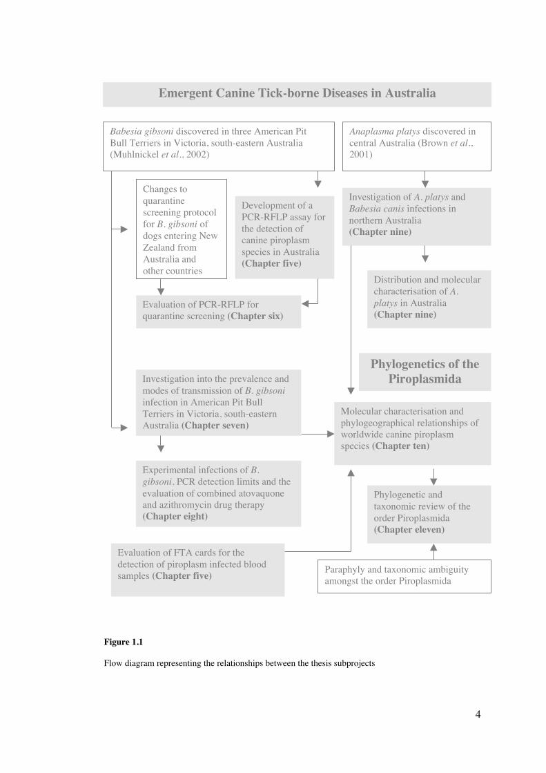
An overview of each subproject investigated and the inter-relationships between each
subproject within this thesis is shown in Figure 1.1.
4
Figure 1.1
Flow diagram representing the relationships between the thesis subprojects
Babesia gibsoni discovered in three American PitBull Terriers in Victoria, south-eastern Australia(Muhlnickel et al., 2002)
Anaplasma platys discovered incentral Australia (Brown et al.,2001)
Emergent Canine Tick-borne Diseases in Australia
Development of aPCR-RFLP assay forthe detection ofcanine piroplasmspecies in Australia(Chapter five)
Changes toquarantinescreening protocolfor B. gibsoni ofdogs entering NewZealand fromAustralia andother countries
Evaluation of PCR-RFLP forquarantine screening (Chapter six)
Phylogenetics of thePiroplasmida
Molecular characterisation andphylogeographical relationships ofworldwide canine piroplasmspecies (Chapter ten)
Phylogenetic andtaxonomic review of theorder Piroplasmida(Chapter eleven)
Investigation into the prevalence andmodes of transmission of B. gibsoniinfection in American Pit BullTerriers in Victoria, south-easternAustralia (Chapter seven)
Experimental infections of B.gibsoni, PCR detection limits and theevaluation of combined atovaquoneand azithromycin drug therapy(Chapter eight)
Evaluation of FTA cards for thedetection of piroplasm infected bloodsamples (Chapter five)
Investigation of A. platys andBabesia canis infections innorthern Australia(Chapter nine)
Distribution and molecularcharacterisation of A.platys in Australia(Chapter nine)
Paraphyly and taxonomic ambiguityamongst the order Piroplasmida

5
1.3 General aims
1. To develop a PCR-RFLP assay for the detection and differentiation of the canine
Piroplasmida species
2. To evaluate Whatman FTA classic cards for the application of canine blood and
subsequent use for PCR detection of piroplasm DNA
3. To evaluate PCR-RFLP for quarantine screening of dogs for B. gibsoni infection
4. To assess the prevalence and transmission dynamics of B. gibsoni infections in
American Pit Bull Terriers in Victoria, Australia
5. To investigate the efficacy of atovaquone and azithromycin drug therapy and
detection limits of PCR using experimental infections of B. gibsoni
6. To determine the distribution of A. platys in Australia and molecularly characterise
isolates from different geographical locations
7. To investigate co-infections of A. platys and B. canis and the efficacy of doxycycline
treatment
8. To molecularly characterise isolates of B. canis and B. gibsoni collected worldwide
and investigate phylogeographical relationships among isolates
9. To review the phylogenetic and taxonomic relationships of the order Piroplasmida
6
Review of Literature on the Canine Piroplasmida
Piroplasmosis is the collective term for diseases caused by ‘piroplasms’; intracellular, blood-
borne protozoan parasites of the order Piroplasmida. These tick-transmitted diseases, many
of which are of veterinary and medical significance, have been described worldwide, in a
large diversity of mammals, birds and reptiles. Piroplasmosis is a significant disease of
members of the Canidae, with multiple species of piroplasm reported to infect dogs and wild
canines. Although some of these piroplasm species cause limited pathogenesis, others can
produce severe illness, often leading to death. Identifying the species and subspecies of
piroplasm infecting dogs is of importance in the accurate management of the disease
including its diagnosis and subsequent treatment.
Piroplasmosis is considered an emerging disease syndrome, with many new species being
described and multiple species showing increasing prevalence and worldwide distributions.
Whether this increase is due to an increased awareness, the use of more sensitive detection
methods, or changing global travel patterns has yet to be determined.
2.1 Taxonomic classification of the canine piroplasms
Members of the order Piroplasmida are apicomplexan protozoa categorized into four main
families; Anthemosomatidae, Babesiidae, Theileriidae and Haemohormidiidae (Levine,
1988). The families Babesiidae and Theileriidae are well documented and include the
genera, Babesia, Entopolypoides, Cytauxzoon and Theileria. Historically, multiple genus
names have described the Piroplasmida including Piroplasma, Pyrosoma, Apiosoma,
CHAPTER TWO
7
Nuttallia, Nicollia, Babesiosoma, Smithia and Rossiella (Levine, 1988), each of which are no
longer generally accepted. It has also been suggested that the genus Entopolypoides is
synonymous with the genus Babesia (Gleason and Wolf, 1974; Bronsdon et al., 1999). There
is currently no consensus regarding correct species allocation within the order Piroplasmida.
Early classification of these blood-borne piroplasms relied heavily upon examination of their
morphological and life cycle characteristics (Allsopp et al., 1994). Initial taxonomic
classification of the canine piroplasms was on the basis of size and allowed for the
separation of two species, the ‘large’ Babesia canis and ‘small’ Babesia gibsoni. Evidently,
there are limitations in such a general consignment to a single species on the basis of host
specificity and morphological similarity. For example, it has now been noted that some
species of Babesia are not host specific, such as B. microti, which can infect a wide range of
vertebrate hosts (Etkind et al., 1980; Moore and Kuntz, 1981). Additionally, B. microti
cannot be reliably differentiated from B. gibsoni when examining Giemsa-stained blood
smears using light microscopy (Conrad et al., 1992).
Molecular characterisation on the basis of conserved gene loci has significantly aided the
accurate identification of a species and also allows further discrimination to a genotypic
level. The classification of the piroplasms has received renewed attention with the advances
in molecular biology and has resulted in the characterisation of more than two canine
piroplasm species and the infection of dogs with piroplasm species formerly considered
specific to other hosts (Table 2.1).
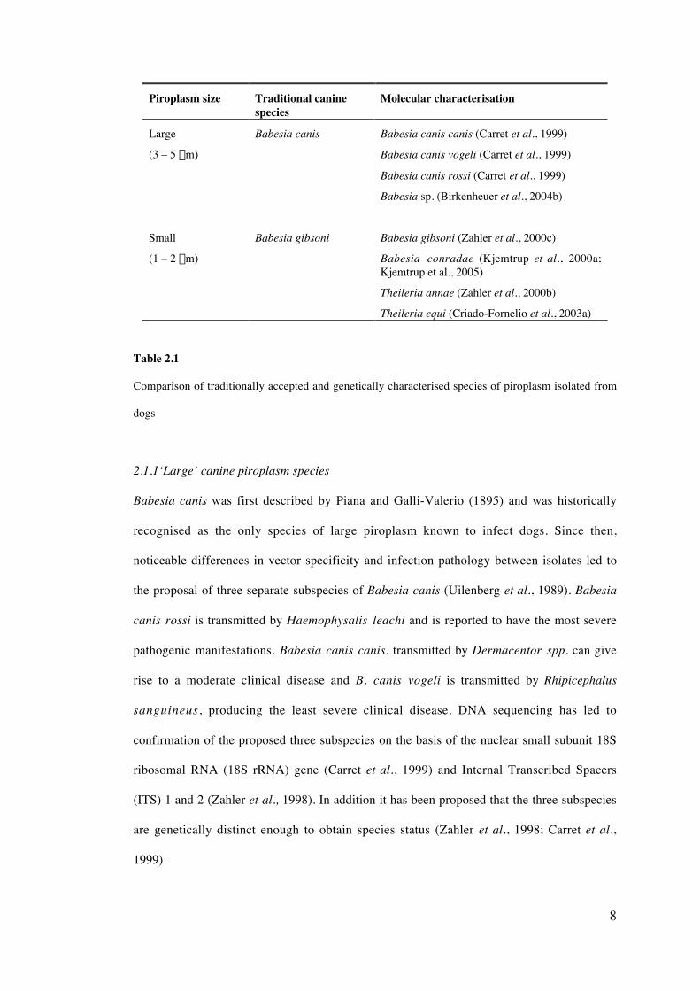
8
Piroplasm size Traditional caninespecies
Molecular characterisation
Large Babesia canis Babesia canis canis (Carret et al., 1999)
(3 – 5 mm) Babesia canis vogeli (Carret et al., 1999)
Babesia canis rossi (Carret et al., 1999)
Babesia sp. (Birkenheuer et al., 2004b)
Small Babesia gibsoni Babesia gibsoni (Zahler et al., 2000c)
(1 – 2 mm) Babesia conradae (Kjemtrup et al., 2000a;Kjemtrup et al., 2005)
Theileria annae (Zahler et al., 2000b)
Theileria equi (Criado-Fornelio et al., 2003a)
Table 2.1
Comparison of traditionally accepted and genetically characterised species of piroplasm isolated from
dogs
2.1.1‘Large’ canine piroplasm species
Babesia canis was first described by Piana and Galli-Valerio (1895) and was historically
recognised as the only species of large piroplasm known to infect dogs. Since then,
noticeable differences in vector specificity and infection pathology between isolates led to
the proposal of three separate subspecies of Babesia canis (Uilenberg et al., 1989). Babesia
canis rossi is transmitted by Haemophysalis leachi and is reported to have the most severe
pathogenic manifestations. Babesia canis canis, transmitted by Dermacentor spp. can give
rise to a moderate clinical disease and B. canis vogeli is transmitted by Rhipicephalus
sanguineus, producing the least severe clinical disease. DNA sequencing has led to
confirmation of the proposed three subspecies on the basis of the nuclear small subunit 18S
ribosomal RNA (18S rRNA) gene (Carret et al., 1999) and Internal Transcribed Spacers
(ITS) 1 and 2 (Zahler et al., 1998). In addition it has been proposed that the three subspecies
are genetically distinct enough to obtain species status (Zahler et al., 1998; Carret et al.,
1999).
9
A fourth subspecies of B. canis has also been proposed, B. canis presentii, which was
identified in domestic cats from Israel (Baneth et al., 2004) and an additional species of
‘large’ canine Babesia has been reported in a Labrador in North Carolina (Birkenheuer et al.,
2004b). This species remains unnamed.
2.1.2‘Small’ canine piroplasm species
Patton (1910) was first to describe Piroplasma gibsoni as a species, when this small
piroplasm was identified within the blood of dogs and jackals in India. This species was later
renamed Babesia gibsoni and subsequent findings of all small piroplasms in canine blood
were also assigned the same species name on the basis of morphology and host specificity
(Botros et al., 1975). Molecular-based characterisation of various isolates of small canine
piroplasms from separate geographical regions of the world has however, identified distinct
genetic variants and resulted in the differentiation of the small canine piroplasms into more
than one species.
Zahler et al. (2000b) first described the existence of a small canine piroplasm from Spain
that was genetically distinct from B. gibsoni and was most genetically similar to a
rodent/human species, B. microti, on the basis of the 18S rRNA gene. This isolate was
taxonomically classified as a member of the genus Theileria and named T. annae.
Isolates of small canine Babesia from Asia and North America were also compared on the
basis of sequence analysis of the 18S rRNA gene (Zahler et al., 2000c). Phylogenetic
comparison of these geographically different isolates suggested that the Asian isolates
belong to different species to the American isolate (obtained from dogs in California).
Further support of the existence of at least three species of small canine Babesia was
developed when strains from Okinawa, Oklahoma, North Carolina, Indiana, Missouri and
Alabama were compared phylogenetically (Kjemtrup et al., 2000a). The isolate from
10
California which has been named Babesia conradae1 (Kjemtrup et al ., 2005) .was shown to
be most closely related to a human species of piroplasm referred to as ‘WA1’ (Quick et al.,
1993) (now described as the species Babesia duncani). The remaining isolates from Asia and
the Midwestern and eastern United States have been classified as B. gibsoni, often with the
qualifier ‘Asian genotype’ to avoid further confusion.
Additionally, there have been reports of Theileria equi, a species normally only associated
with horses, found within dogs (Criado-Fornelio et al., 2003a; Criado-Fornelio et al., 2004).
The pathogenicity of T. equi is currently unknown as only four dogs have been reported to
be infected, one described as ‘symptomatic’, while the remaining three were clinically
normal.
2.2 Phylogeny and evolutionary relationships among the Piroplasmida
An increased understanding of the phylogenetic relationships among the Piroplasmida has
been established through the use of genetic-based analysis. Much confusion still exists over
the correct evolutionary relationships among the canine piroplasms and may not be resolved
until additional isolates and species are genetically characterised on the basis of multiple
gene loci. To date, phylogenetic analyses of the Piroplasmida have concentrated on the
conserved 18S rRNA gene.
A limited number of studies have determined the overall phylogenetic relationships between
members of the phylum Apicomplexa and is likely to be a reflection of the many thousands
of species described (Escalante and Ayala, 1995). Most research has concentrated on
selected genera that have medical or veterinary significance. In general, the Piroplasmida
have been shown to share a common ancestor with members of genus Plasmodium, forming
1 Babesia conradae is synonymous with early reports of B. gibsoni described from California (Conradet al., 1991; Conrad et al., 1992; Wokniak et al., 1997), B. gibsoni (Californian genotype) (Zahler etal., 2000c; Kocan et al., 2000; Kocan et al., 2001, ‘Dog from California’ (GenBank accession No.AF158702)
11
a separate clade with the genera Sarcocystis, Neospora and Toxoplasma. All characterised
species of the Piroplasmida (of the families Babesiidae and Theileriidae) form a distinctive,
individual clade, separate from all other members of the phylum Apicomplexa. No research
has been published on the phylogenetic relationships of the little known Piroplasmida
families, the Anthemosomatidae and Haemohormidiidae.
Early phylogenetic classification of the Piroplasmida relied solely on morphological and/or
life cycle characteristics. Members of the genus Theileria were differentiated from other
species of piroplasm by the presence of a tetrad or ‘maltese cross’ formation of the
intraerythrocytic merozoites and the existence of an exoerythrocytic lifecycle stage
(Mehlhorn and Schein, 1984). The Theileria were also distinguished by transstadial
transmission in the tick vector as opposed to the transovarial transmission found to occur in
the ‘true’ Babesia, termed the Babesia sensu stricto (Mehlhorn and Schein, 1984).
Allsopp et al. (1994) first assessed the phylogenetic and evolutionary relationships of the
piroplasms on the basis of the 18S rRNA gene of a limited number of species of Babesia,
Theileria and Cytauxzoon. This study suggested that most of the Babesia spp (the Babesia
sensu stricto) and the Theileria spp. separated into distinct monophyletic clades. A third
group containing Babesia rodhaini, Babesia equi and Cytauxzoon felis was inferred to be
ancestral to the only the Theileria or both the Theileria and the Babesia sensu stricto groups.
This group was proposed as a new Family, the Nicollidae (Allsopp et al., 1994).
Using an increased number of piroplasm species, including newly described human and
wildlife piroplasm species from western USA, Kjemtrup et al. (2000b) further conducted
phylogenetic analysis using the 18S rRNA gene. Four distinct groups were inferred from
phylogenetic trees. As in previous studies, the Babesia sensu stricto group were distinctly
separated from the Theileria group. An additional group of piroplasms was reported in this
study, termed the western Babesia spp. group, which contained wildlife and human
12
piroplasm spp, in addition to B. conradae (Kjemtrup et al., 2000b). Babesia microti was
found to form a fourth clade, ancestral to all other three groups of piroplasms.
The phylogenetic position of the canine piroplasm species in early analyses related solely to
B. canis. Allsopp et al. (1994) found that B. canis belonged to the Babesia sensu stricto
group. Further support for this was provided when B. canis canis and B. canis rossi were
shown to cluster together and that B. canis vogeli separated into a monophyletic group with
B. odocoilei and B. divergens (Carret et al., 1999). Babesia conradae was found to belong to
the western Babesia spp. group (Kjemtrup et al., 2000b).
Further revision of the phylogenetic relationships among the Piroplasmida has been
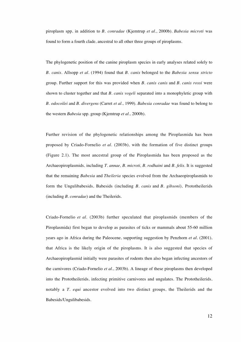
proposed by Criado-Fornelio et al. (2003b), with the formation of five distinct groups
(Figure 2.1). The most ancestral group of the Piroplasmida has been proposed as the
Archaeopiroplasmids, including T. annae, B. microti, B. rodhaini and B. felis. It is suggested
that the remaining Babesia and Theileria species evolved from the Archaeopiroplasmids to
form the Ungulibabesids, Babesids (including B. canis and B. gibsoni), Prototheilerids
(including B. conradae) and the Theilerids.
Criado-Fornelio et al. (2003b) further speculated that piroplasmids (members of the
Piroplasmida) first began to develop as parasites of ticks or mammals about 55-60 million
years ago in Africa during the Paleocene, supporting suggestion by Penzhorn et al. (2001),
that Africa is the likely origin of the piroplasms. It is also suggested that species of
Archaeopiroplasmid initially were parasites of rodents then also began infecting ancestors of
the carnivores (Criado-Fornelio et al., 2003b). A lineage of these piroplasms then developed
into the Prototheilerids, infecting primitive carnivores and ungulates. The Prototheilerids,
notably a T. equi ancestor evolved into two distinct groups, the Theilerids and the
Babesids/Ungulibabesids.
13
Figure 2.1
Distance based phylogenetic tree of the Piroplasmida (adapted from Criado-Fornelio et al., 2003b),
Arrows indicate piroplasm species found in dogs (Babesia sp – North Carolina, not included)
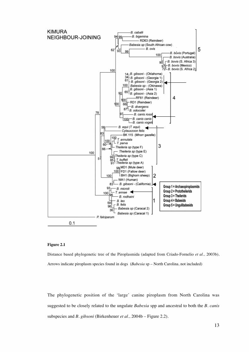
The phylogenetic position of the ‘large’ canine piroplasm from North Carolina was
suggested to be closely related to the ungulate Babesia spp and ancestral to both the B. canis
subspecies and B. gibsoni (Birkenheuer et al., 2004b – Figure 2.2).
14
Figure 2.2
Partial phylogenetic tree identifying the evolutionary relationships between the large Babesia sp. from
a dog (North Carolina) and other Babesia species (adapted from Birkenheuer et al., 2004b)
2.3 Morphology
2.3.1 ‘Large’ canine piroplasm species
All ‘large’ species of canine piroplasms are typically 2 – 5 µm in diameter, with
differentiation of individual species and subspecies difficult on the sole basis of morphology.
Trophozoites of B. canis are characterised by a length of approximately 5.0 mm and a width
of 2.5 - 3.0 mm and are generally described as large canine Babesia (Kuttler, 1988, Conrad et
al., 1992). Babesia canis piroplasms are generally pear-shaped, occurring singularly or as
pairs of dividing trophozoites inside the erythrocyte (Kjemtrup et al., 2000a), but a wide
range of morphological characteristics are recognised.
The Babesia sp. (North Carolina) described by Birkenheuer et al., (2004b), was reported as
being polymorphic, typically singular, with occasional two pyriform-shaped organisms
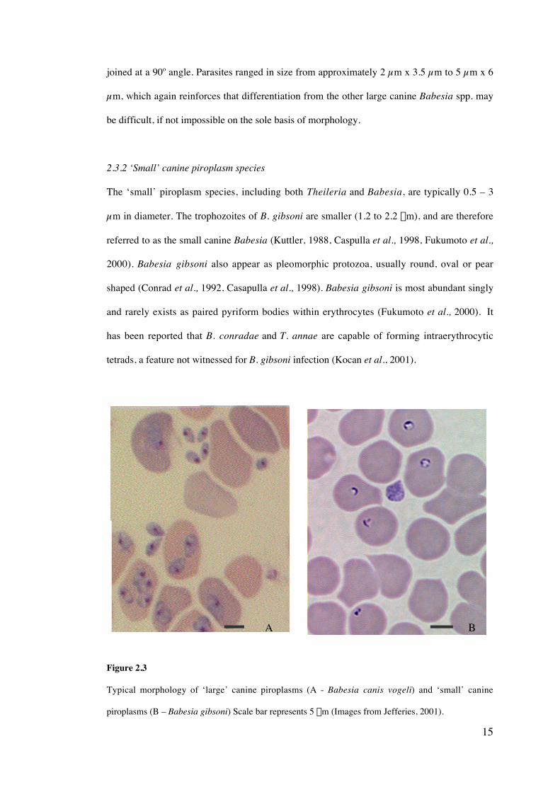
15
joined at a 90o angle. Parasites ranged in size from approximately 2 µm x 3.5 µm to 5 µm x 6
µm, which again reinforces that differentiation from the other large canine Babesia spp. may
be difficult, if not impossible on the sole basis of morphology.
2.3.2 ‘Small’ canine piroplasm species
The ‘small’ piroplasm species, including both Theileria and Babesia, are typically 0.5 – 3
µm in diameter. The trophozoites of B. gibsoni are smaller (1.2 to 2.2 mm), and are therefore
referred to as the small canine Babesia (Kuttler, 1988, Caspulla et al., 1998, Fukumoto et al.,
2000). Babesia gibsoni also appear as pleomorphic protozoa, usually round, oval or pear
shaped (Conrad et al., 1992, Casapulla et al., 1998). Babesia gibsoni is most abundant singly
and rarely exists as paired pyriform bodies within erythrocytes (Fukumoto et al., 2000). It
has been reported that B. conradae and T. annae are capable of forming intraerythrocytic
tetrads, a feature not witnessed for B. gibsoni infection (Kocan et al., 2001).
Figure 2.3
Typical morphology of ‘large’ canine piroplasms (A - Babesia canis vogeli) and ‘small’ canine
piroplasms (B – Babesia gibsoni) Scale bar represents 5 mm (Images from Jefferies, 2001).
A B

16
Electron microscopic examination of intraerythrocytic B. gibsoni, revealed the presence of
four morphologically distinct trophozoite stages; small spheres, small rods, irregular forms
lacking pseudo-inclusions and large spheres with pseudo-inclusions (Radi et al., 2004).
2.4 Transmission
2.4.1 Tick vectors
Tick species are recognized as being the main vector responsible for the transmission of all
species of piroplasm. Each of the species and subspecies of the canine piroplasms is tick
vector specific, with each of the B. canis subspecies infecting a single and separate tick
species. A summary of the known tick species that transmit the different species and
subspecies of the canine piroplasms is given in Table 2.2. It is important to recognise that
many transmission studies carried out may not be reliable and that a definitive list of tick
vector species of the canine Piroplasmida has not been determined.
Piroplasm species Tick vector species Citations
Babesia canis canis Dermacentor reticulatus
Dermacentor marginatus?
Schein et al. (1979); Mehlhornet al. (1980) Jongejan andUilenberg (2004)
Babesia canis rossi Haemophysalis leachi Lewis et al. (1996)
Babesia canis vogeli Rhipicephalus sanguineus
Babaesia gibsoni Haemophysalis bispinosa?,Haemophysalis longicornisRhipicephalus sanguineus?
Higuchi et al. (1991a); (1991b);(1992); (1993a); (1993b);(1995); (1999a); (1999b)
Babesia sp. (NorthCarolina)
Currently undetermined
Babesia conradae Currently undetermined
Theileria annae Ixodes hexagonus Camacho et al. (2003)
Theileria equi Dermacentor variabilis, D.nutalli, Hyalomma spp ,Boophilus microplus,Rhipicephalus turanicus
Moltmann et al. (1983); Zapfand Schien (1994a); (1994b);Battsetseg et al. (2001); Stilleret al. (2002)
Table 2.2
Tick vector candidates of the canine piroplasm species.
17
Babesia infection is generally associated with adult ticks (especially females), however
transmission by larval and nymphal ticks has also been documented for B. canis (Shortt,
1973). The engorged female tick is the only stage capable of acquiring the infection from the
vertebrate host (Friedhoff, 1988). Vertical transmission (transovarial) of B. canis is possible
and has been demonstrated for R. sanguineus (Friedhoff, 1988). This study suggested that B.
canis may remain infective for five successive generations. Mechanical transmission by
most blood-feeding arthropod may also be possible, however limited research has
investigated this possibility.
2.4.2 Vertical transmission
Transplacental or perinatal transmission has been known to occur for both Babesia and
Theileria spp (New et al., 1997; Baek et al., 2003). Initial reports of pernatal transmission of
parasites in utero were noted to occur in humans, suggesting that it was possible for a mother
infected with B. microti, to transmit the infection to her unborn child (Esernio-Jenssen et al.,
1987; New et al., 1997). Further reports have suggested that transplacental transmission of
other species such as Theileria sergenti (Baek et al., 2003) and Theileria equi (Phipps and
Otter, 2004) can occur.
Limited study has assessed transplacental transmission of the canine piroplasms, with most
information being anecdotal. Babesia gibsoni has been found in the blood of young puppies
and in their dams, suggesting that transplacental transmission is the most likely cause of
infection (Harvey et al., 1988) and a recent study proved this mode of transmission
experimentally (Fukomoto et al., 2005a).
2.4.3 Blood transfusion
The role of blood transfusion in the transmission of blood-borne pathogens has become
increasingly recognised in both human and veterinary medicine (Herwaldt et al., 2002;
Kjemtrup et al., 2002; Powell and Grima, 2002; Cable and Leiby, 2003; Leiby and Gill,
18
2004). Transfusion babesiosis was first reported in the USA, when a patient received blood
infected with B. microti (Wittner et al., 1982). The results of a later study indicated that B.
microti parasites can remain infective under normal blood banking conditions (Eberhard et
al., 1995), highlighting the need to screen potential blood donors.
Transfusion-associated transmission has also been reported for at least two species of canine
Babesia, highlighting the need to screen potential blood donor dogs (Wardrop et al., 2005).
Babesia gibsoni has been reported to be transmitted during a whole blood transfusion, with
the donor blood originating from an American Pit Bull Terrier (Stegeman et al., 2003).
Likewise, transfusion-associated transmission has been noted in B. canis rossi infections
(Jacobson and Clark, 1994). While appropriate screening for Babesia and Theileria in
potential blood donor dogs should be carried out, it has also been reported that the treatment
of donor blood with INACTINE PEN110 is highly effective in eradicating B. microti from
human erythrocytes (Zavizion et al., 2004). It is possible that chemical treatment of Babesia
infected donor blood from dogs may be also be effective but requires investigation.
2.4.4 Direct blood-to-blood transmission
The possibility of direct blood-to-blood transmission of piroplasms has also been suggested
when dogs attack and bite one another. The greatest implication of this form of transmission
has been reported in breeds used in dog fighting. A high prevalence of B. gibsoni has been
described in American Pit Bull Terriers in the USA (Birkenheuer et al., 1999; Macintire et
al., 2002; Birkenheuer et al., 2003b) and also in Tosa dogs in Japan (Matsuu et al., 2004a).
In both countries, it has been postulated that direct blood-to-blood transmission of B. gibsoni
may occur during biting or fighting between dogs. Matsuu et al. (2004a) also speculated that
transmission of the parasite may occur during mating.
19
2.4.5 Movement of dogs and ticks from areas of endemicity
A major contributing factor in the increased distribution of canine piroplasm species is the
movement of family-owned and military working dogs between countries (Anderson et al.,
1980; Shaw et al., 2001b). The translocation of chronically infected animals into disease-free
areas has previously been suggested as being of significant importance in the spread of B.
gibsoni in the USA and it is also theorised that military dogs returning from Japan were
responsible for the original introduction of this parasite into the US (Anderson et al., 1980).
International travel of dogs has increased recently, with programs such as the Pet Travel
Scheme (PETS) contributing to the movement of dogs between countries in Europe (Shaw et
al., 2003). Selective analysis of dogs entering the UK revealed many were infected with
exotic pathogens including both B. gibsoni and B. canis canis. This highlights the need to
increase surveillance of dogs entering countries know to be free from piroplasm infection to
avoid the establishment of these diseases in new parts of the world.
2.4.6 Wild Caniidae species as reservoirs for piroplasms
An important feature of piroplasm spp. infection is the facilitation of wild canids as
reservoirs of these parasites. Multiple canine species have been described as potential hosts
for canine Babesia throughout many regions of the world. Jackals (Canis aureus) in India,
foxes (Vulpes vulpes niloticus), jackals (Canis aureus lupaster) and a fenec (Fennecus
zerda) in Egypt (Maronpot and Guindy, 1970; Botros et al., 1975) and coyotes (Canis
latrans) in the USA (Yamane et al., 1994) have all been suggested as reservoirs of B. gibsoni
infection. Notably, coyotes that were experimentally infected with B. gibsoni exhibited only
mild clinical signs (Roher, 1985), suggesting that they may act as carrier animals. Cape
hunting dogs (Lycaon pictus) and silver-backed jackals (Canis mesomelas) have been
associated with B. canis infection (Kuttler, 1988). Additionally, B. canis rossi was found in
the blood of side-striped jackals (Canis adustus) in southern Africa (Lewis et al., 1996) and
20
T. annae has been identified in red foxes (Vulpes vulpes) in Spain (Criado-Fornelio et al.,
2003a) and the USA (Goethert and Telford, 2003).
Wild canines in Australia, most notably dingoes (C. familiaris dingo) have been previously
reported with babesiosis (Callow, 1984) and were probably infected with B. canis vogeli
(Irwin and Hutchinson, 1991). Dingo populations may therefore also represent a potential
reservoir for B. canis vogeli in Australia
2.4.7 Other mammal species as canine piroplasms reservoirs
It has increasingly been reported that many piroplasm species are not host specific and may
be cable of infecting multiple host species (Criado-Fornelio et al., 2003a; Criado-Fornelio et
al., 2003c). Such reports have also been published for some of the canine piroplasm species.
Theileria annae has been found to infect cats and B. canis canis has been detected in the
blood of both cats and horses (Criado-Fornelio et al., 2003a). Theileria annae-like
piroplasms have also been identified in skunks and racoons (Goethert and Telford, 2003;
Kawabuchi et al., 2005). Other carnivores may also harbour species of piroplasm, potentially
capable of infecting dogs such as Babesia missirolii and an unnamed piroplasm species
identified in badgers (Meles meles) (Peirce, 1974; Simsek et al., 2003), Babesia mephitis
from the striped skunk (Mephitis mephitis) (Holbrook and Frerichs, 1970) and Babesia
heischi and Babesia hoarei from Peter’s pigmy mongoose (Helogale undulata rufula)
(Grewal, 1957). Each of these species have never been genetically characterised. Badgers are
reported to be commonly infected with the tick Ixodes hexagonus, also the presumed vector
of T. annae. Further research is therefore warranted to determine whether the badger
piroplasm species described by Peirce (1974) is actually T. annae.
2.5 Life cycles of the Piroplasmida spp.
The life cycle of canine piroplasms is characteristic of that of all apicomplexan parasites in
that it generally involves at least three phases of reproduction; gamogony, sporogony and
21
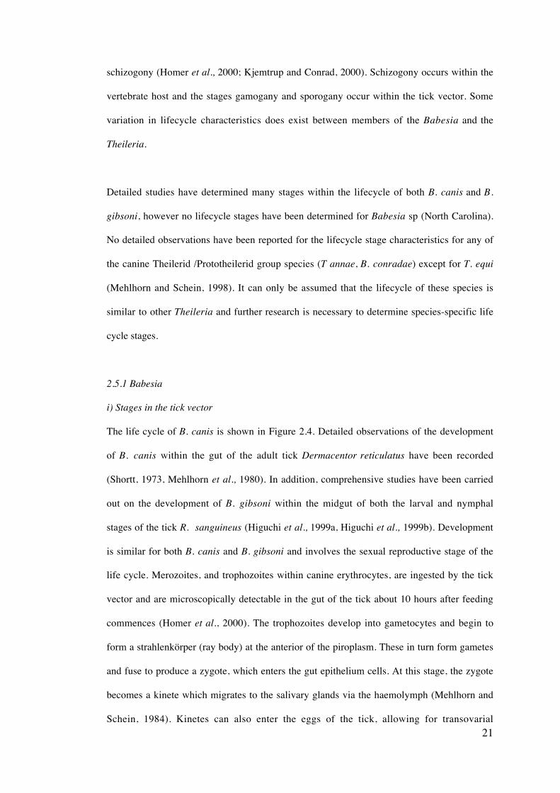
schizogony (Homer et al., 2000; Kjemtrup and Conrad, 2000). Schizogony occurs within the
vertebrate host and the stages gamogany and sporogany occur within the tick vector. Some
variation in lifecycle characteristics does exist between members of the Babesia and the
Theileria.
Detailed studies have determined many stages within the lifecycle of both B. canis and B.
gibsoni, however no lifecycle stages have been determined for Babesia sp (North Carolina).
No detailed observations have been reported for the lifecycle stage characteristics for any of
the canine Theilerid /Prototheilerid group species (T annae, B. conradae) except for T. equi
(Mehlhorn and Schein, 1998). It can only be assumed that the lifecycle of these species is
similar to other Theileria and further research is necessary to determine species-specific life
cycle stages.
2.5.1 Babesia
i) Stages in the tick vector
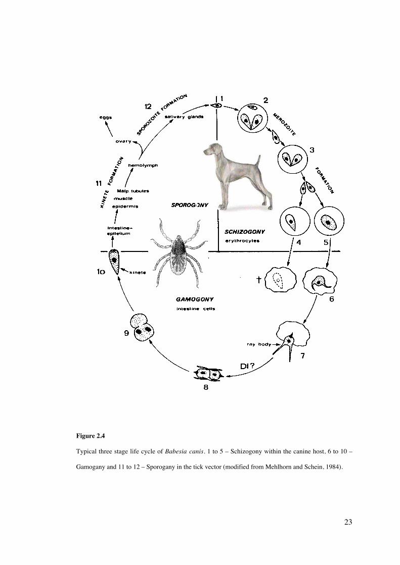
The life cycle of B. canis is shown in Figure 2.4. Detailed observations of the development
of B. canis within the gut of the adult tick Dermacentor reticulatus have been recorded
(Shortt, 1973, Mehlhorn et al., 1980). In addition, comprehensive studies have been carried
out on the development of B. gibsoni within the midgut of both the larval and nymphal
stages of the tick R. sanguineus (Higuchi et al., 1999a, Higuchi et al., 1999b). Development
is similar for both B. canis and B. gibsoni and involves the sexual reproductive stage of the
life cycle. Merozoites, and trophozoites within canine erythrocytes, are ingested by the tick
vector and are microscopically detectable in the gut of the tick about 10 hours after feeding
commences (Homer et al., 2000). The trophozoites develop into gametocytes and begin to
form a strahlenkörper (ray body) at the anterior of the piroplasm. These in turn form gametes
and fuse to produce a zygote, which enters the gut epithelium cells. At this stage, the zygote
becomes a kinete which migrates to the salivary glands via the haemolymph (Mehlhorn and
Schein, 1984). Kinetes can also enter the eggs of the tick, allowing for transovarial
22
transmission (Homer et al., 2000). Sporogony or the formation of sporozoites occurs within
the salivary gland, with many thousands of sporozoites being produced from each initial
kinete.
ii) Stages in the vertebrate host
Transmission of the sporozoites from the tick’s salivary glands to the canine host generally
occurs 2-3 days after tick attachment (Martinod et al., 1985). Once inside the host, the
sporozoites become merozoites and invade the erythrocytes by a process of endocytosis and
form a parasitophorus vacuole (which later disintegrates) within the cell (Homer et al.,
2000). The merozoites transform into trophozoites and divide by binary fission into
additional merozoites, a stage termed schizogony. The newly formed merozoites lyse the
host cell and continue to invade and multiply within other erythrocytes. Some of the
trophozoites become gametocytes, reproducing once inside of the tick gut.
23
Figure 2.4
Typical three stage life cycle of Babesia canis. 1 to 5 – Schizogony within the canine host, 6 to 10 –
Gamogany and 11 to 12 – Sporogany in the tick vector (modified from Mehlhorn and Schein, 1984).
24
2.5.2 Theileria
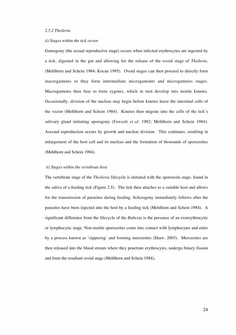
ii) Stages within the tick vector
Gamogony (the sexual reproductive stage) occurs when infected erythrocytes are ingested by
a tick, digested in the gut and allowing for the release of the ovoid stage of Theileria.
(Mehlhorn and Schein 1984; Kocan 1995). Ovoid stages can then proceed to directly form
macrogametes or they form intermediate microgamonts and microgametes stages.
Macrogametes then fuse to form zygotes, which in turn develop into motile kinetes.
Occasionally, division of the nucleus may begin before kinetes leave the intestinal cells of
the vector (Mehlhorn and Schein 1984). Kinetes then migrate into the cells of the tick’s
salivary gland initiating sporogony (Fawcett et al. 1982; Mehlhorn and Schein 1984).
Asexual reproduction occurs by growth and nuclear division. This continues, resulting in
enlargement of the host cell and its nucleus and the formation of thousands of sporozoites
(Mehlhorn and Schein 1984).
ii) Stages within the vertebrate host
The vertebrate stage of the Theileria lifecycle is initiated with the sporozoite stage, found in
the saliva of a feeding tick (Figure 2.5). The tick then attaches to a suitable host and allows
for the transmission of parasites during feeding. Schizogony immediately follows after the
parasites have been injected into the host by a feeding tick (Mehlhorn and Schein 1984). A
significant difference from the lifecycle of the Babesia is the presence of an exoerythrocytic
or lymphocytic stage. Non-motile sporozoites come into contact with lymphocytes and enter
by a process known as ‘zippering’ and forming merozoites (Shaw, 2003). Merozoites are
then released into the blood stream where they penetrate erythrocytes, undergo binary fission
and form the resultant ovoid stage (Mehlhorn and Schein 1984).
25
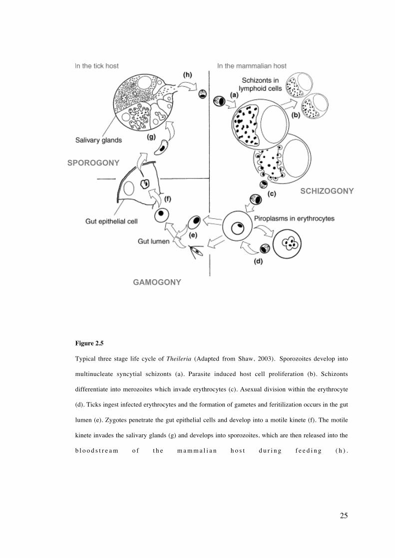
Figure 2.5
Typical three stage life cycle of Theileria (Adapted from Shaw, 2003). Sporozoites develop into
multinucleate syncytial schizonts (a). Parasite induced host cell proliferation (b). Schizonts
differentiate into merozoites which invade erythrocytes (c). Asexual division within the erythrocyte
(d). Ticks ingest infected erythrocytes and the formation of gametes and feritilization occurs in the gut
lumen (e). Zygotes penetrate the gut epithelial cells and develop into a motile kinete (f). The motile
kinete invades the salivary glands (g) and develops into sporozoites, which are then released into the
b l o o d s t r e a m o f t h e m a m m a l i a n h o s t d u r i n g f e e d i n g ( h ) .
SCHIZOGONY
GAMOGONY
SPOROGONY
26
2.6 Distribution
The distribution of each of the canine Piroplasmida species is variable with some showing an
ever emerging, worldwide dispersal, while others seem to have a relatively restricted
distribution, found in a very limited number of countries. The full extent of the distribution
of each of the different species is currently unknown and requires further investigation to
appreciate the complete epidemiological situation among these protozoa.
2.6.1 Large canine piroplasm spp.
Of the large canine piroplasms (Figure 2.6), Babesia canis vogeli has the greatest known
distribution, predominantly in semitropical to tropical areas and has been reported in South
and North America, Africa, Australia, Asia, Southern Europe and the Middle East
(Uilenberg et al., 1989; Taboada et al., 1992; Carret et al., 1999; Caccio et al., 2002;
Jefferies et al., 2003). Babesia canis rossi is believed to have the most confined distribution,
found only in southern Africa (Uilenberg et al., 1989; Carret et al., 1999) and Sudan
(Oyamada et al., 2005). Babesia canis canis has been reported in France, Hungary (Földvari
et al., 2005), The Netherlands (Zandvliet et al., 2004), Slovenia (Duh et al., 2004), Russia
(Rar et al., 2004), Switzerland (Casati et al., 2004) Poland and Croatia (Caccio et al., 2002).
The current distribution of the unnamed large Babesia sp. is unknown and has only been
found within one dog in North Carolina, USA (Birkenheuer et al., 2004b).
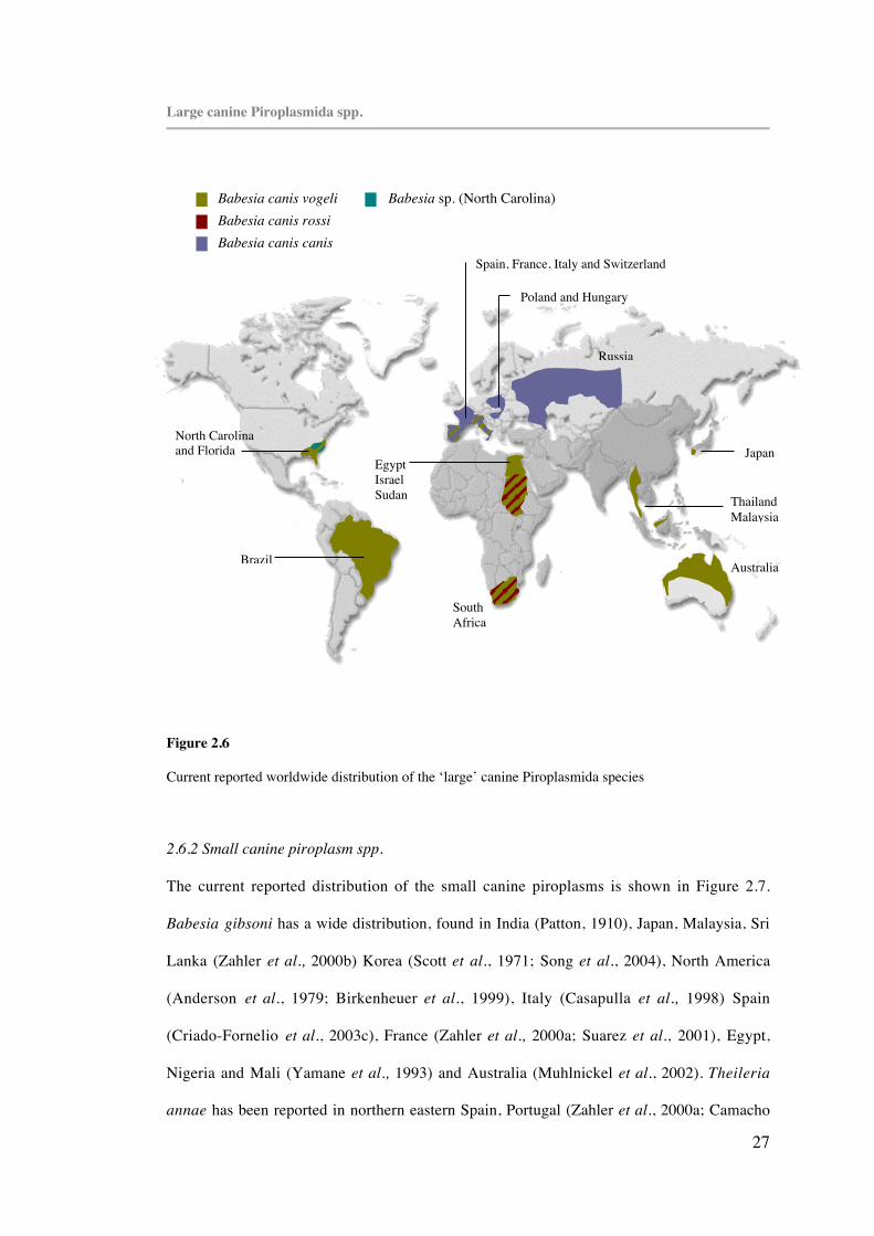
27
Large canine Piroplasmida spp.
Figure 2.6
Current reported worldwide distribution of the ‘large’ canine Piroplasmida species
2.6.2 Small canine piroplasm spp.
The current reported distribution of the small canine piroplasms is shown in Figure 2.7.
Babesia gibsoni has a wide distribution, found in India (Patton, 1910), Japan, Malaysia, Sri
Lanka (Zahler et al., 2000b) Korea (Scott et al., 1971; Song et al., 2004), North America
(Anderson et al., 1979; Birkenheuer et al., 1999), Italy (Casapulla et al., 1998) Spain
(Criado-Fornelio et al., 2003c), France (Zahler et al., 2000a; Suarez et al., 2001), Egypt,
Nigeria and Mali (Yamane et al., 1993) and Australia (Muhlnickel et al., 2002). Theileria
annae has been reported in northern eastern Spain, Portugal (Zahler et al., 2000a; Camacho
Babesia canis vogeliBabesia canis rossiBabesia canis canis
Babesia sp. (North Carolina)
SouthAfrica
Brazil Australia
Spain, France, Italy and Switzerland
Poland and Hungary
Russia
JapanEgyptIsraelSudan Thailand
Malaysia
North Carolinaand Florida
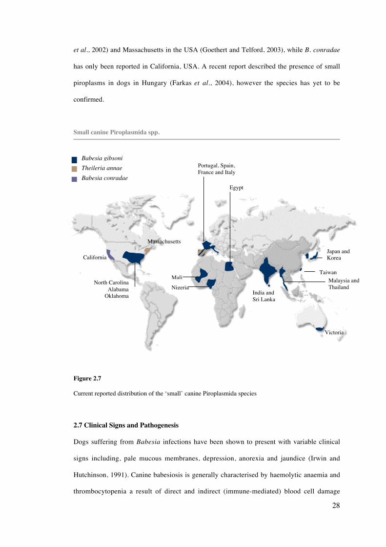
28
et al., 2002) and Massachusetts in the USA (Goethert and Telford, 2003), while B. conradae
has only been reported in California, USA. A recent report described the presence of small
piroplasms in dogs in Hungary (Farkas et al., 2004), however the species has yet to be
confirmed.
Small canine Piroplasmida spp.
Figure 2.7
Current reported distribution of the ‘small’ canine Piroplasmida species
2.7 Clinical Signs and Pathogenesis
Dogs suffering from Babesia infections have been shown to present with variable clinical
signs including, pale mucous membranes, depression, anorexia and jaundice (Irwin and
Hutchinson, 1991). Canine babesiosis is generally characterised by haemolytic anaemia and
thrombocytopenia a result of direct and indirect (immune-mediated) blood cell damage
Babesia gibsoni
Babesia conradaeTheileria annae
Nigeria
Mali
Egypt
India andSri Lanka
Taiwan
Victoria
Japan andKorea
Malaysia andThailand
Portugal, Spain,France and Italy
North CarolinaAlabama
Oklahoma
California
Massachusetts

29
induced by the parasites. Additional complications of the disease are variable depending
upon the strain and species of Babesia involved. Less virulent strains produce a more
transient disease while those that exhibit an increased virulence can produce multiple organ
dysfunction, which can lead to death of the infected host (Lobetti, 1998; Boozer and
Macintire, 2003). Babesiosis can generally be classified as acute, chronic or subclinical
(Breitschwerdt et al., 1984).
It is also suggested in the literature on the Piroplasmida species that recrudescence of
infections is possible (Bronsdon et al., 1999), a feature similarly reported in certain
Plasmodium infections (Mackintosh et al., 2004). Recrudescence, or the re-emergence of
clinical infection in animals previously known to be infected with a pathogen, is often
induced by increased stress levels in the host or by immuno-compromisation. This highlights
the possibility that Piroplasmida species can remain inactive within certain organ systems,
while not being present in the circulatory system (Ilhan et al., 1998). Studies have speculated
that inactive piroplasm may exist within the spleen, liver, kidneys or brain of the host,
producing no illness for months and even years (Dao et al., 1996; O’Connor et al., 1999).
2.7.1 Babesia canis subspecies
The acute phase of the infection of all three subspecies is characterised by haemolytic
anemia. Acute renal failure, cerebral babesiosis, coagulopathy, icterus, hepatopathy,
immune-mediated haemolytic anaemia, acute respiratory distress syndrome and shock have
been reported as complications associated with B. canis infection (Lobetti, 1998). Each of
the B. canis subspecies have been reported to produce different disease syndromes (Irwin
and Hutchinson, 1991; Schetters et al., 1997b). The most virulent subspecies is B. canis
rossi, characterized by a high proliferation rate. Schetters et al. (1997b) reported parasitemia
rates that were greater than 1% and that the level of parasitemia showed a correlation with
the degree of haemolysis and haemoglobinuria. Hypoglycemia and icterus are also
associated with infection (Keller et al. 2004). Babesia canis rossi can also produce mild
30
infections (Malherbe et al., 1976; Moore and Williams, 1979; Reyers et al., 1998). Such
non-anaemic babesiosis has been reported to be associated with severe azotemia, electrolyte
and acid-base disturbances and sometimes leukopenia (Reyers et al., 1998). Differences in
virulence exhibited between cases of B. canis rossi infection in South Africa may relate to
the potential of co-infections with B. canis vogeli (Matjila et al., 2004).
Babesia canis canis exhibits a lesser virulence and comparative studies with B. canis rossi
concluded each of the two subspecies produced a different disease syndrome (Schetters et
al., 1997b). Clinical disease resulting from B. canis canis infection was correlated to changes
in the dog’s coagulation system and not the level of parasitemia. Babesia canis canis may
proliferate in deep tissues and also shows evidence of autoagglutination (Schetters and
Montenegro-James, 1995). Fatal cases have also been reported (Matjila et al., 2005).
Babesia canis vogeli has been shown to be the least virulent of the three subspecies, with the
acute phase of the disease most notable in pups (Irwin and Hutchinson, 1991). Co-infections
of B. canis canis and B. canis vogeli may also occur (Caccio et al., 2002; Duh et al., 2004),
further complicating the disease pathogenesis.
2.7.2 Babesia gibsoni
Inokuma et al. (2005) reported that B. gibsoni infections with low-level parasitaemia
produce clinical and laboratory findings similar to those exhibited by immune mediated
haemolytic anaemia. This similarity can lead to misdiagnosis and has also been reported by
Muhlnickel et al. (2002), when a case of B. gibsoni infection in Australia was initially
misdiagnosed as immune mediated haemolytic anaemia. It has also been suggested that
macrophages phagocytose both parasitised and non-parasitised erythrocytes, causing
extravascular haemolysis and splenomegaly (Murase et al., 1996). This signified that
oxidative damage within the erythrocytes, including those not parasitised, is a result of B.
gibsoni proliferation. Babesia gibsoni parasites have also been shown to preferentially infect
and multiply in younger erythrocytes (Murase et al., 1993).
31
2.7.3 Babesia conradae
Wozniak et al. (1997) demonstrated that haemolytic, regenerative anaemia occurs within all
B. conradae (described as B. gibsoni) infections. It was suggested that the destruction of
erythrocytes was a result of mechanical disruption of the cells by the infecting parasites,
complement-dependent immune-mediated erythrolysis and the phagocytosis of antigenically
altered or opsonized erythrocytes. Hepatic lesions are another distinctive feature of the
infection, characterised by hepatitis, hepatocellular atrophy, perivenular fibrosis and Kupffer
cell hypertrophy (Wozniak et al., 1997). Additionally, vasculitis and glomerulonephritis
have been reported and are believed to be a consequence of the immune mediated
component of the disease (Wozniak et al., 1997).
2.7.4 Theileria annae
Intense anaemia, azotemia and thrombocytopenia, with limited leucocytosis and renal
dysfunction and sometimes death have been reported in T. annae infected dogs in north-west
Spain (Camacho et al., 2001; Guitian et al., 2003; Camacho et al., 2004). In addition, a
splenectomized dog infected with T. annae presented with hypothermia, trembling and dark
urine and clinical signs included pale mucous membranes, tachycardia, tachypnea and
lymphadenopathy (Camacho et al., 2002).
2.8 Detection and diagnosis of canine piroplasm infections
Effective diagnosis of Babesia infections is important in their monitoring, management and
control (McLaughlin et al., 1992). A large diversity of diagnostic techniques exist, each of
which has its own limitations. The diagnostic tests for piroplasmosis can be divided into
three broad categories; traditional methods, including microscopy and culture; serological
techniques, and molecular-based methods. It is increasingly recognised that a combination of
detection techniques is necessary for accurate diagnosis. Limitations of clinical data, parasite
morphology and serological cross-reactivity, have lead to an increased interest in molecular
based methods and highlights the need for their application in clinical medicine.
32
2.8.1 Light Microscopy
The most widely used technique for the detection of Babesia is the examination of thin blood
smears stained with either Wright or Giemsa stain (Homer et al., 2000). Erythrocytes are
scanned for the presence of piroplasms. Parasitaemia levels have been found to be
concentrated in blood taken from ear-tip capillaries (Breitschwerdt, 1984), therefore using
ear-tip blood smears may increase the likelihood of detecting piroplasm infections. The
sensitivity of microscopy is suggested to be one parasite per 105 erythrocytes (Bose et al.,
1995). This technique is limited in that morphologically similar species cannot be
distinguished (Conrad et al., 1992) and accurate diagnosis is dependent on the experience of
the microscopist (Morgan, 2000).
2.8.2 Serological Tests
Multiple immunodiagnostic techniques have been created to detect antibodies to Babesia
spp. (reviewed by Bose et al., 1995). The two that are routinely applied to the diagnosis of
Babesia infections in dogs are the immunofluorescent antibody test and the enzyme-linked
immunosorbent assay.
i) Immunofluorescent Antibody Test (IFAT)
This test is a commonly used method of diagnosing Babesia and Theileria infections by
detecting the presence of antibodies to the parasites within the host serum. The test uses
antigen, in the form of parasite-infected blood applied to glass slides, host serum titrated to
various dilutions and fluorescein-labelled antibodies. The serum and antibodies are added to
the antigen, incubated and analysed using fluorescent microscopy.
Immunofluorescent Antibody Tests have been developed for both B. canis and B. gibsoni,
however limitations have been suggested to exist for such methodology. Levy et al. (1987)
found that 3.8% of dogs analysed in North Carolina were seropositive for B. canis using
IFAT, however recognised the possibility of cross-reactivity with B. gibsoni. Later, the
33
seroprevalence of B. canis in greyhounds from Florida was determined using IFA screening
and was reported to be 46% (Taboada et al., 1992). A B.canis seropositive dog has been
reported to have an antibody titre cut-off of ≥ 1:80 (Levy et al., 1987; Taboada et al., 1992).
IFAT was first developed for the diagnosis of B. gibsoni infections by Anderson et al.
(1980), who set the seropositive cut-off titre at >1:64. The use of IFAT for the diagnosis of
B. gibsoni was later evaluated and the need for optimal cut-off titres to be established to
avoid false-positive results due to antigen cross-reactivity was described. The IFAT for B.
gibsoni was found to be cross-reactive with B. canis, Toxoplasma gondii and Neospora
caninum (Yamane et al., 1993). It is also reported that dogs that are acutely infected with
Babesia may be seronegative (Breitschwerdt et al., 1983) and it is also difficult to assess
whether the dog currently has an infection or has previously been infected.
ii) Enzyme-linked immunosorbent assay (ELISA)
The ELISA was first applied to Babesia diagnosis in the detection of B. bovis and B. caballi
in cattle by utilizing antigens from infected erythrocytes. The earliest application of the
ELISA to canine Babesia was by Martinod et al. (1985). Their study developed the assay to
detect antibodies against B. canis, in addition to antibodies to the vectors D. reticulatus and
I. ricinus. The ELISA is limited by Babesia strain differences eliciting different antibody
responses and producing variable seroreactivity (Reiter and Weiland, 1989). Verdida et al.
(2004) developed an improved ELISA using recombinant truncated P50 surface antigen for
the serodiagnosis of B. gibsoni infection.
2.8.3 Polymerase chain reaction
The advent of the polymerase chain reaction (PCR) has shown significant promise in the
detection of pathogens and the diagnosis of disease over the past decade. PCR is a relatively
new molecular procedure that was first described in 1985 (Saiki et al., 1985; Mullis, 1990)
and involves the in vitro amplification of target nucleic acid sequences by primer directed
DNA synthesis. Initial use of PCR as a detection technique for Babesia was demonstrated in
34
non-canine species, most notably B. bovis, B. microti and B. bigemina and was shown to
have a significant degree of sensitivity and specificity (Fahrimal et al., 1992, Persing et al.,
1992 and Figueroa et al., 1992). Since then, the technique has been applied to many other
species of Babesia.
i) PCR detection of Babesia DNA in canine blood
PCR application to canine Babesia was first demonstrated on B. canis, involving DNA
amplification for sequencing and phylogenetic comparison (Allsopp et al., 1994). Later
studies have shown the ability of PCR to be a useful diagnostic tool for the detection and
phylogenetic analysis of the canine Babesia species. A majority of these studies used
amplification of partial regions of the small subunit ribosomal RNA gene as the basis of
diagnosis (Carret et al., 1999). The small subunit ribosomal RNA gene is useful in that it is a
highly conserved gene, showing limited nucleotide sequence variation. The gene exhibits a
steady accumulation of mutations on an evolutionary scale and is therefore valuable in
distinguishing different species (Hillis and Dixon, 1991). Different regions of the small
subunit ribosomal RNA gene have been amplified by PCR, including the 18S rRNA gene
(Conrad et al., 1992; Allsopp et al., 1994; Kordick et al., 1999; Zahler et al., 2000b; Zahler
et al., 2000c; Kjemtrup et al., 2000a; Ano et al., 2001; Birkenheuer et al., 2003a), the first
and second transcribed spacers (ITS1 and ITS2) and the 5.8S rRNA gene for B. canis
(Zahler et al., 1998).
A partial region of the b-tubulin gene has also shown promise in PCR diagnosis (Caccio et
al., 2000). The gene contains an intron that is extensively variable in length and sequence
among species of Babesia and Theileria. Species could be differentiated on the basis of the
size of the PCR product. This technique has as yet, not been applied to the canine Babesia.
Additionally, the genetic sequences of the heat shock-related proteins 70 and 90, show
promise as PCR target regions as they are highly conserved (Muhlschlegel et al., 1995). Of
35
the Babesia species, amplification of the heat shock protein genes has been applied to B.
bovis, B. microti (Ruef et al., 2000) and B. gibsoni (Yamasaki et al., 2002).
ii) PCR detection of Babesia DNA in Ticks
PCR has also been applied to the detection of pathogen DNA within tick vectors and has
been extensively reviewed by Sparagano et al. (1999). Babesia bigemina and B. bovis
(Sparagano et al., 1999) and B. caballi and B. equi (Battsetseg et al., 2001) have been
detected using PCR but its application to the canine Babesia has not been reported to date.
The main problem associated with PCR analysis on ticks is contamination by non-target
organisms on the surface of the ticks, which can be overcome by ethanol sterilization
(Sparagano et al., 1999).
iii) Specificity and detection limits of PCR
A superior feature of PCR as a diagnostic tool is its high detection limit and specificity.
Primers can be designed to be genus specific or can amplify species-specific sequences of
DNA, allowing for detection of a single species. Assessment of PCR sensitivity for the
detection of canine Babesia has been carried out by serially diluting blood samples of a
known percentage parasitaemia (Ano et al., 2001, Fukumoto et al., 2001; Birkenheuer et al.,
2003a; Jefferies et al., 2003). The tests were shown to detect parasitaemias ranging from
0.000118 to 0.00000073 %. Caution is suggested in interpreting detection limit calculating
using serially diluted blood due to likely variations in erythrocyte levels in the host
(Birkenheuer et al., 2003a). The high degree of sensitivity of PCR is important in effectively
diagnosing acute infections when the parasitaemia is low (>1%). PCR has been found to be
more sensitive than blood smear examination and IFAT for the diagnosis of acute Babesia
infections (Krause et al., 1996).
High levels of sensitivity can also be considered a downfall of PCR as it can produce false
positives due to nucleic acid contamination (Persing, 1991). The use of ultra-violet
36
irradiation of reagents and primers has been shown to be successful in reducing and even
removing all PCR reagent contamination (Sarkar and Sommer, 1990), however this only
offers a treatment to the problem and fails to offer a preventative solution. Contamination is
best controlled by stringent execution of good laboratory practice, including the physical
separation of pre and post amplification procedures, design of species specific primers and
ultra-violet irradiation of laboratory equipment (Persing, 1991).
2.8.4 PCR-Restriction fragment length polymorphism (RFLP)
The use of RFLP allows for the discrimination of amplified DNA products on the basis of
nucleotide differences. Restriction enzymes are used to cleave DNA at specific sites,
producing a series of smaller DNA fragments that can be used as a means of differentiating
species and/ or genotypes. The advantage of this method is that amplified DNA does not
need to be sequenced, reducing the time and cost of detection and differentiation. The
amplified ITS1, ITS2 and the 5.8S rRNA were subjected to restriction-fragment-length
polymorphism analysis and provided the basis for an effective means of discriminating
between the three subspecies of B. canis (Zahler et al., 1998). Each of the B. canis
subspecies have also been differentiated by RFLP using a partial region of the 18S rRNA
gene (Carret et al., 1999).
2.8.5 Quantitative PCR
As separate from traditional PCR, quantitative PCR (Q-PCR) allows for the estimation of the
initial concentration of target DNA within a sample using various fluorescence technologies.
The use of Q-PCR for the detection and quantification of piroplasms was initially developed
for Theileria sergenti using TaqMan chemistry (Jeong et al., 2003). The TaqMan Q-PCR
was reported to detect a parasitaemia of 0.00005%, making it highly useful in detecting
chronic infection and also in the effective determination of parasitaemia status in cattle. Q-
PCR has not yet been reported for the detection of the canine piroplasm spp.
37
2.8.6 Loop-mediated isothermal amplification method (LAMP)
Loop-mediated isothermal amplification was first described by Notomi et al. (2000) and is a
method that allows for the amplification of DNA with high levels of specificity, efficiency
and rapidity under isothermal conditions. Using four primers that recognise six distinct
regions on the target DNA and DNA polymerase, multiple stem-loop DNA structures are
synthesized. In less than an hour, the cycling reaction can produce 109 copies of the target
region of DNA (Notomi et al., 2000). Ikadai et al. (2004) developed a LAMP assay for the
detection of B. gibsoni. One of the most significant advantages of this method is the time
requirement, with the LAMP reaction time limited to one hour, while PCR can take up to
four hours (Ikadai et al., 2004).
2.8.7 Filter paper-based DNA detection
The use of filter paper for the storage and archiving of DNA samples for subsequent DNA
amplification was first developed by Belgrader et al. (1995). A number of commercial filter
papers including Isocode Stix and Whatman® FTA cards were later developed as a methods
of collection, shipment, archiving and purification of DNA from blood and tissue samples
for PCR analysis. FTA treated filter paper contains protein denaturants, chelating agents and
a free radical trap designed to enable the protection and long term binding of the DNA to the
filter matrix (Belgrader et al., 1995). Other substances within the sample, such as potential
PCR inhibitors found in blood, are not bound to the FTA matrix and can be removed during
serial washing of the sample. Samples stored on FTA cards show significant archiving
potential, with DNA stability shown to exist after greater than four years (Li et al., 2004).
The use of filter-based technology has primarily been for forensic applications whereby
DNA can be isolated directly from mammalian and plant tissues (Natarajan et al., 2000;
Ivanov et al., 2002; Raina and Dogra, 2002; Smith and Burgoyne, 2004; Harvey, 2005).
There has also been increased use of this technique for the PCR amplification of pathogen
DNA within a sample, for example, parasite DNA within a human blood sample (Kuboki et
38
al., 2003; Becker et al., 2004; Chappius et al., 2005). This technique has been used for the
detection of B. microti DNA (Okabayashi et al., 2002) but has not yet been applied to the
canine piroplasm species.
2.8.8 Other methods of detection
Other detection techniques that have the potential to be applied to the canine piroplasms
include the inoculation of susceptible animals with blood from a suspected case (Krause et
al., 1996), the hydroethidine-flow cytometry method (Bicalho et al., 2004), reverse line blot
hybridisation assays (Gubbels et al., 1999; Georges et al., 2001; Almeria et al., 2002; Oura
et al., 2004) and the latex agglutination test (Xuan et al., 2001).
2.9 Prevention and Treatment of Piroplasm Infections
The most successful method for the prevention of babesiosis is to avoid exposure to ticks
(Smith and Kakoma, 1989; Homer et al., 2000). Transmission of the parasites can also be
limited by the removal of ticks within 24 hours of attachment, as there is a direct correlation
between attachment time and the transmission of sporozoites (Homer et al., 2000).
2.9.1 Acaricide therapy
Synthetic pyrethroids are suggested to be effective tick control compounds for companion
animals, having both acaricidal and repellant properties. Two recently developed commercial
therapies are a combination of imidacloprid and permethrin (Advantix®; Bayer Healthcare
AG, Germany) and combined fipronil and methoprene (Frontline Combo®; Merial, France),
each showing high efficacy against ticks on naturally infected dogs (Young et al., 2003;
Otranto et al., 2005). Other acaricidal therapies include collars impregnated with flumethrin
and propoxur (Fourie et al., 2003). The use of pheromones, kairomones and allomone have
also been suggested as tick control agents (Sonenshine, 2004). As the complete eradication
of ticks is considered impractical and the continuous application of chemicals not
stustainable (Peter et al., 2005), other control stategies may need to be considered.
39
The development of resistance in dogs to the tick R. sanguineus has been repetitively studied
(Theis and Budwiser, 1974; Bechara et al., 1994). While most research has suggested the
absence of induced resistance, one study reported the possibility of resistance, warranting
further investigation (Inokuma et al., 1997). Vaccination may also be effective against tick
attachment. A vaccine based on the use of a recombinant gut antigen has been developed
against Boophilus microplus, a cattle tick and vector of bovine babesiosis, reducing the
number of engorged females and their larvae (Willadsen and Kemp, 1989; Prichard and Tait,
2001). This technique has potential in the development of a vaccine against the tick vectors
of the canine Babesia.
2.9.2 Drug prophylaxis
Prophylactic chemotherapy for B. canis infection has been demonstrated with imidocarb
(Vercammen et al., 1996a) and doxycycline (Vercammen et al., 1996b). Imidocarb has
previously been shown to give a two-week protection against experimental infection.
Doxycycline was proven to offer some protection, however asymptomatic infection could
not be ruled out.
2.9.3 Drug treatment
i) Babesia canis
Diminazene aceturate and imidocarb are commonly used babesiacidal drugs (Kuttler, 1988;
Jacobson et al., 1996). Imidocarb dipropionate is suggested to be effective against all large
Babesia species, including B. canis and is administered by intramuscular injection, followed
by a second dose 14 days later (Kuttler et al., 1975). Other antibabesial drugs used against B.
canis infection including amicarbalide, euflavine, quinoronium sulfate and chloroquine have
been reported however each has shown poor efficacy and adverse side effects (Lobetti,
1998).
40
Trypan blue is also used to treat dogs presenting with severe shock associated with B. canis
rossi infection (Boozer and Macintire, 2003; Jacobson et al., 1996). Further supportive
treatments for acute renal failure, cerebral babesiosis, immune-mediated haemolytic
anaemia, disseminated intravascular coagulation, pulmonary oedema and shock are
described by Jacobson and Swan (1995). The use of lipotropic drugs, haematinics, vitamins
and glucocorticoids may also aid in the supportive treatment of B. canis infection (Jacobson
and Swan, 1995).
ii) Babesia gibsoni
Drug treatments for B. gibsoni infections have included phenamidine isethionate (Groves
and Vanniasingham, 1970), diminazene aceturate (Farwell et al., 1982) and imidocarb
(Boozer and Macintire, 2003) however each fail to totally eliminate circulating parasites.
Cytidine 5’- monophosphate and inosine 5’ – monophosphate may also have an inhibitory
effect on the replication of B. gibsoni (Hossain et al., 2004). Clindamycin has also been
assessed as a treatment for B. gibsoni infection (Wulansari et al., 2003). No significant
differences in the level of parasitaemia were reported between untreated and treated dogs,
however, parasites within the erythrocytes of treated animals showed signs of morphological
abnormalities. Clindamycin treatment also resolved anaemia and other clinical
manifestations after the acute stage of infection (Wulansari et al., 2003).
Recently, the efficacy of a combined treatment of atovaquone and azithromycin has been
assessed (Birkenheuer et al., 2004a). Results suggested that the combined therapy either
eliminated or suppressed infections to a limit below detection. Some dogs in this study, did
however fail to respond to drug therapy and remained PCR positive for B. gibsoni after
treatment. Thus, it can be speculated that combined atovaquone and azithromycin may only
be effective in some dogs and requires further investigation. The efficacy of atovaquone by
itself was also assessed both in vivo and in vitro (Matsuu et al., 2004b). Although a reduction
in parasite numbers was observed in the presence of atovaquone, complete eradication of
41
infection was not observed in vivo. Drug resistance was also demonstrated for atovaquone
(Matsuu et al., 2004b) and has been found to be associated with mutations within the
cytochrome b gene (Birkenheuer and Marr, 2005; Matsuu et al., 2005).
Interestingly, the effects of plant extracts on B. gibsoni cultured in vivo in mice have also be
investigated using 45 different plant species from central Kalimantan, Indonesia (Subeki et
al., 2004). Five of the plant extracts (sourced from Arcangelisia flava, Curcuma zedoaria,
Garcinia benothamiana, Lansium domesticum and Peronema canescens) showed significant
anti-babesial activity with IC50 values ranging from 5.3 to 49.3 µg/ml. Extracts taken from A.
flava gave the highest antibabesial activity. Further investigation was performed into the
active compounds found within A. flava and their effect on B. gibsoni in culture (Subeki et
al., 2005a). In addition, bioassay-guided fractionation of the Indonesian plant Phyllanthus
niruri identified three possible anti-babesial and anti-malarial compounds (Subeki et al.,
2005b).
The assessment of anti-babesial activity of plants has also been studied in South Africa, with
four ethnoveterinary crude plant extracts being tested against B. caballi in vitro (Naidoo et
al., 2005). Rhoiscissus tridentata, Elephantorrhiza elephantina, Aloe marlothii and Urginea
sanguinea were all assessed, with only E. elephantina acetone extracts shown to be effective
against B. caballi parasites at a concentration of 100 µg/ml. Further study is required to
determine the active compounds within such plant extracts to allow for the development of
possible anti-babesial drugs.
iii) Other canine piroplasm species
Limited study has investigated potential drug therapies for each of the recently described
canine piroplasm species. Treatment of an infection with an unnamed Babesia sp. from
North Carolina resulted in the resolution of clinical signs (Birkenheuer et al., 2004b). Drug
therapy with imidocarb dipropionate has also been reported for T. annae infections but was
42
found to be unsuccessful (Camacho et al., 2002). Anti-theilerial drugs include parvoquone,
buparvaquone, halofunginone lactate, and parvoquone-plus-furosemide (Njau et al. 1985;
Mbwambo et al. 1987; Mbwambo and Mpokwa 1989; Mbwambo et al. 2002) and could
potentially be used against T. annae.
2.9.2 Protective immunity and vaccination
Dogs that are initially infected with Babesia often do not become re-infected due to the
effect of protective immunity. Vercammen et al. (1997) established that immunity existed
for at least 5 months (and even up to 8 months) after an initial B. canis infection. No cross-
protection between the subspecies of B. canis has been observed (Schetters et al., 1995;
Vercammen et al., 1997), which suggests antigenic variation exists between the species.
Protective immunity is also the basis for an effective vaccine. Multiple vaccines based on
soluble parasite antigens have been developed for B. canis infections and some are availably
commercially (Moreau et al., 1989; Schetters et al., 1995; Schetters et al., 1997a).
Immunization of dogs against B. gibsoni has also been suggested using recombinant surface
antigen P50 (Fukumoto et al., 2005b).
43
Review of Literature on Anaplasma platys infection of dogs (Canine
Infectious Cyclic Thrombocytopenia)
Harvey et al. (1978) first described canine infectious cyclic thrombocytopenia (CICT) when
Rickettsia-like organisms were observed within the platelets of dogs. The causative agent of
the disease is now recognised as Anaplasma platys (formerly Ehrlichia platys, French and
Harvey, 1983). CICT generally presents with few clinical signs and is therefore difficult to
diagnose. A number of diagnostic methods to detect A. platys already exist, although many
have their own limitations.
Infection with A. platys is considered an emerging disease, although whether this increasing
distribution of this pathogen is a reflection of increased awareness in addition to the use of
more sensitive detection techniques, rather than a true emergence of disease remains
inconclusive.
3.1 Taxonomic classification
Members of the order Rickettsiales and in particular, the families Anaplasmatacea and
Rickettsiacea have recently been reorganised, while also unifying and redesignating species
belonging to the genera Ehrlichia, Cowdria, Anaplasma and Neorickettsia (Dumler et al.,
2001). This reorganisation of the Rickettsiales, including the abolishment of the tribes
Ehrlichieae and Wolbachiae, has received some level of disagreement with Uilenberg et al.
CHAPTER THREE
44
(2004) suggesting that Anaplasma phogocytophila, Anaplasma platys and Anaplasma bovis
be re-classified under a new genus.
Anaplasma platys belongs to the family Anaplasmataceae and although initially classified as
a member of the genus Ehrlichia, this bacterium has now been reclassified on the basis of
the 16S rRNA gene and is now recognised as belonging to the genus Anaplasma (Dumler et
al., 2001).
3.2 Phylogeny and evolutionary relationships of the Anaplasmacae
Anaplasma platys along with other Anaplasma species such as Anaplasma marginale, A.
centrale, A. ovis and A. phagocytophila form a distinctive phylogenetic clade separate from
the genera Ehrlichia, Wolbachia and Neorickettsia on the basis of the 16S rRNA (Figure 3.1)
and GroESL genes (Dumler et al., 2001; Yu et al., 2001; Lee et al., 2003). Both the citrate
synthase gene (Inokuma et al. 2001a) and the rpoB gene (Taillardat-Bisch et al. 2003) have
also been used for phylogenetic investigation of the Anaplasmacae.
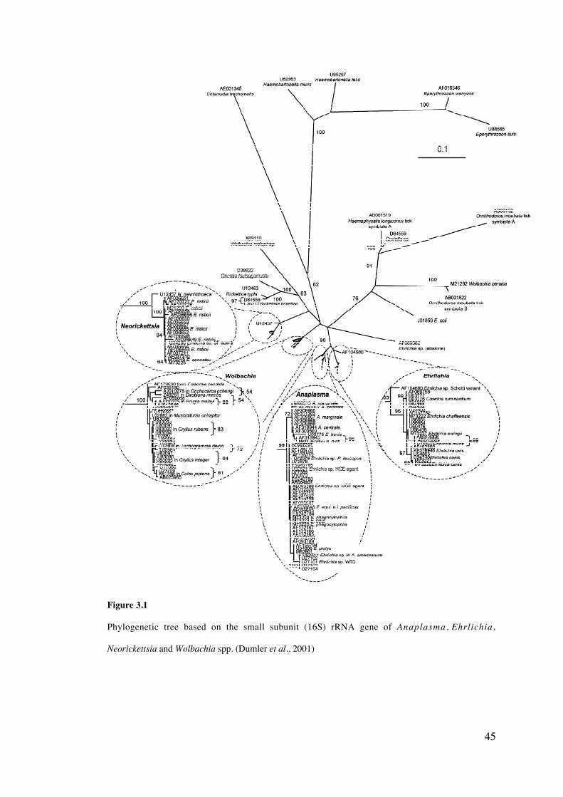
45
Figure 3.1
Phylogenetic tree based on the small subunit (16S) rRNA gene of Anaplasma , Ehrlichia,
Neorickettsia and Wolbachia spp. (Dumler et al., 2001)
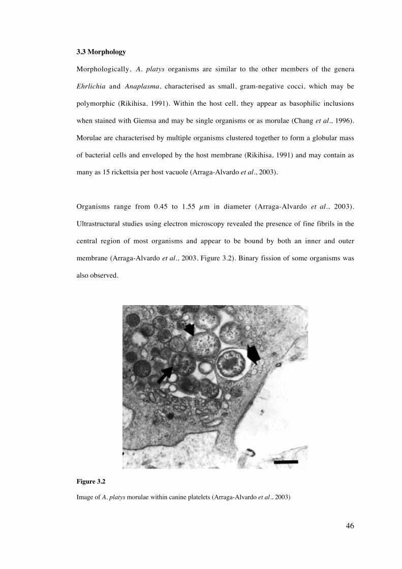
46
3.3 Morphology
Morphologically, A. platys organisms are similar to the other members of the genera
Ehrlichia and Anaplasma, characterised as small, gram-negative cocci, which may be
polymorphic (Rikihisa, 1991). Within the host cell, they appear as basophilic inclusions
when stained with Giemsa and may be single organisms or as morulae (Chang et al., 1996).
Morulae are characterised by multiple organisms clustered together to form a globular mass
of bacterial cells and enveloped by the host membrane (Rikihisa, 1991) and may contain as
many as 15 rickettsia per host vacuole (Arraga-Alvardo et al., 2003).
Organisms range from 0.45 to 1.55 µm in diameter (Arraga-Alvardo et al., 2003).
Ultrastructural studies using electron microscopy revealed the presence of fine fibrils in the
central region of most organisms and appear to be bound by both an inner and outer
membrane (Arraga-Alvardo et al., 2003, Figure 3.2). Binary fission of some organisms was
also observed.
Figure 3.2
Image of A. platys morulae within canine platelets (Arraga-Alvardo et al., 2003)

47
3.4 Transmission
Tentative evidence has suggested that the tick R. sanguineus is the vector responsible for the
transmission of A. platys based on geographic distribution, molecular and serological studies
(Chang et al., 1996; Inokuma et al., 2000; Motoi et al., 2001). Experimental infection of R.
sanguineus with A. platys failed however, and it was suggested that this tick might not act as
the vector for A. platys (Simpson et al., 1991). Further studies need to be carried out to
determine whether R. sanguineus is actually responsible for A. platys transmission.
It has also been suggested that other arthropod species may act as vectors of A. platys.
Martin et al. (2005) have speculated that the louse species, Heterodoxus spiniger may act as
a vector for A. platys in Australia, however it could not be determined whether the A. platys
DNA in the lice was a reflection of the ingested blood meal or whether this pathogen was
actually infecting the louse.
3.5 Life cycle
As the tick vector of A. platys has not been confirmed (Simpson et al, 1991), there have been
no studies on the development of these organisms within the arthropod host. If indeed R.
sanguineus is responsible for the transmission of A. platys it could be inferred that life cycle
events within the tick are similar to those of E. canis infections. Ehrlichia organisms enter
the tick midgut while feeding on vertebrate blood, then move into the tick hemocytes and
into the salivary glands (Smith et al., 1976). The vertebrate host becomes infected during
feeding of the tick vector. Ehrlichia species multiply by binary fission within both the tick
vector and the canine host (Woldehiwet and Ristic, 1993).
3.6 Distribution
Anaplasma platys has an increasingly worldwide distribution (Figure 3.3) and is considered
an emerging pathogen (Rikihisa, 2000). As stated previously, whether this is a reflection of
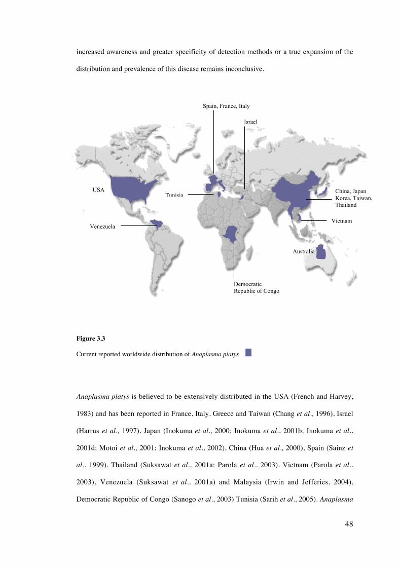
48
increased awareness and greater specificity of detection methods or a true expansion of the
distribution and prevalence of this disease remains inconclusive.
Figure 3.3
Current reported worldwide distribution of Anaplasma platys
Anaplasma platys is believed to be extensively distributed in the USA (French and Harvey,
1983) and has been reported in France, Italy, Greece and Taiwan (Chang et al., 1996), Israel
(Harrus et al., 1997), Japan (Inokuma et al., 2000; Inokuma et al., 2001b; Inokuma et al.,
2001d; Motoi et al., 2001; Inokuma et al., 2002), China (Hua et al., 2000), Spain (Sainz et
al., 1999), Thailand (Suksawat et al., 2001a; Parola et al., 2003), Vietnam (Parola et al.,
2003), Venezuela (Suksawat et al., 2001a) and Malaysia (Irwin and Jefferies, 2004),
Democratic Republic of Congo (Sanogo et al., 2003) Tunisia (Sarih et al., 2005). Anaplasma
USA
Venezuela
DemocraticRepublic of Congo
Spain, France, Italy
Israel
Tunisia
Vietnam
Australia
China, JapanKorea, Taiwan,Thailand
49
platys has also been reported in central Australia (Brown et al., 2001; Brown et al., 2005).
The full extent of its distribution in Australia has not been established.
3.7 Clinical Signs and Pathogenesis
Anaplasma platys infection is characterised by a seven to 14 day incubation after which
time, clinical signs are only occasionally present (Rikihisa, 1991). It has generally been
stated that dogs suffering from CICT are rarely clinically ill and do not often present with
any significant haemorrhage associated with platelet depletion (Harvey et al., 1978; Chang et
al., 1996; Mathew et al., 1997). Such a lack of clinical signs has been disputed by other
studies, in which important clinical signs of weight loss, fever and depression were reported
(Harrus et al., 1997). Harrus et al. (1997) provided an explanation for the differences by
suggesting that there may be strains of A. platys in southern Europe and the Middle East that
are more virulent or pathogenic than the strain present in USA. Further study is required to
assess the level of virulence between strains of A. platys.
As suggested by the name of this disease, the most characteristic feature of A. platys
infection is thrombocytopenia. Thrombocytopenia does not necessarily correlate to the
degree of parasitemia, as it may also be attributed to immune mediation (Baker et al., 1987;
Bradfield et al., 1996). An important feature of CICT is the cyclic nature of platelet
infection, with thrombocytopenia occurring in cycles of approximately 10-14 day intervals
(Harvey et al., 1978; Baker et al., 1987) Such repetitive absence and presence of infected
platelets needs to be considered in effectively diagnosing the disease. Infection of the
platelets and the resulting thrombocytopenia generally persists for seven to 10 days,
followed by a time of recovery (Bradfield et al., 1996). CICT has also been associated with
lymph node hyperplasia and plasmacytosis in various organs (Baker et al., 1987).
50
3.8 Detection and Diagnosis
3.8.1 Light microscopy
The most frequently used method of diagnosing acute A. platys infection is by examination
of infected platelets using light microscopy (Rikihisa, 1991). Blood smears are generally
stained with Giemsa and scanned for blue-staining, cytoplasmic inclusions of A. platys
within the platelets. Notably, diagnosis using such a method is limited due to the failure of
distinguishing between parasitised platelets and platelets with variable granule morphology
or megakaryocyte nuclear remnants (Simpson and Gaunt, 1991). Effective diagnosis is
therefore reliant on the ability and experience of the technician. In an attempt to overcome
these limitations, Simpson and Gaunt (1991) optimised an immunocytochemical stain
procedure for the detection of A. platys antigens. They developed an avidin-biotin
immunoperoxidase complex immunocytochemical stain that allowed for effective
discrimination between A. platys and other platelet inclusions using light microscopy.
Detection of A. platys within platelets using microscopic examination of Giemsa-stained
blood smears has a major limitation in that infection of platelets follows a cyclic pattern
(Chang and Pan, 1996). It was suggested that a single blood smear examination may give
rise to a negative diagnosis, when in fact infection exists. Multiple tests over an extended
period of time may be necessary for increased accuracy.
3.8.2 Electron Microscopy
Mathew et al. (1997) used electron microscopy to reveal the presence of rickettsia-like
inclusions within the platelets. A majority of the infected platelets showed parasitism by an
organism that had a general rickettsial-like morphology. Ultrastructural studies were later
conducted by Arraga-Alvarado et al. (2003). Due to the similarity in morphology between
the Ehrlichia species, such a technique is limited in that it is not possible to diagnose the
bacteria to a species level on the basis of morphology. Visible organisms can only be
assumed to be A. platys due to their presence within platelets.
51
iii) Indirect Fluorescent Antibody Test (IFAT)
Diagnosis can also be carried out using an indirect fluorescent antibody (IFA) test. An A.
platys IFA test was developed by French and Harvey (1983) and proved to be more effective
than examination by light microscopy. This test showed some degree of specificity, as it was
not able to detect antibodies to E. canis. Cross-reactions with antibodies of other Ehrlichia
species have not been reported however, in the case of the E. canis IFAT, cross-reactivity is
a significant limitation (Suksawat et al., 2001b; Waner et al., 2001).
iv) Polymerase Chain Reaction
DNA amplification using PCR has been demonstrated to show much promise as a highly
sensitive and specific test for the diagnosis of Ehrlichia infections. Initial PCR applications
were in equine and human infections (Biswas et al., 1991 and Anderson et al., 1992).
Successive studies showed the successful application of PCR to the diagnosis of canine
Ehrlichia infections. Iqbal et al. (1994) were first to use this technique as an effective means
of diagnosing canine Ehrlichia infections and were able to detect E. canis in canine blood.
Amplification of A. platys DNA was first carried out by Anderson et al. (1992) to allow for
sequencing of the DNA structure and its comparison to E. ewingii and other Ehrlichia
species. This sequence information provided the initial basis for primer development in later
studies. The first use of PCR to diagnose CICT was by two-step PCR in which primers were
developed to target a region of the 16S rRNA gene (Chang and Pan, 1996). Sensitivity
testing showed that the two-step method of PCR was 10 times more sensitive than single
PCR. Chang and Pan (1996) suggest that such a PCR-based detection method could be
applied to clinical use and found that PCR detected the presence of A. platys in blood
samples that were assumed negative after Giemsa stain examination. It was also suggested
that the two-step PCR was less time consuming than Southern Blot hybridisation. The PCR
test was successful in detecting A. platys in both the acute and chronic stages of
52
thrombocytopenia. It has also been suggested that nested PCR (two-step) is more useful in
assessing clearance of organisms after therapeutic treatment than IFAT (Wen et al., 1997).
A later study developed a single step PCR for the amplification of a region of the 16S rRNA
gene and was also found to be an effective diagnostic tool (Mathew et al., 1997). The
amplified region of DNA was sequenced to provide further support for an accurate diagnosis
of A. platys infection. The sensitivity and specificity of this PCR was not assessed and is
therefore difficult to compare with the previous two-step method. Hua et al. (2000) and
Motoi et al. (2001), have more recently applied PCR to the detection of A. platys and
allowed for the first discovery of this organism in dogs in China and Japan. One of these
PCR tests was associated with the amplification of Wolbachia spp. (Motoi et al., 2001). This
suggests that the specificity of the test is limited and requires future attention. Anaplasma
platys was also discovered in Australia, using PCR as the sole basis of detection (Brown et
al., 2001; Brown et al., 2005). Microscopic examination failed to detect any rickettsial
morulae within the platelets of the dogs and was assumed to be a consequence of the stage of
the infection.
Application of reverse transcription-PCR may also have potential in A. platys diagnosis as
this technique offers increased sensitivity than convential PCR and detects only viable
organisms (Felek et al., 2001). Multiplex detection of both Ehrlichia and Anaplasma spp.
has been reported using Real-time reverse transcriptase PCR (Sirigireddy and Ganta, 2005)
Limited information is available on whether A. platys exists in a dormant phase within such
organs as the spleen. If the parasites do involve other organs it may be useful to be able to
detect A. platys in tissue samples. PCR detection of A. platys within tissues has not been
reported, however such a technique has been used in E. canis diagnosis (Iqbal and Rikihisa,
1994; Harrus et al., 1998).
53
vi) PCR detection of A. platys DNA in ticks
Inokuma et al. (2000) were first to successfully detect A. platys DNA in R. sanguineus ticks
by using PCR. All of the ticks which they examined were semi-engorged, making it difficult
to determine whether the ticks were themselves infected or whether the PCR was amplifying
DNA solely within the blood-meal of the tick. Other species of Ehrlichia have been
effectively detected in the tissues of R. sanguineus using PCR (Sparagano et al., 1999).
3.9 Prevention and Treatment
As with all tick-borne pathogens, the most effective prevention of A. platys infection is
avoidance of the tick vector. However, this may be difficult due to the current dispute over
the actual tick species involved. The incidence of infection may be greater in the summer
months due to the increase in the number of feeding ticks (Bradfield et al., 1996). Therefore
control measures should be more vigorous during this time. Control of vector populations by
chemical treatment of dog housing and external treatment of the infected animals is
suggested every 1-2 weeks in endemic areas (Rikihisa, 1991).
Administration of tetracycline hydrochloride to infected dogs resulted in the disappearance
of thrombocytopenia, however some A. platys organisms remained in the platelets (Chang et
al., 1996). Oral treatment with doxycycline (5-10mg/kg for 10-14 days) was suggested to be
effective in eliminating thrombocytopenia (Bradfield et al., 1996). Oral treatment with
doxycycline and intramuscular injection of imidocarb dipropionate resulted in a recovery 48
to 72 hours after the initiation of treatment (Harrus et al., 1997). A similar co-drug treatment
was administered to dogs infected with A. platys by Sainz et al. (1999) and resulted in the
remission of clinical signs.
3.10 Co-infection of Ehrlichia and Anaplasma species
Ehrlichia infection can result from the simultaneous infection of dogs by multiple species.
Understanding co-infection is important in avoiding incorrect diagnosis. Sainz et al., (1999)

54
reported that dogs can be infected with both E. canis and A. platys. The infected animals
were shown to present with cutaneous petechial and ecchymotic haemorrhages and treatment
with doxycycline or imidocarb dipropionate resulted in remission of clinical signs. A later
study demonstrated the co-infection of E. canis, A. platys and E. equi in dogs within
Thailand and Venezuela (Suksawat et al., 2001b).
55
General Materials and Methods
4.1 Identification of piroplasm spp. by light microscopy
Ear-tip blood, venous blood and/or buffy coat smears were stained with a modified Wright-
Giemsa stain using an Ames Hema-Tek“ slide stainer (Bayer AG, Germany). Smears were
examined for intra-erythracytic piroplasm merozoites in the feathered region of the film or
for platelet inclusions throughout the slide, initially with a low-powered objective (40x) and
then at higher power (100x). At least two hundred microscopic fields of view were examined
under oil immersion using the 100x objective before being reported negative (Garcia and
Bruckner, 1988).
4.2 DNA extraction from canine blood
DNA was isolated from blood samples using a QIAamp‚ DNA mini kit (QIAGEN,
Germany). 200 ml of EDTA blood was added to a 1.5 ml microcentrifuge tube containing 20
ml of QIAGEN Protease. 200 ml of AL buffer was added to the sample and mixed by pulse
vortexing for 15 sec. The tube was incubated at 56 C for 10 min. Droplets formed within the
lid of the tube were removed by brief centrifugation. 200 ml of 99.5% ethanol was added to
the sample and mixed by pulse vortexing for 15 sec, then briefly centrifuged. The entire
mixture was applied to a QIAamp spin column without wetting the rim and the closed
column was spun at 6000 xg (half-speed) for 1 min. The spin column was placed in a clean
collection tube and the filtrate and collection tube were discarded. 500 ml of AW1 buffer was
added to the spin column, which was then centrifuged at half-speed for 1 min. After being
placed in a new collection tube, 500 ml of AW2 buffer was added to the spin filter, followed
CHAPTER FOUR
56
by centrifugation at 20 000 xg (full-speed) for 3 min. The filtrate was discarded and the
column spun for a further 1 min. After the addition of 100 ml of AE buffer, the tube was
incubated at room temperature for greater than 30 minutes to increase the DNA yield, before
being centrifuged at half-speed for 1 min. Extracted DNA was frozen at -20 C.
4.3 DNA extraction from animal tissues
DNA was isolated from tissue samples using a QIAamp“ DNA Mini Kit (QIAGEN,
Germany). A maximum of 25 mg of tissue was macerated using a scalpel blade and placed
in a sterile 1.5 ml microcentrifuge tube. Added to the tissue were 180 ml of buffer ATL and
20 ml of Proteinase K, which were then mixed by vortexing. The sample was incubated at 56
C for 4 hrs or until the tissue had completely lysed. Two hundred microliters of buffer AL
was added to the tube, mixed by vortexing and then incubated at 70 C for 10 min. The tube
was again centrifuged briefly and 200 ml of 100 % ethanol was added and mixed by
vortexing. The entire contents of the tube was then transferred to a QIAamp spin column and
the remaining protocol followed that described in section 4.3.
4.4 Gel electrophoresis
Gel electrophoresis of PCR products was performed using 1 % agarose (Promega, Madison,
USA) gels in TAE buffer (40 mM Tris-HCL, 20mM acetate, 2mM EDTA). Gels were pre-
stained with ethidium bromide (Amresco, USA). A 100 bp molecular weight marker (Life
Technologies, Australia) was run for all gels to determine the size of PCR products.
Electrophoresis was performed using a Minisub electrophoretic cell (Biorad) at 90 V for 30
min and DNA was visualised by UV transillumination.
4.5 DNA purification of gel bands
DNA purification was carried out using an UltraClean TM Gelspin DNA Purification Kit (MO
BIO Laboratories, Inc.). Amplified DNA was electrophoresed on a 1% agarose gel (90 V, 40
57
min). The gel was viewed under ultra-violet light and the appropriate sized band was cut out
using a scalpel blade. Each band was cut out using a separate scalpel blade to avoid
contamination of DNA. The bands were each placed in separate 1.5ml centrifuge tubes and
the individual volume of each band was estimated. Three times the volume of the gel band of
Gelbind or gel solubilization buffer was added to the gel slice and incubated for 2 minutes at
65 C. The tube was then inverted once and incubated for a further minute. The tube was
inverted again to ensure mixing. This solution was then transferred to a spin filter basket and
centrifuged for 10 sec at 10000 x g. The spin filter was removed from the collection tube and
the eluted solution was briefly vortexed before reloading into the spin filter. The tube was
centrifuged again for 10 sec at 10000xg and the flow-through liquid discarded. Three
hundred microlitres of GelWash buffer was added to the filter and spun for 10 sec at 10000 x
g. The flow-through liquid was discarded and the spin filter was spun for an additional 30
sec. The spin filter was carefully transferred to a clean collection tube and 50 ml of distilled
water was added. After >5 min incubation at room temperature, the tube was centrifuged for
30 sec at 10000 x g. Eluted DNA was immediately used for sequencing amplification or was
frozen at –20 C for later use.
4.6 Sequencing amplification
DNA was sequenced using an ABI Prism‘ Dye Terminator Cycle Sequencing Kit (Applied
Biosystems, Foster City, California) according to manufacturer’s instructions, with the
following modifications: Amplification was carried out in a 10 m l reaction mixture
containing the following: 0.5 ml each of the forward and reverse primers diluted to 3.25
pmol/ml, 2.0 ml of dye terminator solution, 2.0 ml of half term (Genpak Inc., Stony Brook,
New York) and 5.5ml of purified template (from 3.4). Forty cycles of amplification (94 C for
10 sec, 60 C for 5 sec and 60 C for 4min) was preceded by an intial denaturation of 94 C for 2
min 20 sec and followed by a holding temperature of 15 C.
58
4.7 Purification of sequencing reactions
Twenty-five microlitres of 95% ethanol, 1 ml of 125 mM EDTA and 1 ml of 3M sodium
acetate were added to a 0.6 ml tube. The 10 ml amplified DNA was centrifuged briefly and
added to the ethanol mixture. This solution was mixed gently using a pipette, then incubated
on ice for 20 min and then centrifuged for 30 min. The supernatant was carefully removed
with a pipette and 125 ml of 80 % ethanol was added to the remaining precipitate. The
solution was gently ‘rolled’ to ensure all salt deposits were removed from the sides of the
tube. The tube was spun for 5 min at 20 000 xg. The majority of the ethanol was removed
with a pipette and the sample was vacuumed dry in a vacuum desiccator (Nalgene).
4.8 Analysis of sequence chromatograms
The sequenced products were analysed using the program SeqEd v.1.0.3 (ABI) and were
compared to sequence data available from GenBank‘ , using the BLAST 2.1 program
(http://www.ncbi.nlm.nih.gov/BLAST/).
4.9 Immunofluorescent Antibody Test (IFAT)
IFAT was performed using a modified procedure described by Anderson et al., (1980). The
antigen used was a pre-prepared, ethanol fixed glass slides coated with a thin layer of B.
gibsoni-infected erythrocytes (approximately 6% parasitaemia) obtained from in vivo culture
of parasite from a naturally infected pit bull terrier in Victoria, Eastern Australia. Slides were
stored at -70 C until required. The conjugate used was Rabbit anti-canine IgG globulin
labelled with FITC is diluted in PBS at 1:1000.
Antigen slides were thawed at room temperature. Patient serum was diluted 1: 40, 1: 160, 1;
640, 1: 2560 and 1: 10240 and then placed in demarcated areas of the slide and incubated at
37 C for 60 min, then washed with PBS and rinsed. Diluted conjugate was applied to each
sample, incubated at 37 C for 60 min, washed in PBS for 10min, dried, then overlaid with

59
buffered glycine. Each slide was then cover slipped and examined using a fluorescent
microscope. Positive (serum from a known-infected dog supplied by Onderstepoort
Veterinary Institute, South Africa) and negative controls (SPF-canine serum) were used on
each slide during each test. Samples were considered positive with a titre greater than 1:40.
60
Development of a PCR-RFLP assay for the detection and
differentiation of the canine Piroplasmida species and evaluation of
Whatman® FTA cards
5.1 Introduction
Accurate detection is imperative to the identification of the species of pathogen responsible
for infection. Many reports suggest PCR is a highly effective detection technique and could
potentially be used in the routine diagnosis of diseases such as piroplasmosis (McLaughlin et
al., 1992; Prichard and Tait, 2001). It is important however to consider the low potential of
PCR in regions of the world where economic resources and sophisticated technology are
limited (Hanscheid and Grobusch, 2002). The application of blood samples to FTA cards
may overcome this limitation, by allowing samples to be sent rapidly and safely to
diagnostic facilities capable of PCR-based diagnosis.
Multiple species of piroplasm are now recognised to infect dogs, including three subspecies
of B. canis, B. gibsoni, B. conradae, T. annae, an unnamed Babesia sp. from North Carolina
and possibly T. equi. The morphological similarity between these species and subspecies of
the canine piroplasm has led to much confusion over accurate diagnosis using light
microscopy (Kjemtrup et al., 2000a). Likewise, there are reports that serology-based
diagnosis also lacks specificity, with the existence of antigen cross-reactivity between
species and even between genera with methods such as immunofluorescent antibody tests
(IFAT) (Yamane et al., 1993). Various PCR-based tests have been developed for detection
CHAPTER FIVE
61
of the canine piroplasms (Zahler et al., 1998; Carret et al., 1999; Ano et al, 2001;
Birkenheuer et al., 2003a) however, many are species-specific and fail to detect novel
species and genotypes of canine piroplasm. Some tests require complete sequencing of the
amplified target gene to determine the species or genotype present. Most assays have
targeted the 18S rRNA gene; a conserved, functional gene that contains moderate levels of
genetic variation that can be used to discriminate between species. This gene is also found in
multiple copies within the genome, allowing for increased levels of detection when
amplified using PCR (Hillis and Dixon, 1991). Restriction fragment length polymorphism
(RFLP) analysis offers an effective means of discriminating between species without the
need for DNA sequencing. To date, no PCR-RFLP for the differentiation of all reported
canine piroplasm species has been developed.
Blood samples are often difficult to store and transport from remote locations or places with
limited technology resources (Zhong et al., 2001). It is therefore beneficial to use a method
that requires minimal expertise and equipment, while also being simple and cost effective to
transport samples worldwide. In addition, long-term storage of samples such as whole blood
can be difficult due to the space and freezer requirements. Repetitive freeze thawing of blood
samples can also result in degradation of DNA, decreasing the sensitivity of DNA
amplification (Farnert et al., 1999). The application of samples to FTA cards may help to
overcome these problems. The FTA matrix also contains a substance that allows for the
inactivation of pathogens such as bacteria and viruses (Moscoso et al, 2004). This enables
FTA samples to be sent domestically and internationally without the risk of spreading
disease pathogens and also minimizing the risk of infection to laboratory personnel. Multiple
studies have demonstrated the antimicrobial efficacy of the FTA treated matrix (Moscoso et
al., 2004; Li et al., 2004). Currently, Australian quarantine laws prohibit the importation of
whole canine blood from countries including unless UV sterilized (http://www.aqis.gov.au/,
accessed 5/2002), inactivating potential pathogens but also cross-links DNA. FTA cards
62
offer a method of importing blood samples without the need for UV sterilization and also
minimizes the risk of importing infectious pathogens.
This chapter describes the development of a PCR-RFLP assay for the detection and
differentiation of the canine piroplasm species. It also describes the assessment of FTA cards
for the application of canine blood samples for subsequent PCR amplification of piroplasm
DNA, thereby allowing for infected blood samples to be imported into Australia from
overseas countries for molecular characterisation (refer to Chapter ten). The Whatman“
FTA DNA purification method was also compared to QIAGEN DNA extraction of blood
applied to filter paper and IsoCode‘ Stix DNA isolation techniques.
5.2 Aims
i. To design a PCR-RFLP assay for the detection and differentiation of the canine
piroplasm spp.
ii. To determine the detection limit and specificity of the PCR-RFLP assay
iii. To assess whether piroplasm DNA could be amplified from blood applied to FTA
cards
iv. To determine the possibility of DNA cross-over contamination using the Whatman“
FTA template preparation protocol
v. To compare the detection limit of FTA disc purification, QIAGEN extraction and
Isocode‘ Stix template preparation
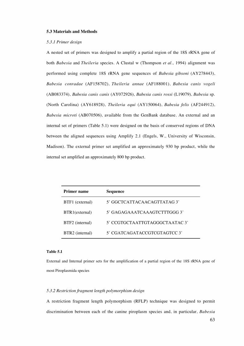
63
5.3 Materials and Methods
5.3.1 Primer design
A nested set of primers was designed to amplify a partial region of the 18S rRNA gene of
both Babesia and Theileria species. A Clustal w (Thompson et al., 1994) alignment was
performed using complete 18S rRNA gene sequences of Babesia gibsoni (AY278443),
Babesia conradae (AF158702), Theileria annae (AF188001), Babesia canis vogeli
(AB083374), Babesia canis canis (AY072926), Babesia canis rossi (L19079), Babesia sp.
(North Carolina) (AY618928), Theileria equi (AY150064), Babesia felis (AF244912),
Babesia microti (AB070506), available from the GenBank database. An external and an
internal set of primers (Table 5.1) were designed on the basis of conserved regions of DNA
between the aligned sequences using Amplify 2.1 (Engels, W., University of Wisconsin,
Madison). The external primer set amplified an approximately 930 bp product, while the
internal set amplified an approximately 800 bp product.
Primer name Sequence
BTF1 (external) 5’ GGCTCATTACAACAGTTATAG 3’
BTR1(external) 5’ GAGAGAAATCAAAGTCTTTGGG 3’
BTF2 (internal) 5’ CCGTGCTAATTGTAGGGCTAATAC 3’
BTR2 (internal) 5’ CGATCAGATACCGTCGTAGTCC 3’
Table 5.1
External and Internal primer sets for the amplification of a partial region of the 18S rRNA gene of
most Piroplasmida species
5.3.2 Restriction fragment length polymorphism design
A restriction fragment length polymorphism (RFLP) technique was designed to permit
discrimination between each of the canine piroplasm species and, in particular, Babesia

64
canis and Babesia gibsoni in blood samples from dogs in Australia. Complete sequences of
the 18S rRNA gene each of the canine species and subspecies available on GenBank
database were imported into the program Amplify 2.1 (Engels, W., University of Wisconsin,
Madison) and the target region of DNA was determined using the internal primer set (BTF2
and BTR2). The sequence of the amplified internal PCR product was then used in DNA
Strider™1.0 (Mark, 1988) to determine the most suitable restriction enzymes for
discriminating between the canine piroplasm species.
5.3.3 DNA extraction and amplification
DNA was isolated from 200 ml aliquots of EDTA blood (stored at -20 C) using a QIAamp‚
DNA mini kit (QIAGEN, Hilden, Germany), according to the manufacturer’s instructions
(refer to Chapter four, section 4.2). One ml of extracted DNA was added to a 24 ml reaction
mixture comprising 0.6875 units of Tth Plus DNA polymerase (Fisher Biotech, Australia),
200 mM of each dNTP, 12.5 pmoles of the forward and reverse primers (Invitrogen,
Australia), 2.5 ml 10x PCR buffer (Fisher Biotech, Australia) and 1.5 ml MgCl2 (Fisher
Biotech, Australia). Positive (1 ml of B. canis vogeli DNA, Australia) and negative (1 ml
dH2O) control samples were included with each set of PCR reactions.
Amplification was performed on a GeneAmp PCR system 2700 thermal cycler (Applied
Biosystems, USA). For the primary round of amplification, an initial activation step at 94 C
for 3 min, 58 C for 1 min and 72 C for 2 min, was followed by 45 cycles of amplification (94
C for 30 sec, 58 C for 20 sec and 72 C for 30 sec) and a final extension step of 72 C for 7
min for 25µl reactions. The same conditions were followed for the secondary round of
amplification, except that the annealing temperature was increased to 62 C, using 1µl of
DNA template from the primary reaction. Amplified DNA was electrophoresed and
visualised according to the method described in Chapter four, section 4.4.
65
5.3.4 Restriction digestion
Six ml of amplified DNA from the secondary PCR reaction was subjected to restriction
enzyme digestion in a reaction mixture of 16.3 ml of dH2O, 2.0 ml of Buffer B (Promega,
Madison, USA), 0.2 ml of Bovine serum albumin acetylated (Promega, USA). The reaction
mixture was gently mixed and 0.5 ml of the appropriate restriction enzyme (either Hinf I,
Hinc II or Ava II) (Promega, USA) was added and then incubated at 37 C for 2 hrs.
Restriction products were then electrophoresed at 80 volts for 1 hr on a 3 % agarose gel
(Promega, USA) stained with ethidium bromide and visualised using UV illumination.
5.3.5 DNA Sequencing
Amplified products were purified using an UltraClean™ Gelspin DNA Purification Kit (MO
Bio Laboratories, Inc., Sohlana Beach, California) and sequenced using an ABI Prism™ Dye
Terminator Cycle Sequencing Kit (Applied Biosystems [ABI], Foster City, California)
(Refer to Chapter four, section 4.6). The sequenced products were analysed using SeqEd
v.1.0.3 (ABI), compared with sequence data available from GenBank™ using the BLAST
2.1 program (http://www.ncbi.nlm.nih.gov/BLAST/), and aligned to sequences available
from GenBank™ using Clustal w (Thompson et al., 1994).
5.3.6 Determination of detection limit of PCR
a) Serial dilution of infected blood
The sensitivity of the PCR was calculated by performing PCR assays on a blood sample with
a known parasitaemia that had been serially diluted in parasite free canine blood as
previously described (Jefferies et al., 2003). A venous blood sample from a dog known to be
infected with Babesia by microscopic examination was collected and EDTA was added. A
thin blood smear was prepared from venous blood and the parasitaemia was calculated
according to Read and Hyde (1993), by counting between 500 to 1000 erythrocytes and
noting the number that were infected with Babesia (cells infected with more than one
66
piroplasm were counted as one). A total of 1187 erythrocytes were counted over a thin blood
smear section of uniform density.
The blood sample of known parasitaemia was then diluted using canine blood considered to
be free from Babesia infection by microscopy, PCR and the dog’s lack of exposure to tick
vectors. Twenty-five microlitres of infected blood was added to 225 ml of uninfected control
blood and mixed. 25 ml of this blood was then added to 225 ml of control blood to form a
1x10⁻² dilution. This procedure of serial dilution was repeated until a 1x10⁻⁷ dilution was
obtained. DNA was extracted from 200 ml of each of the diluted blood samples according to
the described extraction procedure in Chapter four, section 4.2. Amplification of the
extracted DNA was carried out according to the procedure described in section 5.3.3.
b) Serial dilution of quantified plasmid DNA
i) Cloning of primary PCR product
The primary PCR product amplified from an isolate of B. canis vogeli was cloned into a
plasmid vector using a TOPO‚ Cloning Kit (Invitrogen, California). TOPO‚ cloning
reactions were comprised of 4 ml of purified PCR product, 1 ml of salt solution (1.2M NaCl
and 0.06M MgCl2) and 1 ml of TOPO‚ vector. The reactions were mixed gently and
incubated at room temperature for 30 min before being placed on ice for 2 min.
Transformation involved the addition of 2 ml of the TOPO‚ cloning reaction to 1 ml of
OneShot‚ chemically competent E. coli cells, mixed gently, incubated on ice for 30 min and
then heat shocked at 42 C for 30 sec. Cells were again incubated on ice, before 200 ml of
SOC media (at room temperature) was added and incubated at 37 C for 1 hr with continual
shaking. A 100 ml aliquot of the transformation mixture was then spread evenly onto LB
agar plates containing ampicillin (50 mg/ml) and then incubated at 37 C overnight. Ten
colonies were chosen to be screened for the insert, with half of each colony being screened
67
by PCR, while the other half was inoculated into 200 ml of LB media containing 50 mg/ml of
ampicillin and incubated at 37 C overnight with shaking.
ii) PCR screening of colonies
The primers M13 Forward (5’ GTAAAACGACGGCCAG 3’) and M13 Reverse (5’
CAGGAAACAGCTATGAC 3’) were used to amplify positive transformants. Samples from
each of the 10 colonies selected were added to a 48 ml PCR reaction mixture. After an initial
denaturation step at 95 C for 5 min, 35 cycles of amplification were conducted (95 C for 20
sec, 56 C for 20 sec, 72 for 1 min) before a final extension step of 72 C for 7 min.
Transformants were sequenced according to the method described in Chapter four, section
4.6.
iii) Plasmid extraction
Samples determined to be positive for the desired insert were purified using a QIAprep‚
Plasmid Miniprep Kit (QIAGEN, Germany). Cells inoculated into LB media were pelleted
by centrifugation at low speed (1450 xg) for 1 min. Pelleted cells were transferred into a
sterile microcentrifuge tube and resuspended in 250 ml of Buffer P1. Two hundred and fifty
ml of Buffer P2 and then 350 ml of Buffer N3 were added to the sample, gently mixed four to
six times on addition of each buffer. Samples were then centrifuged for 10 min at high speed
(20 000 xg) and the resulting supernatant was transferred into a QIAprep‚ spin column,
centrifuged at high speed (20 000 xg) for 1 min and the flow through liquid was discarded.
The spin column was washed with 750 ml of Buffer PE and then centrifuged at high speed
for 1 min. Flow through was discarded and the spin column was centrifuged for a further 1
min. The QIAprep‚ column was placed into a clean 1.5 ml microcentrifuge tube and 50 ml of
Buffer EB was added to the column, before being left to incubate at room temperature for
1min. The sample was then centrifuged at high speed for 1 min and the flow through
retained for subsequent analysis.
68
iv) Quantification and dilution of plasmid DNA
The transformed plasmid DNA was quantified using a Lambda 25 UV/VIS spectrometer
(PerkinElmer). The purified sample contained 0.035 ug/ml of DNA and was serially diluted
10 fold to a dilution of 1x10⁻¹⁰. The diluted samples were amplified using the nested PCR.
5.3.6 Determination of specificity of PCR-RFLP
The specificity of the primers used was determined by BLAST searching the primer
sequences to make sure that they did not amplify host or human DNA or other blood
microbe DNA. The primers of this PCR assay were designed to amplify most species of the
genera Babesia and Theileria based on 18S rRNA sequences available on the GenBank
database (http://www.ncbi.nlm.nih.gov/entrez/, accessed 12/2002). Speciation is then
achieved using RFLP. In addition, a sample containing both B. canis vogeli and B. gibsoni
DNA was subjected to PCR-RFLP to assess the detection of co-infections of multiple
Babesia species. Sixteen blood samples obtained from dogs in New Zealand, a country
considered free of all canine piroplasm species, were also screened using the PCR-RFLP as
an additional negative control.
The specificity of the PCR-RFLP assay was also determined by amplifying DNA from
various piroplasm species and DNA obtained from cultured Neospora caninum and
Toxoplasma gondii and other parasite species (Table 5.2).
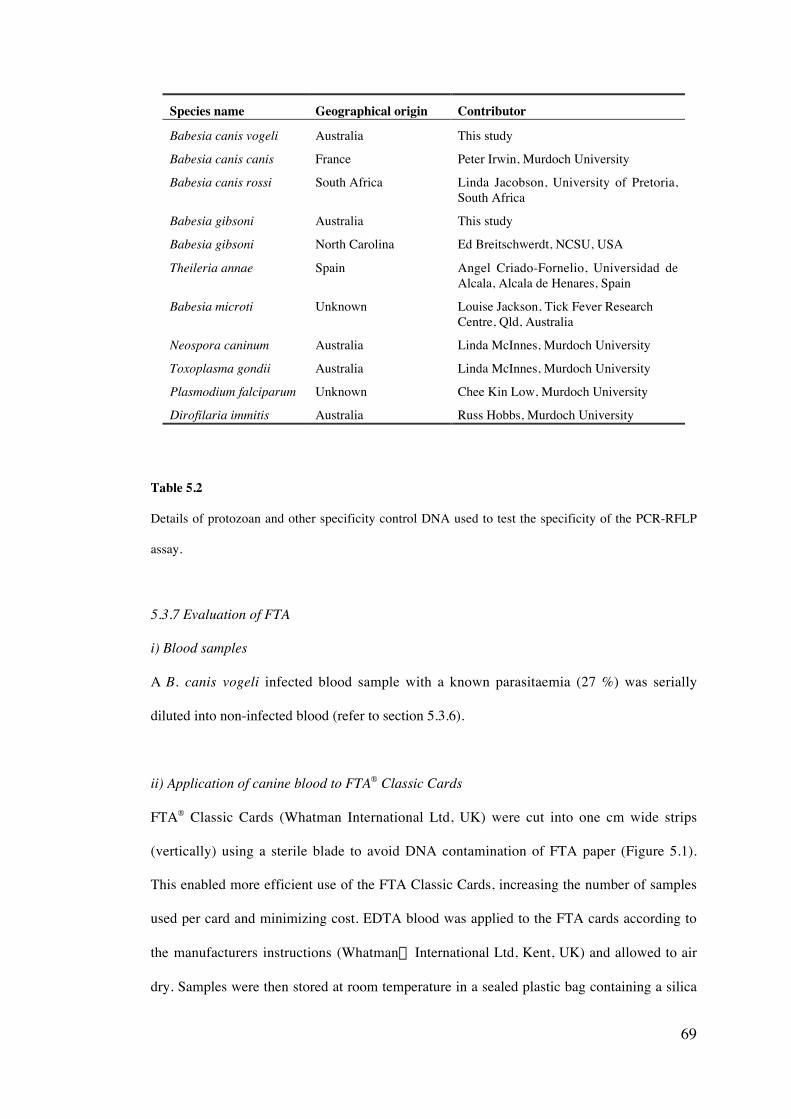
69
Species name Geographical origin Contributor
Babesia canis vogeli Australia This study
Babesia canis canis France Peter Irwin, Murdoch University
Babesia canis rossi South Africa Linda Jacobson, University of Pretoria,South Africa
Babesia gibsoni Australia This study
Babesia gibsoni North Carolina Ed Breitschwerdt, NCSU, USA
Theileria annae Spain Angel Criado-Fornelio, Universidad deAlcala, Alcala de Henares, Spain
Babesia microti Unknown Louise Jackson, Tick Fever ResearchCentre, Qld, Australia
Neospora caninum Australia Linda McInnes, Murdoch University
Toxoplasma gondii Australia Linda McInnes, Murdoch University
Plasmodium falciparum Unknown Chee Kin Low, Murdoch University
Dirofilaria immitis Australia Russ Hobbs, Murdoch University
Table 5.2
Details of protozoan and other specificity control DNA used to test the specificity of the PCR-RFLP
assay.
5.3.7 Evaluation of FTA
i) Blood samples
A B. canis vogeli infected blood sample with a known parasitaemia (27 %) was serially
diluted into non-infected blood (refer to section 5.3.6).
ii) Application of canine blood to FTA® Classic Cards
FTA® Classic Cards (Whatman International Ltd, UK) were cut into one cm wide strips
(vertically) using a sterile blade to avoid DNA contamination of FTA paper (Figure 5.1).
This enabled more efficient use of the FTA Classic Cards, increasing the number of samples
used per card and minimizing cost. EDTA blood was applied to the FTA cards according to
the manufacturers instructions (Whatman“ International Ltd, Kent, UK) and allowed to air
dry. Samples were then stored at room temperature in a sealed plastic bag containing a silica
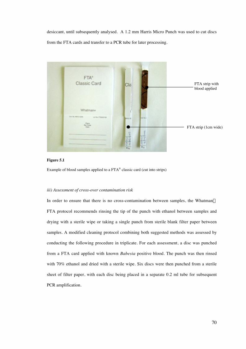
70
desiccant, until subsequently analysed. A 1.2 mm Harris Micro Punch was used to cut discs
from the FTA cards and transfer to a PCR tube for later processing.
Figure 5.1
Example of blood samples applied to a FTA® classic card (cut into strips)
iii) Assessment of cross-over contamination risk
In order to ensure that there is no cross-contamination between samples, the Whatman“
FTA protocol recommends rinsing the tip of the punch with ethanol between samples and
drying with a sterile wipe or taking a single punch from sterile blank filter paper between
samples. A modified cleaning protocol combining both suggested methods was assessed by
conducting the following procedure in triplicate. For each assessment, a disc was punched
from a FTA card applied with known Babesia positive blood. The punch was then rinsed
with 70% ethanol and dried with a sterile wipe. Six discs were then punched from a sterile
sheet of filter paper, with each disc being placed in a separate 0.2 ml tube for subsequent
PCR amplification.
FTA strip withblood applied
FTA strip (1cm wide)
71
iv) Whatman® preparation technique of DNA for PCR analysis
For each sample, a 1.2 mm disc was placed into a 0.2 ml tube, 200 µl of FTA Purification
Reagent was then added to the tube and incubated at room temperature for 10 min. All spent
FTA Purification Reagent was then removed and discarded using a pipette. A further two
washes of the disc using FTA Purification reagent was carried out. 200 µl of TE Buffer was
added to the sample disc and incubated at room temperature for 10 min, before being
removed using a pipette and discarded. A second wash using TE was then performed. The
disc was then dried using a vaccum dessicator (Nalgene) for 30 min before performing PCR.
v) Comparison of FTA with IsoCode‘ Stix and QIAGEN extraction methods
Sensitivity of each method was calculated by serially diluting canine blood with a known
percentage Babesia parasitemia (according to section 5.3.6). Blood of each dilution was
then applied to separate FTA strips or IsoCode‘ Stix and a PCR was conducted from
samples prepared by each of the following two methods.
a) QIAamp DNA isolation protocol
Serially diluted blood was applied to For each sample a 2 cm strip of blood covered FTA
paper was placed in a centrifuge tube with 40 µl of proteinase K and 180 µl ATL buffer,
then incubated at 56 C for 1 hr. 200 µl of AL buffer was then added to the sample, mixed by
pulse-vortexing for 10 sec and incubated at 70 C for 1 hr. The tube was briefly centrifuged,
then all of the lysate was carefully removed and applied onto a QIAamp minelute column
and centrifuged at 6000 xg for 1min.
b) IsoCode‚ Stix preparation
Approximately 10 µl of Babesia infected blood (serially diluted) was applied to the IsoCode
Stix (Schleicher and Scheull, Germany) and dried at room temperature for 3 hrs. Each blood-
covered triangle was placed over an open sterile microcentrifuge tube and detached while
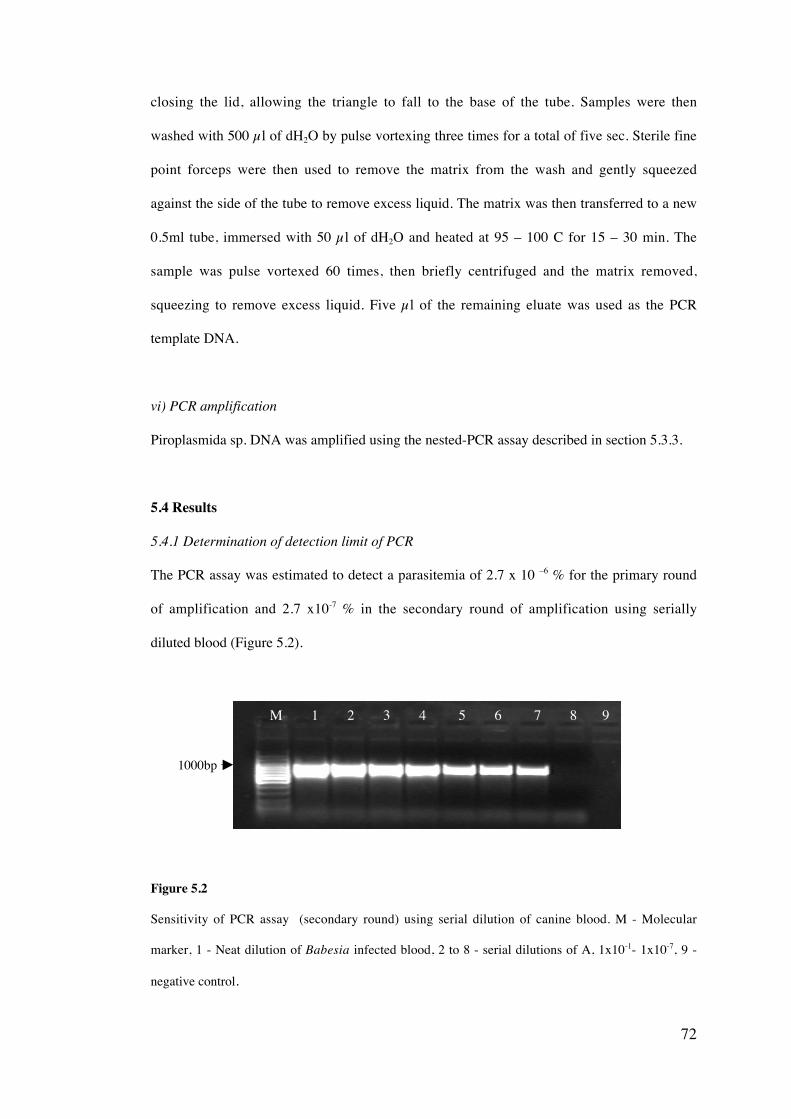
72
closing the lid, allowing the triangle to fall to the base of the tube. Samples were then
washed with 500 µl of dH2O by pulse vortexing three times for a total of five sec. Sterile fine
point forceps were then used to remove the matrix from the wash and gently squeezed
against the side of the tube to remove excess liquid. The matrix was then transferred to a new
0.5ml tube, immersed with 50 µl of dH2O and heated at 95 – 100 C for 15 – 30 min. The
sample was pulse vortexed 60 times, then briefly centrifuged and the matrix removed,
squeezing to remove excess liquid. Five µl of the remaining eluate was used as the PCR
template DNA.
vi) PCR amplification
Piroplasmida sp. DNA was amplified using the nested-PCR assay described in section 5.3.3.
5.4 Results
5.4.1 Determination of detection limit of PCR
The PCR assay was estimated to detect a parasitemia of 2.7 x 10 –6 % for the primary round
of amplification and 2.7 x10⁻⁷ % in the secondary round of amplification using serially
diluted blood (Figure 5.2).
Figure 5.2
Sensitivity of PCR assay (secondary round) using serial dilution of canine blood. M - Molecular
marker, 1 - Neat dilution of Babesia infected blood, 2 to 8 - serial dilutions of A, 1x10⁻¹- 1x10⁻⁷, 9 -
negative control.
1000bp
M 1 2 3 4 5 6 7 8 9
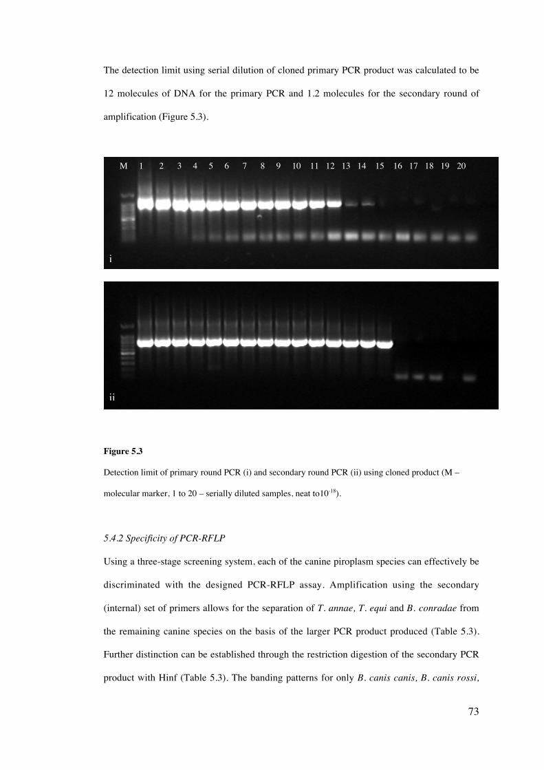
73
The detection limit using serial dilution of cloned primary PCR product was calculated to be
12 molecules of DNA for the primary PCR and 1.2 molecules for the secondary round of
amplification (Figure 5.3).
Figure 5.3
Detection limit of primary round PCR (i) and secondary round PCR (ii) using cloned product (M –
molecular marker, 1 to 20 – serially diluted samples, neat to10-18).
5.4.2 Specificity of PCR-RFLP
Using a three-stage screening system, each of the canine piroplasm species can effectively be
discriminated with the designed PCR-RFLP assay. Amplification using the secondary
(internal) set of primers allows for the separation of T. annae, T. equi and B. conradae from
the remaining canine species on the basis of the larger PCR product produced (Table 5.3).
Further distinction can be established through the restriction digestion of the secondary PCR
product with Hinf (Table 5.3). The banding patterns for only B. canis canis, B. canis rossi,
i
M 1 2 3 4 5 6 7 8 9 10 11 12 13 14 15 16 17 18 19 20
ii
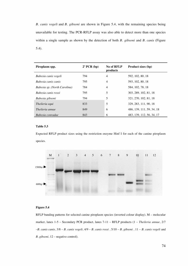
74
B. canis vogeli and B. gibsoni are shown in Figure 5.4, with the remaining species being
unavailable for testing. The PCR-RFLP assay was also able to detect more than one species
within a single sample as shown by the detection of both B. gibsoni and B. canis (Figure
5.4).
Piroplasm spp. 2o PCR (bp) No of RFLPproducts
Product sizes (bp)
Babesia canis vogeli 794 4 592, 102, 80, 18
Babesia canis canis 795 4 593, 102, 80, 18
Babesia sp. (North Carolina) 784 4 584, 102, 78, 18
Babesia canis rossi 795 5 303, 289, 102, 81, 18
Babesia gibsoni 794 5 321, 270, 102, 81, 18
Theileria equi 833 5 329, 283, 111, 90, 18
Theileria annae 849 6 486, 139, 111, 59, 34, 18
Babesia conradae 843 6 483, 139, 112, 56, 34, 17
Table 5.3
Expected RFLP product sizes using the restriction enzyme Hinf I for each of the canine piroplasm
species.
Figure 5.4
RFLP banding patterns for selected canine piroplasm species (inverted colour display). M – molecular
marker, lanes 1-5 – Secondary PCR product, lanes 7-11 – RFLP products (1 – Theileria annae, 2/7
–B. canis canis, 3/8 – B. canis vogeli, 4/9 – B. canis rossi , 5/10 – B. gibsoni , 11 – B. canis vogeli and
B. gibsoni, 12 – negative control).
M A B C
M 1 2 3 4 5 6 7 8 9 10 11 12
600bp
1500bp
75
Differentiation between B. canis canis and B. canis vogeli was produced using the restriction
enzyme HinC II (Table 5.4), while T. annae, T equi and B. conradae were separated using
Ava II (Table 5.4).
Restriction enzyme Species No of products Product sizes
HinC II Babesia canis vogeli 1 794
Babesia canis canis 2 463, 330
Ava II Babesia conradae 3 636, 173, 32
Theileria annae 1 849
Table 5.4
Discrimination of the canine piroplasm species using the restriction enzymes HinC II and Ava II.
The PCR assay was also found to amplify piroplasm species beyond just the canine species
as observed by the amplification of Babesia microti DNA. The secondary product amplified
from B. microti was of similar size to T. annae (851bp). Neospora caninum and T. gondii
was also amplified by the PCR assay, however the secondary product was larger (871bp)
than that amplified for each of the canine piroplasms (which ranged from 784 to 849bp). The
PCR assay did not amplify DNA from the other tested protozoan species, Dirofilaria immitis
or the host (Canis familiaris).
5.4.3 Evaluation of FTA
i) Assessment of punch cross-over contimination
Each of the trials used to assess punch cross-over contamination amplified DNA from the
negative punch discs. The greatest number of blank disc punches taken before no DNA was
amplified was 2 for two trials and 3 within the third trial (Figure 5.5). Six negative control
punches using sterile filter paper were taken between each sample and PCR was carried out
on the sixth sample as an added negative control for all samples that were subsequently
assessed.
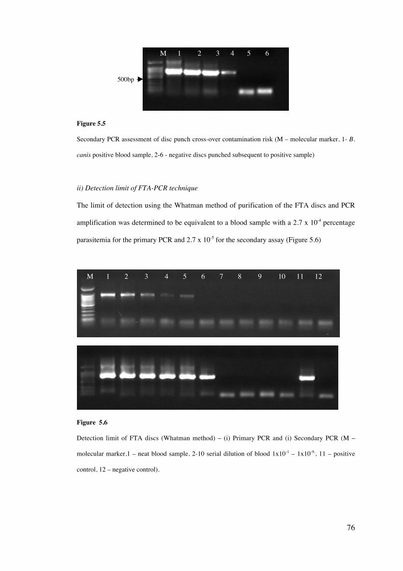
76
Figure 5.5
Secondary PCR assessment of disc punch cross-over contamination risk (M – molecular marker, 1- B.
canis positive blood sample, 2-6 - negative discs punched subsequent to positive sample)
ii) Detection limit of FTA-PCR technique
The limit of detection using the Whatman method of purification of the FTA discs and PCR
amplification was determined to be equivalent to a blood sample with a 2.7 x 10⁻⁴ percentage
parasitemia for the primary PCR and 2.7 x 10⁻⁵ for the secondary assay (Figure 5.6)
Figure 5.6
Detection limit of FTA discs (Whatman method) – (i) Primary PCR and (i) Secondary PCR (M –
molecular marker,1 – neat blood sample, 2-10 serial dilution of blood 1x10⁻¹ – 1x10⁻⁹,, 11 – positive
control, 12 – negative control).
M 1 2 3 4 5 6 7 8 9 10 11 12
500bp
M 1 2 3 4 5 6
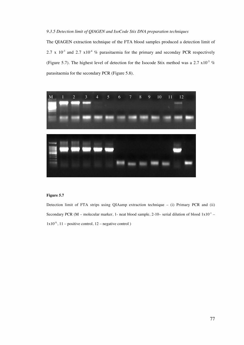
77
9.3.5 Detection limit of QIAGEN and IsoCode Stix DNA preparation techniques
The QIAGEN extraction technique of the FTA blood samples produced a detection limit of
2.7 x 10⁻³ and 2.7 x10⁻⁴ % parasitaemia for the primary and seconday PCR respectively
(Figure 5.7). The highest level of detection for the Isocode Stix method was a 2.7 x10⁻⁵ %
parasitaemia for the secondary PCR (Figure 5.8).
Figure 5.7
Detection limit of FTA strips using QIAamp extraction technique – (i) Primary PCR and (ii)
Secondary PCR (M – molecular marker, 1- neat blood sample, 2-10– serial dilution of blood 1x10⁻¹ –
1x10⁻⁹,, 11 – positive control, 12 – negative control )
M 1 2 3 4 5 6 7 8 9 10 11 12
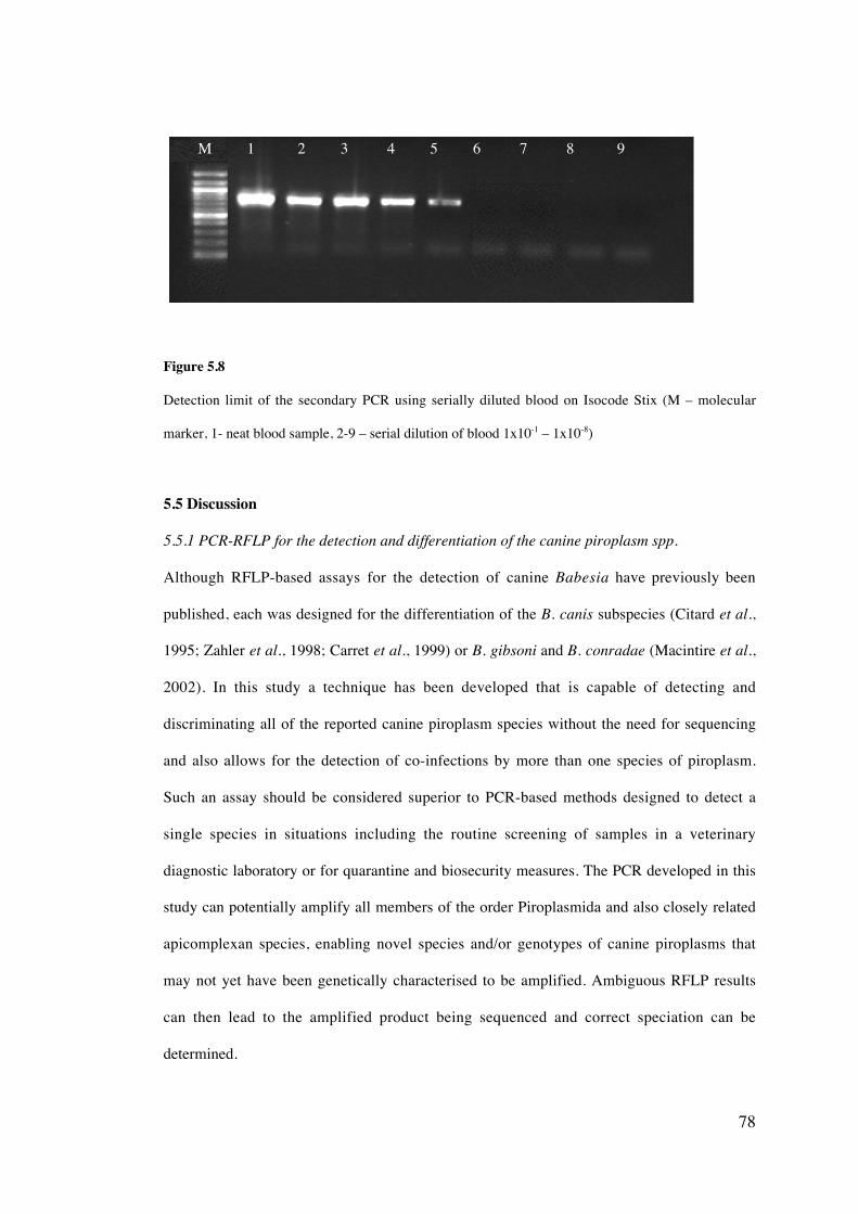
78
Figure 5.8
Detection limit of the secondary PCR using serially diluted blood on Isocode Stix (M – molecular
marker, 1- neat blood sample, 2-9 – serial dilution of blood 1x10⁻¹ – 1x10⁻⁸)
5.5 Discussion
5.5.1 PCR-RFLP for the detection and differentiation of the canine piroplasm spp.
Although RFLP-based assays for the detection of canine Babesia have previously been
published, each was designed for the differentiation of the B. canis subspecies (Citard et al.,
1995; Zahler et al., 1998; Carret et al., 1999) or B. gibsoni and B. conradae (Macintire et al.,
2002). In this study a technique has been developed that is capable of detecting and
discriminating all of the reported canine piroplasm species without the need for sequencing
and also allows for the detection of co-infections by more than one species of piroplasm.
Such an assay should be considered superior to PCR-based methods designed to detect a
single species in situations including the routine screening of samples in a veterinary
diagnostic laboratory or for quarantine and biosecurity measures. The PCR developed in this
study can potentially amplify all members of the order Piroplasmida and also closely related
apicomplexan species, enabling novel species and/or genotypes of canine piroplasms that
may not yet have been genetically characterised to be amplified. Ambiguous RFLP results
can then lead to the amplified product being sequenced and correct speciation can be
determined.
M 1 2 3 4 5 6 7 8 9
79
The moderate level of genetic variation found within the 18S rRNA gene of the canine
piroplasm species allows for effective discrimination between species using the RFLP
technique. While this PCR-RFLP assay is beneficial in discriminating easily between certain
species, such as B. gibsoni and B. canis vogeli through the use of a single restriction digest,
differentiating between all canine species using multiple restriction digestions is somewhat
laborious. A less labour intensive method could be acheived by the modification of the
described PCR assay into a quantitative method, such as quantitative real-time PCR based on
Taqman or SYBR Green chemistries (Giglio et al., 2003; Jeong et al., 2003) and warrants
further study. Loop-mediated isothermal amplification has also been reported as a rapid and
sensitive detection tool (Ikadai et al., 2004) however was reported to be species-specific and
additional research is necessary to determine whether this technique could be used to detect
multiple species including possible novel species and genotypes.
PCR is considered to be one of the most sensitive diagnostic methods currently available for
the detection of species of canine Babesia (Ano et al., 2003; Birkenheuer et al., 2003;
Jefferies et al., 2003; Inokuma et al., 2004). The detection limit of the assay reported in this
chapter, as a value of percentage parasitaemia, is higher than PCR assays previously
described for the detection of Babesia and Theileria species (Roy et al., 2000; Ano et al.,
2001; Jefferies et al., 2003) and comparable to the assays developed by Birkenheuer et al.
(2003a) and Fukomoto et al. (2001). Caution however should be taken in the interpretation
of lowest detectable percentage parasitaemia due to the high variability of red blood cell
counts (Birkenheuer et al., 2003a). The clinical sensitivity of PCR during pre-acute and
chronic stages of infection for each of the canine piroplasm species has not been reported
and would require further investigation using experimentally infected animals. Further study
into the detection limits of PCR is described in Chapter eight.
80
5.5.2 Evaluation of the FTA® Classic Card DNA purification technique
The results of this study suggest that the use of FTA as a template for amplification of
piroplasm DNA from canine blood is more sensitive than using QIAGEN and Isocode
techniques. All three techniques were, however much less sensitive than using DNA
extracted from whole blood (refer to section 5.4.1). DNA extraction using whole blood
should be given priority over FTA-based methods to allow for increased levels of detection.
Previous studies comparing both IsoCode™ Stix and FTA cards to store blood and diagnosis
of malaria by PCR suggested that FTA cards showed the greatest level of sensitivity in the
detection of mixed infections (Zhong et al., 2001). Comparisons between DNA template
from IsoCode™ Stix and QIAamp blood extraction techniques for the detection of pathogens
in various sample types have also been previously conducted and suggested that the
IsoCode™ Stix method was highly sensitive (Henning et al., 1999; Coyne et al., 2004).
The use of FTA cards as a method for transport, storage and DNA template for the PCR
detection of pathogen DNA in blood samples has previously been suggested to be a highly
efficient and sensitive technique (Zhong et al., 2001; Subrungruang et al., 2004). DNA of a
Babesia microti-like parasite has also previously been amplified from blood sampling filter
paper (Okabayashi et al., 2001). While such studies have concentrated on the advantages of
this method, such as the archival potential and high sensitivity, limited study has been
carried on the possibility of cross-contamination between samples and the importance of
sample preparation. The risk of cross-contamination between samples is considerable when
using the suggested protocol by Whatman“. The use of a single punch repetitively between
samples offers a significant means of transferral of DNA between samples. Ultimately, the
use of a new punch for each sample would be ideal, however the high cost (approximately
AU$6 per punch) prohibits this. A modified technique based on the FTA punch cleaning
protocol using sterile filter paper was devised in this study. This allowed for a significant
decrease in cross-contamination and also included using of a negative control for each
81
sample tested. Further research should be carried out to optimise a suitable protocol with a
risk of cross-contamination that is negligible.
The use of FTA paper for the application of arthropod samples for archiving and use for
subsequent detection of pathogens using PCR has been previously reported for fire ants
(Solenopsis invicta) (Snowden et al., 2002; Milks et al., 2004) and other arthropods (Bextine
et al., 2004; Harvey, 2005). There may therefore be potential for using this technique for the
storage and detection of piroplasm DNA in ticks.
5.5.3 Conclusion
This chapter has described the development of a simple nested PCR-RFLP technique for the
detection and discrimination of the canine piroplasms. This assay has the potential to be
implemented into a standardised screening protocol for B. gibsoni in dogs being exported
from Australia and is evaluated in Chapter six. While FTA cards are potentially beneficial in
regions where technology resources are limited, allowing for samples to be sent at an
ambient temperature to a specialist laboratory, this study has shown that some limitations do
exist, including the reduced detection limit and the risk of DNA cross-contamination. It was
therefore decided that FTA was only to be used as a means of importing canine blood
samples from overseas (refer to chapter ten). Further study needs to determine DNA
purification methods from FTA cards that are comparable to DNA extraction from EDTA
whole blood before this technique can be considered for routine diagnosis of infections.
82
Evaluation of PCR-RFLP for the screening of B. gibsoni infections
in dogs being exported from Australia
6.1 Introduction
Following the first report of B. gibsoni infections in three American Pit Bull Terriers in the
south eastern state of Victoria (Muhlnickel et al., 2002), Australia has been defined as
endemic for this infection by the Australian Quarantine and Inspection Services (AQIS). The
risk of importing B. gibsoni into New Zealand (a country reportedly free from this pathogen)
has been assessed by evaluating the import regulations which govern dogs travelling
between the two countries and the likelihood of B. gibsoni infection becoming established
(Beban, 2003). This lead to a change in screening protocols for dogs exported from Australia
to New Zealand (http://aqis.gov.au/ accessed 9/2003).
Thus dogs that are to be imported into New Zealand from Australia must be tested at an
AQIS approved laboratory for B. gibsoni infection according to the following schedule as
specified by the Ministry of Agriculture and Forestry (MAF), New Zealand
(http://www.biosecurity.govt.nz/imports/animals/standards/domaniic.aus.htm, accessed 12/
2004):
i) Within 10 days from the scheduled date of shipment, a blood sample is
collected for serum preparation and a thin blood smear made from a drop of
blood obtained from the ear margin.
ii) The serum sample must test negative (cutoff is 1:40) to the indirect
fluorescence antibody test (IFAT) for B. gibsoni using antigens appropriate
CHAPTER SIX
83
for the strains likely to be present in all the countries where the dog has been
resident.
iii) The blood smear must be negative for B. gibsoni.
Dogs are also required to undergo acaricidal drug therapy before transport to New Zealand.
To further evaluate the nested PCR-RFLP assay described in Chapter five, a comparative
study was conducted with microscopy and IFAT to assess this technique for screening B.
gibsoni infections in dogs being imported into New Zealand from Australia.
6.2 Aim
• To evaluate the current B. gibsoni screening protocol for dogs being
exported from Australia to New Zealand and compare it with PCR-RFLP
based detection.
84
6.3 Material and Methods
6.3.1 Blood samples
Two hundred and thirty five blood samples (EDTA blood, serum and blood smears) were
collected from dogs being screened as a requirement by AQIS and MAFNZ before being
imported into New Zealand from Australia during 2003/04 (n = 229) or were submitted by
Australian veterinarians due to suspected babesiosis (n = 6).
6.3.2 DNA extraction and PCR-RFLP
DNA was extracted from each EDTA canine blood sample according to the method
described in Chapter four, section 4.2 and piroplasm DNA was amplified using the PCR
conditions described in section 5.3.3. RFLP was used to discriminate between species (refer
to Chapter five). Extracted DNA for each PCR negative sample was spiked with DNA of B.
canis vogeli and then amplified to ensure the absence of PCR inhibitors.
6.3.3 Immunofluorescent Antibody Test (IFAT)
Immunofluorescent Antibody testing was performed using a modified procedure described
by Anderson et al., (1980) (refer to Chapter four, section 4.9).
6.3.4 Light microscopy
EDTA thin blood smears were stained with a modified Wright-Giemsa stain using an Ames
Hema-Tek“ slide stainer (Bayer AG, Germany). Smears were examined according to the
procedure described in Chapter four, section 4.1.
6.4 Results
Of the 235 blood samples screened, 11 were found to be positive for B. gibsoni using IFAT.
One sample was microscopy positive and was also IFAT positive (Table 6.1).
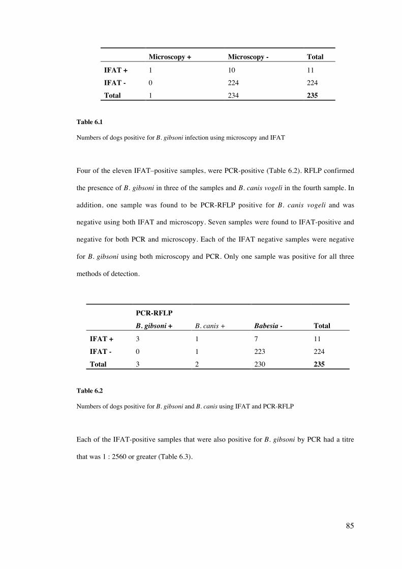
85
Microscopy + Microscopy - Total
IFAT + 1 10 11
IFAT - 0 224 224
Total 1 234 235
Table 6.1
Numbers of dogs positive for B. gibsoni infection using microscopy and IFAT
Four of the eleven IFAT–positive samples, were PCR-positive (Table 6.2). RFLP confirmed
the presence of B. gibsoni in three of the samples and B. canis vogeli in the fourth sample. In
addition, one sample was found to be PCR-RFLP positive for B. canis vogeli and was
negative using both IFAT and microscopy. Seven samples were found to IFAT-positive and
negative for both PCR and microscopy. Each of the IFAT negative samples were negative
for B. gibsoni using both microscopy and PCR. Only one sample was positive for all three
methods of detection.
PCR-RFLPB. gibsoni + B. canis + Babesia - Total
IFAT + 3 1 7 11
IFAT - 0 1 223 224
Total 3 2 230 235
Table 6.2
Numbers of dogs positive for B. gibsoni and B. canis using IFAT and PCR-RFLP
Each of the IFAT-positive samples that were also positive for B. gibsoni by PCR had a titre
that was 1 : 2560 or greater (Table 6.3).
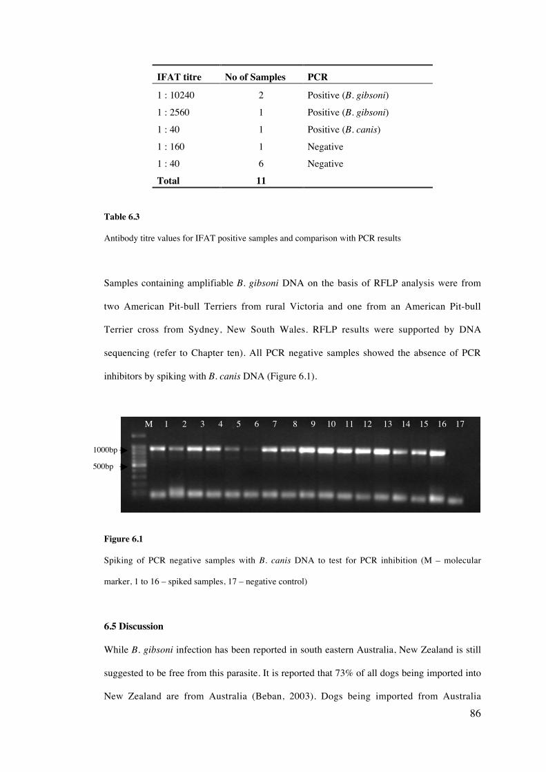
86
IFAT titre No of Samples PCR
1 : 10240 2 Positive (B. gibsoni)
1 : 2560 1 Positive (B. gibsoni)
1 : 40 1 Positive (B. canis)
1 : 160 1 Negative1 : 40 6 Negative
Total 11
Table 6.3
Antibody titre values for IFAT positive samples and comparison with PCR results
Samples containing amplifiable B. gibsoni DNA on the basis of RFLP analysis were from
two American Pit-bull Terriers from rural Victoria and one from an American Pit-bull
Terrier cross from Sydney, New South Wales. RFLP results were supported by DNA
sequencing (refer to Chapter ten). All PCR negative samples showed the absence of PCR
inhibitors by spiking with B. canis DNA (Figure 6.1).
Figure 6.1
Spiking of PCR negative samples with B. canis DNA to test for PCR inhibition (M – molecular
marker, 1 to 16 – spiked samples, 17 – negative control)
6.5 Discussion
While B. gibsoni infection has been reported in south eastern Australia, New Zealand is still
suggested to be free from this parasite. It is reported that 73% of all dogs being imported into
New Zealand are from Australia (Beban, 2003). Dogs being imported from Australia
M 1 2 3 4 5 6 7 8 9 10 11 12 13 14 15 16 17
500bp
1000bp
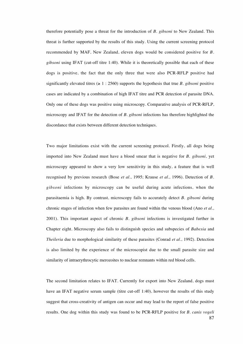
87
therefore potentially pose a threat for the introduction of B. gibsoni to New Zealand. This
threat is further supported by the results of this study. Using the current screening protocol
recommended by MAF, New Zealand, eleven dogs would be considered positive for B.
gibsoni using IFAT (cut-off titre 1:40). While it is theoretically possible that each of these
dogs is positive, the fact that the only three that were also PCR-RFLP positive had
significantly elevated titres (≥ 1 : 2560) supports the hypothesis that true B. gibsoni positive
cases are indicated by a combination of high IFAT titre and PCR detection of parasite DNA.
Only one of these dogs was positive using microscopy. Comparative analysis of PCR-RFLP,
microscopy and IFAT for the detection of B. gibsoni infections has therefore highlighted the
discordance that exists between different detection techniques.
Two major limitations exist with the current screening protocol. Firstly, all dogs being
imported into New Zealand must have a blood smear that is negative for B. gibsoni, yet
microscopy appeared to show a very low sensitivity in this study, a feature that is well
recognised by previous research (Bose et al., 1995; Krause et al., 1996). Detection of B.
gibsoni infections by microscopy can be useful during acute infections, when the
parasitaemia is high. By contrast, microscopy fails to accurately detect B. gibsoni during
chronic stages of infection when few parasites are found within the venous blood (Ano et al.,
2001). This important aspect of chronic B. gibsoni infections is investigated further in
Chapter eight. Microscopy also fails to distinguish species and subspecies of Babesia and
Theileria due to morphological similarity of these parasites (Conrad et al., 1992). Detection
is also limited by the experience of the microscopist due to the small parasite size and
similarity of intraerythrocytic merozoites to nuclear remnants within red blood cells.
The second limitation relates to IFAT. Currently for export into New Zealand, dogs must
have an IFAT negative serum sample (titre cut-off 1:40), however the results of this study
suggest that cross-creativity of antigen can occur and may lead to the report of false positive
results. One dog within this study was found to be PCR-RFLP positive for B. canis vogeli
88
and was also IFAT-positive (1 : 40). All other samples with an IFAT titre of 1 : 40 were
found to be PCR-negative for piroplasm DNA and an additional PCR-negative sample gave
a antibody titre value of 1 : 160. The cross-reactivity of antigen with other Babesia spp., and
even parasites of other genera such as Neospora and Toxoplasma has previously been
reported when using IFAT (Yamane et al., 1993). Cross-reactions were reported for ten dogs
naturally infected with B. canis, with eight giving a antibody titre less than or equal to 1 :
160 and two had titres of 1 : 320 (Yamane et al., 1993). Increasing the currently accepted
cut-off titre (to 1: 160) for B. gibsoni positive samples may help eliminate false positive
cases and give a similar level of agreement to PCR, while retaining the high level of
sensitivity of this detection technique. It has also been reported that IFAT may fail to detect
dogs infected with B. gibsoni during early infections and that some dogs may fail to
seroconvert (Farwell et al., 1982). IFAT also fails to distinguish between current and
previous infections. Further study is therefore necessary to determine the time taken to detect
early B. gibsoni infections and titre levels post-infection using IFAT and is investigated in
Chapter eight.
This study confirms that PCR-RFLP shows promise as an effective detection technique, as it
is capable of detecting various Babesia species with a high level of sensitivity. Employing a
technique that can detect B. gibsoni, in addition to multiple other species of canine piroplasm
is beneficial in preventing other exotic species such as T. annae, B. conradae or the highly
virulent B. canis rossi from entering New Zealand. It also enables dogs with B. canis vogeli
infections that give an IFAT-positive result to be differentiated from those with actual B.
gibsoni infections and preventing the unnecessary restriction of B. canis infected dogs. This
is an important consideration, as Australia is known to be endemic to B. canis vogeli and a
majority of dogs being imported into New Zealand are from Australia (Beban, 2003). PCR-
RFLP was shown to be more sensitive than microscopy for the detection of B. gibsoni
infections, a feature supported by previous studies (Bose et al., 1995).
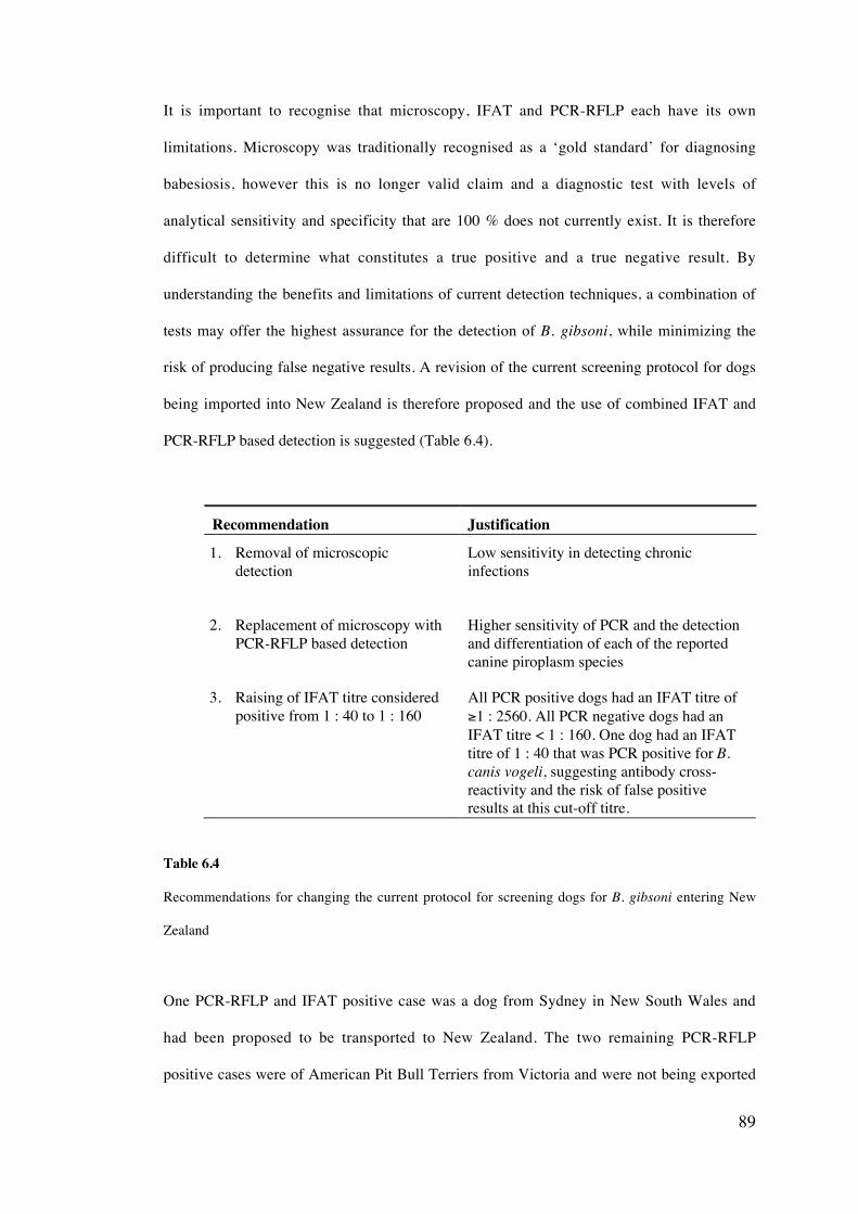
89
It is important to recognise that microscopy, IFAT and PCR-RFLP each have its own
limitations. Microscopy was traditionally recognised as a ‘gold standard’ for diagnosing
babesiosis, however this is no longer valid claim and a diagnostic test with levels of
analytical sensitivity and specificity that are 100 % does not currently exist. It is therefore
difficult to determine what constitutes a true positive and a true negative result. By
understanding the benefits and limitations of current detection techniques, a combination of
tests may offer the highest assurance for the detection of B. gibsoni, while minimizing the
risk of producing false negative results. A revision of the current screening protocol for dogs
being imported into New Zealand is therefore proposed and the use of combined IFAT and
PCR-RFLP based detection is suggested (Table 6.4).
Recommendation Justification
1. Removal of microscopicdetection
Low sensitivity in detecting chronicinfections
2. Replacement of microscopy withPCR-RFLP based detection
Higher sensitivity of PCR and the detectionand differentiation of each of the reportedcanine piroplasm species
3. Raising of IFAT titre consideredpositive from 1 : 40 to 1 : 160
All PCR positive dogs had an IFAT titre of≥1 : 2560. All PCR negative dogs had anIFAT titre < 1 : 160. One dog had an IFATtitre of 1 : 40 that was PCR positive for B.canis vogeli, suggesting antibody cross-reactivity and the risk of false positiveresults at this cut-off titre.
Table 6.4
Recommendations for changing the current protocol for screening dogs for B. gibsoni entering New
Zealand
One PCR-RFLP and IFAT positive case was a dog from Sydney in New South Wales and
had been proposed to be transported to New Zealand. The two remaining PCR-RFLP
positive cases were of American Pit Bull Terriers from Victoria and were not being exported
90
from Australia. Additional research on B. gibsoni infections of dogs in Victoria is described
in Chapter seven. While it is illegal to import dangerous dog breeds such as the American
P i t B u l l T e r r i e r i n t o N e w Z e a l a n d
(http://www.biosecurity.govt.nz/imports/animals/standards/domaniic.aus.htm, accessed
2/2005), the possibility of other dog breeds being infected with B. gibsoni is significant. This
is exemplified by the American Pit Bull Terrier–cross breed that was proposed to be
exported from Australia to New Zealand being found to be positive for B. gibsoni. There
have also been many reports in countries other than Australia of B. gibsoni infection in dogs
of non-American Pit Bull Terrier breeds (Macintire et al., 2002; Birkenheuer et al., 2003b;
Ikadai et al., 2004).
The results of this chapter suggest that nested PCR-RFLP has the potential to be
implemented into a standardised screening protocol for B. gibsoni in dogs being exported
from Australia. A proposal for the change of current screening methods for dogs being
exported from Australia to New Zealand, including the replacement of microscopic
examination of thin blood smears with PCR-RFLP and increase of the IFAT cut-off titre, has
been submitted to MAF, New Zealand2 (Appendix A). This is also the first report of a B.
gibsoni infected dog in New South Wales, extending the current known distribution of this
pathogen in Australia.
2 Subsequent to this investigation, a review of the current screening protocol was undertaken by MAF,New Zealand and a revision of the screening procedure is expected to be implemented late 2005.
91
Enzootic infections of Babesia gibsoni in American Pit Bull Terriers
in south-eastern Australia
7.1 Introduction
An interesting feature of B. gibsoni infection is the high number of reports of this disease
that have been described in fighting dog breeds such as the American Staffordshire Terriers
and American Pit Bull Terrier in USA (Macintire et al., 2002; Birkenheuer et al., 2003b) and
Tosa and American Pit Bull Terriers in Japan (Matsuu et al., 2004; Miyama et al., 2005).
Studies of dogs from the Aomori Prefecture in Japan found that 3.9 % were positive for B.
gibsoni, all were of the Tosa breed (Ikadai et al., 2004) and 29.8% of all Tosa dogs studied
from the same Prefecture were positive for B. gibsoni (Matsuu et al., 2004a). A much
broader study investigating suspected cases of B. gibsoni infection in 13 Prefectures
throughout Japan found that 80 % of all positive dogs were Tosa and 11.4 % were American
Pit Bull Terriers (Miyama et al., 2005). Similarly, a high proportion (55 %) of fighting dog
breeds were found to be positive for B. gibsoni in the southeastern United States (Macintire
et al., 2002). The significance of this breed predisposition to B. gibsoni infections is not yet
fully understood, however the possibility of blood-to-blood transfer occurring between dogs
during fighting has been speculated as a possible mode of transmission.
CHAPTER SEVEN
92
Within Australia to date, B. gibsoni has only been reported in the south-eastern state of
Victoria in three American Pit Bull Terriers (Muhlnickel et al., 2002). Since then, no studies,
have investigated the prevalence or transmission dynamics of this parasitic infection within
Australia or determined if other dog breedshave been infected. This study investigates the
prevalence and epidemiology of B. gibsoni in populations of American Pit Bull Terriers in
Victoria subsequent to the initial case report.
7.2 Aims
i. To determine whether B. gibsoni infection had extended beyond the three dogs
initially reported by Muhlnickel et al. (2002).
ii. If so, to determine the extent of B. gibsoni in American Pit Bull Terriers and other
dog breeds within Victoria, Australia and if possible, to determine the prevalence of
infection.
iii. To investigate possible modes of transmission of B. gibsoni among American Pit
Bull Terriers.
93
7.3 Material and Methods
7.3.1 Dogs sampled3
EDTA blood and serum samples were collected from 151 dogs residing within the State of
Victoria in south-eastern Australia during 2004/05. These included:
i. American Pit Bull Terriers from various rural properties between the towns of
Warrnambool and Ballarat4 (for the purposes of this study, termed non-show
American Pit Bull Terriers) (n = 80). Nine of the dogs had blood collected on two
occasions and one dog had three blood samples taken at separate times.
ii. Jack Russell Terriers and other dogs of non-American Pit Bull Terrier breed closely
associated with (i) (n = 6).
iii. American Pit Bull Terriers at the annual show of the APBT Breeders Association,
Melbourne (for the purposes of this study, termed show American Pit Bull Terriers)
(n = 20).
iv. Non-American Pit Bull Terrier breeds from the same geographical locality as dogs
in (i) that were patients of the Warrnambool Veterinary Clinic (n = 45) and were
referred to as the control group of this study.
7.3.2 DNA extraction, amplification and RFLP
For each sample, DNA was isolated from 200 ml aliquots of EDTA blood (stored at -20 C)
using a QIAamp‚ DNA mini kit (QIAGEN, Hilden, Germany), according to the
manufacturer’s instructions (refer to Chapter four, section 4.2). Amplification of a partial
region of the 18S rRNA gene of Babesia spp. was performed as described in Chapter five,
section 5.3.3. The species of piroplasm present was determined by RFLP analysis of the
3 Ethics approval R1064/044 The locality was chosen for investigation as one of B. gibsoni infected dogs described byMuhlnickel et al. (2002) resided in this district.
94
amplified product (refer to Chapter five, section 5.3.4) and further confirmed using DNA
sequencing.
7.3.3 IFAT and microscopy
Antibodies to B. gibsoni were detected by IFA test (refer to chapter four, section 4.9) and a
positive IFAT titre was considered to be 1:40. Thin blood smears were examined by
microscopy for each sample (described in Chapter four, section 4.1).
7.3.4 Haematological data
The packed cell volume (PCV), red cell count (RCC), haemoglobin (HB), white blood cell
count (WBC), platelet number (PLT) and total protein (TP) were determined for each of the
blood samples using an ADVIA® 120 Haematology System (Bayer Healthcare LLC,
Germany) and a Cell-Dyn 3500 haematology analyser (Abbott Diagnostics, U.S.A.).
7.3.5 Epidemiological data
Owners of each American Pit Bull Terriers involved in this study completed a questionaire
(Appendix B) identifying the following epidemiological parameters:
i. Sex and age of the dog
ii. Kennelling information and whether the dog mixed readily with other dogs.
iii. Breeding history of the dog (i.e. – whether it had ever acted as a sire or
breeding bitch).
iv. Interstate and overseas travel history.
v. Had the dog been witnessed being bitten by or biting another dog?
vi. Had the dog ever received a blood transfusion?
vii. Had ticks ever been found on the dog?
viii. Had any acarcidal treatments ever been given to the dog?
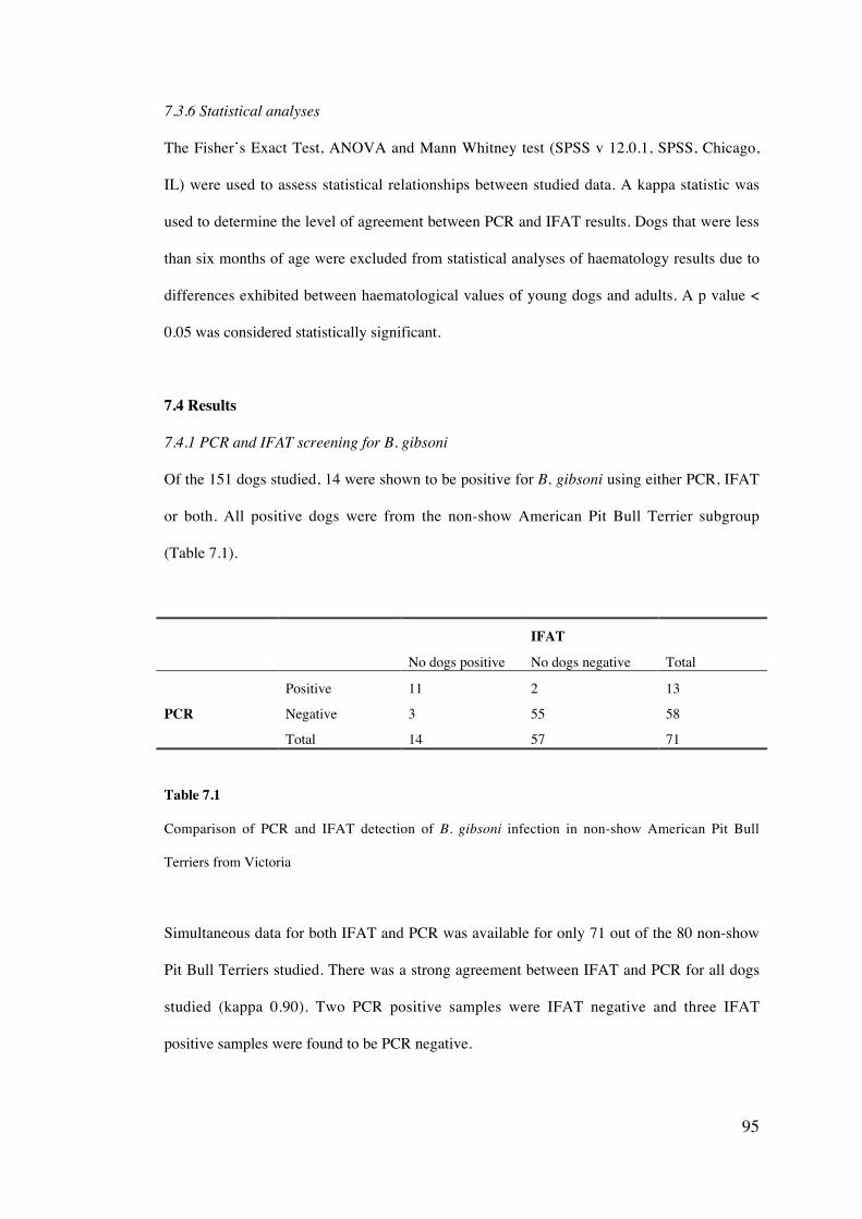
95
7.3.6 Statistical analyses
The Fisher’s Exact Test, ANOVA and Mann Whitney test (SPSS v 12.0.1, SPSS, Chicago,
IL) were used to assess statistical relationships between studied data. A kappa statistic was
used to determine the level of agreement between PCR and IFAT results. Dogs that were less
than six months of age were excluded from statistical analyses of haematology results due to
differences exhibited between haematological values of young dogs and adults. A p value <
0.05 was considered statistically significant.
7.4 Results
7.4.1 PCR and IFAT screening for B. gibsoni
Of the 151 dogs studied, 14 were shown to be positive for B. gibsoni using either PCR, IFAT
or both. All positive dogs were from the non-show American Pit Bull Terrier subgroup
(Table 7.1).
No dogs positive
IFAT
No dogs negative Total
Positive 11 2 13
PCR Negative 3 55 58
Total 14 57 71
Table 7.1
Comparison of PCR and IFAT detection of B. gibsoni infection in non-show American Pit Bull
Terriers from Victoria
Simultaneous data for both IFAT and PCR was available for only 71 out of the 80 non-show
Pit Bull Terriers studied. There was a strong agreement between IFAT and PCR for all dogs
studied (kappa 0.90). Two PCR positive samples were IFAT negative and three IFAT
positive samples were found to be PCR negative.
96
All show American Pit Bull Terriers and non-American Pit Bull Terrier breeds (associated
dogs and all control group dogs) were negative for both detection techniques. Babesia
parasites were not detected by microscopy in any dog during this sudy.
Of the 10 dogs that were sampled on multiple occasions, only one was consistently positive
by both PCR and IFAT. Two dogs that were initially PCR/IFAT positive were treated with
combined azithromycin and atovaquone, becoming PCR negative when tested approximately
two months later but remaining IFAT positive. Two B. gibsoni positive female non-show
American Pit Bull Terriers tested twice over a two-month period had pups. One that was
consistently IFAT positive had five pups, two months of age and the other initially
PCR/IFAT negative had three pups that were eight months of age at the time of sampling.
All pups were negative for B. gibsoni using both IFAT and PCR.
RFLP results were confirmed by genetic sequencing, showing 100% homology with isolates
from the United States and Japan (GenBank accession numbers AF271082, AF205636 and
AF271081) on the basis of a partial region of the 18S rRNA gene (details of molecular
characterisation are presented in Chapter ten).
7.4.2 Clinical signs and haematological data
Two of the B. gibsoni positive dogs had lethargy and bleeding from the mouth at the time of
blood collection and were later euthanased. Unfortunately, no reliable clinical information or
haematological data was available for these two dogs. All remaining dogs were found to be
clinically normal at the time of blood collection, as examined by a veterinarian (Dr P. Irwin).
Dogs that were positive for B. gibsoni had a significantly lower WBC (p = 0.028) and
platelet count (p = 0.002) and a significantly higher total protein level (p = 0.000) than dogs
that were infection-free (Table 7.2). All other haematological parameters were found to be
normal for all dogs studied, whether infected or uninfected (raw data is shown in Appendix
C).
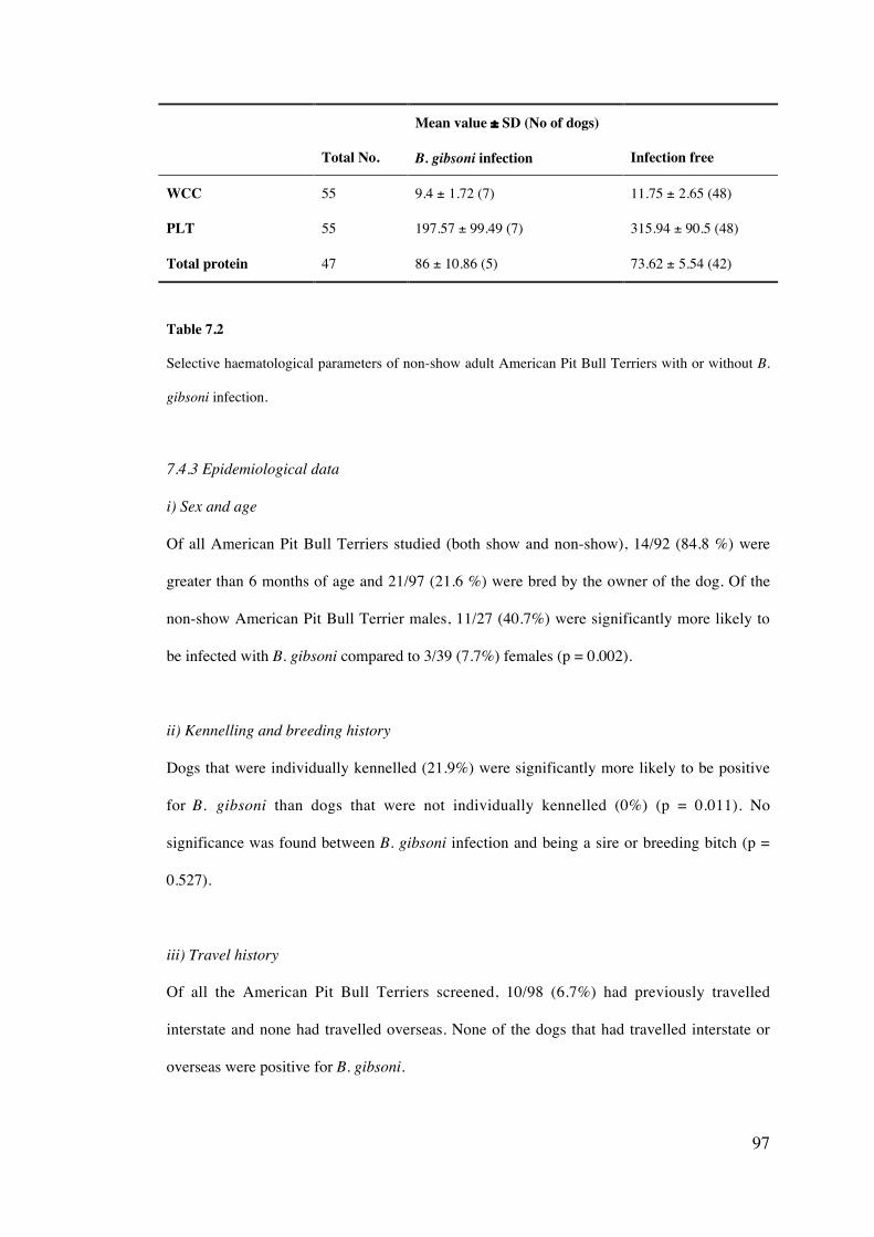
97
Total No.
Mean value ± SD (No of dogs)
B. gibsoni infection Infection free
WCC 55 9.4 ± 1.72 (7) 11.75 ± 2.65 (48)
PLT 55 197.57 ± 99.49 (7) 315.94 ± 90.5 (48)
Total protein 47 86 ± 10.86 (5) 73.62 ± 5.54 (42)
Table 7.2
Selective haematological parameters of non-show adult American Pit Bull Terriers with or without B.
gibsoni infection.
7.4.3 Epidemiological data
i) Sex and age
Of all American Pit Bull Terriers studied (both show and non-show), 14/92 (84.8 %) were
greater than 6 months of age and 21/97 (21.6 %) were bred by the owner of the dog. Of the
non-show American Pit Bull Terrier males, 11/27 (40.7%) were significantly more likely to
be infected with B. gibsoni compared to 3/39 (7.7%) females (p = 0.002).
ii) Kennelling and breeding history
Dogs that were individually kennelled (21.9%) were significantly more likely to be positive
for B. gibsoni than dogs that were not individually kennelled (0%) (p = 0.011). No
significance was found between B. gibsoni infection and being a sire or breeding bitch (p =
0.527).
iii) Travel history
Of all the American Pit Bull Terriers screened, 10/98 (6.7%) had previously travelled
interstate and none had travelled overseas. None of the dogs that had travelled interstate or
overseas were positive for B. gibsoni.
98
iv) Dog bite and history of blood transfusion
More non-show American Pit Bull Terriers that were reportedly bitten or were biters, were
positive for B. gibsoni (12/41, 29.3%) than those not reportedly bitten (2/27, 7.4%) (p =
0.027). One dog that initially tested negative for B. gibsoni using PCR, later tested positive
and had been reportedly attacked by another American Pit Bull Terrier. None of the dogs
were reported to have had a blood transfusion.
v) Tick exposure and treatment
A single tick was found on one American Pit Bull Terrier by the owner subsequent to blood
collection, however it was not available for species identification or PCR analysis. All other
dogs were tick-free at the time of blood collection and had no reported history of tick
exposure. None of the dogs had ever received acaricidal treatment.
7.5 Discussion
This study has demonstrated that B. gibsoni infection in Australia has occurred in more
American Pit Bull Terriers than the three individuals initially described by Muhlnickel et al.
(2002). Without testing many thousands of American Pit Bull Terriers in Australia, it is not
possible to reliably determine the prevalence of this infection. Indeed, the prevalence within
the American Pit Bull Terrier breed itself is likely to vary widely, a feature suggested by the
results of this chapter, with a prevalence of 17.5 % among one group (non-show American
Pit Bull Terriers) compared to the total absence of infection in the second group (show
American Pit Bull Terriers). Thus for practical purposes, this study focussed on a discrete
geographical region within Victoria from where one of the first cases originated (Muhlnickel
et al., 2002).
No other dog breeds were found to be infected with B. gibsoni and further research is
necessary to investigate the possibility of this disease establishing within dogs other than
American Pit Bull Terriers. Notably, the prevalence of this pathogen in non-fighting dog
99
breeds seems to be low in the United States (Macintire et al., 2002; Birkenheuer et al.,
2003b) and all dogs found to be B. gibsoni positive from the Okinawa Prefecture were of
various breeds that did not include either the Tosa or American Pit Bull Terrier (Ikadai et al.,
2004).
7.5.1 Detection of B. gibsoni infection
Both IFAT and PCR showed a general agreement for the detection of B. gibsoni in dogs
within this study however, slight discordance was observed between these two detection
techniques and all samples were microscopy negative. These results further support
suggestions made in Chapter six, that accurate detection of B. gibsoni is problematic without
an established ‘gold standard’ test and that microscopy exhibits a low level of sensitivity.
The identification of two PCR-positive, IFAT-negative dogs and three PCR-negative, IFAT-
positive dogs however, exposes the advantage of using a dual screening techniques
approach. A possible explanation for the negative IFAT titre in two of these cases is that the
infection was pre-acute and the dogs had not yet developed an immune response to the
parasite. The failure of some dogs infected with B. gibsoni to seroconvert has also been
reported (Farwell et al., 1982). PCR-negative, IFAT-positive dogs may have been false-
positive results as a result of antigen cross-reaction or true positive cases. Further study is
therefore necessary to assess such cases, particularly the detection limit of PCR during
chronic B. gibsoni infections, and this is investigated in Chapter eight. It is important to note
that while all dogs were negative for B. gibsoni using light microscopy, this technique is
generally the only detection method available to both veterinarians and commercial
diagnostic laboratories, significantly limiting accurate diagnosis of this disease due to the
low sensitivity and specificity of this method (Conrad et al., 1991; Krause et al., 1996).
Detection using both PCR and IFAT has however been shown to be superior to microscopy
(refer to Chapters five and six) and should be used when dealing with suspected cases of B.
gibsoni infection.
100
7.5.2 American Pit Bull Terrier predisposition to B. gibsoni infection
The discovery of enzootic B. gibsoni infections in a population of American Pit Bull Terriers
within Victoria and the absence of infection in other dogs breeds from the same locality,
provides further support for the B. gibsoni infection predisposition of fighting dog breeds,
such as American Pit Bull Terriers and Tosas reported in the USA and Japan (Macintire et
al., 2002; Birkenheuer et al., 2003b; Ikadai et al., 2004; Matsuu et al., 2004a; Miyama et al.,
2005). The absence of infection in show American Pit Bulls and the presence of B. gibsoni in
non-show dogs is likely to be consequence of different management practices by the owners
of these dogs, a feature supported by analysis of questionnaire data. Perhaps the most
important factor contributing to the disease within non-show American Pit Bull Terriers
from rural localities between Warrnambool and Ballarat in Victoria is the increased risk of
bitting or being bitten by another American Pit Bull Terrier and is further discussed with
regard to transmission dynamics of B. gibsoni infection.
7.5.3 Transmission dynamics
It has been reported by Macintire et al. (2002) and Ikadai et al. (2004) that various modes of
transmission may exist for B. gibsoni, contributing to the spread of this parasite. These
include the role of direct blood contamination during dog fights, tick vectors, transplacental
transmission and blood transfusion associated transmission. The first three of these
mechanisms of transmission can be discussed with respect to the results of this study. The
potential spread and increased distribution of this parasite can also be related to the
movement of dogs from areas of endemicity to areas of non-endemicity.
i) Dog bite
This study supports the hypothesis of transmission occurring during dog fighting due to the
high number of B. gibsoni positive dogs having been reportedly bitten by another dog. Dog
fighting is illegal within Australia, however American Pit Bull Terriers are bred to attack
other dogs, a situation that is likely to occur both accidentally and during illegal
101
‘underground’ dog fighting. Dogs often attack each other by biting the facial region of their
opponent. Tentative evidence suggests that Babesia parasites are concentrated in the
capillaries of its host (Breitschwerdt, 1984) and thus a higher concentration of parasite could
be transmitted during mixing of facial capillary blood. Definitive evidence of blood-to-blood
transmission of B. gibsoni would require controlled experimental fighting to occur between a
positive and non-positive dog, a situation considered both un-ethical and illegal within
Australia.
The first suggestion of possible direct blood-to-blood transmission of B. gibsoni between
dogs, was made by Irizarry-Rovira et al. (2001), who reported B. gibsoni infection in a dog
that had received multiple attack wounds from three American Pit Bull Terriers. The attack
had occurred two months before the development of clinical signs and was consistent with
the pre-patent period of B. gibsoni infection (Macintire et al., 2002). A significant
correlation between dog fighting and B. gibsoni infection has also been reported in Japan
where the practice is still legal, with 26 of 35 positive dogs studied having been bitten by
other dogs (Miyama et al., 2005) and 47.1% of all dogs studied with a history of fighting
being found to be positive (Matsuu et al., 2004a).
ii) Tick vectors
The discovery of an unidentified tick species on only one American Pit Bull Terrier in
Victoria, together with the absence of tick infestation reported by the dog owners suggests
that ticks are potentially not significant in the transmission of B. gibsoni in the studies group
of dogs from Victoria. Other studies have also suggested limited tick exposure in both
American Pit Bull Terriers and Tosa. No ticks were identified on American Pit Bull Terriers
infected with B. gibsoni in southeastern United States (Macintire et al., 2002) and only three
of 35 positive dogs had a confirmed history of tick exposure in a study in Japan (Miyama et
al., 2005).
102
The tick species Haemaphysalis longicornis, H. bispinosa and Rhipicephalus sanguineus
have each been reported as possible vector candidates for the transmission of B. gibsoni
(Swaminath and Shortt, 1937; Otsuka, 1974; Higuchi et al., 1995), although definitive
transmission studies using R. sanguineus have not been conducted. Within Australia, both H.
longicornis and R. sanguineus are known to exist in Australia. Rhipicephalus sanguineus
typically has a tropical to subtropical distribution, although has also been known to establish
in more temperate locations if suitable conditions exist (Roberts, 1970; De Chaneet, 1976).
Haemaphysalis longicornis was originally introduced from Japan to Australia in the early
1900’s (Hoogstraal et al., 1968) and it is now distributed throughout southeast Queensland
(Sutherst and Bourne, 1991), coastal New South Wales, the Murray Valley and Western
Australia (Besier and Wroth, 1985). Both tick species are potential vector candidates for B.
gibsoni in Australia.
The involvement of ticks in disease transmission may however, be more significant than
blood-to-blood transmission during dog fighting in certain countries and regions. It has been
suggested that the main mode of transmission of B. gibsoni in the Okinawa Prefecture was
likely to be by the tick Rhipicephalus sanguineus (Ikadai et al., 2004). If B. gibsoni was to
establish within regions of northern Australia, where dog infestation by R. sanguineus is
extremely common, tick vector transmission may become more significant.
iii) Transplacental transmission
This study has identified two B. gibsoni infected female American Pit Bull Terriers that had
infection-free pups. While it is likely that one of the adult females became infected with B.
gibsoni after giving birth, the other was infected during pregnancy and transplacental
transmission did not appear to occur or at least, patent infection was not maintained. A
recent study however, proved experimental transplacental transmission of B. gibsoni and
excluded the possibility of trans-mammary transmission (Fukumoto et al., 2005). All pups
died from acquired babesiosis. While vertical transmission of B. gibsoni can occur, this study
103
suggests it does not happen in all cases when the pregnant dog is known to be infected. The
reason for case variation is unknown but could relate to the stage of infection and/or the
immune status of the bitch.
iv) Movement of dogs to areas of non-endemicity
Only one dog residing outside of Victoria (located in Sydney, NSW) was reported to be
infected with B. gibsoni (refer to Chapter six). The absence of reported cases in other
Australian states, besides Victoria and New South Wales does not however, rule out the
possibility of this parasite existing in other States and Territories of Australia. There are
currently no restrictions on the movements of dogs throughout Australia and as this study
has shown, dogs from enzootic regions have been reported to travel interstate.
New legislation has now been proposed for most states of Australia including New South
Wales (http://dig.nsw.gov.au/dig/dighome/documents/circulars/05-20.pdf, accessed 5/2005)
and Victoria (http://www.dpc.vic.gov.au/domino/Web_Notes/newmedia.nsf, accessed
4/9/2005), in which it will be an offence to breed, sell, give away or acquire Pit Bull Terriers
and other similar breeds such as Japanese Tosas, Argentinean fighting dogs and Brazilian
fighting dogs. All current owners of such breeds will also have to get their dogs de-sexed.
Such banning of Pit Bull Terriers may help to limit the spread of B. gibsoni.
None of the dogs in this study had been reported to have travelled overseas, which together
with current laws preventing the importation of American Pit Bull Terriers, make it difficult
to speculate on the original source of B. gibsoni, in Australia. Reports suggest that B. gibsoni
was likely to have first been introduced into the United States from military and/or fighting
dogs being imported from Malaysia or Okinawa, Japan (Farwell et al., 1982; Macintire et
al., 2002). Likewise, the initial introduction of this piroplasm into Australia may have also
occurred as a consequence of importation of infected dogs and/or ticks from endemic
countries such as Asia or the United States. Stringent control practices should now be put in
104
place to avoid further spread of this disease to non-endemic countries with screening
protocols already existing for dogs being imported into New Zealand (http://www.
biosecurity.govt.nz/imports/animals/ standards/domaniic.aus.htm, accessed 3/2005) and
South Africa (http://www.aqis.gov.au.htm, accessed 3/2005).
7.5.4 Clinical signs of infections
It appears that most of the American Pit Bull Terriers described in this study had subclinical
B. gibsoni infections, which further contributes to the difficulty of accurate clinical
diagnosis. Indeed, B. gibsoni infection has previously been misdiagnosed as immune
mediated anaemia (Muhlnickel et al., 2002). Two dogs, did however present with bleeding
tendencies. This may reflect the thrombocytopenias found in most positive dogs, yet the
platelet count would have to be severely depressed (< 20 x 109/L) in order for bleeding to
occur. Tosa dogs with subclinical B. gibsoni infections also had significantly lower mean
platelet counts than dogs that were infection free. A significantly lower platelet count in B.
gibsoni infected dogs was also reported in other studies (Macintire et al., 2002; Miyama et
al., 2005).
7.5.5 Conclusion
This study has provided further evidence for the existence of B. gibsoni infections within
Australia beyond the initial report of just three infected dogs. Increased veterinary awareness
of these parasites, in addition to the employment of more effective detection methods such as
PCR and IFAT, need to be considered if this infection is to be managed within Australia.
While infected dog populations seem to be enzootic and restricted to dogs of American Pit
Bull Terrier breed at present, the transmission potential to other dog breeds and other
locations within Australia remains unknown. Careful management of known infected dogs,
including antibabesial drug treatment and prevention of dog fighting is also necessary to help
prevent the spread of this pathogen in Australia.

105
Babesia gibsoni infection should now be considered a significant disease of fighting dog
breeds worldwide, is likely to be transmitted by direct blood exchange occurring during
fighting/biting in these dogs and is a feature evident within American Pit Bull Terriers in
Australia.
106
Experimental Babesia gibsoni infections: The assessment of
combined atovaquone and azithromycin therapy and the detection
limits of PCR during early and chronic stages of infection.
8.1. Introduction
While the in vivo culture of B. gibsoni has been reported previously in numerous studies
(Anderson et al., 1980; Yamane et al., 1993; Wozniak et al., 1997; Wulansari et al., 2003;
Matsuu et al., 2004), the clinical and pathological manifestations of these experimental
infections seem to be varied and study dependent. Similar variation is also exhibited by
natural infections (Irizarry-Rovira et al., 2001; Birkenheuer et al., 2003b; Matsuu et al.,
2004a; Miyama et al., 2005). The disease can be pre-acute, acute, or chronic/subclinical.
Acute infections are often typified by haemolytic anaemia, hemoglobinuria,
thrombocytopenia and splenomegaly (Yamane et al., 1993). Chronic infections can develop,
however this stage of infection can often be asymptomatic, with carriers of B. gibsoni
infection acting as reservoirs of disease. Such subclinical carriers of infection are reported to
maintain high antibody titres (Anderson et al., 1980; Farwell et al., 1982; Conrad et al.,
1991; Yamane et al., 1993).
PCR has been shown to be highly sensitive and specific for the detection of B. gibsoni (Ano
et al., 2001; Fukumoto et al., 2001; Jefferies et al., 2002; Birkenheuer et al., 2003a; Chapter
five, sections 5.4.1, 5.4.2), yet limited study has been carried out on the detection limit of
this technique during the early and chronic stages of infection. It is also not understood
CHAPTER EIGHT
107
whether canine piroplasms are capable of leaving the circulatory system of its host, to
become sequestered within tissues such as the spleen.
Babesia gibsoni infection has a history of being notoriously difficult to treat successfully.
Various treatments for B. gibsoni infection have been described (Farwell et al., 1982;
Wulansari et al., 2003), however, no drugs have been reported to produce total eradication of
circulating parasite. Atovaquone and azithromycin were first used as drug therapies for the
eradication of malaria infections (Looareesuwan et al., 1996; Taylor et al., 1999) and were
subsequently assessed as treatments for B. microti infections (Wittner et al., 1996; Bonoan et
al., 1998). Combined drug treatment was found to be more effective than the use of each
separately (Wittner et al., 1996). While in some patients the drug therapy resulted in the total
elimination of parasitaemia, other studies suggest that the clearance of parasites is
inconsistent, with B. microti parasites sometimes persisting for months after treatment
(Krause et al., 2000).
A recent study assessed the efficacy of combined atovaquone and azithromycin therapy for
the treatment of B. gibsoni and reported that the combined drugs reduced infections to
undetectable levels (Birkenheuer et al., 2004a). This study however did not use controlled
experimental infections, but known naturally infected cases of chronic B. gibsoni infection in
American Pit Bull Terriers. Some of the dogs given the treatment were found to remain PCR
positive for B. gibsoni several months later, questioning whether this combined drug
treatment is effective in all cases. A later report identified the possibility of drug resistence to
atovaquone (Matsuu et al., 2004b). Further study is therefore warranted to investigate the
efficacy of combined atovaquone and azithromycin treatment in experimentally infected
animals.
108
Experimental B. gibsoni infections were established in dogs for the production of IFAT
blood slides, in addition to investigating the detection limits of PCR during various stages of
infection and to assess the efficacy of combined azithromycin and atovaquone drug therapy.
8.2 Aims
i. To establish experimental B. gibsoni infections in dogs.
ii. To evaluate the efficacy of combined azithromycin and atovaquone drug therapy for
B. gibsoni infections.
iii. To determine the detection limit of PCR during early and chronic stages of infection
and compare with IFAT and microscopy.
iv. To assess the presence of B. gibsoni within various tissues when parasites have been
eradicated from the bloodstream.
109
8.3 Materials and Methods
8.3.1 B. gibsoni positive blood samples
Babesia gibsoni-infected blood was collected from naturally infected American Pit Bull
Terriers (refer to epidemiological study in Chapter seven). Blood samples were mixed into
CPD-1 solution (Baxter International Inc., USA) and refrigerated at 4 C. Samples were
screened for the presence of B. gibsoni using PCR-RFLP. DNA sequencing was later used to
confirm the species and genotype of canine piroplam present (refer to Chapter four, section
4.7).
Infected blood was then passaged into experimental Dogs A and C, while Dog B received
infected blood from Dog A (refer to section 8.3.3).
8.3.2 Experimental dogs5
Three five-month old, female beagle foxhound-cross dogs (Dogs A, B and C) were obtained
from the Commonwealth Serum Laboratories, Melbourne. Each dog was sprayed with
Frontline‚ (2.5 g/L fipronil) (Merial, France) as a tick control measure before the initiation
of the experiment and also once during the course of the experiment. Blood was collected at
the initial time of arrival in Perth and screened using PCR and IFAT to confirm the absence
of Babesia parasites. Dogs were housed indoors in the animal isolation facility at Murdoch
University and examined daily.
8.3.3 Experimental infection with B. gibsoni (overview)
Immediately prior to passage of infected blood, a 5 ml blood sample was collected from the
recipient dog for serology, PCR and haematological analysis. Passage details for each dog
are described in Table 8.1. Each dog had 1 ml EDTA and 4 ml clotted for serum was
collected daily for the duration of the experiment and a further 4 ml of clooted blood for
5 Ethics approval R1063/04
110
serum was taken daily for the first 3 weeks of the experiment and then every 5 days. Rectal
temperature, pulse rate, respiratory rate and general physical condition were also assessed
daily for the duration of the experiment.
ExperimentalDog
Passage details Duration ofExperiment
Comments
A 7 ml of blood from a naturallyinfected American Pit BullTerrier (parasitaemia was notcalculated since B. gibsonicould not be visualised).
70 days An additional bloodsample was taken 15hrs post-passage
B 9 ml of blood from Dog A onday 27 post-passage(parasitaemia = 1.51 %).
121 days Additional bloodsamples were takenat 1, 6 and 24hrspost-passage
C 8 ml of blood from a naturallyinfected American Pit BullTerrier (parasitaemia was notcalculated since B. gibsonicould not be visualised).
78 days Additional bloodsamples were takenat 1, 6 and 24hrspost-passage
Table 8.1
Passage details for Dogs A, B and C
8.3.4 Haematological analysis of blood samples
EDTA blood samples were submitted for daily automated haematological analysis
(CBC/DIFF) using an ADVIA® 120 Haematology System (Bayer Healthcare LLC,
Germany). Haematological data were calculated for white blood cell count (WBC), red
blood cell count (RBC), haemoglobin (HGB), haematocrit (HCT), mean cell volume (MCV),
(MCH), (HCHC), (CHCM), (CH), (RDW), HDW, platelet number (PLT), (MDV),
neutrophil number (#NEU), lymphocyte number (#LYMPH), monocyte number (#MONO),
eosinophil number (#EOS), basophil number (#BASO) and leucocyte number (#LUC).
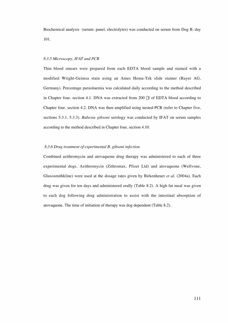
111
Biochemical analysis (serum: panel, electrolytes) was conducted on serum from Dog B, day
101.
8.3.5 Microscopy, IFAT and PCR
Thin blood smears were prepared from each EDTA blood sample and stained with a
modified Wright-Geimsa stain using an Ames Hema-Tek slide stainer (Bayer AG,
Germany). Percentage parasitaemia was calculated daily according to the method described
in Chapter four, section 4.1. DNA was extracted from 200 ml of EDTA blood according to
Chapter four, section 4.2. DNA was then amplified using nested-PCR (refer to Chapter five,
sections 5.3.1, 5.3.3). Babesia gibsoni serology was conducted by IFAT on serum samples
according to the method described in Chapter four, section 4.10.
8.3.6 Drug treatment of experimental B. gibsoni infection
Combined azithromycin and atovaquone drug therapy was administered to each of three
experimental dogs. Azithromycin (Zithromax, Pfizer Ltd) and atovaquone (Wellvone,
Glaxosmithkline) were used at the dosage rates given by Birkenheuer et al. (2004a). Each
drug was given for ten days and administered orally (Table 8.2). A high fat meal was given
to each dog following drug administration to assist with the intestinal absorption of
atovaquone. The time of initiation of therapy was dog dependent (Table 8.2).
112
Dog Weight(kg)
Initiation ofdrug therapy
Stage ofinfection
Azithromycin1
10 mg/kg q 24 hPO
Atovaquone2
13.3 mg/kg q 8hPO
A 13.3 Day 31 Acute 3.3 ml 1.2 ml
B 11.36
11.3
Day 53
Day 95
Acute
Chronic
2.84 ml
2.84 ml
1 ml
1 ml
C 12.1 Day 52 Chronic 3.03 ml 1.06 ml
1. Zithromax‘ (Pfizer Ltd, Australia), 200mg/ml
2. Wellvone‘ Suspension (Glaxosmithkline, Australia), 500mg/ml
Table 8.2
Dosage rates for azithromycin and atovaquone administered to Dogs A, B and C (q =quaque, PO =
per os).
8.3.7 Collection of saliva, urine and tissue samples
Saliva was collected by syringe from Dog A on day 25 and Dog B on day 51 and also by
placing FTA paper directly into the mouth to absorb any saliva present. DNA was extracted
from the saliva collected using a syringe using the QIAGEN blood extraction protocol
(Chapter four, section 4.2). Saliva collected on FTA was allowed to dry and then purified
according to the FTA purification procedure (Chapter five, section 5.3.7). Dogs A, B and C
were euthanised by intra-venous barbiturate overdose on days 70, 121 and 78 respectively.
At necropsy, each dog had tissue samples collected from spleen, pancreas, kidney, heart,
lymph node, salivary gland, bone marrow, skin and intestine using a new scapel blade for the
collection of each tissue. Tissues were stored separately A urine sample was also taken by
cystocentesis. DNA was extracted from tissue samples using a QIAamp DNA mini kit
(QIAGEN, Germany), according to the tissue protocol (refer to Chapter four, section 4.3).
DNA was extracted from urine using a QIAamp DNA mini kit according to the protocol
(refer to Chapter four, section 4.2) for blood and body fluids.
113
8.3.8 Cryopreservation of canine blood infected with B. gibsoni.
For archival purposes, blood samples from Dog A were collected on day 23 and for Dog B
on days 52 and 121 were cryopreserved according to Dalgliesh (1971). An equal volume of
4M dimethyl sulphoxide (DMSO) in PBS was added to blood. Half a ml of cold DMSO was
added to 0.5 ml cold blood at a rate of 1 ml/30 sec, gently mixed and then held at 4 C for 10
min, -20 C for 20 min and –80 C for 20 min. The blood was then transferred to liquid
nitrogen for long-term storage.
8.3.9 Statistical analysis
Statistical analysis was performed using SPSS v12.0.1 (SPSS, Chicago, IL). Correlations
between parasitaemia and haematological data were determined using a Pearson correlation
test and a p-value less then 0.05 was considered significant.
8.4 Results
8.4.1 Clinical observations
Each dog was successfully infected with B. gibsoni. Only two dogs (Dog A and B) exhibited
clinical signs of infection. Dog A developed loss of appetite on days 19-23 and fever on days
23-28. A significant positive correlation was observed between parasitaemia and rectal
temperature (p = 0.000) and heart rate (p = 0.015) for Dog A. Mild icterus was observed on
days 26-28. Splenomegaly developed in Dog B on day 52 and slight icterus was also
observed on day 55. Dog C never developed clinical signs of infection. No significant side
effects were observed during the periods of drug therapy for each of the three dogs, although
fatigue and excessive salivation were witnessed for Dog B during the second course of drug
therapy. Subsequent biochemical analysis revealed no abnormality.
8.4.2 Microscopic detection of B. gibsoni
For the purposes of this study, the early stage of infection was defined to be from the time of
passage to the first detection of B. gibsoni by light microscopy. In both dogs A and B,
114
distinct, pre-acute, acute and chronic stages of infection could be differentiated during the
course of experimental infection (Figure 8.1). Early infection was determined to be from day
0-6 for Dog A and day 0-36 for Dog B, although one parasite was microscopically visualised
on day 5 (Figure 8.1 i and ii). The acute stage of infection was considered as the time during
which B. gibsoni parasites could be detected by microscopy and was from day 7-31 for Dog
A, reaching a maximum of 1.5 % parasitaemia on day 27. Drug treatment was initiated on
day 31 when parasitaemia had dropped to 0.875 % and no parasites were observed
subsequently. Two acute stages of infection were observed for Dog B on days 37-60 and
107-121 (Figure 8.1 ii). The level of parasitaemia reached a maximum of 6.02 % on day 51.
Drug treatment was initiated on day 53 when parasitaemia showed a slight decrease to 5.82
%, then a rapid decrease in parasite number until day 60 when no parasites were observed.
The chronic stage of infection was from day 33-70 for Dog A and day 61-106 for Dog B
(Figure 8.1 i and ii). Dog C failed to develop a parasitaemia detectable by microscopy and
was therefore deemed to have a chronic infection for the duration of the experiment (Figure
8.1 iii).
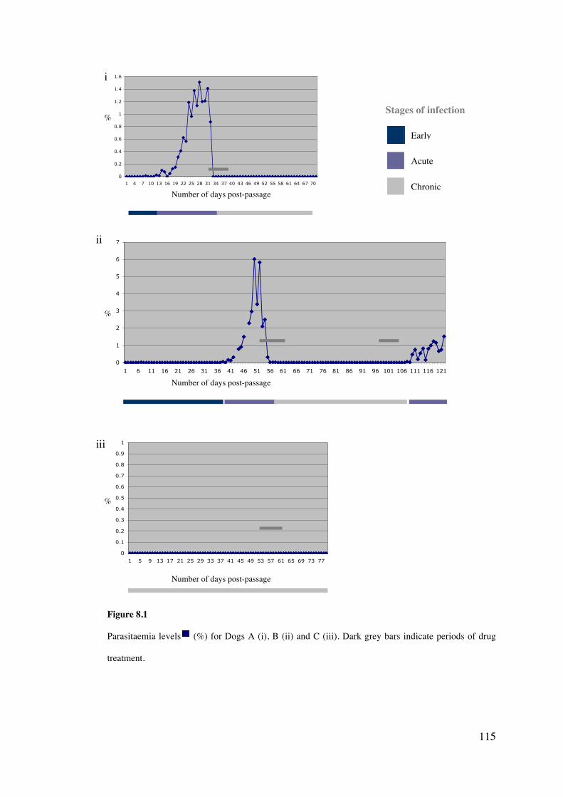
115
0
0.2
0.4
0.6
0.8
1
1.2
1.4
1.6
1 4 7 10 13 16 19 22 25 28 31 34 37 40 43 46 49 52 55 58 61 64 67 70
0
1
2
3
4
5
6
7
1 6 11 16 21 26 31 36 41 46 51 56 61 66 71 76 81 86 91 96 101 106 111 116 121
0
0.1
0.2
0.3
0.4
0.5
0.6
0.7
0.8
0.9
1
1 5 9 13 17 21 25 29 33 37 41 45 49 53 57 61 65 69 73 77
Figure 8.1
Parasitaemia levels (%) for Dogs A (i), B (ii) and C (iii). Dark grey bars indicate periods of drug
treatment.
Early
Chronic
Acute
Stages of infection
Number of days post-passage
Number of days post-passage
Number of days post-passage
i
ii
iii
%
%
%
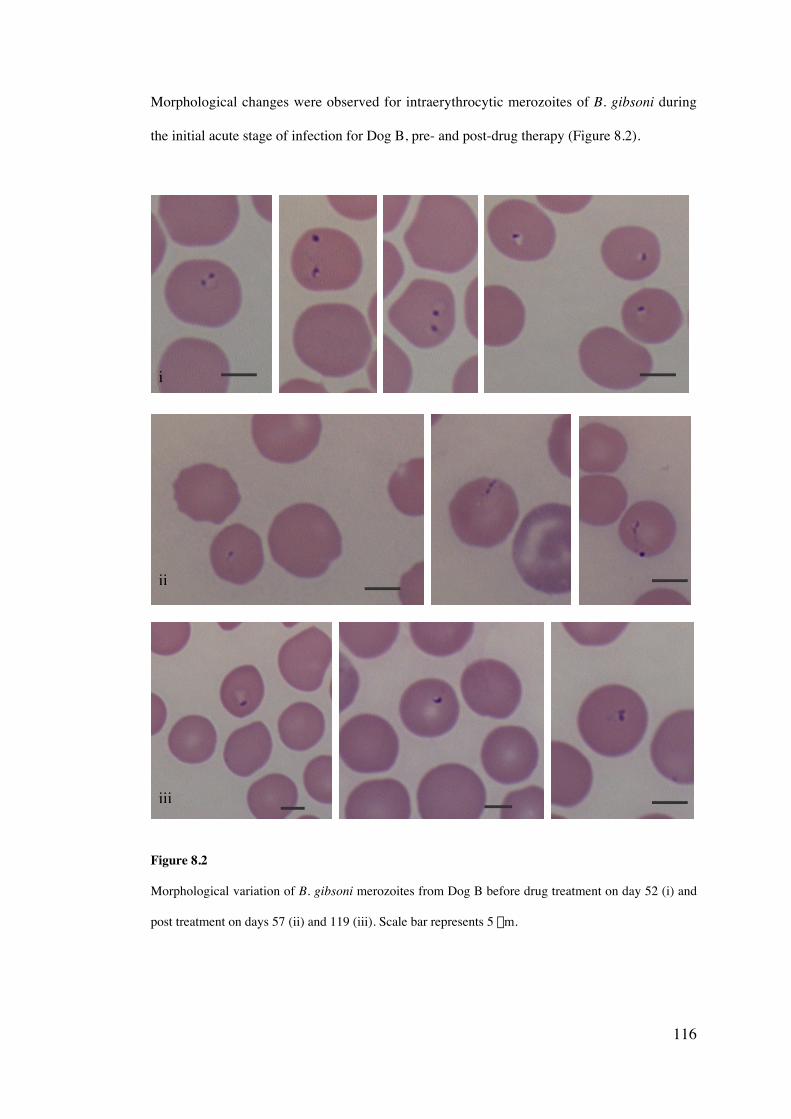
116
Morphological changes were observed for intraerythrocytic merozoites of B. gibsoni during
the initial acute stage of infection for Dog B, pre- and post-drug therapy (Figure 8.2).
2 z z
Figure 8.2
Morphological variation of B. gibsoni merozoites from Dog B before drug treatment on day 52 (i) and
post treatment on days 57 (ii) and 119 (iii). Scale bar represents 5 mm.
i
ii
iii
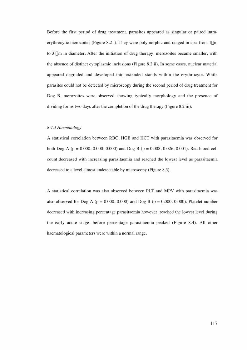
117
Before the first period of drug treatment, parasites appeared as singular or paired intra-
erythrocytic merozoites (Figure 8.2 i). They were polymorphic and ranged in size from 1mm
to 3 mm in diameter. After the initiation of drug therapy, merozoites became smaller, with
the absence of distinct cytoplasmic inclusions (Figure 8.2 ii). In some cases, nuclear material
appeared degraded and developed into extended stands within the erythrocyte. While
parasites could not be detected by microscopy during the second period of drug treatment for
Dog B, merozoites were observed showing typically morphology and the presence of
dividing forms two days after the completion of the drug therapy (Figure 8.2 iii).
8.4.3 Haematology
A statistical correlation between RBC, HGB and HCT with parasitaemia was observed for
both Dog A (p = 0.000, 0.000, 0.000) and Dog B (p = 0.008, 0.026, 0.001). Red blood cell
count decreased with increasing parasitaemia and reached the lowest level as parasitaemia
decreased to a level almost undetectable by microscopy (Figure 8.3).
A statistical correlation was also observed between PLT and MPV with parasitaemia was
also observed for Dog A (p = 0.000, 0.000) and Dog B (p = 0.000, 0.000). Platelet number
decreased with increasing percentage parasitaemia however, reached the lowest level during
the early acute stage, before percentage parasitaemia peaked (Figure 8.4). All other
haematological parameters were within a normal range.
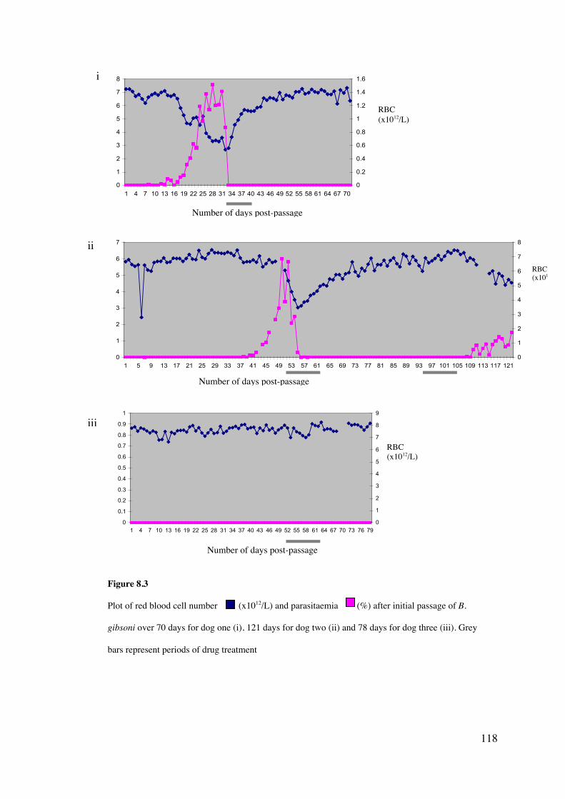
118
0
1
2
3
4
5
6
7
8
1 4 7 10 13 16 19 22 25 28 31 34 37 40 43 46 49 52 55 58 61 64 67 700
0.2
0.4
0.6
0.8
1
1.2
1.4
1.6
0
1
2
3
4
5
6
7
1 5 9 13 17 21 25 29 33 37 41 45 49 53 57 61 65 69 73 77 81 85 89 93 97 101 105 109 113 117 1210
1
2
3
4
5
6
7
8
0
0.1
0.2
0.3
0.4
0.5
0.6
0.7
0.8
0.9
1
1 4 7 10 13 16 19 22 25 28 31 34 37 40 43 46 49 52 55 58 61 64 67 70 73 76 790
1
2
3
4
5
6
7
8
9
Figure 8.3
Plot of red blood cell number (x1012/L) and parasitaemia (%) after initial passage of B.
gibsoni over 70 days for dog one (i), 121 days for dog two (ii) and 78 days for dog three (iii). Grey
bars represent periods of drug treatment
Number of days post-passage
i
RBC(x1012/L)
RBC(x1012/L)
Number of days post-passage
Number of days post-passage
ii
iii
RBC(x1012/L)
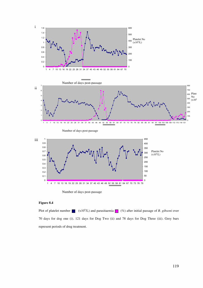
119
0
0.2
0.4
0.6
0.8
1
1.2
1.4
1.6
1 4 7 10 13 16 19 22 25 28 31 34 37 40 43 46 49 52 55 58 61 64 67 700
100
200
300
400
500
600
0
1
2
3
4
5
6
7
1 4 7 10 13 16 19 22 25 28 31 34 37 40 43 46 49 52 55 58 61 64 67 70 73 76 79 82 85 88 91 94 97 100 103 106 109 112 115 118 1210
100
200
300
400
500
600
700
800
0
0.1
0.2
0.3
0.4
0.5
0.6
0.7
0.8
0.9
1
1 4 7 10 13 16 19 22 25 28 31 34 37 40 43 46 49 52 55 58 61 64 67 70 73 76 790
50
100
150
200
250
300
350
400
450
Figure 8.4
Plot of platelet number (x109/L) and parasitaemia (%) after initial passage of B. gibsoni over
70 days for dog one (i), 121 days for Dog Two (ii) and 78 days for Dog Three (iii). Grey bars
represent periods of drug treatment.
Number of days post-passage
i
Platelet No(x109/L)
Number of days post-passage
Number of days post-passage
PlateletNo(x109/L)
Platelet No(x109/L)
ii
iii
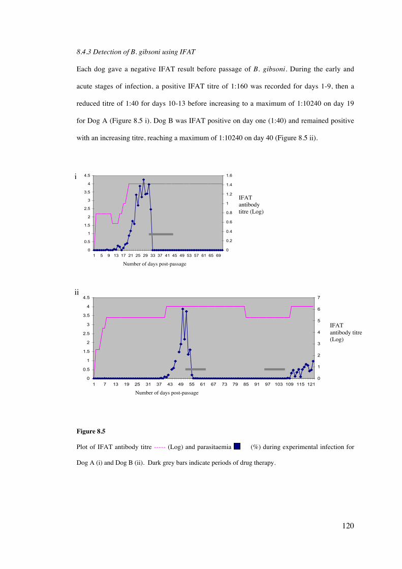
120
8.4.3 Detection of B. gibsoni using IFAT
Each dog gave a negative IFAT result before passage of B. gibsoni. During the early and
acute stages of infection, a positive IFAT titre of 1:160 was recorded for days 1-9, then a
reduced titre of 1:40 for days 10-13 before increasing to a maximum of 1:10240 on day 19
for Dog A (Figure 8.5 i). Dog B was IFAT positive on day one (1:40) and remained positive
with an increasing titre, reaching a maximum of 1:10240 on day 40 (Figure 8.5 ii).
0
0.5
1
1.5
2
2.5
3
3.5
4
4.5
1 5 9 13 17 21 25 29 33 37 41 45 49 53 57 61 65 690
0.2
0.4
0.6
0.8
1
1.2
1.4
1.6
0
0.5
1
1.5
2
2.5
3
3.5
4
4.5
1 7 13 19 25 31 37 43 49 55 61 67 73 79 85 91 97 103 109 115 1210
1
2
3
4
5
6
7
Figure 8.5
Plot of IFAT antibody titre ----- (Log) and parasitaemia (%) during experimental infection for
Dog A (i) and Dog B (ii). Dark grey bars indicate periods of drug therapy.
IFATantibodytitre (Log)
IFATantibody titre(Log)
Number of days post-passage
Number of days post-passage
i
ii
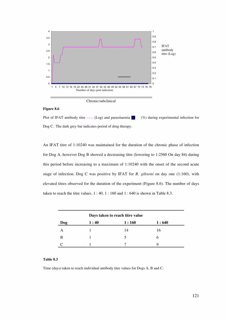
121
0
0.5
1
1.5
2
2.5
3
3.5
4
1 4 7 10 13 16 19 22 25 28 31 34 37 40 43 46 49 52 55 58 61 64 67 70 73 76 790
0.1
0.2
0.3
0.4
0.5
0.6
0.7
0.8
0.9
1
Figure 8.6
Plot of IFAT antibody titre ----- (Log) and parasitaemia (%) during experimental infection for
Dog C. The dark grey bar indicates period of drug therapy.
An IFAT titre of 1:10240 was maintained for the duration of the chronic phase of infection
for Dog A, however Dog B showed a decreasing titre (lowering to 1:2560 On day 84) during
this period before increasing to a maximum of 1:10240 with the onset of the second acute
stage of infection. Dog C was positive by IFAT for B. gibsoni on day one (1:160), with
elevated titres observed for the duration of the experiment (Figure 8.6). The number of days
taken to reach the titre values, 1 : 40, 1 : 160 and 1 : 640 is shown in Table 8.3.
Days taken to reach titre valueDog 1 : 40 1 : 160 1 : 640
A 1 14 16
B 1 5 6
C 1 7 9
Table 8.3
Time (days) taken to reach individual antibody titre values for Dogs A, B and C.
IFATantibodytitre (Log)
Chronic/subclinical
Number of days post-infection
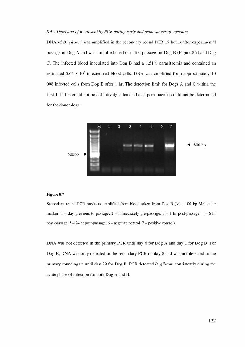
122
8.4.4 Detection of B. gibsoni by PCR during early and acute stages of infection
DNA of B. gibsoni was amplified in the secondary round PCR 15 hours after experimental
passage of Dog A and was amplified one hour after passage for Dog B (Figure 8.7) and Dog
C. The infected blood inoculated into Dog B had a 1.51% parasitaemia and contained an
estimated 5.65 x 10⁷ infected red blood cells. DNA was amplified from approximately 10
008 infected cells from Dog B after 1 hr. The detection limit for Dogs A and C within the
first 1-15 hrs could not be definitively calculated as a parastiaemia could not be determined
for the donor dogs.
Figure 8.7
Secondary round PCR products amplified from blood taken from Dog B (M – 100 bp Molecular
marker, 1 – day previous to passage, 2 – immediately pre-passage, 3 – 1 hr post-passage, 4 – 6 hr
post-passage, 5 – 24 hr post-passage, 6 – negative control, 7 – positive control)
DNA was not detected in the primary PCR until day 6 for Dog A and day 2 for Dog B. For
Dog B, DNA was only detected in the secondary PCR on day 8 and was not detected in the
primary round again until day 29 for Dog B. PCR detected B. gibsoni consistently during the
acute phase of infection for both Dog A and B.
800 bp
500bp
M 1 2 3 4 5 6 7
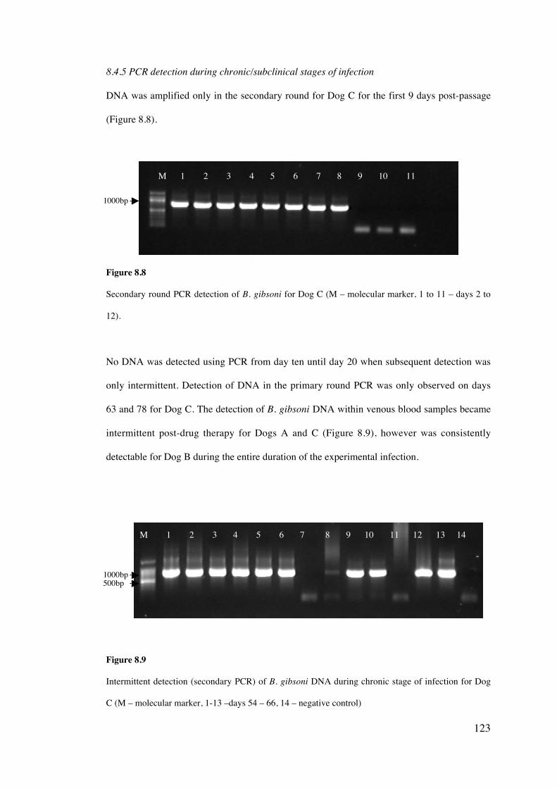
123
8.4.5 PCR detection during chronic/subclinical stages of infection
DNA was amplified only in the secondary round for Dog C for the first 9 days post-passage
(Figure 8.8).
Figure 8.8
Secondary round PCR detection of B. gibsoni for Dog C (M – molecular marker, 1 to 11 – days 2 to
12).
No DNA was detected using PCR from day ten until day 20 when subsequent detection was
only intermittent. Detection of DNA in the primary round PCR was only observed on days
63 and 78 for Dog C. The detection of B. gibsoni DNA within venous blood samples became
intermittent post-drug therapy for Dogs A and C (Figure 8.9), however was consistently
detectable for Dog B during the entire duration of the experimental infection.
Figure 8.9
Intermittent detection (secondary PCR) of B. gibsoni DNA during chronic stage of infection for Dog
C (M – molecular marker, 1-13 –days 54 – 66, 14 – negative control)
M 1 2 3 4 5 6 7 8 9 10 11 12 13 14
1000bp
1000bp
M 1 2 3 4 5 6 7 8 9 10 11
500bp
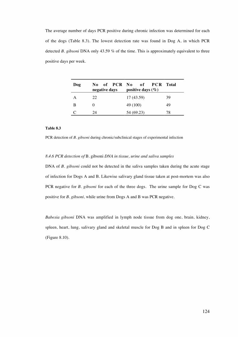
124
The average number of days PCR positive during chronic infection was determined for each
of the dogs (Table 8.3). The lowest detection rate was found in Dog A, in which PCR
detected B. gibsoni DNA only 43.59 % of the time. This is approximately equivalent to three
positive days per week.
Dog No of PCRnegative days
N o o f P C Rpositive days (%)
Total
A 22 17 (43.59) 39
B 0 49 (100) 49
C 24 54 (69.23) 78
Table 8.3
PCR detection of B. gibsoni during chronic/subclinical stages of experimental infection
8.4.6 PCR detection of B. gibsoni DNA in tissue, urine and saliva samples
DNA of B. gibsoni could not be detected in the saliva samples taken during the acute stage
of infection for Dogs A and B. Likewise salivary gland tissue taken at post-mortem was also
PCR negative for B. gibsoni for each of the three dogs. The urine sample for Dog C was
positive for B. gibsoni, while urine from Dogs A and B was PCR negative.
Babesia gibsoni DNA was amplified in lymph node tissue from dog one, brain, kidney,
spleen, heart, lung, salivary gland and skeletal muscle for Dog B and in spleen for Dog C
(Figure 8.10).
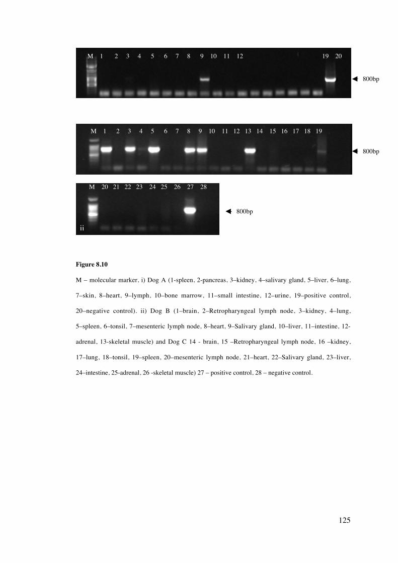
125
Figure 8.10
M – molecular marker, i) Dog A (1-spleen, 2-pancreas, 3–kidney, 4–salivary gland, 5–liver, 6–lung,
7–skin, 8–heart, 9–lymph, 10–bone marrow, 11–small intestine, 12–urine, 19–positive control,
20–negative control). ii) Dog B (1–brain, 2–Retropharyngeal lymph node, 3–kidney, 4–lung,
5–spleen, 6–tonsil, 7–mesenteric lymph node, 8–heart, 9–Salivary gland, 10–liver, 11–intestine, 12-
adrenal, 13-skeletal muscle) and Dog C 14 - brain, 15 –Retropharyngeal lymph node, 16 –kidney,
17–lung, 18–tonsil, 19–spleen, 20–mesenteric lymph node, 21–heart, 22–Salivary gland, 23–liver,
24–intestine, 25-adrenal, 26 -skeletal muscle) 27 – positive control, 28 – negative control.
M 1 2 3 4 5 6 7 8 9 10 11 12 19 20
M 1 2 3 4 5 6 7 8 9 10 11 12 13 14 15 16 17 18 19
M 20 21 22 23 24 25 26 27 28
800bp
800bp
800bp
ii
126
8.5 Discussion
Successful experimental B. gibsoni infections were established in this study and in two cases
were passaged using blood from chronically infected American Pit Bull Terriers that were
microscopically negative for B. gibsoni. Highlighted by this investigation, are both the
variability of infection and the difficulty in detecting B. gibsoni during early and chronic
infections.
8.5.1 Clinical and clinicopathological indicators of infection
The variable clinical signs and clinicopathological parameters exhibited by each of the
experimental dogs are similar to findings in previous studies suggesting some dogs develop a
rapid, acute disease characterised by intermittent fever, thrombocytopenia and haemolytic
anaemia, while other cases can be asymptomatic (Meinkoth et al., 2002; Matsuu et al.,
2004a). In two of the experimentally infected dogs, a sudden decline in platelet number
could be correlated to a rapid rise in parasitaemia. Thrombocytopenia has been reported in
many cases of both experimental and natural B. gibsoni infections (Macintire et al., 2002;
Miyama et al., 2005). This has been explained as an immune-mediated mechanism in which
IgG binds to the platelet surface, resulting in the removal of platelets from circulation and a
decrease in the mean platelet volume (Wilkerson et al., 2001; Matsuu et al., 2004a) or as a
result of excessive release of inflammatory mediators during the process of erythrocyte lysis
(Lobetti, 1998).
One dog (C) developed a subclinical infection for the entire duration of the experiment and
failed to exhibit any physical signs of infection, exposing the unreliability of clinical signs in
diagnosing B. gibsoni infections. No parasites were observed by light microscopy further
highlighting the limitation of this detection method during early and chronic infections.
Numerous reports describe the low detection limit of microscopy and its inabaility to
accurately differentiate species, both of which are discussed in detail in Chapter five, section
5.4.2.
127
8.5.2 Combined atovaquone and azithromycin drug treatment
While combined atovaquone and azithromycin produced a rapid reduction in the number of
circulating parasites, it is probable that B. gibsoni was not totally eradicated in any of the
experimental dogs. The results of this experiment also suggest that in certain cases B. gibsoni
can develop resistance to the drugs atovaquone and azithromycin. This was reflected in one
of the experimental dogs, which showed an increasing circulating parasitaemia after a
second period of drug treatment. Atovaquone resistance has also been reported in other
studies and has been found to be associated with point mutations within the cytochrome b
gene of B. gibsoni (Birkenheuer and Marr, 2005; Matsuu et al., 2005). A single nucleotide
mutation in the cytochrome b gene, resulting in one amino acid replacement, was found in
parasites after atovaquone treatment but was not present in parasites pre-treatment (Matsuu
et al., 2005). Sequencing of the cytochrome b gene of isolates pre- and post-drug therapy
from the experimental dogs described in this chapter would allow for confirmation of
resistance. Further study into more effective drug therapies is warranted and multiple novel
drugs are currently being investigated as possible curative treatments for B. gibsoni
infection, including compounds derived from plant extracts from Indonesia (Subeki et al.,
2005) and Africa (Naidoo et al., 2005).
8.5.3 Detection limits of PCR and IFAT in early and chronic infections
Early and chronic infections with B. gibsoni are difficult to detect using traditional methods,
a feature which is of clinical relevance in natural cases of infection. For example, a recently
infected dog in Australia may be negative by both microscopic examination and IFAT, thus
escaping detection using these tests prior to export to New Zealand (refer to Chapter six).
To date, there have been no studies published that have investigated the dynamics of PCR
detection in these important stages of B. gibsoni infection.
128
i) Early stages of infection
DNA was detectable using nested-PCR within one hour of passage of infected blood and is
likely to correlate directly to the number of parasites injected into the blood of the host
animal and the high detection limit of the nested-PCR targeting the 18S rRNA reported by
the assay used in this study (refer to Chapter five). Caution however should be taken in
interpreting such early detection, as the intravenous passage of B. gibsoni is not comparable
to the transfer of sporozoites by tick vectors or by possible direct blood-to-blood
transmission of this parasite. Further research is therefore necessary to determine the
detection limit of PCR during pre-acute stages of natural infections. Dogs could be
experimentally infected with B. gibsoni using known infective ticks and parasite levels
monitored from the initial time of tick attachment.
While IFAT gave a positive result on day one for each of the three dogs, this is most likely
due to the transferral and detection of antibodies from the donor animal to the experimental
dog and is further supported by the fall in titre during the early phase of infection in two of
the dogs (A and C). The differences in antibody titres between dogs may be a consequence
of Dog A and C receiving blood from chronically infected American Pit Bull Terriers, while
Dog B received blood from Dog A during the acute phase of infection. The time taken to
reach a titre of 1 : 160 was a more accurate indicator of the development of an immune
response by each experimental dog and ranged from five days to two weeks. Previous
studies have reported that host generated antibodies are first detected eight days (Fukomoto
et al., 2001) or two weeks after inoculation with B. gibsoni (Anderson et al., 1980). Some
dogs have also been reported not to seroconvert and IFAT would consequently fail to detect
infection (Farwell et al., 1982). It is therefore important to consider using both PCR and
IFAT to ensure the most accurate means of detecting B. gibsoni during all phases of
infection.
129
ii) Chronic stages of infection
IFAT antibody titres were shown to be consistently greater than 1 : 640 during chronic
stages of infection indicating a 100 % sensitivity of this test during the chronic experimental
stages of experimental infection. However, the potential duration of elevated antibody levels
is unknown. A study on B. canis infection showed a gradual decline in antibody titres and
suggested the previously infected dogs did not remain in a state of premunition (Brandao et
al., 2003), while studies of Plasmodium vivax reported the potential persistence of specific
antibodies against this parasite seven years after brief exposure (Braga, 1998). No study has
definitively shown the total eradication of B. gibsoni once a dog has become infected,
thereby making it difficult to determine whether there is a persistence of antibodies post-
elimination of infection. Dogs in the study described in this chapter were unable to be kept
alive for a longer period of time due to ethical considerations.
In contrast to IFAT, PCR detection was intermittent during some chronic stages of infection
and reveals the potential for false negative results using this technique. The intermittent
detection of B. gibsoni DNA using PCR in two of the experimentally infected dogs
suggested that the level of infection was either below the detection limit of the PCR, or that
parasites were only occasionally present within the venous blood system during these times.
This study has shown that chronic infections are detected by PCR 43.6 to 100 % of the time.
Other studies have however suggested that some dogs remaining consistently PCR positive
during chronic infections, up to 220 days after the dog first became infected (Fukumoto et
al., 2001), further illustrating the variability of infection dynamics. Babesia gibsoni could
also only be detected by secondary round PCR for 76 out of 78 days for one dog highlighting
the importance of using nested PCR to increase the limit of detection (refer to Chapter five,
section 5.4.1). To accurately detect piroplasm DNA using PCR during chronic stages of
infection, it has been suggested that by testing at two or more time points, the diagnostic test
sensitivity can be significantly increased (Calder et al., 1996) and this requires further
investigation regarding B. gibsoni infections.
130
8.5.4 Detection of B. gibsoni in tissues
This study also describes for the first time, a dog with a PCR negative blood result and a
tissue positive result. Interestingly, the only tissue to be shown to be PCR positive was the
lymph node. It is difficult to determine whether the presence of B. gibsoni DNA in the lymph
node of this dog was from viable parasites or circulating degraded DNA present from the
former infection. A recent study has suggested that DNA from non-viable Plasmodium
chaboudi parasites is undetectable after 48 hr from the time when dead parasites were
injected into the blood of mice (Jarra and Snounou, 1998). This suggests that PCR
amplification of parasite DNA within blood is reflective of the presence of viable parasites
and could also be assumed to be similar for piroplasm infections. Dead parasites are rapidly
removed from the circulation by circulating and reticuloendothelial phagocytes and
consequnetly causing the degradation of parasite DNA during phagocytosis (Jarra and
Snounou, 1998).
The absence of circulating parasite within the venous blood but presence within lymphatic
and splenic tissue may also relate to possible sequestration of B. gibsoni infected
erythrocytes within tissues with high levels of capillary vasculature. Many Plasmodium spp.
as well as B. bovis, B. canis and possibly Piroplasmida sp. (WA1) have been shown to
exhibit sequestration through the process of cytoadhesion (Dao et al., 1996; Schetters et al.,
1998; O’Connor et al., 1999; Allred and Al-Khedery, 2004). Erythrocytes infected with
parasite within the venous blood can then become sequestered in the capillaries of multiple
host organs (O’Connor et al., 1999; O’Connor and Allred, 2000). It is also reported that
some species of piroplasm such as B. bigemina (O’Connor et al., 1999), are non-
sequestering, and further research into the possibility of sequestering is of B. gibsoni is
therefore necessary.
The amplification of B. gibsoni DNA in the urine of one experimental dog, when no
circulating parasite DNA was detected within the venous blood, was unexpected and may
131
have been the consequence of contamination occurring when the sample was collected by
cystocentesis during necropsy. Degraded parasite DNA may also be filtered into the urine by
the kidneys.
8.5.5 Conclusion
Overall, this study has suggested that combined atovaquone and azithromycin drug therapy
can significantly lower B. gibsoni parasite levels, however totally eradication is unlikely and
resistance to this drug therapy may develop. This study has also shown that PCR can be an
effective tool in detecting early stages of infection, however can fail to accurately detect
chronic and subclinical B. gibsoni infections due to the absence of circulating parasite in the
venous blood. IFAT by contrast, may not effectively detect infection during the early stages
but is very useful in the detection of B. gibsoni in chronic or carrier animals. It is therefore
suggested that a combination of PCR and IFAT be used to increase the chances of accurately
detecting B. gibsoni. Microscopy should only be considered useful for detecting acute stages
of infection. This chapter has also described the detection of B. gibsoni in tissue samples
using PCR for the first time and further research into tissue sequestration for this protozoan
is necessary.

132
Canine Infectious Cyclic Thrombocytopenia in Australia
9.1 PCR-based investigation of the distribution and genetic variation of A. platys in
Australia
9.1.1 Introduction
The absence of clinical signs in some dogs, the cyclic nature of CICT and low level
parasitaemia have made diagnosis of this disease problematic. Microscopic examination of
thin blood smears has limited use due to the difficulty in distinguishing platelet granules
from A. platys morulae, combined with low sensitivity and specificity (Simpson and Gaunt
1991; Bradfield et al. 1996; Chang and Pan 1996; Inokuma et al. 2002). Reports suggest that
IFA testing appears to be relatively species specific, however, fails to differentiate between
current infection and previous exposure to A. platys (French and Harvey 1983; Chang and
Pan 1996). Molecular based detection shows greater promise over microscopy and
serological methods, exhibiting both high sensitivity and specificity (Chang and Pan 1996;
Inokuma et al., 2001c).
Within Australia, A. platys was first detected in dogs of a remote community in the Tanami
Desert, in central Northern Territory by PCR (Brown et al., 2001). A subsequent study,
investigating a clinical syndrome, often referred to as ‘tick fever’ that includes the clinical
signs of depression, fatigue, fever, pale mucous membranes and bleeding tendencies, in pet
dogs in northern Australia reported infections with A. platys (Jefferies, 2001). The majority
of cases were found to be associated with thrombocytopenia and 27.8 % of these dogs were
CHAPTER NINE
133
infected with A. platys. This study further investigates A. platys infection in regions of
Australia outside of the Northern Territory and the association of this infection with
thrombocytopenia.
9.1.2 Aims
i. To investigate whether A. platys exists in the Australian states of Western Australia,
Queensland, New South Wales and Victoria and assess the prevalence of this
pathogen in thrombocytopenic dogs
ii. To genetically characterise isolates of A. platys from various geographical locations
within Australia on the basis of the 16S rRNA gene and compare to other isolates
worldwide
9.1.3 Materials and methods
i) Sample collection
Canine EDTA blood samples (n = 283) were collected from veterinary diagnostic pathology
laboratories in Western Australia (Perth, Vetpath Laboratories), Queensland (Brisbane,
IDEXX), New South Wales (Sydney, IDEXX) and Victoria (Melbourne, IDEXX) during the
spring and summer months of 2003/04. Blood samples were categorized as either
thrombocytopenic (platelets < 100 x109/L) or as non-thrombocytopenic (platelets >100
x109/L) (Table 9.1).
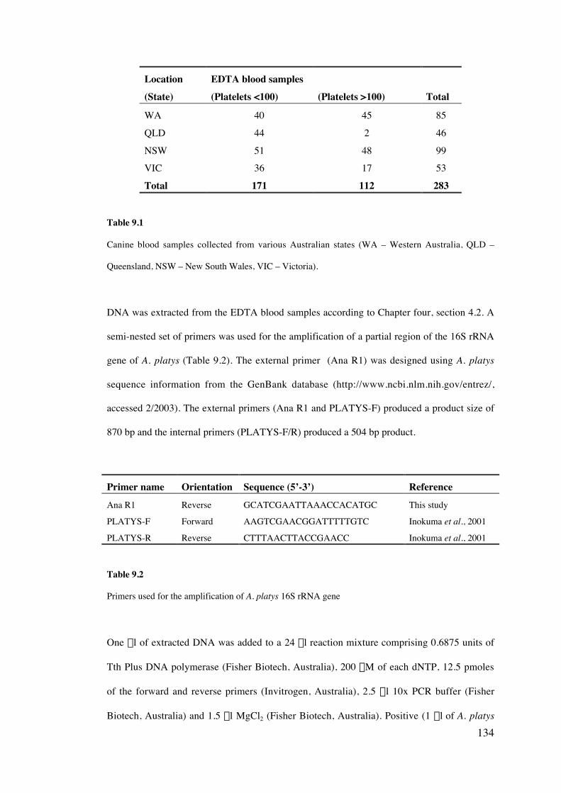
134
Location(State)
EDTA blood samples(Platelets <100) (Platelets >100) Total
WA 40 45 85
QLD 44 2 46
NSW 51 48 99VIC 36 17 53
Total 171 112 283
Table 9.1
Canine blood samples collected from various Australian states (WA – Western Australia, QLD –
Queensland, NSW – New South Wales, VIC – Victoria).
iii) DNA extraction and amplification
DNA was extracted from the EDTA blood samples according to Chapter four, section 4.2. A
semi-nested set of primers was used for the amplification of a partial region of the 16S rRNA
gene of A. platys (Table 9.2). The external primer (Ana R1) was designed using A. platys
sequence information from the GenBank database (http://www.ncbi.nlm.nih.gov/entrez/,
accessed 2/2003). The external primers (Ana R1 and PLATYS-F) produced a product size of
870 bp and the internal primers (PLATYS-F/R) produced a 504 bp product.
Primer name Orientation Sequence (5’-3’) ReferenceAna R1 Reverse GCATCGAATTAAACCACATGC This study
PLATYS-F Forward AAGTCGAACGGATTTTTGTC Inokuma et al., 2001
PLATYS-R Reverse CTTTAACTTACCGAACC Inokuma et al., 2001
Table 9.2
Primers used for the amplification of A. platys 16S rRNA gene
One ml of extracted DNA was added to a 24 ml reaction mixture comprising 0.6875 units of
Tth Plus DNA polymerase (Fisher Biotech, Australia), 200 mM of each dNTP, 12.5 pmoles
of the forward and reverse primers (Invitrogen, Australia), 2.5 ml 10x PCR buffer (Fisher
Biotech, Australia) and 1.5 ml MgCl2 (Fisher Biotech, Australia). Positive (1 ml of A. platys
135
DNA, Darwin, Australia) and negative (1 ml dH2O) control samples were included with each
set of PCR reactions.
Amplification was performed on a GeneAmp PCR system 2700 thermal cycler (Applied
Biosystems, USA). For the primary round of amplification, an initial activation step at 94 C
for 3 min, 62 C for 1 min and 72 C for 2 min, was followed by 45 cycles of amplification (94
C for 30 sec, 62 C for 20 sec and 72 C for 30 sec) and a final extension step of 72 C for 7
min for 25 µl reactions. The same conditions were followed for the secondary round of
amplification, except that the annealing temperature was 55 C, using 1µl of DNA template
from the primary reaction. Amplified DNA was electrophoresed and visualised according to
Chapter four, section 4.4.
iv) Determination of specificity of PCR assay
Primer specificity was determined by using the BLAST 2.1 program
(http://www.ncbi.nlm.nih.gov/BLAST/, accessed 2/2003). The specificity of the A. platys
PCR was also determined by testing the assay against DNA of Ehrlichia canis, Ehrlichia
equi, Bartonella vinsonii, Rickettsia rickettsia (kindly donated by Edward Breitschwerdt,
North Carolina State University, USA) and canine DNA. Anaplasma platys DNA from
Venezuela and Australia was used as positive control samples.
v) DNA sequencing and phylogenetic analysis
Amplified products were sequenced according to Chapter four, sections 4.5 – 4.8. Sequences
were aligned, together with addition sequences from the GenBank database (Table 9.3) using
Clustal W (Thompson et al., 1994).
Phylogenetic analysis was conducted on the basis of distance (Tajima and Nei, 1984)
algorithms and tree topologies were inferred using Neighbour joining (Saitou and Nei, 1987)
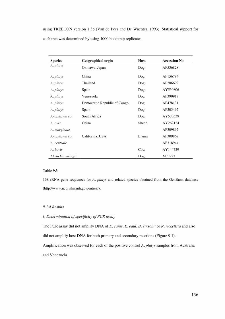
136
using TREECON version 1.3b (Van de Peer and De Wachter, 1993). Statistical support for
each tree was determined by using 1000 bootstrap replicates.
Species Geographical orgin Host Accession NoA. platys Okinawa, Japan Dog AF536828
A. platys China Dog AF156784
A. platys Thailand Dog AF286699
A. platys Spain Dog AY530806
A. platys Venezuela Dog AF399917
A. platys Democratic Republic of Congo Dog AF478131
A. platys Spain Dog AF303467
Anaplasma sp. South Africa Dog AY570539
A. ovis China Sheep AY262124
A. marginale AF309867
Anaplasma sp. California, USA Llama AF309867
A. centrale AF318944
A. bovis Cow AY144729
Ehrlichia ewingii Dog M73227
Table 9.3
16S rRNA gene sequences for A. platys and related species obtained from the GenBank database
(http://www.ncbi.nlm.nih.gov/entrez/).
9.1.4 Results
i) Determination of specificity of PCR assay
The PCR assay did not amplify DNA of E. canis, E. equi, B. vinsonii or R. rickettsia and also
did not amplify host DNA for both primary and secondary reactions (Figure 9.1).
Amplification was observed for each of the positive control A. platys samples from Australia
and Venezuela.
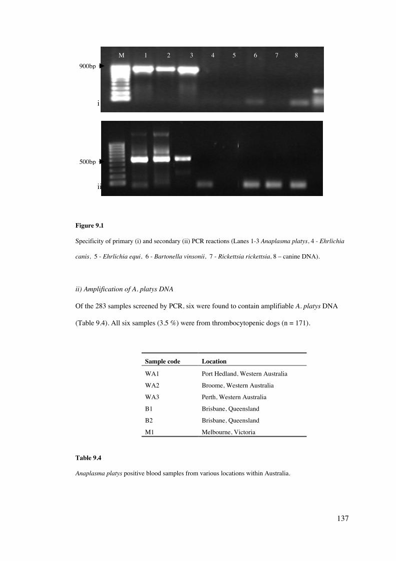
137
Figure 9.1
Specificity of primary (i) and secondary (ii) PCR reactions (Lanes 1-3 Anaplasma platys, 4 - Ehrlichia
canis, 5 - Ehrlichia equi, 6 - Bartonella vinsonii, 7 - Rickettsia rickettsia, 8 – canine DNA).
ii) Amplification of A. platys DNA
Of the 283 samples screened by PCR, six were found to contain amplifiable A. platys DNA
(Table 9.4). All six samples (3.5 %) were from thrombocytopenic dogs (n = 171).
Sample code Location
WA1 Port Hedland, Western Australia
WA2 Broome, Western Australia
WA3 Perth, Western Australia
B1 Brisbane, Queensland
B2 Brisbane, Queensland
M1 Melbourne, Victoria
Table 9.4
Anaplasma platys positive blood samples from various locations within Australia.
M 1 2 3 4 5 6 7 8
500bp
i
ii
900bp
138
iii) Genetic variation and phylogenetic analysis
DNA sequences from each of the six A. platys isolates were all 100 % homologous on the
basis of a partial region of the 16S rRNA gene. Sequences were also identical to those
obtained for isolates from the Northern Territory (refer to section 9.2). Australian sequences
were compared to isolates from other geographical locations from around the world using
phylogenetic analysis (Figure 9.2).
All A. platys isolates formed a single clade and were most closely related to Anaplasma
bovis and an unnamed Anaplasma sp from a dog in South Africa. Two individual groups of
A. platys was observed. The Australian isolates of A. platys clustered together with isolates
from China, Japan, Thailand, Spain and France, while a second group contained isolates
from Venezuela and The Democratic Republic of Congo. Statistic support for the separation
of the formation of a single clade for all A. platys isolates was significant (83 %), however
only moderate support was given for the separation of the A. platys isolates into two distinct
groups (60 –61 %).
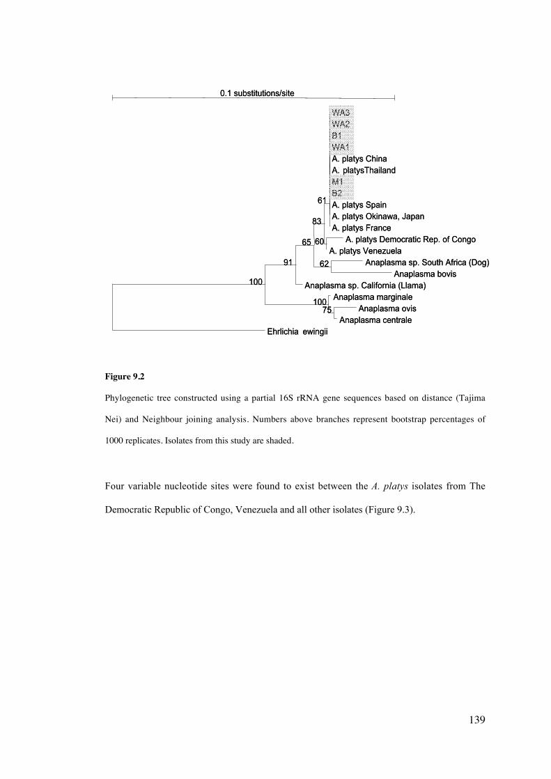
139
Figure 9.2
Phylogenetic tree constructed using a partial 16S rRNA gene sequences based on distance (Tajima
Nei) and Neighbour joining analysis. Numbers above branches represent bootstrap percentages of
1000 replicates. Isolates from this study are shaded.
Four variable nucleotide sites were found to exist between the A. platys isolates from The
Democratic Republic of Congo, Venezuela and all other isolates (Figure 9.3).
0.1 substitutions/site
Ehrlichia ewingii
Anaplasma sp. California (Llama)
A. platys France
Anaplasma marginale
WA3 WA2 B1 WA1 A. platys ChinaA. platysThailandM1 B2 A. platys SpainA. platys Okinawa, Japan
A. platys Democratic Rep. of CongoA. platys Venezuela
Anaplasma sp. South Africa (Dog)Anaplasma bovis
Anaplasma ovisAnaplasma centrale
100
91
65
83
61
100
60
62
75
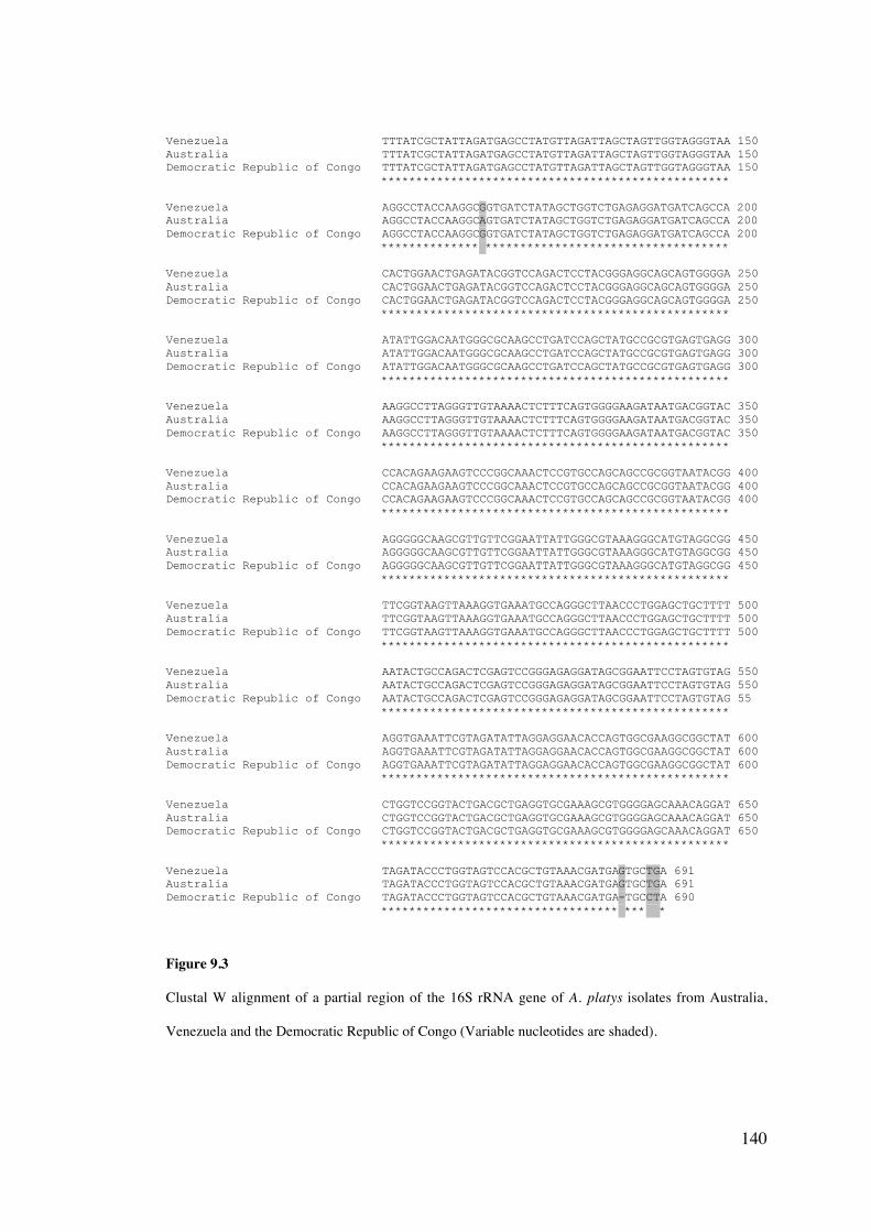
140
Venezuela TTTATCGCTATTAGATGAGCCTATGTTAGATTAGCTAGTTGGTAGGGTAA 150Australia TTTATCGCTATTAGATGAGCCTATGTTAGATTAGCTAGTTGGTAGGGTAA 150Democratic Republic of Congo TTTATCGCTATTAGATGAGCCTATGTTAGATTAGCTAGTTGGTAGGGTAA 150 **************************************************
Venezuela AGGCCTACCAAGGCGGTGATCTATAGCTGGTCTGAGAGGATGATCAGCCA 200Australia AGGCCTACCAAGGCAGTGATCTATAGCTGGTCTGAGAGGATGATCAGCCA 200Democratic Republic of Congo AGGCCTACCAAGGCGGTGATCTATAGCTGGTCTGAGAGGATGATCAGCCA 200 ************** ***********************************
Venezuela CACTGGAACTGAGATACGGTCCAGACTCCTACGGGAGGCAGCAGTGGGGA 250Australia CACTGGAACTGAGATACGGTCCAGACTCCTACGGGAGGCAGCAGTGGGGA 250Democratic Republic of Congo CACTGGAACTGAGATACGGTCCAGACTCCTACGGGAGGCAGCAGTGGGGA 250 **************************************************
Venezuela ATATTGGACAATGGGCGCAAGCCTGATCCAGCTATGCCGCGTGAGTGAGG 300Australia ATATTGGACAATGGGCGCAAGCCTGATCCAGCTATGCCGCGTGAGTGAGG 300Democratic Republic of Congo ATATTGGACAATGGGCGCAAGCCTGATCCAGCTATGCCGCGTGAGTGAGG 300 **************************************************
Venezuela AAGGCCTTAGGGTTGTAAAACTCTTTCAGTGGGGAAGATAATGACGGTAC 350Australia AAGGCCTTAGGGTTGTAAAACTCTTTCAGTGGGGAAGATAATGACGGTAC 350Democratic Republic of Congo AAGGCCTTAGGGTTGTAAAACTCTTTCAGTGGGGAAGATAATGACGGTAC 350 **************************************************
Venezuela CCACAGAAGAAGTCCCGGCAAACTCCGTGCCAGCAGCCGCGGTAATACGG 400Australia CCACAGAAGAAGTCCCGGCAAACTCCGTGCCAGCAGCCGCGGTAATACGG 400Democratic Republic of Congo CCACAGAAGAAGTCCCGGCAAACTCCGTGCCAGCAGCCGCGGTAATACGG 400 **************************************************
Venezuela AGGGGGCAAGCGTTGTTCGGAATTATTGGGCGTAAAGGGCATGTAGGCGG 450Australia AGGGGGCAAGCGTTGTTCGGAATTATTGGGCGTAAAGGGCATGTAGGCGG 450Democratic Republic of Congo AGGGGGCAAGCGTTGTTCGGAATTATTGGGCGTAAAGGGCATGTAGGCGG 450 **************************************************
Venezuela TTCGGTAAGTTAAAGGTGAAATGCCAGGGCTTAACCCTGGAGCTGCTTTT 500Australia TTCGGTAAGTTAAAGGTGAAATGCCAGGGCTTAACCCTGGAGCTGCTTTT 500Democratic Republic of Congo TTCGGTAAGTTAAAGGTGAAATGCCAGGGCTTAACCCTGGAGCTGCTTTT 500 **************************************************
Venezuela AATACTGCCAGACTCGAGTCCGGGAGAGGATAGCGGAATTCCTAGTGTAG 550Australia AATACTGCCAGACTCGAGTCCGGGAGAGGATAGCGGAATTCCTAGTGTAG 550Democratic Republic of Congo AATACTGCCAGACTCGAGTCCGGGAGAGGATAGCGGAATTCCTAGTGTAG 55 **************************************************
Venezuela AGGTGAAATTCGTAGATATTAGGAGGAACACCAGTGGCGAAGGCGGCTAT 600Australia AGGTGAAATTCGTAGATATTAGGAGGAACACCAGTGGCGAAGGCGGCTAT 600Democratic Republic of Congo AGGTGAAATTCGTAGATATTAGGAGGAACACCAGTGGCGAAGGCGGCTAT 600 **************************************************
Venezuela CTGGTCCGGTACTGACGCTGAGGTGCGAAAGCGTGGGGAGCAAACAGGAT 650Australia CTGGTCCGGTACTGACGCTGAGGTGCGAAAGCGTGGGGAGCAAACAGGAT 650Democratic Republic of Congo CTGGTCCGGTACTGACGCTGAGGTGCGAAAGCGTGGGGAGCAAACAGGAT 650 **************************************************
Venezuela TAGATACCCTGGTAGTCCACGCTGTAAACGATGAGTGCTGA 691Australia TAGATACCCTGGTAGTCCACGCTGTAAACGATGAGTGCTGA 691Democratic Republic of Congo TAGATACCCTGGTAGTCCACGCTGTAAACGATGA-TGCCTA 690 ********************************** *** *
Figure 9.3
Clustal W alignment of a partial region of the 16S rRNA gene of A. platys isolates from Australia,
Venezuela and the Democratic Republic of Congo (Variable nucleotides are shaded).
141
9.1.5 Discussion
i) Distribution and prevalence
This study reports for the first time, the presence of A. platys in Western Australia,
Queensland and Victoria. Unfortunately, samples were unavailable from South Australia and
Tasmania, preventing assessment of infection of dogs within these States. Previously, A.
platys has only been detected in dogs from the Northern Territory, in both central (Brown et
al., 2001; Brown et al., 2005) and northern (Jefferies, 2001) geographical regions. It can now
be assumed that A. platys is distributed throughout Australia and is likely to be a
consequence of the extensive distribution of R. sanguineus, the suspected vector of this
pathogen and unrestricted movement of dogs around the country.
Whilst R. sanguineus has not been successfully proven to transmit A. platys experimentally
(Simpson et al., 1991) DNA of A. platys has been detected using PCR within semi-engorged
ticks (Sanogo et al., 2003; Brown et al., 2005). It is difficult to ascertain whether the A.
platys DNA detected was simply a reflection of the ingestion of infected blood or whether A.
platys was actually within the haemolymph of the tick. Limited study has assessed the
distribution of R. sanguineus in Australia, however reports suggest that this tick species is
prevalent throughout northern Western Australia, the Northern Territory, Queensland and
northern New South Wales (Roberts, 1970). While large populations of this tick species are
found within tropical to subtropical regions of Australia, there have also been reports in
more temperate climatic regions in southern Australia including urban areas such as Perth
(De Chaneet, 1976) and Melbourne (Roberts, 1970).
Brown et al. (2005) also postulated that the dog chewing louse (Heterodoxus spiniger) may
be a potential vector of A. platys or may contribute to the spread of this pathogen by
mechanical transmission. Transmission of A. platys is likely to be multifactorial, including
the possibility of transplacental transmission (Brown et al., 2005) and requires additional
research to better understand the epidemiology of this organism.
142
The prevalence of A. platys infection among thrombocytopenic dogs sampled during the
course of this pilot study was 3.5 % (6/171). This is much lower than the 27.8 % (5/18)
prevalence observed in thrombocytopenic dogs in Darwin, Northern Territory (Jefferies,
2001). By selectively targeting a sub-sample of the normal population that presented with
thrombocytopenia, it was anticipated that the likelihood of detecting A. platys would be
increased. The prevalence of A. platys infection may therefore be much lower in the general
dog population of Australia. This was reflected by the absence of infection in all non-
thrombocytopenic blood samples tested, however the number of samples tested, especially
from Queensland, were very small and not statistically significant. The results of this study
however may not be a true reflection of the epidemiology of this disease in the general dog
population of Australia, as while all dogs that were found to have A. platys were within the
thrombocytopenic group, previous studies have suggested that many dogs infected with this
pathogen do not present with thrombocytopenia and indeed may not show any signs of
illness (Brown et al., 2001; Jefferies, 2001; section 9.2). It is also difficult to definitively
correlate the thrombocytopenia observed, with A. platys infection and may have been a
consequence of an unrelated disorder such as immune mediated thrombocytopenia. A more
accurate investigation into the prevalence of A. platys in dog populations in Australia
therefore requires an increased sample size and sampling a much broader population,
including obtaining detailed epidemiological and haematological data from each dog
sampled.
Samples were also only collected from pathology laboratories of major Australian cities,
disproportionately selecting samples from urban rather than rural dog populations. Dogs in
rural areas could potentially have a higher rate of A. platys infection than their urban
counterparts and should be investigated further.
143
ii) Genetic characterisation
This study has also revealed the existence of genetic differences between isolates of A. platys
on the basis of the highly conserved, 16S rRNA gene and supports isolate variation reported
by Mathew et al. (1997). The genetic variation observed between different isolates has not
previously been correlated to geographic origin and the significance of this variation is not
yet understood. The variability of clinical signs and pathogenesis of this infection sometimes
observed in separate geographic regions of the world (Harrus et al., 1997; Sainz et al., 1999)
may be explained by different genotypes and requires further investigation. No genetic
variation in the 16S rRNA gene was observed between isolates from different regions within
Australia and indeed, from multiple other countries. Characterisation on the basis of more
variable gene loci is therefore required to better elucidate phylogeographical relationships
among these isolates. Potential gene candidates include the citrate synthase gene (Raux et
al., 1997) and the major surface protein genes (de la Fuente et al., 2002).
iii) Conclusion
This study has revealed that A. platys is likely to have a widespread distribution throughout
Australia, extending beyond the Northern Territory and including Western Australia,
Queensland and Victoria. All dogs with A. platys infection in this study were
thrombocytopenic. Therefore, the possibility of A. platys infection should be considered by
veterinarians, Australia-wide, when presented with cases of idiopathic thrombocytopenia.
The prevalence of A. platys infection in Australia remains unknown and conformation of the
vector responsible for the transmission of this organism is necessary and would facilitate
future studies.
144
9.2 Anaplasma platys and Babesia canis vogeli infections in military German Shepherds
from northern Australia
9.2.1 Introduction
Canine infections of A. platys are frequently associated with thrombocytopenia, which
occurs in cycles of approximately 7-14 day intervals (Harrus et al., 1997). Canine infectious
cyclic thrombocytopenia is often reported to be subclinical, however this is disputed by other
studies of infected dogs in southern Europe and the Middle East, which suggest that
infection with A. platys results in weight loss, fever and depression (Harrus et al., 1997;
Sainz et al., 1999). Such variable observations may be explained by differences attributable
to the strain of the organism, the immune status of the host and by co-infection by one or
more other organisms. No previous studies have investigated the clinical and pathological
manifestations of A. platys infection in Australia.
Doxycycline has been suggested as an effective drug in the elimination of thrombocytopenia
associated with A. platys infection (Bradfield et al., 1996). Whether this drug therapy can
produce total eradication of A. platys has not been proven and therefore requires additional
investigation.
Anaplasma platys may also co-infect hosts with other pathogens such as Ehrlichia, Babesia
and Hepatozoon spp. and has been reported in the USA, Thailand and (Kordick et al. 1999;
Hua et al. 2000). Preliminary study suggested that co-infection of A. platys and B. canis does
occur in northern Australia (Jefferies, 2001). Limited research however, has assessed the
significance of co-infection and whether it produces any changes in the pathogenesis of these
diseases.
145
9.2.1 Aims
• To investigate infections of A. platys and B. canis within clinically normal, military
German Shepherds
• To assess the efficacy of doxycycline against A. platys infections
9.2.2 Materials and Methods
i) Dogs sampled
Blood samples were taken from fourteen German shepherd dogs in Darwin, Northern
Australia, that were used for military purposes by the Royal Australian Airforce (RAAF).
Blood was PCR screened for the presence of Anaplasma platys and for co-infection with
Babesia species.
Eight of these dogs (six PCR positive for A. platys, two positive for Babesia and two not
infected with A. platys) were involved in a further nine-week study. Blood was taken weekly
and the packed cell volume (PCV), total white blood cells (WBC), platelet count and mean
platelet volume (MPV) were calculated for each blood sample. Thin blood smears were also
prepared for each sample and stained with a modified Giemsa Wright stain. Three dogs (all
PCR positive for A. platys and one also co-infected with Babesia) were treated with
doxycycline at 10 mg/kg once daily for 14 days. The weight, body temperature and food
consumption of each of the dogs was recorded weekly and a score was given weekly for the
dogs' ability and willingness to exercise through a standard agility training course (0 =
unable to excise, 1-5 = reduced willingness to exercise, 5-8 = willingness to exercise and 8-
10 = very willing and eager to exercise). A normal value was considered to be in the range of
5-10.
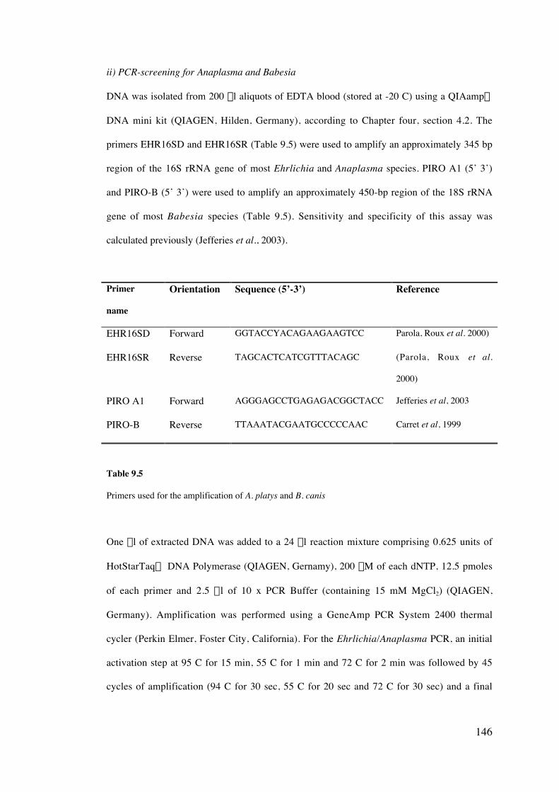
146
ii) PCR-screening for Anaplasma and Babesia
DNA was isolated from 200 ml aliquots of EDTA blood (stored at -20 C) using a QIAamp‚
DNA mini kit (QIAGEN, Hilden, Germany), according to Chapter four, section 4.2. The
primers EHR16SD and EHR16SR (Table 9.5) were used to amplify an approximately 345 bp
region of the 16S rRNA gene of most Ehrlichia and Anaplasma species. PIRO A1 (5’ 3’)
and PIRO-B (5’ 3’) were used to amplify an approximately 450-bp region of the 18S rRNA
gene of most Babesia species (Table 9.5). Sensitivity and specificity of this assay was
calculated previously (Jefferies et al., 2003).
Primer
name
Orientation Sequence (5’-3’) Reference
EHR16SD Forward GGTACCYACAGAAGAAGTCC Parola, Roux et al. 2000)
EHR16SR Reverse TAGCACTCATCGTTTACAGC (Parola, Roux et al.
2000)
PIRO A1 Forward AGGGAGCCTGAGAGACGGCTACC Jefferies et al, 2003
PIRO-B Reverse TTAAATACGAATGCCCCCAAC Carret et al, 1999
Table 9.5
Primers used for the amplification of A. platys and B. canis
One ml of extracted DNA was added to a 24 ml reaction mixture comprising 0.625 units of
HotStarTaq‘ DNA Polymerase (QIAGEN, Gernamy), 200 mM of each dNTP, 12.5 pmoles
of each primer and 2.5 ml of 10 x PCR Buffer (containing 15 mM MgCl2) (QIAGEN,
Germany). Amplification was performed using a GeneAmp PCR System 2400 thermal
cycler (Perkin Elmer, Foster City, California). For the Ehrlichia/Anaplasma PCR, an initial
activation step at 95 C for 15 min, 55 C for 1 min and 72 C for 2 min was followed by 45
cycles of amplification (94 C for 30 sec, 55 C for 20 sec and 72 C for 30 sec) and a final
147
extension step of 7 min at 72 C. Amplification conditions for the Babesia PCR were the
same except for an increased annealing temperature of 62 C.
iii) Sequencing of amplified products
The amplified products for both A. platys and B. canis vogeli were purified and sequenced
according to Chapter four, sections 4.5 - 4.8.
iv) Statistical analyses
Statistical relationships between A. platys infection and selected clinical data were assessed
using SPSS v11.0 (SPSS, Chicago, IL). Data was tested for normality using a test of
homogeneity of variances before using a oneway ANOVA to test for significance between
the presence of infection and clinical data. A Mann-Whitney test was also used to assess the
relationship between Babesia infection and platelet number. A p-value of less than 0.05 was
considered to indicate statistical significance.
9.2.3 Results
i) Presence of Anaplasma and Babesia DNA in blood samples
Six of the 14 dogs screened were PCR positive for Anaplasma / Ehrlichia. All blood samples
were microscopy negative for platelet inclusions. Sequencing of the amplified product
confirmed the presence of A. platys in each of the samples (100% homologous to Anaplasma
platys strain Okinawa, GenBank accession number AF536828). Three of the dogs were PCR
positive for Babesia and two were positive for both Anaplasma and Babesia. Sequencing of
the amplified Babesia PCR product confirmed the presence of Babesia canis vogeli DNA in
the samples (Details of the molecular characterisation of B. canis isolates from Australia are
described in Chapter ten).
Blood samples from eight of the 14 dogs (six PCR positive for A. platys, two positive for
Babesia and two infection free) showed variation in whether they were PCR positive for A.
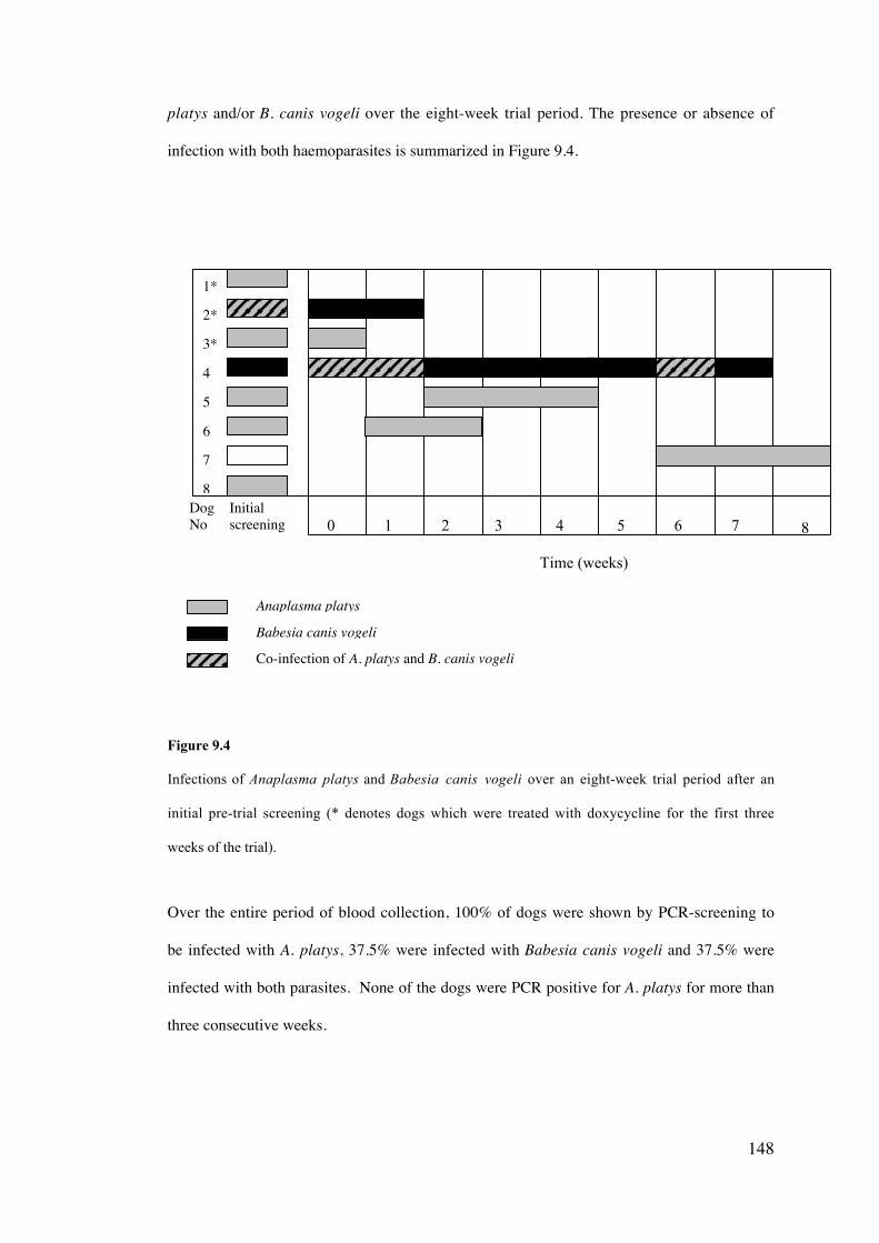
148
platys and/or B. canis vogeli over the eight-week trial period. The presence or absence of
infection with both haemoparasites is summarized in Figure 9.4.
0 1 2 3 4 5 6 7
Time (weeks)
Figure 9.4
Infections of Anaplasma platys and Babesia canis vogeli over an eight-week trial period after an
initial pre-trial screening (* denotes dogs which were treated with doxycycline for the first three
weeks of the trial).
Over the entire period of blood collection, 100% of dogs were shown by PCR-screening to
be infected with A. platys, 37.5% were infected with Babesia canis vogeli and 37.5% were
infected with both parasites. None of the dogs were PCR positive for A. platys for more than
three consecutive weeks.
Anaplasma platys
Babesia canis vogeli
Co-infection of A. platys and B. canis vogeli
1*
2*
3*
4
5
6
7
8DogNo
Initialscreening 8
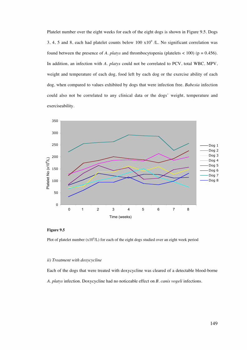
149
Platelet number over the eight weeks for each of the eight dogs is shown in Figure 9.5. Dogs
3, 4, 5 and 8, each had platelet counts below 100 x109 /L. No significant correlation was
found between the presence of A. platys and thrombocytopenia (platelets < 100) (p = 0.456).
In addition, an infection with A. platys could not be correlated to PCV, total WBC, MPV,
weight and temperature of each dog, food left by each dog or the exercise ability of each
dog, when compared to values exhibited by dogs that were infection free. Babesia infection
could also not be correlated to any clinical data or the dogs’ weight, temperature and
exerciseability.
0
50
100
150
200
250
300
350
1 2 3 4 5 6 7 8 9
Dog 1
Dog 2
Dog 3
Dog 4
Dog 5
Dog 6
Dog 7
Dog 8
Figure 9.5
Plot of platelet number (x109/L) for each of the eight dogs studied over an eight week period
ii) Treatment with doxycycline
Each of the dogs that were treated with doxycycline was cleared of a detectable blood-borne
A. platys infection. Doxycycline had no noticeable effect on B. canis vogeli infections.
0 1 2 3 4 5 6 7 8
Pla
tele
t No
(x10
/L)
Time (weeks)
150
9.2.3 Discussion
The results of this study suggest that chronic or repeat infections with A. platys in dogs in
Australia are generally benign and that this organism may not be a significant pathogen. This
concurs with previous studies in other parts of the world where detection of A. platys by
PCR has not correlated with any major clinical signs (Chang et al., 1996; Brown et al., 2001;
Inokuma et al., 2002). The intermittent detection of A. platys was suggestive of the cyclic
nature of this infection and is in agreement with other studies that have reported cycles of
infection occuring every 7– 14 days (Harrus et al., 1997). Daily sampling would be required
to investigate the cyclic nature of the parasitaemia but has been previously studied in other
countries (Chang and Pan, 1996; Chang et al., 1996; Chang et al., 1997).
Interestingly, the presence of A. platys DNA could not be correlated with thrombocytopenia.
This may be a reflection of the low sample size influencing the statistical significance,
inaccurate platelet counts or possibly related to the chronic stage of these infections.
Furthermore, as blood samples were only collected once a week and not daily, it is possible
that blood was not taken during periods of thrombocytopenia.
It still remains unknown, however, whether dogs in Australia exposed to A. platys for the
first time produce clinical signs of increased severity. It could be speculated that the dogs
within this study had been exposed regularly to infected ticks and may have developed some
degree of immunity, hence the absence of ill health. Only by using experimental infections
would it be possible to determine the pathogenesis of A. platys in previously naïve dogs in
Australia.
Other studies have suggested the existence of different strains of A. platys, with one
producing no detectable clinical signs and the other causing anorexia, depression, lethargy,
lymphadenomegaly and fever (Harrus et al., 1997). It is possible that the difference in
clinical signs may also be due to the stage of infection, age, breed, the immune status of the
animal or the genotype of A. platys.
151
Treatment with doxycycline appeared to reduce the infection to an undetectable level after a
week, however some untreated dogs also developed undetectable DNA levels of A. platys. It
is therefore difficult to determine whether these results are a reflection of the cyclic nature of
this disease or an effect of doxycycline. It remains inconclusive whether doxycycline is an
effective treatment for A. platys infection in Australia. One previous study has suggested that
tetracycline is a useful therapy for A. platys infection, however, the duration of treatment is
dependent upon the stage of illness, with chronic infections requiring a longer period of drug
therapy (Chang and Pan, 1997). Doxycycline has also been reported to be a more effective
drug therapy than tetracycline (Chang et al., 1997).
This study is also the first known report of co-infection with A. platys and B. canis vogeli in
dogs in Australia. Both parasites have been reported to be transmitted by the tick R.
sanguineus (A. platys not experimentally confirmed), which is widely distributed across
northern Australia. Babesia canis vogeli in Australia has previously been reported to be
mildly pathogenic, causing severe anaemia and sometimes death in young dogs but may
exist in a state of premunity or carrier state in adult dogs (Irwin and Hutchinson, 1991). Dogs
infected with both A. platys and B. canis vogeli showed no detectable pathogenesis, however
this may again be a reflection of the immune status of the animals. Co-infection in naïve
animals may give rise to detectable clinical signs and pathogenesis and therefore requires
further investigation.
Overall, the results from this study suggest that A. platys infection may be quite prevalent in
dogs in northern Australia, however chronic or repeat infections are unlikely to cause any
major symptoms. Regular tick prevention treatment should be considered as the most
effective method of controlling infections of both A. platys and B. canis vogeli.
152
Molecular characterisation of the Australian canine Babesia spp.
and phylogeographical relationships among worldwide isolates of B.
canis and B. gibsoni
10.1 Introduction
A total of six individual species of piroplasm have been reported to infect dogs, these being
B. canis, B. gibsoni, Babesia sp. (North Carolina), B. conradae, T. annae and T. equi. The
species, B. canis is comprised of four subspecies; three of which; B. canis canis, B. canis
vogeli and B. canis rossi, are each considered to be separate species by some (Uilenberg et
al., 1989; Zahler et al., 1998; Carret et al., 1999), yet have never been taxonomically
elevated. The fourth subspecies, B. canis presentii, has only been reported in cats (Baneth et
al., 2004). Limited study has investigated the concept of the species within the canine
piroplasms and the levels of inter- and intra-species genetic variation that exist within
established species.
Multiple isolates of canine piroplasm have been defined as new species on the basis of
molecular characterisation (Kjemtrup et al., 1999; Zahler et al., 2000; Birkenheuer et al.,
2004), yet the level of genetic variation used to define a species has not been established.
Defining levels of inter-species variation would thus limit the current difficulty and
confusion that exists when describing a new species or genotype. Defining levels of intra-
species genetic variation within individual species of piroplasmid is also important and is
imperative to many fields of research, including diagnosis, epidemiology, chemotherapy,
systematics and taxonomy. PCR is increasingly becoming a widely used detection technique
CHAPTER TEN
153
and accurate amplification of DNA of the target species is paramount. The existence of
genetic variation may also be useful in determining the geographic origin of an isolate,
phylogeographical relationships and also explaining possible variations in pathogenesis and
life cycle characteristics.
Within Australia, both B. canis (Chapter six and nine) and B. gibsoni (Chapters six and
seven) have been reported and provided the basis for the molecular characterisation and
phylogeography described in this chapter. Isolates of both species were collected from
different geographical locations within Australia and from around the world, and were used
to investigate molecular variation, phylogeny and taxonomy.
10.2 Aims
• To collect isolates of canine piroplasms from various geographical locations
worldwide.
• To molecularly characterise B. gibsoni and B. canis vogeli isolates from dogs in
Australia on the basis of 18S rRNA gene, the ITS 1, 5.8S rRNA and ITS 2 loci and
HSP 70 gene.
• To investigate levels of intra-species genetic variation using the 18S rRNA gene and
ITS 1, 5.8S rRNA, ITS 2 loci and HSP 70 gene, among world-wide isolates of B.
gibsoni and B. canis and determine levels of inter-species genetic variation among
the canine piroplasms and other established species of the Piroplasmida
• To review the taxonomic status of the B. canis subspecies
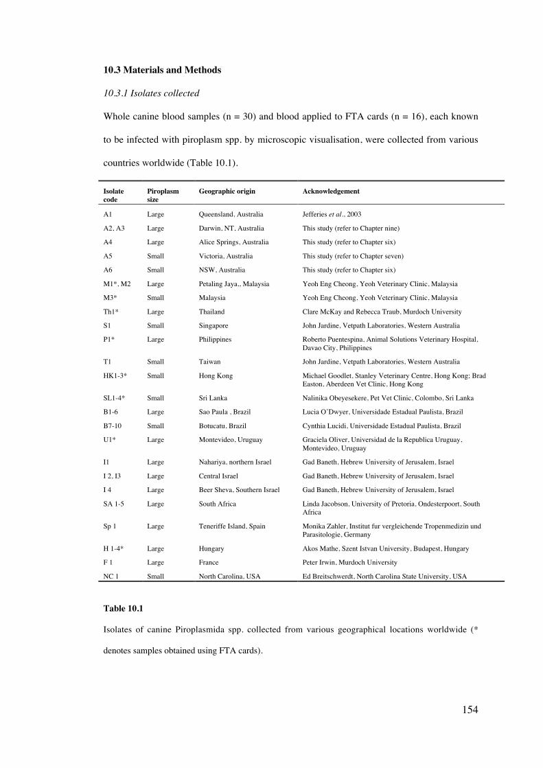
154
10.3 Materials and Methods
10.3.1 Isolates collected
Whole canine blood samples (n = 30) and blood applied to FTA cards (n = 16), each known
to be infected with piroplasm spp. by microscopic visualisation, were collected from various
countries worldwide (Table 10.1).
Isolatecode
Piroplasmsize
Geographic origin Acknowledgement
A1 Large Queensland, Australia Jefferies et al., 2003
A2, A3 Large Darwin, NT, Australia This study (refer to Chapter nine)
A4 Large Alice Springs, Australia This study (refer to Chapter six)
A5 Small Victoria, Australia This study (refer to Chapter seven)
A6 Small NSW, Australia This study (refer to Chapter six)
M1*, M2 Large Petaling Jaya,, Malaysia Yeoh Eng Cheong, Yeoh Veterinary Clinic, Malaysia
M3* Small Malaysia Yeoh Eng Cheong, Yeoh Veterinary Clinic, Malaysia
Th1* Large Thailand Clare McKay and Rebecca Traub, Murdoch University
S1 Small Singapore John Jardine, Vetpath Laboratories, Western Australia
P1* Large Philippines Roberto Puentespina, Animal Solutions Veterinary Hospital,Davao City, Philippines
T1 Small Taiwan John Jardine, Vetpath Laboratories, Western Australia
HK1-3* Small Hong Kong Michael Goodlet, Stanley Veterinary Centre, Hong Kong; BradEaston, Aberdeen Vet Clinic, Hong Kong
SL1-4* Small Sri Lanka Nalinika Obeyesekere, Pet Vet Clinic, Colombo, Sri Lanka
B1-6 Large Sao Paula , Brazil Lucia O’Dwyer, Universidade Estadual Paulista, Brazil
B7-10 Small Botucatu, Brazil Cynthia Lucidi, Universidade Estadual Paulista, Brazil
U1* Large Montevideo, Uruguay Graciela Oliver, Universidad de la Republica Uruguay,Montevideo, Uruguay
I1 Large Nahariya, northern Israel Gad Baneth, Hebrew University of Jerusalem, Israel
I 2, I3 Large Central Israel Gad Baneth, Hebrew University of Jerusalem, Israel
I 4 Large Beer Sheva, Southern Israel Gad Baneth, Hebrew University of Jerusalem, Israel
SA 1-5 Large South Africa Linda Jacobson, University of Pretoria, Ondesterpoort, SouthAfrica
Sp 1 Large Teneriffe Island, Spain Monika Zahler, Institut fur vergleichende Tropenmedizin undParasitologie, Germany
H 1-4* Large Hungary Akos Mathe, Szent Istvan University, Budapest, Hungary
F 1 Large France Peter Irwin, Murdoch University
NC 1 Small North Carolina, USA Ed Breitschwerdt, North Carolina State University, USA
Table 10.1
Isolates of canine Piroplasmida spp. collected from various geographical locations worldwide (*
denotes samples obtained using FTA cards).
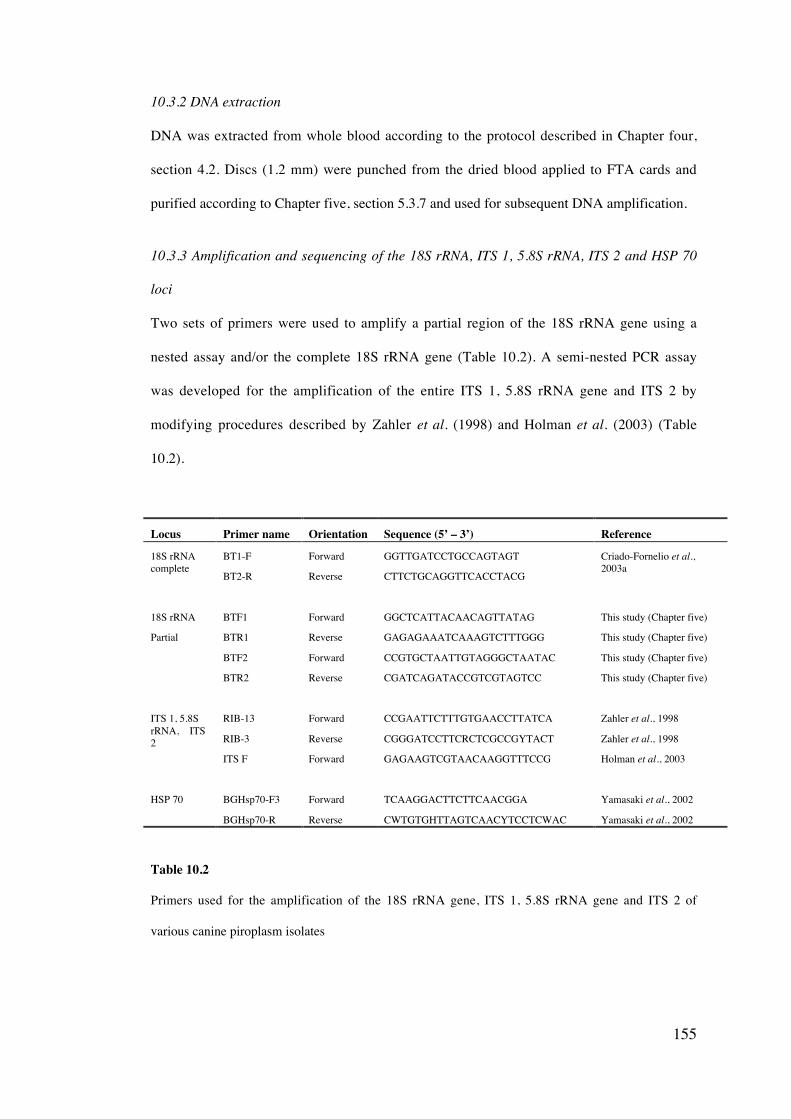
155
10.3.2 DNA extraction
DNA was extracted from whole blood according to the protocol described in Chapter four,
section 4.2. Discs (1.2 mm) were punched from the dried blood applied to FTA cards and
purified according to Chapter five, section 5.3.7 and used for subsequent DNA amplification.
10.3.3 Amplification and sequencing of the 18S rRNA, ITS 1, 5.8S rRNA, ITS 2 and HSP 70
loci
Two sets of primers were used to amplify a partial region of the 18S rRNA gene using a
nested assay and/or the complete 18S rRNA gene (Table 10.2). A semi-nested PCR assay
was developed for the amplification of the entire ITS 1, 5.8S rRNA gene and ITS 2 by
modifying procedures described by Zahler et al. (1998) and Holman et al. (2003) (Table
10.2).
Locus Primer name Orientation Sequence (5’ – 3’) Reference
18S rRNAcomplete
BT1-F
BT2-R
Forward
Reverse
GGTTGATCCTGCCAGTAGT
CTTCTGCAGGTTCACCTACG
Criado-Fornelio et al.,2003a
18S rRNA BTF1 Forward GGCTCATTACAACAGTTATAG This study (Chapter five)
Partial BTR1
BTF2
BTR2
Reverse
Forward
Reverse
GAGAGAAATCAAAGTCTTTGGG
CCGTGCTAATTGTAGGGCTAATAC
CGATCAGATACCGTCGTAGTCC
This study (Chapter five)
This study (Chapter five)
This study (Chapter five)
ITS 1, 5.8SrRNA, ITS2
RIB-13
RIB-3
ITS F
Forward
Reverse
Forward
CCGAATTCTTTGTGAACCTTATCA
CGGGATCCTTCRCTCGCCGYTACT
GAGAAGTCGTAACAAGGTTTCCG
Zahler et al., 1998
Zahler et al., 1998
Holman et al., 2003
HSP 70 BGHsp70-F3 Forward TCAAGGACTTCTTCAACGGA Yamasaki et al., 2002
BGHsp70-R Reverse CWTGTGHTTAGTCAACYTCCTCWAC Yamasaki et al., 2002
Table 10.2
Primers used for the amplification of the 18S rRNA gene, ITS 1, 5.8S rRNA gene and ITS 2 of
various canine piroplasm isolates
156
For each PCR reaction, 1ml of extracted DNA or a purified 1.2 mm FTA disc was added to a
24 ml reaction mixture comprised of 0.6875 units of Tth Plus DNA polymerase (Fisher
Biotech, Australia), 200 mM of each dNTP (Fisher Biotech, Australia), 12.5 pmoles of each
primer, 2.5 m l of 10x PCR buffer (Fisher Biotech, Australia) and 1.5 m l of MgCL2.
Amplification was performed using a GeneAmp PCR thermal cycler (Perkin Elmer,
California, USA). Cycling conditions for BTF1/R1 and BTF2/R2 are described in Chapter
five, section 5.3.3. Cycling conditions for each of the other primer sets were identical except
for different annealing temperatures, which were 58 C for BT-F/BT2-R, 60 C for RIB 3/RIB
13, 65 C for RIB 3/ITS F and 60 C for BGHsp70 F3/R.
Amplified DNA was purified and sequenced according to the protocol described in Chapter
four, section 4.5 – 4.8.
10.3.4 Sequence alignment and phylogenetic analysis
Sequences obtained for each gene, in addition to sequences for the 18S rRNA gene (Table
10.3), the ITS1, 5.8S rRNA gene and ITS2 (Table 10.4) and HSP 70 gene (Table 10.5)
obtained from the GenBank database (http://www.ncbi.nlm.nih.gov/entrez/) were aligned
using Clustal W (Thompson et al., 1994). A partial 18S rRNA gene sequence of Babesia
kiwiensis (Down, 2004) was also included for phylogenetic analysis. Phylogenetic analysis
was conducted on the basis of distance (Tajima and Nei, 1984) algorithms and tree
topologies were inferred using Neighbour joining (Saitou and Nei, 1987) using TREECON
version 1.3b (Van de Peer and De Watcher, 1993). Statistical support for each tree was
determined by using 1000 bootstrap replicates. Percentage identity between isolates and
species was calculated based on Kimura 2-parameter distance method using MEGA v.3
(Kumar et al., 2004).
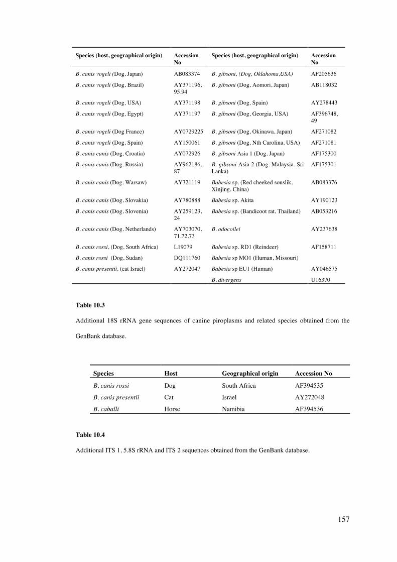
157
Species (host, geographical origin) AccessionNo
Species (host, geographical origin) AccessionNo
B. canis vogeli (Dog, Japan) AB083374 B. gibsoni, (Dog, Oklahoma,USA) AF205636
B. canis vogeli (Dog, Brazil) AY371196,95,94
B. gibsoni (Dog, Aomori, Japan) AB118032
B. canis vogeli (Dog, USA) AY371198 B. gibsoni (Dog, Spain) AY278443
B. canis vogeli (Dog, Egypt) AY371197 B. gibsoni (Dog, Georgia, USA) AF396748,49
B. canis vogeli (Dog France) AY0729225 B. gibsoni (Dog, Okinawa, Japan) AF271082
B. canis vogeli (Dog, Spain) AY150061 B. gibsoni (Dog, Nth Carolina, USA) AF271081
B. canis canis (Dog, Croatia) AY072926 B. gibsoni Asia 1 (Dog, Japan) AF175300
B. canis canis (Dog, Russia) AY962186,87
B. gibsoni Asia 2 (Dog, Malaysia, SriLanka)
AF175301
B. canis canis (Dog, Warsaw) AY321119 Babesia sp. (Red cheeked souslik,Xinjing, China)
AB083376
B. canis canis (Dog, Slovakia) AY780888 Babesia sp. Akita AY190123
B. canis canis (Dog, Slovenia) AY259123,24
Babesia sp. (Bandicoot rat, Thailand) AB053216
B. canis canis (Dog, Netherlands) AY703070,71,72,73
B. odocoilei AY237638
B. canis rossi, (Dog, South Africa) L19079 Babesia sp. RD1 (Reindeer) AF158711
B. canis rossi (Dog, Sudan) DQ111760 Babesia sp MO1 (Human, Missouri)
B. canis presentii, (cat Israel) AY272047 Babesia sp EU1 (Human) AY046575
B. divergens U16370
Table 10.3
Additional 18S rRNA gene sequences of canine piroplasms and related species obtained from the
GenBank database.
Species Host Geographical origin Accession No
B. canis rossi Dog South Africa AF394535
B. canis presentii Cat Israel AY272048
B. caballi Horse Namibia AF394536
Table 10.4
Additional ITS 1, 5.8S rRNA and ITS 2 sequences obtained from the GenBank database.
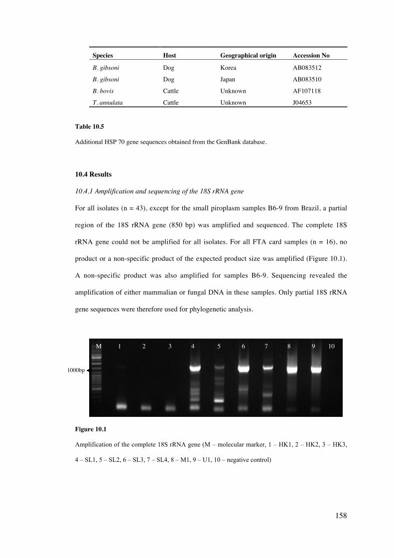
158
Species Host Geographical origin Accession No
B. gibsoni Dog Korea AB083512
B. gibsoni Dog Japan AB083510
B. bovis Cattle Unknown AF107118
T. annulata Cattle Unknown J04653
Table 10.5
Additional HSP 70 gene sequences obtained from the GenBank database.
10.4 Results
10.4.1 Amplification and sequencing of the 18S rRNA gene
For all isolates (n = 43), except for the small piroplasm samples B6-9 from Brazil, a partial
region of the 18S rRNA gene (850 bp) was amplified and sequenced. The complete 18S
rRNA gene could not be amplified for all isolates. For all FTA card samples (n = 16), no
product or a non-specific product of the expected product size was amplified (Figure 10.1).
A non-specific product was also amplified for samples B6-9. Sequencing revealed the
amplification of either mammalian or fungal DNA in these samples. Only partial 18S rRNA
gene sequences were therefore used for phylogenetic analysis.
Figure 10.1
Amplification of the complete 18S rRNA gene (M – molecular marker, 1 – HK1, 2 – HK2, 3 – HK3,
4 – SL1, 5 – SL2, 6 – SL3, 7 – SL4, 8 – M1, 9 – U1, 10 – negative control)
M 1 2 3 4 5 6 7 8 9 10
1000bp
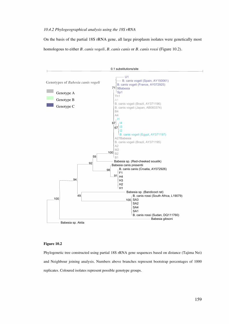
159
10.4.2 Phylogeographical analysis using the 18S rRNA
On the basis of the partial 18S rRNA gene, all large piroplasm isolates were genetically most
homologous to either B. canis vogeli, B. canis canis or B. canis rossi (Figure 10.2).
Figure 10.2
Phylogenetic tree constructed using partial 18S rRNA gene sequences based on distance (Tajima Nei)
and Neighbour joining analysis. Numbers above branches represent bootstrap percentages of 1000
replicates. Coloured isolates represent possible genotype groups.
Genotype A
Genotype B
Genotype C
0.1 substitutions/site
Babesia sp. AkitaBabesia gibsoni
Babesia sp. (Red-cheeked souslik)B1B2M2A2B. canis vogeli (Brazil, AY371195)A27Babesia
B. canis vogeli (Brazil, AY371196)B. canis vogeli (Japan, AB083374)
Babesia canis presentii
Babesia sp. (Bandicoot rat)
A1
B4
B. canis rossi (South Africa, L19079)
Th1
A4I1
Sp19Babesia
SA4
B. canis vogeli (France, AY072925)
U1B. canis vogeli (Spain, AY150061)
I4I3I2B. canis vogeli (Egypt, AY371197)
B. canis canis (Croatia, AY072926)F1H4H3H2H1
SA3SA2
SA1B. canis rossi (Sudan, DQ111760)
100
94
92
59100
98
49100
91
67
71
67
Genotypes of Babesia canis vogeli
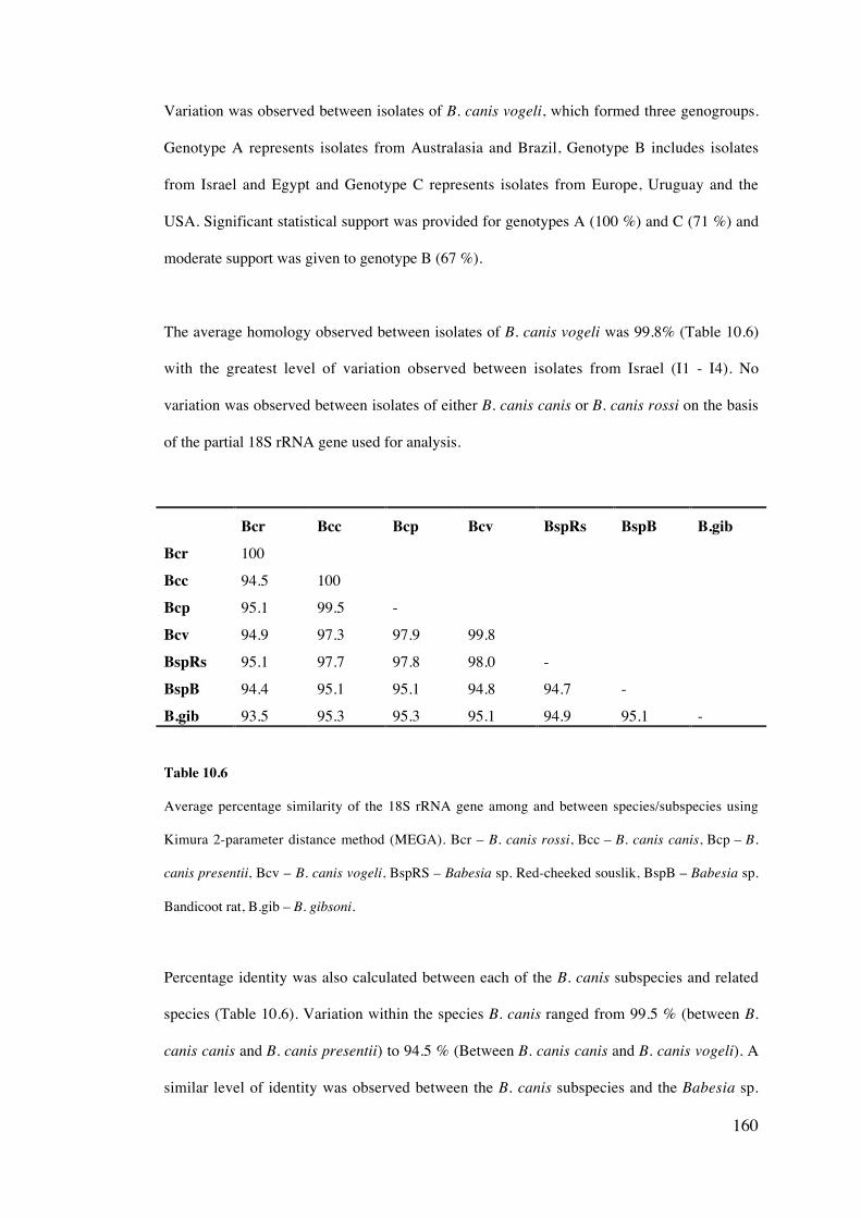
160
Variation was observed between isolates of B. canis vogeli, which formed three genogroups.
Genotype A represents isolates from Australasia and Brazil, Genotype B includes isolates
from Israel and Egypt and Genotype C represents isolates from Europe, Uruguay and the
USA. Significant statistical support was provided for genotypes A (100 %) and C (71 %) and
moderate support was given to genotype B (67 %).
The average homology observed between isolates of B. canis vogeli was 99.8% (Table 10.6)
with the greatest level of variation observed between isolates from Israel (I1 - I4). No
variation was observed between isolates of either B. canis canis or B. canis rossi on the basis
of the partial 18S rRNA gene used for analysis.
Bcr Bcc Bcp Bcv BspRs BspB B.gibBcr 100
Bcc 94.5 100
Bcp 95.1 99.5 -
Bcv 94.9 97.3 97.9 99.8
BspRs 95.1 97.7 97.8 98.0 -
BspB 94.4 95.1 95.1 94.8 94.7 -
B.gib 93.5 95.3 95.3 95.1 94.9 95.1 -
Table 10.6
Average percentage similarity of the 18S rRNA gene among and between species/subspecies using
Kimura 2-parameter distance method (MEGA). Bcr – B. canis rossi, Bcc – B. canis canis, Bcp – B.
canis presentii, Bcv – B. canis vogeli, BspRS – Babesia sp. Red-cheeked souslik, BspB – Babesia sp.
Bandicoot rat, B.gib – B. gibsoni.
Percentage identity was also calculated between each of the B. canis subspecies and related
species (Table 10.6). Variation within the species B. canis ranged from 99.5 % (between B.
canis canis and B. canis presentii) to 94.5 % (Between B. canis canis and B. canis vogeli). A
similar level of identity was observed between the B. canis subspecies and the Babesia sp.
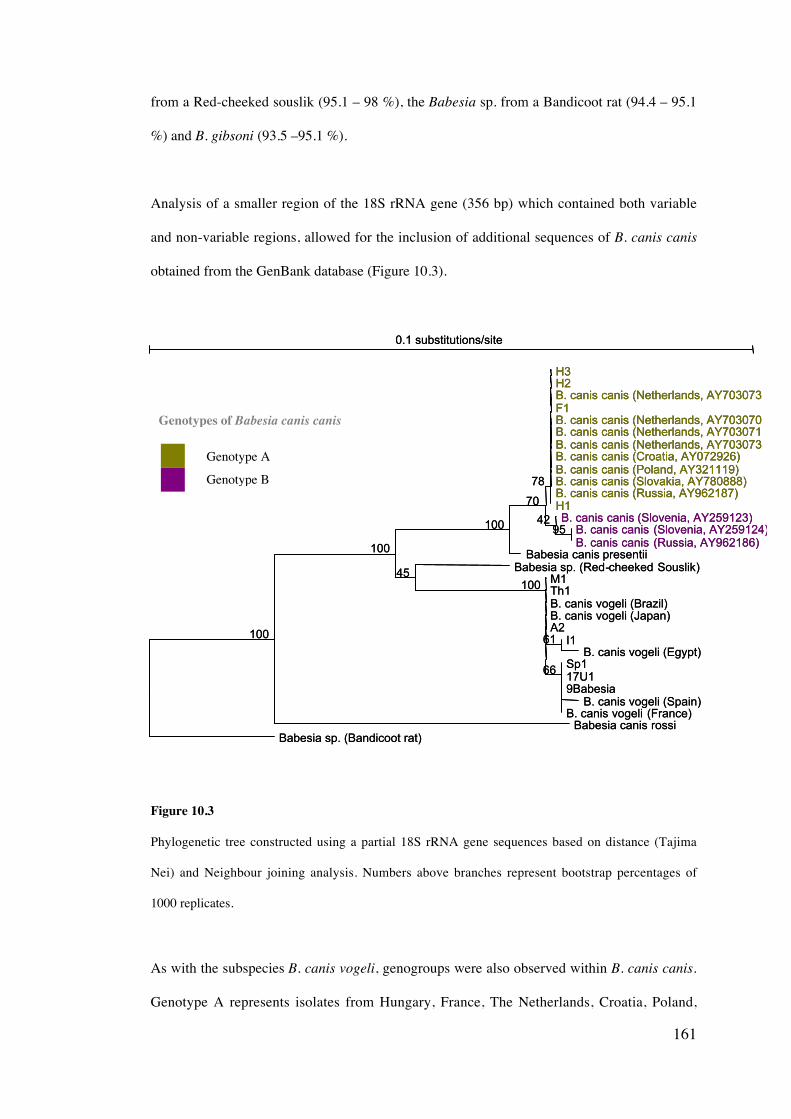
161
from a Red-cheeked souslik (95.1 – 98 %), the Babesia sp. from a Bandicoot rat (94.4 – 95.1
%) and B. gibsoni (93.5 –95.1 %).
Analysis of a smaller region of the 18S rRNA gene (356 bp) which contained both variable
and non-variable regions, allowed for the inclusion of additional sequences of B. canis canis
obtained from the GenBank database (Figure 10.3).
Figure 10.3
Phylogenetic tree constructed using a partial 18S rRNA gene sequences based on distance (Tajima
Nei) and Neighbour joining analysis. Numbers above branches represent bootstrap percentages of
1000 replicates.
As with the subspecies B. canis vogeli, genogroups were also observed within B. canis canis.
Genotype A represents isolates from Hungary, France, The Netherlands, Croatia, Poland,
0.1 substitutions/site
Babesia sp. (Bandicoot rat)Babesia canis rossi
Babesia canis presentiiBabesia sp. (Red-cheeked Souslik)
M1
H1
Th1
B. canis canis (Russia, AY962187)
B. canis vogeli (Brazil)B. canis vogeli (Japan)A2
Sp117U1
B. canis canis (Slovenia, AY259123)
9Babesia
H3H2B. canis canis (Netherlands, AY703073F1B. canis canis (Netherlands, AY703070B. canis canis (Netherlands, AY703071B. canis canis (Netherlands, AY703073B. canis canis (Croatia, AY072926)B. canis canis (Poland, AY321119)B. canis canis (Slovakia, AY780888)
B. canis canis (Slovenia, AY259124)B. canis canis (Russia, AY962186)
I1B. canis vogeli (Egypt)
B. canis vogeli (Spain)B. canis vogeli (France)
100
100
100
70
45100
78
66
4295
61
Genotype A
Genotype B
Genotypes of Babesia canis canis
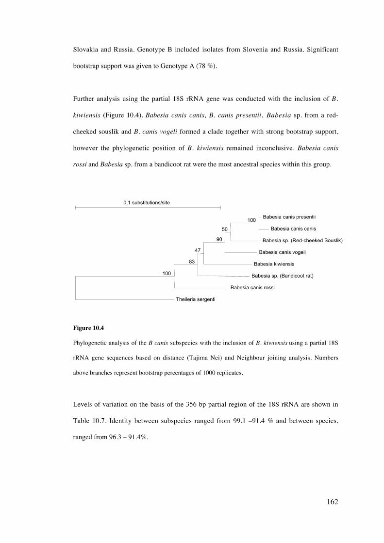
162
Slovakia and Russia. Genotype B included isolates from Slovenia and Russia. Significant
bootstrap support was given to Genotype A (78 %).
Further analysis using the partial 18S rRNA gene was conducted with the inclusion of B.
kiwiensis (Figure 10.4). Babesia canis canis, B. canis presentii, Babesia sp. from a red-
cheeked souslik and B. canis vogeli formed a clade together with strong bootstrap support,
however the phylogenetic position of B. kiwiensis remained inconclusive. Babesia canis
rossi and Babesia sp. from a bandicoot rat were the most ancestral species within this group.
Figure 10.4
Phylogenetic analysis of the B canis subspecies with the inclusion of B. kiwiensis using a partial 18S
rRNA gene sequences based on distance (Tajima Nei) and Neighbour joining analysis. Numbers
above branches represent bootstrap percentages of 1000 replicates.
Levels of variation on the basis of the 356 bp partial region of the 18S rRNA are shown in
Table 10.7. Identity between subspecies ranged from 99.1 –91.4 % and between species,
ranged from 96.3 – 91.4%.
0.1 substitutions/site
Theileria sergenti
Babesia canis rossi
Babesia sp. (Bandicoot rat)
Babesia kiwiensis
Babesia canis vogeli
Babesia sp. (Red-cheeked Souslik)
Babesia canis presentii
Babesia canis canis
100
83
47
90
50
100
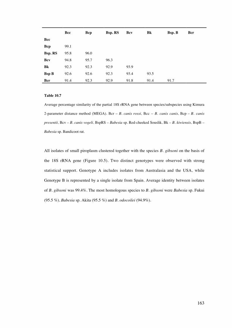
163
Bcc Bcp Bsp. RS Bcv Bk Bsp. B Bcr
Bcc
Bcp 99.1
Bsp. RS 95.8 96.0
Bcv 94.8 95.7 96.3
Bk 92.3 92.3 92.9 93.9
Bsp B 92.6 92.6 92.3 93.4 93.5
Bcr 91.4 92.3 92.9 91.8 91.4 91.7
Table 10.7
Average percentage similarity of the partial 18S rRNA gene between species/subspecies using Kimura
2-parameter distance method (MEGA). Bcr – B. canis rossi, Bcc – B. canis canis, Bcp – B. canis
presentii, Bcv – B. canis vogeli, BspRS – Babesia sp. Red-cheeked Souslik, Bk – B. kiwiensis, BspB –
Babesia sp. Bandicoot rat.
All isolates of small piroplasm clustered together with the species B. gibsoni on the basis of
the 18S rRNA gene (Figure 10.5). Two distinct genotypes were observed with strong
statistical support. Genotype A includes isolates from Australasia and the USA, while
Genotype B is represented by a single isolate from Spain. Average identity between isolates
of B. gibsoni was 99.4%. The most homologous species to B. gibsoni were Babesia sp. Fukui
(95.5 %), Babesia sp. Akita (95.5 %) and B. odocoilei (94.9%).
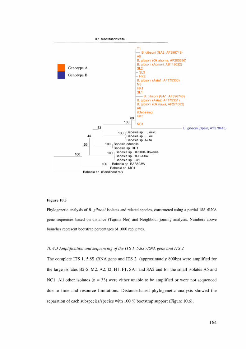
164
Figure 10.5
Phylogenetic analysis of B. gibsoni isolates and related species, constructed using a partial 18S rRNA
gene sequences based on distance (Tajima Nei) and Neighbour joining analysis. Numbers above
branches represent bootstrap percentages of 1000 replicates.
10.4.3 Amplification and sequencing of the ITS 1, 5.8S rRNA gene and ITS 2
The complete ITS 1, 5.8S rRNA gene and ITS 2 (approximately 800bp) were amplified for
the large isolates B2-5, M2, A2, I2, H1, F1, SA1 and SA2 and for the small isolates A5 and
NC1. All other isolates (n = 33) were either unable to be amplified or were not sequenced
due to time and resource limitations. Distance-based phylogenetic analysis showed the
separation of each subspecies/species with 100 % bootstrap support (Figure 10.6).
0.1 substitutions/site
Babesia sp. (Bandicoot rat)
B. gibsoni (Spain, AY278443)NC1IFATHK39BabesiagiA6B. gibsoni (Okinawa, AF271082)B. gibsoni (Asia2, AF175301)
B. gibsoni (Asia1, AF175300)M3
B. gibsoni (Oklahoma, AF205636)B. gibsoni (Aomori, AB118032)
Babesia sp. DD2004 slovenia
Babesia sp. Fukui76
HK1
A5
SL2
T1B. gibsoni (GA2, AF396749)
SL3HK2
SL1B. gibsoni (GA1, AF396748)
Babesia sp. FukuiBabesia sp. Akita
Babesia odocoileiBabesia sp. RD1
Babesia sp. RDS2004Babesia sp. EU1
Babesia sp. BAB693WBabesia sp. MO1
100
56
44
83
10089
100
100
100
100
Genotype A
Genotype B
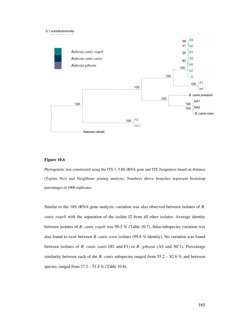
165
Figure 10.6
Phylogenetic tree constructed using the ITS 1, 5.8S rRNA gene and ITS 2sequences based on distance
(Tajima Nei) and Neighbour joining analysis. Numbers above branches represent bootstrap
percentages of 1000 replicates.
Similar to the 18S rRNA gene analysis, variation was also observed between isolates of B.
canis vogeli with the separation of the isolate I2 from all other isolates. Average identity
between isolates of B. canis vogeli was 99.3 % (Table 10.7). Intra-subspecies variation was
also found to exist between B. canis rossi isolates (99.8 % identity). No variation was found
between isolates of B. canis canis (H1 and F1) or B. gibsoni (A5 and NC1). Percentage
similarity between each of the B. canis subspecies ranged from 55.2 – 82.6 % and between
species, ranged from 27.2 – 51.4 % (Table 10.8).
0.1 substitutions/site
Babesia caballi
I2
A2
B. canis presentii
SA1
B3
B4
B2
B5
M2
F1
H1
SA2
B. canis rossi
A5
NC1
100
100
100
100
82
20
100
100
31
68
100
100
100
Babesia canis vogeli
Babesia canis canis
Babesia gibsoni
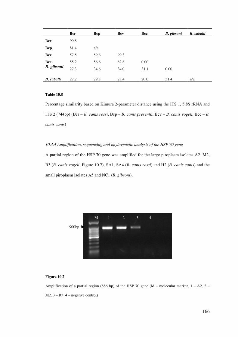
166
Bcr Bcp Bcv Bcc B. gibsoni B. caballi
Bcr 99.8
Bcp 81.4 n/a
Bcv 57.5 59.6 99.3
Bcc 55.2 56.6 82.6 0.00B. gibsoni 27.3 34.6 34.0 31.1 0.00
B. caballi 27.2 29.8 28.4 20.0 51.4 n/a
Table 10.8
Percentage similarity based on Kimura 2-parameter distance using the ITS 1, 5.8S rRNA and
ITS 2 (744bp) (Bcr – B. canis rossi, Bcp – B. canis presentii, Bcv – B. canis vogeli, Bcc – B.
canis canis)
10.4.4 Amplification, sequencing and phylogenetic analysis of the HSP 70 gene
A partial region of the HSP 70 gene was amplified for the large piroplasm isolates A2, M2,
B3 (B. canis vogeli, Figure 10.7), SA1, SA4 (B. canis rossi) and H2 (B. canis canis) and the
small piroplasm isolates A5 and NC1 (B. gibsoni).
Figure 10.7
Amplification of a partial region (886 bp) of the HSP 70 gene (M – molecular marker, 1 – A2, 2 –
M2, 3 – B3, 4 – negative control)
900bp
M 1 2 3 4
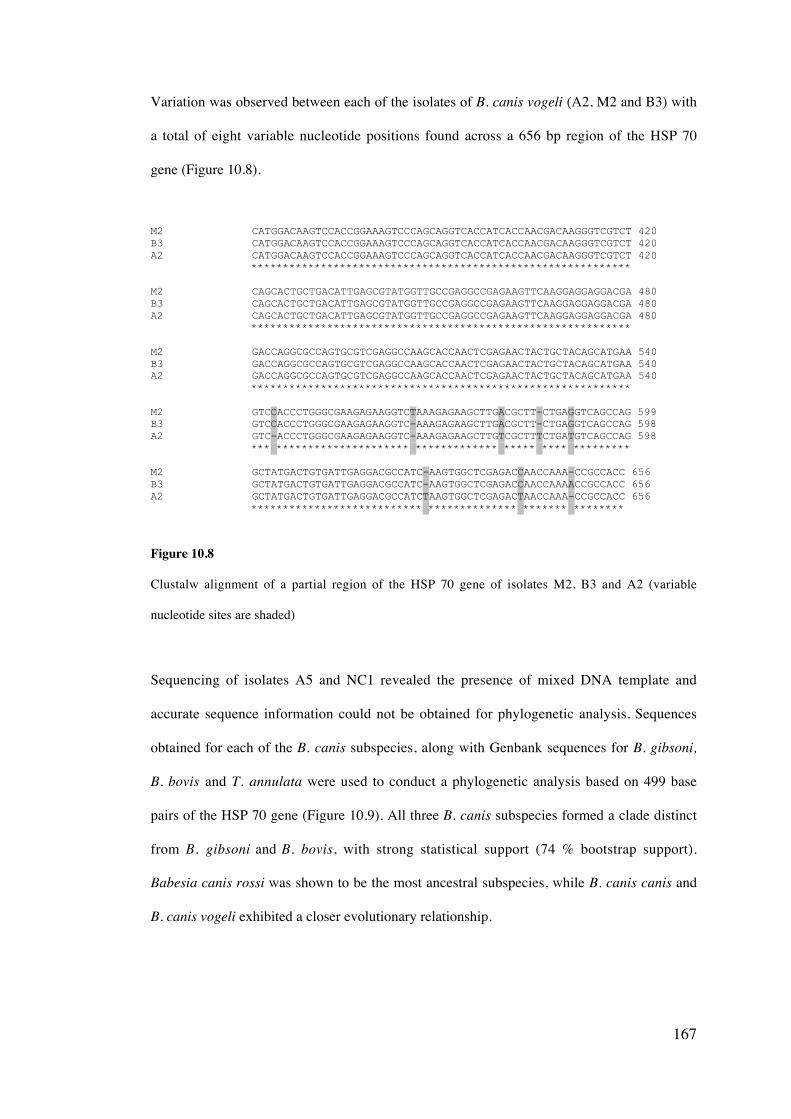
167
Variation was observed between each of the isolates of B. canis vogeli (A2, M2 and B3) with
a total of eight variable nucleotide positions found across a 656 bp region of the HSP 70
gene (Figure 10.8).
M2 CATGGACAAGTCCACCGGAAAGTCCCAGCAGGTCACCATCACCAACGACAAGGGTCGTCT 420B3 CATGGACAAGTCCACCGGAAAGTCCCAGCAGGTCACCATCACCAACGACAAGGGTCGTCT 420A2 CATGGACAAGTCCACCGGAAAGTCCCAGCAGGTCACCATCACCAACGACAAGGGTCGTCT 420 ************************************************************
M2 CAGCACTGCTGACATTGAGCGTATGGTTGCCGAGGCCGAGAAGTTCAAGGAGGAGGACGA 480B3 CAGCACTGCTGACATTGAGCGTATGGTTGCCGAGGCCGAGAAGTTCAAGGAGGAGGACGA 480A2 CAGCACTGCTGACATTGAGCGTATGGTTGCCGAGGCCGAGAAGTTCAAGGAGGAGGACGA 480 ************************************************************
M2 GACCAGGCGCCAGTGCGTCGAGGCCAAGCACCAACTCGAGAACTACTGCTACAGCATGAA 540B3 GACCAGGCGCCAGTGCGTCGAGGCCAAGCACCAACTCGAGAACTACTGCTACAGCATGAA 540A2 GACCAGGCGCCAGTGCGTCGAGGCCAAGCACCAACTCGAGAACTACTGCTACAGCATGAA 540 ************************************************************
M2 GTCCACCCTGGGCGAAGAGAAGGTCTAAAGAGAAGCTTGACGCTT-CTGAGGTCAGCCAG 599B3 GTCCACCCTGGGCGAAGAGAAGGTC-AAAGAGAAGCTTGACGCTT-CTGAGGTCAGCCAG 598A2 GTC-ACCCTGGGCGAAGAGAAGGTC-AAAGAGAAGCTTGTCGCTTTCTGATGTCAGCCAG 598 *** ********************* ************* ***** **** *********
M2 GCTATGACTGTGATTGAGGACGCCATC-AAGTGGCTCGAGACCAACCAAA-CCGCCACC 656B3 GCTATGACTGTGATTGAGGACGCCATC-AAGTGGCTCGAGACCAACCAAAACCGCCACC 656A2 GCTATGACTGTGATTGAGGACGCCATCTAAGTGGCTCGAGACTAACCAAA-CCGCCACC 656 *************************** ************** ******* ********
Figure 10.8
Clustalw alignment of a partial region of the HSP 70 gene of isolates M2, B3 and A2 (variable
nucleotide sites are shaded)
Sequencing of isolates A5 and NC1 revealed the presence of mixed DNA template and
accurate sequence information could not be obtained for phylogenetic analysis. Sequences
obtained for each of the B. canis subspecies, along with Genbank sequences for B. gibsoni,
B. bovis and T. annulata were used to conduct a phylogenetic analysis based on 499 base
pairs of the HSP 70 gene (Figure 10.9). All three B. canis subspecies formed a clade distinct
from B. gibsoni and B. bovis, with strong statistical support (74 % bootstrap support).
Babesia canis rossi was shown to be the most ancestral subspecies, while B. canis canis and
B. canis vogeli exhibited a closer evolutionary relationship.
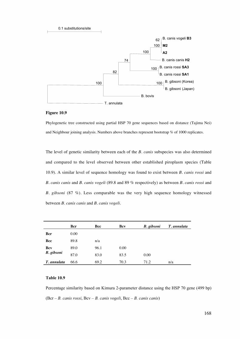
168
Figure 10.9
Phylogenetic tree constructed using partial HSP 70 gene sequences based on distance (Tajima Nei)
and Neighbour joining analysis. Numbers above branches represent bootstrap % of 1000 replicates.
The level of genetic similarity between each of the B. canis subspecies was also determined
and compared to the level observed between other established piroplasm species (Table
10.9). A similar level of sequence homology was found to exist between B. canis rossi and
B. canis canis and B. canis vogeli (89.8 and 89 % respectively) as between B. canis rossi and
B. gibsoni (87 %). Less comparable was the very high sequence homology witnessed
between B. canis canis and B. canis vogeli.
Bcr Bcc Bcv B. gibsoni T. annulata
Bcr 0.00
Bcc 89.8 n/a
Bcv 89.0 96.1 0.00B. gibsoni 87.0 83.0 83.5 0.00
T. annulata 66.6 69.2 70.3 71.2 n/a
Table 10.9
Percentage similarity based on Kimura 2-parameter distance using the HSP 70 gene (499 bp)
(Bcr – B. canis rossi, Bcv – B. canis vogeli, Bcc – B. canis canis)
0.1 substitutions/site
T. annulata
B. bovis
B. canis canis H2
A2
B. canis vogeli B3
M2
B. canis rossi SA3
B. canis rossi SA1
B. gibsoni (Korea)
B. gibsoni (Japan)
100
82
74
100
100
62
100
100
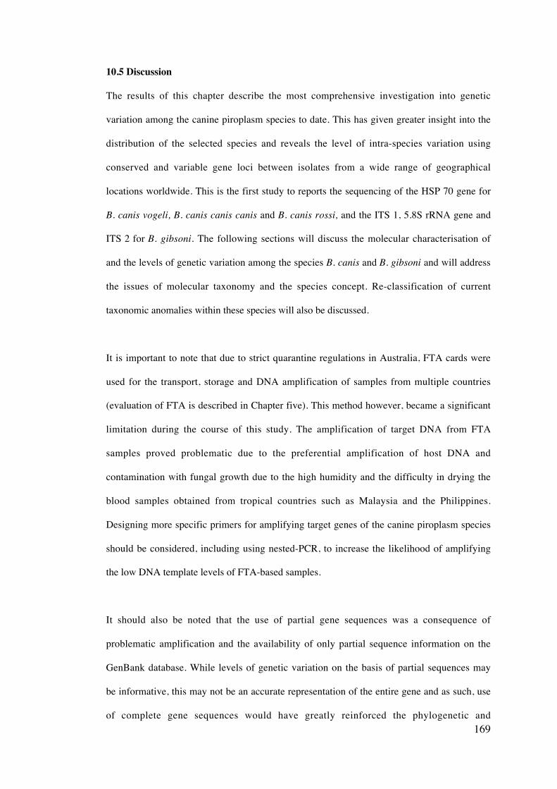
169
10.5 Discussion
The results of this chapter describe the most comprehensive investigation into genetic
variation among the canine piroplasm species to date. This has given greater insight into the
distribution of the selected species and reveals the level of intra-species variation using
conserved and variable gene loci between isolates from a wide range of geographical
locations worldwide. This is the first study to reports the sequencing of the HSP 70 gene for
B. canis vogeli, B. canis canis canis and B. canis rossi, and the ITS 1, 5.8S rRNA gene and
ITS 2 for B. gibsoni. The following sections will discuss the molecular characterisation of
and the levels of genetic variation among the species B. canis and B. gibsoni and will address
the issues of molecular taxonomy and the species concept. Re-classification of current
taxonomic anomalies within these species will also be discussed.
It is important to note that due to strict quarantine regulations in Australia, FTA cards were
used for the transport, storage and DNA amplification of samples from multiple countries
(evaluation of FTA is described in Chapter five). This method however, became a significant
limitation during the course of this study. The amplification of target DNA from FTA
samples proved problematic due to the preferential amplification of host DNA and
contamination with fungal growth due to the high humidity and the difficulty in drying the
blood samples obtained from tropical countries such as Malaysia and the Philippines.
Designing more specific primers for amplifying target genes of the canine piroplasm species
should be considered, including using nested-PCR, to increase the likelihood of amplifying
the low DNA template levels of FTA-based samples.
It should also be noted that the use of partial gene sequences was a consequence of
problematic amplification and the availability of only partial sequence information on the
GenBank database. While levels of genetic variation on the basis of partial sequences may
be informative, this may not be an accurate representation of the entire gene and as such, use
of complete gene sequences would have greatly reinforced the phylogenetic and
170
phylogeographical data obtained in this chapter. The following hypotheses discussed with
regard to genetic characterisation and phylogeography of the selected canine piroplasm
species should be considered as preliminary studies and need to be followed up with
comprehensive studies using complete gene sequences of multiple loci before definitive
theories can be postulated.
10.5.1 Genetic characterisation and phylogeography of the Babesia canis subspecies
i) Babesia canis vogeli
Molecular characterisation of multiple isolates of B. canis from different geographic
locations within Australia further supports that B. canis vogeli is the predominant large
canine piroplasm species in Australia (Jefferies et al., 2003) and is likely to be a reflection of
the cosmopolitan distribution of the tick vector R. sanguineus. The absence of the tick
vectors responsible for the transmission of other subspecies in Australia is likely to prevent
establishment, however stringent tick control measures should be maintained to prevent
exotic canine tick species entering Australia.
Through the use of molecular characterisation, B. canis vogeli has also been confirmed to be
present within many regions of the world for the first time, suggesting that this subspecies is
likely to be the most widely distributed of all the B. canis subspecies. The genetic
characterisation of large piroplasm isolates from Malaysia, Thailand and the Philippines has
been achieved for the first time, providing a greater insight into the canine Babesia in south-
east Asia. Research into B. canis infections in south-east Asia has been extremely limited
(Irwin and Jefferies, 2004). While probable cases of B. canis infection have been reported in
Malaysia (Rajamanickam et al., 1985) and Thailand (Suksawat et al., 2001b) the subspecies
present was not determined. It is now confirmed that B. canis vogeli is present within
multiple south-east Asian countries and again, is likely to be due to the high endemicity of R.
sanguineus throughout the region.
171
Before the commencement of this research, limited study had been conducted on canine
babesiosis in South America. O’Dwyer et al. (2001) reported coinfections of a large
intraerythrocytic piroplasm presumed to be Babesia canis and Hepatozoon canis in Brazil. A
more recent study investigated clinical cases of canine babesiosis in Belo Horizonte, Minas
Gerais, Brazil, suggesting a high infection rate of this disease in dogs surveyed (Bastos et al.,
2004). Babesia canis has also been reported in maned wolves (Chrysocyon brachyurus) at
the Sorocaba Zoo, Brazil (Nunes, 1989). While canine Babesia spp. have been previously
documented to exist in South America, this study, along with Passos et al. (2005) describe
the molecular characterisation of these piroplasms for the first time. All isolates were
determined to be B. canis vogeli, a subspecies now confirmed to be present in both Brazil
and Uruguay. Rhipicephalus sanguineus, the principal vector of B. canis vogeli, has been
reported to exist in both Brazil and Uruguay. This tick was originally introduced from the
Afrotropical region to Brazil, where it has been reported to be an increasingly widespread
pest in urban environments (Evans et al., 2000; Szabo et al., 2001). Likewise, R. sanguineus
has been reported in Uruguay (Rodriguez and Lazaro, 1954; Venzal et al., 2003). This tick
species has also been found in Mexico (Cruz-Vazquez and Garcia-Vazquez, 1999), Panama
(Miller et al., 2001), Venezuela (Unver et al., 2001) and Argentina (Guglielmone et al.,
1991; Ruiz et al., 2003), which suggests that B. canis vogeli may also be widespread
throughout Central and South America.
The existence of B. canis has previously been reported within Israel (Baneth et al., 1998) and
sequencing of a 270 bp region of the 18S rRNA gene suggested that B. canis vogeli is the
subspecies present (Baneth et al., 2004). In the present study, sequencing a larger region of
the 18S rRNA gene from isolates from four separate geographic regions has confirmed that
B. canis vogeli is indeed the subspecies present within Israel. Isolates from Israel showed
greatest homology to an isolate from Egypt that is considered the type specimen for B. canis
vogeli (Passos et al., 2005). A possibly geographic correlation may exist between isolates
from Africa and the Middle East. Babesia canis vogeli has also been reported in South
172
Africa (Matjila et al., 2004) and Sudan (Oyamada et al., 2005), however the small size of the
partial sequences amplified prevented its inclusion in the analysis performed in this chapter.
As a result of this study, it should also be recognized that three distinct genotypes of the
subspecies B. canis vogeli occur on the basis of the 18S rRNA gene and is a possible
reflection of their different geographical origins. The most common genotype described was
in isolates from Australasia and Brazil and was shown to be ancestral to other isolates. A
second from dogs in Egypt and Israel and a third genotype was found to be present in
Europe, USA and Uruguay. Previous studies have recognized the existence of low levels of
genetic variation between isolates of the B. canis subspecies (Zahler et al., 1998; Caccio et
al., 2002; Passos et al., 2005) however these studies did not correlate this variation to
possible phylogeographical relationships between isolates. Also, the possibility of
differences in biology or pathogenicity between each of these genotypes requires further
study.
It is postulated that B. canis vogeli may have originated from Asia and may be correlated to
the geographical origins of the domestic dog. A recent study suggested that the dog
originated from eastern Asia (Savolainen et al, 2002). The similarity in genotype of B. canis
vogeli isolates from Asia and Australia may relate to early dog movement, notably dingoes
(Canis familiaris dingo) from Asia to Australia. Babesia canis has also been reported in
dingoes within Australia (Callow, 1984; Jefferies, 2001) and molecular characterisation is
necessary in order to determine the genotype present within these wild canines. The reason
that isolates from Brazil are genetically distinct from Uruguay and similar to isolates from
the Australasian region remains unknown. Further study investigating B. canis vogeli
isolates from additional geographical locations, in addition to co-evolutionary relationships
with dogs and tick vectors is necessary before definitive conclusions on the phylogeography
of this subspecies can be determined.
173
It is interesting to hypothesise that the clustering of the isolate from Uruguay with sequences
from Spain and France may be a reflection of the European colonization of this country. It is
conceivable that immigrants from Spain or Portugal may have brought with them the first
Babesia canis vogeli infected dogs or ticks to Uruguay. A similar situation may have also
occurred in the USA. Unfortunately, isolates from Uruguay and Spain could not be amplified
on the basis of the ITS 1, 5.8S rRNA gene to confirm the existence of distinct genotypes.
ii) Babesia canis canis
Distinct genotypes were also observed between different isolates of B. canis canis,
supporting the genetic variation observed in previous studies (Caccio et al., 2002; Duh et al.,
2004). In contrast to B. canis vogeli, these genetic differences did not reflect the
geographical origin of the isolate. A possible explanation for the mixture of genotypes
throughout Europe is a reflection of recent increases in dog movement throughout countries
of the European Union, with the introduction of travel schemes such as PETS (Pet Travel
Scheme) (Shaw et al., 2001b). The existence of allopatric (non-overlapping) populations
may have originally enabled distinct genotypes to develop, however subsequent movement
of dogs between various countries could have lead to a mixture of genotypes existing in the
same geographic location. Only small partial regions of the 18S rRNA gene could be used to
investigate isolate heterogeneity and further research using the complete or near complete
18S rRNA gene, in addition to other gene loci is necessary to confirm the existence of
genotypes within B. canis canis and what significance this variation may have on
phylogeography and biological differences.
iii) Babesia canis rossi
This study has revealed very limited genetic variation witnessed among different isolates of
B. canis rossi. Until recently, B. canis rossi was believed only to be present within South
Africa, however with its discovery in Sudan (Oyamada et al., 2005), this subspecies may be
more widely distributed throughout Africa. Further investigation into the distribution of this
174
subspecies is necessary to determine how widespread B. canis rossi is in the African
continent and whether it exists in other countries outside Africa. Recently described in
Australia was a case of a dog being imported from South Africa via Hong Kong that was
found to be infected with B. canis rossi (Ainslie Brown, AQIS, pers. com) suggesting that
sporadic infections with this subspecies may occur outside of Africa and should be
considered when diagnosing canine piroplasmosis worldwide.
10.5.2 Genetic characterisation and phylogeography of Babesia gibsoni
This study has further characterised isolates of B. gibsoni from Australia. While, isolates of
this species in Australia have previously been characterised on the basis of the 18S rRNA
(Muhlnickel et al., 2002; Jefferies et al., 2003), only small partial regions of the gene were
sequenced, limiting accurate comparisons to other isolates. This study, in contrast, has
sequenced isolates from two separate states, including an isolate from New South Wales for
the first time, on the basis of a larger partial region of the 18S rRNA gene. In addition, the
complete ITS 1, 5.8S rRNA gene and ITS 2 sequences were determined for an isolate from
Victoria. This is the first reported sequencing of these combined loci for B. gibsoni.
Also described for the first time, is the presence of B. gibsoni in Singapore, Taiwan and
Hong Kong, on the basis of the molecular detection and characterisation of the 18S rRNA
gene, in addition to further confirming the genotype of this species in Malaysia and Sri
Lanka. (Zahler et al., 2000c). Unfortunately DNA of small piroplasm isolates from Brazil
(B6-9) could not be amplified and while the existence of small piroplasms in Brazil should
now be recognized, the species and/or genotype remains unknown and is currently the
subject of further investigation.
Using the 18S rRNA gene, two main genotypes of B. gibsoni were found to exist, with the
notable phylogenetic separation of the B. gibsoni isolate from Spain from all other isolates.
The distinction of B. gibsoni from Spain from other isolates was first reported by Criado-
175
Fornelio et al. (2003d), who suggested that this species may have originated from Asia and
developed into two sister lineages. Babesia gibsoni has also been reported to exist in Egypt,
Nigeria and Mali (Yamane et al., 1993), however no isolates from Africa have ever been
molecularly characterised. It is important to characterise isolates of small canine piroplasms
from Africa before a hypothesis on the evolution of B. gibsoni can be proposed. It has also
been suggested that the genotypes of B. gibsoni should not be taxonomically elevated
(Criado-Fornelio et al., 2003d). Further research into pathogenesis, geographic distribution
and tick vectors of each genotype is necessary before the significance of the genetic variation
observed is better understood and whether taxonomic revision is appropriate.
The genetic homology that exists on the basis of the 18S rRNA gene between isolates of B.
gibsoni from Australia and certain regions of the USA and Asia is of particular interest with
reference to infections of this species in fighting dog breeds (refer to Chapter seven).
Identical sequences of the ITS 1, 5.8S rRNA gene and ITS 2 were also identified for B.
gibsoni isolates from Australia and the USA. As is presumed with all the piroplasmid
species, the sexual stage of the lifecycle occurs within the tick vector. Sexual reproduction
allows for genetic recombination to occur and the production of genetic heterogeneity within
a population. If the tick vector was absent during the transmission of B. gibsoni, as is
suggested to occur in populations of fighting dogs (Macintire et al., 2002; Birkenheuer et al.,
2003b; Matsuu et al., 2004a), then it would be expected that a clonal lineage of these
protozoa would develop. This indeed may be the case in fighting dog populations from
Australia (Chapter seven), certain regions of Japan and the USA. The use of only partial
gene sequences for analysis and the conserved nature of the selected loci are perhaps too
conserved to draw any definitive conclusions and the sequencing of much less conserved
loci is suggested. The absence of genetic variation within B. gibsoni isolates from Australia,
Japan and certain states of the USA may also simply be a reflection of the recent spread of
this parasite to these countries, not allowing sufficient time for any genetic variation to
develop.
176
10.5.3 Molecular taxonomy; defining species level classification
The problematic concept of defining species level categorization has plagued taxonomists
throughout history and remains a contentious issue. The traditional concept, that ‘species are
groups of actually or potentially interbreeding natural populations, which are reproductively
isolated from other such groups’ (Kunz, 2002), is even more difficult to apply to the
protozoa which often have complex life cycles, involving both asexual and sexual
reproduction. As the concept of a species is an artificial construct, created as a means of
convenient categorization, a universal description should not be applied to all taxa rather
individual definitions of a species should be devised for separate groups of organisms.
General criteria for describing a new species should however be conformed to and follow the
guidelines established by the International Code of Zoological Nomenclature (ICZN) (Ride
et al., 1999). Criteria for describing a new species have been postulated for other
apicomplexans such as Cryptosporidium, including parasite morphology, host specificity and
genetic characterisation (Xiao et al., 2004)
Uilenberg et al. (2004) argued that current molecular taxonomy using gene sequences, rather
than entire genomes, has lead to premature changes in classification systems. They suggested
that polyphasic taxonomy, based upon both genotype and phenotype, should be considered
before making any new taxonomic changes. Within the piroplasms, overzealous naming of
species on the basis of molecular characterisation, such as in the case of the taxonomic
description of T. annae (Zahler et al., 2000b) has been considered inappropriate (Goethert
and Telford, 2003; Reichard et al., 2005). Baneth et al. (2004) chose a far more cautious
approach when taxonomically describing B. canis presentii.
Uilenberg et al. (2004) also suggested that molecular-based taxonomy can lead to repeated
taxonomic changes and subsequently produce confusion rather than clarification. In contrast
to this argument, it should be noted that taxonomic changes were far more confusing during
the pre-molecular taxonomy era, as for example, since the first description of B. canis, at
177
least twelve separate species names have been given to this species before the introduction of
molecular characterisation (Levine, 1988). Since the molecular characterisation of this
species, no taxonomic changes have been made. Such complex pre-molecular taxonomy is
common among many piroplasmids. While it is important to consider all available
information, both genotypic and phenotypic, before making changes to the taxonomy, overly
cautious attitudes by Uilenberg et al. (2004) and Goethert and Telford (2003) hinder
progressive, yet systematic changes in the taxonomic classification of the piroplams.
This chapter has also shown that the level of genetic variation observed between established
Babesia species on the basis of the 18S rRNA gene, ITS 1, 5.8S rRNA gene and ITS 2 is
similar to that which exists between each of the B. canis subspecies. For example, the level
of intra-subspecies genetic variation on the basis of the 18S rRNA gene for B. canis vogeli
and B. canis canis is similar to the intra-species variation observed in isolates of B. gibsoni.
Also for the first time also provides further support with phylogenetic analyses based on a
partial region of the HSP 70 gene. These results further support the suggestion that each of
the B. canis subspecies should be elevated to species level classification (Uilenberg et al.,
1989; Zahler et al., 1998; Carret et al., 1999). Schnittger et al. (2003) proposed that an
isolate should be defined as a new species if the genetic identity using the 18S rRNA gene is
lower than 99.3 % for Theileria and 96.6% for Babesia on the basis of sheep and goat
piroplasms, a feature also observed between each of the B. canis subspecies. Other studies
have delineated piroplasm species level classification using the internal transcribed spaces
(ITS 1 and 2) and the intervening 5.8S rRNA gene (Zahler et al., 1998; Holman et al., 2003).
Phylogenetic analysis in the present study also revealed that two unnamed Babesia species,
one identified from Red-cheeked sousliks (Citella erythrogenys) in China (Zamoto et al.,
2004) and the second from Bandicoot rats (Bandicota indica) in Thailand (Dantrakool et al.,
2004) are closely related to the B. canis subspecies. Babesia kiwiensis also shows a
phylogenetic affiliation to the B. canis group. Further study needs to be carried out on the
178
phylogenetic relationships between each of these species on the basis of multiple gene loci.
Previous studies into the phylogenetic relationships of the B. canis subspecies have found
that each of the subspecies cluster together in a monophyletic clade, separate to all other
Babesia spp. (Carret et al., 1999; Criado-Fornelio et al., 2003b; Baneth et al., 2004; Caccio
et al., 2002) and may have contributed to each of the subspecies not being recognised as
different species. It therefore becomes important to be able to define species level
classification among the piroplasms. Species level classification on the sole basis of a certain
level of genetic variation may be misleading as some separate species may possess identical
genetic sequences in some genomic regions and not in others (Xiao et al., 2004). No
stipulations are given by the ICZN for the description of new species on the basis of
molecular characterisation (Ride et al., 1999).
Significant differences between B. canis canis, B. canis vogeli, B.canis rossi and B. canis
presentii on the basis of one or more of, pathogenesis, vector specificity and genetic
variation suggests that subspecies level classification for B. canis is inappropriate and it may
be deemed necessary to elevate each to assume species level status. Thus taxonomic
classification can be based on both molecular and biological characteristics.
10.5.4 Proposed re-classification of the B. canis subspecies, including the re-description of
B. canis (Piana and Galli-Valeria, 1895), B. rossi (Wenyon, 1926) and B. vogeli
(Reichenow, 1937)
Babesia canis was first described by Piana and Galli-Valeria (1895) and subsequently all
large piroplasms (3 – 5 mm) found in dogs were classified within this species. It then
became evident that differences in vector specificity and cross-immunity existed between
different isolates of this species, leading to the description of three B. canis subspecies by
Uilenberg et al. (1989). It is interesting to note that Uilenberg et al. (1989) actually
suggested that each of the proposed B. canis subspecies were likely to be separate species
but chose to define them as subspecies on the simple basis of convenience rather than
179
consistency within taxonomic procedures. Different disease pathologies were also described
for each subspecies (Irwin and Hutchinson, 1991; Schetters et al., 1997b). Babesia canis
canis, B. canis vogeli and B. canis rossi were each then characterised on the basis of the 18S
rRNA gene (Carret et al., 1999) and the ITS 1, 5.8S rRNA gene and ITS 2 (Zahler et al.,
1998), confirming the separation of each of these subspecies and allowed for further
speculation that a species level of categorization may be more appropriate. Differences
between B. canis canis and B. canis rossi have also been suggested at a genomic level, with
respective genome sizes estimated to be 14.5 Mbp and 16 Mbp (Depoix et al., 2002) which
may further suggest the existence of separate species. Further support for the elevation of
each of the B. canis subspecies to species level classification has been proved by the genetic
characterisation and phylogenetic studies described in this chapter.
The additional subspecies, B. canis presentii was later described as a piroplasm of cats
(Baneth et al., 2004) and was described as having merozoites and trophozoites that were
morphologically smaller than B. canis canis. Further information regarding host specificity
(can this subspecies infect dogs?), pathogenicity and molecular characterisation of multiple
genes is required before it can be concluded whether B. canis presentii is indeed a separate
species or simply a subspecies of B. canis. This is combined with current phylogenetic
ambiguity of this subspecies when comparing 18S rRNA gene and ITS based analysis, and
suggest that no taxonomic changes be made to this subspecies until further study is
conducted.
It is proposed that each of the B. canis subspecies (excluding B. canis presentii) should
assume a species level of classification on the basis of the following six criteria, which
should be considered when describing any new species of piroplasm:
i) Host
ii) Vector specificity
iii) Morphology
180
iv) Pathogenesis
v) Genetic characterisation
vi) Geographic distribution
The proposed taxonomic changes are as follows:
• Babesia canis (Piana and Galli-Valeria, 1895)
Host/s Dogs, foxes, cats and horses
Vector Dermacentor reticulatus
Pathogenicity Moderate disease
Babesia canis presentii (Baneth et al., 2004)
Host Cats
Vector unknown
Pathogenicity unknown
• Babesia vogeli (Reichenow, 1937)
Host Dogs, possibly other Canidae
Vector Rhipicephalus sanguineus
Pathogenicity Mild to moderate disease
• Babesia rossi ([Nutall, 1910], Wenyon, 1926)
Host Dogs, Jackals
Vector Haemaphysalis leachi
Pathogenicity Highly virulent, moderate to severe haemolytic disease
10.5.5 Conclusions
An attempt has been made to clarify the species concept among the canine and related
piroplasms using molecular characterisation. The level of genetic variation distinguishing
181
species is dependent upon the group of piroplasms investigated and perhaps more
specifically the gene loci used for analysis. While the importance of defining genetic
variation among species and between species is of great significance, a universal species
concept remains elusive. It is therefore imperative that criteria for describing new piroplasm
species be established to allow for less confusion when describing new isolates and it is
suggested that the host, vector specificity, morphology, pathogenesis, genetic
characterisation and geographic distribution are considered as potential criteria. This chapter
has also revealed that a number of genotypes are likely to exist within each canine piroplasm
species, however the taxonomic or pathological significance of these genotypes is yet to be
determined. Further investigation into the phylogeography of the canine piroplasms using
less conserved gene loci may allow for a better understanding of the complex epidemiology
of these protozoa.
182
Phylogenetic and taxonomic status of the order Piroplasmida:
Defining family level classification
11.1 Introduction
Traditional schemes of taxonomic classifications have, in the past, concentrated on
phenotypic features such as life cycle and morphological characteristics as well as host-
parasite relationships, however the classification of many taxa is currently under review with
the introduction of genetic sequencing and phylogenetic-based analysis. This is a common
feature of the classification of most protozoa belonging to the phylum Apicomplexa
(Cavalier-Smith, 1993; Escante and Ayala, 1995; Bernhard et al., 2001; Tenter et al., 2002;
Xiao et al., 2004), including the order Piroplasmida (Allsopp et al., 1994; Reichard et al.,
2005).
DNA sequencing of target genes has become a highly effective means of characterising
established species within the order Piroplasmida and has also given rise to the discovery of
multiple new species and genotypes (Zahler et al., 2000b; Dantrakool et al., 2004). While
the use of molecular technology has allowed for increased diagnostic accuracy, it has also
led to some confusion over the taxonomic position of many species of piroplasm. For
example, certain species initially described as belonging to the genus Babesia have later
been found to be more genetically similar to Theileria species, suggesting that the taxonomy
may need to be clarified. It is now reported that both the Theileria and Babesia are
paraphyletic taxa (Allsopp et al., 1994; Zahler et al., 2000b; Reichard et al., 2005).
CHAPTER ELEVEN
183
There is also current debate over the phylogeny and taxonomy of the B. microti group of
piroplasms, described as the Archeopiroplasmids (Criado-Fornelio et al., 2003b) which
include the species, T. annae, B. felis and B. leo. While it has been suggested that this group
of piroplasms is ancestral to both the genera Babesia and Theileria, their taxonomic position
has not been determined with absolute certainty. Also suffering taxonomic ambiguity are the
Western USA Babesia spp. described by Kjemtrup et al. (2000b), later referred to as the
Prototheilerids (Criado-Fornelio et al., 2003b). It is not clear whether both the
Archeopiroplasmid and Prototheilerid groups should be allocated to a separate genus and
also possibly to a new family group. Re-defining family level classification within the Order
Piroplasmida is therefore overdue and needs to be determined to limit current taxonomic
confusion.
One of the problematic features of current phylogeny and taxonomy on the basis of gene
sequences is the predominant use of the 18S rRNA gene. While the 18S rRNA gene is
commonly used as a ‘molecular clock’ for determining rates of evolution among various
organisms due to its highly conserved nature, analysis using a single gene locus is simply a
reflection of evolution of that gene. Phylogeny based on other gene loci, such as the less
conserved ITS regions and the intervening 5.8S rRNA gene may offer greater insight into the
evolutionary relationships of the piroplasmids. Another group of genes that have
phylogenetic potential are the HSP 90 and HSP 70 genes that encode for the heat shock
proteins, synthesized as a response to an elevation in temperature in all organisms ranging
from archaebacteria to plants and animals (Lindquist and Craig, 1988; Hendrick et al.,
1995). The highly conserved nature of these genes, therefore make them a desirable
candidate for determining phylogenetic relationships between selected taxa. Multiple species
of piroplasm have been characterised on the basis of the HSP 70 gene (Daubenberger et al.,
1997; Ruef et al., 2000; Yamasaki et al., 2002), however few studies have used this gene to
infer evolutionary relationships among the Piroplasmida.
184
Phylogenetic analysis of combined gene loci has been suggested as a more accurate method
for determining the evolution of a species, rather than just of a single gene, and has been
used for a variety of organisms (Devulder et al., 2005; Hypsa et al., 2005). Multi-locus
analyses can also accurately reflect the evolution of entire genomes (Zeigler, 2003).
By incorporating new gene sequences of B. canis and B. gibsoni derived earlier in this study
(refer to Chapter ten), the phylogenetic relationships and taxonomy of the Piroplasmida will
be investigated using multiple, including combined, gene loci analysis.
11.2 Aims
i. To determine the phylogenetic relationships among members of the Piroplasmida on
the basis of the 18S rRNA gene, ITS 1, 5.8S rRNA gene and HSP 70 gene.
ii. To investigate the phylogenetic relationships of the Piroplasmida using a combined
gene loci approach.
iii. To investigate the phylogenetic relationships between the Piroplasmida,
Haemosporida and other Apicomplexan taxa on the basis of the 18S rRNA gene, ITS
1, 5. 8S rRNA gene and HSP 70 gene.
iv. To re-evaluate the taxonomic status of members of the Piroplasmida, including
redefining family level classification.
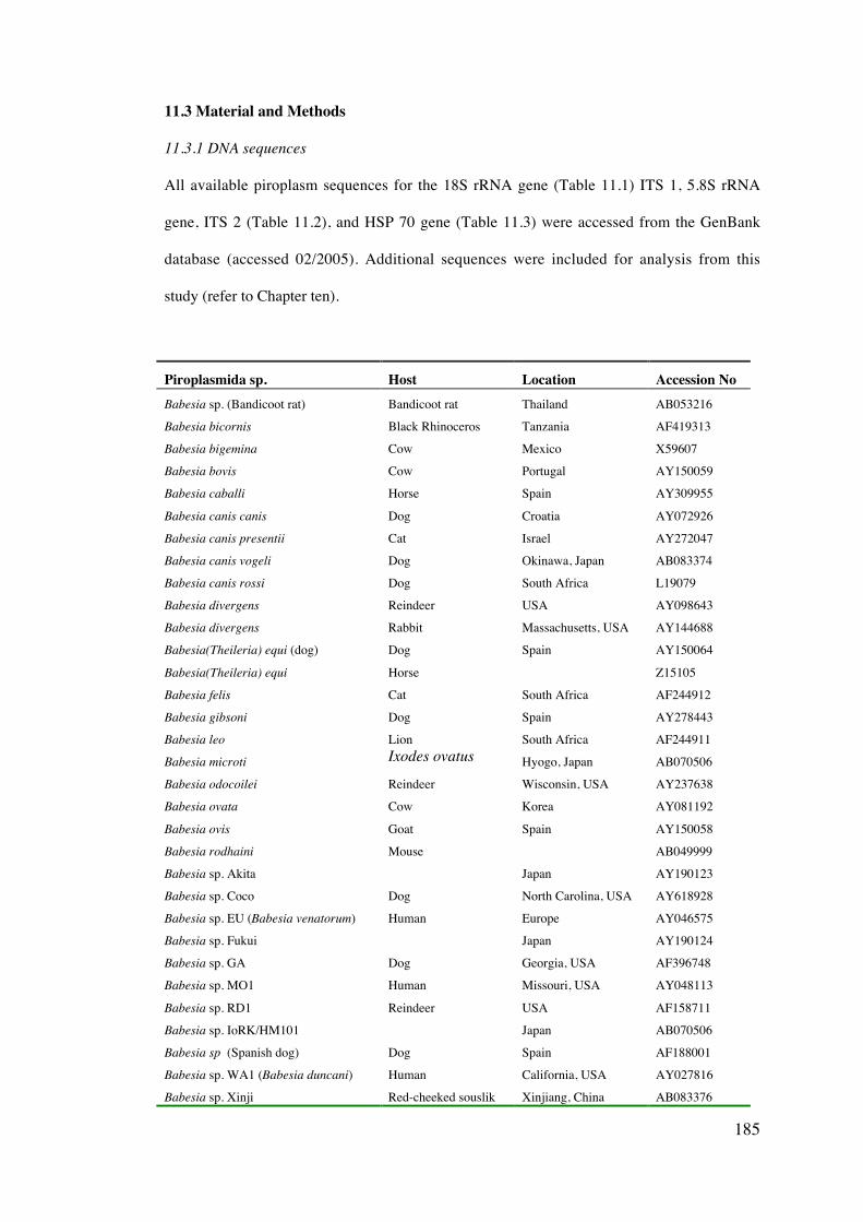
185
11.3 Material and Methods
11.3.1 DNA sequences
All available piroplasm sequences for the 18S rRNA gene (Table 11.1) ITS 1, 5.8S rRNA
gene, ITS 2 (Table 11.2), and HSP 70 gene (Table 11.3) were accessed from the GenBank
database (accessed 02/2005). Additional sequences were included for analysis from this
study (refer to Chapter ten).
Piroplasmida sp. Host Location Accession NoBabesia sp. (Bandicoot rat) Bandicoot rat Thailand AB053216
Babesia bicornis Black Rhinoceros Tanzania AF419313
Babesia bigemina Cow Mexico X59607
Babesia bovis Cow Portugal AY150059
Babesia caballi Horse Spain AY309955
Babesia canis canis Dog Croatia AY072926
Babesia canis presentii Cat Israel AY272047
Babesia canis vogeli Dog Okinawa, Japan AB083374
Babesia canis rossi Dog South Africa L19079
Babesia divergens Reindeer USA AY098643
Babesia divergens Rabbit Massachusetts, USA AY144688
Babesia(Theileria) equi (dog) Dog Spain AY150064
Babesia(Theileria) equi Horse Z15105
Babesia felis Cat South Africa AF244912
Babesia gibsoni Dog Spain AY278443
Babesia leo Lion South Africa AF244911
Babesia microti Ixodes ovatus Hyogo, Japan AB070506
Babesia odocoilei Reindeer Wisconsin, USA AY237638
Babesia ovata Cow Korea AY081192
Babesia ovis Goat Spain AY150058
Babesia rodhaini Mouse AB049999
Babesia sp. Akita Japan AY190123
Babesia sp. Coco Dog North Carolina, USA AY618928
Babesia sp. EU (Babesia venatorum) Human Europe AY046575
Babesia sp. Fukui Japan AY190124
Babesia sp. GA Dog Georgia, USA AF396748
Babesia sp. MO1 Human Missouri, USA AY048113
Babesia sp. RD1 Reindeer USA AF158711
Babesia sp. IoRK/HM101 Japan AB070506
Babesia sp (Spanish dog) Dog Spain AF188001
Babesia sp. WA1 (Babesia duncani) Human California, USA AY027816
Babesia sp. Xinji Red-cheeked souslik Xinjiang, China AB083376
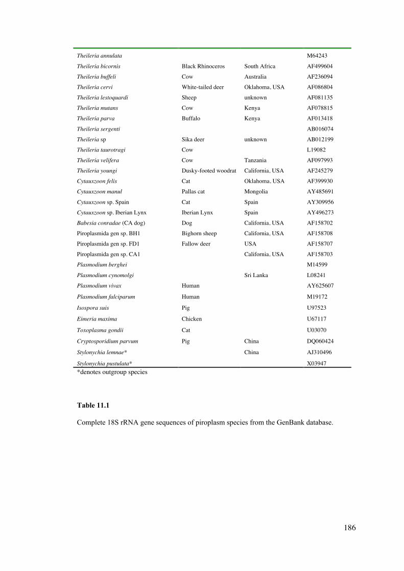
186
Theileria annulata M64243
Theileria bicornis Black Rhinoceros South Africa AF499604
Theileria buffeli Cow Australia AF236094
Theileria cervi White-tailed deer Oklahoma, USA AF086804
Theileria lestoquardi Sheep unknown AF081135
Theileria mutans Cow Kenya AF078815
Theileria parva Buffalo Kenya AF013418
Theileria sergenti AB016074
Theileria sp Sika deer unknown AB012199
Theileria taurotragi Cow L19082
Theileria velifera Cow Tanzania AF097993
Theileria youngi Dusky-footed woodrat California, USA AF245279
Cytauxzoon felis Cat Oklahoma, USA AF399930
Cytauxzoon manul Pallas cat Mongolia AY485691
Cytauxzoon sp. Spain Cat Spain AY309956
Cytauxzoon sp. Iberian Lynx Iberian Lynx Spain AY496273
Babesia conradae (CA dog) Dog California, USA AF158702
Piroplasmida gen sp. BH1 Bighorn sheep California, USA AF158708
Piroplasmida gen sp. FD1 Fallow deer USA AF158707
Piroplasmida gen sp. CA1 California, USA AF158703
Plasmodium berghei M14599
Plasmodium cynomolgi Sri Lanka L08241
Plasmodium vivax Human AY625607
Plasmodium falciparum Human M19172
Isospora suis Pig U97523
Eimeria maxima Chicken U67117
Toxoplasma gondii Cat U03070
Cryptosporidium parvum Pig China DQ060424
Stylonychia lemnae* China AJ310496
Stylonychia pustulata* X03947*denotes outgroup species
Table 11.1
Complete 18S rRNA gene sequences of piroplasm species from the GenBank database.
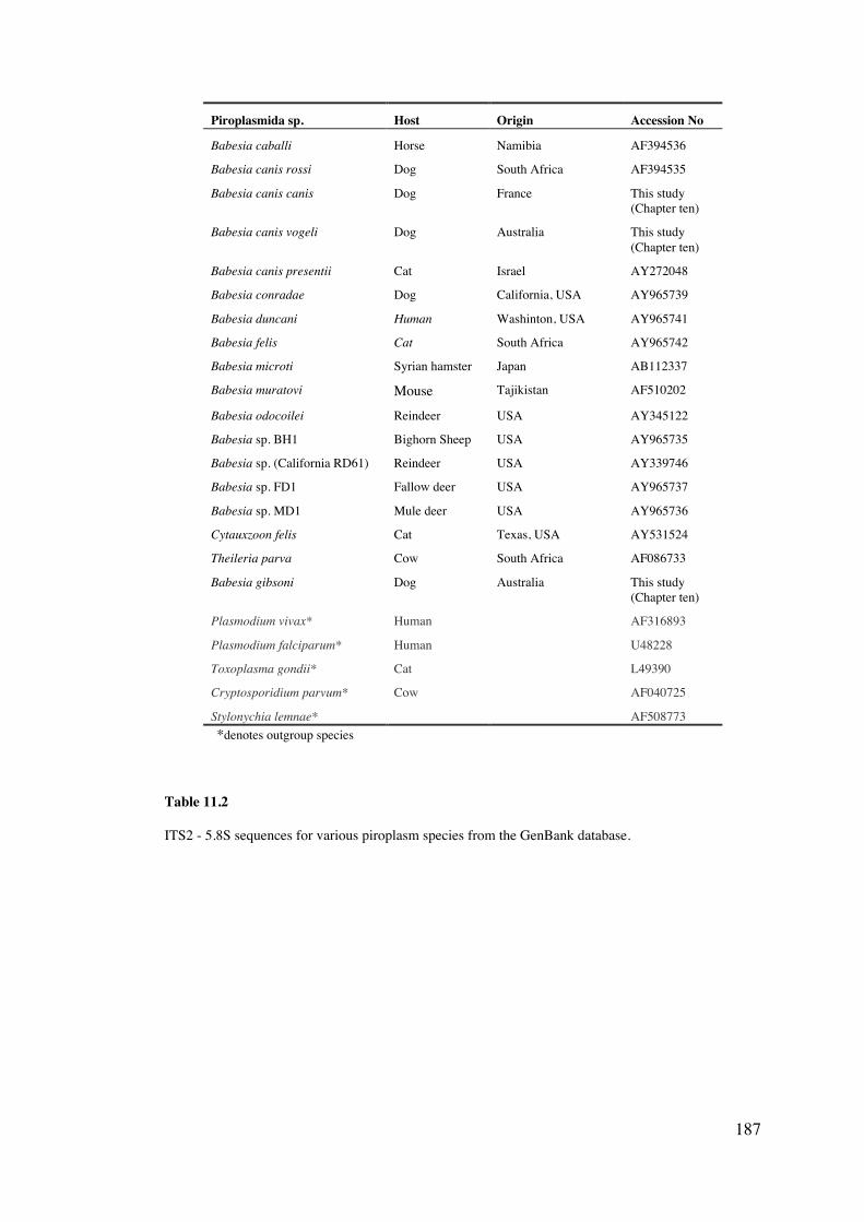
187
Piroplasmida sp. Host Origin Accession No
Babesia caballi Horse Namibia AF394536
Babesia canis rossi Dog South Africa AF394535
Babesia canis canis Dog France This study(Chapter ten)
Babesia canis vogeli Dog Australia This study(Chapter ten)
Babesia canis presentii Cat Israel AY272048
Babesia conradae Dog California, USA AY965739
Babesia duncani Human Washinton, USA AY965741
Babesia felis Cat South Africa AY965742
Babesia microti Syrian hamster Japan AB112337
Babesia muratovi Mouse Tajikistan AF510202
Babesia odocoilei Reindeer USA AY345122
Babesia sp. BH1 Bighorn Sheep USA AY965735
Babesia sp. (California RD61) Reindeer USA AY339746
Babesia sp. FD1 Fallow deer USA AY965737
Babesia sp. MD1 Mule deer USA AY965736
Cytauxzoon felis Cat Texas, USA AY531524
Theileria parva Cow South Africa AF086733
Babesia gibsoni Dog Australia This study(Chapter ten)
Plasmodium vivax* Human AF316893
Plasmodium falciparum* Human U48228
Toxoplasma gondii* Cat L49390
Cryptosporidium parvum* Cow AF040725
Stylonychia lemnae* AF508773*denotes outgroup species
Table 11.2
ITS2 - 5.8S sequences for various piroplasm species from the GenBank database.
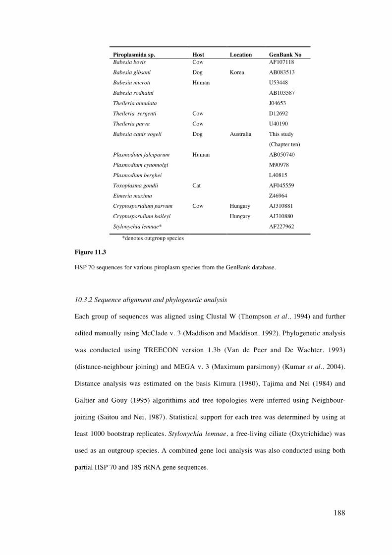
188
Piroplasmida sp. Host Location GenBank NoBabesia bovis Cow AF107118
Babesia gibsoni Dog Korea AB083513
Babesia microti Human U53448
Babesia rodhaini AB103587
Theileria annulata J04653
Theileria sergenti Cow D12692
Theileria parva Cow U40190
Babesia canis vogeli Dog Australia This study
(Chapter ten)
Plasmodium falciparum Human AB050740
Plasmodium cynomolgi M90978
Plasmodium berghei L40815
Toxoplasma gondii Cat AF045559
Eimeria maxima Z46964
Cryptosporidium parvum Cow Hungary AJ310881
Cryptosporidium baileyi Hungary AJ310880
Stylonychia lemnae* AF227962
*denotes outgroup species
Figure 11.3
HSP 70 sequences for various piroplasm species from the GenBank database.
10.3.2 Sequence alignment and phylogenetic analysis
Each group of sequences was aligned using Clustal W (Thompson et al., 1994) and further
edited manually using McClade v. 3 (Maddison and Maddison, 1992). Phylogenetic analysis
was conducted using TREECON version 1.3b (Van de Peer and De Wachter, 1993)
(distance-neighbour joining) and MEGA v. 3 (Maximum parsimony) (Kumar et al., 2004).
Distance analysis was estimated on the basis Kimura (1980), Tajima and Nei (1984) and
Galtier and Gouy (1995) algorithims and tree topologies were inferred using Neighbour-
joining (Saitou and Nei, 1987). Statistical support for each tree was determined by using at
least 1000 bootstrap replicates. Stylonychia lemnae, a free-living ciliate (Oxytrichidae) was
used as an outgroup species. A combined gene loci analysis was also conducted using both
partial HSP 70 and 18S rRNA gene sequences.
189
11.4 Results
11.4.1 Phylogeny of the Piroplasmida – 18S rRNA gene analysis
The separation of the Piroplasmida species into four distinct clades (Groups 1-46) on the
basis of the 18S rRNA gene was produced for maximum parsimony analyses, with strong
bootstrap support for Groups 1, 3 and 4 (Figure 11.1). Group 4 was most distantly related to
all other piroplasmid groups and included the B. microti related spp. Group 3, containing the
Babesia sp. WA1 related spp., was ancestral to both Groups 1 (B. bovis and B. canis related
spp.) and 2 (T. parva and Cytauxzoon related spp.). Distance based analysis also produced
four distinct piroplasmid clades with strong bootstrap support (Figures 11.2, 11.3). The
position of Groups 3 and 4 was shown to be influenced by the outgroup spp. used. When all
outgroup spp. were included in the analysis (Figure 11.2), both Groups 3 and 4 clustered
with Group 2 to form a separate clade to Group 1. A second analysis (Figure 11.3), using
only Eimeria, Isospora and Toxoplasma as outgroup spp. gave a tree structure more similar
to the maximum parasimony tree, with both Groups 3 and 4 placed ancestral to Groups 1 and
2.
The existence of additional clades within Groups 1 and 2 allowed for further sub-
categorization for both maximum parasimony (Figure 11.1) and distance (Figures 11.2, 11.3)
analysis. Group 1 was divided into two subgroups, 1a that includes B. canis and B. odocoilei
and 1b that includes B. bovis and B. bigemina. Group 2 was divided into 2a (the Cyauxzoon
spp. and B. bicornis) and 2b (the T. equi related spp.) both of which were ancestral to 2c (the
T. parva related spp.) Theileria youngi did not cluster with any of these subgroups but was
ancestral to group 2c. Limited bootstrap support was produced for the phylogenetic position
of 2a, 2b and T. youngi using maximum parsimony.
6 Group notation is consistently used for the same groups of species for all analyses throughoutsection 11.4.
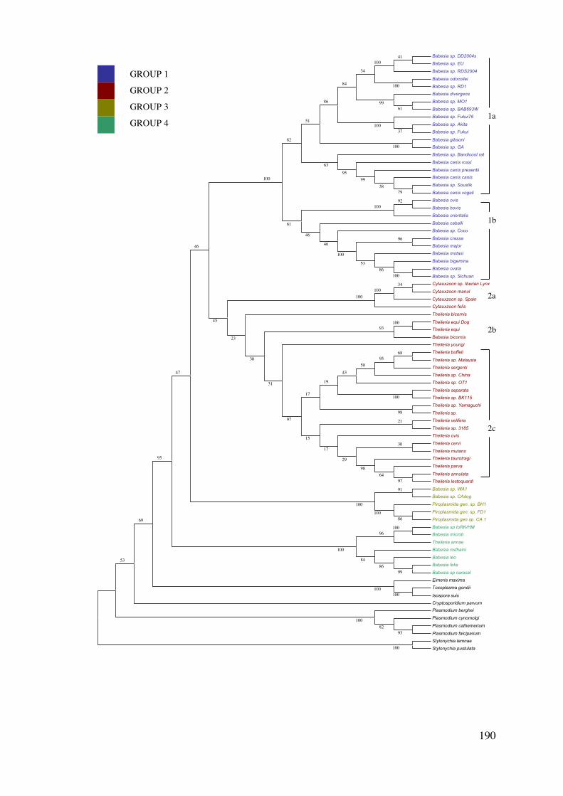
190
Babesia sp. DD2004s
Babesia sp. EU
Babesia sp. RDS2004
Babesia odocoilei
Babesia sp. RD1
Babesia divergens
Babesia sp. MO1
Babesia sp. BAB693W
Babesia sp. Fukui76
Babesia sp. Akita
Babesia sp. Fukui
Babesia gibsoni
Babesia sp. GA
Babesia sp. Bandicoot rat
Babesia canis rossi
Babesia canis presentii
Babesia canis canis
Babesia sp. Souslik
Babesia canis vogeli
Babesia ovis
Babesia bovis
Babesia orientalis
Babesia caballi
Babesia sp. Coco
Babesia crassa
Babesia major
Babesia motasi
Babesia bigemina
Babesia ovata
Babesia sp. Sichuan
Cytauxzoon sp. Iberian Lynx
Cytauxzoon manul
Cytauxzoon sp. Spain
Cytauxzoon felis
Theileria bicornis
Theileria equi Dog
Theileria equi
Babesia bicornis
Theileria youngi
Theileria buffeli
Theileria sp. Malaysia
Theileria sergenti
Theileria sp. China
Theileria sp. OT1
Theileria separata
Theileria sp. BK115
Theileria sp. Yamaguchi
Theileria sp.
Theileria velifera
Theileria sp. 3185
Theileria ovis
Theileria cervi
Theileria mutans
Theileria taurotragi
Theileria parva
Theileria annulata
Theileria lestoquardi
Babesia sp. WA1
Babesia sp. CAdog
Piroplasmida gen. sp. BH1
Piroplasmida gen. sp. FD1
Piroplasmida gen sp. CA 1
Babesia sp IoRK/HM
Babesia microti
Theileria annae
Babesia rodhaini
Babesia leo
Babesia felis
Babesia sp caracal
Eimeria maxima
Toxoplasma gondii
Isospora suis
Cryptosporidium parvum
Plasmodium berghei
Plasmodium cynomolgi
Plasmodium cathemerium
Plasmodium falciparium
Stylonychia lemnae
Stylonychia pustulata
100100
86100
100
37100
100
96
8653
100
92100
91
100
34100
100
99
96
8684
100
100
100
41100
100
6199
7938
9995
34
84
86
63
51
82
46
46
61
100
100
100
9764
98
98
6895
5043
30
29
21
19
17
17
15
97
93
31
30
23
45
46
47
95
69
53
9382
100
1b
2a
2b
2c
GROUP 1
GROUP 2
GROUP 3
GROUP 41a
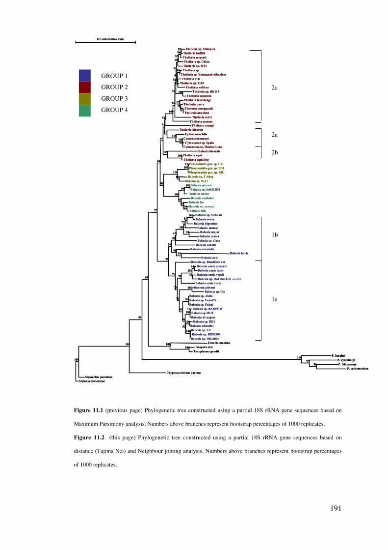
191
Figure 11.1 (previous page) Phylogenetic tree constructed using a partial 18S rRNA gene sequences based on
Maximum Parsimony analysis. Numbers above branches represent bootstrap percentages of 1000 replicates.
Figure 11.2 (this page) Phylogenetic tree constructed using a partial 18S rRNA gene sequences based on
distance (Tajima Nei) and Neighbour joining analysis. Numbers above branches represent bootstrap percentages
of 1000 replicates.
2c
2a
2b
1b
1a
0.1 substitutions/site
Stylonychia lemnaeStylonchia pustulata
Cryptosporidium parvum
Babesia sp. Bandicoot rat
Theileria youngiTheileria mutans
Theileria cervi
Babesia caballiBabesia sp. Coco
Babesia canis rossi
Theileria bicornis
Theileria sp. OT1
Babesia rodhaini
P. berghei
Babesia motasi
Cytauxzoon felis
Theileria taurotragi
Theileria sp. China
Theileria annae
Babesia leo
Babesia bigemina
Babesia sp. EU
Eimeria maxima
Babesia divergens
Babesia sp. Fukui
Babesia orientalis
Babesia bicornis
Cytauxzoon manul
Theileria parva
P. cynomolgi
Theileria sergenti
Piroplasmida gen. sp. BH1
Theileria ovis
Theileria sp. MalaysiaTheileria buffeli
Theileria sp.Theileria sp. Yamaguchi sika deer
Theileria sp. 3185Theileria velifera
Theileria sp. BK115Theileria separata
Theileria lestoquardiTheileria annulata
Cytauxzoon sp. SpainCytauxzoon sp. Iberian Lynx
Theileria equiTheileria equi Dog
Piroplasmida gen. sp. CAPiroplasmida gen. sp. FD1
Babesia sp. CAdogBabesia sp. WA1
Babesia microtiBabesia sp. IoR/KHM
Babesia sp. caracalBabesia felis
Babesia sp. SichuanBabesia ovata
Babesia majorBabesia crassa
Babesia bovis
Babesia ovis
Babesia canis presentiiBabesia canis canisBabesia canis vogeliBabesia sp. Red cheeked souslik
Babesia gibsoniBabesia sp. GA
Babesia sp. AkitaBabesia sp. Fukui76
Babesia sp. BAB693WBabesia sp MO1
Babesia sp. RD1Babesia odocoilei
Babesia sp. RDS2004Babesia sp. DD2004s
Isospora suisToxoplasma gondii
P. falciparumP. cathemerium
100
100
99
65
97
84
99
87
74
97
51
75
100
74
36
70
88
58
99
56
23
96
91
100
45
100
100
53
90
81
59
41
95
100
72
100
99
53
100
99
93
83
100
100
100
100
98
95
100
89
97
96
100
16
52
99
26
100
98
99
100
91
80
100
99
100
98
59
94
48
100
71
100
100
98
0.1 substitutions/site
Stylonychia lemnaeStylonchia pustulata
Cryptosporidium parvum
Babesia sp. Bandicoot rat
Theileria youngiTheileria mutans
Theileria cervi
Babesia caballiBabesia sp. Coco
Babesia canis rossi
Theileria bicornis
Theileria sp. OT1
Babesia rodhaini
P. berghei
Babesia motasi
Cytauxzoon felis
Theileria taurotragi
P. berghei
Babesia motasi
Cytauxzoon felis
Theileria taurotragi
Theileria sp. China
Theileria annae
Babesia leo
Babesia bigemina
Babesia sp. EU
Eimeria maxima
Babesia divergens
Babesia sp. Fukui
Babesia orientalis
Babesia bicornis
Cytauxzoon manul
Theileria parva
P. cynomolgi
Theileria sergenti
Piroplasmida gen. sp. BH1
Theileria ovis
Theileria sp. MalaysiaTheileria buffeli
Theileria sp.Theileria sp. Yamaguchi sika deer
Theileria sp. 3185Theileria velifera
Theileria sp. BK115Theileria separata
Theileria lestoquardiTheileria annulata
Cytauxzoon sp. SpainCytauxzoon sp. Iberian Lynx
Theileria equiTheileria equi Dog
Piroplasmida gen. sp. CAPiroplasmida gen. sp. FD1
Babesia sp. CAdogBabesia sp. WA1
Babesia microtiBabesia sp. IoR/KHM
Babesia sp. caracalBabesia felis
Babesia sp. SichuanBabesia ovata
Babesia majorBabesia crassa
Babesia bovisBabesia ovis
Babesia canis presentiiBabesia canis canisBabesia canis vogeliBabesia sp. Red cheeked
Babesia gibsoniBabesia sp. GA
Babesia sp. AkitaBabesia sp. Fukui76
Babesia sp. BAB693WBabesia sp MO1
Babesia sp. RD1Babesia odocoilei
Babesia sp. RDS2004Babesia sp. DD2004s
Isospora suisToxoplasma gondii
P. falciparumP. cathemerium
100
100
99
65
97
84
99
87
74
97
51
75
100
74
36
70
88
58
99
56
23
96
91
100
45
100
100
53
90
81
59
41
95
100
72
100
99
53
100
99
93
83
100
100
100
100
98
95
100
89
97
96
100
16
52
99
26
100
98
99
100
91
80
100
99
100
98
59
94
48
100
71
100
100
98
GROUP 1
GROUP 2
GROUP 3
GROUP 4
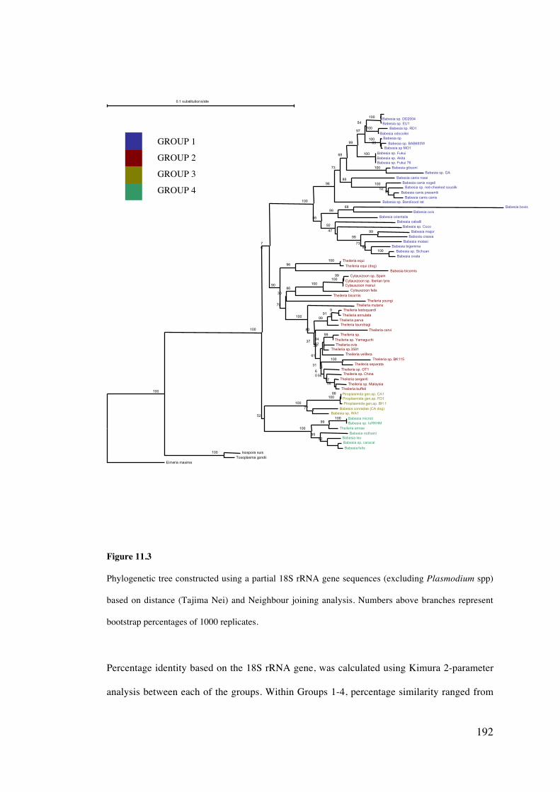
192
Figure 11.3
Phylogenetic tree constructed using a partial 18S rRNA gene sequences (excluding Plasmodium spp)
based on distance (Tajima Nei) and Neighbour joining analysis. Numbers above branches represent
bootstrap percentages of 1000 replicates.
Percentage identity based on the 18S rRNA gene, was calculated using Kimura 2-parameter
analysis between each of the groups. Within Groups 1-4, percentage similarity ranged from
0.1 substitutions/site
Eimer ia maxima
Babesia sp. Bandicoot rat
Theileria mutans
Theileria cervi
Babesia caballiBabesia sp. Coco
Theileria youngi
Babesia canis rossi
Theileria bicornis
Theileria sp. OT1
Theileria velifera
Babesia motasi
Babesia rodhaini
Cytauxzoon felis
Babesia canis vogeli
Theileria taurotragi
Theileria sp. China.
Babesia bigemina
Theileria annae
Babesia sp
Babesia sp. EU1
Babesia leo
Cytauxzoon manul
Babesia sp. Fukui
Babesia orientalis
Babesia bicornis
Babesia sp. red-cheeked souslik
Theileria parva
Theileria sergenti
Piroplasmida gen sp. BH 1.
Babesia sp. DD2004
Babesia sp. RD1
Babesia odocoilei
Babesia sp. BAB693WBabesia sp MO1
Babesia sp. AkitaBabesia sp. Fukui 76
Babesia gibsoniBabesia sp. GA
Babesia canis presentiiBabesia canis canis
Babesia bovisBabesia ovis
Babesia majorBabesia crassa
Babesia sp. SichuanBabesia ovata
Theileria equi
Theileria equi (dog)
Cytauxzoon sp. Spain.Cytauxzoon sp. Iberian lynx
Theileria lestoquardi
Theileria annulata
Theileria sp.
Theileria sp. YamaguchiTheileria ovis
Theileria sp 3581.
Theileria sp. BK115.Theileria separata
Theileria sp. MalaysiaTheileria buffeli
Piroplasmida gen sp. CA1.Piroplasmida gen sp. FD1.
Babesia conradae (CA dog)Babesia sp. WA1
Babesia microtiBabesia sp. IoRKHM
Babesia sp. caracal
Babesia felisIsospora suis
Toxoplasma gondii
100
100
76
100
90
70
96
73
100
80
68
37
52
61
99
56
97
92
100
47
31
99
30
54
88
86
100
60
38
73
95
100
100
99
56
34
84
99
100
100
92
100
100
99
96
52
91
97
100
100
69
100
93
68
99
100
100
99
99
99
32
100
58
88
77
100
100
GROUP 1
GROUP 2
GROUP 3
GROUP 4
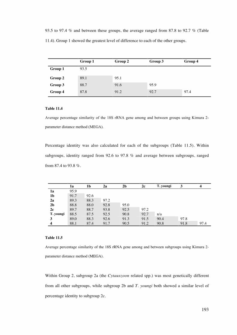
193
93.5 to 97.4 % and between these groups, the average ranged from 87.8 to 92.7 % (Table
11.4). Group 1 showed the greatest level of difference to each of the other groups.
Group 1 Group 2 Group 3 Group 4
Group 1 93.5
Group 2 89.1 95.1
Group 3 88.7 91.6 95.9
Group 4 87.8 91.2 92.7 97.4
Table 11.4
Average percentage similarity of the 18S rRNA gene among and between groups using Kimura 2-
parameter distance method (MEGA).
Percentage identity was also calculated for each of the subgroups (Table 11.5). Within
subgroups, identity ranged from 92.6 to 97.8 % and average between subgroups, ranged
from 87.4 to 93.8 %.
1a 1b 2a 2b 2c T. youngi 3 41a 95.91b 91.7 92.62a 89.3 88.3 97.22b 88.8 88.0 92.8 95.02c 89.7 88.7 93.8 92.5 97.2T. youngi 88.5 87.5 92.5 90.8 92.7 n/a3 89.0 88.3 92.6 91.3 91.5 90.4 97.84 88.1 87.4 91.7 90.5 91.2 90.8 91.8 97.4
Table 11.5
Average percentage similarity of the 18S rRNA gene among and between subgroups using Kimura 2-
parameter distance method (MEGA).
Within Group 2, subgroup 2a (the Cytauxzoon related spp.) was most genetically different
from all other subgroups, while subgroup 2b and T. youngi both showed a similar level of
percentage identity to subgroup 2c.
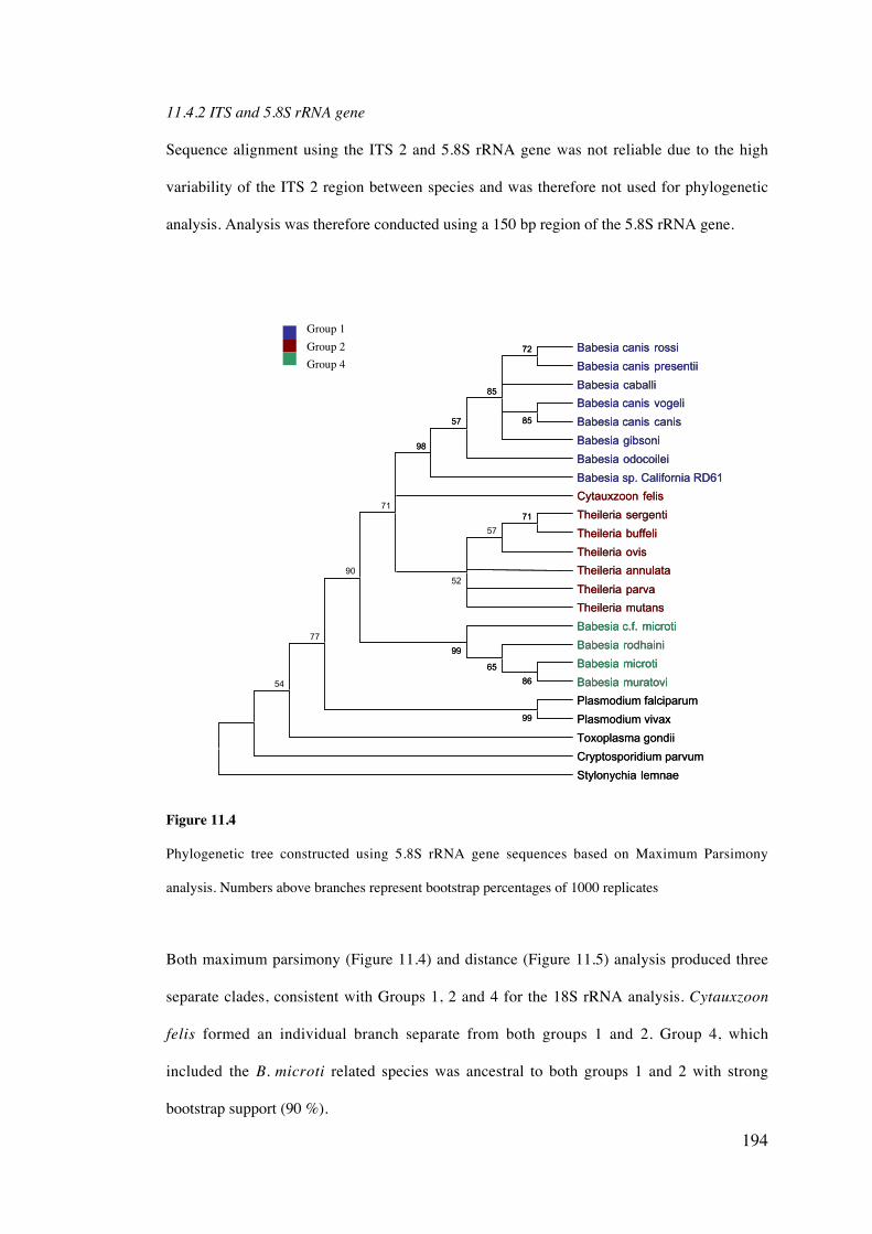
194
11.4.2 ITS and 5.8S rRNA gene
Sequence alignment using the ITS 2 and 5.8S rRNA gene was not reliable due to the high
variability of the ITS 2 region between species and was therefore not used for phylogenetic
analysis. Analysis was therefore conducted using a 150 bp region of the 5.8S rRNA gene.
Figure 11.4
Phylogenetic tree constructed using 5.8S rRNA gene sequences based on Maximum Parsimony
analysis. Numbers above branches represent bootstrap percentages of 1000 replicates
Both maximum parsimony (Figure 11.4) and distance (Figure 11.5) analysis produced three
separate clades, consistent with Groups 1, 2 and 4 for the 18S rRNA analysis. Cytauxzoon
felis formed an individual branch separate from both groups 1 and 2. Group 4, which
included the B. microti related species was ancestral to both groups 1 and 2 with strong
bootstrap support (90 %).
Babesia canis rossi
Babesia canis presentii
Babesia caballi
Babesia canis vogeli
Babesia canis canis
Babesia gibsoni
Babesia odocoilei
Babesia sp. California RD61
Cytauxzoon felis
Theileria sergenti
Theileria buffeli
Theileria ovis
Theileria annulata
Theileria parva
Theileria mutans
Babesia c.f. microti
Babesia rodhaini
Babesia microti
Babesia muratovi
Plasmodium falciparum
Plasmodium vivax
Toxoplasma gondii
Cryptosporidium parvum
Stylonychia lemnae
99
86
65
99
85
72
85
57
98
71
57
52
71
90
77
54
Babesia canis rossi
Babesia canis presentii
Babesia caballi
Babesia canis vogeli
Babesia canis canis
Babesia gibsoni
Babesia odocoilei
Babesia sp. California RD61
Cytauxzoon felis
Theileria sergenti
Theileria buffeli
Theileria ovis
Theileria annulata
Theileria parva
Theileria mutans
Babesia c.f. microti
Babesia rodhaini
Babesia microti
Babesia muratovi
Plasmodium falciparum
Plasmodium vivax
Toxoplasma gondii
Cryptosporidium parvum
Stylonychia lemnae
99
86
65
99
85
72
85
57
98
71
Group 1Group 2Group 4
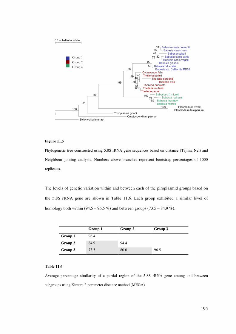
195
Figure 11.5
Phylogenetic tree constructed using 5.8S rRNA gene sequences based on distance (Tajima Nei) and
Neighbour joining analysis. Numbers above branches represent bootstrap percentages of 1000
replicates.
The levels of genetic variation within and between each of the piroplasmid groups based on
the 5.8S rRNA gene are shown in Table 11.6. Each group exhibited a similar level of
homology both within (94.5 – 96.5 %) and between groups (73.5 – 84.9 %).
Group 1 Group 2 Group 3
Group 1 96.4
Group 2 84.9 94.4
Group 3 73.5 80.0 96.5
Table 11.6
Average percentage similarity of a partial region of the 5.8S rRNA gene among and between
subgroups using Kimura 2-parameter distance method (MEGA).
0.1 substitutions/site
Stylonychia lemnaeCryptosporidium parvum
Toxoplasma gondii
Cytauxzoon felis
Babesia gibsoni
Babesia c.f. microti
Theileria annulataTheileria ovis
Babesia rodhaini
Babesia caballi
Babesia canis presentiiBabesia canis rossi
Babesia canis canisBabesia canis vogeli
Babesia odocoileiBabesia sp. California RD61
Theileria buffeliTheileria sergenti
Theileria mutansTheileria parva
Babesia muratoviBabesia microti
Plasmodium vivaxPlasmodium falciparium100
61
59
99
68
99
49
50
78
47
100
72
43
78
8063
82
58
40
63
82
100
Group 1Group 2Group 4
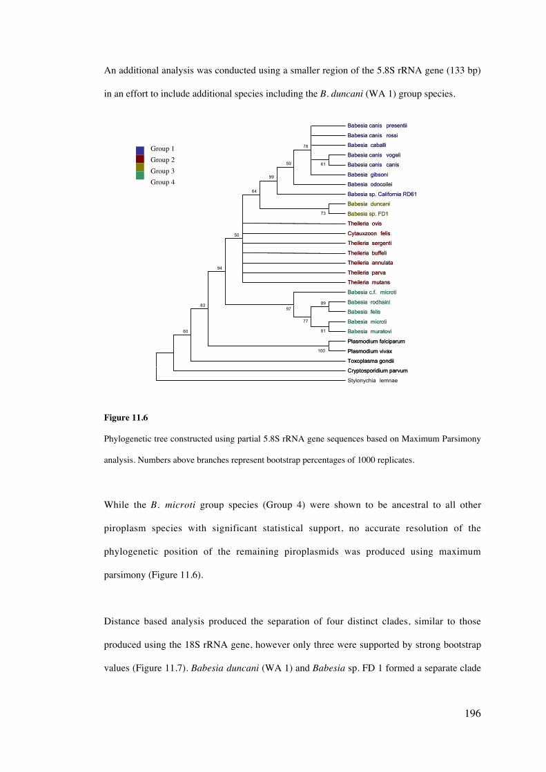
196
An additional analysis was conducted using a smaller region of the 5.8S rRNA gene (133 bp)
in an effort to include additional species including the B. duncani (WA 1) group species.
Figure 11.6
Phylogenetic tree constructed using partial 5.8S rRNA gene sequences based on Maximum Parsimony
analysis. Numbers above branches represent bootstrap percentages of 1000 replicates.
While the B. microti group species (Group 4) were shown to be ancestral to all other
piroplasm species with significant statistical support, no accurate resolution of the
phylogenetic position of the remaining piroplasmids was produced using maximum
parsimony (Figure 11.6).
Distance based analysis produced the separation of four distinct clades, similar to those
produced using the 18S rRNA gene, however only three were supported by strong bootstrap
values (Figure 11.7). Babesia duncani (WA 1) and Babesia sp. FD 1 formed a separate clade
Babesia canis presentii
Babesia canis rossi
Babesia caballi
Babesia canis vogeli
Babesia canis canis
Babesia gibsoni
Babesia odocoilei
Babesia sp. California RD61
Babesia duncani
Babesia sp. FD1
Theileria ovis
Cytauxzoon felis
Theileria sergenti
Theileria buffeli
Theileria annulata
Theileria parva
Theileria mutans
Babesia c.f. microti
Babesia rodhaini
Babesia felis
Babesia microti
Babesia muratovi
Plasmodium falciparum
Plasmodium vivax
Toxoplasma gondii
Cryptosporidium parvum
Stylonychia lemnae
100
61
78
59
99
89
81
77
97
73
64
50
94
83
60
Babesia canis presentii
Babesia canis rossi
Babesia caballi
Babesia canis vogeli
Babesia canis canis
Babesia gibsoni
Babesia odocoilei
Babesia sp. California RD61
Babesia duncani
Babesia sp. FD1
Theileria ovis
Cytauxzoon felis
Theileria sergenti
Theileria buffeli
Theileria annulata
Theileria parva
Theileria mutans
Babesia c.f. microti
Babesia rodhaini
Babesia felis
Babesia microti
Babesia muratovi
Plasmodium falciparum
Plasmodium vivax
Toxoplasma gondii
Cryptosporidium parvum
Group 1Group 2Group 3Group 4
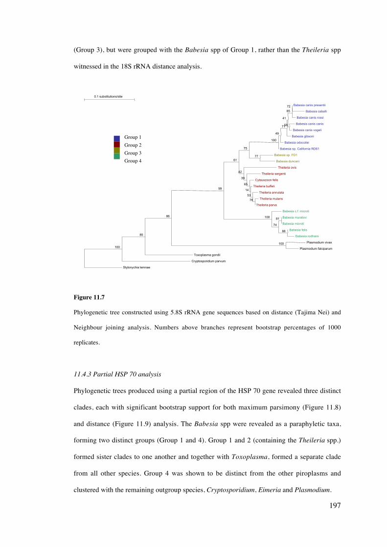
197
(Group 3), but were grouped with the Babesia spp of Group 1, rather than the Theileria spp
witnessed in the 18S rRNA distance analysis.
Figure 11.7
Phylogenetic tree constructed using 5.8S rRNA gene sequences based on distance (Tajima Nei) and
Neighbour joining analysis. Numbers above branches represent bootstrap percentages of 1000
replicates.
11.4.3 Partial HSP 70 analysis
Phylogenetic trees produced using a partial region of the HSP 70 gene revealed three distinct
clades, each with significant bootstrap support for both maximum parsimony (Figure 11.8)
and distance (Figure 11.9) analysis. The Babesia spp were revealed as a paraphyletic taxa,
forming two distinct groups (Group 1 and 4). Group 1 and 2 (containing the Theileria spp.)
formed sister clades to one another and together with Toxoplasma, formed a separate clade
from all other species. Group 4 was shown to be distinct from the other piroplasms and
clustered with the remaining outgroup species, Cryptosporidium, Eimeria and Plasmodium.
0.1 substitutions/site
Stylonychia lemnae
Cryptosporidium parvum
Toxoplasma gondii
Babesia sp. California RD61
Babesia odocoilei
Theileria ovis
Babesia gibsoni
Theileria sergenti
Babesia c.f. microti
Cytauxzoon felis
Theileria buffeli
Babesia canis rossi
Theileria annulata
Babesia canis presentii
Babesia caballi
Babesia canis canis
Babesia canis vogeli
Babesia sp. FD1
Babesia duncani
Theileria mutans
Theileria parva
Babesia muratovi
Babesia microti
Babesia felis
Babesia rodhaini
Plasmodium vivax
Plasmodium falciparum100
85
86
99
61
75
100
49
42
71
39
100
41
65
74
14
85
53
72
58
77
76
81
88
100
Group 1Group 2Group 3Group 4
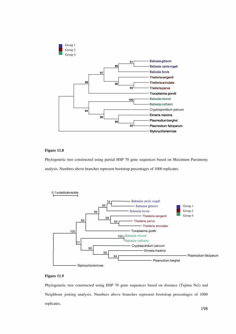
198
Figure 11.8
Phylogenetic tree constructed using partial HSP 70 gene sequences based on Maximum Parsimony
analysis. Numbers above branches represent bootstrap percentages of 1000 replicates.
Figure 11.9
Phylogenetic tree constructed using HSP 70 gene sequences based on distance (Tajima Nei) and
Neighbour joining analysis. Numbers above branches represent bootstrap percentages of 1000
replicates.
Babesia gibsoni
Babesia canisvogeli
Babesia bovis
Theileriasergenti
Theileriaannulata
Theileriaparva
Toxoplasma gondii
Babesia microti
Babesia rodhaini
Cryptosporidium parvum
Eimeria maxima
Plasmodiumberghei
Plasmodium falciparum
Stylonychialemnae
100
65
98
81
88
97
89
84
86
69
99
Babesia gibsoni
Babesia canisvogeli
Babesia bovis
Theileriasergenti
Theileriaannulata
Theileriaparva
Toxoplasma gondii
Babesia microti
Babesia rodhaini
Eimeria maxima
Plasmodiumberghei
Plasmodium falciparum
Stylonychialemnae
100
65
98
81
88
97
89
84
86
69
99
0.1 substitutions/site
Stylonychia lemnae
Toxoplasma gondii
Cryptosporidium parvum
Eimeria maxima
Theileria sergenti
Babesia bovis
Babesia canis vogeli
Babesia gibsoni
Theileria parva
Theileria annulata
Babesia microti
Babesia rodhaini
Plasmodium falciparum
Plasmodium berghei
100
89
77
99
9999
99
9978
94
100
94
Group 1Group 2Group 4
Group 1Group 2Group 4
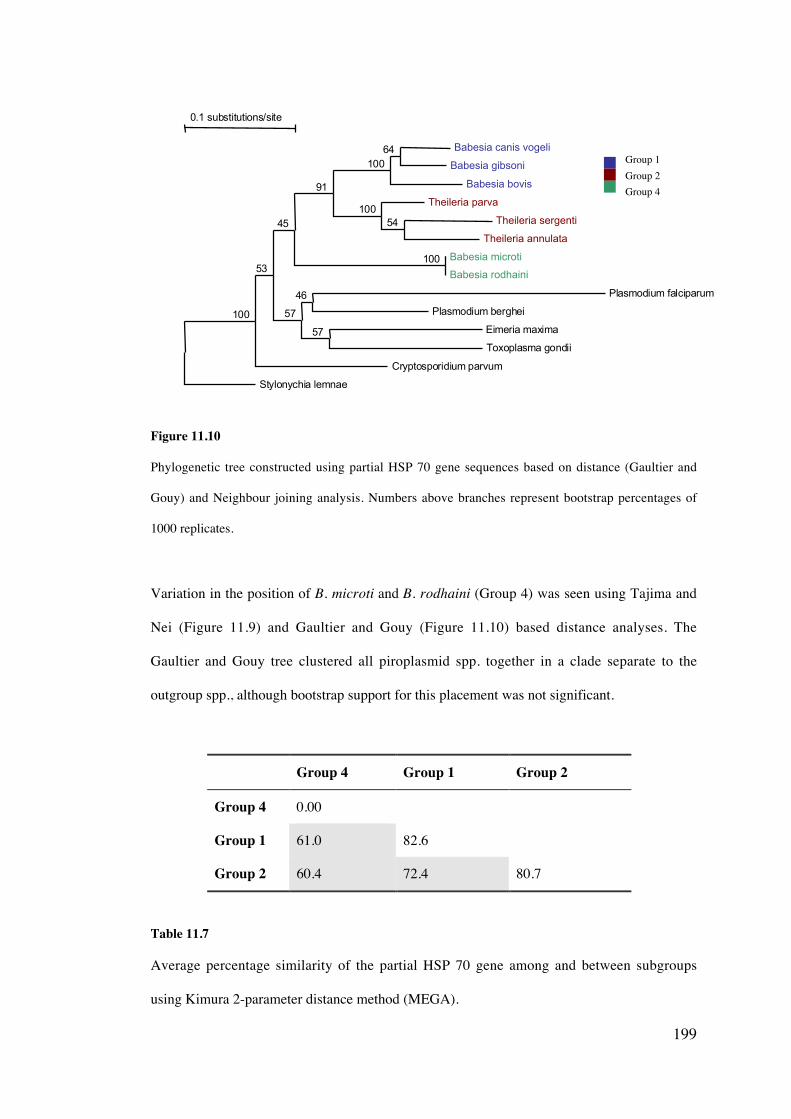
199
Figure 11.10
Phylogenetic tree constructed using partial HSP 70 gene sequences based on distance (Gaultier and
Gouy) and Neighbour joining analysis. Numbers above branches represent bootstrap percentages of
1000 replicates.
Variation in the position of B. microti and B. rodhaini (Group 4) was seen using Tajima and
Nei (Figure 11.9) and Gaultier and Gouy (Figure 11.10) based distance analyses. The
Gaultier and Gouy tree clustered all piroplasmid spp. together in a clade separate to the
outgroup spp., although bootstrap support for this placement was not significant.
Group 4 Group 1 Group 2
Group 4 0.00
Group 1 61.0 82.6
Group 2 60.4 72.4 80.7
Table 11.7
Average percentage similarity of the partial HSP 70 gene among and between subgroups
using Kimura 2-parameter distance method (MEGA).
0.1 substitutions/site
Stylonychia lemnae
Cryptosporidium parvum
Babesia bovis
Theileria parva
Babesia canis vogeli
Babesia gibsoni
Theileria sergenti
Theileria annulata
Babesia microti
Babesia rodhaini
Plasmodium falciparum
Plasmodium berghei
Eimeria maxima
Toxoplasma gondii
100
53
45
91
57
100
100
64
54
100
46
57
Group 1Group 2Group 4
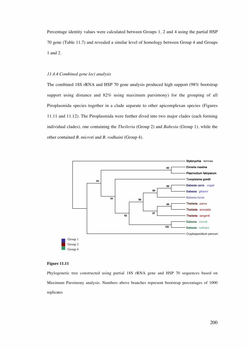
200
Percentage identity values were calculated between Groups 1, 2 and 4 using the partial HSP
70 gene (Table 11.7) and revealed a similar level of homology between Group 4 and Groups
1 and 2.
11.4.4 Combined gene loci analysis
The combined 18S rRNA and HSP 70 gene analysis produced high support (98% bootstrap
support using distance and 82% using maximum parsimony) for the grouping of all
Piroplasmida species together in a clade separate to other apicomplexan species (Figures
11.11 and 11.12). The Piroplasmida were further dived into two major clades (each forming
individual clades), one containing the Theileria (Group 2) and Babesia (Group 1), while the
other contained B. microti and B. rodhaini (Group 4).
Figure 11.11
Phylogenetic tree constructed using partial 18S rRNA gene and HSP 70 sequences based on
Maximum Parsimony analysis. Numbers above branches represent bootstrap percentages of 1000
replicates
Stylonychia lemnae
Eimeria maxima
Plasmodium falciparum
Toxoplasma gondii
Babesia canis vogeli
Babesia gibsoni
Babesia bovis
Theileria parva
Theileria annulata
Theileria sergenti
Babesia microti
Babesia rodhaini
Cryptosporidium parvum
100
88
99
96
97
99
82
34
44
60
Stylonychia
Eimeria maxima
Plasmodium falciparum
Toxoplasma gondii
Babesia canis
Babesia
Theileria
Theileria
Theileria
Babesia
Babesia100
88
99
96
97
99
82
34
44
60
Group 1Group 2Group 4
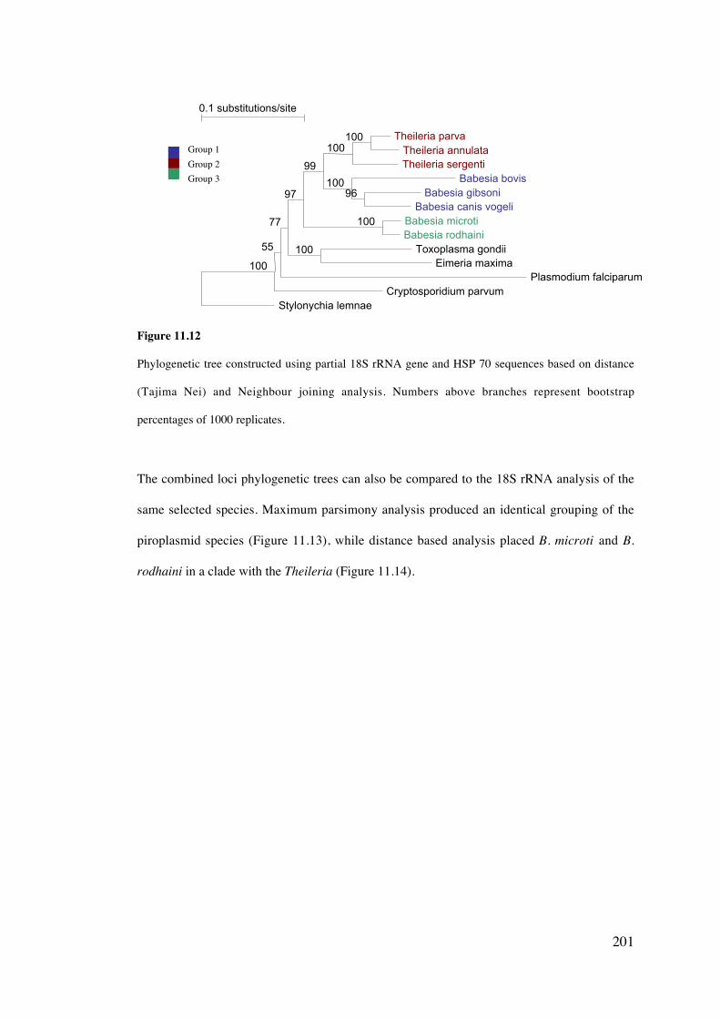
201
Figure 11.12
Phylogenetic tree constructed using partial 18S rRNA gene and HSP 70 sequences based on distance
(Tajima Nei) and Neighbour joining analysis. Numbers above branches represent bootstrap
percentages of 1000 replicates.
The combined loci phylogenetic trees can also be compared to the 18S rRNA analysis of the
same selected species. Maximum parsimony analysis produced an identical grouping of the
piroplasmid species (Figure 11.13), while distance based analysis placed B. microti and B.
rodhaini in a clade with the Theileria (Figure 11.14).
0.1 substitutions/site
Stylonychia lemnaeCryptosporidium parvum
Plasmodium falciparum
Babesia bovisTheileria sergenti
Theileria parvaTheileria annulata
Babesia gibsoniBabesia canis vogeli
Babesia microtiBabesia rodhaini
Toxoplasma gondiiEimeria maxima100
55
77
97
99
100
100100
96
100
100
Group 1Group 2Group 3
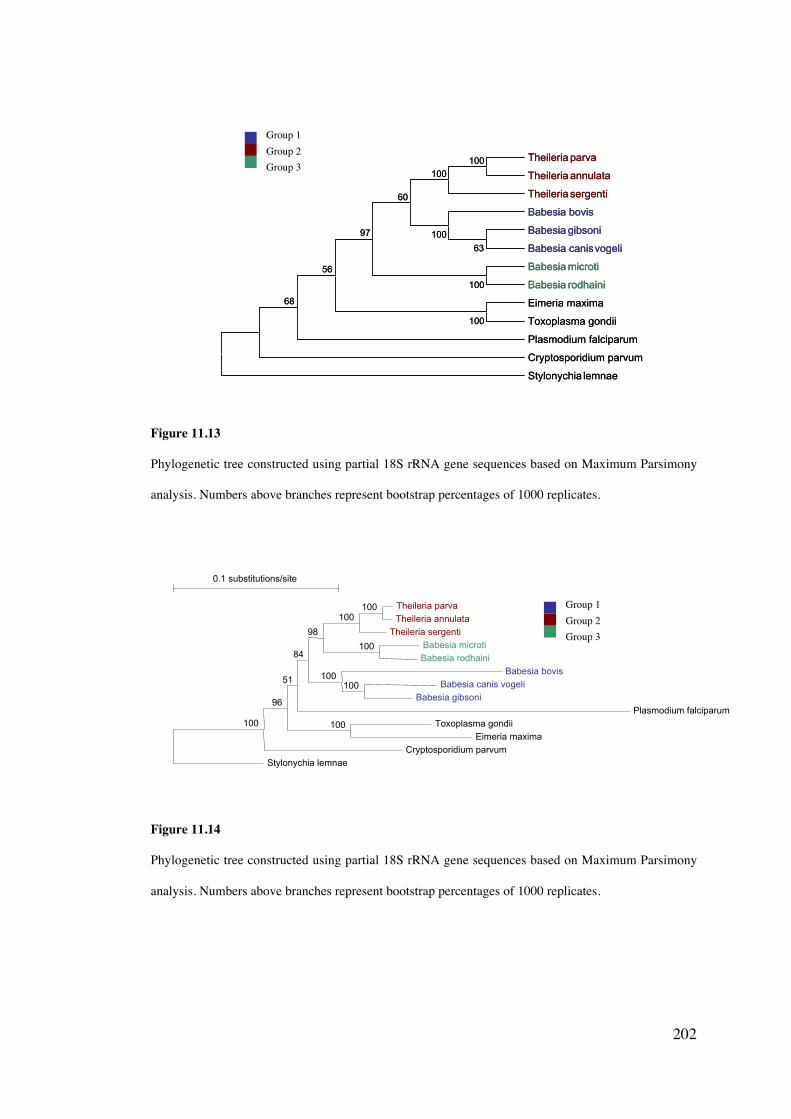
202
Figure 11.13
Phylogenetic tree constructed using partial 18S rRNA gene sequences based on Maximum Parsimony
analysis. Numbers above branches represent bootstrap percentages of 1000 replicates.
Figure 11.14
Phylogenetic tree constructed using partial 18S rRNA gene sequences based on Maximum Parsimony
analysis. Numbers above branches represent bootstrap percentages of 1000 replicates.
0.1 substitutions/site
Stylonychia lemnae
Cryptosporidium parvum
Plasmodium falciparum
Theileria sergenti
Babesia bovis
Theileria parva
Theileria annulata
Babesia microti
Babesia rodhaini
Babesia canis vogeli
Babesia gibsoni
Toxoplasma gondii
Eimeria maxima
100
96
51
84
98
100
100
100
100
100
100
Theileria parva
Theileria annulata
Theileria sergenti
Babesia bovis
Babesia gibsoni
Babesia canisvogeli
Babesia microti
Babesia rodhaini
Eimeria maxima
Toxoplasma gondii
Plasmodium falciparum
Cryptosporidium parvum
Stylonychia lemnae
100
100
100
100
63
100
60
97
56
68
Theileria parva
Theileria annulata
Theileria sergenti
Babesia bovis
Babesia gibsoni
Babesia canisvogeli
Babesia microti
Babesia rodhaini
Eimeria maxima
Toxoplasma gondii
Plasmodium falciparum
Cryptosporidium parvum
Stylonychia lemnae
100
100
100
100
63
100
60
97
56
68
Group 1Group 2Group 3
Group 1Group 2Group 3
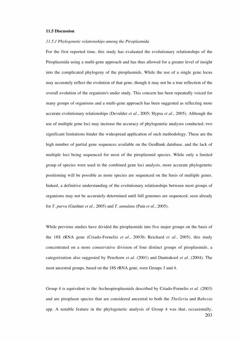
203
11.5 Discussion
11.5.1 Phylogenetic relationships among the Piroplasmida
For the first reported time, this study has evaluated the evolutionary relationships of the
Piroplasmida using a multi-gene approach and has thus allowed for a greater level of insight
into the complicated phylogeny of the piroplasmids. While the use of a single gene locus
may accurately reflect the evolution of that gene, though it may not be a true reflection of the
overall evolution of the organism/s under study. This concern has been repeatedly voiced for
many groups of organisms and a multi-gene approach has been suggested as reflecting more
accurate evolutionary relationships (Devulder et al., 2005; Hypsa et al., 2005). Although the
use of multiple gene loci may increase the accuracy of phylogenetic analyses conducted, two
significant limitations hinder the widespread application of such methodology. These are the
high number of partial gene sequences available on the GenBank database, and the lack of
multiple loci being sequenced for most of the piroplasmid species. While only a limited
group of species were used in the combined gene loci analysis, more accurate phylogenetic
positioning will be possible as more species are sequenced on the basis of multiple genes.
Indeed, a definitive understanding of the evolutionary relationships between most groups of
organisms may not be accurately determined until full genomes are sequenced, seen already
for T. parva (Gardner et al., 2005) and T. annulata (Pain et al., 2005).
While previous studies have divided the piroplasmids into five major groups on the basis of
the 18S rRNA gene (Criado-Fornelio et al., 2003b; Reichard et al., 2005), this study
concentrated on a more conservative division of four distinct groups of piroplasmids, a
categorization also suggested by Penzhorn et al. (2001) and Dantrakool et al. (2004). The
most ancestral groups, based on the 18S rRNA gene, were Groups 3 and 4.
Group 4 is equivalent to the Archeopiroplasmids described by Criado-Fornelio et al. (2003)
and are piroplasm species that are considered ancestral to both the Theileria and Babesia
spp. A notable feature in the phylogenetic analysis of Group 4 was that, occasionally,
204
particularly when using distance-based analysis of the 18S rRNA gene, this group was
shown only to be ancestral to the Theileria spp. of Group 2 and not Group 1. Such placement
of this group has also been replicated in other studies (Penzhorn et al., 2001) but is likely to
be misleading, a feature pointed out by Criado-Fornelio et al. (2003b) who suggested that
using the substitution rate calibration method (Van de Peer and De Wachter, 1996) allows
for a more accurate tree topology to be generated. The choice of outgroup species may also
influence accurate phylogenetic positioning and further investigation into the relationship
between the Piroplasmida and other apicomplexans is necessary (refer to 11.5.2).
Further verification that this group (Group 4) of piroplasms are indeed ancestral to all other
characterised species was provided by analysis of the 5.8S rRNA gene, HSP 70 gene and
combined 18S rRNA and HSP loci. Interestingly, on the basis of analysis of the HSP 70
gene, Group four showed a greater affinity to other Apicomplexan species such as
Cryptosporidium, Eimeria and Plasmodium, offering additional support for the primative
nature of this group of piroplasmids.
Another feature of this Group 4 is that the most ancestral species, notably B. rodhaini and B.
leo are both piroplasms from Africa, thereby agreeing with the theory that Africa is the
possible origin of all piroplasmids (Penzhorn et al., 2001; Criado-Fornelio et al., 2003b).
Species belonging to this group have so far only been described in three broad mammalian
taxa, the Rodentia, the Primates and the Carnivora, but it is difficult to determine whether a
correlation between host and piroplasm evolution exists due to the limited number of species
included in this analysis. Interestingly, the Rodentia and Primates are considered sister taxa
within mammalian evolution (Jow et al., 2002; Reyes et al., 2004) and could provide a
possible link within this group of piroplasms and explain the ability of B. microti to infect
both rodents and humans, further raising questions about the zoonotic potential of other
species within this group, such as B. felis, B. leo and T. annae. Limited information is
205
available on the tick vectors of the members of this group, preventing speculation into any
evolutionary relationship between these and their respective hosts.
Group 3 corresponds to the Western USA Babesia clade described by Kjemtrup et al.
(2000b) and later referred to as the Prototheilerids by Criado-Fornelio et al. (2003b).
Unfortunately, no further support for the phylogenetic position of this group produced by
18S rRNA gene analysis was possible on the basis of other gene loci, due to only small
sequence fragments being available for the 5.8S rRNA gene and the absence of any other
published gene sequence data for this group. Further research into the molecular
characterisation of species of this group using multiple gene loci needs to be investigated to
clarify its position.
Group 1 and 2 were generally confirmed to be sister taxa using multiple analyses in this
study. Group 1 should be considered homologous to the Babesids and Unguilibabesids
described by Criado-Fornelio et al. (2003b) and has been suggested to represent the genus
Babesia – sensu stricto (Reichard et al., 2005). The species within this group are the most
recently evolved of all the piroplasmids. Group 2 is consistent with the Theilerids described
by Criado-Fornelio et al. (2003b). The most ancestral species within this group were the
Cytauxzoon species, the T. equi like species, B. bicornis and T. youngi. Early investigations
suggest that the marsupial piroplasms are ancestors to the Theileriidae and closely related to
the Theileria equi and Cytauxzoon spp groups (Lee, 2004) and may provide greater insight
into the evolution of this group.
It is important to understand that a definitive theory on the evolution of the Piroplasmida is
difficult to achieve without the inclusion of key piroplasm species from fish, amphibians,
reptiles, birds, marsupials and other mammals. The majority of species that have been
molecularly characterised are of veterinary and medical significance, with regrettable neglect
206
of the wildlife piroplasms. No species described by Levine (1988) as belonging to the family
Haemohormidiiae, which include piroplasms of fish, amphibia, reptiles and birds or the
family Anthemosomatidae of mammals have been molecularly characterised and therefore
future study to investigate these species is pertinent to this discussion of phylogeny. While
lineages of piroplasm species are likely to follow the evolutionary patterns of their vertebrate
hosts, the role of invertebrate hosts should not be underestimated. Piroplasm species may
also follow the evolutionary patterns of ticks. This may include decreased host specificity of
some tick species that results in a rapid change in piroplasm hosts, for example a bird tick
that evolves to also parasitize mammalian hosts. Thus, the evolution of the piroplasmids
becomes increasingly complicated and difficult to define and conclusions based solely on
current host-parasite relationships may not be sufficiently robust.
11.5.2 Phylogenetic relationship of the Piroplasmida to other Apicomplexan Taxa
This study has also shown that on the basis of the 18S rRNA gene, HSP 70 gene and
combined loci, the relationship of the Piroplasmida to other Apicomplexan taxa is somewhat
inconclusive and is likely to be a reflection of the small number of Coccidian and other
Apicomplexan species analysed. Historically, both the Piroplasmida and Haemosporida,
which include the genera Plasmodium, Hepatocystis, Haemoproteus and Leucocytozoon
have been considered to be sister orders (together forming the Haematozoa) as both groups
of protozoa have similar life cycle stages, including the existence of an arthropod vector
stage and an intraerythrocytic stage within the vertebrate host (Levine, 1988). Early
phylogenetic analysis of species from each of the two orders on the basis of the 18S rRNA
gene was inconclusive, as the Theileria and Babesia species were not statistically more
closely grouped with Plasmodium than the coccidia Toxoplasma, Neospora and Sarcocytis
(Escalante and Ayala, 1995). Other studies, also using the 18S rRNA gene have suggested
that the coccidians are indeed ancestral to both the Piroplasmida and the Haemosporida (Van
de Peer and De Wachter, 1997; Dantrakool et al., 2004). The uncertain phylogenetic
relationships between the Piroplasmida and Haemosporida is also reflected in analyses of the

207
HSP 90 gene (Stechmann and Cavalier-Smith, 2003). Further analysis of multiple gene loci
from a large number of species is needed to better understand the evolutionary relationships
among the apicomplexan taxa and may also allow for a more accurate view of the phylogeny
of the Piroplasmida.
11.5.3 Taxonomic relationships among the Piroplasmida
This study has shown that phylogenetic analysis of the Piroplasmida using the 18S rRNA,
5.8S rRNA and HSP 70 genes exposes the current paraphyly that exists among multiple taxa,
with members of the genus Babesia being located within three separate clades and the
Theileria belonging to two separate groups. This is a concept supported by a number of
previous investigations (Allsopp et al., 1994; Zahler et al., 2000a; Ruef et al., 2000;
Reichard et al., 2005).
A re-occurring problem with the piroplasmids is the difficulty in accurately assigning a
newly described species to a genus and combined with complex historical classification
systems, has resulted in both the genera Babesia and Theileria becoming paraphyletic taxa.
There is consequently an overwhelming need to verify the taxonomic status of multiple new
species of piroplasm before they are named. Careless assignment of new species of
piroplasm to incorrect genera and the continual description of new species with simple code
names, adds to the ambivalent nature of the current taxonomic scheme for the order
Piroplasmida. The practicality of continually referring to a species as for example, WA 1,
BH 1 or Cytauxzoon sp. (Iberian Lynx) is limited and especially confusing in the latter case
if more than one piroplasm spp. infect the described host. A more useful description of new
species, following the International Code of Zoological Nomenclature (Ride et al., 1999) is
suggested, including verification of the genus on the basis of two or more phylogenetically
informative gene loci. Perhaps the most significant problem with describing a new species
on the sole basis of molecular characterisation is the possibility that this species has already
previously been described using phenotypic characteristics during the pre-molecular
208
biological era. It is impossible to ascertain whether this is indeed the case, without any of the
originally described protozoa being available for subsequent molecular analysis.
A means of alleviating the current taxonomic discordance within the order Piroplasmida is
by establishing three families and re-categorising all species within five genera. It is difficult
to definitively define a family level classification, an artificial construct designed purely for
convenience, although molecular based analysis may offer a solution. Previous studies that
have determined the taxonomic status of selected piroplasmids using molecular analysis
have proposed percentage identity as the basis of discimination (Schnittiger et al., 2003) and
is also a feature used for species level separation (discussed in Chapter ten).
Groups 1 and 2 are consistently recognised as separate groups in this study and in multiple
other analyses (Penzhorn et al., 2001; Reichard et al., 2005). It is also postulated to
correspond to the families the Babesiidae (Group 1) and the Theileriidae (Group 2). All other
groups can therefore be defined as a family or not based on the mean percentage identity
comparison with these two established families. Group 1 displayed a similar average
percentage identity using the 18S rRNA between Groups 2, 3 and 4. Likewise, the average
percentage identity between Group 2 and Groups 3 and 4 were also similar. This was a
feature that also existed between Groups 1, 2 and 4 based on the 5.8S rRNA and HSP 70
genes. The phylogenetic separation and the percentage identity using multiple gene loci
therefore provides the basis of the proposed taxonomic changes to each group of piroplasmid
and is discussed in section 10.5.5.
10.5.4 Limitations of phenotypic characters as the basis for taxonomic classification
It has been argued that molecular-based taxonomy can result in the over-zealous creation of
new taxa, often at the expense of many decades of classification using traditional criteria
(Uilenberg et al., 2004). Preference to the use of combined genotypic and phenotypic
characters is therefore suggested, however traditional characters used to define the
209
Piroplasmida currently show significant limitations by disagreeing with the results of
phylogenetic analyses and failing to allow for the accurate taxonomic classification of this
group of protozoa. The separation of the piroplasmids into three or possibly four families is
not supported at present by any phenotypic characteristics due to the following reasons:
i) Morphology
The morphological similarity of all the piroplasm species makes it difficult to distinguish
each of the proposed family groups on the basis of phenotype. Both ‘small’ (typically 1-3
mm in diameter) and ‘large’ (3-5 mm) species of piroplasm are known to exist. All large
piroplasms reported to date are confined to the Babesiidae, yet small-type species also exist
within this family, such as the small B. gibsoni. In each of the three remaining families, only
small morphological species have been reported. Intracellular organelles have also been used
to distinguish families within the Piroplasmida with the Babesiidae reported to have an
apical complex reduced to a polar ring and the presence of rhoptries and subpellicular
microtubules (Levine, 1988). The Theileriidae have reduced elements of the apical complex,
always include rhoptries, are without polar ring or conoid and usually are without
subpellicular microtubules (Levine, 1988). It is important to note that such detailed
morphological descriptions of many piroplasm spp are absent or incomplete. Further
investigation of the morphology of members of the Piroplasmida is therefore necessary to
determine whether possible phenotypic differences exist between each Family group.
ii) Number of merozoites
A traditional characteristic used to separate certain taxa within the Piroplasmida is the
number of dividing merozoites formed within a single erythrocyte of the vertebrate host.
Members of the family Anthemosomatidae are reported to form between five and 32
merozoites within the host erythrocyte, while members of all other piroplasm families are
suggested to produce two to four merozoites (Levine, 1981; Levine, 1988). Recent
investigations have shown that five merozoites are observed in infections of a new Babesia
210
sp. of bandicoot rats in Thailand (Dantrakool et al., 2004) and in severe combined immune
deficiency (SCID) mice, as many as 32 merozoites of B. gibsoni were observed in a single
erythrocyte (Fukumoto et al., 2000).
Previous studies have also speculated that the Theileridae may be differentiated from the
Babesiidae on the basis of merozoite morphology. It was suggested that only Theileria spp
developed a tetrad of dividing merozoites, which produced a ‘maltese cross’ formation. The
discovery of maltese cross forms of B. microti, then lead to speculation that these piroplasms
were possibly more closely related to the Theileriidae (Zahler et al., 2000a). Merozoites in a
maltese cross formation have however now been reported in multiple species. Within Group
4, B. microti (Yokoyama et al., 2003), B. leo (Penzhorn et al., 2001) and Entopolypoides
macaci (Bronsdon et al., 1999). Within the Group 3, the maltese cross formation has been
observed for Babesia sp. WA 1 (Thomford et al., 1993) and within the Theileriidae, T. parva
(Fawcett et al., 1987) and T. equi (Mehlhorn and Schein, 1998). A recent report has also
described maltese cross forms of Babesia kiwiensis (Pierce et al., 2003), a probable member
of the Babesiidae (Down, 2004).
iii) Lifecycle characteristics
Very few detailed studies of the lifecycles of individual piroplasm spp have been published,
making it difficult to correlate any characteristics between family groups with any level of
assurity. The presence or absence of an exoerythrocytic stage has traditionally been a key
characteristic used in defining the Theileriidae, which show invasion of the lymphocytes
before intraerythrocytic development (Shaw et al., 2003) and the Babesiidae that develop
solely within the erythrocytes of the vertebrate host (Mehlhorn and Schein, 1984). An
exoerythrocytic stage is suspected to exist for B. microti (Homer et al., 2000) requiring
further investigation. Theileria buffeli and T. sergenti have also been suggested to belong to
an evolutionary lineage of non-lymphoproliferative Theileria spp. (Schnittger et al., 2000).
Criado-Fornelio et al., (2003b) suggested that invasion of lymphocytic cells is a primative
211
characteristic of the piroplamids, a feature lost in the Babesia spp. as they became more
specialised and cell specific.
iv) Host species
Defining families and genera among the piroplasmids on the basis of their vertebrate host is
also futile, as a number of host species have now been reported to potentially become
infected with multiple piroplasm species. For example, B. canis presentii, B. canis canis, C.
felis and B. felis in cats (Criado-Fornelio et al., 2003a; Baneth et al., 2004; Reichard et al.,
2005), B. bigemina, B. bovis, T. buffeli, T. mutans and T. velifera in cattle and eight different
piroplasm species have been found to infect dogs. Many piroplasm species are also capable
of infecting multiple host spp., a feature that is increasingly being reported, such as T. equi
infecting both horses and dogs (Criado-Fornelio et al., 2003a), B. divergens infecting rabbits,
rats, humans, sheep and cattle (Chauvin et al., 2002; Goethert and Telford, 2003; Musa and
Abdel Gawad, 2004) and B. microti being capable of infecting humans and rodents (Goethert
and Telford, 2003).
11.5.4 Proposed taxonomic changes to the Order Piroplasmida
The following section describes two separate ways to attempt to resolve the current
paraphyly and taxonomic confusion that exist in recent phylogenetic analyses and schemes
of systematic classification. Both are tentative approaches and by no means can offer
definitive solutions to the taxonomy of the order Piroplasmida, especially in light of the use
of partial gene sequences and absence of gene sequences for many piroplasm species in the
phylogenetic analyses within this chapter.
212
a) Proposal One: The re-organization of the Families Babesiidae and Theileriidae and
establishment of the new Family Piroplasmiidae and resurrection of the genera Piroplasma
(Patton, 1895) and Achromaticus (Dionisi, 1899)
• Family Babesiidae (Group 1 in this study)
• Genus Babesia - type species is B. bovis (Babes, 1888)
The Babesiidae comprises one of the most genetically variable and most recently evolved
groups of piroplasms. They have been described in multiple mammalian and possibly bird
host species, with the probable inclusion of Babesia kiwiensis upon further phylogenetic
analysis (Down, 2004). Two distinct subgroups were found to separate the Babesiidae,
homologous to the Babesids and Ungulibabesids clades proposed by Criado-Fornelio et al.
(2003b). It is suggested however, that both of these descriptions by Criado-Fornelio et al.
(2003b) are misleading due to the existence of ungulate species of Babesia in both groups, a
view also supported by Reichard et al. (2005). Also in light of new species sequence data,
the Ungulibabesid group also includes a species from a dog (Birkenheuer et al., 2004b).
While the existence of subgroups should be recognized, the taxonomic significance of these
separations remains uncertain. All the Babesiidae should therefore remain classified within a
single genus, until the inclusion of additional species and/or genetic analysis allows for
further clarification of these subgroups.
• Family Theileriidae (Group 2 in this study)
• Genus Theileria – type species is T. parva (Theiler, 1904),
• Genus Cytauxzoon – type species proposed as C. felis.
The Theileriidae also represents a diverse group of piroplasmids, with the possible need for
the sub-categorisation of this family into multiple genera to accurately reflect evolutionary
relationships. The Cytauxzoon spp., with the possible inclusion of T. bicornis need to be
formally described under the genus Cytauxzoon. This genus has previously been considered
a synonym of the genus Theileria (Levine, 1988) and therefore must be redefined to avoid
213
confusion. It may also be deemed necessary to elevate the T. equi clade (Group 2b) to a
genus level of classification, however as a similar level of genetic difference is seen in T.
youngi when compared to other Theileria groups, it is suggested that these species remain
within the genus Theileria until further analysis suggests otherwise. This includes
reclassifying B. bicornis within the genus Theileria and the additional allocation of a new
species name due to the pre-existence of the distinct species T. bicornis (Nijhof et al., 2003).
• Family Piroplasmiidae nov. Fam. (Group 4 in this study)
• Genus Piroplasma (Patton, 1895) - type species proposed as Piroplasma microti
(Franca, 1910)
The family Piroplasmiidae is likely to represent the most ancestral of the piroplasmids
described to date and hence was described by Criado-Fornelio et al. (2003b) as the
Archeopiroplasmids. This group of piroplasm species currently exists under a paraphyletic
taxonomic system and is comprised of two genera, both Theileria and Babesia. While the
need for the separation of this group of piroplasmids into a new genus or even family has
been speculated before (Zahler et al., 2000a), no definitive taxonomic solution has been
postulated and to further add to the confusion, many species within this group have had
multiple name changes in the past.
As an example, Babesia microti (Franca, 1912) was originally described as Nicollia microti
(Franca, 1910) and this genus was also later suggested for B. rodhaini due to its phylogenetic
separation from other Babesia and Theileria species (Ellis et al., 1992). The family
Nicolliidae was then first proposed by Allsopp et al. (1994) for the species B. rodhaini, B.
equi and C. felis as a result of the suggested reclassification of B. equi to Nicollia equi
(Krylov, 1981). Babesia equi was later re-classified as Theileria equi (Mehlhorn and Schein,
1998) and all subsequent genetic studies have found that both T. equi and C. felis show a
closer affinity to the Theileriidae (Kjemtrup et al., 2000b; Penzhorn et al., 2001; Criado-
Fornelio et al., 2003b; Reichard et al., 2005). Zahler et al. (2000a) also supported the notion
214
that B. microti and related piroplasm species should be classified under a third taxonomic
entity at the family level of classification, a feature later supported by Criado-Fornelio et al.
(2003b). To complicate matters further, the genus Nicollia has also been used to describe a
Trychostrongilid nematode (Durette-Desset and Cassone, 1983).
A more suitable means of renaming this group is by selecting the oldest synonym7 of the
genus Babesia, not currently in use according to the ICZN (Ride et al., 1999). It is therefore
proposed that B. microti, T. annae, B. rodhaini, B. leo, B. felis and unnamed species/isolates
Babesia sp. IoRK/HM101 (Saito-Ito et al., 2004) and Babesia sp. Caracal be re-classified
under the genus Piroplasma (Patton, 1895) in the newly proposed family, Piroplasmiidae,
with the possible inclusion of the Baboon piroplasm Entopolypoides macaci (Bronsdon et
al., 1999) and B. microti-like isolates from raccoons (Goethert and Telford, 2003;
Kawabuchi et al., 2005), foxes (Goethert and Telford, 2003; Criado-Fornelio et al., 2003a),
skunks and humans (Goethert and Telford, 2003) upon further phylogenetic analysis.
• Family incertae sedis (possible creation of the Achromaticiidae nov. fam.)
• Genus Achromaticus (Dionisi, 1899) – type species proposed as Achromaticus
duncani (formerly Babesia sp. WA1, Babesia duncani)
This group of piroplasmid species is somewhat problematic in both phylogenetic and
taxonomic schemes of classification. The main reason for this is the limited biological and
genetic studies conducted on these species and as a consequence it is difficult to allocate this
group to an already established taxonomic entity or postulate that a new level of
classification may be necessary. It has recently been suggested that the species commonly
referred to as WA1 and the small canine piroplasm described in California should be
classified within the genus Babesia as B. duncani and B. conradae (Kjemtrup et al., 2005)
respectively, yet is a feature disputed by the analyses described in this chapter.
7 Article 60 of the International Code of Zoological Nomenclature. 60.2. Junior homonyms with synonyms. If the rejectedjunior homonym has one or more available and potentially valid synonyms, the oldest of these becomes the valid name of thetaxon [Art. 23.3.5] with its own authorship and date.
215
While less genetic information is available for the WA1 related species, the 18S rRNA gene
based identity between this group and the Babesiidae (88.7%) and the Theileriidae (91.6%)
is similar to average identity shown between the Piroplasmiidae and these two families
(87.8% and 91.2% resepectively). As only one gene could be accurately analysed, it is
speculated that this group may represent a separate piroplasmid Family, but further research
needs to be conducted to produce additional support for this idea. As a way of defining the
phylogenetic separation of this group, a new genus is proposed in accordance to guidelines
established by the ICZN, without the definitive inclusion of this group within an established
family. The species, Babesia sp. WA1 (Babesia duncani), Babesia conradae, Piroplasmida
gen. sp. FD1, CA1, CA2 and BH1 should each be reclassified under the genus Achromaticus
(Dionisi, 1899), the third oldest synonym for the genus Babesia. The second oldest synonym
is Haematococcus (Babes, 1889), however this genus is already established as a genus of
algae (Hepperle et al., 1998) and therefore should not be considered for this group of
piroplasmids. Interestingly, Uilenberg (1967) previously gave priority to the genus
Achromaticus to describe members of the Babesiidae that formed tetrads, a feature observed
for the species WA1 (Thomford et al., 1993).

216
Family Current classification*
18S rRNA 5.8S rRNA HSP 70Taxonomicchanges
Babesiidae Babesia bicornis B. bigeminaBabesia bigemina B. bovis B. bovisBabesia bovis B. caballi B. caballiBabesia caballi B. canis canis B. canis canis B. c. canis Babesia canisBabesia canis canis B. canis presentii B. canis presentii Babesia presentiiBabesia canis presentii B. canis vogeli B. canis vogeli B. c. vogeli Babesia vogeliBabesia canis vogeli B. canis rossi B. c. rossi Babesia rossiBabesia canis rossi B. divergensBabesia conradae* B. gibsoni B. gibsoni B. gibsoniBabesia divergens B. odecoilei B. odocoileiBabesia duncani* B. ovataBabesia felis B. ovisBabesia gibsoni B . v e n a t o r u m
(EU1)Babesia leoBabesia microtiBabesia muratoviBabesia odecoileiBabesia ovataBabesia ovisBabesia rodhainiBabesia venatorum EU1
Theileriidae Theileria annae T. annulata T. annulata T. annulataTheileria annulata T. bicornis Cytauxzoon
bicornis?Theileria bicornis T. buffeli T. buffeliTheileria buffeli T. cerviTheileria cervi T. lestoquardiTheileria lestoquardi T. mutans T. mutansTheileria mutans T. ovis T. ovis T. parvaTheileria ovis T. parva T. parvaTheileria parva T. sergenti T. sergenti T. sergentiTheileria separata T. separataTheileria sergenti T. taurotragiTheileria taurotragi T. veliferaTheileria velifera T. youngiTheileria youngi Cytauxzoon felis C. felisCytauxzoon felis Cytauxzoon manulCytauxzoon manul Babesia bicornis Theileria sp.
Piroplasmiidae Theileria annae Piroplasma annaeBabesia felis B. felis Piroplasma felisBabesia leo Piroplasma leoBabesia microti B. microti B. microti Piroplasma microtiBabesia rodhaini B. rodhaini B. rodhaini Piroplasma
rodhainiB. muratovi Piroplasma
muratoviUncertain Piroplasmida sp. BH1 BH1 Achromaticus sp.
Piroplasmida sp. FD1 FD1 FD1 Achromaticus sp.Babesia conradae Achromaticus
conradaeBabesia duncani B. duncani Achromaticus
duncani
Table 11.8
Proposed taxonomic changes to the order Piroplasmida (*most un-named species have been omitted)
Proposed Classification
217
b) Proposal Two: Reorganization of all species of order Piroplasmida into two families; the
Theileriidae and the Babesiidae.
The second option for a proposal for changes to the taxonomic classification of the order
Piroplasmida offers a more simplified and less ambiguous approach by dividing the
piroplasms into just two separate family groups. Support for the proposed taxonomy is
provided by both phylogenetic and traditional biological characteristics.
• Family Babesiidae (Group 1 in this study)
• Genus Babesia - type species is B. bovis (Babes, 1888)
Members of the family Babesiidae and genus Babesia under the second proposed scheme of
classification is identical to that described under the first proposal.
• Family Theileriidae (Groups 2, 3 and 4 in this study)
• Genus Theileria – type specimen is T. parva (Theiler, 1904),
The second proposal for the family Theileriidae suggests that all piroplasm species
ancestral to the Babesiidae (groups 2, 3 and 4) are included in the one family and are all
classified as belonging to the genus Theileria and the removal of the currently
synonymous genus Cytauxzoon. This is perhaps the most practical solution to the current
paraphyly that exists, yet may not be the most scientifically sound. Support for this
proposal is provided by the similar morphology observed between each of the included
piroplasm species, all are typically small (1-3 mm in diameter), are capable of producing
a maltese cross lifecycle stage and the general existence of a lymphocyctic stage in most
species. While both small size and the maltese cross formation are also observed in
members of the Babesiidae, no large form species have been described for the
Theileriiidae. These biological characteristics can be considered primitive in accordance
with suggestions made by Criado-Fornelio et al. (2003b) and the ancestral phylogenetic
position of these protozoa.
218
11.5.5 Conclusion
This chapter has provided strong evidence to support the increased application of molecular-
based analysis to better understand the phylogenetic relationships, and clarify taxonomic
discordance amongst the order Piroplasmida. This study has also highlighted the advantages
of using multiple genes, including combined loci, to reflect the evolution of the selected
organisms, rather than just of a selected gene. Further biological and genetic evidence is
needed before definitive taxonomic changes can be made for this group of protozoa as
suggested with both proposed schemes of classification in this chapter. It is anticipated that
reclassifying the Piroplasmida into re-defined families and genera, will help to alleviate the
current paraphyly and overall taxonomic confusion among this important group of
apicomplexan parasites.
219
General Discussion
12.1 Emergent tick-borne pathogens in Australia and quarantine implications
This thesis has encompassed two emergent tick-borne diseases of dogs in Australia and has
allowed for a significant increase in the current knowledge regarding the molecular
epidemiology of these infections. Previous to this study, B. gibsoni had only been reported in
three dogs in Victoria, eastern Australia (Muhlnickel et al., 2002). Subsequently, infections
of B. gibsoni have been described in many American Pit Bull Terriers between the localities
of Warrnambool and Ballarat in Victoria (Chapter seven) and the first case of this infection
in New South Wales has also been reported (Chapter six). A greater insight has also been
provided into the transmission dynamics of enzootic B. gibsoni infections in Victoria and has
suggested that blood-to-blood transmission occurring during dog fighting may be a
significant factor in the spread of this disease (Chapter seven). Changes to the management
practices of certain populations of American Pit Bull Terriers could be considered in an
attempt to control the spread of this disease but as dog fighting is already an illegal activity
in Australia, this is unlikely to be a feasible goal. The definitive role of tick vectors remains
unknown and requires further investigation, as both R. sanguineus and H. longicornis are
present within Australia (Hoogstraal et al., 1968; Roberts, 1970). Additional research into an
effective drug treatment for B. gibsoni is also paramount if this disease is to be controlled.
Highlighted by studies in this thesis, are the limitations that exist for each diagnostic method
currently available for the detection of B. gibsoni and indeed that no ‘gold standard’
detection technique is available. Evaluation of the current screening protocol, which includes
CHAPTER TWELVE
220
detection using microscopic examination of a blood smear and IFAT, for dogs being
imported into New Zealand, revealed the low sensitivity of microscopic detection. PCR-
RFLP is suggested as a suitable replacement of microscopy. The use of both IFAT and PCR
offer the greatest assurance currently available in accurately detecting B. gibsoni during all
stages of infection, particularly by PCR during early stage infections and by IFAT during
chronic stage infections. Increasing the cut-off titre of IFA testing for positive B. gibsoni
infections from 1: 40 to 1 : 160 is also suggested as a means of reducing the number of false
positive produced by antigen cross-reaction. The combination of different IFAT and PCR
results for dogs being imported into New Zealand are each given in Table 12.1, along with
possible explanations and recommendations for each result. Each recommendation is the
culmination of results presented in Chapters five, six, seven and eight.
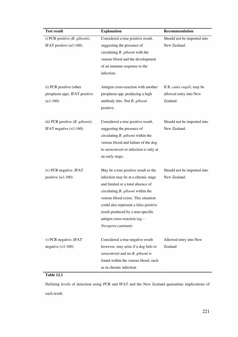
221
Test result Explanation Recommendation
i) PCR positive (B. gibsoni),
IFAT positive (≥1:160)
Considered a true positive result,
suggesting the presence of
circulating B. gibsoni with the
venous blood and the development
of an immune response to the
infection.
Should not be imported into
New Zealand.
ii) PCR positive (other
piroplasm spp), IFAT positive
(≥1:160)
Antigen cross-reaction with another
piroplasm spp, producing a high
antibody titre. Not B. gibsoni
positive.
If B. canis vogeli, may be
allowed entry into New
Zealand.
iii) PCR positive (B. gibsoni),
IFAT negative (<1:160)
Considered a true positive result,
suggesting the presence of
circulating B. gibsoni within the
venous blood and failure of the dog
to seroconvert or infection is only at
an early stage.
Should not be imported into
New Zealand.
iv) PCR negative, IFAT
positive (≥1:160)
May be a true positive result as the
infection may be at a chronic stage
and limited or a total absence of
circulating B. gibsoni within the
venous blood exists. This situation
could also represent a false positive
result produced by a non-specific
antigen cross-reaction (eg –
Neospora caninum)
Should not be imported into
New Zealand.
v) PCR negative, IFAT
negative (<1:160)
Considered a true negative result
however, may arise if a dog fails to
seroconvert and no B. gibsoni is
found within the venous blood, such
as in chronic infection
Allowed entry into New
Zealand
Table 12.1
Defining levels of detection using PCR and IFAT and the New Zealand quarantine implications of
each result.
222
This thesis also reports for the first time, the detection of A. platys in Western Australia,
Queensland and Victoria (Chapter nine). It is probable that A. platys is widespread
throughout Australia and could be a reflection of the wide distribution of R. sanguineus.
Further study must however be conducted to verify this tick as the vector of A. platys. Also
investigated for the first time was the pathogenesis of A. platys infection in dogs in northern
Australia, the influence of co-infection with B. canis vogeli and the efficacy of doxycycline
drug therapy. While studies conducted suggested that A. platys infection is somewhat
benign, this could simply be a reflection of the immune status and previous exposure to this
infection by the host. As there still remains reports of dogs developing signs of lethargy,
fever and bleeding tendencies (Jefferies, 2001), additional research into dogs naive to A.
platys exposure is necessary to further understand the pathogenesis of this infection. The role
of sylvatic reservoirs in Australia, such as dingoes, feral dogs and foxes is poorly understood
and may contribute to the transmission dynamics of both A. platys and the canine Babesia
species. Further investigation into tick-borne disease of wild canine species should therefore
also be considered.
Biosecurity is a pertinent issue in Australia and New Zealand, with both countries reportedly
free from many significant pathogens of dogs such as rabies virus and Ehrlichia canis
(Irwin, 2001; Mason et al., 2001; Davidson, 2002). With increased levels of pet travel
worldwide, the surveillance for exotic diseases in animals being imported and exported is of
great importance. Both the recent discovery of B. gibsoni and A. platys in Australia and the
limitations of current screening protocols for tick-borne diseases in dogs entering Australia
and New Zealand, also expose the need to review current quaratine measures in an effort to
prevent the importation and possible establishment of exotic pathogens into these countries.
By implicating both serological and molecular-based detection methods for screening dogs,
the risk of importing exotic tick-borne diseases including the highly pathogenic B. canis
rossi and E. canis, can be minimized and should be considered by quarantine authorities in
the future.
223
12.2 Molecular phylogeny and taxonomy of the Piroplasmida
This thesis has described the molecular characterisation of canine piroplasm isolates from
many countries for the first time, giving a greater insight into the levels of molecular
variation and worldwide distribution of the canine Babesia species, including the possible
existence of genotypes associated with separate geographic locations. DNA sequencing of
the HSP 70 for B. canis vogeli and the ITS 1, 5.8S, ITS 2 loci for B. gibsoni has also been
achieved for the first time and has allowed for phylogenetic relationships to be established
using multiple gene loci. The increased use of multiple gene loci for phylogenetic analysis
and molecular characterisation is recommended to allow for a more accurate view of the
evolutionary relationships among the piroplasms to be established.
Study conducted within this thesis has highlighted the current discordance and general
confusion in the taxonomic allocation among the canine piroplasms and within the order
Piroplasmida at a species, genus and family level of classification (Chapters ten and eleven).
The establishment of specific criteria for determining different levels of taxonomic
allocation is overdue within this important group of protozoan parasites. A general
consensus of criteria would minimize misguided and often premature descriptions of new
piroplasm species. While traditional phenotypic characteristics should always be considered
when classifying members of the Piroplasmida, genetic characterisation and phylogeny
shows promise as a means of delineating taxa. By reorganising the order Piroplasmida into
three families, the Theileriidae, Babesiidae and Piroplasmiidae and the establishment of new
genera including the Piroplasma and Achromaticus, an attempt has been made to alleviate
the taxonomic anomalies and paraphyly that currently exist.
12.3 Conclusion
Overall, this thesis has revealed the benefits of molecular-based techniques to monitor,
manage and control emerging canine tick-borne disease, while also giving a greater insight
into evolutionary relationships and taxonomic classification of these organisms. The

224
increased application of PCR in veterinary diagnosis will not only allow for increased
diagnostic accuracy but has the potential to be implemented in the quarantine screening of
imported animals. Combined with other detection methods, molecular technology will help
to ensure the high levels of biosecurity of countries such as Australia and New Zealand.
225
References
Allred DR, Al-Khedery B (2004) Antigenic variation and cytoadhesion in Babesia bovis and
Plasmodium falciparum: different logics achieve the same goal. Mol Biochem Parasitol
134:27-35
Allsopp MT, Cavalier-Smith T, De Waal DT, Allsopp BA (1994) Phylogeny and evolution of the
piroplasms. Parasitology 108:147-52.
Almeria S, Castella J, Ferrer D, Gutierrez JF, Estrada-Pena A, Sparagano O (2002) Reverse line blot
hybridization used to identify hemoprotozoa in Minorcan cattle. Ann N Y Acad Sci 969:78-
82
Anderson JF, Magnarelli LA, Donner CS, Spielman A, Piesman J (1979) Canine Babesia new to
North America. Science 204:1431-2.
Anderson JF, Magnarelli LA, Sulzer AJ (1980) Canine babesiosis: indirect fluorescent antibody test
for a North American isolate of Babesia gibsoni. Am J Vet Res 41:2102-5
Ano H, Makimura S, Harasawa R (2001) Detection of Babesia species from infected dog blood by
polymerase chain reaction. J Vet Med Sci 63:111-3.
Arraga-Alvarado C, Palmar M, Parra O, Salas P (2003) Ehrlichia platys (Anaplasma platys) in dogs
from Maracaibo, Venezuela: an ultrastructural study of experimental and natural infections.
Vet Pathol 40:149-56
Babes V. (1888) Sur l'hemoglobinurie bacterienne du boeuf, CR Acad. Sci. 107:692.
Baek BK, Soo KB, Kim JH, Hur J, Lee BO, Jung JM, Onuma M, Oluoch AO, Kim CH,
KaMinasHkoma I (2003) Verification by polymerase chain reaction of vertical transmission
of Theileria sergenti in cows. Can J Vet Res 67:278-82
Baker DC, Simpson M, Gaunt SD, Corstvet RE (1987) Acute Ehrlichia platys infection in the dog.
Vet Pathol 24:449-53.
Baneth G, Breitschwerdt EB, Hegarty BC, Pappalardo B, Ryan J (1998) A survey of tick-borne
bacteria and protozoa in naturally exposed dogs from Israel. Vet Parasitol 74:133-42.
Baneth G, Kenny MJ, Tasker S, Anug Y, Shkap V, Levy A, Shaw SE (2004) Infection with a
proposed new subspecies of Babesia canis, Babesia canis subsp. presentii, in domestic cats.
J Clin Microbiol 42:99-105
Bastos C, Moreira SM, Passos MF. (2004) Retrospective study (1998-2001) on canine babesisos in
Belo Horizonte, Minas Gerais, Brazil. Ann N Y Acad Sci 1026:158-60.
CHAPTER THIRTEEN
226
Battsetseg B, Xuan X, Ikadai H, Bautista JL, Byambaa B, Boldbaatar D, Battur B, Battsetseg G,
Batsukh Z, Igarashi I, Nagasawa H, Mikami T, Fujisaki K (2001) Detection of Babesia
caballi and Babesia equi in Dermacentor nuttalli adult ticks. Int J Parasitol 31:384-6.
Beban H. (2003) Absence of Babesia gibsoni in New Zealand dogs. Surveillance 30:9-11.
Bechara GH, Szabo MP, Mukai LS, Rosa PC (1994) Immunisation of dogs, hamsters and guinea pigs
against Rhipicephalus sanguineus using crude unfed adult tick extracts. Vet Parasitol 52:79-
90
Becker S, Franco JR, Simarro PP, Stitch A, Abel PM, Steverding D (2004) Real-time PCR for
detecting Trypanosoma brucei in human blood samples. Diag Microbiol Inf Dis 50:193-99
Belgrader P, Del Rio SA, Turner KA, Marino MA, Weaver KR, Williams PE (1995) Automated DNA
purification and amplification from blood-stained cards using a robotic workstation.
Biotechniques 19:426-32
Bernhard D, Stechmann A, Foissner W, Ammermann D, Hehn M, Schlegel M. (2001) Phylogenetic
relationships within the class Spirotrichea (Ciliophora) inferred from small subunit rRNA
gene sequences. Molecular Phylogenetics and Evolution 21:86-92.
Besier RB, Wroth R (1985) Discovery of the tick Haemaphysalis longicornis in Western Australia.
Aust Vet J 62:205-6
Bextine B, Tuan SJ, Shaikh H, Blua M, Miller TA (2004) Evaluation of methods for extracting
Xylella fastidiosa DNA from the glassy-winged sharpshooter. J Econ Entomol 97:757-63
Bicalho KA, Ribeiro MF, Martins-Filho OA (2004) Molecular fluorescent approach to assessing
intraerythrocytic hemoprotozoan Babesia canis infection in dogs. Vet Parasitol 125:221-35
Birkenheuer A, Marr H. (2005) Babesia gibsoni cytochrome b gene analysis. ACVIM abstracts
Birkenheuer AJ, Levy MG, Breitschwerdt EB (2003a) Development and evaluation of a seminested
PCR for detection and differentiation of Babesia gibsoni (Asian genotype) and B. canis DNA
in canine blood samples. J Clin Microbiol 41:4172-7
Birkenheuer AJ, Levy MG, Breitschwerdt EB (2004a) Efficacy of combined atovaquone and
azithromycin for therapy of chronic Babesia gibsoni (Asian genotype) infections in dogs. J
Vet Intern Med 18:494-8
Birkenheuer AJ, Levy MG, Savary KC, Gager RB, Breitschwerdt EB (1999) Babesia gibsoni
infections in dogs from North Carolina. J Am Anim Hosp Assoc 35:125-8
Birkenheuer AJ, Levy MG, Stebbins M, Poore M, Breitschwerdt E (2003b) Serosurvey of
AntiBabesia Antibodies in Stray Dogs and American Pit Bull Terriers and American
Staffordshire Terriers From North Carolina. J Am Anim Hosp Assoc 39:551-7
Birkenheuer AJ, Neel J, Ruslander D, Levy MG, Breitschwerdt EB (2004b) Detection and molecular
characterization of a novel large Babesia species in a dog. Vet Parasitol 124:151-60
Biswas B, Mukherjee D, Mattingly-Napier BL, Dutta SK (1991) Diagnostic application of polymerase
chain reaction for detection of Ehrlichia risticii in equine monocytic ehrlichiosis (Potomac
horse fever). J Clin Microbiol 29:2228-33.
Bonoan JT, Johnson DH, Cunha BA (1998) Life-threatening babesiosis in an asplenic patient treated
with exchange transfusion, azithromycin, and atovaquone. Heart Lung 27:424-8
227
Boozer AL, Macintire DK (2003) Canine babesiosis. Vet Clin North Am Small Anim Pract 33:885-
904, viii
Bose R, Jorgensen WK, Dalgliesh RJ, Friedhoff KT, de Vos AJ (1995) Current state and future trends
in the diagnosis of babesiosis. Vet Parasitol 57:61-74.
Botros BA, Moch RW, Barsoum IS (1975) Some observations on experimentally induced infection of
dogs with Babesia gibsoni. Am J Vet Res 36:293-6.
Bradfield JF, Vore SJ, Pryor WH, Jr. (1996) Ehrlichia platys infection in dogs. Lab Anim Sci 46:565-
8.
Braga EM, Fontes CJ, Krettli AU (1998) Persistence of humoral response against sporozoite and
blood-stage malaria antigens 7 years after a brief exposure to Plasmodium vivax. J Infect Dis
177:1132-5
Brandao LP, Hagiwara MK, Myiashiro SI (2003) Humoral immunity and reinfection resistance in
dogs experimentally inoculated with Babesia canis and either treated or untreated with
imidocarb dipropionate. Vet Parasitol 114:253-65
Breitschwerdt EB (1984) Babesiosis. In Clinical Microbiology and Infectious Diseases of the Dog and
Cat (Edited by Greene CE), p. 796-805. WB Saunders, Philadelphia.
Bronsdon MA, Homer MJ, Magera JM, Harrison C, Andrews RG, Bielitzki JT, Emerson CL, Persing
DH, Fritsche TR (1999) Detection of enzootic babesiosis in baboons (Papio cynocephalus)
and phylogenetic evidence supporting synonymy of the genera Entopolypoides and Babesia.
J Clin Microbiol 37:1548-53
Brown GK, Martin AR, Roberts TK, Aitken RJ (2001) Detection of Ehrlichia platys in dogs in
Australia. Aust Vet J 79:554-8.
Brown GK, Martin AR, Roberts TK, Dunstan RH (2005) Molecular detection of Anaplasma platys in
lice collected from dogs in Australia. Aust Vet J 83:101-2
Cable RG, Leiby DA (2003) Risk and prevention of transfusion-transmitted babesiosis and other tick-
borne diseases. Curr Opin Hematol 10:405-11
Caccio S, Camma C, Onuma M, Severini C (2000) The beta-tubulin gene of Babesia and Theileria
parasites is an informative marker for species discrimination. Int J Parasitol 30:1181-5.
Caccio SM, Antunovic B, Moretti A, Mangili V, Marinculic A, Baric RR, Slemenda SB, Pieniazek NJ
(2002) Molecular characterisation of Babesia canis canis and Babesia canis vogeli from
naturally infected European dogs. Vet Parasitol 106:285-92
Calder JA, Reddy GR, Chieves L, Courtney CH, Littell R, Livengood JR, Norval RA, Smith C, Dame
JB (1996) Monitoring Babesia bovis infections in cattle by using PCR-based tests. J Clin
Microbiol 34:2748-55
Callow LL (1984) Protozoal and rickettsial diseases. In Animal Health in Australia, Vol 5, p 161.
Australian Bureau of Animal Helath/Australian Government Publishing Service, Canberrra.
Camacho AT, Guitian EJ, Pallas E, Gestal JJ, Olmeda AS, Goethert HK, Telford SR, 3rd, Spielman A
(2004) Azotemia and mortality among Babesia microti-like infected dogs. J Vet Intern Med
18:141-6

228
Camacho AT, Pallas E, Gestal JJ, Guitian FJ, Olmeda AS (2002) Natural infection by a Babesia
microti-like piroplasm in a splenectomised dog. Vet Rec 150:381-2
Camacho AT, Pallas E, Gestal JJ, Guitian FJ, Olmeda AS, Goethert HK, Telford SR (2001) Infection
of dogs in north-west Spain with a Babesia microti-like agent. Vet Rec 149:552-5
Camacho AT, Pallas E, Gestal JJ, Guitian FJ, Olmeda AS, Telford SR, Spielman A (2003) Ixodes
hexagonus is the main candidate as vector of Theileria annae in northwest Spain. Vet
Parasitol 112:157-63
Carret C, Walas F, Carcy B, Grande N, Precigout E, Moubri K, Schetters TP, Gorenflot A (1999)
Babesia canis canis, Babesia canis vogeli, Babesia canis rossi: differentiation of the three
subspecies by a restriction fragment length polymorphism analysis on amplified small
subunit ribosomal RNA genes. J Eukaryot Microbiol 46:298-303.
Casapulla R, Baldi L, Avallone V, Sannino R, Pazzanese L, Mizzoni V (1998) Canine piroplasmosis
due to Babesia gibsoni: clinical and morphological aspects. Vet Rec 142:168-9.
Casati S, Sager H, Gern L, Piffaretti JC. (2004) Diversity of Babesia sp. infecting Ixodes ricinus ticks
in Switzerland (http://www.ncbi.nih.gov/entrez/, accessed 7/2004).
Cavalier-Smith T (1993) Kingdom protozoa and its 18 phyla. Microbiol Rev 57:953-94
Chang AC, Chang WL, Lin CT, Pan MJ, Lee SC (1996) Canine infectious cyclic thrombocytopenia
found in Taiwan. J Vet Med Sci 58:473-6.
Chang WL, Pan MJ (1996) Specific amplification of Ehrlichia platys DNA from blood specimens by
two-step PCR. J Clin Microbiol 34:3142-6.
Chang WL, Su WL, Pan MJ (1997) Two-step PCR in the evaluation of antibiotic treatment for
Ehrlichia platys infection. J Vet Med Sci 59:849-51
Chansiri K, Sarataphan N (2002) Molecular phylogenetic study of Theileria sp. (Thung Song) based
on the thymidylate synthetase gene. Parasitol Res 88:S33-5
Citard T, Mahl P, Boulouis HJ, Chavigny C, Druilhe P (1995) Babesia canis: evidence for genetic
diversity among isolates revealed by restriction fragment length polymorphism analysis.
Trop Med Parasitol 46:172-9
Colgrove DJ (1976) Canine babesiosis in Hawaii. J Am Vet Med Assoc 168:591-2
Conrad P, Thomford J, Yamane I, Whiting J, Bosma L, Uno T, Holshuh HJ, Shelly S (1991)
Hemolytic anemia caused by Babesia gibsoni infection in dogs. J Am Vet Med Assoc
199:601-5
Conrad PA, Thomford JW, Marsh A, Telford SR, 3rd, Anderson JF, Spielman A, Sabin EA, Yamane
I, Persing DH (1992) Ribosomal DNA probe for differentiation of Babesia microti and B.
gibsoni isolates. J Clin Microbiol 30:1210-5
Coyne SR, Craw PD, Norwood DA, Ulrich MP. (2004) Comparative analysis of the Schleicher and
Schuell IsoCode Stix DNA isolation device and the Qiagen QIAamp DNA mini kit. J. Clin.
Microbiol. 42:4859-62.
Criado-Fornelio A, Gonzalez-del-Rio MA, Buling-Sarana A, Barba-Carretero JC (2003d) Molecular
characterization of a Babesia gibsoni isolate from a Spanish dog. Vet Parasitol 117:123-9
229
Criado-Fornelio A, Gonzalez-del-Rio MA, Buling-Sarana A, Barba-Carretero JC (2004) The
"expanding universe" of piroplasms. Vet Parasitol 119:337-45
Criado-Fornelio A, Martinez-Marcos A, Buling-Sarana A, Barba-Carretero JC (2003a) Molecular
studies on Babesia, Theileria and Hepatozoon in southern Europe. Part I. Epizootiological
aspects. Vet Parasitol 113:189-201
Criado-Fornelio A, Martinez-Marcos A, Buling-Sarana A, Barba-Carretero JC (2003b) Molecular
studies on Babesia, Theileria and Hepatozoon in southern Europe. Part II. Phylogenetic
analysis and evolutionary history. Vet Parasitol 114:173-94
Criado-Fornelio A, Martinez-Marcos A, Buling-Sarana A, Barba-Carretero JC (2003c) Presence of
Mycoplasma haemofelis, Mycoplasma haemominutum and piroplasmids in cats from
southern Europe: a molecular study. Vet Microbiol 93:307-17
Cruz-Vazquez C, Garcia-Vazquez Z (1999) Seasonal distribution of Rhipicephalus sanguineus ticks
(Acari: Ixodidae) on dogs in an urban area of Morelos, Mexico. Exp Appl Acarol 23:277-80
Dalgliesh RJ (1971) Dimethyl sulphoxide in the low-temperature preservation of Babesia bigemina.
Res Vet Sci 12:469-71
Dantrakool A, Somboon P, Hashimoto T, Saito-Ito A (2004) Identification of a new type of Babesia
species in wild rats (Bandicota indica) in Chiang Mai Province, Thailand. J Clin Microbiol
42:850-4
Dao AH, Eberhard ML (1996) Pathology of acute fatal babesiosis in hamsters experimentally infected
with the WA-1 strain of Babesia. Lab Invest 74:853-9
Daubenberger C, Heussler V, Gobright E, Wijngaard P, Clevers HC, Wells C, Tsuji N, Musoke A,
McKeever D (1997) Molecular characterisation of a cognate 70 kDa heat shock protein of
the protozoan Theileria parva. Mol Biochem Parasitol 85:265-9
Davidson (2002) Control and eradication of animal diseases in New Zealand. NZ Vet. J. 50 (3
suppl):6-12
De Chaneet G. (1976) Extension of the range of Rhipicephalus sanguineus in Western Australia. Aust
Vet J. 52:433.
de la Fuente J, Van Den Bussche RA, Garcia-Garcia JC, Rodriguez SD, Garcia MA, Guglielmone
AA, Mangold AJ, Friche Passos LM, Barbosa Ribeiro MF, Blouin EF, Kocan KM (2002)
Phylogeography of New World isolates of Anaplasma marginale based on major surface
protein sequences. Vet Microbiol 88:275-85
Depoix D, Carcy B, Jumas-Bilak E, Pages M, Precigout E, Schetters TP, Ravel C, Gorenflot A (2002)
Chromosome number, genome size and polymorphism of European and South African
isolates of large Babesia parasites that infect dogs. Parasitology 125:313-21
Down J. (2004) Identification of a novel blood parasite in the Brown Kiwi of New Zealand (Honors
thesis) Murdoch University, Perth, Western Australia.
Devulder G, Perouse de Montclos M, Flandrois JP (2005) A multigene approach to phylogenetic
analysis using the genus Mycobacterium as a model. Int J Syst Evol Microbiol 55:293-302
Duh D, Tozon N, Petrovec M, Strasek K, Avsic-Zupanc T (2004) Canine babesiosis in Slovenia:
molecular evidence of Babesia canis canis and Babesia canis vogeli. Vet Res 35:363-8
230
Dumler JS, Barbet AF, Bekker CP, Dasch GA, Palmer GH, Ray SC, Rikihisa Y, Rurangirwa FR
(2001) Reorganization of genera in the families Rickettsiaceae and Anaplasmataceae in the
order Rickettsiales: unification of some species of Ehrlichia with Anaplasma, Cowdria with
Ehrlichia and Ehrlichia with Neorickettsia, descriptions of six new species combinations and
designation of Ehrlichia equi and 'HGE agent' as subjective synonyms of Ehrlichia
phagocytophila. Int J Syst Evol Microbiol 51:2145-65
Durette-Desset MC, Cassone J (1983) A taxonomic revision of the Trychostrongilid nematode
parasites of the echidna, Tachyglossus aculeatus (Monotrema). Aust J Zool 31 : 257-84.
Eberhard ML, Walker EM, Steurer FJ (1995) Survival and infectivity of Babesia in blood maintained
at 25 C and 2-4 C. J Parasitol 81:790-2
Egea N, Lang-Unnasch N (1995) Phylogeny of the large extrachromosomal DNA of organisms in the
phylum Apicomplexa. J Eukaryot Microbiol 42:679-84
Ellis J, Hefford C, Baverstock PR, Dalrymple BP, Johnson AM (1992) Ribosomal DNA sequence
comparison of Babesia and Theileria. Mol Biochem Parasitol 54:87-95
Emerson CL, Tsai CC, Holland CJ, Ralston P, Diluzio ME (1990) Recrudescence of Entopolypoides
macaci Mayer, 1933 (Babesiidae) infection secondary to stress in long-tailed macaques
(Macaca fascicularis). Lab Anim Sci 40:169-71
Esernio-Jensson D, Scimeca PG, Benach JL, Tenenbaum MJ. (1987) Transplacental/perinatal
babesiosis. J. Pediatr. 110:570-2.
Escalante AA, Ayala FJ (1995) Evolutionary origin of Plasmodium and other Apicomplexa based on
rRNA genes. Proc Natl Acad Sci U S A 92:5793-7
Etkind P, Piesman J, Ruebush TK, 2nd, Spielman A, Juranek DD (1980) Methods for detecting
Babesia microti infection in wild rodents. J Parasitol 66:107-10
Evans DE, Martins JR, Guglielmone AA (2000) A review of the ticks (Acari, ixodida) of Brazil, their
hosts and geographic distribution - 1. The state of Rio Grande do Sul, southern Brazil. Mem
Inst Oswaldo Cruz 95:453-70
Fahrimal Y, Goff WL, Jasmer DP (1992) Detection of Babesia bovis carrier cattle by using
polymerase chain reaction amplification of parasite DNA. J Clin Microbiol 30:1374-9.
Farkas R, Foldvari G, Fenyves B, Kotai I, Szilagyi A, Hegedus GT (2004) First detection of small
babesiae in two dogs in Hungary. Vet Rec 154:176-8
Farnert A, Arez AP, Correia AT, Bjorkman A, Snounou G, do Rosario V (1999) Sampling and
storage of blood and the detection of malaria parasites by polymerase chain reaction. Trans R
Soc Trop Med Hyg 93:50-3.
Fawcett DW, Young AS, Leitch BL (1982) Sporogony in Theileria (Apicomplexa: Piroplasmida). A
comparative ultrastructural study J. Submicroscop. Cytol. Vol. 17, pp 299-314
Farwell GE, LeGrand EK, Cobb CC (1982) Clinical observations on Babesia gibsoni and Babesia
canis infections in dogs. J Am Vet Med Assoc 180:507-11.
Felek S, Unver A, Stich RW, Rikihisa Y (2001) Sensitive detection of Ehrlichia chaffeensis in cell
culture, blood, and tick specimens by reverse transcription-PCR. J Clin Microbiol 39:460-3.

231
Figueroa JV, Chieves LP, Johnson GS, Buening GM (1992) Detection of Babesia bigemina-infected
carriers by polymerase chain reaction amplification. J Clin Microbiol 30:2576-82.
Foldvari G, Hell E, Farkas R (2005) Babesia canis canis in dogs from Hungary: detection by PCR and
sequencing. Vet Parasitol 127:221-6
Fourie LJ, Stanneck D, Horak IG (2003) The efficacy of collars impregnated with flumethrin and
propoxur against experimental infestations of adult Rhipicephalus sanguineus on dogs. J S
Afr Vet Assoc 74:123-6
Franca C. (1910) Sur la classification des pioplasmes et description des deux formes de ces parasites.
Arq. Inst. Bacteriol. Camara Pestana Lisbon. 3:11.
Franca C. (1912) Sur la classification des pioplasmes et description des deux formes de ces parasites.
Arq. Inst. Bacteriol. Camara Pestana Lisbon. 3:11.
French TW, Harvey JW (1983) Serologic diagnosis of infectious cyclic thrombocytopenia in dogs
using an indirect fluorescent antibody test. Am J Vet Res 44:2407-11.
Friedhoff K. (1988) Transmission of Babesia. In Babesiosis of domestic animals and man. ed. Ristic
M, p 23-45, CRC Press, Florida.
Fukumoto S, Suzuki H, Igarashi I, Xuan X (2005a) Fatal experimental transplacental Babesia gibsoni
infections in dogs. Int J Parasitol 35:1031-1035
Fukumoto S, Tamaki Y, Shirafuji H, Harakawa S, Suzuki H, Xuan X (2005b) Immunization with
recombinant surface antigen P50 of Babesia gibsoni expressed in insect cells induced
parasite growth inhibition in dogs. Clin Diagn Lab Immunol 12:557-9
Fukumoto S, Xuan X, Igarashi I, Zhang S, Mugisha J, Ogata T, Nagasawa H, Fujisaki K, Suzuki N,
Mikami T (2000) Morphological changes of Babesia gibsoni grown in canine red blood cell-
substituted severe combined immune deficiency mice. J Parasitol 86:956-8.
Fukumoto S, Xuan X, Shigeno S, Kimbita E, Igarashi I, Nagasawa H, Fujisaki K, Mikami T (2001)
Development of a polymerase chain reaction method for diagnosing Babesia gibsoni
infection in dogs. J Vet Med Sci 63:977-81
Fukumoto S, Xuan X, Takabatake N, Igarashi I, Sugimoto C, Fujisaki K, Nagasawa H, Mikami T,
Suzuki H (2004) Inhibitory effect of antiserum to surface antigen P50 of Babesia gibsoni on
growth of parasites in severe combined immunodeficiency mice given canine red blood cells.
Infect Immun 72:1795-8
Galtier N, Gouy M. (1995) Inferring phylogenies from DNA sequences of unequal base compositions.
Proc Natl Acad Sci USA 92:11317-21
Garcia, L. S. and Bruckner, D. A. (1988) Procedures for detecting blood parasites. In Diagnostic
midical parasitology (Elsevier, New York). pp. 412-420.
Georges K, Loria GR, Riili S, Greco A, Caracappa S, Jongejan F, Sparagano O (2001) Detection of
haemoparasites in cattle by reverse line blot hybridisation with a note on the distribution of
ticks in Sicily. Vet Parasitol 99:273-86.
Giglio S, Monis PT, Saint CP (2003) Demonstration of preferential binding of SYBR Green I to
specific DNA fragments in real-time multiplex PCR. Nucleic Acids Res 31:e136
232
Gleason NN, Wolf RE (1974) Entopolypoides macaci (Babesiidae) in Macaca mulatta. J Parasitol
60:844-7
Goethert HK, Telford SR, 3rd (2003) What is Babesia microti? Parasitology 127:301-9
Gonzalez A, Castro Ddel C, Gonzalez S (2004) Ectoparasitic species from Canis familiaris (Linne) in
Buenos Aires province, Argentina. Vet Parasitol 120:123-9
Grewal MS (1957) Two new piroplasms, Babesia (Babesiella) heischi and Babesia (Babesia) hoarei
from Peter's pigmy mongoose, Helogale undulata rufula (demonstrated by Professor
Garnham). Trans R Soc Trop Med Hyg 51:290-1
Groves MG, Vanniasingham JA (1970) Treatment of Babesia gibsoni infections with phenamidine
isethionate. Vet Rec 86:8-10
Groves MG, Yap LF (1968) Babesia gibsoni (Patton, 1910) from a dog in Kuala Lumpur. Med J
Malaya 22:229
Gubbels JM, de Vos AP, van der Weide M, Viseras J, Schouls LM, de Vries E, Jongejan F (1999)
Simultaneous detection of bovine Theileria and Babesia species by reverse line blot
hybridization. J Clin Microbiol 37:1782-9.
Guglielmone AA, Mangold AJ, Vinabal AE (1991) Ticks (Ixodidae) parasitizing humans in four
provinces of north-western Argentina. Ann Trop Med Parasitol 85:539-42
Guitian FJ, Camacho AT, Telford SR, 3rd (2003) Case-control study of canine infection by a newly
recognised Babesia microti-like piroplasm. Prev Vet Med 61:137-45
Hancock SI, Breitschwerdt EB, Pitulle C (2001) Differentiation of Ehrlichia platys and E. equi
infections in dogs by using 16S ribosomal DNA-based PCR. J Clin Microbiol 39:4577-8
Hanscheid T, Grobusch MP (2002) How useful is PCR in the diagnosis of malaria? Trends Parasitol
18:395-8
Harrus S, Aroch I, Lavy E, Bark H (1997) Clinical manifestations of infectious canine cyclic
thrombocytopenia. Vet Rec 141:247-50.
Harrus S, Waner T, Aizenberg I, Bark H (1998a) Therapeutic effect of doxycycline in experimental
subclinical canine monocytic ehrlichiosis: evaluation of a 6-week course. J Clin Microbiol
36:2140-2.
Harrus S, Waner T, Aizenberg I, Foley JE, Poland AM, Bark H (1998b) Amplification of ehrlichial
DNA from dogs 34 months after infection with Ehrlichia canis. J Clin Microbiol 36:73-6.
Harvey JW. (1990) Ehrlichia platys infection. In Infectious diseases of the dog and cat. ed. Greene
CE., p 415-18, WB Saunders, Philadelphia.
Harvey JW, Taboada J, Lewis JC. (1988) Babesiosis in a litter of pups. J. Am. Vet. Med. Assoc.
192:1751-2.
Harvey ML (2005) An alternative for the extraction and storage of DNA from insects in forensic
entomology. J Forensic Sci 50: 3
Harvey JW, Simpson CF, Gaskin JM (1978) Cyclic thrombocytopenia induced by a Rickettsia-like
agent in dogs. J Infect Dis 137:182-8.
233
Hauschild S, Shayan P, Schein E (1995) Characterization and comparison of merozoite antigens of
different Babesia canis isolates by serological and immunological investigations. Parasitol
Res 81:638-42
Hendrick JP, Hartl FU (1995) The role of molecular chaperones in protein folding. Faseb J 9:1559-69.
Henning L, Felgar I, Beck H. (1999) Rapid DNA extraction for molecular epidemiological studies of
malaria. Acta Tropica 72:149-55.
Herwaldt BL, Neitzel DF, Gorlin JB, Jensen KA, Perry EH, Peglow WR, Slemenda SB, Won KY,
Nace EK, Pieniazek NJ, Wilson M (2002) Transmission of Babesia microti in Minnesota
through four blood donations from the same donor over a 6-month period. Transfusion
42:1154-8
Higuchi S, Fujimori M, Hoshi F, Kawamura S, Yasuda Y (1995) Development of Babesia gibsoni in
the salivary glands of the larval tick, Rhipicephalus sanguineus. J Vet Med Sci 57:117-9
Higuchi S, Hoshina H, Hoshi F, Kawamura S, Yasuda Y (1993a) Development of Babesia gibsoni in
the salivary glands of the tick, Haemaphysalis longicornis. Kitasato Arch Exp Med 65
Suppl:147-51
Higuchi S, Izumitani M, Hoshi H, Kawamura S, Yasuda Y (1999a) Development of Babesia gibsoni
in the midgut of larval tick, Rhipicephalus sanguineus. J Vet Med Sci 61:689-91.
Higuchi S, Konno H, Hoshi F, Kawamura S, Yasuda Y (1993b) Observations of Babesia gibsoni in
the ovary of the tick, Haemaphysalis longicornis. Kitasato Arch Exp Med 65 Suppl:153-8
Higuchi S, Kuroda H, Hoshi H, Kawamura S, Yasuda Y (1999b) Development of Babesia gibsoni in
the midgut of the nymphal stage of the tick, Rhipicephalus sanguineus. J Vet Med Sci
61:697-9.
Higuchi S, Oya H, Hoshi F, Kawamura S, Yasuda Y (1992) Observations of Babesia gibsoni in
midgut epithelial cells of the tick, Haemaphysalis longicornis. Kitasato Arch Exp Med
65:143-7
Higuchi S, Simomura S, Yoshida H, Hoshi F, Kawamura S, Yasuda Y (1991a) Development of
Babesia gibsoni in the gut epithelium of the tick, Haemaphysalis longicornis. J Vet Med Sci
53:129-31
Higuchi S, Simomura S, Yoshida H, Hoshi F, Kawamura S, Yasuda Y (1991b) Development of
Babesia gibsoni in the hemolymph of the vector tick, Haemaphysalis longicornis. J Vet Med
Sci 53:491-3
Hill MWM, Bolton BL (1966) Canine Babesiosis in Queensland. Australian Veterinary Journal 42:84-
86
Hillis DM, Dixon MT (1991) Ribosomal DNA: molecular evolution and phylogenetic inference. Q
Rev Biol 66:411-53.
Holbrook AA, Frerichs WM (1970) Babesia mephitis sp. N. (Protozoa: Piroplasmida), a hematozoan
parasite of the striped skunk, Mephitis mephitis. J Parasitol 56:930-1
Holman PJ, Bendele KG, Schoelkopf L, Jones-Witthuhn RL, Jones SO (2003) Ribosomal RNA
analysis of Babesia odocoilei isolates from farmed reindeer (Rangifer tarandus tarandus)
and elk (Cervus elaphus canadensis) in Wisconsin. Parasitol Res 91:378-83
234
Homer MJ, Aguilar-Delfin I, Telford SR, 3rd, Krause PJ, Persing DH (2000) Babesiosis. Clin
Microbiol Rev 13:451-69.
Hoogstraal H, Roberts FH, Kohls GM, Tipton VJ (1968) Review of Haemaphysalis (kaiseriana)
Longicornis Neumann (resurrected) of Australia, New Zealand, New Caledonia, Fiji, Japan,
Korea, and Northeastern China and USSR, and its parthenogenetic and bisexual populations
(Ixodoidea, Ixodidae). J Parasitol 54:1197-213
Hossain MA, Yamato O, Yamasaki M, Maede Y (2004) Inhibitory effect of pyrimidine and purine
nucleotides on the multiplication of Babesia gibsoni: possible cause of low parasitemia and
simultaneous reticulocytosis in canine babesiosis. J Vet Med Sci 66:389-95
Hua P, Yuhai M, Shide T, Yang S, Bohai W, Xiangrui C (2000) Canine ehrlichiosis caused
simultaneously by Ehrlichia canis and Ehrlichia platys. Microbiol Immunol 44:737-9
Hypsa V, Skerikova A, Scholz T (2005) Phylogeny, evolution and host-parasite relationships of the
order Proteocephalidea (Eucestoda) as revealed by combined analysis and secondary
structure characters. Parasitology 130:359-71
Ikadai H, Tanaka H, Shibahara N, Matsuu A, Uechi M, Itoh N, Oshiro S, Kudo N, Igarashi I,
Oyamada T (2004) Molecular evidence of infections with Babesia gibsoni parasites in Japan
and evaluation of the diagnostic potential of a loop-mediated isothermal amplification
method. J Clin Microbiol 42:2465-9
Ilhan T, Williamson S, Kirvar E, Shiels B, Brown CG (1998) Theileria annulata: carrier state and
immunity. Ann N Y Acad Sci 849:109-25
Inokuma H, Brouqui P, Drancourt M, Raoult D (2001a) Citrate synthase gene sequence: a new tool
for phylogenetic analysis and identification of ehrlichia. J Clin Microbiol 39:3031-9.
Inokuma H, Fujii K, Matsumoto K, Okuda M, Nakagome K, Kosugi R, Hirakawa M, Onishi T (2002)
Demonstration of Anaplasma (Ehrlichia) platys inclusions in peripheral blood platelets of a
dog in Japan. Vet Parasitol 110:145-52
Inokuma H, Nane G, Uechi T, Yonahara Y, Brouqui P, Okuda M, Onishi T (2001b) Survey of tick
infestation and tick-borne ehrlichial infection of dogs in Ishigaki Island, Japan. J Vet Med
Sci 63:1225-7
Inokuma H, Ohno K, Onishi T, Raoult D, Brouqui P (2001c) Detection of ehrlichial infection by PCR
in dogs from Yamaguchi and Okinawa Prefectures, Japan. J Vet Med Sci 63:815-7
Inokuma H, Okuda M, Yoshizaki Y, Hiraoka H, Miyama T, Itamoto K, Une S, Nakaichi M, Taura Y
(2005) Clinical observations of Babesia gibsoni infection with low parasitaemia confirmed
by PCR in dogs. Vet Rec 156:116-8
Inokuma H, Parola P, Raoult D, Brouqui P (2001d) Molecular survey of Ehrlichia infection in ticks
from animals in Yamaguchi Prefecture, Japan. Vet Parasitol 99:335-9
Inokuma H, Raoult D, Brouqui P (2000) Detection of Ehrlichia platys DNA in brown dog ticks
(Rhipicephalus sanguineus) in Okinawa Island, Japan. J Clin Microbiol 38:4219-21.
Inokuma H, Tamura K, Onishi T (1997) Dogs develop resistance to Rhipicephalus sanguineus. Vet
Parasitol 68:295-7
235
Inokuma H, Yoshizaki Y, Matsumoto K, Okuda M, Onishi T, Nakagome K, Kosugi R, Hirakawa M
(2004) Molecular survey of Babesia infection in dogs in Okinawa, Japan. Vet Parasitol
121:341-6
Inokuma H, Yoshizaki Y, Shimada Y, Sakata Y, Okuda M, Onishi T (2003) Epidemiological survey
of Babesia species in Japan performed with specimens from ticks collected from dogs and
detection of new Babesia DNA closely related to Babesia odocoilei and Babesia divergens
DNA. J Clin Microbiol 41:3494-8
Iqbal Z, Chaichanasiriwithaya W, Rikihisa Y (1994) Comparison of PCR with other tests for early
diagnosis of canine ehrlichiosis. J Clin Microbiol 32:1658-62.
Iqbal Z, Rikihisa Y (1994) Application of the polymerase chain reaction for the detection of Ehrlichia
canis in tissues of dogs. Vet Microbiol 42:281-7.
Irizarry-Rovira AR, Stephens J, Christian J, Kjemtrup A, DeNicola DB, Widmer WR, Conrad PA
(2001) Babesia gibsoni Infection in a Dog from Indiana. Vet Clin Pathol 30:180-188
Irwin PJ (2001) The first report of canine ehrlichiosis in Australia. Australian Veterinary Journal
79:552-553
Irwin PJ, Hutchinson GW (1991) Clinical and pathological findings of Babesia infection in dogs. Aust
Vet J 68:204-9.
Irwin PJ, Jefferies R (2004) Arthropod-transmitted diseases of companion animals in Southeast Asia.
Trends Parasitol 20:27-34
Ivanov PL, Zemskova EY, Tarasova TA, Frolova SA, Mikhalkovich LS, Koriyenko IV (2002) The
experience of using the FTA cards for blood samples storage at conducting the molecular and
genetic identification of non-identified killed persons - victims of military actions in the
territory of Chechen Republic. Sudebno-Meditinskaya Ekspertiza 45:20-27
Jacobson LS, Clark IA. (1994) The pathophysiology of canine babesiosis: new approches to an old
puzzle. J S Afr Vet Assoc 65:134-45.
Jacobson LS, Reyers F, Berry WL, Viljoen E (1996) Changes in haematocrit after treatment of
uncomplicated canine babesiosis: a comparison between diminazene and trypan blue, and an
evaluation of the influence of parasitaemia. J S Afr Vet Assoc 67:77-82
Jacobson LS, Swan GE (1995) Supportive treatment of canine babesiosis. J S Afr Vet Assoc 66:95-
105
Jarra W, Snounou G (1998) Only viable parasites are detected by PCR following clearance of rodent
malarial infections by drug treatment or immune responses. Infect Immun 66:3783-7
Jefferies R. (2001) PCR-based diagnosis and characterisation of canine babesiosis and canine
infectious cyclic thrombocytopenia (Honours thesis), Murdoch University, Perth, Western
Australia
Jefferies R, Ryan UM, Muhlnickel CJ, Irwin PJ (2003) Two species of canine Babesia in Australia:
detection and characterization by PCR. J Parasitol 89:409-12
Jeong W, Kweon CH, Kang SW, Paik SG (2003) Diagnosis and quantification of Theileria sergenti
using TaqMan PCR. Vet Parasitol 111:287-95
236
Johnson JG, Epstein N, Shiroishi T, Miller LH (1980) Factors affecting the ability of isolated
Plasmodium knowlesi merozoites to attach to and invade erythrocytes. Parasitology 80:539-
50
Jongejan F, Uilenberg G (2004) The global importance of ticks. Parasitology 129 Suppl:S3-14
Jow H, Hudelot C, Rattray M, Higgs PG (2002) Bayesian phylogenetics using an RNA substitution
model applied to early mammalian evolution. Mol Biol Evol 19:1591-601
Kakuda T, Shiki M, Kubota S, Sugimoto C, Brown WC, Kosum C, Nopporn S, Onuma M (1998)
Phylogeny of benign Theileria species from cattle in Thailand, China and the U.S.A. based
on the major piroplasm surface protein and small subunit ribosomal RNA genes. Int. J.
Parasitol 28:1261-7
Kasahara K, Nomura S, Subeki, Matsuura H, Yamasaki M, Yamato O, Maede Y, Katakura K, Suzuki
M, Trimurningsih, Chairul, Yoshihara T (2005) Anti-babesial compounds from Curcuma
zedoaria. Planta Med 71:482-4
Katzer F, McKellar S, Kirvar E, Shiels B (1998) Phylogenetic analysis of Theileria and Babesia equi
in relation to the establishment of parasite populations within novel host species and the
development of diagnostic tests. Mol Biochem Parasitol 95:33-44
Keller N, Jacobson LS, Nel M, de Clerq M, Thompson PN, Schoeman JP (2004) Prevalence and risk
factors of hypoglycemia in virulent canine babesiosis. J Vet Intern Med 18:265-70
Kimura M. (1980) A simple method for estimateing evolutional rates of base substitutions through
comparative studies of nucleotide sequence. J. Mol. Evol. 16:111-20
Kjemtrup AM, Conrad PA (2000) Human babesiosis: an emerging tick-borne disease. Int J Parasitol
30:1323-37.
Kjemtrup AM, Kocan AA, Whitworth L, Meinkoth J, Birkenheuer AJ, Cummings J, Boudreaux MK,
Stockham SL, Irizarry-Rovira A, Conrad PA (2000a) There are at least three genetically
distinct small piroplasms from dogs. Int J Parasitol 30:1501-5.
Kjemtrup AM, Lee B, Fritz CL, Evans C, Chervenak M, Conrad PA (2002) Investigation of
transfusion transmission of a WA1-type babesial parasite to a premature infant in California.
Transfusion 42:1482-7
Kjemtrup AM, Thomford J, Robinson T, Conrad PA (2000b) Phylogenetic relationships of human and
wildlife piroplasm isolates in the western United States inferred from the 18S nuclear small
subunit RNA gene. Parasitology 120:487-93.
Kjemtrup AM, Wainwright K, Miller M, Carreno RA. (2005) Babesia conradae sp. nov., a small
canine Babesia identified in California (http://wwwncbi.nlm.nih.gov/entrez/, accessed
4/2005).
Kocan AA, Kjemtrup A, Meinkoth J, Whitworth LC, Murphy GL, Decker L, Lorenz M (2001) A
genotypically unique Babesia gibsoni-like parasite recovered from a dog in Oklahoma. J
Parasitol 87:437-8.
Kocan AA, Levesque GC, Whitworth LC, Murphy GL, Ewing SA, Barker RW (2000) Naturally
occurring Ehrlichia chaffeensis infection in coyotes from Oklahoma. Emerg Infect Dis
6:477-80
237
Kordick SK, Breitschwerdt EB, Hegarty BC, Southwick KL, Colitz CM, Hancock SI, Bradley JM,
Rumbough R, McPherson JT, MacCormack JN (1999) Coinfection with multiple tick-borne
pathogens in a Walker Hound kennel in North Carolina. J Clin Microbiol 37:2631-8.
Kawabuchi T, Tsuji M, Sado A, Matoba Y, Asakawa M, Ishihara C. (2005) Babesia microti-like
parasites detected in feral raccoons (Procyon lotor) captured in Hokkaida, Japan. J. Vet. Med.
Sci. 67:825-27.
Krause PJ, Lepore T, Sikand VK, Gadbaw J, Jr., Burke G, Telford SR, 3rd, Brassard P, Pearl D,
Azlanzadeh J, Christianson D, McGrath D, Spielman A (2000) Atovaquone and
azithromycin for the treatment of babesiosis. N Engl J Med 343:1454-8
Krause PJ, Spielman A, Telford SR, 3rd, Sikand VK, McKay K, Christianson D, Pollack RJ, Brassard
P, Magera J, Ryan R, Persing DH (1998) Persistent parasitemia after acute babesiosis. N
Engl J Med 339:160-5
Krause PJ, Telford S, 3rd, Spielman A, Ryan R, Magera J, Rajan TV, Christianson D, Alberghini TV,
Bow L, Persing D (1996) Comparison of PCR with blood smear and inoculation of small
animals for diagnosis of Babesia microti parasitemia. J Clin Microbiol 34:2791-4.
Krylov MV (1981) Piroplasmids. In Protozoa, vol 2, Nauka, Leningrad
Kuboki N, Inoue N, Sakurai T, Di Cello F, Grab DJ, Suzuki H, Sugimoto C, Igarashi I (2003) Loop-
mediated isothermal amplification for detection of African trypanosomes. J Clin Microbiol
41:5517-24
Kumar S, Tamura K, Nei M. (2004) MEGA 3 : Integrated software for molecular evolutionary
genetics analysis and sequence alignment. Briefings in Bioinformatics 5:150-63.
Kunz W (2002) When is a parasite species a species? Trends Parasitol 18:121-4
Kuttler KL. (1988) Chemotherapy of Babesiosis. In Babesiosis of domestic animals and man, edited
by Ristic M, CRC Press, Florida, USA.
Kuttler K. L. (1975) The effect of Imidocarb treatment on Babesia in the bovine and the tick
(Boophilus microplus). Research in Veterinary Science 18:198-200.
Labruna MB, de Paula CD, Lima TF, Sana DA (2002) Ticks (Acari: Ixodidae) on wild animals from
the Porto-Primavera Hydroelectric power station area, Brazil. Mem Inst Oswaldo Cruz
97:1133-6
Lee J. (2004) Molecular characterisation of a novel piroplasm infecting Glibert's Potoroo (Potorous
gilbertii) (Honours thesis), Murdoch University, Perth, Western Australia.
Lee JH, Park HS, Jang WJ, Koh SE, Kim JM, Shim SK, Park MY, Kim YW, Kim BJ, Kook YH, Park
KH, Lee SH (2003) Differentiation of rickettsiae by groEL gene analysis. J Clin Microbiol
41:2952-60
Leiby DA, Gill JE (2004) Transfusion-transmitted tick-borne infections: a cornucopia of threats.
Transfus Med Rev 18:293-306
Levine ND. (1981) Anthemosomatidae fam. n. (Protozoa: Apicomplexa) J. Parasitol. 67:440.
Levine ND. (1988) The protozoan phylum apicomplexa, Vol II, CRC press Inc, Boca Raton, Florida.
Levy MG, Breitschwerdt E, Moncol DJ. (1987) Antibody activity to Babesia canis in dogs in North
Carolina. Am, J Vet. Res. 48:339-41.
238
Lewis BD, Penzhorn BL, Lopez-Rebollar LM, De Waal DT (1996) Isolation of a South African
vector-specific strain of Babesia canis. Vet Parasitol 63:9-16.
Li C, Beck IA, Seidel KD, Frenkel LM (2004) Persistence of Human Immunodeficiency Virus Type 1
Subtype B DNA in dried-blood samples on FTA paper. J Clin Microbiol 42:3847-49
Lindquist S, Craig EA. (1988) The heat-shock proteins. Annu. Rev. Genet. 22:631-77.
Lobetti RG. (1998) Canine Babesiosis. The Compendium 20:418-30.
Looareesuwan S, Viravan C, Webster HK, Kyle DE, Hutchinson DB, Canfield CJ (1996) Clinical
studies of atovaquone, alone or in combination with other antimalarial drugs, for treatment of
acute uncomplicated malaria in Thailand. Am J Trop Med Hyg 54:62-6
Macintire DK, Boudreaux MK, West GD, Bourne C, Wright JC, Conrad PA (2002) Babesia gibsoni
infection among dogs in the southeastern United States. J Am Vet Med Assoc 220:325-9
Mackintosh CL, Beeson JG, Marsh K (2004) Clinical features and pathogenesis of severe malaria.
Trends Parasitol 20:597-603
Maddison WP, Maddison DR. (1992) MacClade : Analysis of phylogeny and character evolution.
Version 3.0, Sinauer Associates, Sunderland, Massachusetts.
Magnarelli LA, Stafford KC, 3rd, Mather TN, Yeh MT, Horn KD, Dumler JS (1995) Hemocytic
rickettsia-like organisms in ticks: serologic reactivity with antisera to Ehrlichiae and
detection of DNA of agent of human granulocytic ehrlichiosis by PCR. J Clin Microbiol
33:2710-4.
Makala LH, Mangani P, Fujisaki K, Nagasawa H (2003) The current status of major tick borne
diseases in Zambia. Vet Res 34:27-45
Malherbe WD, Immelman A, Haupt WH, Walzl HJ. (1976) The diagnosis and treatment of acid-base
deranged dogs infected with Babesia canis. J. S. Afr. Vet. Assoc. 47:29-33.
Marck C (1988) 'DNA Strider': a 'C' program for the fast analysis of DNA and protein sequences on
the Apple Macintosh family of computers. Nucleic Acids Res 16:1829-36
Markoulatos P, Siafakas N, Moncany M (2002) Multiplex polymerase chain reaction: a practical
approach. J Clin Lab Anal 16:47-51
Maronpot RR, Guindy E (1970) Preliminary study of Babesia gibsoni Patton in wild carnivores and
domesticated dogs in Egypt. Am J Vet Res 31:797-9
Martin AR, Brown GK, Dunstan RH, Roberts TK (2005) Anaplasma platys: an improved PCR for its
detection in dogs. Exp Parasitol 109:176-80
Martinod S, Brossard M, Moreau Y (1985) Immunity of dogs against Babesia canis, its vector tick
Dermacentor reticulatus, and Ixodes ricinus in endemic area. J Parasitol 71:269-73.
Martinod S, Laurent N, Moreau Y (1986) Resistance and immunity of dogs against Babesia canis in
an endemic area. Vet Parasitol 19:245-54
Mason RJ, Lee JM, Curran JM, Moss A, Van Der Heide B, Daniels PW (2001) Serological survey for
Ehrlichia canis in urban dogs from the major population centres of northern Australia. Aust
Vet J 79:559-62.
239
Mathew JS, Ewing SA, Murphy GL, Kocan KM, Corstvet RE, Fox JC (1997) Characterization of a
new isolate of Ehrlichia platys (Order Rickettsiales) using electron microscopy and
polymerase chain reaction. Vet Parasitol 68:1-10.
Matjila TP, Nijhof AM, Taoufik A, Houwers D, Teske E, Penzhorn BL, de Lange T, Jongejan F
(2005) Autochthonous canine babesiosis in The Netherlands. Vet Parasitol 131:23-9
Matsuu A, Ikadai H, Okano S, Higuchi S. (2005) Analysis of Babesia gibsoni recurrence after
atovaquone treatment. ACVIM abstracts.
Matsuu A, Kawabe A, Koshida Y, Ikadai H, Okano S, Higuchi S (2004a) Incidence of canine Babesia
gibsoni infection and subclinical infection among Tosa dogs in Aomori Prefecture, Japan. J
Vet Med Sci 66:893-7
Matsuu A, Koshida Y, Kawahara M, Inoue K, Ikadai H, Hikasa Y, Okano S, Higuchi S (2004b)
Efficacy of atovaquone against Babesia gibsoni in vivo and in vitro. Vet Parasitol 124:9-18
Matthewman LA, Kelly PJ, Bobade PA, Tagwira M, Mason PR, Majok A, Brouqui P, Raoult D
(1993) Infections with Babesia canis and Ehrlichia canis in dogs in Zimbabwe. Vet Rec
133:344-6.
Mbwambo HA, Mpokwa MH (1989) East Coast Fever chemotherapy: treatment of natural Theileria
parva infections with Butalex(R) Coopers Proceedings of the Int. Conf. Inst. Trop. Vet. Med.
on Livestock Production and Diseases in the Tropics, Wageningen Agricultural University,
The Netherlands, pp 337-340
Mbwambo HA, Mkonyi PA, Chua RB (1987) Field evaluation of parvaquone against naturally
occurring East Coast fever. Vet Parasitol 23:161-8
Mbwambo HA, Sudi FF, Mkonyi PA, Mfinanga JM, Mella ES, Ngovi CJ (2002) Comparative studies
of the efficacy of parvaquone and parvaquone-plus-frusemide in the treatment of Theileria
parva infection (East Coast fever) in cattle. Vet Parasitol 108:195-205
McBride JW, Corstvet RE, Breitschwerdt EB, Walker DH (2001) Immunodiagnosis of Ehrlichia
canis infection with recombinant proteins. J Clin Microbiol 39:315-22.
McLaughlin GL, Montenegro-James S, Vodkin MH, Howe D, Toro M, Leon E, Armijos R, Kakoma
I, Greenwood BM, Hassan-King M, et al. (1992) Molecular approaches to malaria and
babesiosis diagnosis. Mem Inst Oswaldo Cruz 87:57-68.
Mehlhorn H, Schein E (1998) Redescription of Babesia equi Laveran, 1901 as Theileria equi
Mehlhorn, Schein 1998. Parasitol Res 84:467-75
Mehlhorn H, Schein E, Voigt WP (1980) Light and electron microscopic study on developmental
stages of Babesia canis within the gut of the tick Dermacentor reticulatus. J Parasitol
66:220-8.
Mehlhorn H, Shein E (1984) The piroplasms: life cycle and sexual stages. Adv Parasitol 23:37-103
Meinkoth JH, Kocan AA, Loud SD, Lorenz MD (2002) Clinical and hematologic effects of
experimental infection of dogs with recently identified Babesia gibsoni-like isolates from
Oklahoma. J Am Vet Med Assoc 220:185-9
Milks ML, Sokolova YY, Isakova I, Fuxa JR, Mitchell F, Snowden KF, Vinson SB. (2004)
Comparative effectiveness of light-microscopic techniques and PCR in detecting Thelohania
240
solenopsae (Microsporidia) infections in red imported fire ants (Solenopsis invicta). J
Eukaryot Microbiol. 51:187-91.
Miller RJ, George JE, Guerrero F, Carpenter L, Welch JB (2001) Characterization of acaricide
resistance in Rhipicephalus sanguineus (latreille) (Acari: Ixodidae) collected from the
Corozal Army Veterinary Quarantine Center, Panama. J Med Entomol 38:298-302
Miyama T, Sakata Y, Shimada Y, Ogino S, Watanabe M, Itamoto K, Okuda M, Verdida RA, Xuan X,
Nagasawa H, Inokuma H (2005) Epidemiological survey of Babesia gibsoni infection in
dogs in eastern Japan. J Vet Med Sci 67:467-71
Moltmann UG, Mehlhorn H, Schein E, Voigt WP, Friedhoff KT (1983) Ultrastructural study on the
development of Babesia equi (Coccidia: Piroplasmia) in the salivary glands of its vector
ticks. J Protozool 30:218-25
Moore JA, Kuntz RE. (1981) Babesia microti infections in nonhuman primates. J. Parasitol. 67:454-6.
Moore DJ, Williams MC. (1979) Disseminated intravascular coagulation: a complication of Babesia
canis infection in the dog. J. S. Afr. Vet. Assoc. 50:265-75.
Moreau Y, Vidor E, Bissuel G, Dubreuil N (1989) Vaccination against canine babesiosis: an overview
of field observations. Trans R Soc Trop Med Hyg 83:95-6.
Morgan UM (2000) Detection and characterisation of parasites causing emerging zoonoses. Int J
Parasitol 30:1407-21.
Morita T, Saeki H, Imai S, Ishii T (1996) Erythrocyte oxidation in artificial Babesia gibsoni infection.
Vet Parasitol 63:1-7.
Moscoso H, Thayer SG, Hofacre CL, Kleven SH (2004) Inactivation, storage and PCR detection of
Mycoplasma on FTA filter paper. Avian Diseases 48:841-50.
Motoi Y, Satoh H, Inokuma H, Kiyuuna T, Muramatsu Y, Ueno H, Morita C (2001) First detection of
Ehrlichia platys in dogs and ticks in Okinawa, Japan. Microbiol Immunol 45:89-91
Muhlnickel CJ, Jefferies R, Morgan-Ryan UM, Irwin PJ (2002) Babesia gibsoni infection in three
dogs in Victoria. Aust Vet J 80:606-10
Muhlschlegel F, Frosch P, Castro A, Apfel H, Muller A, Frosch M (1995) Molecular cloning and
characterization of an Echinococcus multilocularis and Echinococcus granulosus stress
protein homologous to the mammalian 78 kDa glucose regulated protein. Mol Biochem
Parasitol 74:245-50.
Mullis KB. (1990) The unusual origin of the polymerase chain reation. Sci Am. 262:56-61.
Murase T, Iwai M, Maede Y (1993) Direct evidence for preferential multiplication of Babesia gibsoni
in young erythrocytes. Parasitol Res 79:269-71
Murase T, Ueda T, Yamato O, Tajima M, Maede Y (1996) Oxidative damage and enhanced
erythrophagocytosis in canine erythrocytes infected with Babesia gibsoni. J Vet Med Sci
58:259-61.
Murphy GL, Ewing SA, Whitworth LC, Fox JC, Kocan AA (1998) A molecular and serologic survey
of Ehrlichia canis, E. chaffeensis, and E. ewingii in dogs and ticks from Oklahoma. Vet
Parasitol 79:325-39.
Mylonakis E (2001) When to suspect and how to monitor babesiosis. Am Fam Physician 63:1969-74
241
Naidoo V, Zweygarth E, Eloff JN, Swan GE (2005) Identification of anti-babesial activity for four
ethnoveterinary plants in vitro. Vet Parasitol 130:9-13
Natarajan P, Trinh T, Mertz L, Goldsborough M, Fox DK (2000) Paper-based archiving of
mammalian and plant samples for RNA analysis. Bio Techniques 29:1328-33
New DL, Quinn JB, Qureshi MZ, Sigler SJ (1997) Vertically transmitted babesiosis. J Pediatr
131:163-4
Nietfeld JC, Pollock C (2002) Fatal cytauxzoonosis in a free-ranging bobcat (Lynx rufus). J Wildl Dis
38:607-10
Nijhof AM, Penzhorn BL, Lynen G, Mollel JO, Morkel P, Bekker CP, Jongejan F (2003) Babesia
bicornis sp. nov. and Theileria bicornis sp. nov.: tick-borne parasites associated with
mortality in the black rhinoceros (Diceros bicornis). J Clin Microbiol 41:2249-54
Nishi A, Ishida K, Endoh H (2005) Reevaluation of the Evolutionary Position of Opalinids Based on
18S rDNA,and alpha- and beta-Tubulin Gene Phylogenies. J Mol Evol 60:695-705
Njau BC, Mkonyi PA, Mleche WC, Kitaly JI, Maiseli NC (1985) Treatment of field cases of East
Coast fever with halofuginone lactate. Trop Anim Health Prod 17:193-200
Notomi T, Okayama H, Masubuchi H, Yonekawa T, Watanabe K, Amino N, Hase T (2000) Loop-
mediated isothermal amplification of DNA. Nucleic Acids Res 28:E63
Nunes ALV. (1989) - Babesiose em lobo-guará ( Chrysocyon brachyurus): ocorrência e recuperação
em dois casos clínicos. In Proceedings of the XIII Congresso da SZB, Bauru-SP, Brazil, 18-
22
Nuttall GH. (1910) On haemoprotozoa occurring in wild animals in Africa, I. Piroplasma rossi n. sp.
and Haemogregarina canis adusti n. sp. found in the jackal. Parasitology, 5:61.
O'Connor RM, Allred DR (2000) Selection of Babesia bovis-infected erythrocytes for adhesion to
endothelial cells coselects for altered variant erythrocyte surface antigen isoforms. J
Immunol 164:2037-45
O'Connor RM, Long JA, Allred DR (1999) Cytoadherence of Babesia bovis-infected erythrocytes to
bovine brain capillary endothelial cells provides an in vitro model for sequestration. Infect
Immun 67:3921-8
O'Dwyer LH, Massard CL, Pereira de Souza JC (2001) Hepatozoon canis infection associated with
dog ticks of rural areas of Rio de Janeiro State, Brazil. Vet Parasitol 94:143-50.
Ogden NH, Gwakisa P, Swai E, French NP, Fitzpatrick J, Kambarage D, Bryant M (2003) Evaluation
of PCR to detect Theileria parva in field-collected tick and bovine samples in Tanzania. Vet
Parasitol 112:177-83
Okabayashi T, Hagiya J, Tsuji M, Ishihara C, Satoh H, Morita C (2002) Detection of Babesia microti-
like parasite in filter paper-absorbed blood of wild rodents. J Vet Med Sci 64:145-7
Oliveira-Sequeira TC, Oliveira MC, Araujo JP, Jr., Amarante AF (2005) PCR-based detection of
Babesia bovis and Babesia bigemina in their natural host Boophilus microplus and cattle. Int
J Parasitol 35:105-11
242
Olmeda AS, Armstrong PM, Rosenthal BM, Valladares B, del Castillo A, de Armas F, Miguelez M,
Gonzalez A, Rodriguez Rodriguez JA, Spielman A, Telford SR, 3rd (1997) A subtropical
case of human babesiosis. Acta Trop 67:229-34
Otranto D, Lia RP, Cantacessi C, Galli G, Paradies P, Mallia E, Capelli G (2005) Efficacy of a
combination of imidacloprid 10%/permethrin 50% versus fipronil 10%/(S)-methoprene 12%,
against ticks in naturally infected dogs. Vet Parasitol 130:293-304
Oura CA, Bishop RP, Wampande EM, Lubega GW, Tait A (2004) Application of a reverse line blot
assay to the study of haemoparasites in cattle in Uganda. Int J Parasitol 34:603-13
Oura CA, Odongo DO, Lubega GW, Spooner PR, Tait A, Bishop RP (2003) A panel of microsatellite
and minisatellite markers for the characterisation of field isolates of Theileria parva. Int J
Parasitol 33:1641-53
Oyamada M, Davoust B, Boni M, Dereure J, Bucheton B, Hammad A, Itamoto K, Okuba M, Inokuma
H. (2005) Detection of Babesia canis rossi, B. canis vogeli and Hepatozoon canis in dogs in
eastern Sudan using screening by PCR and sequencing method
(http://www.ncbi.noh.gov/entrez/, accessed 7/2005).
Pain A, Renauld H, Berriman M, Murphy L, Yeats CA, Weir W, Kerhornou A, et al. (2005) Genome
of the host-cell transforming parasite Theileria annulata compared with T. parva. Science
309:131-3
Parola P, Cornet JP, Sanogo YO, Miller RS, Thien HV, Gonzalez JP, Raoult D, Telford IS,
Wongsrichanalai C (2003) Detection of Ehrlichia spp., Anaplasma spp., Rickettsia spp., and
other eubacteria in ticks from the Thai-Myanmar border and Vietnam. J Clin Microbiol
41:1600-8
Parola P, Inokuma H, Camicas JL, Brouqui P, Raoult D (2001) Detection and identification of spotted
fever group Rickettsiae and Ehrlichiae in African ticks. Emerg Infect Dis 7:1014-7
Parola P, Roux V, Camicas JL, Baradji I, Brouqui P, Raoult D (2000) Detection of ehrlichiae in
African ticks by polymerase chain reaction. Trans R Soc Trop Med Hyg 94:707-8.
Passos LM, Geiger SM, Ribeiro MF, Pfister K, Zahler-Rinder M (2005) First molecular detection of
Babesia vogeli in dogs from Brazil. Vet Parasitol 127:81-5
Patton WS. (1910) Preliminary report on a new piroplasm (Piroplasma gibsoni sp. Nov.) found in the
blood of the hounds of Madras Hunt and subsequently discovered in the blood of the jackel
Canis aureus. Bulletin de la Societe de Pathologie Exotique 3: 274-280.
Peirce MA, Anderson MD, Penzhorn BL (2001) Piroplasmosis in the aardwolf (Proteles cristatus).
Vet Rec 149:561-2
Peirce MA, Jakob-Hoff RM, Twentyman C. (2003) New species of haematozoa from Apterygidae in
New Zealand. Journal of Natural History 37:1797-1804.
Peirce MA, Neal C (1974) Piroplasmosis in British badgers (Meles meles). Vet Rec 94:493-4
Penzhorn BL, Kjemtrup AM, Lopez-Rebollar LM, Conrad PA (2001) Babesia leo n. sp. from lions in
the Kruger National Park, South Africa, and its relation to other small piroplasms. J Parasitol
87:681-5
243
Perez M, Rikihisa Y, Wen B (1996) Ehrlichia canis-like agent isolated from a man in Venezuela:
antigenic and genetic characterization. J Clin Microbiol 34:2133-9.
Persing DH (1991) Polymerase chain reaction: trenches to benches. J Clin Microbiol 29:1281-5.
Persing DH, Mathiesen D, Marshall WF, Telford SR, Spielman A, Thomford JW, Conrad PA (1992)
Detection of Babesia microti by polymerase chain reaction. J Clin Microbiol 30:2097-103.
Peter RJ, Van den Bossche P, Penzhorn BL, Sharp B (2005) Tick, fly, and mosquito control-Lessons
from the past, solutions for the future. Vet Parasitol
Phipps LP, Otter A (2004) Transplacental transmission of Theileria equi in two foals born and reared
in the United Kingdom. Vet Rec 154:406-8
Piana GP, Galli-Valerio B. (1895) Su di un' infezione del cane con parassiti endoglobulari nel sangue.
Mod. Zooiatr. 6:163.
Playford G, Whitby M (1996) Tick-borne diseases in Australia. Aust Fam Physician 25:1841-5
Powell VI, Grima K (2002) Exchange transfusion for malaria and Babesia infection. Transfus Med
Rev 16:239-50
Prichard R, Tait A (2001) The role of molecular biology in veterinary parasitology. Vet Parasitol
98:169-94.
Pusterla N, Deplazes P, Braun U, Lutz H (1999a) Serological evidence of infection with Ehrlichia
spp. in red foxes (Vulpes vulpes) in Switzerland. J Clin Microbiol 37:1168-9.
Pusterla N, Huder JB, Leutenegger CM, Braun U, Madigan JE, Lutz H (1999b) Quantitative real-time
PCR for detection of members of the Ehrlichia phagocytophila genogroup in host animals
and Ixodes ricinus ticks. J Clin Microbiol 37:1329-31.
Quick RE, Herwaldt BL, Thomford JW, Garnett ME, Eberhard ML, Wilson M, Spach DH, Dickerson
JW, Telford SR, 3rd, Steingart KR, et al. (1993) Babesiosis in Washington State: a new
species of Babesia? Ann Intern Med 119:284-90.
Radi ZA, Styer EL, Frazier KS (2004) Electron microscopic study of canine Babesia gibsoni
infection. J Vet Diagn Invest 16:229-33
Raina A, Dogra TD (2002) Application of DNA fingerprinting in medicolegal practice. J Indian Med
Assoc 100:688-94
Rajamanickam C, Wiesenhutter E, Zin FM, Hamid J (1985) The incidence of canine haematozoa in
Peninsular Malaysia. Vet Parasitol 17:151-7
Rampersad J, Cesar E, Campbell MD, Samlal M, Ammons D (2003) A field evaluation of PCR for
the routine detection of Babesia equi in horses. Vet Parasitol 114:81-7
Rar VA, Zakharenko LP, Dobrotvorsky AK, Morozova OV (2004) Detection of Babesia canis canis
in ticks and dogs with symptoms of babesiosis in Novosibirsk region, West Siberia, Russia
(http://ncbi.nlm.nih.gov/entrez/, accessed 6/2004).
Read M. and Hyde J.E. (1993) Simple in vitro cultivation of the malaria parasite Plasmodium
falciparum (erythrocytic stages) suitable for large-scale preparations. In Methods in
Molecular Biology, vol. 12, p. 43-55, Humana Press Inc., Totowa

244
Reichard MV, Van Den Bussche RA, Meinkoth JH, Hoover JP, Kocan AA (2005) A new species of
Cytauxzoon from Pallas' cats caught in Mongolia and comments on the systematics and
taxonomy of piroplasmids. J Parasitol 91:420-6
Reichenow E. (1937) Lehrbuch der Protozoenkunde, 5th ed., Gustav Fischer, Jena, East Germany.
Reiter I, Weiland G (1989) Recently developed methods for the detection of babesial infections. Trans
R Soc Trop Med Hyg 83:21-3.
Reyers F, Leisewitz AL, Lobetti RG, Milner RJ, Jacobson LS, van Zyl M (1998) Canine babesiosis in
South Africa: more than one disease. Does this serve as a model for falciparum malaria? Ann
Trop Med Parasitol 92:503-11
Ride WDL, Cogger HG, Dupuis C, Kraus O, Minelli A, Thompson FC, Tubbs PK. (1999)
International code of zoological nomenclature, 4th ed. International Trust for Zoological
Nomenclature, The Natural History Museum, London, UK.
Rikihisa Y (1991) The tribe Ehrlichieae and ehrlichial diseases. Clin Microbiol Rev 4:286-308.
Rikihisa Y (2000) Diagnosis of emerging ehrlichial diseases of dogs, horses, and humans. J Vet Intern
Med 14:250-1.
Rikihisa Y, Ewing SA, Fox JC, Siregar AG, Pasaribu FH, Malole MB (1992) Analyses of Ehrlichia
canis and a canine granulocytic Ehrlichia infection. J Clin Microbiol 30:143-8.
Roberts, F. H. S. (1970) Australian ticks. (Commonwealth Scientific and Industrial Research
Organisation, Australia).
Rogerson SJ (2003) Sequestration: causes and consequences. Redox Rep 8:295-9
Roher DP, Anderson JF, Nielsen SW (1985) Experimental babesiosis in coyotes and coydogs. Am J
Vet Res 46:256-62.
Rosenthal BM (2001) Defining and interpreting intraspecific molecular variation. Vet Parasitol
101:187-200
Roy KC, Ray D, Bansal GC, Singh RK (2000) Detection of Theileria annulata carrier cattle by PCR.
Indian J Exp Biol 38:283-4
Ruebush TK, 2nd, Chisholm ES, Sulzer AJ, Healy GR (1981) Development and persistence of
antibody in persons infected with Babesia microti. Am J Trop Med Hyg 30:291-2
Ruef BJ, Ward TJ, Oxner CR, Conley PG, Brown WC, Rice-Ficht AC (2000) Phylogenetic analysis
with newly characterized Babesia bovis hsp70 and hsp90 provides strong support for
paraphyly within the piroplasms. Mol Biochem Parasitol 109:67-72.
Ruiz MF, Orcellet V, Iriarte JA, Carrera M, Guglielmone A. (2003) The presence of Rhipicephalus
sanguineus (Latreille, 1806) and Amblyomma tigrinum Koch, 1844 (Acar: Ixodidae) in
southern Argintina. Systematic and Applied Acarology 8:89-91.
Rydkina E, Roux V, Raoult D (1999) Determination of the genome size of Ehrlichia spp., using
pulsed field gel electrophoresis. FEMS Microbiol Lett 176:73-8
Saiki RK, Scharf S, Faloona, Mullis KB, Horn, Erlich HA, Arnheim N. (1985) Enzymatic
amplification of beta-globin genomic sequences and restriction site analysis for diagnosis of
sickle cell anemia. Science 230:1350-4.

245
Sainz A, Amusategui I, Tesouro MA (1999) Ehrlichia platys infection and disease in dogs in Spain. J
Vet Diagn Invest 11:382-4.
Saito-Ito A, Yano Y, Dantrakool A, Hashimoto T, Takada N (2004) Survey of rodents and ticks in
human babesiosis emergence area in Japan: first detection of Babesia microti-like parasites
in Ixodes ovatus. J Clin Microbiol 42:2268-70
Saitou N, Nei M (1987) The neighbor-joining method: a new method for reconstructing phylogenetic
trees. Mol Biol Evol 4:406-25
Sakihama N, Mitamura T, Kaneko A, Horii T, Tanabe K (2001) Long PCR amplification of
Plasmodium falciparum DNA extracted from filter paper blots. Exp Parasitol 97:50-4.
Sanogo YO, Davoust B, Inokuma H, Camicas JL, Parola P, Brouqui P (2003) First evidence of
Anaplasma platys in Rhipicephalus sanguineus (Acari: Ixodida) collected from dogs in
Africa. Onderstepoort J Vet Res 70:205-12
Sarih M, M'Ghirbi Y, Bouattour A, Gern L, Baranton G, Postic D (2005) Detection and identification
of Ehrlichia spp. in ticks collected in Tunisia and Morocco. J Clin Microbiol 43:1127-32
Sarkar G, Sommer SS (1990) Shedding light on PCR contamination. Nature 343:27.
Savolainen P, Zhang YP, Luo J, Lundeberg J, Leitner T (2002) Genetic evidence for an East Asian
origin of domestic dogs. Science 298:1610-3
Schein E, Mehlhorn H, Voigt WP (1979) Electron microscopical studies on the development of
Babesia canis (Sporozoa) in the salivary glands of the vector tick Dermacentor reticulatus.
Acta Trop 36:229-41
Schetters TH, Kleuskens J, Scholtes N, Bos HJ (1995) Strain variation limits protective activity of
vaccines based on soluble Babesia canis antigens. Parasite Immunol 17:215-8.
Schetters TP, Kleuskens J, Scholtes N, Gorenflot A (1998) Parasite localization and dissemination in
the Babesia-infected host. Ann Trop Med Parasitol 92:513-9
Schetters TP, Kleuskens JA, Scholtes NC, Gorenflot A, Moubri K, Vermeulen AN (2001)
Vaccination of dogs against heterologous Babesia canis infection using antigens from culture
supernatants. Vet Parasitol 100:75-86
Schetters TP, Kleuskens JA, Scholtes NC, Pasman JW, Goovaerts D (1997a) Vaccination of dogs
against Babesia canis infection. Vet Parasitol 73:35-41.
Schetters TP, Montenegro-James S. (1995) Vaccines against babesiosis using soluble parasite
antigens. Parasitol. Today 11:456-62.
Schetters TP, Moubri K, Precigout E, Kleuskens J, Scholtes NC, Gorenflot A (1997b) Different
Babesia canis isolates, different diseases. Parasitology 115:485-93.
Schnittger L, Hong Y, Jianxun L, Ludwig W, Shayan P, Rahbari S, Voss-Holtmann A, Ahmed JS
(2000) Phylogenetic analysis by rRNA comparison of the highly pathogenic sheep-infecting
parasites Theileria lestoquardi and a Theileria species identified in China. Ann N Y Acad
Sci 916:271-5
Schuster FL (2002) Cultivation of Babesia and Babesia-like blood parasites: agents of an emerging
zoonotic disease. Clin Microbiol Rev 15:365-73
246
Scott MV, Fowler JL, Ruff MD. (1971) Babesia gibsoni (Patton, 1910) infections in Korea. JAVMA
159:1122-23.
Shaw MK (2003) Cell invasion by Theileria sporozoites. Trends Parasitol 19:2-6
Shaw S, Kenny M, Day M, Birtles R, Holden D, German A, Craven M, Chandler M, Garosi L (2001a)
Canine granulocytic ehrlichiosis in the UK. Vet Rec 148:727-8.
Shaw SE, Day MJ, Birtles RJ, Breitschwerdt EB (2001b) Tick-borne infectious diseases of dogs.
Trends Parasitol 17:74-80.
Shaw SE, Lerga AI, Williams S, Beugnet F, Birtles RJ, Day MJ, Kenny MJ (2003) Review of exotic
infectious diseases in small animals entering the United Kingdom from abroad diagnosed by
PCR. Vet Rec 152:176-7
Shortt HE (1973) Babesia canis: the life cycle and laboratory maintenance in its arthropod and
mammalian hosts. Int J Parasitol 3:119-48
Simpson RM, Gaunt SD (1991) Immunocytochemical detection of Ehrlichia platys antigens in canine
blood platelets. J Vet Diagn Invest 3:228-31.
Simpson RM, Gaunt SD, Hair JA, Kocan KM, Henk WG, Casey HW (1991) Evaluation of
Rhipicephalus sanguineus as a potential biologic vector of Ehrlichia platys. Am J Vet Res
52:1537-41.
Simsek S, Dumanli N, Koroglu E (2003) Piroplasmosis in a badger from Turkey. Vet Rec 153:372
Singh B (1997) Molecular methods for diagnosis and epidemiological studies of parasitic infections.
Int J Parasitol 27:1135-45.
Sirigireddy KR, Ganta RR (2005) Multiplex detection of Ehrlichia and Anaplasma species pathogens
in peripheral blood by real-time reverse transcriptase-polymerase chain reaction. J Mol
Diagn 7:308-16
Smith LM, Burgoyne LA (2004) Collecting, archiving and processing DNA from wildlife samples
using FTA databasing paper. BMC Ecology 4:4
Smith RD, Kakoma I (1989) A reappraisal of vector control strategies for babesiosis. Trans R Soc
Trop Med Hyg 83 Suppl:43-52
Smith RD, Sells DM, Stephenson EH, Ristic MR, Huxsoll DL (1976) Development of Ehrlichia
canis, causative agent of canine ehrlichiosis, in the tick Rhipicephalus sanguineus and its
differentiation from a symbiotic Rickettsia. Am J Vet Res 37:119-26.
Snowden KF, Logan KS, Vinson SB (2002) Simple, filter-based PCR detection of Thelohania
solenopsae (Microspora) in Fire Ants (Solenopsis invicta). J Eukaryotic Micro 49:447-48
Sogin ML, Silberman JD (1998) Evolution of the protists and protistan parasites from the perspective
of molecular systematics. Int J Parasitol 28:11-20
Sonenshine DE (2004) Pheromones and other semiochemicals of ticks and their use in tick control.
Parasitology 129 Suppl:S405-25
Song KH, Kim DH, Hayasaki M. (2004) The PCR-based detection of Babesia gibsoni infection in
dogs (German shepherds) reared in South Korea. Annals of Tropical Medicine and
Parasitology, 98:149-53.
247
Sparagano OAE, Allsopp MTEP, Mank RA, Rijpkema SGT, Figueroa JV, Jongejan F (1999)
Molecular detection of pathogen DNA in ticks (Acari: Ixodidae): A review. Experimental
and Applied Acarology 23:929-960
Stechmann A, Cavalier-Smith T (2003) Phylogenetic analysis of eukaryotes using heat-shock protein
Hsp90. J Mol Evol 57:408-19
Stegeman JR, Birkenheuer AJ, Kruger JM, Breitschwerdt EB (2003) Transfusion-associated Babesia
gibsoni infection in a dog. J Am Vet Med Assoc 222:959-63, 952
Stiller D, Goff WL, Johnson LW, Knowles DP (2002) Dermacentor variabilis and boophilus
microplus (Acari: Ixodidae): experimental vectors of Babesia equi to equids. J Med Entomol
39:667-70
Storer E, Sheridan AT, Warren L, Wayte J (2003) Ticks in australia. Australas J Dermatol 44:83-9
Suarez ML, Espino L, Goicoa A, Fidalgo LE, Santamarina G (2001) Fatal Babesia gibsoni infection
in a dog from Spain. Vet Rec 148:819-20.
Subeki, Matsuura H, Takahashi K, Yamasaki M, Yamato O, Maede Y, Katakura K, Suzuki M,
Trimurningsih, Chairul, Yoshihara T (2005a) Antibabesial activity of protoberberine
alkaloids and 20-hydroxyecdysone from Arcangelisia flava against Babesia gibsoni in
culture. J Vet Med Sci 67:223-7
Subeki, Matsuura H, Yamasaki M, Yamato O, Maede Y, Katakura K, Suzuki M, Trimurningsih,
Chairul, Yoshihara T (2004) Effects of Central Kalimantan plant extracts on intraerythrocytic
Babesia gibsoni in culture. J Vet Med Sci 66:871-4
Subeki S, Matsuura H, Takahashi K, Yamasaki M, Yamato O, Maede Y, Katakura K, Kobayashi S,
Trimurningsih T, Chairul C, Yoshihara T (2005b) Anti-babesial and anti-plasmodial
compounds from Phyllanthus niruri. J Nat Prod 68:537-9
Subrungruang I, Mungthin M, Chavalitshewinkoon-Petmitri P, Rangsin R, Naaglor T, Leelayoova S.
(2004) Evaluation of DNA extraction and PCR methods for detection of Enterocytozoon
bienuesi in stool specimens. J. Clin. Microbiol. 42:3490-94
Suksawat J, Hegarty BC, Breitschwerdt EB (2000) Seroprevalence of Ehrlichia canis, Ehrlichia equi,
and Ehrlichia risticii in sick dogs from North Carolina and Virginia. J Vet Intern Med 14:50-
5
Suksawat J, Pitulle C, Arraga-Alvarado C, Madrigal K, Hancock SI, Breitschwerdt EB (2001a)
Coinfection with three Ehrlichia species in dogs from Thailand and Venezuela with
emphasis on consideration of 16S ribosomal DNA secondary structure. J Clin Microbiol
39:90-3.
Suksawat J, Xuejie Y, Hancock SI, Hegarty BC, Nilkumhang P, Breitschwerdt EB (2001b) Serologic
and molecular evidence of coinfection with multiple vector-borne pathogens in dogs from
Thailand. J Vet Intern Med 15:453-62
Sumner JW, Nicholson WL, Massung RF (1997) PCR amplification and comparison of nucleotide
sequences from the groESL heat shock operon of Ehrlichia species. J Clin Microbiol
35:2087-92.
248
Sutherst RW, Bourne AS (1991) Development, survival, fecundity and behaviour of Haemaphysalis
(Kaiseriana) longicornis (Ixodidae) at two locations in southeast Queensland. Int J Parasitol
21:661-72
Szabo MP, Cunha TM, Pinter A, Vicentini F (2001) Ticks (Acari: Ixodidae) associated with domestic
dogs in Franca region, Sao Paulo, Brazil. Exp Appl Acarol 25:909-16
Taboada J, Harvey JW, Levy MG, Breitschwerdt EB (1992) Seroprevalence of babesiosis in
Greyhounds in Florida. J Am Vet Med Assoc 200:47-50.
Taillardat-Bisch AV, Raoult D, Drancourt M (2003) RNA polymerase beta-subunit-based phylogeny
of Ehrlichia spp., Anaplasma spp., Neorickettsia spp. and Wolbachia pipientis. Int J Syst
Evol Microbiol 53:455-8
Tajima F, Nei M. (1984) Estimation of evolutionary distance between nucleotide sequences. Mol.
Biol. Evol. 1:11-21.
Taylor-Robinson AW (2000) The sequestration hypothesis: an explanation for the sensitivity of
malaria parasites to nitric oxide-mediated immune effector function in vivo. Med Hypotheses
54:638-41
Telford SR, 3rd, Goethert HK (2004) Emerging tick-borne infections: rediscovered and better
characterized, or truly 'new' ? Parasitology 129 Suppl:S301-27
Templeton AR (1998) Nested clade analyses of phylogeographic data: testing hypotheses about gene
flow and population history. Mol Ecol 7:381-97
Tenter AM, Barta JR, Beveridge I, Duszynski DW, Mehlhorn H, Morrison DA, Thompson RC,
Conrad PA (2002) The conceptual basis for a new classification of the coccidia. Int J
Parasitol 32:595-616
Theiler A. (1904) Beitrag zur frage der immunität bei der piroplasmosis des hundes Zentralbl.
Bakteriol. Parasitenkd. Infektionskr. (Abteilung I, Originale),!37!pp.!401-405
Theis JH, Budwiser PD (1974) Rhipicephalus sanguineus: sequential histopathology at the host-
arthropod interface. Exp Parasitol 36:77-105
Thomford JW, Conrad PA, Telford SR, 3rd, Mathiesen D, Bowman BH, Spielman A, Eberhard ML,
Herwaldt BL, Quick RE, Persing DH (1994) Cultivation and phylogenetic characterization of
a newly recognized human pathogenic protozoan. J Infect Dis 169:1050-6.
Thompson, J. D., Higgins, D. G. and Gibson, T. J. (1994). Clustal w: Improving the sensitivity of
progressive multiple sequence alignment through sequence weighting, position-specific gap
penalties and weight matrix choice. Nucleic Acids Res. 22 (22): 4673-4680.
Tourneur N, Scherf A, Wahlgren M, Gysin J (1992) The squirrel monkey as an experimental model
for Plasmodium falciparum erythrocyte rosette formation. Am J Trop Med Hyg 47:633-42
Trotz-Williams LA, Trees AJ (2003) Systematic review of the distribution of the major vector-borne
parasitic infections in dogs and cats in Europe. Vet Rec 152:97-105
Tsuji M, Wei Q, Zamoto A, Morita C, Arai S, Shiota T, Fujimagari M, Itagaki A, Fujita H, Ishihara C
(2001) Human babesiosis in Japan: epizootiologic survey of rodent reservoir and isolation of
new type of Babesia microti-like parasite. J Clin Microbiol 39:4316-22
249
Uilenberg G, Franssen FF, Perie NM, Spanjer AA (1989) Three groups of Babesia canis distinguished
and a proposal for nomenclature. Vet Q 11:33-40.
Uilenberg G, Thiaucourt F, Jongejan F (2004) On molecular taxonomy: what is in a name? Exp Appl
Acarol 32:301-12
Van de Peer Y, De Wachter R. (1993) TREECON: a software package for the construction and
drawing of evolutionary trees. Comp. Applic. Biosci. 9:177-82
Van de Peer Y, De Wachter R. (1996) Construction of evolutionary distance trees with TREECON for
Windows: accounting for variation in nucleotide substutition rate among sites. Comput.
Applic. Biosci. 13:227-30.
Van de Peer Y, De Wachter R (1997) Evolutionary relationships among the eukaryotic crown taxa
taking into account site-to-site rate variation in 18S rRNA. J Mol Evol 45:619-30
Venzal JM, Guglielmone AA, Estrada Pena A, Cabrera PA, Castro O (2003) Ticks (Ixodida:
Ixodidae) parasitising humans in Uruguay. Ann Trop Med Parasitol 97:769-72
Vercammen F, De Deken R, Maes L (1996a) Prophylactic activity of imidocarb against experimental
infection with Babesia canis. Vet Parasitol 63:195-8.
Vercammen F, De Deken R, Maes L (1996b) Prophylactic treatment of experimental canine
babesiosis (Babesia canis) with doxycycline. Vet Parasitol 66:251-5.
Vercammen F, De Deken R, Maes L (1997) Duration of protective immunity in experimental canine
babesiosis after homologous and heterologous challenge. Vet Parasitol 68:51-5.
Verdida RA, Hara OA, Xuan X, Fukumoto S, Igarashi I, Zhang S, Dong J, Inokuma H, Kabeya H,
Sato Y, Moritomo T, Maruyama S, Claveria F, Nagasawa H (2004) Serodiagnosis of Babesia
gibsoni infection in dogs by an improved enzyme-linked immunosorbent assay with
recombinant truncated P50. J Vet Med Sci 66:1517-21
Wanduragala L, Kakoma I, Clabaugh GW, Abeygunawardena I, Levy MG, Ristic M (1987)
Development of Dot-enzyme immunoassay for diagnosis of canine babesiosis. Am J Trop
Med Hyg 36:20-1.
Waner T, Harrus S, Jongejan F, Bark H, Keysary A, Cornelissen AW (2001) Significance of
serological testing for ehrlichial diseases in dogs with special emphasis on the diagnosis of
canine monocytic ehrlichiosis caused by Ehrlichia canis. Vet Parasitol 95:1-15.
Wardrop KJ, Reine N, Birkenheuer A, Hale A, Hohenhaus A, Crawford C, Lappin MR (2005) Canine
and feline blood donor screening for infectious disease. J Vet Intern Med 19:135-42
Wei Q, Tsuji M, Zamoto A, Kohsaki M, Matsui T, Shiota T, Telford SR, 3rd, Ishihara C (2001)
Human babesiosis in Japan: isolation of Babesia microti-like parasites from an asymptomatic
transfusion donor and from a rodent from an area where babesiosis is endemic. J Clin
Microbiol 39:2178-83
Weisburg WG, Dobson ME, Samuel JE, Dasch GA, Mallavia LP, Baca O, Mandelco L, Sechrest JE,
Weiss E, Woese CR (1989) Phylogenetic diversity of the Rickettsiae. J Bacteriol 171:4202-6.
Wenyon CM. (1926) Protozoology, Vol 2, Wm Wood, New York.
250
Wilkerson MJ, Shuman W, Swist S, Harkin K, Meinkoth J, Kocan AA (2001) Platelet size, platelet
surface-associated IgG, and reticulated platelets in dogs with immune-mediated
thrombocytopenia. Vet Clin Pathol 30:141-149
Willadsen P, Kemp DH (1989) Novel vaccination for control of the Babesia vector, Boophilus
microplus. Trans R Soc Trop Med Hyg 83:107.
Wittner M, Lederman J, Tanowitz HB, Rosenbaum GS, Weiss LM. (1996) Atovaquone in the
treatment of Babesia microti infections in hamsters. Am J Trop Med Hyg. 55:219-22.
Wittner M, Rowin KS, Tanowitz HB, Hobbs JF, Saltzman S, Wenz B, Hirsch R, Chisholm E, Healy
GR (1982) Successful chemotherapy of transfusion babesiosis. Ann Intern Med 96:601-4
Woldehiwet Z, Ristic M. (1993) The rickettsiae. In Rickettsial and Chlamydial Diseases of Domestic
Animals. ed. Woldehiwet Z, Ristic M, Pergamon Press, Oxford, p1-26.
Wozniak EJ, Barr BC, Thomford JW, Yamane I, McDonough SP, Moore PF, Naydan D, Robinson
TW, Conrad PA (1997) Clinical, anatomic, and immunopathologic characterization of
Babesia gibsoni infection in the domestic dog (Canis familiaris). J Parasitol 83:692-9.
Wulansari R, Wijaya A, Ano H, Horii Y, Nasu T, Yamane S, Makimura S (2003) Clindamycin in the
Treatment of Babesia gibsoni Infections in Dogs. J Am Anim Hosp Assoc 39:558-62
Xiao L, Fayer R, Ryan U, Upton SJ (2004) Cryptosporidium taxonomy: recent advances and
implications for public health. Clin Microbiol Rev 17:72-97
Xuan X, Igarashi I, Tanaka T, Fukumoto S, Nagasawa H, Fujisaki K, Mikami T (2001) Detection of
antibodies to Babesia equi in horses by a latex agglutination test using recombinant EMA-1.
Clin Diagn Lab Immunol 8:645-6
Yamane I, Gardner IA, Ryan CP, Levy M, Urrico J, Conrad PA (1994) Serosurvey of Babesia canis,
Babesia gibsoni and Ehrlichia canis in pound dogs in California. Preventive Veterinary
Medicine 18:293-304
Yamane I, Thomford JW, Gardner IA, Dubey JP, Levy M, Conrad PA (1993) Evaluation of the
indirect fluorescent antibody test for diagnosis of Babesia gibsoni infections in dogs. Am J
Vet Res 54:1579-84.
Yamasaki M, Asano H, Otsuka Y, Yamato O, Tajima M, Maede Y (2000) Use of canine red blood
cell with high concentrations of potassium, reduced glutathione, and free amino acid as host
cells for in vitro cultivation of Babesia gibsoni. Am J Vet Res 61:1520-4.
Yamasaki M, Hossain MA, Jeong JR, Chang HS, Satoh H, Yamato O, Maede Y (2003) Babesia
gibsoni-specific isoenzymes related to energy metabolism of the parasite in infected
erythrocytes. J Parasitol 89:1142-6
Yamasaki M, Tajima M, Lee KW, Jeong JR, Yamato O, Maede Y (2002) Molecular cloning and
phylogenetic analysis of Babesia gibsoni heat shock protein 70. Vet Parasitol 110:123-9
Yin H, Luo J, Schnittger L, Lu B, Beyer D, Ma M, Guan G, Bai Q, Lu C, Ahmed J (2004)
Phylogenetic analysis of Theileria species transmitted by Haemaphysalis qinghaiensis.
Parasitol Res 92:36-42
251
Yokoyama N, Bork S, Nishisaka M, Hirata H, Matsuo T, Inoue N, Xuan X, Suzuki H, Sugimoto C,
Igarashi I (2003) Roles of the Maltese cross form in the development of parasitemia and
protection against Babesia microti infection in mice. Infect Immun 71:411-7
Young AS, Grootenhuis JG, Leitch BL, Schein E (1980) The development of Theileria=Cytauxzoon
taurotragi (Martin and Brocklesby, 1960) from eland in its tick vector Rhipicephalus
appendiculatus. Parasitology 81:129-44
Young DR, Arther RG, Davis WL (2003) Evaluation of K9 Advantix vs. Frontline Plus topical
treatments to repel brown dog ticks (Rhipicephalus sanguineus) on dogs. Parasitol Res 90
Suppl 3:S116-8
Yu XJ, McBride JW, Diaz CM, Walker DH (2000) Molecular cloning and characterization of the 120-
kilodalton protein gene of Ehrlichia canis and application of the recombinant 120- kilodalton
protein for serodiagnosis of canine ehrlichiosis. J Clin Microbiol 38:369-74.
Yu XJ, Zhang XF, McBride JW, Zhang Y, Walker DH (2001) Phylogenetic relationships of
Anaplasma marginale and 'Ehrlichia platys' to other Ehrlichia species determined by GroEL
amino acid sequences. Int J Syst Evol Microbiol 51:1143-6.
Zahler M, Rinder H, Gothe R (2000a) Genotypic status of Babesia microti within the piroplasms.
Parasitol Res 86:642-6
Zahler M, Rinder H, Schein E, Gothe R (2000b) Detection of a new pathogenic Babesia microti-like
species in dogs. Vet Parasitol 89:241-8.
Zahler M, Rinder H, Zweygarth E, Fukata T, Maede Y, Schein E, Gothe R (2000c) 'Babesia gibsoni'
of dogs from North America and Asia belong to different species. Parasitology 120:365-9.
Zahler M, Schein E, Rinder H, Gothe R (1998) Characteristic genotypes discriminate between
Babesia canis isolates of differing vector specificity and pathogenicity to dogs. Parasitol Res
84:544-8.
Zamoto A, Tsuji M, Kawabuchi T, Wei Q, Asakawa M, Ishihara C (2004a) U.S.-type Babesia microti
isolated from small wild mammals in Eastern Hokkaido, Japan. J Vet Med Sci 66:919-26
Zamoto A, Tsuji M, Wei Q, Cho SH, Shin EH, Kim TS, Leonova GN, Hagiwara K, Asakawa M,
Kariwa H, Takashima I, Ishihara C (2004b) Epizootiologic survey for Babesia microti
among small wild mammals in northeastern Eurasia and a geographic diversity in the beta-
tubulin gene sequences. J Vet Med Sci 66:785-92
Zandvliet MM, Teske E, Piek CJ. (2004) Ehrlichia and Babesia infections in dogs in The
Netherlands. Tijdschr Diergeneeskd. 129:740-5.
Zapf F, Schein E (1994a) The development of Babesia (Theileria) equi (Laveran, 1901) in the gut and
the haemolymph of the vector ticks, Hyalomma species. Parasitol Res 80:297-302
Zapf F, Schein E (1994b) New findings in the development of Babesia (Theileria) equi (Laveran,
1901) in the salivary glands of the vector ticks, Hyalomma species. Parasitol Res 80:543-8
Zarlenga DS, Higgins J (2001) PCR as a diagnostic and quantitative technique in veterinary
parasitology. Vet Parasitol 101:215-30
252
Zavizion B, Pereira M, de Melo Jorge M, Serebryanik D, Mather TN, Chapman J, Miller NJ, Alford
B, Bzik DJ, Purmal A (2004) Inactivation of protozoan parasites in red blood cells using
INACTINE PEN110 chemistry. Transfusion 44:731-8
Zhong KJ, Salas CJ, Shafer R, Gubanov A, Gasser RA, Magill AJ, Forney JR, Kain KC (2001)
Comparison of IsoCode STIX and FTA gene guard collection matrices as whole-blood
storage and processing devices for diagnosis of malaria by PCR. J Clin Microbiol 39:1195-
96
Zintl A, Gray JS, Skerrett HE, Mulcahy G (2005) Possible mechanisms underlying age-related
resistance to bovine babesiosis. Parasite Immunol 27:115-20
Zweygarth E, Lopez-Rebollar LM (2000) Continuous in vitro cultivation of Babesia gibsoni. Parasitol
Res 86:905-7
253
Response to NZMAF Document: Amendment to all Canine Import Health Standards:Babesia gibsoni
By Peter Irwin1, Ryan Jefferies1 and John Jardine2
1) School of Veterinary and Biomedical Sciences, Murdoch University, Murdoch, WA, 61502) VETPATH Laboratory Services P.O. Box 18, Belmont, WA, 6984
Our research of naturally infected and experimental cases of Babesia gibsoni, to be
published later this year, addresses some of the aspects of B. gibsoni epidemiology and
diagnosis that are critical to assessing import risk. However, due to limited funding and
ethical considerations, exhaustive studies (e.g. experimental infection under ‘natural’
conditions [tick transmission or fighting] and the use of large numbers of dogs) have not
been possible. Our responses to certain aspects of the NZMAF document are given below.
• PCR should always be performed concurrently with IFAT, and vice versa. Dogs with
chronic B. gibsoni infection may be PCR negative in our experience, yet the vast
majority of these will be seroreactive. The fact that PCR alone is not an adequate screen
is noted in the document (2nd page), so we therefore cannot see any logic for the
recommended 30 day test with PCR alone.
• We agree that PCR should replace blood examination.
• Persisting with an IFAT cut-off titre of 1:40 will continue to detect non-specific
reactions. We will be publishing data to suggest that increasing the IFAT cut-off titre
and/or co-testing at 1:40 and 1:160 (while concurrently testing with PCR) will remove
these false positives and also reduce the chance of detecting cross reaction with B. canis.
• With the testing protocol as proposed, dogs with B. canis would be prohibited from
travelling. In the section “Other Requirements” it is stated that “appropriate primers”
must be used for B. gibsoni. We suggest:
1) The use of a PCR capable of amplifying all members of the genera Babesia and
Theileria with an additional speciation step (eg RFLP) to determine the species
present. This will facilitate the detection of all canine piroplasm species including B.
APPENDIX A
254
gibsoni (Asian genotype), Theileria annae, un-named piroplasm species (Californian
genotype) and Babesia canis.
2) The use of either nested or semi-nested PCR, allowing for increased sensitivity of
the PCR procedure.
• As the document notes, short stay dogs <10 days that are never tested theoretically pose
a risk for B. gibsoni entry into New Zealand. There is little doubt that biting is the main
mode of transmission of B. gibsoni between dogs in certain countries (including
Australia). We therefore recommend that either such short stay without testing is not
allowed, or that these dogs are tested by both PCR and IFAT 20-30 days after travelling.
In addition, these dogs should be restricted in their movements and contact with other
dogs during this time.
• The time requirement for acaricide treatment seems excessive and appears to be based
on data reflecting visual/microscopic detection of parasitaemia. In our experiments the
dogs seroconverted between 1 and 3 weeks post-infection and remained positive for the
duration of the experiment. Although these are experimental data, it appears that direct
transmission may result in earlier detection of positive cases with IFAT and PCR. Under
these circumstances consideration should be given to beginning the acaricide treatment
at the date of the first PCR/IFAT blood test and continuing through until departure.

255
Dear Owner,
As part of our research into tick-transmitted diseases of dogs, we are currently investigatingcanine Babesiosis in Australia. This disease is caused by the blood parasite, Babesia gibsoniand can cause severe anaemia and sickness in dogs. Our research aims to gain an increasedunderstanding of the distribution and prevalence of this disease and will help in bettercontrol and treatment.
By completing the following questionnaire you will be helping with this much neededresearch.
Babesia gibsoni in American Pit Bull Terriers in Victoria
Owner Questionnaire
OWNER CODE__________________________________________________________
DOG CODE/NAME_______________________________________________________
Breed (if different to American Pit Bull Terrier)_________________________________
Age____________________________________________________________________
Sex_____________________________________________________________________
Housing
Individually penned
Group penned
Free run
Other (please specify)_______________________________________________Contact with other dogs
Number of other dogs on property?___________________________________________
APPENDIX B
Australasian Centre for Companion Animal Research
Division of Health Sciences
School of Veterinary Biology & Biomedical Sciences

256
Does the dog ever mix with other dogs from different properties/owners?
Yes No
Breeding and Travel history
Did you breed this dog? Or was it from another breeder?
Has the dog ever travelled:
Interstate Specifystate/s_____________________________________________________
Internationally Specifycountry_________________________________________________
Health Yes No
Has the dog ever been bitten by another dog?
Has your dog ever had a blood transfusion?
Have you ever seen ticks on the dog?
Has the dog been treated for ticks?
If yes, whichtreatment?___________________________________________________________
Thankyou for your time and your help is greatly appreciated!
Ryan Jefferies, A/Prof Una Ryan and Dr Peter Irwin
For further information please contact Dr Peter Irwin ([email protected], Ph (08)9360 2590) or Ryan Jefferies ([email protected] , Ph (08) 9360 6718)

257