pathogens
Article
Electrolyzed Oxidizing Water Modulates the ImmuneResponse in BALB/c Mice Experimentally Infectedwith Trypanosoma cruzi
Olivia Rodríguez-Morales 1 , Juan José Cabrera-Mata 1, Silvia del C. Carrillo-Sánchez 1,Rodolfo A. Gutiérrez-Ocejo 1, Lidia Baylón-Pacheco 2, Olga L. Pérez-Reyes 3,José Luis Rosales-Encina 2 , Alberto Aranda-Fraustro 3, Sergio Hernández-García 4 andMinerva Arce-Fonseca 1,*
1 Laboratory of Molecular Immunology and Proteomics, Department of Molecular Biology, National Instituteof Cardiology “Ignacio Chávez”, Juan Badiano No. 1, Col. Sección XVI, Tlalpan, Mexico City 14080, Mexico;[email protected] (O.R.-M.); [email protected] (J.J.C.-M.); [email protected] (S.d.C.C.-S.);[email protected] (R.A.G.-O.)
2 Department of Infectomics and Molecular Pathogenesis, Center for Research and Advanced Studies of theNational Polytechnic Institute, Av. Instituto Politécnico Nacional No. 2508, Col. San Pedro Zacatenco,Gustavo A. Madero, Mexico City 07360, Mexico; [email protected] (L.B.-P.);[email protected] (J.L.R.-E.)
3 Department of Pathology, National Institute of Cardiology “Ignacio Chávez”, Juan Badiano No. 1,Col. Sección XVI, Tlalpan, Mexico City 14080, Mexico; [email protected] (O.L.P.-R.);[email protected] (A.A.-F.)
4 Department of Cell Biology, Center for Research and Advanced Studies of the National Polytechnic Institute,Av. Instituto Politécnico Nacional No. 2508, Col. San Pedro Zacatenco, Gustavo A. Madero,Mexico City 07360, Mexico; [email protected]
* Correspondence: [email protected]
Received: 15 September 2020; Accepted: 20 November 2020; Published: 23 November 2020 �����������������
Abstract: Chagas disease is a major public health problem in Latin America. The mixed Th1/Th2immune response is required against Trypanosoma cruzi. Electrolyzed oxidizing water (EOW) has beenshown to have germicidal efficacy. The objective of this study was to evaluate the EOW effectivenessin T. cruzi-infected BALB/c mice clinically, immunologically, and histologically. The severity ofthe infection was assessed by parasitaemia, general health condition, mortality, mega syndromes,and histological lesions. IgG, TNF-alpha, IFN-gamma, and IL-1 beta levels were quantified. The EOWadministration showed a beneficial effect on parasitaemia, general physical condition, and mortality.High levels of IgG1 at 50 days postinfection were observed. Prophylactic EOW treatment was able toinduce a predominantly TH1 immune response based on an IgG2a levels increase at the late acutephase, and a 10-fold increase of IFN-gamma in whole acute phase. EOW was able to control the acutephase infection as effectively as benznidazole. Splenomegaly was caused by EOW treatment andlymphadenopathy was stimulated by T. cruzi infection in all groups. Severe tissue damage was notprevented by EOW treatments. Moderate efficacy may be due to immunomodulatory properties andnot to a direct toxic effect on the parasite.
Keywords: Trypanosoma cruzi; Chagas disease; murine model; electrolyzed oxidizing water;prophylaxis; therapy; immunomodulator
1. Introduction
Chagas disease (ChD), or American trypanosomiasis, is an infection caused by the Trypanosomacruzi parasite. The World Health Organization (WHO) estimates that there are approximately 6–7
Pathogens 2020, 9, 974; doi:10.3390/pathogens9110974 www.mdpi.com/journal/pathogens
Pathogens 2020, 9, 974 2 of 20
million infected people worldwide, mostly in Latin America. It causes more than 10,000 deaths peryear [1] and represents a loss of USD 10,000 to 11,000 per patient per year for health systems [2].Mexico, together with Brazil and Bolivia top the list of countries with more cases reported per year [3];however, there are no official statistics. For a long time, ChD was considered exclusive to the southerncone of America; however, the increase in migration was able to facilitate the spread to countries inEurope and North America, making it an emerging disease [4].
ChD has two phases: acute and chronic. The acute phase begins seven to ten days after infectionwith nonspecific clinical manifestations [5]. In less than 1% of cases, a severe form capable of causingdeath can develop. The chronic phase can occur in two ways: the asymptomatic (latent) and thesymptomatic form. Approximately one third of these patients will develop the symptomatic formwithin 20 years with specific organs being affected such as heart, esophagus and/or colon [3].
Because T. cruzi develops in a circulating and intracellular form within its host, both humoraland cellular immune responses play a fundamental role in the body’s defense against this parasite.The cellular immune response is undoubtedly the most important; however, the ability of T. cruzi toescape and/or modulate the immune response was already described. The cellular immune responseis mediated by the production of Th1-type cytokines such as Interleukin-10 (IL-10), Tumor NecrosisFactor-alpha (TNF-α) and Interferon-gamma (IFN-γ) produced by CD4+ T lymphocytes; the increasein these cytokines was associated with reduction of parasitaemia, absence or decrease of symptoms andless histological damage [6]. B lymphocytes and antibodies or immunoglobulins (Ig) are also importantin the control of T. cruzi infection. Some IgG subclasses are responsible for the local and systemicalparasite elimination by mechanisms of complement fixation, agglutination, and cytotoxicity [7–9].In this regard, the normal function of various immune system cells such as B and T lymphocytes isineffective and even beneficial to the parasite. It was also shown that T. cruzi is capable of triggering anautoimmune response, which seems to explain the pathophysiology of ChD [8–11].
There are currently no prophylactic measures available that are totally effective [12]. The only twodrugs that were effective against ChD are nifurtimox and benznidazole, both with cure rates of 80% to90% when used in the acute phase of the disease; however, when they are used in the chronic phase,limited and still controversial effect was shown. One of the great disadvantages is a wide range of sideeffects that force their interruption immediately in most cases [13]. Even more worrying, in recentyears the development of drug resistance of various T. cruzi strains was exposed, limiting the use ofthese specific treatments [14].
In Mexico, none of these two drugs are included in the basic health scheme; therefore, they needto be imported, generating additional costs, and delaying the start of treatment up to one year [15].Consequently, despite the proven efficacy of the current treatment, it is vital to search for newcompounds or strategies that provide a better prognosis for chronic ChD, reduce costs, are easilyaccessible and have a less aggressive pharmacological profile.
Electrolyzed oxidizing water (EOW) is an innovative high-level disinfectant, which has beendisplacing conventional products in wound cleaning and sterilization of medical instruments. EOW isproduced through electrolysis of purified water plus a saturated sodium chloride solution, obtaining asolution with neutral pH and stable ion amounts. Properties of controlled concentration and chemicalstability make it safe for the body’s cells, therefore it is even registered with the Federal Commissionfor Protection against Health Risks (COFEPRIS, for the acronym in Spanish) as a food-grade solution.In this regard, there are multiple studies and reviews describing its effectiveness against microorganismssuch as E. coli, Listeria, Salmonella, Bacillus anthracis, Pseudomonas aeruginosa, etc. [16].
Other studies demonstrated the usefulness of EOW against intracellular organisms, such asthe influenza A virus [17]. It was shown in cell cultures that both the cytopathic effect and thepresence of viral antigens and genome were reduced, while preserving cellular integrity [17]. However,to date there are no studies reported in protozoa or other parasites that prove their efficacy in vivo.The mechanism of action of EOW is attributed to oxidation of the sulfhydryl groups and amino acidsof the bacterial wall that affects the respiration and nutrition process, inhibition in the synthesis of

Pathogens 2020, 9, 974 3 of 20
proteins, breaking of the RNA chains and repression in the synthesis of cell metabolism moleculeswith decreased production of adenosine triphosphate (high-energy phosphates). At viral level, EOWproduces alteration of the capside, DNAse and RNAse enzymes [17,18]; microbicidal properties thatsuggest having an effect against T. cruzi. Therefore, EOW use is a candidate as an innovative treatmentagainst ChD, since there is no drug or product with a curative effect on the market and that does notcause adverse effects as nifurtimox or benznidazole do [19,20].
BALB/c mice were used for the study of ChD for many years by our working group, since itwas shown that they share most of the immunological and pathophysiological characteristics of ChDin humans, and they are easy to handle and maintain at low cost, which allows for having a goodnumber of samples that would be a limitation if larger animal models were used [21]. In previous trials,we tested the safety of EOW inoculating it intramuscularly, intravenously, and orally in mice whichwere followed up for one year, evaluating survival and body condition with positive results, such as100% survival and excellent body condition similar to healthy control animals (unpublished data).
For these reasons, the objective of this study was to evaluate the immunopathological featuresin T. cruzi experimentally infected BALB/c mice treated prophylactically and therapeutically with apH-neutral EOW. A timeline is included to facilitate tracking of the procedures that were carried out(Figure 1) in this study.
Pathogens 2020, 9, x FOR PEER REVIEW 3 of 20
there are no studies reported in protozoa or other parasites that prove their efficacy in vivo. The mechanism of action of EOW is attributed to oxidation of the sulfhydryl groups and amino acids of
the bacterial wall that affects the respiration and nutrition process, inhibition in the synthesis of
proteins, breaking of the RNA chains and repression in the synthesis of cell metabolism molecules
with decreased production of adenosine triphosphate (high‐energy phosphates). At viral level, EOW
produces alteration of the capside, DNAse and RNAse enzymes [17,18]; microbicidal properties that
suggest having an effect against T. cruzi. Therefore, EOW use is a candidate as an innovative
treatment against ChD, since there is no drug or product with a curative effect on the market and that
does not cause adverse effects as nifurtimox or benznidazole do [19,20].
BALB/c mice were used for the study of ChD for many years by our working group, since it was
shown that they share most of the immunological and pathophysiological characteristics of ChD in
humans, and they are easy to handle and maintain at low cost, which allows for having a good
number of samples that would be a limitation if larger animal models were used [21]. In previous
trials, we tested the safety of EOW inoculating it intramuscularly, intravenously, and orally in mice
which were followed up for one year, evaluating survival and body condition with positive results,
such as 100% survival and excellent body condition similar to healthy control animals (unpublished
data).
For these reasons, the objective of this study was to evaluate the immunopathological features
in T. cruzi experimentally infected BALB/c mice treated prophylactically and therapeutically with a
pH‐neutral EOW. A timeline is included to facilitate tracking of the procedures that were carried out
(Figure 1) in this study.
Figure 1. Schematic representation of the methodological design. Six groups of BALB/c mice (n = 5)
were evaluated: HEALTHY, control noninfected/nontreated; H‐EOW, control noninfected and EOW
treated; W/O‐T, positive control of infection with T. cruzi without treatment; P‐EOW, infected with T.
cruzi and treated with prophylactic EOW; T‐EOW, infected with T. cruzi and treated with therapeutic
EOW; and benznidazole (BZN), infected with T. cruzi and treated with benznidazole. The infected
groups were intraperitoneally infected with 150 blood trypomastigotes of the Ninoa T. cruzi strain to
evaluate the effectiveness of the prophylactic and therapeutic EOW treatment.
IP infection
0 d 10 d
Orogastrical therapeutic treatment
25 d 63 d
Antibodies and cytokines determination
30 d
EuthanasiaOrgan indices
Histology
40 d 53 d 60 d
Groups
1) HEALTHY2) H-EOW3) W/O-T4) P-EOW5) T-EOW6) BZN
Prior to infection
-15 d
Orogastrical prophylactic
treatment
15 d
Parasitaemia quantification
Cytokines determination
W/O-T and P-EOWantibodies
determination
W/O-T, T-EOW and BZN antibodies
determination
Figure 1. Schematic representation of the methodological design. Six groups of BALB/c mice (n = 5)were evaluated: HEALTHY, control noninfected/nontreated; H-EOW, control noninfected and EOWtreated; W/O-T, positive control of infection with T. cruzi without treatment; P-EOW, infected withT. cruzi and treated with prophylactic EOW; T-EOW, infected with T. cruzi and treated with therapeuticEOW; and benznidazole (BZN), infected with T. cruzi and treated with benznidazole. The infectedgroups were intraperitoneally infected with 150 blood trypomastigotes of the Ninoa T. cruzi strain toevaluate the effectiveness of the prophylactic and therapeutic EOW treatment.

Pathogens 2020, 9, 974 4 of 20
2. Results
2.1. Beneficial Effects of EOW on Parasitaemia, Health Condition and Survival Rate
To assess the influence of EOW as experimental treatment on the clinical presentation andlethality of ChD in the acute phase, parasitaemia levels were quantified, clinical follow-up wasperformed, and daily mortality was recorded. Parasitaemia was detectable in all groups between13 and 15 days post infection (dpi), ending on days 58 (for the W/O-T group), 56 (for the T-EOWgroup), 52 (for the P-EOW group) and 51 (for the BZN group). Only one peak of parasitaemiain the P-EOW and BZN groups was observed: 4.3 × 10⁶ parasites/mL of blood (on day 32) in thefirst group; and 2.08 × 10⁶ parasites/mL of blood (on day 30) in the second one. Two parasitaemiapeaks occurred in the W/O-T group (on days 28 and 36), the first being of greater magnitude with8.87 × 106 parasites/mL of blood. There were three peaks of parasitaemia in the T-EOW group (on days30, 35 and 44), the first being greater with 3.4 × 10⁶ parasites/mL of blood (Figure 2A). A statisticallysignificant difference was found in the T-EOW (p = 0.034) and BZN (p = 0.002) groups when comparingtheir maximum peaks of parasitaemia to that of the W/O-T group; but no statistically significantdifference was found between each other (p = 1.000). These results demonstrated that therapeutic EOWadministration had an effect similar to that of benznidazole treatment on the control of parasites inblood during the acute stage of infection.
A B
**
0
5
10
15
20
25
30
HEALTHY H-EOW W/O-T P-EOW T-EOW BZN
Bod
y w
eigh
t (g)
Groups
0
1
2
3
4
5
6
7
8
9
10
0 10 20 30 40 50 60
Para
sites
/mL (
106 )
Days post-infection
T-EOWP-EOWW/O-TBZN
Pathogens 2020, 9, x FOR PEER REVIEW 5 of 20
Figure 2. Parasitaemia (A), body weight (B), and survival rate (C) of BALB/c mice experimentally
infected with T. cruzi and treated with prophylactic or therapeutic EOW. The values plotted on (A)
show the mean of five mice per group and are representative of two independent experiments. The
differences were considered significant (*) at p ≤ 0.05 (Kruskal‐Wallis test was used, comparing the
highest peaks in the treated groups and the one observed in the W/O‐T group). The values in (B) show
the mean ± standard deviation (S.D.). The one‐way ANOVA test was used, comparing the data of the
different groups vs. the healthy group and vs. the healthy and EOW treated group. The values plotted
on (C) show the mean ± S.D. of five mice per group and are representative of two independent
experiments with similar results. Statistically significant difference (p ≤ 0.05) among W/O‐T versus the
rest of the groups were observed by Kaplan‐Meier.
2.2. EOW Treatment Stimulates a Th2 Immune Response in the Early Acute Phase and Th1 in the Late One.
With the serum levels determination of total IgG and its subclasses IgG1 and IgG2a, the humoral
immune response induced by the prophylactic and therapeutic EOW administration was
demonstrated (Figure 3). Anti‐T. cruzi IgG production was significantly increased at 15 dpi in the P‐
EOW group, suggesting a positive effect on the early production of this immunoglobulin (Figure 3A).
IgG1 levels in the T‐EOW and BZN groups showed a gradual increase during the course of the acute
phase, reaching significantly high levels at 53 dpi in the group therapeutically treated with EOW
(Figure 3B). With the prophylactic EOW treatment, the production of IgG2a increased significantly
from 30 dpi and maintaining high levels until the beginning of the chronic stage (Figure 3C). With
these data it is possible to suggest that EOW administration stimulates a Th2 polarized immune
response in the early phase of infection, and then toward a Th1‐type response at the beginning of the
chronic phase of ChD, better than infection alone or than treatment with benznidazole.
(A)
0
0.5
1
1.5
2
2.5
15 25 30 40 53 63
Ab
sorb
anc
e (
O.D
. 49
0 n
m)
Days post-infection
A
W/O-T
P-EOW
T-EOW
BZN
*
Figure 2. Parasitaemia (A), body weight (B), and survival rate (C) of BALB/c mice experimentallyinfected with T. cruzi and treated with prophylactic or therapeutic EOW. The values plotted on (A) showthe mean of five mice per group and are representative of two independent experiments. The differenceswere considered significant (*) at p ≤ 0.05 (Kruskal-Wallis test was used, comparing the highest peaksin the treated groups and the one observed in the W/O-T group). The values in (B) show the mean ±standard deviation (S.D.). The one-way ANOVA test was used, comparing the data of the differentgroups vs. the healthy group and vs. the healthy and EOW treated group. The values plotted on (C)show the mean ± S.D. of five mice per group and are representative of two independent experimentswith similar results. Statistically significant difference (p ≤ 0.05) among W/O-T versus the rest of thegroups were observed by Kaplan-Meier.

Pathogens 2020, 9, 974 5 of 20
During clinical follow-up by examining general physical condition, there were no signs of diseasein the four groups that received treatment; while in the W/O-T group signs such as adynamia, archedback and piloerection were observed from 29 dpi. The greatest intergroup discrepancy of body weightwas recorded between the W/O-T and the HEALTHY and H-EOW groups, showing differences of 9.9%and 12.6%, respectively (18.88 ± 3.41 g in the W/O-T group vs. 20.96 ± 1.88 g and 21.60 ± 2.22 g inthe HEALTHY and H-EOW groups) (Figure 2B). However, no statistically significant differences werefound. This suggests that the treatments did not affect the animals’ health status evaluated by physicalexamination and body condition through weight.
The mice of the W/O-T group showed 80% (4/5) survival rate at the end of the experiment,presenting one death on 42 dpi. Regarding the survival rate in the four groups that received treatment,this was 100% (5/5) at 60 dpi (Figure 2C), which suggests that the treatments were effective in avoidingmortality of mice with acute T. cruzi infection, and they did not cause toxicity leading to death(p = 0.025).
2.2. EOW Treatment Stimulates a Th2 Immune Response in the Early Acute Phase and Th1 in the Late One
With the serum levels determination of total IgG and its subclasses IgG1 and IgG2a, the humoralimmune response induced by the prophylactic and therapeutic EOW administration was demonstrated(Figure 3). Anti-T. cruzi IgG production was significantly increased at 15 dpi in the P-EOW group,suggesting a positive effect on the early production of this immunoglobulin (Figure 3A). IgG1 levelsin the T-EOW and BZN groups showed a gradual increase during the course of the acute phase,reaching significantly high levels at 53 dpi in the group therapeutically treated with EOW (Figure 3B).With the prophylactic EOW treatment, the production of IgG2a increased significantly from 30 dpiand maintaining high levels until the beginning of the chronic stage (Figure 3C). With these data it ispossible to suggest that EOW administration stimulates a Th2 polarized immune response in the earlyphase of infection, and then toward a Th1-type response at the beginning of the chronic phase of ChD,better than infection alone or than treatment with benznidazole.
2.3. Prophylactic EOW Administration Induces a 10-fold Increase of IFN-γ Production in the Acute Phase
To establish if there was any variation in the cytokine profile expressed during the acute phaseof infection in those groups that received experimental treatment, TNF-α, IFN-γ, and IL-1β serumlevels were quantified (Figure 4). The cytokine profile of group H-EOW was similar to that of groupHEALTHY, so it is inferred that treatment with EOW has no effect on these cytokines evaluated inhealthy mice.
Pathogens 2020, 9, x FOR PEER REVIEW 5 of 20
Figure 2. Parasitaemia (A), body weight (B), and survival rate (C) of BALB/c mice experimentally
infected with T. cruzi and treated with prophylactic or therapeutic EOW. The values plotted on (A)
show the mean of five mice per group and are representative of two independent experiments. The
differences were considered significant (*) at p ≤ 0.05 (Kruskal‐Wallis test was used, comparing the
highest peaks in the treated groups and the one observed in the W/O‐T group). The values in (B) show
the mean ± standard deviation (S.D.). The one‐way ANOVA test was used, comparing the data of the
different groups vs. the healthy group and vs. the healthy and EOW treated group. The values plotted
on (C) show the mean ± S.D. of five mice per group and are representative of two independent
experiments with similar results. Statistically significant difference (p ≤ 0.05) among W/O‐T versus the
rest of the groups were observed by Kaplan‐Meier.
2.2. EOW Treatment Stimulates a Th2 Immune Response in the Early Acute Phase and Th1 in the Late One.
With the serum levels determination of total IgG and its subclasses IgG1 and IgG2a, the humoral
immune response induced by the prophylactic and therapeutic EOW administration was
demonstrated (Figure 3). Anti‐T. cruzi IgG production was significantly increased at 15 dpi in the P‐
EOW group, suggesting a positive effect on the early production of this immunoglobulin (Figure 3A).
IgG1 levels in the T‐EOW and BZN groups showed a gradual increase during the course of the acute
phase, reaching significantly high levels at 53 dpi in the group therapeutically treated with EOW
(Figure 3B). With the prophylactic EOW treatment, the production of IgG2a increased significantly
from 30 dpi and maintaining high levels until the beginning of the chronic stage (Figure 3C). With
these data it is possible to suggest that EOW administration stimulates a Th2 polarized immune
response in the early phase of infection, and then toward a Th1‐type response at the beginning of the
chronic phase of ChD, better than infection alone or than treatment with benznidazole.
(A)
0
0.5
1
1.5
2
2.5
15 25 30 40 53 63
Ab
sorb
anc
e (
O.D
. 49
0 n
m)
Days post-infection
A
W/O-T
P-EOW
T-EOW
BZN
*
Figure 3. Cont.
Pathogens 2020, 9, 974 6 of 20Pathogens 2020, 9, x FOR PEER REVIEW 6 of 20
(B)
(C)
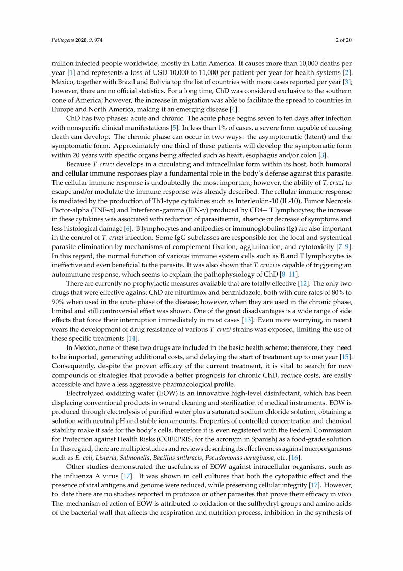
Figure 3. Immunoglobulin levels in sera of BALB/c mice experimentally infected with T. cruzi and
treated with prophylactic or therapeutic EOW. The values represent the means of duplicate assays ±
S.D. for detection of IgG (A), IgG1 (B) and IgG2a (C) titers. The apparent zero values of some groups
are determinations that were not carried out at certain timepoints as shown in Figure 1. The
differences were considered significant (*) at p ≤ 0.05 (Kruskal‐Wallis and one‐way ANOVA tests were
used).
2.3. Prophylactic EOW Administration Induces a 10‐fold Increase of INF‐γ Production in the Acute Phase.
To establish if there was any variation in the cytokine profile expressed during the acute phase
of infection in those groups that received experimental treatment, TNF‐α, INF‐γ, and IL‐1β serum
levels were quantified (Figure 4). The cytokine profile of group H‐EOW was similar to that of group
HEALTHY, so it is inferred that treatment with EOW has no effect on these cytokines evaluated in
healthy mice.
TNF‐α levels decreased at 30 days post‐treatment (dpt) (40 dpi) in the T‐EOW, BZN and W/O‐T
groups compared to the healthy and H‐EOW control groups (Figure 4A); however, there was no
statistically significant difference. Subsequently, at 60 dpi, the T‐EOW group showed a clear statistical
difference by further decreasing their TNF‐α levels compared to the both healthy control groups
(HEALTHY and H‐EOW) (p = 0.029). In the P‐EOW group, the TNF‐α production remained at very
similar levels at all detection times compared to the healthy control groups in despite of the infection
as seen in the W/O‐T group, which showed a significant decrease (p = 0.038) at 60 dpi. This suggests
that to a certain extent, the prophylactic EOW treatment had a positive effect on modulation of the
inflammatory response in infected animals in a late acute phase (Figure 4B).
In the therapeutic scheme, INF‐γ levels behaved similarly in all infected groups with or without
treatment 30 days after administration (40 dpi); and later, they showed a decrease at 60 dpi; however,
0
0.5
1
1.5
2
2.5
3
15 25 30 40 53 63
Ab
sorb
ance
(O
.D.
490
nm
)
Days post-infection
B
W/O-T
P-EOW
T-EOW
BZN
*
*
0
0.5
1
1.5
2
2.5
3
3.5
15 25 30 40 53 63
Ab
sorb
an
ce (
O.D
. 49
0 n
m)
Days post-infection
C
W/O-T
P-EOW
T-EOW
BZN
*
Figure 3. Immunoglobulin levels in sera of BALB/c mice experimentally infected with T. cruzi andtreated with prophylactic or therapeutic EOW. The values represent the means of duplicate assays ± S.D.for detection of IgG (A), IgG1 (B) and IgG2a (C) titers. The apparent zero values of some groups aredeterminations that were not carried out at certain timepoints as shown in Figure 1. The differenceswere considered significant (*) at p ≤ 0.05 (Kruskal-Wallis and one-way ANOVA tests were used).
TNF-α levels decreased at 30 days post-treatment (dpt) (40 dpi) in the T-EOW, BZN and W/O-Tgroups compared to the healthy and H-EOW control groups (Figure 4A); however, there was nostatistically significant difference. Subsequently, at 60 dpi, the T-EOW group showed a clear statisticaldifference by further decreasing their TNF-α levels compared to the both healthy control groups(HEALTHY and H-EOW) (p = 0.029). In the P-EOW group, the TNF-α production remained at verysimilar levels at all detection times compared to the healthy control groups in despite of the infectionas seen in the W/O-T group, which showed a significant decrease (p = 0.038) at 60 dpi. This suggeststhat to a certain extent, the prophylactic EOW treatment had a positive effect on modulation of theinflammatory response in infected animals in a late acute phase (Figure 4B).
In the therapeutic scheme, IFN-γ levels behaved similarly in all infected groups with or withouttreatment 30 days after administration (40 dpi); and later, they showed a decrease at 60 dpi; however,there was no statistically significant difference when compared to the levels of the healthy and H-EOWcontrol groups (Figure 4C). In the prophylactic scheme, the P-EOW group showed dramatically lowlevels of IFN-γ at the end of the preventive treatment (before infection); later, at 30 dpi, its productionincreased approximately 10-fold until reaching levels close to 1000 pg/mL, which subtly decreased at60 dpi (Figure 4D). No statistically significant differences were found when the experimental groupswere compared to the HEALTHY and H-EOW groups. With these data it is possible to propose that

Pathogens 2020, 9, 974 7 of 20
the prophylactic EOW administration contributed to a positive trend in the IFN-γ production duringthe acute phase, which could be maintained even up to 60 dpi.
Pathogens 2020, 9, x FOR PEER REVIEW 7 of 20
there was no statistically significant difference when compared to the levels of the healthy and H‐
EOW control groups (Figure 4C). In the prophylactic scheme, the P‐EOW group showed dramatically
low levels of INF‐γ at the end of the preventive treatment (before infection); later, at 30 dpi, its
production increased approximately 10‐fold until reaching levels close to 1000 pg/mL, which subtly
decreased at 60 dpi (Figure 4D). No statistically significant differences were found when the
experimental groups were compared to the HEALTHY and H‐EOW groups. With these data it is
possible to propose that the prophylactic EOW administration contributed to a positive trend in the
INF‐γ production during the acute phase, which could be maintained even up to 60 dpi.
IL‐1β levels decreased in the groups therapeutically treated with EOW and benznidazole at 40
dpi in the same way as the infected group, and its production continued to decrease until day 60 in
all three groups (Figure 4E), without statistical significance. Prophylactic EOW administration
showed low IL‐1β levels similar to those of W/O‐T group at all detection times; however, neither
demonstrated significant differences when compared to the HEALTHY and H‐EOW control groups;
therefore, it is suggested that prophylactic EOW treatment does not have an effect on this cytokine
(Figure 4F).
As expected, the EOW administration in healthy animals does not have an effect on any of the
three cytokines since the levels found were very similar to those of the HEALTHY control group;
while in infected animals a difference was seen in the levels of some cytokines, thus their
immunomodulatory effect is suggested.
(A) (B)
(C) (D)
0
20
40
60
80
100
120
140
TN
F-
se
rum
leve
ls (
pg
/mL
)
Detection time
HEALTHYH-EOWW/O-TT-EOWBZN
30 dpt/40dpi 60 dpi
*
0
20
40
60
80
100
120
140
160
180
200
TN
F-
seru
m le
vel
s (
pg
/mL
)
Detection time
HEALTHYH-EOWW/O-TP-EOW
15 dpt (pre-infection) 30 dpi 60 dpi
*
0
100
200
300
400
500
600
700
800
30 dpt/40 dpi 60 dpi
IFN
-se
rum
leve
ls (
pg
/mL
)
Detection time
HEALTHYH-EOWW/O-TT-EOWBZN
0
200
400
600
800
1000
1200
1400
15 dpt (pre-infection) 30 dpi 60 dpi
IFN
-se
rum
leve
ls (
pg
/mL
)
Detection time
HEALTHYH-EOWW/O-TP-EOW
Pathogens 2020, 9, x FOR PEER REVIEW 8 of 20
(E) (F)
Figure 4. TNF‐α, IFN‐γ and IL‐1β quantification by enzyme‐linked immunosorbent assay (ELISA) in
sera from T. cruzi‐infected in BALB/c mice experimentally infected with T. cruzi and treated with
prophylactic (B,D,F) or therapeutic EOW (A,C,E). The results are shown as group mean ± S.D. The
differences were considered significant (*) at p ≤ 0.05 when comparing infected and treated groups vs.
the healthy control group (Kruskal‐Wallis and one‐way ANOVA tests were used).
2.4. Effects of EOW on Cardiomegaly, Splenomegaly and Lymphadenopathy.
To evaluate the effect of the treatment on the development of mega syndromes, the indices of
three organs were determined: heart, spleen, and popliteal lymph nodes (Figure 5). Cardiomegaly
(Figure 5A) was not observed in any of the experimental groups demonstrated by the average cardiac
index compared to the healthy control group (p = 0.056). However, a marked trend of increased heart
index could be observed in the W/O‐T group compared to the healthy control group with a difference
of 26.6% (0.68 ± 0.06 in the W/O‐T group vs. 0.49 ± 0.04 in the healthy group), which suggested that
untreated animals might develop dilated cardiomyopathy or hypertrophy if the disease had
progressed to the late chronic stage.
The BZN, P‐EOW and T‐EOW groups showed splenomegaly (Figure 5B) and lymphadenopathy
(Figure 5C) at the end of the course of the acute phase demonstrated by the average splenic and
lymph node index, respectively, compared to the healthy control group (p = 0.000). This suggested
that infected animals, whether treated or not, were responding immunologically to the parasiteʹs
antigenic stimulus in a similar way.
When comparing the splenic index of all the groups that received treatment with that of the
W/O‐T group, there was also statistically significant differences in the P‐EOW (p = 0.016) and T‐EOW
(p = 0.001) group; while the splenic index of the BZN group (p = 0.141) showed values similar to those
of the W/O‐T group (Figure 5B). These data indicated that the treatments caused an enlarged spleen,
which may suggest that an exacerbation of the immune response may be occurring at a higher level
than the infection alone.
Regarding lymphadenopathy, no statistically significant differences were observed between the
peripheral lymph node index of those groups that received treatment compared to the infected
untreated group (Figure 5C), demonstrating that even with treatment, the immune response in
secondary lymphoid organs showed a behavior with a similar pattern in all infected groups.
The organic indices to determine cardiomegaly, splenomegaly and lymphadenopathy did not
show to be different between the uninfected animals (HEALTHY group) and EOW treated non‐
infected mice (H‐EOW group); this demonstrates that oral EOW treatment has no effect on the
macroscopic characteristics of these organs.
0
50
100
150
200
250
300
350
400
30 dpt/40 dpi 60 dpi
IL-1
seru
m l
evel
s (
pg
/mL
)
Detection time
HEALTHYH-EOWW/O-TT-EOWBZN
0
50
100
150
200
250
300
350
400
15 dpt (pre-infection) 30 dpi 60 dpi
IL-1
ser
um
leve
ls(p
g/m
L)
Detection time
HEALTHYH-EOWW/O-TP-EOW
Figure 4. TNF-α, IFN-γ and IL-1β quantification by enzyme-linked immunosorbent assay (ELISA)in sera from T. cruzi-infected in BALB/c mice experimentally infected with T. cruzi and treated withprophylactic (B,D,F) or therapeutic EOW (A,C,E). The results are shown as group mean ± S.D.The differences were considered significant (*) at p ≤ 0.05 when comparing infected and treated groupsvs. the healthy control group (Kruskal-Wallis and one-way ANOVA tests were used).
IL-1β levels decreased in the groups therapeutically treated with EOW and benznidazole at 40 dpiin the same way as the infected group, and its production continued to decrease until day 60 in allthree groups (Figure 4E), without statistical significance. Prophylactic EOW administration showedlow IL-1β levels similar to those of W/O-T group at all detection times; however, neither demonstratedsignificant differences when compared to the HEALTHY and H-EOW control groups; therefore, it issuggested that prophylactic EOW treatment does not have an effect on this cytokine (Figure 4F).
As expected, the EOW administration in healthy animals does not have an effect on any of the threecytokines since the levels found were very similar to those of the HEALTHY control group; while in

Pathogens 2020, 9, 974 8 of 20
infected animals a difference was seen in the levels of some cytokines, thus their immunomodulatoryeffect is suggested.
2.4. Effects of EOW on Cardiomegaly, Splenomegaly and Lymphadenopathy
To evaluate the effect of the treatment on the development of mega syndromes, the indices ofthree organs were determined: heart, spleen, and popliteal lymph nodes (Figure 5). Cardiomegaly(Figure 5A) was not observed in any of the experimental groups demonstrated by the average cardiacindex compared to the healthy control group (p = 0.056). However, a marked trend of increased heartindex could be observed in the W/O-T group compared to the healthy control group with a differenceof 26.6% (0.68 ± 0.06 in the W/O-T group vs. 0.49 ± 0.04 in the healthy group), which suggested thatuntreated animals might develop dilated cardiomyopathy or hypertrophy if the disease had progressedto the late chronic stage.
Pathogens 2020, 9, x FOR PEER REVIEW 9 of 20
(A) (B)
(C)
Figure 5. Cardiomegaly, splenomegaly, and lymphadenopathy in BALB/c mice experimentally
infected with T. cruzi and treated with prophylactic or therapeutic EOW. The values represent the
means of the heart index, the splenic index, and the lymph node index of duplicate assays (± S.D.)
Cardiomegaly (A), splenomegaly (B), and lymphadenopathy (C) was considered when the organ
index was significantly higher than that observed in those from healthy control mice (*) at p ≤ 0.05).
(**) Significant differences in (B) were also shown when comparing treated groups vs. the infected
untreated one. The Kruskal‐Wallis and one‐way ANOVA tests were used.
2.5. EOW Administration Ameliorated the Inflammation Degree and Prevented Tissue Parasitism
With the purpose of evaluating the effect of the experimental treatment on the tissue alterations
caused by T. cruzi, sections of different typical ChD target organs and others that are indirectly
involved were analyzed. A predominantly subepicardial mononuclear inflammatory infiltrate was
observed in the myocardial tissue of all infected mice (Figure 6B–E). The inflammation degree in the
groups prophylactically and therapeutically treated with EOW had a very similar pattern, being
classified with scores between 2 and 3, mostly (Figure 6B–D); the W/O‐T group showed very high
scores, being classified as the most severe grade (score 4) (Figure 6E), and even amastigote nests were
observed in one of the mice (Figure 6F). Inflammation was much lower in the BZN group, as shown
by the statistical significance (p = 0.027) when compared to the W/O‐T group (Figure 6G).
In skeletal muscle, inflammatory infiltrate with the same cellular characteristics as that seen in
heart was observed. All groups showed similar inflammation degrees with low scores without
statistically significant difference when compared among them (p = 0.603) (Figure 6H).
0
0.1
0.2
0.3
0.4
0.5
0.6
0.7
0.8
HEALTHY H-EOW W/O-T P-EOW T-EOW BZN
H.I.
= h
eart
wei
gh
t (g
) / b
od
y w
eig
ht
(g)
x 1
00
Groups
0
0.5
1
1.5
2
2.5
HEALTHY H-EOW W/O-T P-EOW T-EOW BZN
S.I.
= s
ple
en
wei
gh
t (g
) /
bo
dy
w
eig
ht
(g)
x 10
0
Groups
****
**
*
0
0.005
0.01
0.015
0.02
0.025
0.03
0.035
0.04
0.045
HEALTHY H-EOW W/O-T P-EOW T-EOW BZN
Ln
.I. =
Lym
ph
no
de
wei
gh
t (g
) / b
od
y w
eig
ht
(g)
x 1
00
Groups
**
**
Figure 5. Cardiomegaly, splenomegaly, and lymphadenopathy in BALB/c mice experimentally infectedwith T. cruzi and treated with prophylactic or therapeutic EOW. The values represent the means ofthe heart index (H.I.), the splenic index (S.I.), and the lymph node index (Ln.I.) of duplicate assays (±S.D.) Cardiomegaly (A), splenomegaly (B), and lymphadenopathy (C) was considered when the organindex was significantly higher than that observed in those from healthy control mice (*) at p ≤ 0.05).(**) Significant differences in (B) were also shown when comparing treated groups vs. the infecteduntreated one. The Kruskal-Wallis and one-way ANOVA tests were used.
The BZN, P-EOW and T-EOW groups showed splenomegaly (Figure 5B) and lymphadenopathy(Figure 5C) at the end of the course of the acute phase demonstrated by the average splenic and lymphnode index, respectively, compared to the healthy control group (p = 0.000). This suggested thatinfected animals, whether treated or not, were responding immunologically to the parasite’s antigenicstimulus in a similar way.

Pathogens 2020, 9, 974 9 of 20
When comparing the splenic index of all the groups that received treatment with that of theW/O-T group, there was also statistically significant differences in the P-EOW (p = 0.016) and T-EOW(p = 0.001) group; while the splenic index of the BZN group (p = 0.141) showed values similar to thoseof the W/O-T group (Figure 5B). These data indicated that the treatments caused an enlarged spleen,which may suggest that an exacerbation of the immune response may be occurring at a higher levelthan the infection alone.
Regarding lymphadenopathy, no statistically significant differences were observed betweenthe peripheral lymph node index of those groups that received treatment compared to the infecteduntreated group (Figure 5C), demonstrating that even with treatment, the immune response insecondary lymphoid organs showed a behavior with a similar pattern in all infected groups.
The organic indices to determine cardiomegaly, splenomegaly and lymphadenopathy did notshow to be different between the uninfected animals (HEALTHY group) and EOW treated non-infectedmice (H-EOW group); this demonstrates that oral EOW treatment has no effect on the macroscopiccharacteristics of these organs.
2.5. EOW Administration Ameliorated the Inflammation Degree and Prevented Tissue Parasitism
With the purpose of evaluating the effect of the experimental treatment on the tissue alterationscaused by T. cruzi, sections of different typical ChD target organs and others that are indirectly involvedwere analyzed. A predominantly subepicardial mononuclear inflammatory infiltrate was observedin the myocardial tissue of all infected mice (Figure 6B–E). The inflammation degree in the groupsprophylactically and therapeutically treated with EOW had a very similar pattern, being classifiedwith scores between 2 and 3, mostly (Figure 6B–D); the W/O-T group showed very high scores, beingclassified as the most severe grade (score 4) (Figure 6E), and even amastigote nests were observedin one of the mice (Figure 6F). Inflammation was much lower in the BZN group, as shown by thestatistical significance (p = 0.027) when compared to the W/O-T group (Figure 6G).
In skeletal muscle, inflammatory infiltrate with the same cellular characteristics as that seenin heart was observed. All groups showed similar inflammation degrees with low scores withoutstatistically significant difference when compared among them (p = 0.603) (Figure 6H).
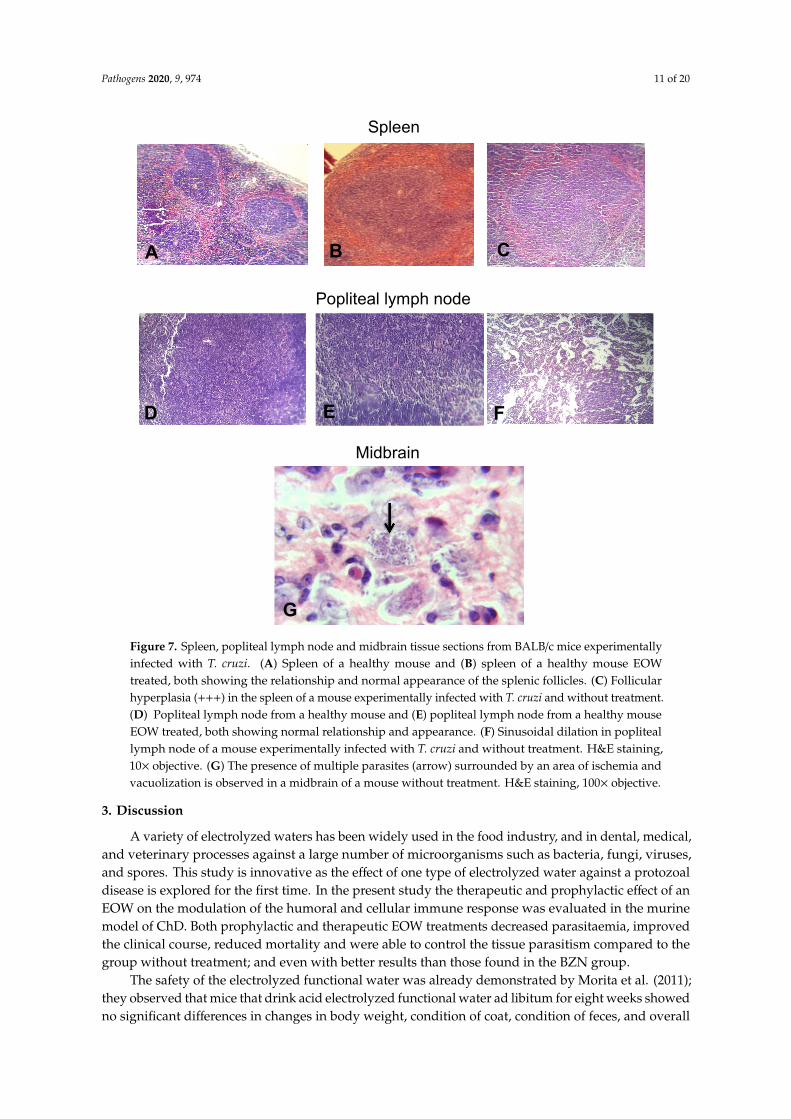
In the W/O-T group, the microscopic findings were not limited to the heart and skeletal muscle,in the majority of the mice splenic follicular hyperplasia was evident (Figure 7C), while in popliteallymph nodes, sinusoidal dilation was found (Figure 7F). In the midbrain of one of the mice thatreceived no treatment, the parasite load could be corroborated by the presence of trypomastigote forms,which were surrounded by an ischemic zone with vacuolization (Figure 7G). In the gastrointestinaltract (esophagus, small and large intestine), no microscopic changes were observed in any of the groups(data not shown). These findings suggested that both prophylactic and therapeutic EOW administrationcontributed to the partial control of inflammation and the total control of tissue parasitism, resultsthat resemble those found in the group of mice treated with benznidazole. The histological sections ofhealthy mice treated with EOW showed no apparent pathological features, which proves its safety.

Pathogens 2020, 9, 974 10 of 20Pathogens 2020, 9, x FOR PEER REVIEW 10 of 20
Figure 6. Histological findings from BALB/c mice experimentally infected with T. cruzi and treated
with prophylactic or therapeutic EOW. Representative micrographs in (A–E) are shown. (A) Score 0
(normal) from a healthy mouse; (B) Score 1 (one focus of inflammatory cells/field) from a mouse
treated with benznidazole; (C) Score 2 (more than one or a few foci of inflammatory cells/field) from
a mouse treated prophylactically or therapeutically with EOW; (D) Score 3 (generalized coalescent
inflammation foci or disseminated inflammation with minimal cell necrosis and preserved tissue
integrity) from a mouse treated prophylactically or therapeutically with EOW; (E) Score 4 (diffuse
inflammation, severe tissue necrosis, interstitial edema, hemorrhage, and/or loss of tissue integrity)
from an untreated infected mouse. H&E staining, 40× objective. (F) Amastigote nest (arrow) in a heart
tissue section from a mouse of the W/O‐T group. H&E staining, 100× objective. Inflammatory lesion
(inflammatory cell infiltrates) scores of heart (G) and skeletal muscle (H) are shown. The score was
converted into logarithm (base 10) and then 1 was added to each result to correct the values of 0. The
results were expressed as group means ± S.D. No differences were observed (Kruskal‐Wallis test when
comparing the infected and treated groups vs. the infected group without treatment).
In the W/O‐T group, the microscopic findings were not limited to the heart and skeletal muscle,
in the majority of the mice splenic follicular hyperplasia was evident (Figure 7C), while in popliteal
lymph nodes, sinusoidal dilation was found (Figure 7F). In the midbrain of one of the mice that
received no treatment, the parasite load could be corroborated by the presence of trypomastigote
forms, which were surrounded by an ischemic zone with vacuolization (Figure 7G). In the
gastrointestinal tract (esophagus, small and large intestine), no microscopic changes were observed
in any of the groups (data not shown). These findings suggested that both prophylactic and
therapeutic EOW administration contributed to the partial control of inflammation and the total
A B C
D E F
00.20.40.60.8
11.21.41.61.8
2
W/O-T P-EOW T-EOW BZN
Hea
rt i
nfl
amm
ati
on
sc
ore
Groups
0
0.2
0.4
0.6
0.8
1
1.2
1.4
1.6
W/O-T P-EOW T-EOW BZN
Sk
elet
al m
us
cle
infl
amm
atio
n
sco
re
Groups
(G) (H)
Figure 6. Histological findings from BALB/c mice experimentally infected with T. cruzi and treated withprophylactic or therapeutic EOW. Representative micrographs in (A–E) are shown. (A) Score 0 (normal)from a healthy mouse; (B) Score 1 (one focus of inflammatory cells/field) from a mouse treated withbenznidazole; (C) Score 2 (more than one or a few foci of inflammatory cells/field) from a mouse treatedprophylactically or therapeutically with EOW; (D) Score 3 (generalized coalescent inflammation foci ordisseminated inflammation with minimal cell necrosis and preserved tissue integrity) from a mousetreated prophylactically or therapeutically with EOW; (E) Score 4 (diffuse inflammation, severe tissuenecrosis, interstitial edema, hemorrhage, and/or loss of tissue integrity) from an untreated infectedmouse. H&E staining, 40× objective. (F) Amastigote nest (arrow) in a heart tissue section from a mouseof the W/O-T group. H&E staining, 100× objective. Inflammatory lesion (inflammatory cell infiltrates)scores of heart (G) and skeletal muscle (H) are shown. The score was converted into logarithm (base10) and then 1 was added to each result to correct the values of 0. The results were expressed as groupmeans ± S.D. No differences were observed (Kruskal-Wallis test when comparing the infected andtreated groups vs. the infected group without treatment).
Pathogens 2020, 9, 974 11 of 20
Pathogens 2020, 9, x FOR PEER REVIEW 11 of 20
control of tissue parasitism, results that resemble those found in the group of mice treated with
benznidazole. The histological sections of healthy mice treated with EOW showed no apparent
pathological features, which proves its safety.
Figure 7. Spleen, popliteal lymph node and midbrain tissue sections from BALB/c mice
experimentally infected with T. cruzi. (A) Spleen of a healthy mouse and (B) spleen of a healthy mouse
EOW treated, both showing the relationship and normal appearance of the splenic follicles. (C)
Follicular hyperplasia (+++) in the spleen of a mouse experimentally infected with T. cruzi and without
treatment. (D) Popliteal lymph node from a healthy mouse and (E) popliteal lymph node from a
healthy mouse EOW treated, both showing normal relationship and appearance. (F) Sinusoidal
dilation in popliteal lymph node of a mouse experimentally infected with T. cruzi and without
treatment. H&E staining, 10× objective. (G) The presence of multiple parasites (arrow) surrounded by
an area of ischemia and vacuolization is observed in a midbrain of a mouse without treatment. H&E
staining, 100× objective.
3. Discussion
A variety of electrolyzed waters has been widely used in the food industry, and in dental,
medical, and veterinary processes against a large number of microorganisms such as bacteria, fungi,
viruses, and spores. This study is innovative as the effect of one type of electrolyzed water against a
protozoal disease is explored for the first time. In the present study the therapeutic and prophylactic
effect of an EOW on the modulation of the humoral and cellular immune response was evaluated in
the murine model of ChD. Both prophylactic and therapeutic EOW treatments decreased
parasitaemia, improved the clinical course, reduced mortality and were able to control the tissue
A
Spleen
Popliteal lymph node
Midbrain
B C
D F
G
E
Figure 7. Spleen, popliteal lymph node and midbrain tissue sections from BALB/c mice experimentallyinfected with T. cruzi. (A) Spleen of a healthy mouse and (B) spleen of a healthy mouse EOWtreated, both showing the relationship and normal appearance of the splenic follicles. (C) Follicularhyperplasia (+++) in the spleen of a mouse experimentally infected with T. cruzi and without treatment.(D) Popliteal lymph node from a healthy mouse and (E) popliteal lymph node from a healthy mouseEOW treated, both showing normal relationship and appearance. (F) Sinusoidal dilation in popliteallymph node of a mouse experimentally infected with T. cruzi and without treatment. H&E staining,10× objective. (G) The presence of multiple parasites (arrow) surrounded by an area of ischemia andvacuolization is observed in a midbrain of a mouse without treatment. H&E staining, 100× objective.
3. Discussion
A variety of electrolyzed waters has been widely used in the food industry, and in dental, medical,and veterinary processes against a large number of microorganisms such as bacteria, fungi, viruses,and spores. This study is innovative as the effect of one type of electrolyzed water against a protozoaldisease is explored for the first time. In the present study the therapeutic and prophylactic effect of anEOW on the modulation of the humoral and cellular immune response was evaluated in the murinemodel of ChD. Both prophylactic and therapeutic EOW treatments decreased parasitaemia, improvedthe clinical course, reduced mortality and were able to control the tissue parasitism compared to thegroup without treatment; and even with better results than those found in the BZN group.
The safety of the electrolyzed functional water was already demonstrated by Morita et al. (2011);they observed that mice that drink acid electrolyzed functional water ad libitum for eight weeks showedno significant differences in changes in body weight, condition of coat, condition of feces, and overall
Pathogens 2020, 9, 974 12 of 20
visual findings compared to the control group. They also found no abnormalities in terms of visualinspections of the oral cavity, histopathological tests, or tooth surface enamel roughness measurements.They only observed enamel attrition in some molars in the test group with the electrolyzed water.This research group suggested the use of acid pH electrolyzed water does not have a systemic effectand is safe for mouthwash [22].
Electrolyzed reducing water (ERW) was shown to protect the cellular redox balance, reducing therisk of various diseases with altered cellular homeostasis such as inflammation [23]. Several biologicalresults of ERW were reported, such as protective effect of oxidative damage to DNA by reactiveoxygen species [24], stimulator of growth of anaerobic microflora in the human gut [25], antidiabeticeffect in rats and mice [26,27], antioxidant effect [28], growth-promoting effect of the fetus in rats [29],therapeutic effect in patients with end-stage renal disease [30] and reduction in blood viscosity due todehydration in humans [31].
Currently, there are multiple studies that describe the interaction among different protectivemechanisms of the immune system in response to T. cruzi infection. These mechanisms involve differentcell lines activation, pro-inflammatory cytokines (TNF-α and IL-1β) and Th1-type (IFN-γ and IL-12)production, and increased reactive oxygen species production [32]. Although these elements favor thecontrol of parasitism in the acute phase, their overexpression during the chronic phase is the cause ofcardiac remodeling and progression to chronic chagasic cardiomyopathy, a clear example of this is thedual role of IFN-γ in different phases of ChD [33].
Among the objectives of this research was to determine the effect of EOW treatment on thehumoral immune response in mice infected with T. cruzi. Although extensive research was done withthe use of EOW, the effect on antibody production was not previously explored in any animal model.In this regard, the total IgG levels and the IgG1 and IgG2a subclasses were quantified. Our resultsindicated that the treatment induced IgG2a antibodies related to a Th1-type response, which correlateswith the profile of cytokines found.
It was shown that several recombinant T. cruzi proteins can generate Th1-type antibodiesin vaccinated mice and this kind of antibodies is required to eliminate the parasite in infectedanimals [34,35]. Although an immunogen was not used in the present study, the EOW had animmunomodulatory effect similar to that of recombinant vaccines, based on IgG2a subclass antibodiespredominantly produced in infected animals prophylactically treated with EOW.
On the other hand, the mice treated prophylactically with EOW had a lower amount of IL-1β andTNF-α and higher levels of IFN-γ than untreated animals. Something similar was found in other studies,which demonstrate the useful of electrolyzed water as treatment in two types of conditions in mousemodel; Lee et al. (2009) showed that electrolyzed reduced water decreased the production of IL-1βand TNF-α in C57BL/6 mice infected with Echinostoma hortense (intestinal helminth) metacercariae [36].Low levels of intestinal TNF-α expression were similar in mice that given free access to tap water asdrinking water to those that were given free access to acid electrolyzed functional water as drinkingwater [22]. More recently, You’s group (2017) observed that treatment with slightly electrolyzed acidicwater in cutaneous wounds in hairless mice decreased serum proinflammatory cytokines such as IL-1β,IL-6, keratinocyte chemoattractant and TNF-α [37].
The IFN-γ peak in the P-EOW group at 30 dpi could not be directly correlated with a beneficialeffect on other parameters determined in this study, since this group expressed characteristics verysimilar to those treated therapeutically with EOW, and even those of the BZN group, which hadless histological damage and better control of parasitaemia even without this IFN-γ overexpression.The foregoing does not agree with what was found by others who studied the effects of benznidazoleon the immune system, since it was described that there is a synergistic action of this drug when it isadministered in the acute phase together with IL-12, which in turn triggers the IFN-γoverproduction [38].While being administered in the chronic phase, it is capable of modulating IFN-γ levels and reducingits production [39].

Pathogens 2020, 9, 974 13 of 20
The cytokines’ expression determined in this study also differs from that described by otherauthors who used the same T. cruzi strain (MHOM/MX/1994/Ninoa) and described how these cytokinesproduction is triggered during the first half of the acute phase during the natural course of T. cruziinfection, and then gradually decreases. Otherwise, the clinical course of the infection observed here,as well as the low levels of parasitaemia and mortality do agree with those reported by others, despitethe use of different infection inoculums [40].
The absence of cardiomegaly and the presence of splenomegaly and lymphadenopathy wereconstant in most of the experimental groups evaluated. These results are consistent with those found inother animal models [41], in which it is described that while the acute phase is characterized by this typeof findings, in the chronic phase a slight splenomegaly, absence of lymphadenopathy and a markedcardiomegaly predominate. Therefore, the splenic and secondary lymphatic organs responses in thelate acute phase of the infection were similar in all groups treated with EOW and with benznidazole.However, while in the W/O-T group it was not possible to show the splenomegaly, there were histologicalchanges that showed an inflammatory response, resulting in reactive lymphoid hyperplasia.
On the other hand, the presence of a diffuse mononuclear inflammatory infiltrate in the heartand skeletal muscle was constant, which is consistent with the findings already described by differentgroups in other animal models during the acute phase, and even in human samples [42]. In this regard,the results obtained in the present study showed that the W/O-T group had the highest inflammationseverity scores in heart tissue, and it was possible to identify the parasite’s presence in this organ andin the central nervous system. However, cytokine production was found in low ranges and similar tothe other treated groups, which differs from that described in the literature, where it is establishedthat the exaggerated increase in the inflammatory infiltrate characteristic of ChD in myocardial tissuecorrelates with increased production of cytokines such as IFN-γ during the acute phase [33,41] andother proinflammatory ones. It is suggested that the immunomodulation observed in the other treatedgroups was associated with the antigenic stimulus of T. cruzi infection and not with the administrationof EOW alone by showing a very similar cytokine profile between HEALTHY and H-EOW groups.
In recent years, the study of the polarization between the Th1- and Th2-type responses wasaddressed, which are closely related by mutually inhibiting each other, and it was observed that theTh1-type cytokines decrease makes the host more susceptible to infection [43]. In this study, Th2-typecytokines were not determined based on the determination of IgG1 subclass to evaluate the TH2immune response, so we cannot confirm whether the low levels of TNF-α, IFN-γ and IL-1β were dueto an overproduction of other antagonists such as IL-10 and IL-4. However, if the increase in the latterwere present, this would be paradoxical with what was described by our research group previously [44],in which high mortalities in experimental animals when the immune response is polarized toward Th2were reported; this did not happen in the present study, where the clinical course was favorable inthe groups treated with EOW and benznidazole. This could suggest two possible scenarios: that theinfection control was elicited by the direct toxic effect of the administered products on the parasite, or thatthere could be synergy with another type of immune response such as humoral or complement throughother signaling pathways unexplored, placing EOW as an immunomodulatory agent. Regarding thepreferred subclass of antibodies generated (IgG2a) and cytokine profile (Th1) induced, it is possible topostulate that EOW leads to immunological modulation that greatly ameliorated T. cruzi infection.This agrees with other authors’ reports, which demonstrate that electrolyzed solutions helped toincrease the body’s immune defenses against oxidative stress by eliminating in particular hydrogenperoxide and the hypochlorite anion, as well as improved the inflammatory response and the controlof blood pressure in hemodialysis patients [45]. Kapur and Marwaha (2011) showed that there wasremarkable reduction in common signs of inflammation such as edema, erythema, and drastic increaseof granulation and fibrin formation in patients with different types of wounds treated with EOWdemonstrating an immunomodulatory effect [46]. In this regard, immunotherapy with the EOW couldprovide a survival advantage by reducing the clinical signs of infection and ameliorating the cardiacdamage of Chagas disease by avoiding disease progression as seen with other immunotherapeutic

Pathogens 2020, 9, 974 14 of 20
agents against various pathologies, such as allergies, injury to the peripheral nervous system, Chagasdisease, leishmaniosis, and other intracellular pathogens [47–51]. In this study, high parasitaemia,general physical condition (adynamia, arched back and piloerection), lethality and tissue parasite loadconsidered to be typical clinical manifestations of ChD were ameliorated in mice treated with EOW.
New questions arise about our findings, which could be answered by complementing this studywith the determination of a more extensive panel of Th1-and Th2-type cytokines, or lymphoproliferationtests; and the search for lytic antibodies mediated by the action of the complement. It is also proposedto extend the study to later stages of the chronic phase of ChD in order to obtain more data that allowsus to define the effect of EOW on the pathogenesis of the disease more precisely.
4. Materials and Methods
4.1. Animals and Experimental Groups
Sixty 6 to 8-week old female BALB/c mice (from the Center for Research and Advanced Studies ofthe National Polytechnic Institute CINVESTAV-IPN, for the acronym in Spanish) were divided intosix groups with five animals per group: (1) healthy mice without infection or treatment (HEALTHY),(2) healthy mice without infection treated with EOW (H-EOW), (3) infected mice without treatment(W/O-T), (4) infected mice prophylactically treated with EOW (P-EOW), (5) infected mice therapeuticallytreated with EOW (T-EOW) and 6) infected mice treated with benznidazole (BZN). Experiments werecarried out in duplicate to confirm reproducibility. All mice were housed with light/dark cycles of 12h/12 h, under temperature conditions of 22–24 ◦C, with food and water ad libitum. All procedures wereperformed under the protocols established by the Guide for Care and Use of Experimental Animals,National Institutes of Health [52] and the official Mexican norm (NOM-062-ZOO-1999) about TechnicalSpecifications for the Production, Care and Use of Laboratory Animals [53], with approval by theInternal Committee for the Care and Use of Laboratory Animals (CICUAL, for the acronym in Spanish)registered under the number INC/CICUAL/001/2017.
4.2. Infection
Mice were inoculated with blood trypomastigotes (BT) of the Ninoa T. cruzi strain(MHOM/MX/1994/Ninoa (T. cruzi)). The parasites were obtained from blood samples from thetail vein of previously infected mice (approximately at 21 days after infection). The parasites werecounted in a Neubauer chamber and the volume was adjusted to infect each mouse with 150 BTintraperitoneally, using sterile syringes with a 27 G × 13 mm needle.
4.3. Parasitaemia and Survival
Parasitaemia curves were elaborated by parasite counts per blood sample every third day from10 dpi and until the blood sample was negative for the presence of parasitic forms by the modifiedPetana method [54] as follows: 10 µL of blood was obtained from the tail vein; they were diluted in490 µL of physiological 0.9% saline solution (SS) to obtain a 1:50 dilution. The Neubauer chamberwas loaded with 10 µL of this dilution, and BT was counted in the four squares used for leukocytecounting using 25× and 40× objectives. The total number of parasites counted was multiplied by 50(dilution factor), divided by 0.4 resulting from the product of the chamber surface (4 mm2) and itsdepth (0.1 mm); finally, this result was multiplied by 1000 to obtain the number of parasites in 1 mLof blood. Also, survival was recorded daily and the mortality rate (number of mice that died duringacute infection) was obtained.
4.4. Prophylactic EOW and Therapeutic EOW Treatments by Orogastric Route
The pH-neutral EOW used in this study was SoluVet®, a commercial product provided byEsteripharma México, S.A. de C.V. SoluVet® is an EOW with neutral pH at 0.0006% (60 parts permillion, ppm) of free active chlorine. For the prophylactic treatment, 500 µL of EOW was administered

Pathogens 2020, 9, 974 15 of 20
every 24 h for 15 days prior to infection, and for the therapeutic treatment the same amount of EOWevery 24 h for 30 days, starting on the tenth day after infection; both by orogastric route. The H-EOWcontrol group was treated under the same scheme of administration as the T-EOW group. As a positivecontrol, an active ingredient obtained from 100 mg benznidazole LaFepe® (Laboratório Farmacêuticodo Estado de Pernambuco, São José Recife, Brazil) tablets was used preparing a suspension withTween 20 at 5%, adjusting the dose of 10 mg/kg to the volume used per dose (500 µL); this dose wasadministered every 24 h for 30 days by orogastric route, starting on the tenth day after infection.The treatments were performed with a No. 18 straight stainless-steel feeding cannula (Cadence ScienceInc.®, Pike, Cranston, RI, USA).
4.5. Collection, Handling and Storage of Blood Samples
A 250–300 µL blood sample was taken from each mouse prior to the experiments to be used asa reference (preimmune serum). For the T-EOW, BZN and W/O-T groups, samples were collectedtwice: at 40 dpi, equivalent to 30 dpt, and 60 dpi. In the P-EOW group, samples were collected threetimes: on day 15 after the start of preventive treatment (preinfection), and at 30 and 60 dpi; and inthe H-EOW group, samples were collected on days 15, 30 and 60 dpt. Likewise, an additional bloodsample was extracted from the group without treatment at 30 dpi. The blood collection was carriedout at 3 h post-treatment from the tail vein and collected in 1.5 mL tubes for microcentrifuge withoutanticoagulant, kept at room temperature until clot retraction and then centrifuged at 3500 rpm for 15 minat a temperature of 4 ◦C using a refrigerated microcentrifuge (model RMC-14, Sorvall®/DuPont®).Finally, aliquots of 50–100 µL of sera were made in 0.6 mL tubes for microcentrifuge and stored infreezing at −20 ◦C until use.
4.6. Immunoglobulins Determination
Total IgG antibody and subclasses IgG1 and IgG2a, were evaluated at 15, 25 (at the middleof the therapeutic EOW and benznidazole administrations), 30, 40 (at the end of the therapeuticEOW and benznidazole administration), 53 and 63 dpi by the enzyme-linked immunosorbent assay(ELISA) method according to the manufacturer’s instructions (Novus Biologicals, Centennial, CO,USA). Briefly, plates were coated with the whole T. cruzi INC-9 isolate extract (1 µg/mL) in 200 µL ofcarbonate buffer (NaCO3/NaHCO3 pH 9.6) and incubated for 1 h at 37 ◦C. Plates were washed fivetimes with PBS 1×-0.05% Tween-20 (PBS-T), incubated for 30 min at 37 ◦C with a blocking solution(PBS containing 0.5% bovine serum albumin fraction V). Serum samples were diluted in blockingbuffer at dilution of 1:200 in 200 µL/well and incubated (1 h, 37 ◦C). As a negative control, a pool ofpreimmune sera was used in all experiments. The plates were washed five times with PBS-T. Afterwashing, the peroxidase-labeled rabbit anti-mouse IgG and IgG subclasses (IgG1 and IgG2a) secondaryantibodies (Novus Biologicals, Centennial, CO, USA) were added at 1:10,000 dilution in PBS-T andincubated for 1 h at room temperature. The plates were washed five times with PBS-T, 150 µL ofperoxidase substrate OPD (orto-phenylenediamine dihydrochloride, Sigma Aldrich, St. Louis, MO,USA) in citrate buffer at pH 4.5–0.03% H2O2 was added, and incubated for 10 min at room temperature.The reaction was stopped 10 min later by the addition of 50 µL of 5 N H2SO4. Absorbance values weredetermined at 495 nm in a Microplate Reader (Bio-Rad, Hercules, CA, USA). All measurements wereperformed twice.
4.7. Cytokine Determination
Th1 cytokine levels (TNF-α, IFN-γ, and IL-1β,) in the mice’s serum at 15 dpt (preinfection inthe prophylactic EOW treatment), 40 dpi (at the end of the therapeutic EOW and benznidazoleadministration) and 60 dpi were measured by duplicate by ELISA using commercial kits (EnzoLife Sciences, Inc.®, East Farmingdale, NY, USA and R&D Systems, Inc.®, Minneapolis, MN, USA)according to the manufacturer’s instructions. Briefly, the capture antibody was diluted to 1.0 µg/mLwith PBS, 50 µL/well was added to the 96 Maxisorb plates (R&D Systems Inc.®, Minneapolis, MN, USA)

Pathogens 2020, 9, 974 16 of 20
and incubated overnight at room temperature. The plates were washed three times with 250 µL PBS-T,blocked with 200 µL of blocking buffer per well for 1 h at room temperature and washed four times. Thestandard sample was prepared for each cytokine and 50 µL/well was added; then, 50 µL of the serumsamples were added to each well and incubated for 2 h at room temperature. The plates were washedthree times, and then 50 µL of the previously diluted detection antibody (1.5 µg/mL) was added andincubated at room temperature for 2 h. The plates were washed, and then 50 µL of previously dilutedavidin were added and incubated for 20 min at room temperature. The plates were washed again,and then 50 µL of peroxidase substrate TMB- H2O2 (3,3’,5,5’-Tetramethylbenzidine, Sigma Aldrich,St. Louis, MO, USA, 30% hydrogen peroxide) was added. The plates were incubated for 20 min atroom temperature, and the reaction was stopped with 50 µL of 5N H2SO4 per well. The reading wasperformed at 450 nm using a Microplate Reader (model Opsys MR, Dynex Technologies®, Chantilly,VA, USA). Each cytokine concentration was determined on the basis of each corresponding standardcurve and expressed in pg/mL.
4.8. Splenomegaly, Cardiomegaly and Lymphadenopathy
Prior to euthanasia (at 60 dpi), the body weight of each mouse was obtained, and the weightsof the heart, the spleen, and the peripheral lymph nodes (poplitei) were collected during necropsyand were weighed. The organ indices were calculated by applying the following formula: organweight/body weight ×100. The presence of cardiomegaly, splenomegaly and/or lymphadenopathy wasconsidered when the calculated organ index was significantly higher than that observed in those fromhealthy uninfected control animals [41].
4.9. Histology
The animals were subjected to euthanasia at 60 dpi to aseptically remove heart, intestines (ileumand colon sections), popliteal lymph nodes, skeletal muscle, esophagus, spleen, and brain. The organswere rinsed with SS, fixed in 10% formaldehyde (pH 7.4) and kept at room temperature until theirprocessing. Samples were dehydrated with absolute ethanol, rinsed with xylene, and embeddedin paraffin. Sections of 5 µm were made, stained with hematoxylin and eosin and evaluated byoptical microscopy (Carl Zeiss® K7, Oberkochen, BW, Germany). The walls of cardiac tissue wereanalyzed from the upper, middle, and lower parts: subepicardium, myocardium, and subendocardium.The severity of inflammation was scored on a scale of 0 to 4, where 0 = no alterations; 1 = one focus ofinflammatory cells/field (400×); 2 = more than one focus of inflammatory cells/field; 3 = generalizedcoalescing foci of inflammation or disseminated inflammation with minimal cell necrosis and retentionof tissue integrity; and 4 = diffuse inflammation, tissue necrosis, interstitial edema, hemorrhage andloss of tissue integrity [55].
4.10. Statistical Analysis
All results were analyzed using the IBM® SPSS software (v. 20.0.0) (Armonk, NY, USA). Datawere expressed as means ± standard deviation (S.D.) of each group. Prior to the comparison betweengroups and possible interactions between them, all the variables were analyzed by the Shapiro-Wilktest considering those with a value of p ≥ 0.05 as normal distribution. Data with a normal distributionwere analyzed by the one-way ANOVA test, followed by Tukey analysis as a post-hoc test. Those thatdid not fit into a normal distribution were analyzed by the Kruskal-Wallis test. The survival analysiswas performed using the Kaplan-Meier method. All differences were considered significant whenp ≤ 0.05. The inflammation grade score was converted into logarithms (base 10) and then 1 was addedto each result to correct the values of 0 [56].
5. Conclusions
Therapeutic EOW administration was useful for the control of the acute phase of ChD by observingparameters of parasitaemia, mortality, clinical status, histological damage, and production of IgG-type

Pathogens 2020, 9, 974 17 of 20
antibodies similar to those obtained with benznidazole treatment. However, it was not possible tocorrelate these results with a synergy in the cellular-type immune response when determining thelevels of the three cytokines. The efficacy observed by EOW could be due to immunomodulatoryproperties and not to a toxic effect on the parasite.
Author Contributions: Conceptualization, O.R.-M., J.J.C.-M. and M.A.-F.; data curation, J.J.C.-M., S.d.C.C.-S.,R.A.G.-O., L.B.-P., O.L.P.-R., J.L.R.-E., A.A.-F. and S.H.-G.; formal analysis, O.R.-M., J.J.C.-M., and M.A.-F.; fundingacquisition, O.R.-M. and M.A.-F.; methodology, J.J.C.-M., S.d.C.C.-S., R.A.G.-O., L.B.-P., O.L.P.-R., J.L.-R.E.,A.A.-F and S.H.-G.; project administration, O.R.-M. and M.A.-F.; resources, L.B.-P., O.L.P.-R, J.L.R.-E. andA.A.-F.; supervision, O.R.-M., S.d.C.C.-S. and M.A.-F.; validation, O.R.-M., J.J.C.-M., S.d.C.C.-S. and M.A.-F.;writing-original draft, O.R.-M. and M.A.-F; writing-review & editing, O.R.-M. and M.A.-F. All authors have readand agreed to the published version of the manuscript.
Funding: This research received no external funding. The affiliation institutes’ resources were the only ones thatfinanced this study.
Acknowledgments: We thank Esteripharma® Company (Esteripharma México, S.A. de C.V.) for providing theSoluVet® for carrying out the experiments in this study. We thank Fernando Martínez Castillo, an English languageinstructor from the National School of Languages, Linguistics and Translation (ENALLT, for the acronym inSpanish) at the National Autonomous University of Mexico for his assistance with the English language editing ofthe manuscript. The authors have not declared a specific grant for this research from any funding agency in thepublic, commercial, or not-for-profit sectors.
Conflicts of Interest: The authors declare no conflict of interest.
Abbreviations
BZN, benznidazole treatment; ChD, Chagas disease; CICUAL, Comité para el Cuidado y Uso de los Animalesde Laboratorio; CINVESTAV-IPN, Centro de Investigación y de Estudios Avanzados del Instituto PolitécnicoNacional; COFEPRIS, Comisión Federal para la Protección contra los Riesgos Sanitarios; dpi, days postinfection;dpt, days post-treatment; ELISA, enzyme-linked immunosorbent assay; EOW, electrolyzed oxidizing water;IFN-gamma, interferon gamma; IgG, immunoglobulin G; IL-1 beta, interleukin 1 beta; P-EOW, prophylacticelectrolyzed oxidizing water treatment; ERW, electrolyzed reducing water; SS saline solution; T-EOW, therapeuticelectrolyzed oxidizing water treatment; TNF-alpha, tumor necrosis factor alpha; USD, United States dollar; WHO,World Health Organization; W/O-T, without treatment.
References
1. World Health Organization (WHO). Chagas Disease (American Trypanosomiasis). 2018. Available online:http://www.who.int/mediacentre/factsheets/fs340/en/ (accessed on 15 June 2018).
2. Ramsey, J.M.; Elizondo-Cano, M.; Sanchez-González, G.; Peña-Nieves, A.; Figueroa-Lara, A. OpportunityCost for Early Treatment of Chagas Disease in Mexico. PLoS Negl. Trop. Dis. 2014, 8, e2776.
3. Molina, I.; Salvador, F.; Sánchez-Montalvá, A. Actualización en enfermedad de Chagas. Enferm. Infecc.Microbiol. Clin. 2016, 34, 132–138.
4. Salvador, F.; Treviño, B.; Sulleiro, E.; Sánchez-Montalva, A.; Cabezos, J.; Soriano, A.; Serre, N.; Gómez iPrat, J.; Pahissa, A.; Molina, I. Trypanosoma cruzi infection in a non-endemic country: Epidemiological andclinical profile. Clin. Microbiol. Infect. 2014, 20, 706–712.
5. Carabarin-Lima, A.; González-Vázquez, M.C.; Rodríguez-Morales, O.; Baylón-Pacheco, L.; Rosales-Encina, J.L.;Reyes-López, P.A.; Arce-Fonseca, M. Chagas disease (American tripanosomiasis) in Mexico: An update. Acta Trop.2013, 127, 126–135. [PubMed]
6. Arce-Fonseca, M.; Ballinas-Verdugo, M.A.; Abreu Zenteno, E.R.; Suárez-Flores, D.; Carrillo-Sánchez, S.C.;Alejandre-Aguilar, R.; Rosales-Encina, J.L.; Reyes, P.A.; Rodríguez-Morales, O. Specific humoral and cellularimmunity induced by Trypanosoma cruzi DNA immunization in a canine model. Vet. Res. 2013, 44, 1–9.
7. Teixeira, A.R.L.; Hecht, M.M.; Guimaro, M.C.; Sousa, A.O.; Nitz, N. Pathogenesis of Chagas’ Disease: ParasitePersistence and Autoinmunity. Clin. Microbiol. Rev. 2011, 24, 592–630.
8. Cardillo, F.; Teixeira, P.R.; Zuquim, A.P.R.; Mengel, J. Immunity and immune modulation in Trypanosomacruzi infection. Pathog. Dis. 2015, 73, ftv082.
9. Basso, B. Modulation of immune response in experimental Chagas disease. World J. Exp. Med. 2013, 3, 1–10.[PubMed]

Pathogens 2020, 9, 974 18 of 20
10. Geiger, A.; Bossard, G.; Sereno, D.; Pissarra, J.; Lemesre, J.-P.; Vincendeau, P.; Holzmuller, P. EscapingDeleterious Immune Response in Their Hosts: Lessons from Trypanosomatids. Front. Immunol. 2016, 7, 1–21.
11. Cardoso, M.S.; Reis-Cunha, J.L.; Bartholomeu, D.C. Evasion of the Immune Response by Trypanosoma cruziduring Acute Infection. Front. Immunol. 2016, 6, 1–15.
12. Rodríguez-Morales, O.; Monteón-Padilla, V.; Carrillo-Sánchez, S.C.; Rios-Castro, M.; Martínez-Cruz, M.;Carabarin-Lima, A.; Arce-Fonseca, M. Experimental Vaccines against Chagas Disease: A Journey throughHistory. J. Immunol. Res. 2015, 2015, e489758.
13. Bern, C. Chagas’ Disease. N. Engl. J. Med. 2015, 373, 456–466. [PubMed]14. Caldas, S.; Caldas, I.S.; Cecílio, A.B.; Diniz, L.D.; Talvani, A.; Ribeiro, I.; Bahia, M.T. Therapeutic responses to
different anti-Trypanosoma cruzi drugs in experimental infection by benznidazole-resistant parasite stock.Parasitology 2014, 141, 1628–1637.
15. Manne, J.M.; Snively, C.S.; Ramsey, J.M.; Salgado, M.O.; Bärnighausen, T.; Reich, M.R. Barriers to TreatmentAccess for Chagas Disease in Mexico. PLoS Negl. Trop. Dis. 2013, 7, e2488.
16. Nachón-García, F.J.; Díaz-Tellez, J.; Nachón-García, M.G. Tolerancia peritoneal a la solución de alta selectividadiónica con pH neutro en ratas macho Wistar. Rev. Med. Univ. Veracruzana 2005, 5, 15–20.
17. Cabello, G.C.; Rosete, O.D.P.; Manjarrez, Z.M.E. Efecto de una solución electrolizada de superoxidación conpH neutro sobre la infección del virus de influenza A en células MDCK. Rev. Inst. Nal. Enf. Resp. 2009, 22,280–287.
18. García, F.J.N.; Téllez, J.D.; Espinoza, V.R.; González, J.S.; García, M.N.; García, F.G.; García, J.S. Esterilizaciónpor inmersión. Estudio comparativo entre glutaraldehído al 2%, agua electrolizada superoxidada con pHneutro y solución electrolizada por selectividad iónica con pH neutro. Rev. Med. Univ. Veracruzana 2008, 8,5–10.
19. Rassi, A., Jr.; Rassi, A.; de Rezende, J.M. American trypanosomiasis (Chagas disease). Infect. Dis. Clin. 2012,26, 275–291.
20. Pérez-Molina, J.A.; Molina, I. Chagas disease. Lancet 2018, 391, 82–94.21. Jelicks, L.A.; Tanowitz, H.B. Advances in Imaging of Animal Models of Chagas Disease. Adv. Parasitol. 2011,
75, 193–208.22. Morita, C.; Nishida, T.; Ito, K. Biological toxicity of acid electrolyzed functional water: Effect of oral
administration on mouse digestive tract and changes in body weight. Arch. Oral. Biol. 2011, 56, 359–366.23. Franceschelli, S.; Gatta, D.M.; Pesce, M.; Ferrone, A.; Patruno, A.; de Lutiis, M.A.; Grilli, A.; Felaco, M.;
Croce, F.; Speranza, L. New Approach in Translational Medicine: Effects of Electrolyzed Reduced Water(ERW) on NF-κB/iNOS Pathway in U937 Cell Line under Altered Redox State. Int. J. Mol. Sci. 2016, 17, 1–14.
24. Shirahata, S.; Kabayama, S.; Nakano, M.; Miura, T.; Kusumoto, K.; Gotoh, M.; Hayashi, H.; Otsuo, K.;Morisawa, S.; Katakura, Y. Electrolyzed-reduced water scavenges active oxygen species and protects DNAfrom oxidative damage. Biochem. Biophys. Res. Commun. 1997, 234, 269–274.
25. Vorobjeva, N.V. Selective stimulation of the growth of anaerobic microflora in the human intestinal tract byelectrolyzed reducing water. Med. Hypotheses 2005, 64, 543–546.
26. Jin, D.; Ryu, S.H.; Kim, H.W.; Yang, E.J.; Lim, S.J.; Ryang, Y.S.; Chung, C.H.; Park, S.K.; Lee, K.J. Anti-diabeticeffect of alkaline-reduced water on OLETF rats. Biosci. Biotechnol. Biochem. 2006, 70, 31–37. [PubMed]
27. Kim, M.J.; Kim, H.K. Anti-diabetic effects of electrolyzed reduced water in streptozotocin-induced andgenetic diabetic mice. Life Sci. 2006, 79, 2288–2292.
28. Hanaoka, K.; Sun, D.; Lawrence, R.; Kamitani, Y.; Fernandes, G. The mechanism of the enhanced antioxidanteffects against superoxide anion radicals of reduced water produced by electrolysis. Biophys. Chem. 2004,107, 71–82.
29. Watanabe, T. Effect of alkaline ionized water on reproduction in gestational and lactational rats. J. Toxicol.Sci. 1995, 20, 135–142.
30. Huang, K.C.; Yang, C.C.; Lee, K.T.; Chien, C.T. Reduced hemodialysis-induced oxidative stress in end-stagerenal disease patients by electrolyzed reduced water. Kidney Int. 2003, 64, 704–714.
31. Weidman, J.; Holsworth, R.E., Jr.; Brossman, B.; Cho, D.J.; Cyr, J.S.; Fridman, G. Effect of electrolyzed high-pHalkaline water on blood viscosity in healthy adults. J. Int. Soc. Sports Nutr. 2016, 13, 1–13.
32. Machado, F.S.; Dutra, W.O.; Esper, L.; Gollob, K.J.; Teixeira, M.M.; Factor, S.M.; Weiss, L.M.; Nagajyouthi, F.;Tanowithz, H.B.; Garg, N.J. Current Understanding of Immunity to Trypanosoma cruzi infection andPathogenesis of Chagas Disease. Semin. Immunopathol. 2012, 34, 753–770. [PubMed]

Pathogens 2020, 9, 974 19 of 20
33. Gomes, J.A.S.; Bahia-Oliveira, L.M.G.; Rocha, M.O.C.; Martins-Filho, O.A.; Gazzinelli, G.; Correa-Oliveira, R.Evidence that Development of Severe Cardiomyopathy in Human Chagas’ Disease Is Due to a Th1-SpecificImmune Response. Infect. Immun. 2003, 71, 1185–1193. [PubMed]
34. Hoft, D.F.; Eickhoff, C.S. Type 1 immunity provides optimal protection against both mucosal and systemicTrypanosoma cruzi challenges. Infect. Immun. 2002, 70, 6715–6725. [PubMed]
35. Paiva, C.N.; Castelo-Branco, M.T.; Lannes-Vieira, J.; Gattass, C.R. Trypanosoma cruzi: Protective responseof vaccinated mice is mediated by CD8+ cells, prevents signs of polycolonal T lymphocyte activation,and allows restoration of a resting immune state after challenge. Exp. Parasitol. 1999, 91, 7–19. [PubMed]
36. Lee, K.J.; Jin, D.; Chang, B.S.; Teng, Y.C.; Kim, D.H. The immunological effects of electrolyzed reduced wateron the Echinostoma hortense infection in C57BL/6 mice. Biol. Pharm. Bull. 2009, 32, 456–462.
37. You, H.S.; Fadriquela, A.; Sajo, M.E.J.; Bajgai, J.; Ara, J.; Kim, C.S.; Kim, S.K.; Oh, J.R.; Shim, K.Y.; Lim, H.K.;et al. Wound Healing Effect of Slightly Acidic Electrolyzed Water on Cutaneous Wounds in Hairless Micevia Immune-Redox Modulation. Biol. Pharm. Bull. 2017, 40, 1423–1431.
38. Michailowsky, V.; Mursta, S.M.F.; Carvalho-Oliveira, L.; Pereira, M.E.; Ferreira, L.R.; Brener, Z.; Romanha, A.J.;Gazzinelli, R.T. Interleukin-12 Enhances In Vivo Parasiticidal Effect of Benznidazole during AcuteExperimental Infection with a Naturally Drug-Resistant Strain of Trypanosoma cruzi. Antimicrob. AgentsChemother. 1998, 42, 2549–2556.
39. Cutrullis, R.A.; Moscatelli, G.F.; Moroni, S.; Volta, B.J.; Cardoni, R.L.; Altcheh, J.M.; Corral, R.S.; Freilij, H.L.;Petray, P.B. Benznidazole Therapy Modulates Interferon-γ and M2 Muscarinic Receptor AutoantibodyResponses in Trypanosoma cruzi-Infected Children. PLoS ONE 2011, 6, e27133.
40. Espinoza, B.; Rico, T.; Sosa, S.; Oaxaca, E.; Vizcaino-Castillo, A.; Caballero, M.L.; Martínez, I. MexicanTrypanosoma cruzi T. cruzi I Strains with Different Degrees of Virulence Induce Diverse Humoral and CellularImmune Responses in a Murine. J. Biomed. Biotechnol. 2010, 2010, 1–10.
41. Guedes, P.M.M.; Veloso, V.M.; Afonso, L.C.C.; Caliari, M.V.; Carneiro, C.M.; Diniz, L.F.; Marques-da-Silva, E.A.;Caldas, I.S.; Do Valle, M.M.A.; Souza, S.M.; et al. Development of chronic cardiomyopathy in canine Chagasdisease correlates with high IFN-γ, TNF-α, and low IL-10 production during the acute infection phase.Vet. Immunol. Immunopathol. 2009, 130, 43–52.
42. Parada, H.; Carrasco, H.A.; Añez, N.; Fuenmayor, C.; Inglessis, I. Cardiac involvement is a constant findingin acute Chagas’ disease: A clinical, parasitological and histopathological study. Int. J. Cardiol. 1997, 60,49–54. [PubMed]
43. Gupta, S.; Garg, N.J. Prophylactic Efficacy of TcVac2 against Trypanosoma cruzi in Mice. PloS Negl. Trop. Dis.2010, 4, e797.
44. Arce-Fonseca, M.; Ramos-Ligonio, A.; López-Monteón, A.; Salgado-Jiménez, B.; Talamás-Rohana, P.;Rosales-Encina, J.L. A DNA Vaccine Encoding for TcSSP4 Induces Protection against Acute and ChronicInfection in Experimental Chagas Disease. Int. J. Biol. Sci. 2011, 7, 1230–1238.
45. Henry, M.; Chambron, J. Physico-Chemical, Biological and Therapeutic Characteristics of ElectrolyzedReduced Alkaline Water (ERAW). Water 2013, 5, 2094–2115.
46. Kapur, V.; Marwaha, A.K. Evaluation of effect and comparison of superoxidised solution (oxum) v/s povidoneiodine (betadine). Indian J. Surg. 2011, 73, 48–53.
47. Ezz-Eldin, Y.M.; Aboseif, A.A.; Khalaf, M.M. Potential anti-inflammatory and immunomodulatory effects ofcarvacrol against ovalbumin-induced asthma in rats. Life Sci. 2020, 242, 117222.
48. Bombeiro, A.L.; Pereira, B.T.N.; Bonfanti, A.P.; Oliveira, A.L.R. Immunomodulation by dimethyl fumaratetreatment improves mouse sciatic nerve regeneration. Brain Res. Bull. 2020, 160, 24–32.
49. Hegazy-Hassan, W.; Zepeda-Escobar, J.A.; Ochoa-García, L.; Contreras-Ortiz, J.M.E.; Tenorio-Borroto, E.;Barbabosa-Pliego, A.; Aparicio-Burgos, J.E.; Oros-Pantoja, R.; Rivas-Santiago, B.; Díaz-Albiter, H.; et al.TcVac1 vaccine delivery by intradermal electroporation enhances vaccine induced immune protection againstTrypanosoma cruzi infection in mice. Vaccine 2019, 37, 248–257.
50. Pereira, I.R.; Vilar-Pereira, G.; Marques, V.; da Silva, A.A.; Caetano, B.; Moreira, O.C.; Machado, A.V.;Bruna-Romero, O.; Rodrigues, M.M.; Gazzinelli, R.T.; et al. A human type 5 adenovirus-based Trypanosomacruzi therapeutic vaccine re-programs immune response and reverses chronic cardiomyopathy. PloS Pathog.2015, 11, e1004594.
51. da Silva, T.A.; Oliveira-Brito, P.K.M.; de Thomaz, S.M.O.; Roque-Barreira, M.C. Artin M: Purification andEvaluation of Biological Activities. Methods Mol. Biol. 2020, 2132, 349–358.
Pathogens 2020, 9, 974 20 of 20
52. Garber, J.C.; Wayne Barbee, R.; Bielitzki, J.T.; Clayton, L.A.; Donovan, J.C.; Hendriksen, C.F.M.; Kohn, D.F.;Lipman, N.S.; Locke, P.A.; Melcher, J.; et al. National Research Council (US) Committee for the Update of theGuide for the Care and Use of Laboratory Animals, 8th ed.; National Academies Press (US): Washington, DC,USA, 2011.
53. Norma Oficial Mexicana NOM-062-ZOO-1999: Especificaciones para el Cuidado y uso de Animales deLaboratorio; Diario Oficial de la Federación: Mexico City, Mexico. 1999. Available online: https://www.gob.mx/cms/uploads/attachment/file/203498/NOM-062-ZOO-1999_220801.pdf (accessed on 16 December 2019).
54. Urribarrí, R.S. Estudio comparativo de dos Métodos para valoración Cuantitativa de la Parasitemia porTripanosomas. KASMERA 1974, 5, 103–110.
55. Rodríguez-Morales, O.; Carrillo-Sánchez, S.C.; García-Mendoza, H.; Aranda-Fraustro, A.; Ballinas-Verdugo, M.A.;Alejandre-Aguilar, R.; Rosales-Encina, J.L.; Vallejo, M.; Arce-Fonseca, M. Effect of the Plasmid-DNA Vaccinationon Macroscopic and Microscopic Damage Caused by the Experimental Chronic Trypanosoma cruzi Infection in theCanine Model. Biomed. Res. Int. 2013, 2013, e826570.
56. Barr, S.C.; Schmidt, S.P.; Brown, C.C.; Klei, T.R. Pathologic features of dogs inoculated with North AmericanTrypanosoma cruzi isolates. Am. J. Vet. Res. 1991, 52, 2033–2039. [PubMed]
Publisher’s Note: MDPI stays neutral with regard to jurisdictional claims in published maps and institutionalaffiliations.
© 2020 by the authors. Licensee MDPI, Basel, Switzerland. This article is an open accessarticle distributed under the terms and conditions of the Creative Commons Attribution(CC BY) license (http://creativecommons.org/licenses/by/4.0/).