


Does Swimming Exercise Affect Experimental ChronicKidney Disease in Rats Treated with Gum Acacia?Badreldin H. Ali1*, Suhail Al-Salam2, Mohammed Al Za’abi1, Khalid A. Al Balushi1, Aishwarya Ramkumar1,
Mostafa I. Waly3, Javid Yasin4, Sirin A. Adham5, Abderrahim Nemmar6
1 Department of Pharmacology and Clinical Pharmacy, College of Medicine and Health Sciences, Sultan Qaboos University, Alkhod, Muscat, Oman, 2 Department of
Pathology, College of Medicine and Health Sciences, United Arab Emirates, Alin, United Arab Emirates, 3 Department of Food Sciences and Nutrition, College of
Agricultural and Marine Sciences, Sultan Qaboos University, Alkhod, Muscat, Oman, 4 Department of Medicine, College of Medicine and Health Sciences, United Arab
Emirates, Alin, United Arab Emirates, 5 Department of Biology, College of Science, Sultan Qaboos University, Sultan Qaboos University, Alkhod, Muscat, Oman,
6 Department of Physiology, College of Medicine and Health Sciences, United Arab Emirates, Alin, United Arab Emirates
Abstract
Different modes of exercise are reported to be beneficial in subjects with chronic kidney disease (CKD). Similar benefits havealso been ascribed to the dietary supplement gum acacia (GA). Using several physiological, biochemical, immunological,and histopathological measurements, we assessed the effect of swimming exercise (SE) on adenine –induced CKD, andtested whether SE would influence the salutary action of GA in rats with CKD. Eight groups of rats were used, the first four ofwhich were fed normal chow for 5 weeks, feed mixed with adenine (0.25% w/w) to induce CKD, GA in the drinking water(15% w/v), or were given adenine plus GA, as above. Another four groups were similarly treated, but were subjected to SEduring the experimental period, while the first four groups remained sedentary. The pre-SE program lasted for four days(before the start of the experimental treatments), during which the rats were made to swim for 5 to 10 min, and thengradually extended to 20 min per day. Thereafter, the rats in the 5th, 6th, 7th, and 8th groups started to receive theirrespective treatments, and were subjected to SE three days a week for 45 min each. Adenine induced the typical signs ofCKD as confirmed by histopathology, and the other measurements, and GA significantly ameliorated all these signs. SE didnot affect the salutary action of GA on renal histology, but it partially improved some of the above biochemical andphysiological analytes, suggesting that addition of this mode of exercise to GA supplementation may improve further thebenefits of GA supplementation.
Citation: Ali BH, Al-Salam S, Al Za’abi M, Al Balushi KA, Ramkumar A, et al. (2014) Does Swimming Exercise Affect Experimental Chronic Kidney Disease in RatsTreated with Gum Acacia? PLoS ONE 9(7): e102528. doi:10.1371/journal.pone.0102528
Editor: Utpal Sen, University of Louisville, United States of America
Received March 26, 2014; Accepted June 11, 2014; Published July 21, 2014
Copyright: � 2014 Ali et al. This is an open-access article distributed under the terms of the Creative Commons Attribution License, which permits unrestricteduse, distribution, and reproduction in any medium, provided the original author and source are credited.
Data Availability: The authors confirm that all data underlying the findings are fully available without restriction. Relevant data are included within the paper.
Funding: This work was financially supported by a grant from the Research Council of Oman (RC/Med/Phar/10/01), and the Sultan Qaboos University (SQU). Thefunders had no role in study design, data collection and analysis, decision to publish, or preparation of the manuscript.
Competing Interests: The authors have declared that no competing interests exist.
* Email: [email protected]
Introduction
Chronic kidney disease (CKD) is a major public health concern
in both developed and developing countries because of the high
prevalence of morbidity and mortality associated with it, mainly
due to cardiovascular dysfunction [1,2]. It has been suggested that
CKD leads to reduced physical activity and an increased risk of
cardiovascular disease (CVD) [2,3]. A sedentary lifestyle increases
the risk of CVD, but CVD can be ameliorated by physical fitness
[3,4,5].
Aerobic exercise has been shown to improve renal and cardiac
function in individuals with CKD [6] and in overweight rats with
metabolic and cardiac dysfunction [7], and exercise has gained
more attention as a possible tool for preventing, reducing or
delaying CKD progression [3,4,5,6,7]. It has been suggested that
appropriate exercise may improve a patient’s physical strength and
quality of life [8,9]. Swimming has been increasingly prescribed as
a non-pharmacological treatment for arterial hypertension, obesity
and coronary heart disease [10,11]. Thus, improving our
knowledge of the effects of swimming training in animal models
is relevant for CKD patients [5,6,12,13,14].
Because of the rise in recent decades of CKD incidence and its
associated cardiovascular risks and damage [15], we thought it of
importance to assess the effect of swimming exercise on a relevant
rodent model of human CKD [16], and further, to evaluate the
effect of co- administration of a natural product, gum acacia,
which has recently been shown to ameliorate CKD in patients
[17,18] and rats [19,20,21,22,23].
Methods
AnimalsMale Wistar rats (9–10 weeks old, weighing 249610 g) were
housed in a room at a temperature of 2262uC, relative humidity
of about 60%, with a 12 h light–dark cycle (lights on 6 00), and
free access to standard pellet chow diet containing 0.85%
phosphorus, 1.12% calcium, 0.35% magnesium, 25.3% crude
protein and 2.5 IU/g vitamin D3 (Oman Flour Mills, Muscat,
Oman) and water. Ethical approval of this work was obtained
PLOS ONE | www.plosone.org 1 July 2014 | Volume 9 | Issue 7 | e102528

Figure 1. Body weight change (%), relative kidney weight (%), water intake, urine volume, feed intake, and fecal weight in ratstreated with saline (C); saline + swimming exercise, (C + swim); adenine (A); A + swim; gum acacia (GA); GA + swim; A + GA; and A + G+ swim. Each column is mean 6 SEM (n = six rats). Statistical analysis by ANOVA followed by Newman– Keuls test.doi:10.1371/journal.pone.0102528.g001
Swimming Exercise in Adenine–Induced Chronic Kidney Disease in Rat
PLOS ONE | www.plosone.org 2 July 2014 | Volume 9 | Issue 7 | e102528

from our University Animal Research Ethics committee, and all
procedures involving animals and their care were carried out in
accordance with international laws and policies (EEC Council
directives 86/609, OJL 358, 1 December, 12, 1987; NIH Guide
for the Care and Use of Laboratory Animals, NIH Publications
No. 85–23, 1985), and ethical clearance was obtained from the
Small Animal Research Ethics Committee of Sultan Qaboos
University.
Experimental DesignAfter an acclimatization period of one week, rats (n = 48) were
randomly divided into eight equal groups and treated for five
consecutive weeks. The 1st group continued to receive the same
diet without treatment until the end of the study (control group).
The 2nd group was switched to a powder diet containing adenine
(0.25%w/w in feed for 5 weeks). The 3rd group was given normal
food and GA in drinking water at a concentration of 15% w/v for
5 weeks. The 4th group was given adenine in the feed as in group
two, plus GA in drinking water at a concentration of 15% w/v.
The dose of adenine was chosen from previous reports [19–21].
The 5th, 6th, 7th and 8th groups were treated in the same manner as
the 1st, 2nd, 3rd and 4th group, respectively, except that these latter
four groups were also subjected to swimming exercise (SE) (see
below)
Swimming Exercise (SE) Training ProtocolRats were subjected first to a pre-SE for acclimation in an
experimental swimming pool (,30uC, water depth: 30 cm; radius
120 cm), as described by others [14,22,24]. The pre SE program
lasted for an acclimation period of four days (before the start of the
experimental treatments), during which the rats were made to
swim for 5 to 10 min, and then gradually extended to 20 min per
day. After the acclimation to swimming, the rats in the 5th, 6th, 7th
and 8th groups started to receive their respective treatments, and
were subjected to SE three days a week for 45 min each.
Figure 2. The plasma concentrations of urea, creatinine and indoxyl sulfate, and the creatinine clearance in rats treated with saline(C); saline + swimming exercise, (C + swim); adenine (A); A + swim; gum acacia (GA); GA + swim; A + GA; and A + G + swim. Eachcolumn is mean 6 SEM (n = six rats). Statistical analysis by ANOVA followed by Newman–Keuls test.doi:10.1371/journal.pone.0102528.g002
Swimming Exercise in Adenine–Induced Chronic Kidney Disease in Rat
PLOS ONE | www.plosone.org 3 July 2014 | Volume 9 | Issue 7 | e102528

TreatmentsDuring the treatment period, the rats were weighed weekly. For
the collection of urine, they were placed individually in metabolic
cages for 24 h, after the 35 days treatment period. On the morning
after the metabolic sampling, the rats were anesthetized with an
intraperitoneal injection of ketamine (75 mg/kg) and xylazine
(5 mg/kg), and blood (about 4 mL) was collected from the anterior
vena cava and placed into heparinized tubes. The blood and urine
were centrifuged at 900 g at 4uC for 15 min. The plasma
obtained, together with the urine specimens, was stored at 280uCto await analysis within 4 weeks after the end of the treatment. The
two kidneys were excised, blotted on filter paper and weighed. A
part of the right kidney was placed in formalin, awaiting
histopathological studies. The rest of the kidneys were kept frozen
at 280uC pending biochemical analysis within three days. The left
kidney was homogenized in ice-cold Tris buffer (pH 7.4) to give a
10% w/v homogenate. The latter was centrifuged at 1500 g at
4uC for 15 min, and the supernatant obtained was used to
measure superoxide dismutase (SOD) and catalase (CAT) activ-
ities, the concentrations of glutathione (GSH), and total antiox-
idant capacity (TAC).
Biochemical and Physiological MeasurementsTraditional and novel biochemical urinary, plasma and renal
biomarkers were measured. Creatinine, urea, uric acid, calcium
(Ca), phosphorus (P) and protein concentrations in plasma and/or
urine were measured spectrophotometrically using commercial
kits. In renal cortex homogenates, protein concentration was
measured by Lowry’s method using albumin as a standard. TAC,
and GSH concentration, as well as CAT and SOD activities in
plasma, and urinary 8-oxo-2’-deoxyguanosine(8-OHDG) were
measured using ELISA kits, as described before [19–22].
In plasma, nephrin, tumor necrosis factor a (TNFa), 8-
isoprostane, adiponectin and cystatin C were measured using
Figure 3. The effect of treatment with saline (C); saline + swimming exercise (C + swim); adenine (A); A + swim; gum acacia (GA); GA+ swim; A + GA; and A + G + swim on urinary uric acid concentration, and the plasma concentration of calcium, phosphorus and uricacid in treated with saline, A, GA and A + GA in rats subjected to swimming exercise. Each column is mean 6 SEM (n = six rats). Statisticalanalysis by ANOVA followed by Newman–Keuls test.doi:10.1371/journal.pone.0102528.g003
Swimming Exercise in Adenine–Induced Chronic Kidney Disease in Rat
PLOS ONE | www.plosone.org 4 July 2014 | Volume 9 | Issue 7 | e102528
Swimming Exercise in Adenine–Induced Chronic Kidney Disease in Rat
PLOS ONE | www.plosone.org 5 July 2014 | Volume 9 | Issue 7 | e102528

ELISA - based commercial kits. The uremic toxin indoxyl sulfate
was measured by a validated HPLC method developed in this
laboratory [25].
HistopathologyAfter weighing, the kidneys were sampled and fixed in 10%
neutral-buffered formalin for 24–48 hrs, dehydrated in increasing
concentrations of ethanol, cleared with xylene and embedded in
paraffin. Four micrometer (mm) sections were prepared from
kidney paraffin blocks and stained with hematoxylin and eosin (H
& E). The microscopic scoring of the kidney sections was carried
out in a blinded fashion by a pathologist who was unaware of the
treatment groups, and assigned a score, as described before [19],
which represents the approximate extent of the necrotic area in
the cortical and medullary tubules, and assigned a score on a scale
of 0–4 (0, no necrosis; 1, a few focal necrotic areas of #25% of the
kidney; 2, necrotic area was about 26–50% of kidney; 3, necrotic
area was 51–75% of kidney; 4, nearly the entire area was necrotic,
necrotic area was 76–100% of kidney).
The size of the necrosis was also estimated, and values were
presented as means 6 SEM.
Four-mm sections were prepared from paraffin blocks and
stained with Masson trichrome stain to assess the degree of
interstitial fibrosis. Image J software (NIH, USA) was used to
measure the extent of necrosis and fibrosis.
Staining for apoptosis was performed with a signal stain-cleaved
caspase-3immuno-histochemical detection kit. This was used to
detect the activation of caspase using the avidin–biotin immuno-
peroxidase method to detect intracellular caspase-3 protein.
Staining was performed on 5 mm paraffin sections from the left
kidney by a standard technique using rabbit anti-cleaved caspase 3
(clone Asp175, 1:50) [16]. Known positive control sections for
apoptosis were used. For negative control, primary antibody was
replaced with normal rabbit serum. The apoptotic index was
calculated by dividing the number of positive tubular epithelial
cells for anti-casapase-3 per 100 tubular epithelial cells. The
calculation was repeated in at least 10 random high power fields
and the total was divided by 10 to get the apoptotic index.
Western blot analysis for caspase-3 and its cleavedisoform
Since caspase cascade activation is a known feature of apoptosis
which is associated with CKD [16], we measured here the
proteolytic activity of caspase-3 in the rat kidneys collected from
the eight different groups. The kidneys were homogenized by
crushing 0.5 mg of the tissue using a micro size mortar and pistol in
cold lysis buffer (Cell Signaling Technologies, USA) containing
protease inhibitor cocktail (Sigma, Aldrich, USA). Kidney lysates
were centrifuged and quantified using BCA protein assay system
(Pierece, USA). Aliquots of total protein from each sample
(100 mg) were loaded into a 15% SDS-PAGE gel. Protein was
transferred to PVDF membrane (Millipore, Belgium). The
membranes were blocked with 5% nonfat milk in TBST
(10 mM Tris, pH 7.5, 150 mM NaCl, 0.05% Tween 20) and
probed with 1:1000 dilution of caspase-3 primary monoclonal
rabbit antibody which was prepared to detect both caspase-3
bands, not cleaved (37 KDa) and cleaved band (25 KDa) (Cell
Signaling Technology, USA) in the same blot. The antibody was
added to 5% nonfat milk/TBST solution. Immunoblots were then
processed with horseradish–peroxidase-conjugated anti-rabbit
immunoglobulin G (IgG) (secondary antibody using the enhanced
BM Chemiluminescence Western Blotting Kit (Mouse/Rabbit)
(Roche, USA). The membranes were stripped off and re- blotted
using beta Actin primary antibody (Cat # 4970 from Cell
Signaling Technology, USA). The blots were exposed to X-ray
film (Roche, U.S.A) at room temperature. Densitometery was
carried out on the scanned X-ray film using Image J software
which measures the relative intensity of the test band in respect to
the loading control beta actin.
Drugs, Chemicals and KitsGA used was SUPERGUMTMEM10, Lot 101008, 1.1.11 (San
– Ei Gen F. F. I.; Sanwa-Cho, Toyonaka, Osaka, Japan); aqueous
solutions were prepared freshly every day. The chemical
properties of GA have been fully reported before [20,21]. The
SUPERGUMTM EM 10 used was characterized by size
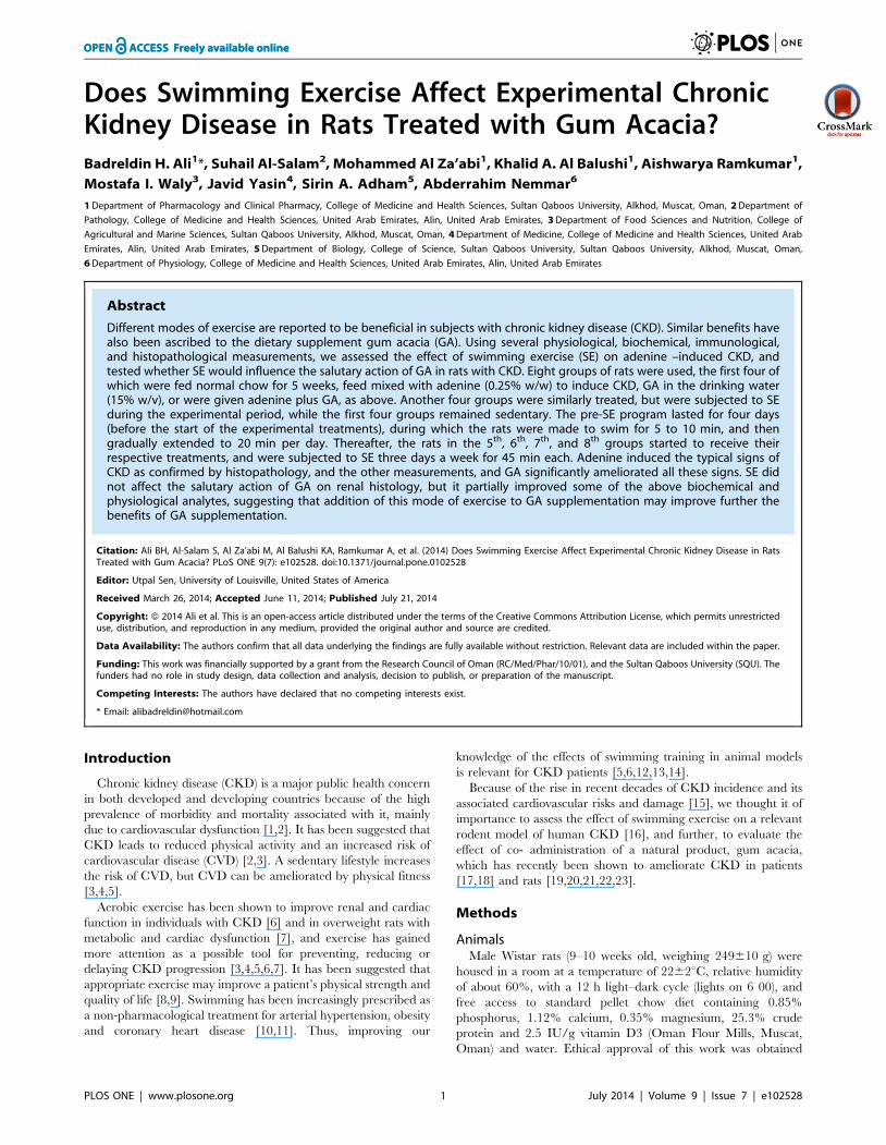
Figure 4. The concentrations of cystatin C, nephrin and adiponectinin plasma of rats treated with saline (C); saline + swimmingexercise (C + swim); adenine (A); A + swim; gum acacia (GA); GA + swim; A + GA; and A + G + swim. Each column is mean 6 SEM (n = sixrats). Statistical analysis by ANOVA followed by Newman–Keuls test.doi:10.1371/journal.pone.0102528.g004
Table 1.The effect of swimming exercise (SE) on some cytokines, antioxidant markers and proteins in plasma or urine from ratswith chronic kidney disease induced by feeding adenine (A) [0.25% w/w, 5 weeks], and the influence of gum acacia (GA) [15% w/vin drinking water, 5 weeks] thereon.
GroupsPlasma TNFa(pg/mL)
Plasma 8-isoprostone(pg/mL)
Urine OHdG(ng/mL)
Plasma adiponectin(ng/L)
Plasma cystatin C(pg/L)
Plasma nephrin(pg/mL)
Control (C) 49.363.2a 51.063.9a 181.2614.7a 3.760.2a 0.5560.03a 0.4760.02a
C + SE 47.764.0a 47.263.8a 178.9615.9a 3.360.2a 0.5760.03a 0.4560.03a
A 66.966.1a 83.467.5a 223.1619.2a 6.160.4b 0.8760.06a 0.7060.05b
A + SE 54.064.2a 61.264.3a 190.1615.3a 5.760.3b 0.6460.05a 0.6560.02c
GA 48.364.0a 50.765.2a 172.3616.3a 3.960.3a 0.4860.04a 0.4660.02a
GA + SE 45.964.2a 47.965.0a 169.9617.1a 3.660.3a 0.560.05a 0.4460.03a
A + GA 56.164.8a 59.264.7a 188.1615.3a 4.760.4c 0.6660.06a 0.660.05c
A + GA + SE 52.164.9a 55.264.1a 179.2615.4a 4.060.3a 0.5960.05a 0.5560.04d
Values in the table are means 6 SEM (n = 6 rats). Values with similar superscripts are not statistically different (level of significance set at P,0.05).TNFa = Tumor necrosis factor alpha; 8- OHdg = 8 – hydroxo – 2- deoxyguanisone;doi:10.1371/journal.pone.0102528.t001
Swimming Exercise in Adenine–Induced Chronic Kidney Disease in Rat
PLOS ONE | www.plosone.org 6 July 2014 | Volume 9 | Issue 7 | e102528
fractionation followed by multiple angle laser light scattering
(GPC-MALLS) to give its molecular profile. The average
molecular weight was 3.436106, and the content of arabinoga-
lactan protein (AGP) 26.4%. Adenine was obtained from Sigma
(St. Louis, MO, USA). Creatinine, urea and protein kits were
bought from Human GmbH (Mannheim, Germany) and SOD,
CAT and AO kits from Randox (Antrim, UK). TAC kits were
from Cayman Chemical, Ann Arbor, MI, USA. Nephrin was
obtained from Novatein Biosciences, Cambridge, MA, USA,
tumor necrosis factor a (TNF a) from Cayman Chemical, Ann
Arbor, MI, USA, 8-isoprostane and 8-oxo-2’-deoxyguanosine (8-
OHDG) from Statok Kino, Shizuoka, Japan, adiponectin from
Cayman Chemical, Ann Arbor, MI, USA, and cystatin C from R
&D Systems, Abingdon, UK.
Statistical AnalysisAll data were analyzed with GraphPad Prism Version 4.01 for
Windows software (Graphpad Software Inc., San Diego, CA).
Data were analyzed for normal distribution using the D’Agostino
and Pearson omnibus normality test. Data are expressed as means
6 SEM.
Comparisons between groups were performed by one-way
ANOVA, followed by Newman- Keuls test for comparing treated
with control data; P values of less than 0.05 are considered
significant.
Results
Physiological ResultsRats that had undergone SE in the different groups appeared
more active than their sedentary counterparts. As shown in Fig 1,
SE did not significantly change body weight of control rats, but it
significantly reduced that of rats with CKD. Treatment with GA
reduced the body weight, an effect that was potentiated by SE.
When GA treatment was combined with SE and adenine, the
body weight of rats was depressed even further.
The weights of the kidneys relative to the final body weight of
adenine –treated rats were significantly higher than those of the
control rats. This action was not significantly affected by the SE.
Water intake and urine volume in the adenine –treated rats
were significantly higher than in control rats (P,0.05), and this
was significantly abated by SE and GA treatment.
Feed intake but not fecal weight was reduced by adenine
treatment. In all groups SE reduced both the feed intake and fecal
weight.
Biochemical ResultsFig 2 shows the plasma concentrations of indoxyl sulfate,
creatinine and urea, as well as the creatinine clearance in the eight
groups. Adenine treatment significantly increased the concentra-
tions of indoxyl sulfate, creatinine and urea, and decreased that of
the creatinine clearance. This effect was significantly but not
completely reversed by GA treatment. Concomitant SE did not
significantly affect any of the above analytes.
Adenine treatment significantly decreased Ca, but increased P
and uric acid concentrations (data not shown). However, as shown
in Fig 3, in rats similarly treated but subjected to SE, Ca and uric
acid concentrations were significantly increased, and P remained
higher than the control (sedentary and subjected to SE). SE in
control rats had no significant effect on urinary uric acid excretion,
but in adenine –treated rats SE induced a significant rise (P,0.01).
The adenine – induced significant decrease in urinary uric acid
excretion was significantly (P,0.01) but not completely antago-
nized by GA treatment.
The effects of SE and GA treatments in adenine –treated rats on
Cystatin C, nephrin and adiponectin concentrations in plasma are
shown in Fig 4. Adenine treatment significantly increased the
concentration of cystatin C, while GA caused the opposite effect.
However, in all the treated groups, SE significantly increased the
concentration of cystatin C. The plasma nephrin concentration
was significantly reduced by adenine treatment, an effect which
was further enhanced by SE in all groups. Adenine treatment
significantly increased adiponectin concentration, and this was not
significantly affected by SE in any of the groups (Fig 4).
The effect of SE and GA treatments in adenine –treated rats on
the concentration of some proteins, cytokines and antioxidants in
plasma is shown in Table 1.
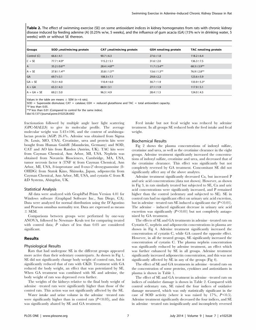
The effect of SE and GA treatment in adenine –treated rats on
indices of oxidative damage is shown in Table 2. Compared with
control sedentary rats, SE raised the four indices of oxidative
damage measured, which was only statistically significant in the
case of SOD activity (where it was raised by 17%, P,0.5).
Adenine treatment significantly decreased the four indices, and SE
in adenine –treated rats insignificantly and incompletely reversed
Table 2. The effect of swimming exercise (SE) on some antioxidant indices in kidney homogenates from rats with chronic kidneydisease induced by feeding adenine (A) [0.25% w/w, 5 weeks], and the influence of gum acacia (GA) [15% w/v in drinking water, 5weeks] with or without SE thereon.
Groups SOD mmol/min/mg protein CAT mmol/min/mg protein GSH nmol/mg protein TAC nmol/mg protein
Control (C) 66.664.1 90.766.3 27.661.8 118.266.4
C + SE 77.764.9* 115.265.1 31.662.0 136.367.5
A 35.260.6** 28.464.8** 11.763.4** 68.363.9**
A + SE 37.861.4** 35.861.5** 13.661.3** 76.962.8**
GA 69.763.1 108.367.1 29.862.2 123.665.9
GA + SE 73.364.0 110.466.0 30.761.9 133.965.3*
A + GA 65.364.3 88.963.1 27.161.9 117.965.1
A + GA + SE 69.263.0 96.364.9 28.461.5 124.064.5
Values in the table are means 6 SEM (n = 6 rats).SOD = Superoxide dismutase; CAT = catalase; GSH = reduced glutathione and TAC = total antioxidant capacity.*P less than 0.05.**P less than 0.01 (Compared to control for the same index).doi:10.1371/journal.pone.0102528.t002
Swimming Exercise in Adenine–Induced Chronic Kidney Disease in Rat
PLOS ONE | www.plosone.org 7 July 2014 | Volume 9 | Issue 7 | e102528
that action. Treatment of rats with either GA alone or together
with SE had no significant effect on any of the indices of oxidative
damage. GA treatment significantly restored these indices to near
normal levels, and this action was not significantly affected by SE.
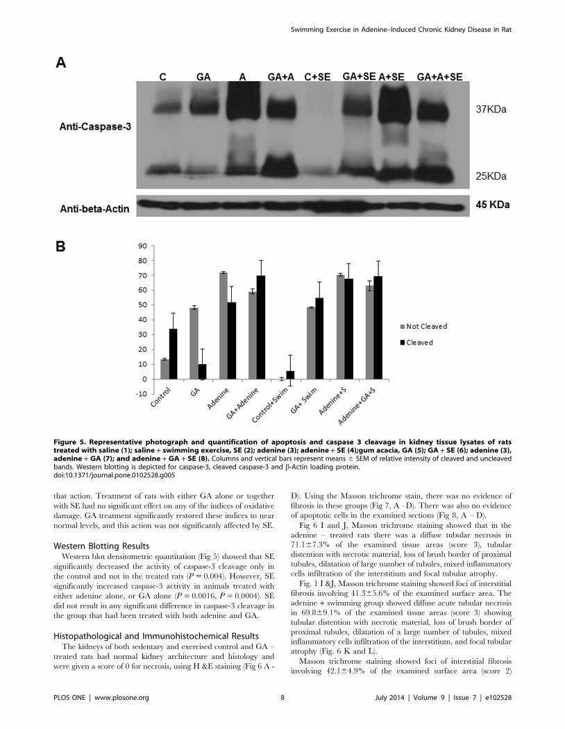
Western Blotting ResultsWestern blot densitometric quantitation (Fig 5) showed that SE
significantly decreased the activity of caspase-3 cleavage only in
the control and not in the treated rats (P = 0.004). However, SE
significantly increased caspase-3 activity in animals treated with
either adenine alone, or GA alone (P = 0.0016, P = 0.0004). SE
did not result in any significant difference in caspase-3 cleavage in
the group that had been treated with both adenine and GA.
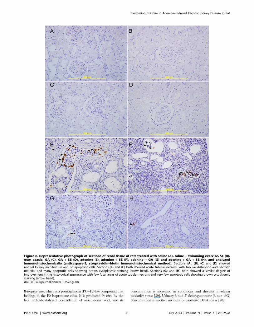
Histopathological and Immunohistochemical ResultsThe kidneys of both sedentary and exercised control and GA –
treated rats had normal kidney architecture and histology and
were given a score of 0 for necrosis, using H &E staining (Fig 6 A -
D). Using the Masson trichrome stain, there was no evidence of
fibrosis in these groups (Fig 7, A –D). There was also no evidence
of apoptotic cells in the examined sections (Fig 8, A – D).
Fig 6 I and J, Masson trichrome staining showed that in the
adenine – treated rats there was a diffuse tubular necrosis in
71.167.3% of the examined tissue areas (score 3), tubular
distention with necrotic material, loss of brush border of proximal
tubules, dilatation of large number of tubules, mixed inflammatory
cells infiltration of the interstitium and focal tubular atrophy.
Fig. 1 I &J, Masson trichrome staining showed foci of interstitial
fibrosis involving 41.365.6% of the examined surface area. The
adenine + swimming group showed diffuse acute tubular necrosis
in 69.869.1% of the examined tissue areas (score 3) showing
tubular distention with necrotic material, loss of brush border of
proximal tubules, dilatation of a large number of tubules, mixed
inflammatory cells infiltration of the interstitium, and focal tubular
atrophy (Fig. 6 K and L).
Masson trichrome staining showed foci of interstitial fibrosis
involving 42.164.9% of the examined surface area (score 2)
Figure 5. Representative photograph and quantification of apoptosis and caspase 3 cleavage in kidney tissue lysates of ratstreated with saline (1); saline + swimming exercise, SE (2); adenine (3); adenine + SE (4);gum acacia, GA (5); GA + SE (6); adenine (3),adenine + GA (7); and adenine + GA + SE (8). Columns and vertical bars represent means 6 SEM of relative intensity of cleaved and uncleavedbands. Western blotting is depicted for caspase-3, cleaved caspase-3 and b-Actin loading protein.doi:10.1371/journal.pone.0102528.g005
Swimming Exercise in Adenine–Induced Chronic Kidney Disease in Rat
PLOS ONE | www.plosone.org 8 July 2014 | Volume 9 | Issue 7 | e102528
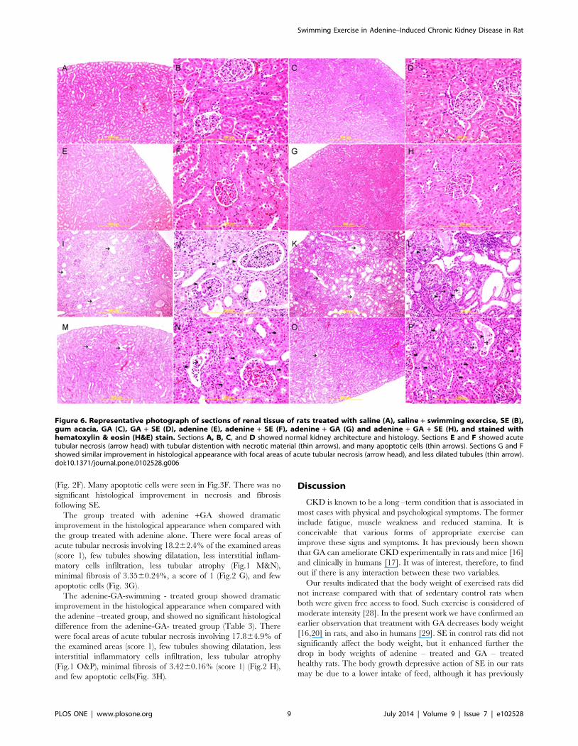
(Fig. 2F). Many apoptotic cells were seen in Fig.3F. There was no
significant histological improvement in necrosis and fibrosis
following SE.
The group treated with adenine +GA showed dramatic
improvement in the histological appearance when compared with
the group treated with adenine alone. There were focal areas of
acute tubular necrosis involving 18.262.4% of the examined areas
(score 1), few tubules showing dilatation, less interstitial inflam-
matory cells infiltration, less tubular atrophy (Fig.1 M&N),
minimal fibrosis of 3.3560.24%, a score of 1 (Fig.2 G), and few
apoptotic cells (Fig. 3G).
The adenine-GA-swimming - treated group showed dramatic
improvement in the histological appearance when compared with
the adenine –treated group, and showed no significant histological
difference from the adenine-GA- treated group (Table 3). There
were focal areas of acute tubular necrosis involving 17.864.9% of
the examined areas (score 1), few tubules showing dilatation, less
interstitial inflammatory cells infiltration, less tubular atrophy
(Fig.1 O&P), minimal fibrosis of 3.4260.16% (score 1) (Fig.2 H),
and few apoptotic cells(Fig. 3H).
Discussion
CKD is known to be a long –term condition that is associated in
most cases with physical and psychological symptoms. The former
include fatigue, muscle weakness and reduced stamina. It is
conceivable that various forms of appropriate exercise can
improve these signs and symptoms. It has previously been shown
that GA can ameliorate CKD experimentally in rats and mice [16]
and clinically in humans [17]. It was of interest, therefore, to find
out if there is any interaction between these two variables.
Our results indicated that the body weight of exercised rats did
not increase compared with that of sedentary control rats when
both were given free access to food. Such exercise is considered of
moderate intensity [28]. In the present work we have confirmed an
earlier observation that treatment with GA decreases body weight
[16,20] in rats, and also in humans [29]. SE in control rats did not
significantly affect the body weight, but it enhanced further the
drop in body weights of adenine – treated and GA – treated
healthy rats. The body growth depressive action of SE in our rats
may be due to a lower intake of feed, although it has previously
Figure 6. Representative photograph of sections of renal tissue of rats treated with saline (A), saline + swimming exercise, SE (B),gum acacia, GA (C), GA + SE (D), adenine (E), adenine + SE (F), adenine + GA (G) and adenine + GA + SE (H), and stained withhematoxylin & eosin (H&E) stain. Sections A, B, C, and D showed normal kidney architecture and histology. Sections E and F showed acutetubular necrosis (arrow head) with tubular distention with necrotic material (thin arrows), and many apoptotic cells (thin arrows). Sections G and Fshowed similar improvement in histological appearance with focal areas of acute tubular necrosis (arrow head), and less dilated tubules (thin arrow).doi:10.1371/journal.pone.0102528.g006
Swimming Exercise in Adenine–Induced Chronic Kidney Disease in Rat
PLOS ONE | www.plosone.org 9 July 2014 | Volume 9 | Issue 7 | e102528

been reported that energy intake in the hemodialysis patients of
Koufaki et al [30] was slightly (5%) but significantly increased.
In this work, the adenine – treated rats exhibited the urinary and
plasma profile of several traditional and novel markers of renal
damage, as reported by us and others [20,36,37]. Most of these were
improved in rats given either GA or SE, and even more so in rats
given GA and subjected to SE at the same time, supporting our
hypothesis that the ameliorative action of GA on adenine- induced
CRF is further enhanced by SE. The use of novel urinary and
plasma biomarkers has been recently highlighted as being able to
detect subtle and early renal changes in both chronic and acute
renal injury [38]. In this work, both traditional and novel
biomarkers measured in urine and plasma were nearly all in full
agreement. Examples of these novel biomarkers used included
Figure 7. Representative photograph of sections of renal tissue of rats treated with saline (A), saline + swimming exercise, SE (B),gum acacia, GA (C), GA + SE (D), adenine (E), adenine + SE (F), adenine + GA (G) and adenine + GA + SE (H), and stained with Massontrichrome stain. Sections A, B, C, and D showed normal kidney architecture and histology and no evidence of fibrosis. Sections E and F showedlarge areas of interstitial fibrosis (thick arrows). Sections G and F showed similar improvements in histological appearance with dramatic decrease infibrosis (thick arrows).doi:10.1371/journal.pone.0102528.g007
Swimming Exercise in Adenine–Induced Chronic Kidney Disease in Rat
PLOS ONE | www.plosone.org 10 July 2014 | Volume 9 | Issue 7 | e102528
8-isoprostane, which is a prostaglandin (PG)-F2-like compound that
belongs to the F2 isoprostane class. It is produced in vivo by the
free radical-catalyzed peroxidation of arachidonic acid, and its
concentration is increased in conditions and diseases involving
oxidative stress [39]. Urinary 8-oxo-2’-deoxyguanosine (8-oxo- dG)
concentration is another measure of oxidative DNA stress [28].
Figure 8. Representative photograph of sections of renal tissue of rats treated with saline (A), saline + swimming exercise, SE (B),gum acacia, GA (C), GA + SE (D), adenine (E), adenine + SE (F), adenine + GA (G) and adenine + GA + SE (H), and analyzedimmunohistochemically (anticaspase-3, streptavidin–biotin immunohistochemical method). Sections (A), (B), (C) and (D) showednormal kidney architecture and no apoptotic cells. Sections (E) and (F) both showed acute tubular necrosis with tubular distention and necroticmaterial and many apoptotic cells showing brown cytoplasmic staining (arrow head). Sections (G) and (H) both showed a similar degree ofimprovement in the histological appearance with few focal areas of acute tubular necrosis and very few apoptotic cells showing brown cytoplasmicstaining (arrow head).doi:10.1371/journal.pone.0102528.g008
Swimming Exercise in Adenine–Induced Chronic Kidney Disease in Rat
PLOS ONE | www.plosone.org 11 July 2014 | Volume 9 | Issue 7 | e102528
Different modes of exercise, including SE, are established to be
beneficial in CKD and its cardiovascular and other complications
in humans [27,31] and animals [31,28]. The mechanism by which
SE ameliorates CKD is not known with certainty, but it has been
hypothesized that the basis of the obtained benefits are probably
multifactorial [40], and include the beneficial effect of SE on the
oxidative status of the tissues. Although there is no unanimity in
the literature regarding the influence of exercise on inflammation
and oxidative stress, moderate SE is believed to be effective in
preventing inflammation and oxidative damage in tissues of rats
[32,33], but severe/acute exercise has been shown to produce the
opposite effect in humans and rats [34,35]. In our present
experiments employing moderate SE, we found that SE did not
significantly alter the renal concentration/activity of the measured
incidence of oxidative stress (except SOD activity, which was
increased), probably reflecting the adequacy of the defensive
antioxidant oxidative abilities in these animals. Adenine – induced
CKD, as reported before, significantly and markedly decreased the
anti-oxidants measured [20,37], an action that was significantly
abrogated by either GA or SE given alone, and even more when
combined.
In conclusion, we aimed to ascertain experimentally if
combining two strategies for mitigating the effects of CKD (viz
administration of GA, a nephroprotectant [26] and SE [14] would
influence the effects of CKD. Judging by the results of several
biochemical and physiological (but not all) parameters measured,
there seems to be a significant positive impact in the condition with
SE. Therefore, on the whole, these results suggest that the
ameliorative action of GA can be enhanced by SE. Previously, it
has been reported that significant clinical benefits are obtained
from GA treatment in CKD patients who are on a low-protein diet
[26]. In future experiments, it would be of interest to see the effect
of other modes of exercise with different intensities on the same
parameters, and also the possible effect of SE on CKD patients on
GA both with and without a low-protein diet.
Author Contributions
Conceived and designed the experiments: BHA. Contributed reagents/
materials/analysis tools: BHA MZ MIW SAA JY AN. Contributed to the
writing of the manuscript: BHA MZ KAB AN. Conducted the
histopathology and immunohistochemistry: SA. Conducted the biochem-
ical experiments: MZ KAB MIW JY AN. Conducted the animal
experimentation: AR. Carried out the Western blotting: SAA.
References
1. Harambat J, van Stralen KJ, Kim JJ, Tizard E (2012) Epidemiology of chronickidney disease in children. Pediatr Nephrol 27: 363–373.
2. Hossain MP, Goyder EC, Rigby JE, El Nahas M (2009) CKD and poverty: a
growing global challenge. Am J Kidney Dis 53: 166–174.
3. Briasoulis A, Bakris GL (2013) Chronic kidney disease as a coronary artery
disease risk equivalent. Curr Cardiol Rep 15: 340.
4. Clapp EL, Bevington A, Smith AC (2012) Exercise for children with chronic
kidney disease and end-stage renal disease. Pediatr Nephrol 27: 165–172.
5. Johansen KL, Painter P (2012) Exercise in individuals with CKD. Am J Kidney
Dis 59: 126–134.
6. Bronas UG (2009) Exercise training and reduction of cardiovascular disease riskfactors in patients with chronic kidney disease. Adv Chronic Kidney Dis 16:
449–458.
7. Sakr HF (2013) Modulation of metabolic and cardiac dysfunctions by swimming
in overweight rats on a high cholesterol and fructose diet: possible role of
adiponectin. J Physiol Pharmacol 64: 231–240.
8. Heiwe S, Jacobson SH (2011) Exercise training for adults with chronic kidneydisease. Cochrane Database Syst Rev 5: CD003236.
9. Howden EJ, Fassett RG, Isbel NM, Coombes JS (2012) Exercise training inchronic kidney disease patients. Sports Med 42: 473–488.
10. Meredith-Jones K, Waters D, Legge M, Jones L (2011) Upright water-basedexercise to improve cardiovascular and metabolic health: a qualitative review.
Complement Ther Med 19: 93–103.
11. Tanaka H (2009) Swimming exercise: impact of aquatic exercise on
cardiovascular health. Sports Med 39: 377–387.
12. Segura-Orti E, Johansen KL (2010) Exercise in end-stage renal disease. Semi
Dial 23: 422–30.
13. Luiz Rda S, Silva KA, Rampaso RR, Antonio EL, Montemor J, et al. (2013)
Exercise attenuates renal dysfunction with preservation of myocardial function
in chronic kidney disease. PLoS One 8(2): e55363.
14. Peng CC, Chen KC, Hsieh CL, Peng RY (2012) Swimming exercise prevents
fibrogenesis in chronic kidney disease by inhibiting the myofibroblast
transdifferentiation. PLoS One 7(6): e37388.
15. Kucukkoylu S, Rump LC (2013) Cardiovascular morbidity and mortality in
renal diseases. Dtsch Med Wochenschr 138: 721–724.
16. Ali BH, Al-Salam S, Al Za’abi M, Waly MI, Ramkumar A, et al. (2013) New
model for adenine-induced chronic renal failure in mice, and the effect of gum
acacia treatment thereon: Comparison with rats. J Pharmacol Toxicol Methods
68: 384–393.
17. Ali AA, Ali KE, Fadlalla AE, Khalid KE (2008) The effects of gum arabic oral
treatment on the metabolic profile of chronic renal failure patients under regular
haemodialysis in Central Sudan. Nat Prod Res 22: 12–21.
18. Al Mosawi AJ (2009) Six-year dialysis freedom in end-stage renal disease. Clin
Exp Nephrol 13: 494–500.
19. Ali BH, Al-Salam S, Al Husseni I, Kayed RR, Al-Masroori N, et al. (2010)
Effects of Gum Arabic in rats with adenine-induced chronic renal failure. Exp
Biol Med (Maywood) 235: 373–382.
20. Ali BH, Al-Husseni I, Beegam S, Al-Shukaili A, Nemmar A, et al. (2013) Effect
of gum arabic on oxidative stress and inflammation in adenine-induced chronic
renal failure in rats. PLoS One 8: e55242.
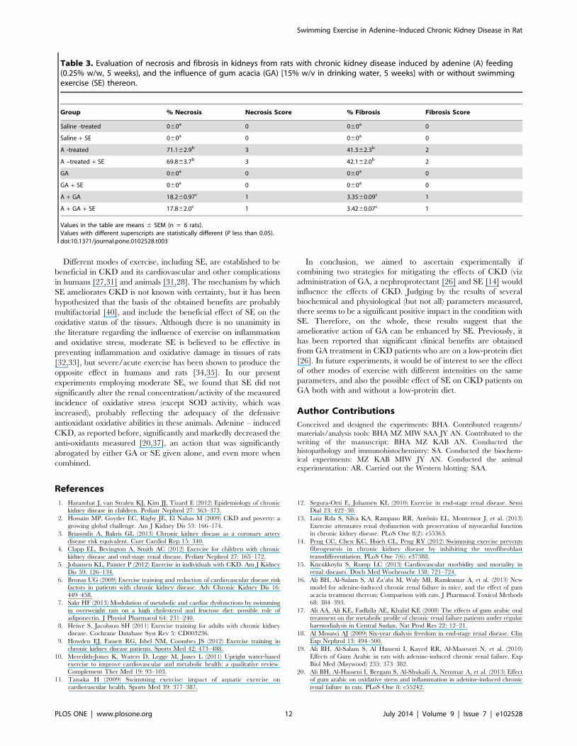
Table 3. Evaluation of necrosis and fibrosis in kidneys from rats with chronic kidney disease induced by adenine (A) feeding(0.25% w/w, 5 weeks), and the influence of gum acacia (GA) [15% w/v in drinking water, 5 weeks] with or without swimmingexercise (SE) thereon.
Group % Necrosis Necrosis Score % Fibrosis Fibrosis Score
Saline -treated 060a 0 060a 0
Saline + SE 060a 0 060a 0
A -treated 71.162.9b 3 41.362.3b 2
A –treated + SE 69.863.7b 3 42.162.0b 2
GA 060a 0 060a 0
GA + SE 060a 0 060a 0
A + GA 18.260.97c 1 3.3560.09c 1
A + GA + SE 17.862.0c 1 3.4260.07c 1
Values in the table are means 6 SEM (n = 6 rats).Values with different superscripts are statistically different (P less than 0.05).doi:10.1371/journal.pone.0102528.t003
Swimming Exercise in Adenine–Induced Chronic Kidney Disease in Rat
PLOS ONE | www.plosone.org 12 July 2014 | Volume 9 | Issue 7 | e102528
21. Ali BH, Al Za’abi M, Ramkumar A, Yasin J, Nemmar A (2014) Anemia in
adenine-induced chronic renal failure and the influence of treatment with gum
acacia thereon. Physiol Res In press.
22. Ali BH, Ziada A, Al Husseni I, Beegam S, Al-Ruqaishi B, et al. (2011) Effect
ofAcacia gum on blood pressure in rats with adenine-induced chronic renal
failure. Phytomedicine 18: 1176–1180.
23. Neto OB, Abate DT, Junior MM, Mota GR, Orsatti FL, et al. (2013) Exercise
training improves cardiovascular autonomic activity and attenuates renal
damage in spontaneously hypertensive rats. J Sports Sci Med 12: 52–59.
24. Gegentonglaga, Yoshizato H, Higuchi Y, Toyota Y, Hanai Y, et al. (2013)
Variable alteration of regional tissue oxygen pressure in rat hippocampus by
acute swimming exercise. Life Sci 93: 773–777.
25. Al Za’abi M, Ali BH, Al Toubi M (2013) HPLC-fluorescence method for
measurement of the uremic toxin indoxyl sulfate in plasma. J Chromatogr Sci
51: 40–43.
26. Ali BH, Ziada A, Blunden G (2009) Biological effects of gum arabic: a review of
some recent research. Food Chem Toxicol 47: 1–8.
27. Pechter U, Ots M, Mesikepp S, Zilmer K, Kullissaar T, et al. (2003) Beneficial
effects of water-based exercise in patients with chronic kidney disease.
Int J Rehabil Res 26: 153–156.
28. Ishikawa Y, Gohda T, Tanimoto M, Omote K, Furukawa M, et al. (2012) Effect
of exercise on kidney function, oxidative stress, and inflammation in type 2
diabetic KK-A(y) mice. Exp Diabetes Res 2012: 702948.
29. Babiker R, Merghani TH, Elmusharaf K, Badi RM, Lang F, et al. (2012) Effects
of Gum Arabic ingestion on body mass index and body fat percentage in healthy
adult females: two-arm randomized, placebo controlled, double-blind trial.
Nutr J 11: 111.
30. Koufaki P, Mercer TH, Naish PF (2002) Effects of exercise training on aerobic
and functional capacity of end-stage renal disease patients. Clin Physiol FunctImaging 22: 115–124.
31. Kutner NG (2007) How can exercise be incorporated into the routine care of
patients on dialysis? Int Urol Nephrol 39: 1281–1285.32. Smart N, Steele M (2011) Exercise training in haemodialysis patients: a
systematic review and meta-analysis. Nephrology (Carlton) 16: 626–632.33. Cechella JL, Leite MR, Dobrachinski F, da Rocha JT, Carvalho NR, et al.
(2014) Moderate swimming exercise and caffeine supplementation reduce the
levels of inflammatory cytokines without causing oxidative stress in tissues ofmiddle-aged rats. Amino Acids In press.
34. Ranadive SM, Kappus RM, Cook MD, Yan H, Lane AD, et al. (2014) Effect ofacute moderate exercise on induced inflammation and arterial function in older
adults. Exp Physiol In press.35. Deminice R, Trindade CS, Degiovanni GC, Garlip MR, Portari GV, et al.
(2010) Oxidative stress biomarkers response to high intensity interval training
and relation to performance in competitive swimmers. J Sports Med Phys Fitness50: 356–362.
36. Deminice R, Jordao AA (2012) Creatine supplementation reduces oxidativestress biomarkers after acute exercise in rats. Amino Acids 43: 709–715.
37. Diwan V, Mistry A, Gobe G, Brown L (2013) Adenine-induced chronic kidney
and cardiovascular damage in rats. J Pharmacol Toxicol Methods 68: 197–207.38. Fuchs TC, Hewitt P (2011) Biomarkers for drug-induced renal damage and
nephrotoxicity-an overview for applied toxicology. AAPS J 3: 615–631.39. Knight SF, Yuan J, Roy S, Imig JD (2010) Simvastatin and tempol protect
against endothelial dysfunction and renal injury in a model of obesity andhypertension. Am J Physiol Renal Physiol 298: F86–94.
40. Heiwe S, Jacobson SH (2011) Exercise training for adults with chronic kidney
disease. Cochrane Database Syst Rev 10: CD003236.
Swimming Exercise in Adenine–Induced Chronic Kidney Disease in Rat
PLOS ONE | www.plosone.org 13 July 2014 | Volume 9 | Issue 7 | e102528


![The Healthy, Natural Way · shipping. This product is sold by weight not volume. Ingredients: Plant protein [(pea protein, brown rice protein) (57%)], fructose, gum acacia, guar gum,](https://static.cupdf.com/doc/110x72/602043cee3e5224ea33f862f/the-healthy-natural-way-shipping-this-product-is-sold-by-weight-not-volume-ingredients.jpg)


