Charles University in Prague, Faculty of Pharmacy
In Hradec Kralove
Department of Pharmaceutical Botany and Ecology
______________________________________________________________________
DIPLOMA THESIS
Biological Activity of Plant Metabolites XVII.
Alkaloids of Corydalis yanhusuo W.T. Wang
Supervisor of Diploma Work: Assoc. Prof. RNDr. Lubomir Opletal, CSc.
Head of Department: Prof. RNDr. Luděk Jahodář, CSc.
Hradec Králové April, 2011 Gabriella Cipra

Declaration
I declare that this thesis is my original copyrighted work. All literature and other sources
from which I extracted my research in the process are listed in the bibliography and all
work is properly cited. This work has not been used to gain another or same title.

Acknowledgements
I wish to express my deepest gratitude, first and foremost to Assoc. Prof. RNDr. Lubomír
Opletal, CSc. for all his guidance, support and enthusiasm with the preparation of this
thesis. I would also like to thank the Department of Pharmaceutical Botany and Ecology
for the pleasant working environment, as well as Assoc. Prof. PharmDr. Jiří Kuneš, Ph.D.
for preparation and interpretation of NMR spectra, Ing. Kateřina Macáková for biological
activity measurements, and Ing. Lucie Cahlíková, Ph.D. for MS spectra measurements
and interpretations.
This work was financially supported by Specific University Research Foundation No
SVV-2011-263002 (Study of biologically active compounds in prespective of their
prevention and treatment in civil diseases)
4
Table of Contents
1 INTRODUCTION 7
2 AIM OF WORK 10
3 THEORETICAL PART 12
3.1 Corydalis yanhusuo 13
3.1.1 History and origin 14
3.1.2 Morphological description 14
3.1.3 Chemical constituents 15
3.1.4 Pharmacological activity of main alkaloid constituents 15
3.1.4.1 Tetrahydropalmatine 16
3.1.4.2 Dehydrocorydaline 17
3.1.4.3 Protopine 18
3.1.4.4 Pseudocoptisine 19
3.1.4.5 Pseudoberberine 20
3.2 Other Corydalis species 21
3.2.1 Corydalis speciosa 21
3.2.2 Corydalis cava 22
3.2.3 Corydalis saxicola 22
3.3 Alzheimer‟s Disease 24
3.3.1 Pathological hallmarks of Alzheimer‟s disease 25
3.3.1.1 β-Amyloid plaques 25
3.3.1.2 Presenilins and tau-phosphorylation 25
3.3.2 The cholinergic hypothesis 26
3.3.3 Current therapy in Alzheirmer‟s disease 29
3.4 Natural compounds influencing metabolism of AChE and BuChE 30
3.4.1 Alkaloids 31
3.4.1.1 Physostigmine 31
3.4.1.2 Galanthamine 31
3.4.1.3 Huperzine A 32
3.4.1.4 Chelidonium majus 32
5
3.4.2 Terpenoids 32
3.4.2.1 Salvia lavandulaefolia 32
3.4.2.2 Melissa officinalis 32
3.4.2.3
3.4.3
Origanum majorana
Withanolides
33
33
3.4.3.1 Withania somnifera 33
4 EXPERIMENTAL PART 34
4.1 General methods 35
4.1.1 Distillation and evaporation 35
4.1.2 Chromatography 35
4.1.2.1 Thin layer chromatography 35
4.1.2.2 Column chromatography 35
4.2 Plant material and equipment 36
4.2.1 Chemicals and solvents 36
4.2.2 Chemicals and material for analysis of AChE and BuChE (IC50) 37
4.2.3 Chemicals and material for analysis of antioxidant activity (EC50) 38
4.2.4 Detection reagents 38
4.2.5 Chromatographic plates and adsorbents 38
4.3 Description and methods of alkaloid isolation 39
4.3.1 Origin of herbal drug 39
4.3.2 Preparation of summary extract 39
4.3.3 Preparation of extract A from primary extract 39
4.3.4 Preparation of extract A on particular groups of alkaloids 40
4.3.5 Separation of mixture of non-phenolic alkaloids from chlorides soluble in
chloroform
41
4.3.6 Isolation of alkaloid from combined fractions 57-63 43
4.4 Method for determining MS Spectra 44
4.5 Method for determining NMR spectra 44
4.6 Method for determining antioxidant activity 44
4.6.1 DPPH free-radical scavenging assay (EC50) 44

6
4.7 Method for determining inhibitory activity against human AChE and
BuChE
45
4.7.1 Preparation of red blood cell ghosts for AChE and BuChE 45
4.7.2
4.8
AChE and BuChE assay (IC50)
Method for determining optical rotation
46
46
5 RESULTS 47
5.1 Structural analysis of compound GC-1 48
5.1.1 MS analysis of (+)-corydaline 48
5.1.2 MS/MS analysis of (+)-corydaline 49
5.1.3 NMR analysis of (+)-corydaline 50
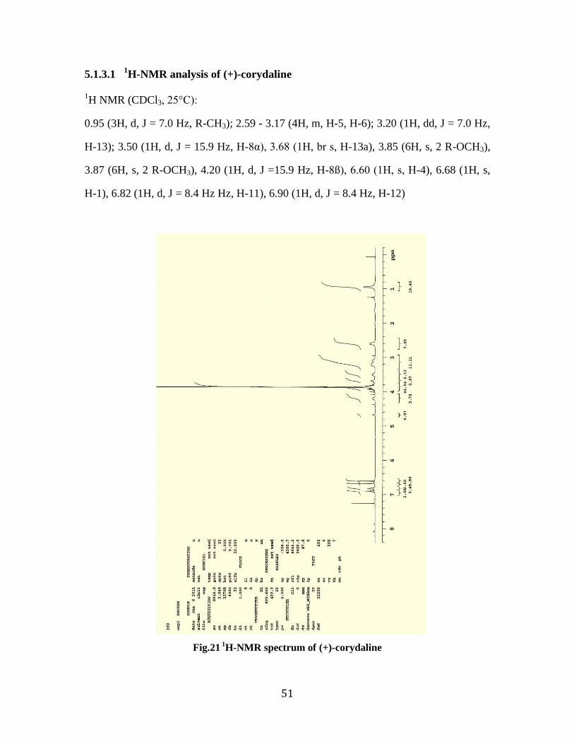
5.1.3.1 1H-NMR analysis of (+)-corydaline 51
5.1.3.2 13
C-NMR analysis of (+)-corydaline 52
5.1.3.3 Optical rotation 53
5.2 Inhibitory activity of corydaline against human AChE and BuChE 53
5.3 Antioxidant activity of (+)-corydaline 53
6 DISCUSSION 54
7 LITERATURE 57
8 ABBREVIATIONS KEY 67

7
1. INTRODUCTION
8
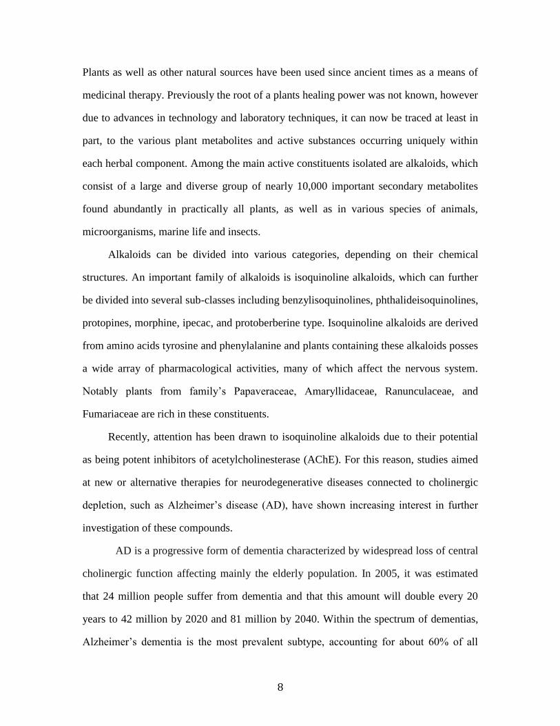
Plants as well as other natural sources have been used since ancient times as a means of
medicinal therapy. Previously the root of a plants healing power was not known, however
due to advances in technology and laboratory techniques, it can now be traced at least in
part, to the various plant metabolites and active substances occurring uniquely within
each herbal component. Among the main active constituents isolated are alkaloids, which
consist of a large and diverse group of nearly 10,000 important secondary metabolites
found abundantly in practically all plants, as well as in various species of animals,
microorganisms, marine life and insects.
Alkaloids can be divided into various categories, depending on their chemical
structures. An important family of alkaloids is isoquinoline alkaloids, which can further
be divided into several sub-classes including benzylisoquinolines, phthalideisoquinolines,
protopines, morphine, ipecac, and protoberberine type. Isoquinoline alkaloids are derived
from amino acids tyrosine and phenylalanine and plants containing these alkaloids posses
a wide array of pharmacological activities, many of which affect the nervous system.
Notably plants from family‟s Papaveraceae, Amaryllidaceae, Ranunculaceae, and
Fumariaceae are rich in these constituents.
Recently, attention has been drawn to isoquinoline alkaloids due to their potential
as being potent inhibitors of acetylcholinesterase (AChE). For this reason, studies aimed
at new or alternative therapies for neurodegenerative diseases connected to cholinergic
depletion, such as Alzheimer‟s disease (AD), have shown increasing interest in further
investigation of these compounds.
AD is a progressive form of dementia characterized by widespread loss of central
cholinergic function affecting mainly the elderly population. In 2005, it was estimated
that 24 million people suffer from dementia and that this amount will double every 20
years to 42 million by 2020 and 81 million by 2040. Within the spectrum of dementias,
Alzheimer‟s dementia is the most prevalent subtype, accounting for about 60% of all

9
dementias [1]. Since there still remains no cure to prevent or treat AD, current therapy is
based on the symptomatic treatment by use of AChE inhibitors.
One genus known to contain several species with AChE inhibitory activity is the
genus Corydalis. Classified within this genus is Corydalis yanhusuo, a plant that has
been used in traditional Chinese medicine for hundreds of years owing to its vast array of
therapeutic indications. The tuber of C. yanhusuo is known to encompass various
biologically active constituents, including isoquinoline alkaloids. Thus, it will be the
topic of this diploma thesis to further investigate the extracts isolated from the tubers of
C. yanhusuo and evaluate their inhibitory activity on AChE for potential use as natural
alternatives in AD therapy.

10
2. AIM OF WORK

11
1) Isolation of one alkaloid in pure form from chromatographic fraction. The fraction
was prepared from primary extract of tubers from Corydalis yanhusuo. Methods of
isolation were carried out on column chromatography and thin-layer chromatography.
2) Determination of physico-chemical properties of isolated compounds (optical rotation
and Rf values in two chromatographic systems – TLC). Determination of MS and
NMR spectra.
3) Determination of antioxidant activity (DPPH test) and influence on human
cholinesterases – acetylcholinesterase and butyrylcholinesterase.
4) Calculation of IC50, EC50 (statistical program GraphPad from faculty web pages).

12
3. THEORETICAL PART

13
3.1 Corydalis yanhusuo
Kingdom: Plantae
Phyllum: Tracheophyta
Class: Magnoliopsida
Order: Papaverales
Family: Fumariaceae
Genus: Corydalis
Botanical name: Corydalis yanhusuo W.T.Wang
Fig. 1 Corydalis yanhusuo [2]

14
3.1.1 History and origin
The genus Corydalis (Fumariaceae) of roughly 320 species is widely distributed in the
northern hemisphere and about 70 species are known to be used in traditional herbal
remedies [3]. Corydalis yanhusuo W.T.Wang, a perennial herb belonging to the
Fumariaceae family and important species of genus Corydalis, has been used in
traditional herbal remedies in China, Japan, and Korea. C. yanhusuo grows wild in
Siberia and Northern China and is cultivated principally in the Zhejiang province, where
it is collected in the early summer season after the stems and leaves have wilted. The
dried and pulverized tuber is also referred to as Rhizoma Corydalis. It is officially listed
in the Chinese Pharmacopoeia, and in traditional Chinese medicine it has been used for
hundreds of years in the treatment of gastric and duodenal ulcers, cardiac arrhythmia,
rheumatism and dysmenorrhea [4]. C. yanhusuo has also been used to promote blood
circulation, reinforce vital energy, move qi, and alleviate pain such as headache, chest
pain, hypochondriac pain, epigastric pain, abdominal pain, backache, arthralgia, or
trauma [5, 6].
3.1.2. Morphological description
The herbs of C. yanhusuo are perennial. The tuber is yellow, rounded, and about 1-2.5
cm in diameter. Stems are erect, 10-30 cm, with one or sometimes two scale leaves.
Leaves are biternate or nearly triternate with leaflets measuring approximately 2-2.5 cm ×
5-8 mm. Flowers are usually between 5-15 and bloom between April and June. Bracts are
lanceolate or narrowly ovate, measuring 5-12 × 2-5 mm and sometimes lower bracts are
slightly divided. The pedicel measures about 10 mm at flowering and in fruit up to
20mm. Outer petals are broad with dentate limbs and the spur of the upper petal is up
curved, cylindrical, and measures about 11-13 mm. Lower petals have a short claw and
inner petals measure 8-9 mm with claw longer than petal lobes. The stigma is nearly

15
orbicular while papillae are longer than in preceding species. Capsule are linear and
measure between 20-28 mm and seeds are found in one row [7].
3.1.3. Chemical Constituents
The tuber of C. yanhusuo contains several tertiary and quaternary alkaloids that form the
main bioactive components. However, there are still many alkaloids in the tubers that
remain un-investigated, especially those in the micro, or even trace concentrations, which
cannot be easily separated and identified by traditional phytochemical methods [8].
Among those identified, nearly 20 alkaloids of the tertiary and quaternary types have
been isolated from C. yanhusuo thus far, which may be responsible for the biological
activities of the drug. Their chemical structures belong to the isoquinoline family of
alkaloids and can be divided into various skeletal structures [9]. They include protopine
type: protopine and allocryptopine, and protoberberine/aporphine type:
tetrahydropalmatine, palmatine, corydaline, dehydrocorydaline, berberine,
pseudoberberine, canadine, columbamine, tetrahydrocolumbamine, glaucine,
dehydroglaucine, corybulbine, dehydrocorybulbine, tetrahydrocoptisine, pseudocoptisine,
and fumaricine [10].
3.1.4. Pharmacological Activity of Main Alkaloid Constituents
Numerous alkaloids displaying a wide range of pharmacological actions including
analgesic [11, 12], anxiolytic [5], hypnotic [8], antiamnestic [9, 13] anti-inflammatory
[14-16], antiplatelet [17] and cardioprotective [3, 18, 19] have been isolated form the
tuber of Corydalis.

16
3.1.4.1 Tetrahydropalmatine
One of the main active constituents isolated from C. yanhusuo is dl- tetrahydropalmatine
(dl-THP). Derived from the tetrahydroprotoberberine backbone structure, dl-THP belongs
to the isoquinoline alkaloid family [5]. It can also be directly synthesized from
laudanosine, a benzylisoquinoline alkaloid via chemical conversion [20].
Pharmacologically, it has been shown that dl-THP exerts marked analgesic,
sedative-tranquilizing and hypnotic action and it has been listed in the Chinese
Pharmacopoeia since 1977 for these indications [8, 11]. It was found that dl-THP could
display such effect, probably due to its antagonistic action on the D1/D2 receptor in the
brain [21]. However, in a study by Leung et al., 2003, it was proposed that dl-THP could
also act on the benzodiazepine site (BDS) of the GABAA receptor in the mouse brain.
The main findings from the animal behavioral tests concluded that dl-THP manifests
anxiolysis at defined low dosages at least in part, and this effect is mediated through the
BDS, thus concluding that the anxiolytic effects of dl-THP could be produced by a
combination of effects from several receptors in the CNS including D1/D2 receptor and
GABAA receptor [5].
Studies aiming to distinguish between the various mechanisms of action by which
dl-THP functions in the CNS have been carried out and in one such study, it was shown
that dl-THP depletes levels of the neurotransmitters dopamine, noradrenaline and
serotonin in the central nervous system [22]. In addition, it has been reported that the two
enantiomers of dl-THP act on different targets – d-THP depletes dopamine while l-THP
functions as a dopamine antagonist [23]. Interestingly, it was also found that dl-THP
decreases both arterial pressure and heart rate through a serotonergic release process in
the hypothalamus [24].
In a recent study by Oh et al., 2010, on the inhibition of pro-inflammatory
mediators, THP inhibited lipopolysaccharide (LPS)-induced interleukin (IL)-8 production

17
in a dose-dependent manner. Furthermore, THP inhibited extracellular signal-regulated
kinase and p38 mitogen-activated protein kinase (MAPK) phosphorylation, which
suggests that THP inhibits IL-8 secretion by blocking MAPK phosphorylation [14].
Fig. 2 Tetrahydropalmatine
3.1.4.2 Dehydrocorydaline
In a study by Kubo et al., 1993, on the potential anti-inflammatory activities of
methanolic extract from Corydalis tuber, it was found that among the tested alkaloidal
components, dehydrocorydaline showed a stronger inhibitory effect than that of the
standard drug, in this case disodium cromoglycate [15]. The methanolic extract of
Corydalis tuber showed an inhibitory effect against histamine release from mast cells but
also inhibitory effect on the released histamine when administered to isolated guinea pig
ileum. It was therefore suggested as having a potentially important future implication in
the therapeutic field of inflammatory disease [15].
Studies also show that dehydrocorydaline not only inhibits antibody-mediated
allergic reactions but also influences cell-mediated allergic reactions [25].

18
Fig. 3 Dehydrocorydaline
3.1.4.3 Protopine
Protopine was reported to exhibit an inhibitory activity on platelet aggregation [17].
Protopine was also found to possess potent anti-nociceptive effects due to its ability to
function as an inhibitor of both serotonin and noradrenaline transporters [12]. It has also
been described that following treatment with protopine, a significant decrease in
glutamate level and an increase in glutamate dehydrogenase activity was observed in rat
brains [26]. Since glutamate plays a significant role in nociceptive processing in central
and peripheral nervous systems [27, 28], the decrease in glutamate level might also be
associated with the anti-nociceptive effects of protopine [10].
Fig. 4 Protopine

19
3.1.4.4 Pseudocoptisine
Based on a study conducted by Hung et al., 2008, it was found that psuedocoptisine
displayed remarkable cognitive-enhancing activity mediated in part by its ability to
inhibit adult male rat AChE activity in a dose dependent manner (IC50 = 12.8 μM) [13].
Pseudocoptisine treatment (2.0, 5.0 mg/kg) also reversed the deficits produced by
scopolamine treatment in the comparison with the vehicle-treated group on passive
avoidance task. At a concentration of 2.0 mg/kg, pseudocoptisine significantly shortened
the escape latency and improved swimming time within the zone of platform on water
maze task [13]. The passive avoidance test is generally accepted as an indicator of long-
term memory in animals [29], and the water maze-learning task was used to asses
hippocampal-dependent spatial learning ability [30, 31].
Pseudocoptisine caused dose-dependent reductions in the levels of inducible nitric-
oxide synthase (iNOS) and cyclooxygenase-2 (COX-2) at both protein and mRNA levels
and concomitant decrease in PGE2 and NO production. In addition, it was found that
pseudocoptisine suppressed the production and mRNA expressions of pro-inflammatory
cytokines, such as, TNF-α and IL-6 [16].
Fig. 5 Pseudocoptisine

20
3.1.4.5 Pseudoberberine
In a recent in vivo experiment, Hung et al., 2008, found that pseudoberberine inhibited
mouse brain cortex AChE activity in a dose-dependent manner with an IC50 value of 4.5
μM. Also, treatment in mice with 5.0mg/kg could reverse the deficits produced by
scopolamine in comparison with vehicle-treated group on passive avoidance task, and
significantly shortened the escape latency and improved the swimming time within the
zone of platform on water maze task when evaluating spatial learning [9].
Fig. 6 Pseudoberberine

21
3.2 Other Corydalis species
In addition to the officially listed C. yanhusuo, there are many species of the genus
Corydalis known to be of use in traditional Chinese medicine or folk medicine.
3.2.1 Corydalis speciosa
Corydalis speciosa has been used in Korea and China as a folk medicine for its
antipyretic, analgesic and diuretic properties. In a study by Kim et al., 2004, the
methanolic extracts of the aerial parts of C. speciosa were found to exhibit significant
AChE inhibitory activity. Four compounds were separated as active constituents and
identified as corynoxidine, protopine, palmatine and berberine [32]. All four compounds
inhibited male mouse AChE in a dose-dependent manner with IC50 values of 89.0, 16.1,
5.8, and 3.3 μM respectively [32].
Fig. 7 Corynoxidine Fig. 8 Protopine

22
Fig. 9 Palmitine Fig. 10 Berberine
3.2.2 Corydalis cava
In a study using plants from Danish folk medicine described as memory enhancers, a
crude methanolic extract of tubers from Corydalis cava demonstrated that corydaline, a
tetrahydroberberine skeletal type alkaloid, inhibited AChE in a dose-dependent manner.
The heads of Drosophila melanogaster were used as the enzyme source and an IC50 value
of 15μM± 3μM was obtained along with bulbocapnine with an IC50 value of 40 ± 2 μM
from [33].
Fig. 11 Bulbocapine
3.2.3 Corydalis saxicola
Corydalis saxicola is a perennial herb native to China, and in traditional Chinese
medicine it has been noted for its use in the treatment of inflammation, pain, and hepatic
diseases. In a recent study by Cheng et al., 2008, it was determined through a DNA
cleavage assay that the alkaloids specifically inhibited topoisomerase through

23
stabilization of the enzyme–DNA complex. Among the isolated alkaloids, pallidine and
scoulerine showed strong inhibitory activities toward topoisomerase I that were
comparable to camptothecin, an atypical topoisomerase I inhibitor [34]. Interestingly, in
another study on C. saxicola, it was found that alkaloidal constituents within this plant
exhibited potential anti-hepatitis B activity [35].
Fig. 13 Pallidine Fig. 14 Scoulerine
Another of the main active constituents of C. saxicola, dehydrocavidine, was
found to exhibit a potent hepatoprotective effect on CCl4-induced liver injury in rats
owing to its antioxidant activity. In a recent study by Wang et al., 2008, both pre- and
post-treatment with dehydrocavidine prior to CCl4 administration significantly prevented
increases in serum enzymatic activities of alanine aminotransferase, aspartate
aminotransferase, lactate dehydrogenase, alkaline phosphatase and total bilirubin. Thus it
was concluded that dehydrocavidine displays a potent hepatoprotective effect on CCl4-
induced liver injury in rats mediated through its antioxidant activity [36].

24
Fig. 12 Dehydrocavidin
3.3. Alzheimer’s disease
Although age-related loss of memory and cognitive decline have been documented for
thousands of years in human history, AD has only existed as a defined medical condition
for roughly 100 years. As with many other conditions, ancient writings suggest remedies
based on natural compounds and plant extracts. An example of such historical indications
is Withania somnifera, or Ashwagandha in ancient Sanskrit, which was renowned in
Ayurvedic medicine as „medharasayan‟ or promoter of learning and memory retrieval in
ancient India almost 4,000 years ago [37].
AD is a progressive and fatal neurodegenerative disorder manifested by cognitive
and memory deterioration, progressive impairment of activities of daily living, and a
variety of neuropsychiatric symptoms and behavioral disturbances [38]. In AD, the
progressive nature of neurodegeneration suggests an age-dependent process that
ultimately leads to degeneration of the synaptic afferent system, dendritic and neuronal
damage, and formation of abnormal protein aggregates throughout the brain [39]. The
main neuropathological changes associated with AD are β-amyloid (βA) plaques,
neurofibrillary tangles (NFT‟s), and neuronal loss or dysfunction. The NFTs accumulate
as abnormal components of the neuronal cytoskeleton aggregated into paired helical

25
filaments, whereas the plaques are comprised of dystrophic neurites and glial elements
and have a core of amyloid peptide, which is derived from a larger amyloid precursor
protein (APP).
Although the senile plaques and NFTs are considered to be the pathological
hallmarks, there is strong evidence that multiple neurotransmitter systems are affected in
the AD brain. Since the most prominent abnormalities are seen to arise in the cholinergic
system, the cholinergic hypothesis of AD was suggested, and further became the leading
strategy for the development of AD medication.
3.3.1 Pathological hallmarks of Alzheimer’s disease
3.3.1.1 β-Amyloid plaques
The amyloid cascade is a sequence of events typically seen in AD which leads to the
abnormal processing of the APP causing production, aggregation, deposition and toxicity
of its Aβ derivative [40]. The production of Aβ from APP is dependent upon the
activities of two enzymes, β-secretases and γ-secretases. The APP molecule is cleaved at
different positions by two individual proteases, α- and β-secretase, further leading to the
release of the large soluble N-terminal fragments, α-APPs and β-APPs, respectively.
Cleavage by α-secretase occurs within the region containing Aβ, consequently preventing
the formation of Aβ. However, since β-secretase cleavage generates the free N-terminus
of Aβ, it is considered the first critical step in amyloid formation [41].
3.3.1.2 Presenilins and tau-phosphorylation
Presenilins are two proteins, presenilin 1 (PS1) and presenilin 2 (PS2), located in
intracellular membranes, which are primarily expressed in neurons and universally
expressed in the brain [42]. PS1 is required for proper formation of the axial skeleton and

26
is involved in normal neurogenesis and survival of progenitor cells and neurons on
specific brain regions. PS1 also takes part in γ-secretase activity and binding of PS
proteins to APP may play an important role in inducing intercellular signaling. Two
conserved transmembrane aspartate residues in PS1 are critical for Aβ production,
suggesting that PS1 either functions as an essential cofactor for γ-secretase, or is itself γ-
secretase [43].
Mutations in genes of APP and presenilins have been shown to modify the
processing of APP, through alteration of secretase activities. This process leads to an
increase in Aβ and may trigger aggregation and induce the path to neurodegeneration
[44]. The majority of early onset familial AD cases are caused by mutations within the PS
genes [45].
Tau is a phosphoprotein containing multiple phosphorylation sites belonging to
the family of microtubule associated proteins and widely expressed in the brain [45]. The
primary function of tau is to maintain microtubule stability [46]. It is also found as the
major component of NFTs. According to the tau and tangle theory, in AD the natural role
of tau in stabilizing microtubules is impaired. Aggregated tau becomes
hyperphosphorylated, where by reducing its ability to bind microtubules, and
consequently diseased neurons microtubules are gradually replaced by tangles [45, 47].
3.3.2 The Cholinergic hypothesis
The cholinergic hypothesis of AD evolved from original observations by Davies and
Maloney in 1976, which first reported decreased numbers of cholinergic neurons in
autopsy brain tissue from patients with AD [48, 49]. The cholinergic hypothesis was
supported by observations in loss of cholinergic markers, such as choline
acetyltransferase and AChE, in patients with AD at post-mortem [50, 51], along with the

27
correlation of mental test scores and severity of dementia with cholinergic abnormalities
in late-stage AD [48, 52].
Acetylcholine (ACh) is produced in cholinergic neurons from acetyl coenzyme A
(CoA) and choline by the action of the enzyme choline acetyltransferase. ACh is
concentrated in vesicles by the action of the vesicular ACh transporter and released from
presynaptic cells following depolarization. The activation of postsynaptic muscarinic
ACh and nicotinic receptors leads to the activation of biochemical pathways or
depolarization of the target cell and thus, the propagation of the nerve impulse.
In the synaptic cleft, ACh is quickly inactivated by ChEs, breaking it down to
acetate and choline. Two ChEs are present in mammals: AChE, which selectively
hydrolyses ACh, and BuChE, which is capable of hydrolyzing ACh as well as other
choline esters [53].
Both AChE and BuChE exist in several globular and asymmetrical forms. A G4
tetrameric form comprised of four globular protein subunits, and a G1 monomeric form
with a single globular protein moiety are known to coexist [54]. The proportions of G1
and G4 forms vary in different human brain regions [55], but for both enzymes, G4 is
found as the predominant isoform in the mature healthy brain [56].
At the molecular level, the structure of BuChE is similar to that of AChE,
displaying only a slight difference in its amino acid sequence [57]. Both enzymes have a
primarily hydrophobic active gorge, shown by X-ray crystallography to be 20 Å deep for
AChE, into which ACh diffuses and is cleaved [58]. Once ACh enters this active site, it
binds at two locations, a catalytic region near the base of the gorge and a choline-binding
site midway up. Structural features of the two ChE enzymes explain the differences in
their substrate specificity.
It has been proposed that the efficiency with which AChE and BuChE hydrolyze
ACh is dependent on the substrate concentration. AChE shows greater catalytic activity

28
at low ACh concentrations, resulting in substrate inhibition at higher doses, whereas
BuChE is more efficient at high substrate concentration [59]. These differences in the
enzymatic kinetic properties and locations of brain AChE and BuChE propose that in the
normal brain, AChE is the main enzyme responsible for ACh hydrolysis while BuChE
maintains a supportive function [60]. However, it is interesting to note that in patients
with AD, BuChE levels in the brain and CSF are found to increase whereas those of
AChE decrease [61].
BuChE is widely distributed in the brain regions affected in AD, such as the
temporal cortex, hippocampus and amygdala. Nuclei expressing high proportions of
BuChE are implicated in working memory, attention, executive function and behavior, all
of which are universal deficits in AD [62]. What is more, ACh metabolism may become
increasingly dependent on BuChE activity as AD progresses [63, 64] and thus the
inhibition of BuChE in addition to AChE would be expected as a valuable therapeutic
approach [65].
It is proposed that both AChE and BuChE may also have a role in the aggregation
of Aβ that occurs in the early stages of senile plaque formation [66]. As AD progresses,
there is evidence indicating that both G1 forms AChE and BuChE become increasingly
accumulated within the amyloid plaques and NFT‟s [66, 67]. Since levels of the G1 form
of both enzymes are found to be positively correlated with plaque density and
pathogenicity, inhibiting these enzymes could potentially augment cholinergic function in
AD [68]. Thus, classical cholinergic signal transduction pathways may protect against
neuronal degeneration by various routes including modifications in the formation of
amyloidogenic compounds and reductions in tau-phosphorylation [69], as well as
reductions in neuronal vulnerability to Aβ toxicity [70].
29
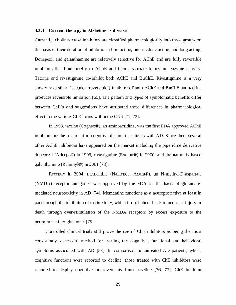
3.3.3 Current therapy in Alzheimer’s disease
Currently, cholinesterase inhibitors are classified pharmacologically into three groups on
the basis of their duration of inhibition- short acting, intermediate acting, and long acting.
Donepezil and galanthamine are relatively selective for AChE and are fully reversible
inhibitors that bind briefly to AChE and then dissociate to restore enzyme activity.
Tacrine and rivastigmine co-inhibit both AChE and BuChE. Rivastigmine is a very
slowly reversible („pseudo-irreversible‟) inhibitor of both AChE and BuChE and tacrine
produces reversible inhibition [65]. The pattern and types of symptomatic benefits differ
between ChE´s and suggestions have attributed these differences in pharmacological
effect to the various ChE forms within the CNS [71, 72].
In 1993, tacrine (Cognex®), an aminoacridine, was the first FDA approved AChE
inhibitor for the treatment of cognitive decline in patients with AD. Since then, several
other AChE inhibitors have appeared on the market including the piperidine derivative
donepezil (Aricept®) in 1996, rivastigmine (Exelon®) in 2000, and the naturally based
galanthamine (Reminyl®) in 2001 [73].
Recently in 2004, memantine (Namenda, Axura®), an N-methyl-D-aspartate
(NMDA) receptor antagonist was approved by the FDA on the basis of glutamate-
mediated neurotoxicity in AD [74]. Memantine functions as a neuroprotective at least in
part through the inhibition of excitoxicity, which if not halted, leads to neuronal injury or
death through over-stimulation of the NMDA receptors by excess exposure to the
neurotransmitter glutamate [75].
Controlled clinical trials still prove the use of ChE inhibitors as being the most
consistently successful method for treating the cognitive, functional and behavioral
symptoms associated with AD [53]. In comparison to untreated AD patients, whose
cognitive functions were reported to decline, those treated with ChE inhibitors were
reported to display cognitive improvements from baseline [76, 77]. ChE inhibitor

30
treatment has also been proven to enhance quantitative electroencephalogram coherence
with decreased slow-wave activity and increased faster frequencies, reflecting increased
cortical arousal, improvements in concentration, sensory processing, learning, and
memory [78, 79]. A recent meta-analysis by Trinh et al., 2003, involving six randomized,
double-blind, placebo-control trials of ChE inhibitors, concluded that as a class, these
agents display a modest, beneficial impact on neuropsychiatric symptoms in patients with
mild-to-moderate probable AD [80]. Although further investigations into evaluating the
effectiveness of AChE and/or BuChE inhibitors are still needed, they currently remain
and hold a promising future as the drugs of choice in treating the symptoms associated
with mild-to-moderately severe forms of AD.
3.4 Natural compounds influencing the metabolism of AChE and
BuChE
From 1981 to 2006, 63% of all low molecular weight drugs developed where from
natural products or natural product-derived compounds [81]. In the quest for additional
AChE inhibitors, various medicinal plants and natural resources have been screened in
the hope of finding substances with comparative IC50 values to those of currently
approved drugs on the market. The search for plant derived inhibitors of AChE has
accelerated inlight of the benefits of these drugs not only in the treatment of AD but in
other forms of dementia [82]. There are currently only a few synthetic medicines for the
treatment of cognitive dysfunction and memory loss associated with mild-to-moderate
AD [83]. Many of these compounds have been reported to present adverse effects
including GIT disturbances, hepatotoxicity and problems associated with bioavailability
[84-86], which further promotes the interest in finding more effective AChE inhibitors

31
from natural resources. Supplementary details beyond this text of natural product
inhibitors of AChE can be found in reviews by Hostettmann et al., 2006, [87] and
Mukherjee et al., 2007, [83].
3.4.1 Alkaloids
3.4.1.1 Physostigmine, Physostigma venenosum, Fabaceae
Physostigma venenosum was traditionally used in Africa as a ritual poison. Treatment
with the indole alkaloid physostigmine, a short-acting reversible AChE inhibitor, isolated
from P. venenosum, has shown cognitive benefits in both normal and AD patients [83].
However, due to its short half-life, physostigmine was found clinically impractical since a
multiple dosing scheme would be required. Instead, the chemical structure of
physostigmine was used as a prototype for the development of rivastigmine, a carbamate
based AChE inhibitor now approved under the trade name Exelon® for symptomatic
treatment of mild-to-moderately severe AD.
3.4.1.2 Galanthamine, Galanthus nivalis, Amaryllidaceae
Galanthus nivalis was used traditionally in Bulgaria and Turkey for neurological
conditions. Initially derived from the extracts of snowdrop and daffodil bulbs,
galanthamine is now a synthetically produced AChE inhibitor. In a randomized, 6-month,
multicenter clinical trial, galanthamine showed improvements in activities of daily living
and behavioral symptom when compared to placebo in patients with probable AD or
vascular dementia [82]. Galanthamine can be taken as a novel representative for
successful natural product substitution in place of synthetic drug treatment in AD.

32
3.4.1.3 Huperzine A, Huperzia serrata, Lycopodiaceae
Huperzia serrata is a moss used to treat contusions, strains, hematuria and swelling in
traditional Chinese medicine [88]. The sesquiterpene alkaloid huperzine A is a potent, yet
reversible inhibitor of AChE. In a study by Raves et al., 1997, huperzine A improved
memory retention process in cognitively impaired aged and adult rats [89]. In China,
studies conducted by Wang et al., 2006, showed enhancement in memory, cognitive
skills, and improvements in daily activities, after administration of huperzine A to
patients with AD [90].
3.4.1.4 Chelidonium majus, Papaveraceae
Chelidonium majus has traditionally been used as an herbal medicine in the treatment of
gastric ulcer, gastric cancer, oral infections and general pain in Asian and European
countries. In a recent study, Cahlikova et al., 2010, demonstrated that the most active of
the natural occurring alkaloids was chelidonine, which inhibited both human AChE and
BuChE in a dose-dependent manner with IC50 values of 26.8 ±1.2μM and 31.9 ±1.4μM
respectively [91].
3.4.2 Terpenoids
3.4.2.1 Salvia lavandulaefolia, Lamiaceae
The ChE inhibition produced by Salvia lavandulaefolia oil was shown to be partly due to
the cyclic monoterpenes 1,8-cineole and α-pinene, which were found to inhibit AChE in
vitro. Upon oral administration of S. lavandulaefolia essential oil to rat‟s, a decreased
striatal AChE activity in both the striatum and the hippocampus was observed and
therefore it was postulated that the in vitro and also in vivo inhibition of AChE in select
brain regions was connected to the activity of either constituents or their metabolites [92].

33
3.4.2.2 Melissa officinalis, Lamiaceae
Melissa officinalis has been used for more than 2,000 years owing in part to its reputation
for restoring memory and promoting long life. Although the constituents have not been
thoroughly investigated, the plant is known to possess monoterpenes in its essential oil,
including citral (a mixture of isomers geraniol and nerol), and it is known from previous
studies that these compounds possess a weak inhibitory effect on AChE [93].
3.4.2.3 Origanum majorana, Lamiaceae
Origanum majorana is a plant found in Indian medicine and also more commonly known
as a spice. In a study testing its inhibitory effect on AChE, the main active component,
identified as the triterpene ursolic acid, exhibited an IC50 value of 7.5 nM [94].
3.4.3 Withanolides
3.4.3.1 Withania somnifera, Solanaceae
The root of this plant, also known as Indian ginger, is one of the most highly regarded
herbs in Ayurvedic medicine where it is classified among the rejuvenating tonics known
as „Rasayanas‟. Compounds present in W. somnifera are structurally related to steroids
and more commonly referred to as withanolides. In a study aimed at the cholinesterase
inhibitory effect of withanolides, Choudhary et al.,2004, isolated and identified six
compounds, of which four dislayed inhibitory effect against electric eel AChE, while the
remaining two inhibited horse-serum BuChE [95].

34
4. EXPERIMENTAL PART

35
4.1 General methods
4.1.1 Distillation and evaporation
Prior to use the solvents were distilled. First, the substances were applied (approx. 5%),
and then the remaining solvent, about 90% in total, was distilled. Solvents were stored in
brown glass containers. Evaporation of the chromatograph fractions was carried out on a
vacuum evaporator under reduced pressure at 40 C.
4.1.2 Chromatography
4.1.2.1 Thin layer chromatography
Chromatography was carried out in a standard chamber system. Chambers were saturated
with the mobile phase. The time of saturation was approx. 30 minutes and in the case of
preparative TLC, approx. 60 minutes. The chromatography was carried out in ascending
order.
4.1.2.2 Column Chromatography
Column chromatography was carried out under gradient elution on a silica gel system,
0.1–0.25mm, deactivated in 10% water. A suspension of the adsorbent in the solvent was
then poured into the chromatographic column. The prepared column was coated with the
sample diluted in a small amount of the solvent. The sample was dried in the exsiccator
and then applied with a small amount of silica gel.

36
4.2 Plant material and equipment
4.2.1 Chemicals and solvents
Solvents:
Cyclohexane
Diethylamine
Diethylether without stabilizer
Ethanol 95%, denatured with methanol (EtOH)
Chloroform (CHCl3)
Methanol (MeOH)
Petrol (ČL 2006)
Toluene
Chemicals:
Acetic acid 99% p. a.
Bismuth subnitrate purum
Hydrochloric acid 36% p. a. (HCl)
Potassium iodide p. a. (KI)
Sodium carbonate anhydrous purum
Sodium hydroxide p. a. (NaOH)
Sodium sulfate anhydrous purum
Sulfuric acid 96% purum
Tartaric acid purum

37
4.2.2 Chemicals and material for analysis of AChE and BuChE (IC50)
Chemicals:
0,1 M phosphate buffer pH 7.4
10 mM acetylcholine iodide (Sigma-Aldrich)
10 mM butyrylcholine iodide (Sigma-Aldrich)
Dimethylsulphoxide p. a. (Sigma-Aldrich)
5,5 -Dithiobis(2-nitrobenzoic acid) (DTND) p. a. (Sigma-Aldrich)
Eserine (Sigma-Aldrich)
Galanthamine hydrobromide (Changsha Organic Herb Inc., China)
Huperzin A (Tazhonghui Co., Ltd., China)
Sodium dihydrogenphosphate dihydrate p. a. (Lachema)
Sodium hydrogen dodecahydrate p. a. (Lachema)
Material:
Source of AChE: hemolysed human erythrocytes
Whole blood was centrifuged for 15 minutes at 10,000 rev / min. The mass obtained
from red blood cells was washed 3 times with 0.1 M phosphate buffer at pH 7.4 to
remove residual plasma, 10% (v/v) lysate was prepared in water.
Source of BuChE: human plasma
Single semi-micro polystyrene cuvette 1.5 ml (PLASTIBRAND)
Equipment:
Centrifuge type MPW–340 (Mechanika precyzyjna,Warszaw, Poland)
Instrument for measurement of optical rotation: ADP 220 POLARIMETER B+S
Micro-heated apparatus Boetius
pH meter Φ 72 METER (Beckmann, USA)

38
UV-Spectrophotometer UVIKON 942 (Kontron Instruments, Switzerland)
4.2.3 Chemicals and material for analysis of antioxidant activity
2,2-Diphenyl-1-picrylhydrazyl radical purum (Sigma-Adrich)
Quercetin (Sigma-Aldrich)
Trolox p.a. (Sigma-Aldrich)
4.2.4 Detection reagents
Dragendorf‟s reagent modified according to Munier:
Solution A: prepared by dissolving 1.7 g bismuth subnitrate and 20 g tartaric acid in
80 ml of water.
Solution B: prepared by dissolving 16 g potassium iodide in 40 ml water.
Stock solution: prepared by mixing solution A and B in ratio of 1:1.Stock solution
may be stored for some months in a refrigerator (4 °C).
Solution for analysis: prepared by adding 5 ml stock solution to 5 ml tartaric acid
dissolved in 50 ml of water.
4.2.5 Chromatographic plates and adsorbents
Aluminum oxide neutral, 100-250 μm (fy Across)
Commercial chromatographic adsorbent was activated in a layer maximally 2 cm thick at
200 °C in the dryer for 8 hours. After cooling to ∼ 80 °C, the adsorbent was poured into a
flask and sealed. After cooling at room temperature, 5% water (w/w) was added and
equilibrated upon periodic shaking.

39
Kieselgel 60 GF254, plates for TLC
For preparation of poured plates (90 x 150 mm), 3.9 g of commercial adsorbent were
mixed with 13.5 ml water and mixture was homogenized for 30 seconds by using a
micro-homogenizer. The suspension was poured on the plate, the surface layer was
planed, and plates were stored in horizontal position for 24 hours at room temperature.
Solution of fraction was spotted in the form of a line about 10 mm from the upper edge of
the plate by using an application-tube.
4.3 Description of alkaloid and its isolation
4.3.1 Origin of herbal drug
Ground tubers of C. yanhusuo were supplied by the company Pragon s.r.o., Prague, and
verification of the herbal drug was conducted by Assoc. Prof. L. Opletal.
4.3.2 Preparation of summary extract
10.8 kg of dry tuber were percolated with 120 liters of 95% ethanol ( 1:11). Collected
extract was evaporated to a viscous residue, heated at 50 °C, and 2.5 liters of 2%
hydrochloric acid was added. The brown solution was decanted and the solid residue in
the flask was homogenized with 1 liter of 2% hydrochloric acid and sonified at 50 °C on
level 10 (apparatus Sonorex 10HP) for 30 minutes. The suspension was then filtered
through viscous cellulose and the filtrate was diluted with water to 4.7 liters.
4.3.3 Preparation of extract A from primary extract
4.7 liters of acidic solution (pH 1) was alkalized by 10% Na2CO3 to pH 9.7 (approx. 7
liters of solution were obtained). The suspension with alkaloids was operated by ether (5×
1.6 liters). The organic layer was then desiccated by sodium sulfate, filtered and
evaporated to dryness.

40
4.3.4 Separation of extract A on particular groups of alkaloids
Dry residue of extract A was dissolved in 2% hydrochloric acid. This solution was
filtered and shaken with chloroform. In this manner the mixture of alkaloids was divided
on chlorides soluble and insoluble in chloroform. Each group of alkaloids was transferred
into alkaloidal bases, which were dissolved in ether and divided into bases of phenolic
and non-phenolic origin. Upon completion, four groups of alkaloids from extract A were
obtained.
The mixture of non-phenolic alkaloids, which were obtained from the mixture of
chlorides soluble in chloroform were examined in this diploma work. The alkaloid
fraction was obtained from the diploma supervisor (Assoc. Prof. L. Opletal). The first
part of the separation was performed together with diploma co-worker Buleza Koci.
Fig. 15 TLC of alkaloidal bases from extract A

41
Non-phenolic bases obtained from chlorides soluble in chloroform. Silica gel plates for
TLC 60F254 (Merck), 50 x 75 mm, toluene + chloroform + diethylamine (70 : 25 : 5),
developing chamber was saturated by vapor of solvent system, developed 1x, detection
UV λ = 254 nm, Dragendorf reagent (modified by Munier).
4.3.5 Separation of mixture of non-phenolic alkaloids from chlorides soluble in
chloroform
9.73 g of yellow-orange oily residue was dissolved in a small volume of chloroform
(minimal quantity only for dissolution). 30 g of Aluminum oxide neutral was added and
the mixture was dried. After drying, 5 g of Celite 545 was added and the mixture was
homogenized. This dry trituration was then applied to the chromatographic column.
Table 1. Column chromatography of non-phenolic bases of alkaloids from
chlorides soluble in chloroform
Adsorbent: Aluminium oxide neutral, 100-250 μm, grade 3 activity
Quantity of adsorbent 350 g
Layer with extract 3 6 cm
Layer with adsorbent 3 39 cm
Dead volume 220 ml
Time of fraction collection 15-20 minutes
Fraction volume 100 ml
Each of the fractions was monitored by TLC (Silica gel for TLC 60F254 (Merck),
plates 50 x 75 mm, solvent system: toluene + chloroform + diethylamine (70 : 25 : 5),
developing tank was saturated by vapor of solvent system, developed 1×, detected UV λ
= 254 nm, Dragendorf reagent (modif. according to Munier).

42
Fractions of the same quality were combined, evaporated under decreased
pressure and temperature and desiccated in vacuum-desiccator over granulated silica gel.
Table 2. Results of column chromatography of non-phenolic alkaloids from
chlorides soluble in chloroform
Fraction Eluent Weight Description
1-42 1-12
13-22
23-42
Petrol+CHCl3 95:5
Petrol+CHCl3 92.5:7.5
Petrol+CHCl3 90:10
0.050 g Yellow oil
43-56 43-56 Petrol+CHCl3 85:15 1.82 g Bright brown, crystals
57-63
57-59
60-63
Petrol+CHCl3 85:15
Petrol+CHCl3 80:20
0.57 g Brown-red, crystals
64-67 64-65
66-67
Petrol+CHCl3 80:20
Petrol+CHCl3 75:25
0.28 g Brown, crystals
68-71 68-71 Petrol+CHCl3 75:25 0.86 g Brown, very viscous
72-75 72-75 Petrol+CHCl3 75:25 0.86 g Dark brown, viscous
76-97
76-85
86-95
96-97
Petrol+CHCl3 75:25
Petrol+CHCl3 70:30
Petrol+CHCl3 25:75
3.20 g Yellow-brown, crystals
98-107 98-107 Petrol+CHCl3 25:75 0.19 g Dark brown, very viscous
108-113 108-113 Petrol+CHCl3 25:75 0.16 g Black, very viscous

43
Fig. 16 Results of column chromatography on aluminum oxide
Silica gel plates for TLC 60F254 (Merck), 50 75 mm, toluene + chloroform +
diethylamine 70 : 25 : 5, developing chamber saturated by vapor of solvent system,
developing 1x, detection UV λ = 254 nm, Dragendorf reagent (modif. according to
Munier).
4.3.6 Isolation of alkaloid from combined fractions 57-63
0.57 g of brown-reddish crystalline mass was dissolved in a 15 ml of mixture chloroform
+ toluene 95 : 5 (w/w). The solution was filtered through 6.0 g of Aluminum oxide
neutral in a micro-chromatographic column (diameter × height 8 : 12 mm) and from the
compound, 20 ml of the mentioned mixture of solvents was eluted. After evaporation of
filtrate, 0.50 g of yellowish crystalline mass was obtained and designated GC-1.

44
4.4 Method for determining MS spectra
The spectra were measured on the LC/MS Thermo Finningan LCQDuo, ion trap,
electrospray ionization in positive mode (ESI+). MS/MS spectra were measured at
collision energy of 40 eV and the substance was dissolved in methanol.
4.5 Method for determining NMR spectra
The spectra were measured on a Varian Inova 500 spectrometer with a working
frequency of 499.9 MHz for 1H and 125.7 MHz for 13C nuclei. 13C NMR spectra were
measured in 5 mm broadband probe SW, 1 H and all 2D spectra in inverse 5 mm ID PFG
probe using a modified version of standard pulse sequences. Experiments were measured
in deuterochloroform at 25 ˚ C.
Values of chemical shifts are in ppm and are relative to internal standard
(hexamethyldisilane, 0.04 ppm in 1H spectra) or the solvent signal (76.99 ppm in 13C
spectra)(Dr. M. Kurfürst, Ph.D., Institute of Chemical Process Fundamentals, ASCR,
Prague).
4.6 Method for determining antioxidant activity- DPPH free-radical scavenging
assay (EC50)
Radical scavenging activity of extracts and pure compounds were evaluated by means of
the DPPH (2,2´-diphenyl-1-picrylhydrazyl radical) test using an SIA (PC-controlled
Sequential Injection Analysis system) method developed in our laboratory [96]. The
stock solution of extract/pure compound was prepared by dissolving 4 mg of the
extract/pure compound in 4 ml of aqueous 50% w/w ethanol during 10 minutes of
sonication; the same solvent was used for appropriate dilution of the extract/pure
compound stock solution (1, 0.5, 0.25, 0.01 mg/mL). DPPH solution (0.1 mM) was
prepared by dissolving 3.9 mg DPPH in 100 mL 50 % w/w ethanol. The automated

45
method was based on the known reaction of stable DPPH with antioxidants resulting in
bleaching of DPPH due to its “quenching” by interaction with the analytes. The decrease
in the absorbance of DPPH measured at 525 nm was related to the concentration of
antioxidant in the tested solution. The percentage of inhibition of DPPH was estimated by
using the formula: % QDPPH = (1-Ax/A0) x 100, where A0 was the height of the peak of the
blank sample and Ax was the height of the peak after the extract/pure compound was
added. All measurements were made in triplicate. The DPPH radical scavenging activity
of samples was expressed as EC50 (mg/mL for extracts, µM for pure compounds), which
was the amount of sample necessary to decrease by 50% the light absorbance.
4.7 Methods for determinating inhibitory activity against AChE and BuChE
4.7.1 Preparation of red blood cells ghost for AChE and BuChE
Ghosts were prepared from freshly drawn blood (taken from healthy volunteers), to
which 1 mL of sodium citrate per 10 mL of blood was added, according to a slightly
modified method of Steck and Kant [97]. Briefly plasma (HuBuChE) was removed from
the whole blood by centrifugation at 4000 rpm in a Boeco U-32R centrifuge with a
Hettich 1611 rotor. Red blood cells were transferred to 50 mL tubes and washed 3 times
with 5 mM phosphate buffer (pH 7.4) containing 150 mM sodium chloride (12000 rpm,
Avanti J-30I, rotor JA-30.50). The washed erythrocytes were stirred with 5 mM
phosphate buffer (pH 7.4) for 10 min to ensure lysis. The lysed cells were centrifuged at
20,000 rpm for 10 minutes and then the ghosts (HuAChE) were washed 3 times with
phosphate buffer.

46
4.7.2 AChE and BuChE assay (IC50)
HuAChE and HuBuChE activities were determined with a modified method of Ellman et
al.,[98] at concentrations 500, 250, 125, 50, 25, 12.5, 5, 2.5, 0.5, and 0.25 μg/mL using
acetylthiocholine iodide (ATChI) and butyrylthiocholine iodide (BTChI) as substrates,
respectively. Briefly, 25-50 μL of ghosts or plasma, 650 μL of DTNB and 25 μL of either
the sample or appropriate solvent, as a blank sample, were added to the semi-micro
cuvette. The reaction was initiated by addition of substrate (ATChI or BTChI). The final
proportion of DTNB to substrate was 1 : 1. The increase of absorbance at 436 nm (∆A)
was measured for one minute using a Shimadzu UV-1611 spectrophotometer. Each
measurement was repeated three times. Galanthamine and huperzine A were used as
positive controls. The IC50 and EC50 values were calculated with the use of GraphPad
Prism 5.02 software. The inhibition (in %) was calculated according to the formula: %I =
100-(∆ABL/∆ASA)*100, where ∆ABL= increase of absorbance of a blank sample and
∆ASA= increase of absorbance of the measured sample.
4.8 Method for determining optical rotation
The optical rotation was measured on polarimeter ADP 220 BS ethanol.

47
5. RESULTS

48
5.1 Structural analysis of compound GC-1
The structure of the isolated compound was determined by comparing spectral data with
those reported in the literature as (+)-corydaline (CAS: 3907-48-0).
5.1.1 MS analysis of (+)-corydaline
ESI-MS m/z [M+H]+ 370.24 (100).
Fig.17 MS Spectrum of (+)-corydaline

49
5.1.2 MS/MS analysis of (+)-corydaline
MS/MS m/z 355.16 (20; [M-CH3]+), 338.14 (15), 308.11 (12), 218.14 (22), 192.14 (100;
[C11H14O2N]+), 165.07 (25; [C10H13O2]
+).
Fig. 18 MS/MS spectrum of (+)-corydaline

50
Fig.19 MS/MS spectrum of corydaline and its proposed retro-Diels-Alder (RDA) pathway
5.1.3 NMR analysis of (+)-corydaline
Fig. 20 Structure of isolated compound (+)-corydaline
51
5.1.3.1 1H-NMR analysis of (+)-corydaline
1H NMR (CDCl3, 25°C):
0.95 (3H, d, J = 7.0 Hz, R-CH3); 2.59 - 3.17 (4H, m, H-5, H-6); 3.20 (1H, dd, J = 7.0 Hz,
H-13); 3.50 (1H, d, J = 15.9 Hz, H-8α), 3.68 (1H, br s, H-13a), 3.85 (6H, s, 2 R-OCH3),
3.87 (6H, s, 2 R-OCH3), 4.20 (1H, d, J =15.9 Hz, H-8ß), 6.60 (1H, s, H-4), 6.68 (1H, s,
H-1), 6.82 (1H, d, J = 8.4 Hz Hz, H-11), 6.90 (1H, d, J = 8.4 Hz, H-12)
Fig.21 1H-NMR spectrum of (+)-corydaline

52
5.1.3.2 13
C-NMR analysis of (+)-corydaline
13C NMR (CDCl3, 25°C):
29.23, C-5; 36.45, C-13; 51.72, C-6; 54.18, C-8; 56.09, C-3a; 56.12, C-10a; 56.32, C-2a;
59.54, C-14; 60.42, C-9a; 108.83, C-1; 111.26, C-11; 111.60, C-4; 124.08, C-12; 126.93,
C-4a; 127.88, C-12a; 128.75, C-8a; 129.75, C-14a; 145.34, C-9; 147.71, C-2; 147.80, C-
3; 150.54, C-1
Fig. 22 13
C NMR spectrum of (+)-corydaline

53
5.1.3.3 Optical rotation
[α]D23
+ 312 ° (EtOH; c = 0.2)
5.2 Inhibitory activity of (+)- corydaline against AChE and BuChE
Table 3 In vitro HuAChE and HuBuChE inhibitory activity of isolated compound
Compound IC50 (μM)a
AChE BuChE
(+)-Corydaline 40.5 ± 1.9 >1000
Galanthaminb
6.9 ± 0.3 156 ± 6.9
Huperzinb
0.25 ± 0.01 >1000
a Results are the mean of free replications;
b Reference compounds
5.3 Antioxidant activity of (+)-corydaline
Table 4 Antioxidant activity of isolated compound
Compound EC50 (μM)a
(+)-Corydaline >1000
Quercetin b
25.3 ± 1.2
Trolox b
27.8± 0.8
a Results are the mean of free replications;
b Reference compounds

54
6. DISCUSSION

55
On the department of pharmaceutical botany and ecology, Faculty of Pharmacy in Hradec
Králové, of Charles University, focus has been aimed at testing the biological activity of
various alkaloids, notably isoquinoline type, with regard to their inhibitory activity on
ChEs. This particular interest stemmed from the ever growing knowledge supporting the
theory that these natural compounds could be employed as potential structural prototypes
in the future development of more active ChE inhibitors. This class of agents is renowned
for their use as the treatment of choice in mild-to-moderate symptomatic therapy of AD.
The aim of this work was to isolate alkaloids from the crude extract A of C.
yanhusuo (from the part of chlorides non-phenolic alkaloids soluble in chloroform) by
column chromatography and preparative thin-layer chromatography, and testing the
biological activities related to ChE inhibition and antioxidant effect.
After chromatographic separation and fractionation of the extract A, one pure
alkaloid was isolated. The alkaloid was termed GC-1 and further identified as (+)-
corydaline on the basis of MS and NMR spectral studies. (+)-Corydaline is a tertiary
alkaloid belonging to the structural group of isoquinoline alkaloids and can be further
classified as having a tetrahydroberberine skeletal backbone (see Fig. 20).
The isolated alkaloid (+)-corydaline was further investigated on its inhibitory
effect against human erythrocytic AChE and BuChE using the spectrophotometric
method devised by Ellman [98], as well as antioxidant activity by use of the DPPH test.
(+)-Corydaline was found to inhibit AChE in a dose-dependent manner with an IC50 value
of 40.5 ± 1.9 μM, with huperzine A and galanthamine as positive controls. However, (+)-
corydaline was found inactive against BuChE due to its IC50 value >1000 μM. The
antioxidant activity was also considered negligible due to a value of EC50. >1000 μM.
Corydaline was isolated from C. yanhusuo in previous investigations [6, 9, 15, 96-
98]. In a study by Hung et al., 2008, corydaline was found to weakly inhibit AChE with
an IC50 value of 30.7 ± 1.5 μM when mouse brain cortex was used as the source of

56
enzyme [9]. Interestingly, in another study on corydaline isolated from Corydalis cava, it
was found to be the most active compound, inhibiting electric eel AChE in a dose-
dependent manner with an IC50 value of 15 ± 3 μM [101]. Moreover, in the same study
by Adsersen et al., 2007, corydaline was also found inactive against BuChE due to IC50 >
100 μM [101]. When comparing the spectrum of results obtained in this work with that of
previous studies, it is important to note that differences in IC50 values could be due, at
least in part, to the choice in enzyme origin.
In addition to possessing inhibitory activity against AChE, previous studies have
demonstrated other unique pharmacological properties of corydaline. In one such study,
corydaline was found to inhibit the enzyme GABA-transaminase, thus dispaling the basic
mechanism of drug action used in the treatment of convulsive disorders [102]. Further
more, corydaline derived from C. yanhusuo was found to exhibit significant
antinociceptive effects with particularly high concentrations in the striatum [10].
The potential use of isoquinoline alkaloids against neurodegenerative processes in
the human brain are concerned not only with their activity against AChE and BuChE, but
another working theory in this area is on the inhibition of β- secretase (BACE1), as well
as inhibition of NMDA receptors. From these reasons, it was also necessary to isolate 56
alkaloids that have previously been reported in literature for activity against human ChEs.
Based on the results obtained from this work along with comparisons from previous
studies mentioned above, it can be said that (+)-corydaline does not prove sufficiently
effective in the inhibition of AChE due to its weak IC50 values, and therefore may not be
used as a direct means of alternative therapy in AD. However, the knowledge obtained
with regard to the biological activity of (+)-corydaline can be considered as an important
base in future production of more active synthetic analogues, where novel therapeutic
strategies for dementia treatment may benefit from the combination of conventional
Western approach and traditional Oriental medical.

57
7. LITERATURE

58
1. Ferri, C.P., et al.: Global prevalence of dementia: a Delphi consensus study.
Lancet, 2005, 366(9503), 2112-2117.
2. Dharmananda, S.: Simple traditional formulas for pain. 2002; Available from:
http://www.itmonline.org/arts/pain.htm (11-03-12).
3. Ling, H.Y., Wu, L.M., Li, L.D.: Corydalis yanhusuo rhizoma extract reduces
infarct size and improves heart function during myocardial ischemia/reperfusion
by inhibiting apoptosis in rats. Phytother. Res. 2006, 20(6), 448-453.
4. Sagare, A.P., et al.: Cytokinin-induced somatic embryogenesis and plant
regeneration in Corydalis yanhusuo (Fumariaceae) - a medicinal plant. Plant Sci.
2000, 160(1), 139-147.
5. Leung, W.C., et al.: Anxiolytic-like action of orally administered dl-
tetrahydropalmatine in elevated plus-maze. Progr. Neuro-Psychopharmacol. Biol.
Psychiatry 2003, 27(5), 775-779.
6. Lee, Y.L., et al.: Formation of protoberberine-type alkaloids by the tubers of
somatic embryo-derived plants of Corydalis yanhusuo. Planta Med. 2001, 67(9),
839-842.
7. eFloras.org. Chinese Plant Names - Corydalis yanhusuo. Available from:
http://www.efloras.org/florataxon.aspx?flora_id=3&taxon_id=200009146 (11-03-
12)
8. Zhang, J., et al.: Systematic screening and characterization of tertiary and
quaternary alkaloids from corydalis yanhusuo W.T. Wang using ultra-
performance liquid chromatography-quadrupole-time-of-flight mass
spectrometry. Talanta 2009, 78(2), 513-522.
9. Hung, T.M., et al.: Cholinesterase inhibitory and anti-amnesic activity of
alkaloids from Corydalis turtschaninovii. J. Ethnopharmacol. 2008, 119(1), 74-80.
10. Wang, C., et al.: Screening of antinociceptive components in Corydalis yanhusuo
W.T. Wang by comprehensive two-dimensional liquid chromatography/tandem
mass spectrometry. Anal. Bioanal. Chem. 2010, 396(5), 1731-1740.
11. Hsu, B., Kin, K.C.: Pharmacological study of tetrahydropalmatine and its analogs.
A new type of central depressants. Arch. Int. Pharmacodyn. Ther. 1962, 139, 318-
327.

59
12. Xu, L.F., et al.: Protopine inhibits serotonin transporter and noradrenaline
transporter and has the antidepressant-like effect in mice models.
Neuropharmacology 2006, 50(8), 934-940.
13. Hung, T.M., et al.: Anti-amnestic activity of pseudocoptisine from Corydalis
Tuber. Biol. Pharm. Bull. 2008, 31(1), 159-162.
14. Oh, Y.C., et al.: Tetrahydropalmatine Inhibits Pro-Inflammatory Mediators in
Lipopolysaccharide-Stimulated THP-1 Cells. J. Med. Food 2010, 13(5), 1125-
1132.
15. Kubo, M., et al.: Antiinflammatory activities of methanolic extract and alkaloidal
components from Corydalis tuber. Biol. Pharm. Bull. 1994, 17(2), 262-265.
16. Yun, K.J., et al.: Quaternary alkaloid, pseudocoptisine isolated from tubers of
Corydalis turtschaninovi inhibits LPS-induced nitric oxide, PGE(2), and pro-
inflammatory cytokines production via the down-regulation of NF-kappa B in
RAW 264.7 murine macrophage cells. Int. Immunopharmacol. 2009, 9(11), 1323-
1331.
17. Shiomoto, H., et al.: Effects of protopine on blood-platelet aggregation 3. effects
of protopine on the metabolic system of arachadonic acid in platelets. Chem.
Pharm. Bull. 1991, 39(2), 474-477.
18. Wu, L.M., et al.: Beneficial effects of the extract from Corydalis yanhusuo in rats
with heart failure following myocardial infarction. J. Pharm. Pharmacol. 2007,
59(5), 695-701.
19. Huang, K., et al.: Blocking L-calcium current by l-tetrahydropalmatine in single
ventricular myocyte of guinea pigs. Acta Pharmacol. Sin. 1999, 20(10), 907-911.
20. Narasimhan, N.S., Bhide, B.H.: A novel synthesis of tetrahydropalmatine. Chem.
Ind. 1969, 19, 621-622.
21. Jin, G.Z., et al.: Different effects of enantiomers of tetrahydropalmatine on
dopaminergic system. Sci. Sin. B 1986, 29(10), 1054-1064; Chem. Abstr. 1987,
131533.
22. Liu, G.Q., et al.: D-L-tetrahydropalmatine as monoamine depletor. Arch. Int.
Pharmacodyn. Ther. 1982, 258(1), 39-50.

60
23. Xu, S.X., et al.: Brain dopamine depleted by d-tetrahydropalmatine Acta
Pharmacol. Sin. 1987, 8(3), 207-212; Chem. Abstr. 1987, 417796.
24. Chueh, F.Y., et al.: Hypotensive and bradycardic effects of dl-tetrahydropalmatine
mediated by decrease in hypothalamic serotonin release in the rat. Jpn. J.
Pharmacol. 1995, 69(2), 177-180.
25. Matsuda, H., et al.: Inhibitory effects of dehydrocorydaline isolated from
Corydalis Tuber against type I-IV allergic models. Biol. Pharm. Bull. 1997, 20(4),
431-434.
26. Lee, K.H., et al.: Regulation of glutamate level in rat brain through activation of
glutamate dehydrogenase by Corydalis ternata. Exp. Mol. Med. 2005, 37(4), 371-
377.
27. Millan, M.J.: The induction of pain: an integrative review. Prog. Neurobiol. 1999,
57(1), 1-164.
28. Fundytus, M.E.: Glutamate receptors and nociception: implications for the drug
treatment of pain. CNS Drugs 2001, 15(1), 29-58.
29. Ledoux, J.E.: Emotional memory systems in the brain. Behav. Brain Res. 1993,
58(1-2), 69-79.
30. Morris, R.: Developments of a water-maze procedure for studying spatial-learning
in the rat. J. Neurosci. Methods 1984, 11(1), 47-60.
31. Barnes, C.A., et al.: Effects of the uncompetitive NMDA receptor antagonist
memantine on hippocampal long-term potentiation, short-term exploratory
modulation and spatial memory in awake, freely moving rats. Eur. J. Neurosci.
1996, 8(3), 565-571.
32. Kim, D.K., et al.: Acetylcholinesterase inhibitors from the aerial parts of
Corydalis speciosa. Arch. Pharmacal Res. 2004, 27(11), 1127-1131.
33. Adsersen, A., et al.: Acetylcholinesterase and butyrylcholinesterase inhibitory
compounds from Corydalis cava Schweigg. & Kort. J. Ethnopharmacol. 2007,
113(1), 179-82.
34. Cheng, X.X., et al.: DNA topoisomerase I inhibitory alkaloids from Corydalis
saxicola. Chem. Biodivers. 2008, 5(7), 1335-1344.
61
35. Li, H.L., et al.: Alkaloids from Corydalis saxicola and their anti-hepatitis B virus
activity. Chem. Biodivers. 2008, 5(5), 777-783.
36. Wang, T., et al.: Protective effects of dehydrocavidine on carbon tetrachloride-
induced acute hepatotoxicity in rats. J. Ethnopharmacol. 2008, 117(2), 300-308.
37. Houghton, P.J., Howes, M.J.: Natural products and derivatives affecting
neurotransmission relevant to Alzheimer's and Parkinson's disease. Neurosignals
2005, 14(1-2), 6-22.
38. Cummings, J.L.: Alzheimer's disease. N. Engl. J. Med. 2004, 351(1), 56-67.
39. Isacson, O., et al.: Alzheimer's disease and Down's syndrome: roles of APP,
trophic factors and ACh. Trends Neurosci. 2002, 25(2), 79-84.
40. Checler, F., Vincent, B.: Alzheimer's and prion diseases: distinct pathologies,
common proteolytic denominators. Trends Neurosci. 2002, 25(12), 616-620.
41. Citron, M.: Secretases as targets for the treatment of Alzheimer's disease. Mol.
Med. Today 2000, 6(10), 392-397.
42. Kovacs, D.M., et al.: Alzheimer-associated presenilins 1 and 2: Neuronal
expression in brain and localization to intracellular membranes in mammalian
cells. Nat. Med. 1996, 2(2), 224-229.
43. Kimberly, W.T., et al.: The transmembrane aspartates in presenilin 1 and 2 are
obligatory for gamma-secretase activity and amyloid beta-protein generation. J.
Biol. Chem. 2000, 275(5), 3173-3178.
44. Scheuner, D., et al.: Secreted amyloid beta-protein similar to that in the senile
plaques of Alzheimer's disease is increased in vivo by the presenilin 1 and 2 and
APP mutations linked to familial Alzheimer's disease. Nat. Med. 1996, 2(8), 864-
870.
45. Parihar, M.S., Hemnani, T.: Alzheimer's disease pathogenesis and therapeutic
interventions. J. Clin. Neurosci. 2004, 11(5), 456-467.
46. Geula, C., et al.: Aging renders the brain vulnerable to amyloid beta-protein
neurotoxicity. Nat. Med. 1998, 4(7), 827-831.
47. Frank, R.A., et al.: Biological markers for therapeutic trials in Alzheimer's disease
- Proceedings of the biological markers working group; NIA initiative on
neuroimaging in Alzheimer's disease. Neurobiol. Aging 2003, 24(4, 521-536.

62
48. Bartus, R.T., et al.: The cholinergic hypothesis of geriatric memory dysfunction.
Science 1982, 217(4558), 408-417.
49. Davies, P., Maloney, A.J.: Selective loss of central cholinergic neurons in
Alzheimer's disease. Lancet 1976, 2(8000), 1403.
50. Perry, E.K., et al.: Necroscopy evidence of central cholinergic deficits in senile
dementia. Lancet 1977, 1(8004), 189-189.
51. Wilcock, G.K., et al.: Alzheimer´s disease-correlation of cortical choline-
acetyltransferase activity with the severity of dementia and histological
abnormalities. J. Neurol. Sci. 1982, 57(2-3), 407-417.
52. Perry, E.K., et al.: Correlation of cholinergic abnormatlities with senile plaques
and mental test scores in senile dementia. Brit. Med. J. 1978, 2(6150), 1457-1459.
53. Wilkinson, D.G., et al.: Cholinesterase inhibitors used in the treatment of
Alzheimer's disease the relationship between pharmacological effects and clinical
efficacy. Drugs Aging 2004, 21(7), 453-478.
54. Massoulie, J., Bon, S.: The molecular forms of cholinesterase and
acetylcholinesterase in vertebrates. Ann. Rev. Neurosci. 1982, 5, 57-106.
55. Atack, J.R., et al.: Molecular forms of acetylcholinesterase and
butyrlcholinesterase in the aged human central nervous system. J. Neurochem.
1986, 47(1), 263-277.
56. Arendt, T., et al.: Changes in acetylcholinesterase and butyrylcholinesterase in
Alzheimer's disease resemble embryonic-development - a study of molecular
forms. Neurochem. Int. 1992, 21(3), 381-396.
57. Jann, M.W., et al.: Clinical pharmacokinetics and pharmacodynamics of
cholinesterase inhibitors. Clin. Pharmacokinetics 2002, 41(10), 719-739.
58. Sussman, J.L., et al.: Atomic structure of acetylcholinesterase from Torpedo
californica-a prototypic acetylcholine binding protein. Science 1991, 253(5022),
872-879.
59. Taylor, P., Radic, Z.: The cholinesterases- from genes to proteins. Ann. Rev.
Pharmacol. Toxicol. 1994, 34, 281-320.
60. Lane, R.M., et al.: Targeting acetylcholinesterase and butyrylcholinesterase in
dementia. In. J. Neuropsychopharmacol. 2006, 9(1), 101-124.

63
61. Perry, E.K., et al.: Changes in brain cholinesterases in senile dmeentia of
Alzheimer type. Neuropathol. Appl. Neurobiol. 1978, 4(4), 273-277.
62. Darvesh, S., Hopkins, D.A.: Differential distribution of butyrylcholinesterase and
acetylcholinesterase in the human thalamus. J. Compar. Neurol. 2003, 463(1), 25-
43.
63. Greig, N.H., et al.: Butyrylcholinesterase: An important new target in Alzheimer's
disease therapy. Int. Psychogeriatr. 2002, 14, 77-91.
64. Ballard, C.G.: Advances in the treatment of Alzheimer's disease: Benefits of dual
cholinesterase inhibition. Eur. Neurol. 2002, 47(1), 64-70.
65. Ballard, C.G., et al.: Cholinesterases: roles in the brain during health and disease.
Curr. Alzheimer Res. 2005, 2(3), 307-18.
66. Guillozet, A.L., et al.: Butyrylcholinesterase in the life cycle of amyloid plaques.
Ann. Neurol. 1997, 42(6), 909-918.
67. Mesulam, M.M., Geula, C.: Butyrylcholinesterase reactivity differentiates the
amyloid plaques of aging from those of dementia. Ann. Neurol. 1994, 36(5), 722-
727.
68. Greig, N.H., et al.: A new therapeutic target in Alzheimer's disease treatment:
Attention to butyrylcholinesterase. Curr. Med. Res. Opin. 2001, 17(3), 159-165.
69. Hellstrom-Lindahl, E.: Modulation of beta-amyloid precursor protein processing
and tau phosphorylation by acetylcholine receptors. Eur. J. Pharmacol. 2000,
393(1-3), 255-263.
70. Kihara, T., et al.: Alpha 7 nicotinic receptor transduces signals to
phosphatidylinositol 3-kinase to block a beta-amyloid-induced neurotoxicity. J.
Biol. Chem. 2001, 276(17), 13541-13546.
71. Enz, A., et al.: Brain selective inhibtion of acetylcholinesterase - a novel approach
to therapy for Alzheimer's disease. Progr. Brain Res. 1993, 98, 431-438.
72. Poirier, J.: Evidence that the clinical effects of cholinesterase inhihitors are related
to potency and targeting of action. Int. J. Clin. Pract. 2002, 6-19.
73. van Marum, R.J.: Current and future therapy in Alzheimer's disease. Fund. Clin.
Pharmacol. 2008, 22(3), 265-274.

64
74. Lipton, S.A.: Pathologically-activated therapeutics for neuroprotection:
Mechanism of NMDA receptor block by memantine and S-nitrosylation. Curr.
Drug Targets 2007, 8(5), 621-632.
75. Sonkusare, S.K., et al.: Dementia of Alzheimer's disease and other
neurodegenerative disorders - memantine, a new hope. Pharmacol. Res. 2005,
51(1), 1-17.
76. Tariot, P.N., et al.: A 5-month, randomized, placebo-controlled trial of
galantamine in AD. Neurology, 2000, 54(12), 2269-2276.
77. Rosler, M., et al.: Efficacy and safety of rivastigmine in patients with Alzheimer's
disease: international randomised controlled trial. Brit. Med. J. 1999, 318(7184),
633-638.
78. Adler, G., Brassen, S.: Short-term rivastigmine treatment reduces EEG slow-wave
power in Alzheimer patients. Neuropsychobiology 2001, 43(4), 273-276.
79. Adler, G., et al.: EEG coherence in Alzheimer's dementia. J Neural Transm.
2003, 110(9), 1051-8.
80. Trinh, N.H., et al.: Efficacy of cholinesterase inhibitors in the treatment of
neuropsychiatric symptoms and functional impairment in Alzheimer disease - A
meta-analysis. JAMA 2003, 289(2), 210-216.
81. Newman, D.J., Cragg, G.M.: Natural products as sources of new drugs over the
last 25 years. J. Nat. Prod. 2007, 70(3), 461-477.
82. Erkinjuntti, T., et al.: Efficacy of galantamine in probable vascular dementia and
Alzheimer's disease combined with cerebrovascular disease: a randomised trial.
Lancet 2002, 359(9314), 1283-1290.
83. Mukherjee, P.K., et al.: Acetylcholinesterase inhibitors from plants.
Phytomedicine 2007, 14(4), 289-300.
84. Melzer, D.: Personal paper - New drug treatment for Alzheimer's disease: lessons
for healthcare policy. Brit. Med. J. 1998, 316(7133), 762-764.
85. Schulz, V.: Ginkgo extract or cholinesterase inhibitors in patients with dementia:
What clinical trials and guidelines fail to consider. Phytomedicine 2003, 10, 74-
79.

65
86. Lleo, A., et al.: Current pharmacotherapy for Alzheimer's disease. Ann. Rev.
Med. 2006, 57, 513-533.
87. Hostettmann, K., et al.: Natural product inhibitors of acetylcholinesterase. Curr.
Org. Chem. 2006, 10(8), 825-847.
88. Wang, T., Tang, X.C.: Reversal of scopolamine-induced deficits in radial maze
performance by (-)-huperzine A: comparison with E2020 and tacrine. Eur. J.
Pharmacol. 1998, 349(2-3), 137-142.
89. Raves, M.L., et al.: Structure of acetylcholinesterase complexed with the
nootropic alkaloid, (-)-huperzine A. Nat. Struct. Biol. 1997, 4(1), 57-63.
90. Wang, R., Yan, H., Tang, X.C.: Progress in studies of huperzine A, a natural
cholinesterase inhibitor from Chinese herbal medicine. Acta Pharmacol. Sin.
2006, 27(1), 1-26; Chem. Abstr. 2006, 43615.
91. Cahlikova, L., et al.: Acetylcholinesterase and Butyrylcholinesterase Inhibitory
Compounds from Chelidonium majus (Papaveraceae). Nat. Prod. Commun. 2010,
5(11), 1751-1754.
92. Perry, N.S.L., et al.: Salvia lavandulaefolia essential oil inhibits cholinesterase in
vivo. Phytomedicine 2002, 9(1), 48-51.
93. Ryan, M.F., Byrne, O.: Plant-insect coevolution and inhibition of
acetylcholinesterase. J. Chem. Ecol. 1988, 14(10), 1965-1975.
94. Chung, Y.K., et al.: Inhibitory effect of ursolic acid purified from Origanum
majorana L. on the acetylcholinesterase. Mol. Cells 2001, 11(2), 137-143.
95. Schliebs, R., et al.: Systemic administration of defined extracts from Withania
somnifera (Indian Ginseng) and Shilajit differentially affects cholinergic but not
glutamatergic and gabaergic markers in rat brain. Neurochem. Int. 1997, 30(2),
181-190.
96. Polašek, M., et al.: Rapid automated assay of anti-oxidation/radical scavenging
activity of natural substances by sequential injection technique (SIA) using
spectrophotometric detection. Anal Bioanal. Chem. 2004, 379 (5), 754-758.
97. Steck T.L., Kant J.A.: Preparation of imperneable ghosts and inside-out vesicles
from human erythrocyte membranes. In: Methods in Enzymology. Volume 31,
edn. Edited by Sidney Fleischer LP: Academic Press, 1974, 172-180.

66
98. Ellman G.L., et al.: A new rapid colorimetricdetermination of acetylcholinesterase
activity. Biochem. Pharmacolog., 1961, 7, 88-95.
99. Ding, B., et al.: Qualitative and quantitative determination of ten alkaloids in
traditional Chinese medicine Corydalis yanhusuo WT Wang by LC-MS/MS and
LC-DAD. J. Pharm. Biomed. Anal. 2007, 45(2), 219-226.
100. Ma, Z.Z., et al.: Isoquinoline alkaloids isolated from Corydalis yanhusuo and their
binding affinities at the dopamine D1 receptor. Molecules 2008, 13(9), 2303-
2312.
101. Adsersen, A., et al.: Acetylcholinesterase and butyrylcholinesterase inhibitory
compounds from Corydalis cava Schweigg. & Kort. J. Ethnopharmacol. 2007,
113(1), 179-182.
102. Choi, S.Y., et al.: Human brain GABA-T (γ-aminobutyric acid transaminase)
inhibitory alkaloids from Corydalis tuber. Agric. Chem. Biotechnol., 2003, 46(2),
67-72.

67
8. ABBREVIATIONS KEY

68
β-A - β-amyloid
AChE - acetylcholinesterase
AD - Alzheimer‟s disease
APP - amyloid precursor protein
BACE1 - β-secretase
BuChE - butyrylcholinesterase
ChE - cholinesterase
CNS - central nervous system
COX-2 - cyclooxygenase 2
D1/D2 - dopamine 1/ dopamine 2 receptor
DA - dopamine
DPPH - 2,2´-diphenyl-1-picrylhydrazyl
EC50 - effective concentration
GABAA - gamma amino butyric acid A subtype
HuAChE -human acetylcholinesterase
HuBuChE -human butyrylcholinesterase
IC50 -inhibitory concentration
IL-6/ IL-8 - interleukin
iNOS - inducible nitric oxide synthase
MAPK - mitogen activated protein kinase
NMDA - N-methyl-D-aspartate (receptor)
NFT(s) - neurofibrillary tangle(s)
PGE2 - prostaglandin E2
PS1, PS2 - presenilin 1, presenilin 2
THP - tetrahydropalmatine
TNF-α - tumor necrosis factor-α

69
Abstrakt
Univerzita Karlova v Praze
Farmaceutická fakulta v Hradci Králové
Katedra farmaceutické botaniky a ekologie
Kandidát: Gabriella Cipra
Konzultant: Doc. RNDr. Lubomir Opletal, CSc.
Název diplomové práce: Biologická aktivita obsahových látek rostlin XVII. Alkaloidy
Corydalis yanhusuo W.T.Wang.
V rámci studia rostlin s obsahem alkaloid , které inhibují aktivitu lidské erytrocytární
acetylcholinesterasy a sérové butyrylcholinesterasy byl studován taxon Corydalis
yanhusuo.
K izolaci bylo pou ito 10.8 kg suchých hlíz. Primární extrakt byl připraven
perkolací 95% EtOH. V této diplomové práci byl zpracován výtřepek A-Et2O (pH 9,7).
Alkaloidy tohoto výtřepku byly rozděleny na baze, jejich chloridy jsou rozpustné a
nerozpustné v chloroformu. Z ka dé uvedené frakce byly dále získány alkaloidy
fenolické a nefenolické. Práce spo ívala v dělení alkaloid výtřepku s obsahem
nefenolických alkaloid , jejich chloridy jsou rozpustné v CHCl3. Z této směsi byl
pomocí sloupcové chromatografie na Al2O3, preparativní TLC izolován (+)-korydalin.
Látka byla identifikována na základě hmotnostního spektra, NMR spekter, optické
otá ivosti a srovnáním údaj s literárními daty. Při sledování vlivu (+)-korydalinu na
lidskou AChE a BuChE a následném matematickém výpo tu byla pro (+)-korydalin
zjištěno: IC50 40,5 ± 1,9 μM. Antioxida ní aktivita (DPPH test) vykázala hodnotu EC50 >
1000 μM.
Klíčová slova: acetylcholinesterasa, butyrylcholinesterasa, alkaloidy, Alzheimerova
choroba, Corydalis yanhusuo

70
Abstract
Charles University in Prague
Faculty of Pharmacy in Hradec Králové
Department of Pharmaceutical Botany and Ecology
Candidate: Gabriella Cipra
Consultant: Assoc. Prof. RNDr. Lubomir Opletal, CSc.
Title of Thesis: Biological activity of plant metabolites XVII. Alkaloids of Corydalis
yanhusuo W.T. Wang
In the process of screening for plants containing alkaloids potentially inhibiting human
erythrocytic AChE and human BuChE, Corydalis yanhusuo was studied.
10.8 kg of dried tuber was percolated with 120 liters of 95% ethanol. From the
primary extract, extracts with individual types of alkaloids were prepared.
In this diploma thesis only one extract was processed (extract type A-ether, pH
9.7). Alkaloids from this extract were separated into bases which chlorides were either
soluble or insoluble in chloroform. From each of the above mentioned fractions phenolic
and non-phenolic alkaloids were obtained. Alkaloids were separated from extract of non-
phenolic chlorides soluble in chloroform. From this mixture (+)-corydaline (labeled GC-
1) was isolated by the use of column chromatography on alumina and preparative TLC on
silica gel. This compound was preliminary identified according to data of MS spectra,
NMR spectra, and optical rotation and by comparison with literature data. The biological
activity of (+)-corydaline on human AChE and BuChE was found to be: IC50 40.5 ± 1.9
μM and IC50 >1000 μM, respectively, and antioxidative activity (DPPH test) was EC50
>1000 μM.
Key words: acetylcholinesterase, butyrylcholinesterase, alkaloids, Alzheimer‟s disease,
Corydalis yanhusuo.


![Naturally occurring triterpene Lupane exerts anticancer ... · biological functions [1]. These naturally occurring compounds fall in broad categories of primary and secondary metabolites.](https://static.cupdf.com/doc/110x72/5fd8c7dbf631af59543bd391/naturally-occurring-triterpene-lupane-exerts-anticancer-biological-functions.jpg)
![DETERMINATION OF ARSENIC SPECIES IN … · Determination of arsenic species in biological samples 577 metabolites [21,22]. Although such experiments are 'artificial' when compared](https://static.cupdf.com/doc/110x72/5afc485e7f8b9a944d8bef69/determination-of-arsenic-species-in-of-arsenic-species-in-biological-samples.jpg)