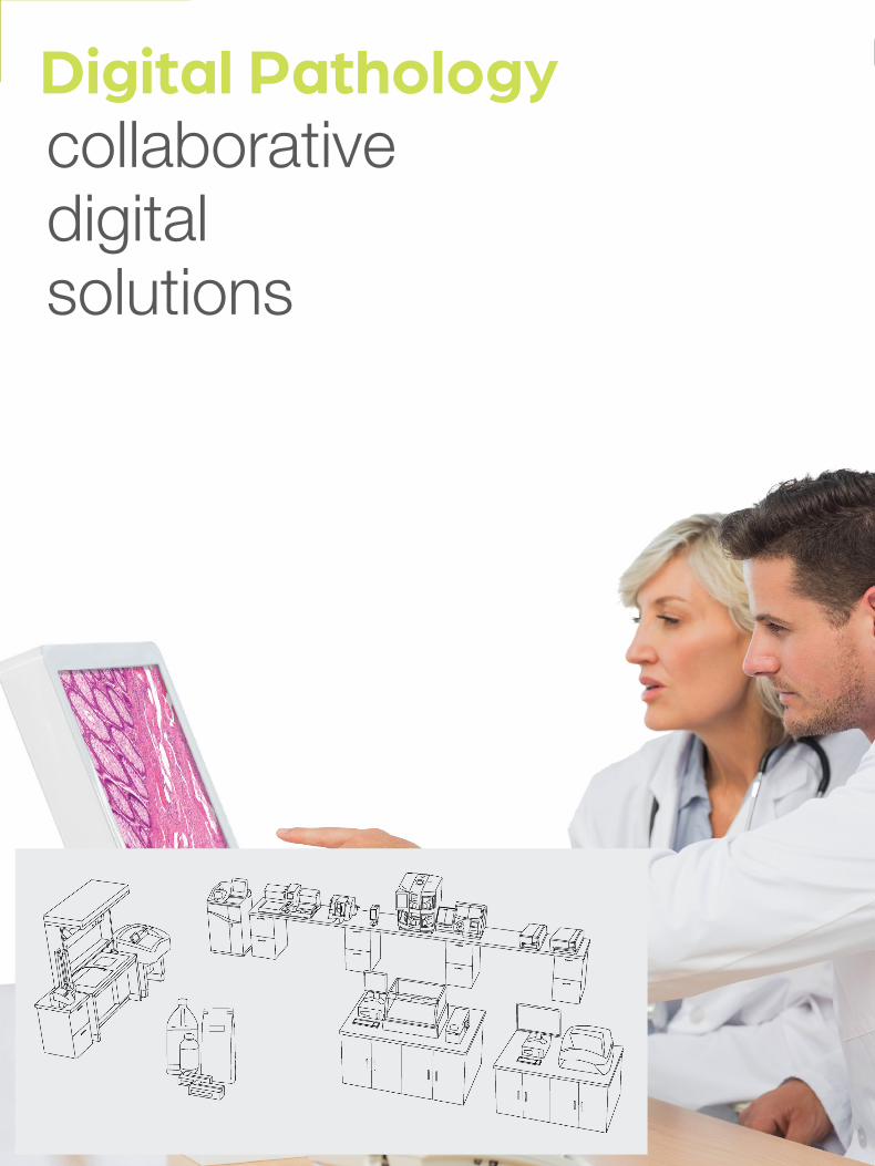
Digital Pathologycollaborativedigitalsolutions
Contact Us | www.epredia.com 205
Digital Pathology
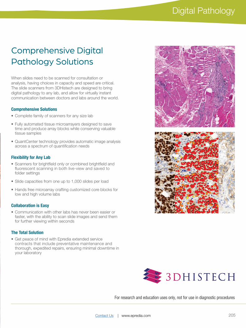
Comprehensive Digital Pathology Solutions
When slides need to be scanned for consultation or analysis, having choices in capacity and speed are critical. The slide scanners from 3DHistech are designed to bring digital pathology to any lab, and allow for virtually instant communication between doctors and labs around the world.
Comprehensive Solutions• Complete family of scanners for any size lab
• Fully automated tissue microarrayers designed to save time and produce array blocks while conserving valuable tissue samples
• QuantCenter technology provides automatic image analysis across a spectrum of quantification needs
Flexibility for Any Lab• Scanners for brightfield only or combined brightfield and
fluorescent scanning in both live-view and saved to folder settings
• Slide capacities from one up to 1,000 slides per load
• Hands free microarray crafting customized core blocks for low and high volume labs
Collaboration is Easy• Communication with other labs has never been easier or
faster, with the ability to scan slide images and send them for further viewing within seconds
The Total Solution• Get peace of mind with Epredia extended service
contracts that include preventative maintenance and thorough, expedited repairs, ensuring minimal downtime in your laboratory
For research and education uses only, not for use in diagnostic procedures
Contact Us | www.epredia.com206
Digital Pathology
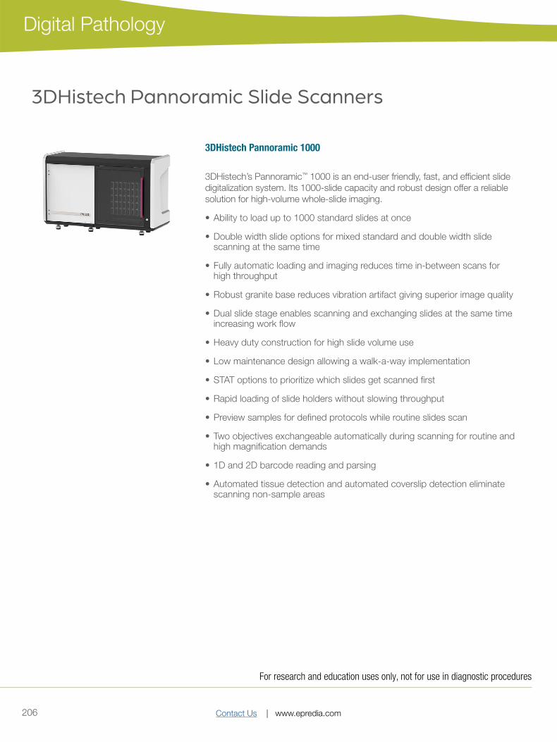
3DHistech Pannoramic 1000
3DHistech’s Pannoramic™ 1000 is an end-user friendly, fast, and efficient slide digitalization system. Its 1000-slide capacity and robust design offer a reliable solution for high-volume whole-slide imaging.
• Ability to load up to 1000 standard slides at once
• Double width slide options for mixed standard and double width slide scanning at the same time
• Fully automatic loading and imaging reduces time in-between scans for high throughput
• Robust granite base reduces vibration artifact giving superior image quality
• Dual slide stage enables scanning and exchanging slides at the same time increasing work flow
• Heavy duty construction for high slide volume use
• Low maintenance design allowing a walk-a-way implementation
• STAT options to prioritize which slides get scanned first
• Rapid loading of slide holders without slowing throughput
• Preview samples for defined protocols while routine slides scan
• Two objectives exchangeable automatically during scanning for routine and high magnification demands
• 1D and 2D barcode reading and parsing
• Automated tissue detection and automated coverslip detection eliminate scanning non-sample areas
3DHistech Pannoramic Slide Scanners
For research and education uses only, not for use in diagnostic procedures

Contact Us | www.epredia.com 207
Digital Pathology
3DHistech Pannoramic 250 FLASH III
3DHistech™ Pannoramic 250 Flash III, an all-in-one solution for digital pathology research and archiving. Enjoy increased speed and efficiency in routine digital pathology with 60 slides per hour.
• 250-slide capacity and continuous loading with vertical slide arrangement
• Award-winning, exceptional image quality for both brightfield and fluorescent scanning with up to nine single-band or multi-band filter slots with advanced FISH scanning techniques
• Pulsed Xenon FLASH light source for high-speed brightfield scanning
• Single or dual objective options for both low and high magnification needs up to 116x
• Darkfield preview for easy optimization of fluorescent samples
• Brightfield slide scanning in one minute at 40x resolution
• Motorized objective and camera changer
• Automatic slide loading, previewing, barcode reading and scanning
• All-round system for high-volume slide scanning in brightfield only or brightfield and fluorescent options
3DHistech Pannoramic SCAN II
Save time in routine pathology and enjoy brightfield only or brightfield and fluorescent scanning solutions in the same machine.
• 150-slide capacity and continuous loading with vertical slide arrangement
• Award-winning, exceptional image quality for both brightfield and fluorescent scanning with up to nine single-band or multi-band filter slots with advanced FISH scanning techniques
• Single or dual objective options for both low and high magnification needs up to 116x
• Motorized objective changer
• One high-quality monochrome camera is used for both brightfield and fluorescence with unique three-channel brightfield light source and Lumencor FL Light Engines
• Automatic slide loading, previewing, barcode reading and scanning
• All-around system for medium volume slide scanning
• Darkfield preview for easy optimization of fluorescent samples
For research and education uses only, not for use in diagnostic procedures
Contact Us | www.epredia.com208
Digital Pathology
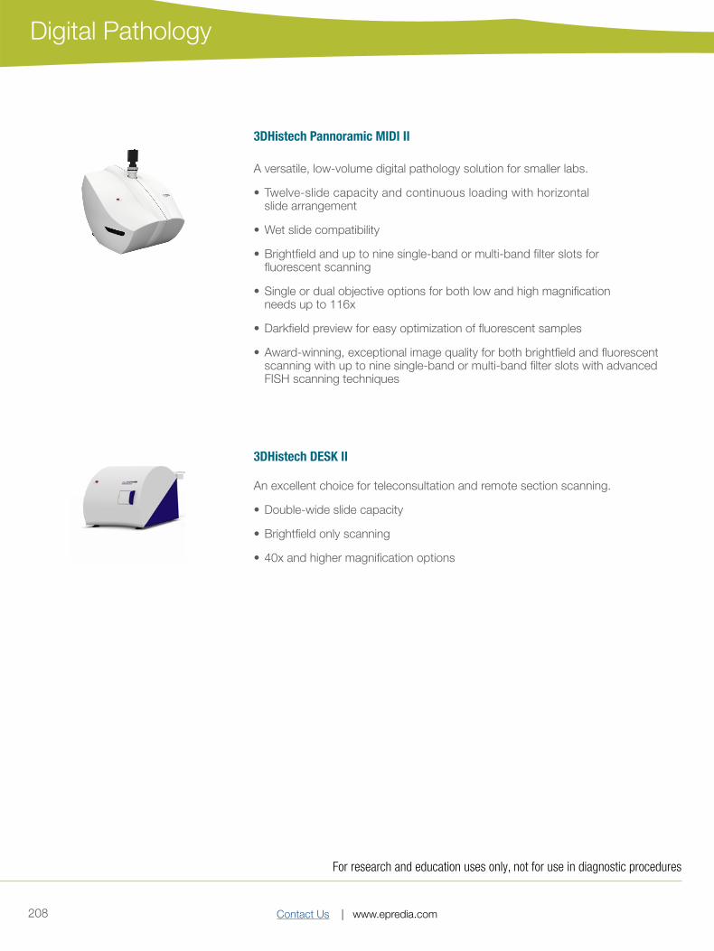
3DHistech Pannoramic MIDI II
A versatile, low-volume digital pathology solution for smaller labs.
• Twelve-slide capacity and continuous loading with horizontal slide arrangement
• Wet slide compatibility
• Brightfield and up to nine single-band or multi-band filter slots for fluorescent scanning
• Single or dual objective options for both low and high magnification needs up to 116x
• Darkfield preview for easy optimization of fluorescent samples
• Award-winning, exceptional image quality for both brightfield and fluorescent scanning with up to nine single-band or multi-band filter slots with advanced FISH scanning techniques
3DHistech DESK II
An excellent choice for teleconsultation and remote section scanning.
• Double-wide slide capacity
• Brightfield only scanning
• 40x and higher magnification options
For research and education uses only, not for use in diagnostic procedures
Contact Us | www.epredia.com 209
Digital Pathology
3DHistech Frozen Section Solutions
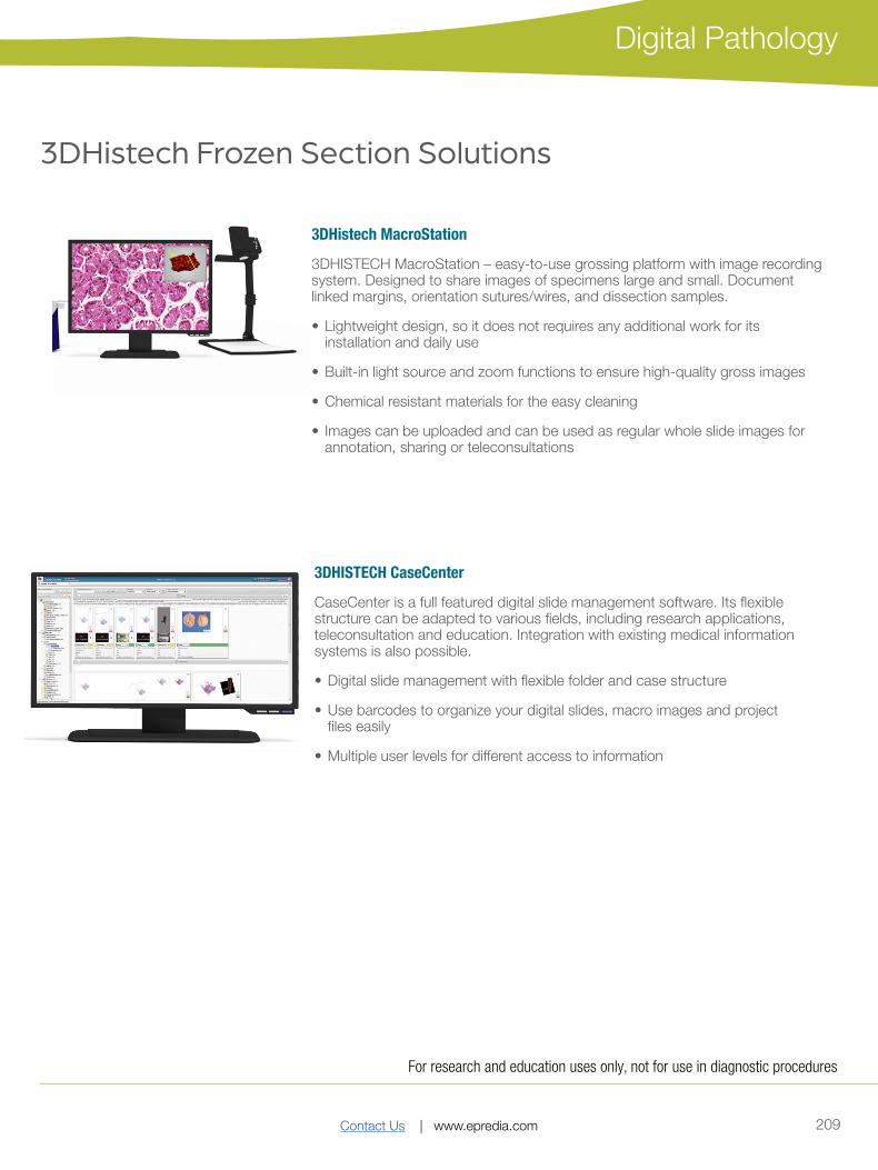
3DHistech MacroStation
3DHISTECH MacroStation – easy-to-use grossing platform with image recording system. Designed to share images of specimens large and small. Document linked margins, orientation sutures/wires, and dissection samples.
• Lightweight design, so it does not requires any additional work for its installation and daily use
• Built-in light source and zoom functions to ensure high-quality gross images
• Chemical resistant materials for the easy cleaning
• Images can be uploaded and can be used as regular whole slide images for annotation, sharing or teleconsultations
3DHISTECH CaseCenter
CaseCenter is a full featured digital slide management software. Its flexible structure can be adapted to various fields, including research applications, teleconsultation and education. Integration with existing medical information systems is also possible.
• Digital slide management with flexible folder and case structure
• Use barcodes to organize your digital slides, macro images and project files easily
• Multiple user levels for different access to information
For research and education uses only, not for use in diagnostic procedures

Contact Us | www.epredia.com210
Digital Pathology
Digital Image Analysis
QuantCenter is a powerful, automatic image analysis platform designed for digital whole slide quantification process.
Designed to fit seamlessly in the conventional microscopic investigation process, QuantCenter includes algorithms from tissue classification to cell-based FISH analysis that can be freely combined. It offers computer-aided image analysis allowing accurate, high-quality analytical results to be generated quickly.
The QuantCenter framework allows the connection of a variety of image analysis applications to generate a unique image analysis scenario. By using this feature, as the first step tissue classification modules can be applied
Molecular pathology
FISHQuant• A powerful cancer and cytogenetic application dedicated to quantify FISH (Fluorescence In Sytu Hybridization) signals on
tissue samples of solid tumor diseases like: breast and lung cancer, sarcomas, and lymphomas.
• This module is suitable for examination of hematologycal tumors, FISHQuant classifies the interphase and metaphase cells individually for a comprehensive evaluation.
CISHQuant• Quantify CISH (Chromogenic In Sytu Hybridization) stained samples. The algorithm can be calibrated to the stain protocoll
and quality by using an integrated color setting tool. This module is suitable for examining gene amplification, deletion and chromosome aberration.
CISH-RNAQuant• Detects RNA virus in virus-infected cell nuclei (Epstein-Barr vírus, HPV, HHV8).
• The application contains a color adjustment module which can be calibrated to the applied stain protocol and quality.
Histopathology Tissue classification
HistoQuant• A histological segmentation module which identifies tissue elements based on the color and intensity of the image pixels.
• This module could be run as a standalone application or could be combined with any of our IHC quantification modules for brightfield or fluorescence analysis.
PatternQuant • A trainable pattern recognition module for tissue classification, tissue pre-segmentation and identification of different
tissue structures.
• The machine-learning-based algorithm is able to classify different tissue types based on their texture pattern and color features.
For research and education uses only, not for use in diagnostic procedures

Contact Us | www.epredia.com 211
Digital Pathology
IHC quantification
NuclearQuant• A cell nuclei detection module designed for cell nuclei detection and quantification of IHC stained samples. The algorithm
can be calibrated to the stain quality (from multiple automated stainers or manual procedures) by using an integrated color setting tool.
MembraneQuant• A membrane detection software application can be used for IHC stained histological sample quantification. The algorithm
can be calibrated to the stain quality (from multiple automated stainers or manual procedures) by using an integrated color setting tool.
CellQuant • A cell detection application which is optimal for several IHC quantification.
• The application is adequate for cell nuclei, cytoplasmatic and membrane marker quantification. The software reports results based on dedicated scores and positivity ranges of cell nuclei, cytoplasm or membrane signals.
DensitoQuant• An easy to use, fast and accurate, stain-intensity-based IHC quantification tool.
• The application identifies the positive stain, based on an automatic color separation method through which individual positive pixels are counted and classified based on intensity and threshold ranges.
For research and education uses only, not for use in diagnostic procedures
Contact Us | www.epredia.com212
Digital Pathology
Whole-Slide Confocal Microscopy and 3D Histology
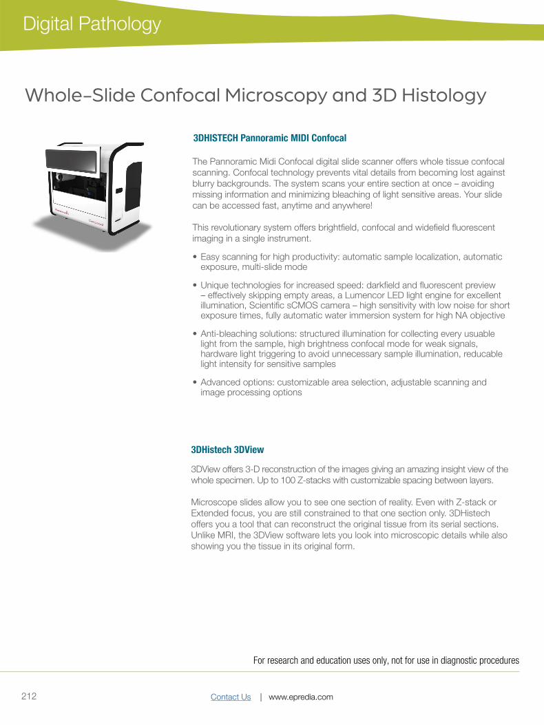
3DHISTECH Pannoramic MIDI Confocal
The Pannoramic Midi Confocal digital slide scanner offers whole tissue confocal scanning. Confocal technology prevents vital details from becoming lost against blurry backgrounds. The system scans your entire section at once – avoiding missing information and minimizing bleaching of light sensitive areas. Your slide can be accessed fast, anytime and anywhere!
This revolutionary system offers brightfield, confocal and widefield fluorescent imaging in a single instrument.
• Easy scanning for high productivity: automatic sample localization, automatic exposure, multi-slide mode
• Unique technologies for increased speed: darkfield and fluorescent preview – effectively skipping empty areas, a Lumencor LED light engine for excellent illumination, Scientific sCMOS camera – high sensitivity with low noise for short exposure times, fully automatic water immersion system for high NA objective
• Anti-bleaching solutions: structured illumination for collecting every usuable light from the sample, high brightness confocal mode for weak signals, hardware light triggering to avoid unnecessary sample illumination, reducable light intensity for sensitive samples
• Advanced options: customizable area selection, adjustable scanning and image processing options
3DHistech 3DView
3DView offers 3-D reconstruction of the images giving an amazing insight view of the whole specimen. Up to 100 Z-stacks with customizable spacing between layers.
Microscope slides allow you to see one section of reality. Even with Z-stack or Extended focus, you are still constrained to that one section only. 3DHistech offers you a tool that can reconstruct the original tissue from its serial sections. Unlike MRI, the 3DView software lets you look into microscopic details while also showing you the tissue in its original form.
For research and education uses only, not for use in diagnostic procedures
Contact Us | www.epredia.com 213
Digital Pathology
For research and education uses only, not for use in diagnostic procedures
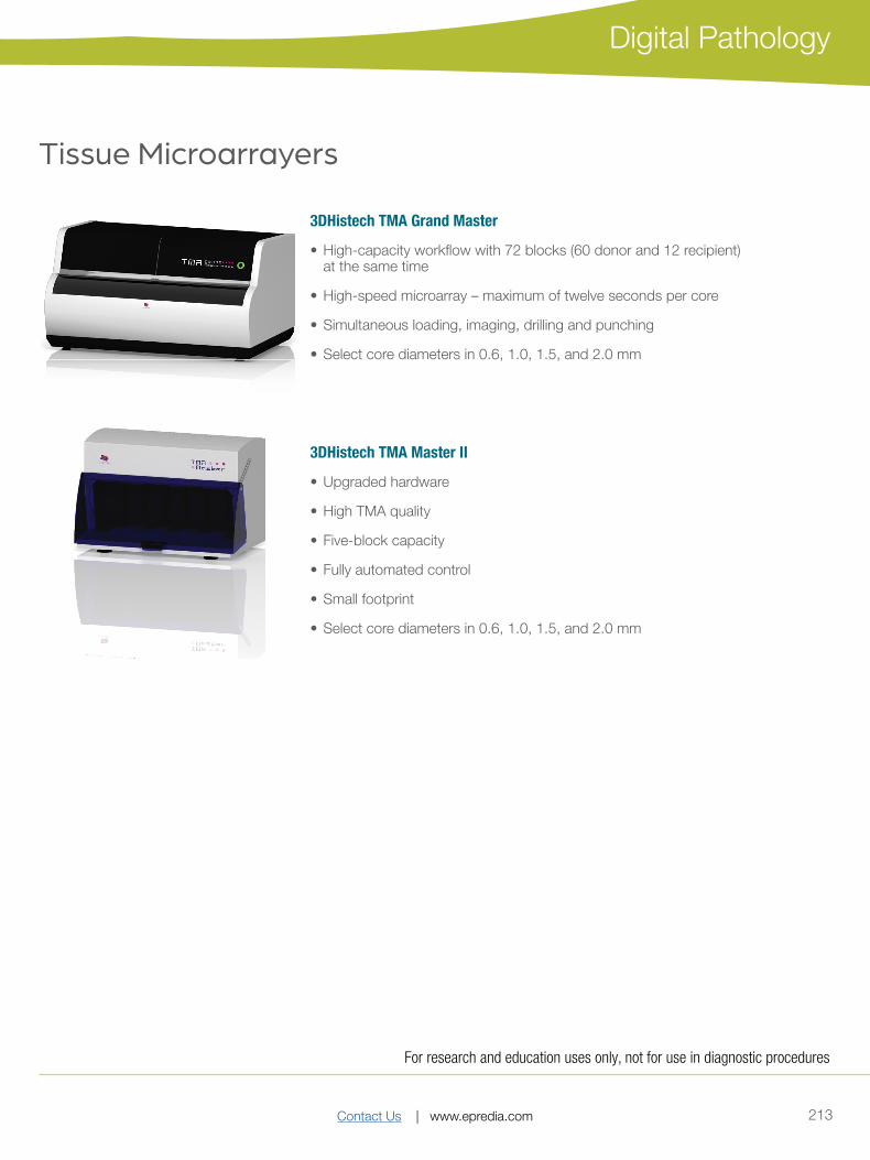
3DHistech TMA Grand Master
• High-capacity workflow with 72 blocks (60 donor and 12 recipient) at the same time
• High-speed microarray – maximum of twelve seconds per core
• Simultaneous loading, imaging, drilling and punching
• Select core diameters in 0.6, 1.0, 1.5, and 2.0 mm
Tissue Microarrayers
3DHistech TMA Master II
• Upgraded hardware
• High TMA quality
• Five-block capacity
• Fully automated control
• Small footprint
• Select core diameters in 0.6, 1.0, 1.5, and 2.0 mm
Contact Us | www.epredia.com214
Digital Pathology
3DHistech TMA Control Software
An easy-to-use solution for TMA block design and creation.
• Project based workflow
• Recipient block layout designer
• Ability to import donor block ID and additional sample data from Excel file
• Barcode-based donor block identification
• Automated digital slide search from CaseCenter or local drive
• Automated digital slide overlay with TMA markers from viewer
• Ability to place tissue cores in a clean cuvette tube
• Customizable export tool: export TMA data with donor block images
3DHistech TMA Module• For high-throughput tissue microarray analysis
• Project based: multi-user, multi-slide
• Flexible gallery
• Works with Excel™ database created by the TMA Master or the TMA Grand Master
For research and education uses only, not for use in diagnostic procedures

Contact Us | www.epredia.com 215
Digital Pathology
For research and education uses only, not for use in diagnostic procedures