Differential Expression of IL-17RC Isoforms in Androgen-Dependentand Androgen-Independent Prostate Cancers1
Zongbing You*, Ying Dong*, Xiangtian Kong y, Yi Zhang*, Robert L. Vessella z and Jonathan Melamed y
*The Lawrence Ellison Center for Tissue Regeneration and Repair, Department of Orthopedic Surgery, School ofMedicine, University of California-Davis, Sacramento, CA 95817, USA; yDepartment of Pathology, New YorkUniversity School of Medicine, New York, NY 10016, USA; zGenitourinary Cancer Research Laboratories,Department of Urology, University of Washington Medical Center, Seattle, WA 98195, USA
Abstract
IL-17RC (interleukin-17 receptor–like) gene codes for
a transmembrane protein, the full length of which
inhibits apoptosis in prostate cancer cells. IL-17RC
gene transcribes over a dozen different splice variants
of mRNA. However, it is not known whether there are
relevant protein isoforms. Here we report that different
IL-17RC protein isoforms were detected by two differ-
ent antibodies. The isoformas detected by anti–IL-17RC
intracellular domainantibodies (anti-ICD)wasexpressed
at higher levels in androgen-independent prostate
cancer cell lines (PC3 and DU145) than in androgen-
dependent prostatic cell lines (RWPE-1, pRNS-1-1, MLC-
SV40, and LNCaP). In contrast, several isoforms as
detected by anti–IL-17RC extracellular domain anti-
bodies (anti-ECD) were expressed at significantly higher
levels in androgen-dependent prostatic cell lines than
in androgen-independent ones. Furthermore, immuno-
histochemical staining of prostate tissue microarrays
showed that IL-17RC protein expression was signifi-
cantly higher in androgen-independent prostate cancers
than in androgen-dependent ones when anti-ICD was
used, whereas the trend was reversed using anti-ECD.
These observations provide evidence that IL-17RC pro-
tein isoforms are differentially expressed in prostatic
cells and cancer tissues and may play a negative or
positive role in the initiation and progression of pros-
tate cancer.
Neoplasia (2007) 9, 464–470
Keywords: Interleukin-17RC, protein isoforms, immunohistochemistry,tissue microarray, prostate cancer.
Introduction
Prostate cancer is the most common malignant disease
among men and the third most common cause of cancer-
related death in the United States [1]. Its is initially androgen-
dependent (androgen-naı̈ve) but progressively becomes
androgen-independent (androgen refractory), and patients
finally succumb to widespread metastases particularly to
the bone [2,3]. Potential molecular mechanisms underlying
prostate cancer progression involve mutations/amplifications
of androgen receptor or its signaling pathways, neuroendocrine
differentiation, and alterations of apoptosis-related genes [4–6].
The IL-17RC (previously named IL-17RL or interleukin-17
receptor– like) gene is a member of the interleukin-17 family of
cytokines and receptors [7,8]. IL-17RC was identified as a
single-pass transmembrane protein with 22% identity to the
interleukin-17A receptor (IL-17RA) [8]. The full-length IL-17RC
mRNA transcript includes 19 exons, whereas there are more
than a dozen different splice variants that alternatively splice
out a single or several exons [9]. Some of the splice variants
are predicted to translate truncated soluble proteins lacking the
transmembrane and/or intracellular domain. However, it is not
clear whether IL-17RC protein isoforms exist under physiologi-
cal or pathological conditions.
We have previously demonstrated that the full-length
IL-17RC protein inhibits tumor necrosis factor a–induced apop-
tosis in a human prostate cancer cell line, LNCaP. Using rabbit
anti– IL-17RC intracellular domain antibodies (anti-ICD), IL-
17RC protein was detected at increased levels in several
androgen-independent prostate cancer cell lines compared to
two androgen-dependent prostate cell lines [10]. However, due
to small sample size (a total of 14 samples), we did not find a
statistically significant difference between androgen-dependent
and androgen-independent prostate cancers [10]. In this study,
we performed immunohistochemical staining on prostate tis-
sue microarrays that contained over a hundred samples of
androgen-dependent/androgen-independent prostate cancers.
We found that IL-17RC protein expression, as detected by anti-
ICD, is significantly increased in androgen-independent pros-
tate cancer compared to androgen-dependent prostate cancer
Abbreviations: anti-ECD, anti – IL-17RC extracellular domain antibodies; anti-ICD, anti – IL-
17RC intracellular domain antibodies; GAPDH, glyceraldehyde-3-phosphate dehydrogenase;
IL-17RA, interleukin-17A receptor; IL-17RB, IL-17B receptor or IL-17 receptor homologue 1;
IL-17RC (IL-17RL), interleukin-17 receptor – like
Address all correspondence to: Zongbing You, The Lawrence Ellison Center for Tissue
Regeneration and Repair, Department of Orthopedic Surgery, School of Medicine, University
of California-Davis, 4635 Second Avenue, Room 2000, Sacramento, CA 95817.
E-mail: [email protected] work was supported, in part, by a grant from the US Army Medical Research and
Material Command, Department of Defense (W81XWH-05-1-0567).
Received 4 January 2007; Revised 26 April 2007; Accepted 27 April 2007.
Copyright D 2007 Neoplasia Press, Inc. All rights reserved 1522-8002/07/$25.00
DOI 10.1593/neo.07109
Neoplasia . Vol. 9, No. 6, June 2007, pp. 464–470 464
www.neoplasia.com
BRIEF ARTICLE
(P < .05). Furthermore, we found that IL-17RC protein ex-
pression, as detected by goat anti–IL-17RC extracellular
domain antibodies (anti-ECD), was significantly decreased
in androgen-independent prostate cancer compared to
androgen-dependent prostate cancer (P < .01). These results
suggest that IL-17RC protein isoforms are differentially ex-
pressed in prostate cancer and might play different roles in
the progression of prostate cancer and its transformation to
androgen independence.
Materials and Methods
Antibodies and Reagents
Rabbit anti-ICD that recognize an intracellular domain
(DSYFHPPGTPAPGR) of IL-17RC protein were affinity-
purified [8]. Goat anti-ECD were generated using the extra-
cellular domain of human IL-17RC isoform 3 (exon 7 was
spliced out) as immunogen (catalog no. AF2269; R&D Sys-
tems, Inc., Minneapolis, MN). Mouse anti-polyHis antibodies
were obtained from Sigma-Aldrich (St. Louis, MO). Goat anti-
actin antibodies and horseradish peroxidase–conjugated
anti-goat IgG antibodies were from Santa Cruz Biotechnology
(Santa Cruz, CA). Mouse anti–glyceraldehyde-3-phosphate
dehydrogenase (GAPDH) antibodies were obtained from
IMGENEX (San Diego, CA). Horseradish peroxidase–
conjugated anti-mouse IgG antibodies and Precision Plus
Protein Dual Color Standards were from Bio-Rad Labo-
ratories, Inc. (Hercules, CA). Horseradish peroxidase–
conjugated anti-rabbit IgG antibodies were from Promega
(Madison, WI). Enhanced chemiluminescence reagent
(SuperSignal West Pico Chemiluminescent Substrate) was
from Pierce Biotechnology, Inc. (Rockford, IL). VECTSTAIN
Elite ABC Reagent and DAB Substrate Kit were from Vector
Laboratories (Burlingame, CA). Recombinant human full-
length IL-17RC protein was purified as a 6-His–tagged pro-
tein from 293RC cells in our laboratory, and protein identity
was confirmed by protein sequencing.
Cell Culture
Human prostate cancer cell lines LNCaP (androgen-
dependent), PC3, and DU145 (androgen-independent), and
human embryo kidney 293 cells were from the American
Type Culture Collection (Manassas, VA). LNCaP cells were
maintained in T medium (custom formula 02-0056) with 5%
fetal bovine serum (FBS). PC3 cells were maintained in
Ham’s F12Kmedium with 10% FBS. DU145 cells were main-
tained in Earle’s minimal essential medium with 10% FBS.
293C (control cells; IL-17RC expression not detectable) and
293RC (overexpression of His-tagged human full-length
IL-17RC) cells were derived from 293 cells and maintained
in Dulbecco’s modified Eagle’s medium with 10% FBS [10].
MLC-SV40 (an immortalized human prostatic epithelial cell
line; androgen-dependent) cells [11] were maintained in
PrEGM medium (Cambrex, Walkersville, MD). RWPE-1
[12] and pRNS-1-1 [11] cells (immortalized human prostatic
epithelial cell lines; androgen-dependent) were maintained
in keratinocyte serum-free medium. Media and supplements
were from Invitrogen (Carlsbad, CA), unless noted otherwise.
The cells were cultured in a 5% CO2 humidified incubator
at 37jC.
Generation of Plasmid Constructs and Transfection
of 293 Cells
Human IL-17RC cDNA isoforms with in-frame deletion
of exon 7 or exons 4 and 7 were subcloned into pcDNA6/
V5-His vector (Invitrogen). V5-His–tagged cDNA were sub-
cloned into pLNCX2 retrovirus vector (Clontech, Palo Alto,
CA) by polymerase chain reaction (PCR) and generated
pLNCX2-IL-17RC-7-V5-His and pLNCX2-IL-17RC-4/7-V5-
His constructs. The insert sequences of all constructs were
confirmed by DNA sequencing (Davis Sequencing, Inc.,
Davis, CA). Detailed maps and sequences are available
on request. pLNCX2-IL-17B receptor (IL-17RB)-V5-His con-
struct has been described [10]. pCDNA3.1-IL-17RA-myc-
His construct was a gift from Dr. Reen Wu (University of
California-Davis). 293 cells were transfected with the above
constructsorwithempty vector byLipofectamine (Invitrogen)–
mediated transfection. For transient expression, the cells were
harvested 48 hours after transfection.
Western Blot Analysis
Proteins were extracted from cultured cells in RIPA lysis
buffer [50 mM sodium fluoride, 0.5% Igepal CA-630 (NP-40),
10 mM sodium phosphate, 150 mM sodium chloride, 25 mM
Tris pH 8.0, 1 mM phenylmethylsulfonyl fluoride, 2 mM
ethylenediaminetetraacetic acid, and 1.2 mM sodium vana-
date] supplemented with protease inhibitor cocktail (Sigma-
Aldrich). An equal amount of proteins was subjected to 10%
sodium dodecyl sulfate (SDS)–polyacrylamide gel electro-
phoresis and transferred to polyvinylidene difluoride mem-
brane. Themembranes were blocked with 5% nonfat dry milk
in TBST (25 mM Tris–HCl, 125 mM NaCl, and 0.1% Tween-
20) for 2 hours and probed with indicated primary antibodies
overnight and horseradish peroxidase–conjugated sec-
ondary antibodies for 1 hour. The results were visualized
by enhanced chemiluminescence reagent according to the
manufacturer’s instructions [13]. For loading control, the
membranes were stripped and probed for actin or GAPDH.
A piece of gel with purified recombinant IL-17RC was stained
with 0.5% Coomassie blue to show the full-length IL-17RC.
Analysis of IL-17RC mRNA Expression by Real-Time
Quantitative Reverse Transcription (RT) PCR
Total RNA was extracted from cells using RNeasy Mini Kit
(Qiagen, Valencia, CA) with on-membrane DNase I digestion
to avoid genomic DNA contamination. cDNA was made from
total RNA using Superscript First-Strand Synthesis System
with oligo dT primers (Invitrogen). Human GAPDH primers
were obtained from Applied Biosystems (Foster City, CA).
PCR primers specific for each interexon region and within
exon 19 (the longest exon of IL-17RC; therefore, two primer
sets were used) are shown in Table 1. Real-time quantitative
PCR was performed in triplicate with an ABI 7700 Sequence
Detector and SYBR-Green reagent (Applied Biosystems) fol-
lowing the recommended protocols. Results were normalized
IL-17RC Isoforms in Prostate Cancers You et al. 465
Neoplasia . Vol. 9, No. 6, 2007

to GAPDH levels using the formula: DCt (cycle threshold) = Ct
of IL-17RC � Ct of GAPDH. We arbitrarily chose the IL-17RC
mRNA level of RWPE-1 cells detected by the primer set
specific for exons 5 and 6 as baseline; therefore, DDCt was
calculated using the formula: DDCt = DCt of any group � DCt
of baseline. The fold change of IL-17RC mRNA level was
calculated as: fold = 2DDCt.
Immunohistochemical Staining
Human prostate tissue microarray slides were provided by
the Cooperative Prostate Cancer Tissue Resource, and hu-
man normal tissue microarray slides (version CHTN2002N1)
were provided by the Cooperative Human Tissue Network.
Both resources are funded by the National Cancer Insti-
tute. The use of these archival tissues was approved by
the University of California-Davis Institution Review Board.
Each prostate tissue microarray slide contained 58 cases of
androgen-dependent primary prostate cancers and 58 cases
of androgen-independent primary prostate cancers. Tumor
tissues were collected from specimens obtained through
either radical prostatectomy or transurethral resection of
the prostate. The determination of androgen-dependent or
androgen-independent status was as follows: 1) patients who
did not receive hormonal therapy before the collection of
tumor tissues were considered as androgen-dependent; and
(2) patients who had earlier undergone surgical orchiectomy
or medical hormone-suppressive therapy at least 6 months
before the collection of tumor tissues were considered as
androgen-independent. A normal tissue microarray slide con-
tains 66 types of tissues.
Tissue microarray slides were stained with 7.5 mg/ml anti-
ICD and 0.25 mg/ml anti-ECD against IL-17RC, using the
VECTSTAIN Elite ABC Reagent and DAB Substrate Kit
according to the manufacturer’s protocol [10,13,14]. The
stained slides were assessed independently by two pathol-
ogists (X.K. and J.M.), and a consensus of grading was
reached for prostate tissue microarrays. We adopted a two-
score system based on a proportion score and an intensity
score, as described by Allred et al. [15]. The proportion score
represented the estimated fraction of positive staining: 0 =
none; 1 = less than one hundredth; 2 = one hundredth to one
tenth; 3 = one tenth to one third; 4 = one third to two thirds; 5 =
greater than two thirds. The intensity score represented the
estimated average staining intensity of positive staining: 0 =
none; 1 = weak; 2 = intermediate; 3 = strong. The overall
amount of IL-17RC protein present in each tumor was then
expressed as the sum (Allred score) of the proportion and
intensity scores for negative and positive tumors (ranges, 0
and 2–8, respectively). Representative photomicrographs
of tumors with different Allred scores were captured under a
microscope with a digital camera.
Statistical Analysis
The difference in Allred scores between androgen-
dependent prostate cancers and androgen-independent
prostate cancers was analyzed by Mann-Whitney U test
when anti-ECD were used for immunohistochemical stain-
ing. Because the majority of prostate cancers were nega-
tive when anti-ICD were used for immunohistochemical
staining and thus generated many numbers with identical
rank (Allred score = 0), chi-square test was used for this set
of data (negative versus positive). Student’s t test was used
to analyze the average fold changes of IL-17RC mRNA
levels as detected by different primer sets. P < .05 was
considered statistically significant.
Results
Validation of Antibodies
Anti-ICD against IL-17RC were generated in our labora-
tory and well characterized previously [8–10]. The commer-
cial anti-ECD against IL-17RC were generated using the
extracellular domain of human IL-17RC isoform 3 (in-frame
deletion of exon 7) as immunogen. To test if anti-ECD
recognized human IL-17RC, we performed Western blot
analysis using proteins isolated from 293C (no detectable
IL-17RC) and 293RC (overexpressing IL-17RC) cells. As
shown in Figure 1A, anti-ECD recognized a protein band
(between 75 and 100 kDa) in 293RC cells but not in 293C
cells. This protein band was also recognized by anti-ICD
(Figure 1A) and corresponded to the purified full-length
IL-17RC (Figure 1A). Because IL-17RC shares about 20%
homology with IL-17RA and IL-17RB [7], it is possible that
antibodies against IL-17RC may cross-react with IL-17RA or
IL-17RB. To test this possibility, His-tagged IL-17RA or
IL-17RB was transiently expressed in 293 cells (293RA
and 293RB, respectively) and detected with anti-polyHis
Table 1. Sequences of PCR Primers Used for qRT-PCR of IL-17RC.
Domain Exon Order Sequence (5V to 3V)
Extracellular 5–6 Forward CTCCTTCCAGGCCTACCCTACT
Reverse GTCATATACCACAGAGCCCACAGA
Junction of extracellular domain 16–17 Forward CTATGGGACGATGACTTGGGAG
and transmembrane Reverse AGCGCAGCGGCAAAGAGTA
Junction of intracellular domain 17–19 Forward CGCTTTCCCTCATCCTCCTT
and transmembrane Reverse CGCTCGAAACCCGAGTCAT
Intracellular domain 19-1 Forward CCGGGCGGCTCCA
Reverse CCAGGGCTGGCTGAAGG
19-2 Forward CGTGGTGGTCTTGCTCTTCTCT
Reverse CATCCTGTAGCCACTCGCTG
Exon 17 encodes the transmembrane domain of IL-17RC.
466 IL-17RC Isoforms in Prostate Cancers You et al.
Neoplasia . Vol. 9, No. 6, 2007
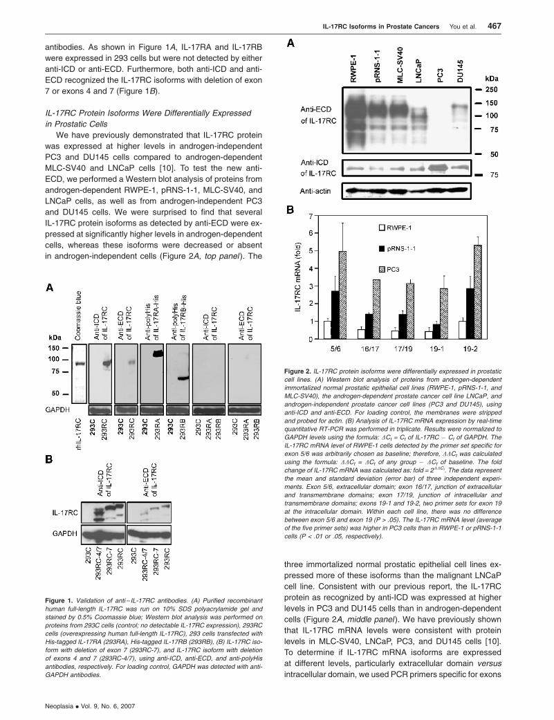
antibodies. As shown in Figure 1A, IL-17RA and IL-17RB
were expressed in 293 cells but were not detected by either
anti-ICD or anti-ECD. Furthermore, both anti-ICD and anti-
ECD recognized the IL-17RC isoforms with deletion of exon
7 or exons 4 and 7 (Figure 1B).
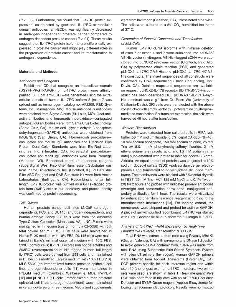
IL-17RC Protein Isoforms Were Differentially Expressed
in Prostatic Cells
We have previously demonstrated that IL-17RC protein
was expressed at higher levels in androgen-independent
PC3 and DU145 cells compared to androgen-dependent
MLC-SV40 and LNCaP cells [10]. To test the new anti-
ECD, we performed a Western blot analysis of proteins from
androgen-dependent RWPE-1, pRNS-1-1, MLC-SV40, and
LNCaP cells, as well as from androgen-independent PC3
and DU145 cells. We were surprised to find that several
IL-17RC protein isoforms as detected by anti-ECD were ex-
pressed at significantly higher levels in androgen-dependent
cells, whereas these isoforms were decreased or absent
in androgen-independent cells (Figure 2A, top panel ). The
three immortalized normal prostatic epithelial cell lines ex-
pressed more of these isoforms than the malignant LNCaP
cell line. Consistent with our previous report, the IL-17RC
protein as recognized by anti-ICD was expressed at higher
levels in PC3 and DU145 cells than in androgen-dependent
cells (Figure 2A, middle panel ). We have previously shown
that IL-17RC mRNA levels were consistent with protein
levels in MLC-SV40, LNCaP, PC3, and DU145 cells [10].
To determine if IL-17RC mRNA isoforms are expressed
at different levels, particularly extracellular domain versus
intracellular domain, we used PCR primers specific for exons
Figure 1. Validation of anti – IL-17RC antibodies. (A) Purified recombinant
human full-length IL-17RC was run on 10% SDS polyacrylamide gel and
stained by 0.5% Coomassie blue; Western blot analysis was performed on
proteins from 293C cells (control; no detectable IL-17RC expression), 293RC
cells (overexpressing human full-length IL-17RC), 293 cells transfected with
His-tagged IL-17RA (293RA), His-tagged IL-17RB (293RB), (B) IL-17RC iso-
form with deletion of exon 7 (293RC-7), and IL-17RC isoform with deletion
of exons 4 and 7 (293RC-4/7), using anti-ICD, anti-ECD, and anti-polyHis
antibodies, respectively. For loading control, GAPDH was detected with anti-
GAPDH antibodies.
Figure 2. IL-17RC protein isoforms were differentially expressed in prostatic
cell lines. (A) Western blot analysis of proteins from androgen-dependent
immortalized normal prostatic epithelial cell lines (RWPE-1, pRNS-1-1, and
MLC-SV40), the androgen-dependent prostate cancer cell line LNCaP, and
androgen-independent prostate cancer cell lines (PC3 and DU145), using
anti-ICD and anti-ECD. For loading control, the membranes were stripped
and probed for actin. (B) Analysis of IL-17RC mRNA expression by real-time
quantitative RT-PCR was performed in triplicate. Results were normalized to
GAPDH levels using the formula: DCt = Ct of IL-17RC � Ct of GAPDH. The
IL-17RC mRNA level of RWPE-1 cells detected by the primer set specific for
exon 5/6 was arbitrarily chosen as baseline; therefore, DDCt was calculated
using the formula: DDCt = DCt of any group � DCt of baseline. The fold
change of IL-17RC mRNA was calculated as: fold = 2DDCt. The data represent
the mean and standard deviation (error bar) of three independent experi-
ments. Exon 5/6, extracellular domain; exon 16/17, junction of extracellular
and transmembrane domains; exon 17/19, junction of intracellular and
transmembrane domains; exons 19-1 and 19-2, two primer sets for exon 19
at the intracellular domain. Within each cell line, there was no difference
between exon 5/6 and exon 19 (P > .05). The IL-17RC mRNA level (average
of the five primer sets) was higher in PC3 cells than in RWPE-1 or pRNS-1-1
cells (P < .01 or .05, respectively).
IL-17RC Isoforms in Prostate Cancers You et al. 467
Neoplasia . Vol. 9, No. 6, 2007
5 and 6 (extracellular domain), exons 16 and 17 (junction of
extracellular domain and transmembrane domain), exons 17
and 19 (junction of intracellular domain and transmembrane
domain), and exon 19 (intracellular domain). As shown in
Figure 2B, in each of the RWPE-1, pRNS-1-1, and PC3 cells,
IL-17RC mRNA levels varied slightly when different primer
sets were used. Particularly, there was no significant differ-
ence between the extracellular domain and the intracellular
domain (P > .05). Of note, average IL-17RC mRNA levels
were significantly higher in PC3 cells than in RWPE-1 or
pRNS-1-1 cells (P < .01 or .05, respectively).
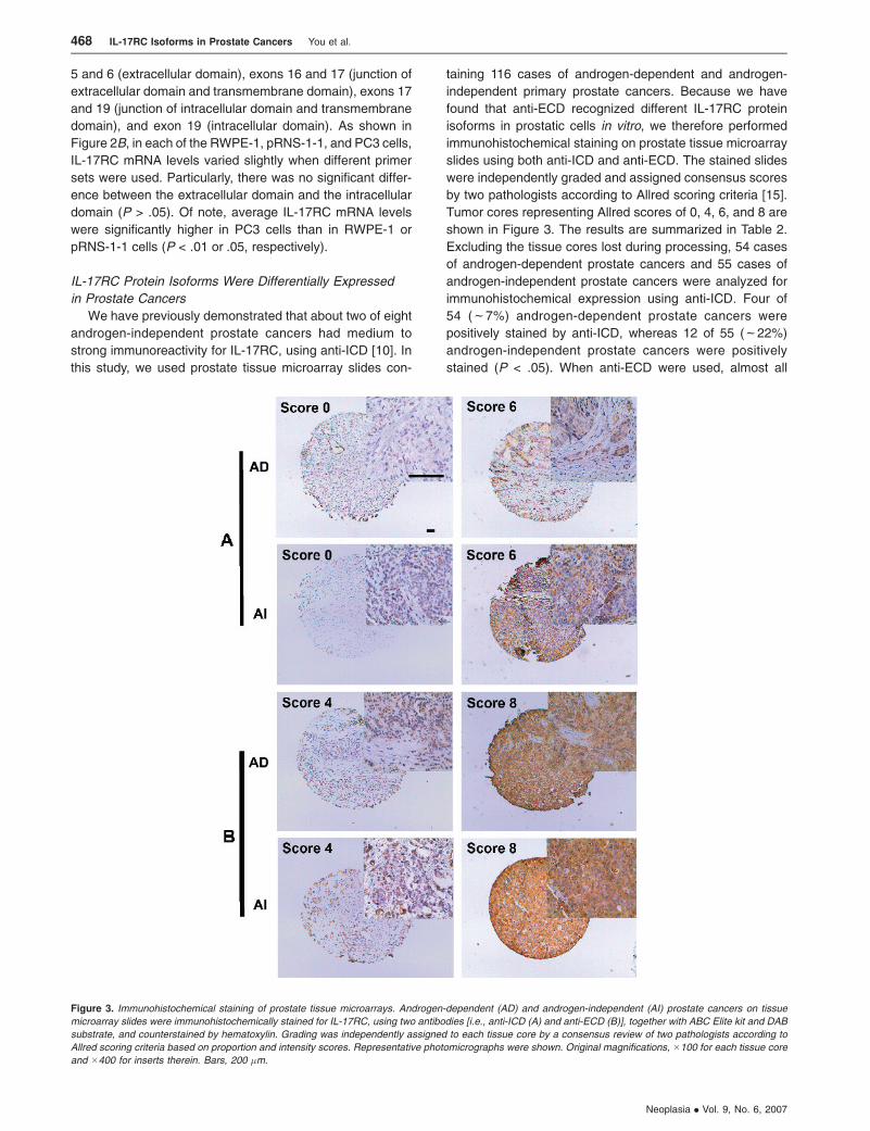
IL-17RC Protein Isoforms Were Differentially Expressed
in Prostate Cancers
We have previously demonstrated that about two of eight
androgen-independent prostate cancers had medium to
strong immunoreactivity for IL-17RC, using anti-ICD [10]. In
this study, we used prostate tissue microarray slides con-
taining 116 cases of androgen-dependent and androgen-
independent primary prostate cancers. Because we have
found that anti-ECD recognized different IL-17RC protein
isoforms in prostatic cells in vitro, we therefore performed
immunohistochemical staining on prostate tissue microarray
slides using both anti-ICD and anti-ECD. The stained slides
were independently graded and assigned consensus scores
by two pathologists according to Allred scoring criteria [15].
Tumor cores representing Allred scores of 0, 4, 6, and 8 are
shown in Figure 3. The results are summarized in Table 2.
Excluding the tissue cores lost during processing, 54 cases
of androgen-dependent prostate cancers and 55 cases of
androgen-independent prostate cancers were analyzed for
immunohistochemical expression using anti-ICD. Four of
54 (f7%) androgen-dependent prostate cancers were
positively stained by anti-ICD, whereas 12 of 55 (f22%)
androgen-independent prostate cancers were positively
stained (P < .05). When anti-ECD were used, almost all
Figure 3. Immunohistochemical staining of prostate tissue microarrays. Androgen-dependent (AD) and androgen-independent (AI) prostate cancers on tissue
microarray slides were immunohistochemically stained for IL-17RC, using two antibodies [i.e., anti-ICD (A) and anti-ECD (B)], together with ABC Elite kit and DAB
substrate, and counterstained by hematoxylin. Grading was independently assigned to each tissue core by a consensus review of two pathologists according to
Allred scoring criteria based on proportion and intensity scores. Representative photomicrographs were shown. Original magnifications, �100 for each tissue core
and �400 for inserts therein. Bars, 200 �m.
468 IL-17RC Isoforms in Prostate Cancers You et al.
Neoplasia . Vol. 9, No. 6, 2007
prostate cancers were positively stained, but the proportions
and intensities of staining were different. The median Allred
score of androgen-dependent prostate cancers was statisti-
cally significantly higher than that of androgen-independent
prostate cancers (P < .01).
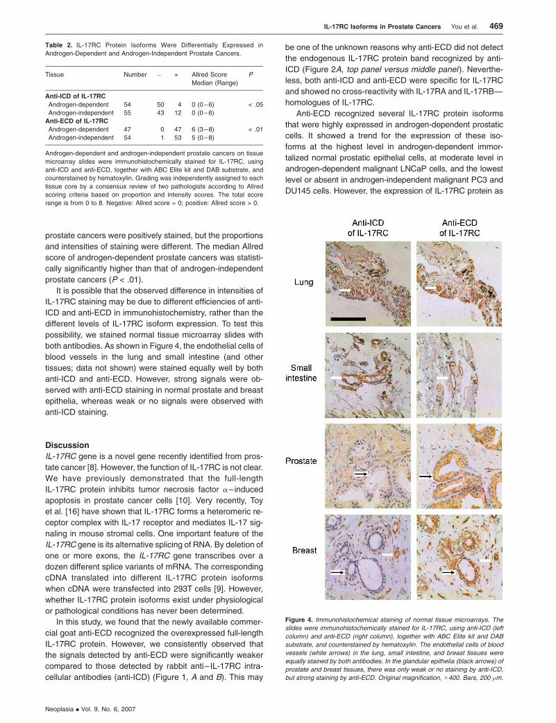
It is possible that the observed difference in intensities of
IL-17RC staining may be due to different efficiencies of anti-
ICD and anti-ECD in immunohistochemistry, rather than the
different levels of IL-17RC isoform expression. To test this
possibility, we stained normal tissue microarray slides with
both antibodies. As shown in Figure 4, the endothelial cells of
blood vessels in the lung and small intestine (and other
tissues; data not shown) were stained equally well by both
anti-ICD and anti-ECD. However, strong signals were ob-
served with anti-ECD staining in normal prostate and breast
epithelia, whereas weak or no signals were observed with
anti-ICD staining.
Discussion
IL-17RC gene is a novel gene recently identified from pros-
tate cancer [8]. However, the function of IL-17RC is not clear.
We have previously demonstrated that the full-length
IL-17RC protein inhibits tumor necrosis factor a–induced
apoptosis in prostate cancer cells [10]. Very recently, Toy
et al. [16] have shown that IL-17RC forms a heteromeric re-
ceptor complex with IL-17 receptor and mediates IL-17 sig-
naling in mouse stromal cells. One important feature of the
IL-17RC gene is its alternative splicing of RNA. By deletion of
one or more exons, the IL-17RC gene transcribes over a
dozen different splice variants of mRNA. The corresponding
cDNA translated into different IL-17RC protein isoforms
when cDNA were transfected into 293T cells [9]. However,
whether IL-17RC protein isoforms exist under physiological
or pathological conditions has never been determined.
In this study, we found that the newly available commer-
cial goat anti-ECD recognized the overexpressed full-length
IL-17RC protein. However, we consistently observed that
the signals detected by anti-ECD were significantly weaker
compared to those detected by rabbit anti– IL-17RC intra-
cellular antibodies (anti-ICD) (Figure 1, A and B). This may
be one of the unknown reasons why anti-ECD did not detect
the endogenous IL-17RC protein band recognized by anti-
ICD (Figure 2A, top panel versus middle panel ). Neverthe-
less, both anti-ICD and anti-ECD were specific for IL-17RC
and showed no cross-reactivity with IL-17RA and IL-17RB—
homologues of IL-17RC.
Anti-ECD recognized several IL-17RC protein isoforms
that were highly expressed in androgen-dependent prostatic
cells. It showed a trend for the expression of these iso-
forms at the highest level in androgen-dependent immor-
talized normal prostatic epithelial cells, at moderate level in
androgen-dependent malignant LNCaP cells, and the lowest
level or absent in androgen-independent malignant PC3 and
DU145 cells. However, the expression of IL-17RC protein as
Table 2. IL-17RC Protein Isoforms Were Differentially Expressed in
Androgen-Dependent and Androgen-Independent Prostate Cancers.
Tissue Number � + Allred Score
Median (Range)
P
Anti-ICD of IL-17RC
Androgen-dependent 54 50 4 0 (0–6) < .05
Androgen-independent 55 43 12 0 (0–6)
Anti-ECD of IL-17RC
Androgen-dependent 47 0 47 6 (3–8) < .01
Androgen-independent 54 1 53 5 (0–8)
Androgen-dependent and androgen-independent prostate cancers on tissue
microarray slides were immunohistochemically stained for IL-17RC, using
anti-ICD and anti-ECD, together with ABC Elite kit and DAB substrate, and
counterstained by hematoxylin. Grading was independently assigned to each
tissue core by a consensus review of two pathologists according to Allred
scoring criteria based on proportion and intensity scores. The total score
range is from 0 to 8. Negative: Allred score = 0; positive: Allred score > 0.
Figure 4. Immunohistochemical staining of normal tissue microarrays. The
slides were immunohistochemically stained for IL-17RC, using anti-ICD (left
column) and anti-ECD (right column), together with ABC Elite kit and DAB
substrate, and counterstained by hematoxylin. The endothelial cells of blood
vessels (white arrows) in the lung, small intestine, and breast tissues were
equally stained by both antibodies. In the glandular epithelia (black arrows) of
prostate and breast tissues, there was only weak or no staining by anti-ICD,
but strong staining by anti-ECD. Original magnification, �400. Bars, 200 �m.
IL-17RC Isoforms in Prostate Cancers You et al. 469
Neoplasia . Vol. 9, No. 6, 2007

recognized by anti-ICD showed an opposite trend. The true
nature of these different IL-17RC protein bands (full length
versus different isoforms) is yet to be determined. The band
(slightly larger than 75 kDa) recognized by anti-ICD must
contain the C-terminal intracellular domain of IL-17RC be-
cause the immunogen used to generate anti-ICD was a short
peptide of the C-terminal intracellular domain. Anti-ECD were
generated by using the extracellular domain of IL-17RC iso-
form with deletion of exon 7. It bound to IL-17RC isoform with
deletion of exon 7 and/or exon 4 (Figure 1B); therefore,
polyclonal anti-ECD may recognize epitopes out of exons 4
and 7. It is not known why anti-ECD recognized several
bands that were larger than the full-length IL-17RC over-
expressed in 293 cells. We speculate that it may be due to
glycosylation or other modifications of these isoforms, as nine
potential sites for N-linked glycosylation were found within the
extracellular domain [8].
The mechanisms underlying the different protein levels
of IL-17RC isoforms as detected by the two antibodies are
not clear. We performed real-time quantitative RT-PCR using
different primer sets specific for exons that are not often
spliced [8]. Within each cell line tested, there was a small
variation of IL-17RC mRNA levels among the five primer
sets, most likely due to different PCR efficiencies of the
primers. This was particularly true when the two primer sets
specific for a nonspliced region of exon 19 were compared
(Figure 2B; similar data not shown for MLC-SV40, LNCaP,
and DU145 cells). However, no difference in IL-17RC mRNA
levels was observed between the extracellular domain and
the intracellular domain. These data suggest that the differ-
ence at protein levels may be due to translational or post-
translational mechanisms, which are currently being studied
in our laboratory.
Furthermore, these in vitro findings were corroborated
by results of the immunohistochemical staining of prostate
cancer tissue microarrays. When anti-ICD were used,
IL-17RC protein expression was significantly higher in
androgen-independent prostate cancer than in androgen-
dependent cancer, whereas IL-17RC protein expression
detected by anti-ECD was significantly higher in androgen-
dependent prostate cancer than in androgen-independent
prostate cancer. It is unlikely that the difference in the inten-
sities of IL-17RC staining was due to different efficiencies of
anti-ICD and anti-ECD in immunohistochemistry because
both antibodies stained equally well in the endothelial cells
of blood vessels in the lung and small intestines, even within
the same breast tissue where the glandular epithelium was
negative by anti-ICD staining (Figure 4). Taken together, these
findings demonstrate, for the first time, differential expression
of IL-17RC protein isoforms in prostatic cell lines and cancer
tissues. Based on the trend for differential expression, it is
speculated that the IL-17RC protein isoform recognized by
anti-ICD may play a promoting role in the progression of
normal to malignant status and/or androgen-dependent to
androgen-independent status, whereas the IL-17RC protein
isoforms recognized by anti-ECDmay play an inhibitory role in
this process. Although the identities of these IL-17RC protein
isoforms are yet to be revealed in future studies, it is an
intriguing hypothesis that a single IL-17RC gene may posi-
tively or negatively regulate the initiation and progression of
prostate cancer through different alternatively expressing
protein isoforms.
Acknowledgements
The authors thank A. Hari Reddi (University of California-
Davis) for helpful suggestions and critical reading of the
manuscript; Xu-Bao Shi (University of California-Davis) for
providing the RWPE-1 and pRNS-1-1 cell lines; Johng S.
Rhim (Uniformed Services University of the Health Sciences)
who was the original source of MLC-SV40, RWPE-1, and
pRNS-1-1 cell lines; and Reen Wu (University of California-
Davis) for the pCDNA3.1-IL-17RA-myc-His construct.
References[1] Jemal A, Siegel R, Ward E, Murray T, Xu J, Smigal C, and Thun MJ
(2006). Cancer statistics, 2006. CA Cancer J Clin 56, 106–130.
[2] Chodak GW, Vogelzang NJ, Caplan RJ, Soloway M, and Smith JA
(1991). Independent prognostic factors in patients with metastatic
(stage D2) prostate cancer. The Zoladex Study Group. JAMA 265,
618–621.
[3] Roudier MP, True LD, Higano CS, Vesselle H, Ellis W, Lange P, and
Vessella RL (2003). Phenotypic heterogeneity of end-stage prostate
carcinoma metastatic to bone. Hum Pathol 34, 646–653.
[4] De La Taille A, Vacherot F, Salomon L, Druel C, Gil Diez De Medina S,
Abbou C, Buttyan R, and Chopin D (2001). Hormone-refractory prostate
cancer: a multi-step and multi-event process. Prostate Cancer Prostatic
Dis 4, 204–212.
[5] Craft N and Sawyers CL (1999). Mechanistic concepts in androgen-
dependence of prostate cancer. Cancer Metastasis Rev 17, 421–427.
[6] Hirano D, Okada Y, Minei S, Takimoto Y, and Nemoto N (2004). Neuro-
endocrine differentiation in hormone refractory prostate cancer following
androgen deprivation therapy. Eur Urol 45, 586–592.
[7] Moseley TA, Haudenschild DR, Rose L, and Reddi AH (2003). Interleukin-
17 family and IL-17 receptors. Cytokine Growth Factor Rev 14, 155–174.
[8] Haudenschild D, Moseley T, Rose L, and Reddi AH (2002). Soluble and
transmembrane isoforms of novel interleukin-17 receptor – like protein
by RNA splicing and expression in prostate cancer. J Biol Chem 277,
4309–4316.
[9] Haudenschild DR, Curtiss SB, Moseley TA, and Reddi AH (2006). Gen-
eration of interleukin-17 receptor – like protein (IL-17RL) in prostate by
alternative splicing of RNA. Prostate 66, 1268–1274.
[10] You Z, Shi XB, DuRaineG, Haudenschild D, Tepper CG, Lo SH, Gandour-
Edwards R, de Vere White RW, and Reddi AH (2006). Interleukin-17
receptor – like gene is a novel antiapoptotic gene highly expressed in
androgen-independent prostate cancer. Cancer Res 66, 175–183.
[11] Lee MS, Garkovenko E, Yun JS, Weijerman PC, Peehl DM, Chen LS,
and Rhim JS (1994). Characterization of adult human prostatic epi-
thelial cells immortalized by polybrene-induced DNA transfection with
a plasmid containing an origin-defective SV40 genome. Int J Oncol 4,
821–830.
[12] Bello D, Webber MM, Kleinman HK, Wartinger DD, and Rhim JS (1997).
Androgen responsive adult human prostatic epithelial cell lines immor-
talized by human papillomavirus 18. Carcinogenesis 18, 1215–1223.
[13] You Z, DuRaine G, Tien JY, Lee C, Moseley TA, and Reddi AH (2005).
Expression of interleukin-17B in mouse embryonic limb buds and reg-
ulation by BMP-7 and bFGF. Biochem Biophys Res Commun 326,
624–631.
[14] Haudenschild DR, Palmer SM, Moseley TA, You Z, and Reddi AH
(2004). Bone morphogenetic protein (BMP)-6 signaling and BMP an-
tagonist noggin in prostate cancer. Cancer Res 64, 8276–8284.
[15] Allred DC, Clark GM, Elledge R, Fuqua SA, Brown RW, Chamness GC,
Osborne CK, and McGuire WL (1993). Association of p53 protein ex-
pression with tumor cell proliferation rate and clinical outcome in node-
negative breast cancer. J Natl Cancer Inst 85, 200–206.
[16] Toy D, Kugler D, Wolfson M, Vanden Bos T, Gurgel J, Derry J, Tocker J,
and Peschon J (2006). Cutting edge: interleukin 17 signals through a
heteromeric receptor complex. J Immunol 177, 36–39.
470 IL-17RC Isoforms in Prostate Cancers You et al.
Neoplasia . Vol. 9, No. 6, 2007