“Did you see that?” …a case by case look at the pediatric eye exam
- Wade Mincher MD- Sangeeta Jain MD
*not ophthalmologists

TESTING INSTRUCTIONS
Go to website: b.socrative.com
Room name: TPSEYE

No financial disclosuresPolicies and standards of the Texas Medical Association, the Accreditation Council for Continuing Medical Education, and the American Medical Association require that speakers and planners for continuing medical education activities disclose any relevant financial relationships they may have with any entity producing, marketing, re-selling, or distributing health care goods or services consumed by, or used on, patients whose products, devices or services may be discussed in the content of the CME activity.
The planners and speakers have no relevant relationships to disclose.
We do not intend to discuss an unapproved/investigative use of a commercial product/device in our presentation.
Website: b.socrative.com Room Name: TPSEYE

Objectives•Review components of the pediatric eye exam using an interactive, case-based approach
•Discuss common ophthalmologic conditions and their presentation in both the inpatient and outpatient setting
•Highlight techniques for specific eye exam components including the “difficult” eye exam
Website: b.socrative.com Room Name: TPSEYE

�������“that pesky Red Reflex”

CaseDischarge newborn exam with assymetric RR,
unilaterally dull.
• Referred to ophthalmology• Diagnosis: ???

Abnormal RR = opacification1)Cornea
2)Lens• Cataract
3)Vitreous
4)Retina• Retinoblastoma

Cataracts1-6/10,000 Live Births
50-60% Idiopathic10-25% Genetic15-30% Other
Associated with:Infections: TORCHGenetic: Trisomy 13, 18, 21, TurnerMetabolic: galactossemiaEndocrine: hypoparathyroid, hypocalcemia, diabetesTraumaDrugs/toxins
Congenital Cataract management…
Treatment: • Surgical removal
• 4-6 weeks of age• Aphakia
IOL (intraocular lens) implantation• After 2 years of age
QUESTION: What is the most common late complication of cataract surgery?
• A) Retinal detachment• B) Glaucoma• C) Retinal hemorrhages• D) Endophthalmitis
Website: b.socrative.com Room Name: TPSEYE
A Needle in the haystack!Treatment is urgent!
• Early diagnosis is key!
If bilateral cataracts are not treated by 2 months of age:• Sensory (acquired) nystagmus• Bilateral poor visual acuity (<20/200)
Later signs/symptoms of undiagnosed mild cataracts:•Lack of reaction to light•Strabismus•Failure to notice toys and faces•Developmental delay•Photophobia in bright lights
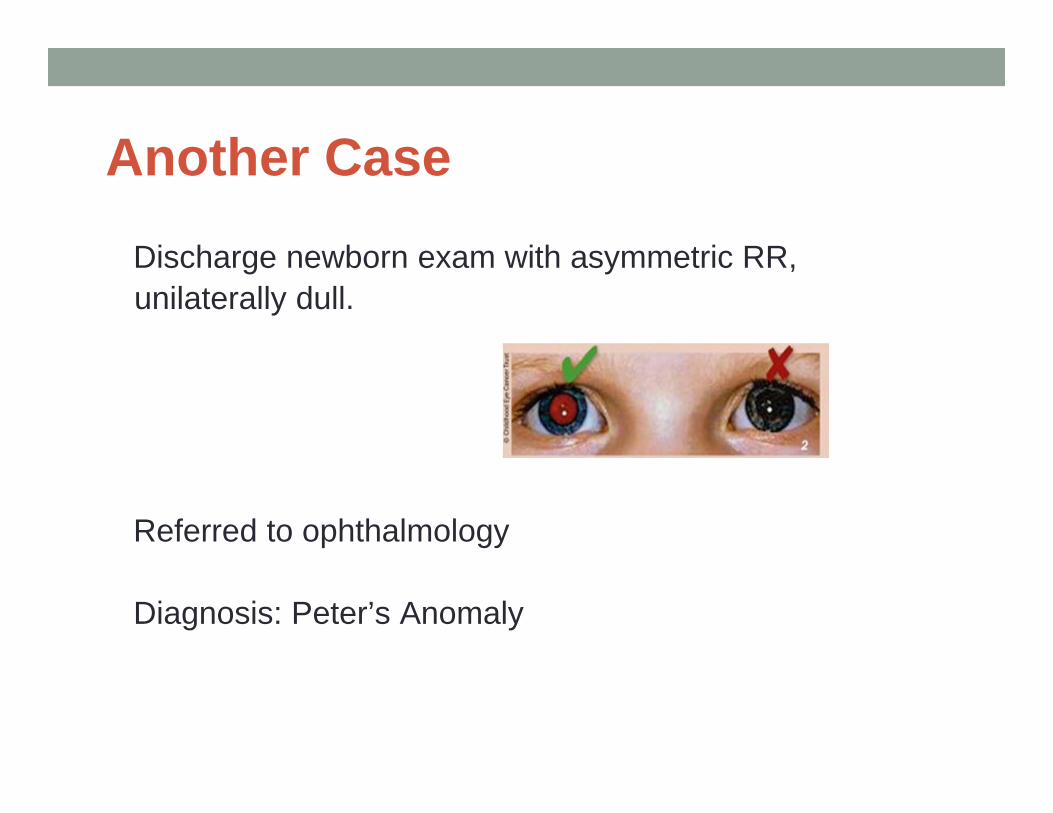
Another CaseDischarge newborn exam with asymmetric RR,unilaterally dull.
Referred to ophthalmology
Diagnosis: Peter’s Anomaly
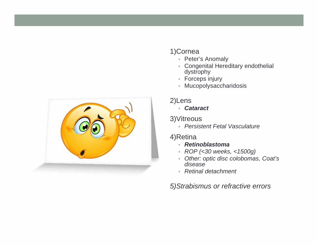
1)Cornea• Peter’s Anomaly• Congenital Hereditary endothelial
dystrophy• Forceps injury• Mucopolysaccharidosis
2)Lens• Cataract
3)Vitreous• Persistent Fetal Vasculature
4)Retina• Retinoblastoma• ROP (<30 weeks, <1500g)• Other: optic disc colobomas, Coat’s
disease• Retinal detachment
5)Strabismus or refractive errors

Peter’s Anomaly• Rare
• Dense opacity in center of cornea
• 50% develop glaucoma
• Longterm: corneal transplant
• Outcomes: “uniformly poor”
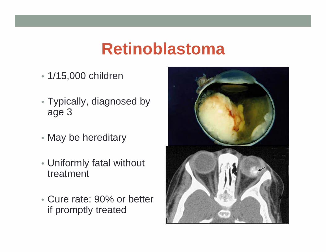
Retinoblastoma• 1/15,000 children
• Typically, diagnosed by age 3
• May be hereditary
• Uniformly fatal without treatment
• Cure rate: 90% or better if promptly treated

QUESTION: Aside from leukocoria, what is the second most common presenting sign for retinoblastoma?
• A) Eye tearing• B) Hyphema• C) Red, painful eye• D) Strabismus
Website: b.socrative.com Room Name: TPSEYE

Tips• Use a darkened room
• Set the ophthalmic lens power at “0” diopter
• Observe the eyes-both together (Bruckner test)
-individually*straight on*obliquely

QUESTION: At what distance should you examine the baby’s red reflex?
• A) 6-12 inches• B) 12-18 inches• C) 18-24 inches• D) 24-30 inches
Website: b.socrative.com Room Name: TPSEYE

Abnormal findings
1)absent, dulled, or asymmetric RR
2)white or opaque reflex
3)dark spots

Additional tips
• At minimum: Check the eyes at initial exam, discharge exam, newborn WCC, and every WCC thereafter!
• “Push the lids open to the edges of the orbital rim”
• Parents might bring you the diagnosis.
• Refer if unsure

QUESTION: Within what time frame should a baby with an abnormal red reflex be evaluated by an ophthalmologist?
• A) <1 week• B) < 2 weeks• C) < 1 month• D) whenever
Website: b.socrative.com Room Name: TPSEYE
Significant Family History
• Retinoblastoma• Infantile Cataracts• Congenital Glaucoma• Blindness from birth• Any other ocular disorders that presented early in life
•Refer to Ophthalmology

������“why do his eyes keep
crossing?”

CASE 12mo male presenting with concerns for “eye turning in”
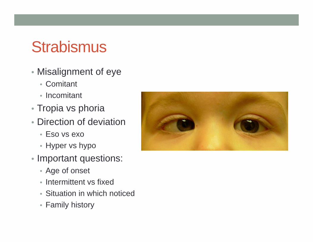
Strabismus• Misalignment of eye
• Comitant• Incomitant
• Tropia vs phoria• Direction of deviation
• Eso vs exo• Hyper vs hypo
• Important questions: • Age of onset• Intermittent vs fixed• Situation in which noticed• Family history

QUESTION: A large percentage of newborns have intermittent strabismus. What is the most common type and at what age should you expect it to resolve?
• A) Exotropic, 4mo• B) Esotropic, 6mo• C) Exotropic, 8mo• D) Esotropic, 2mo
Website: b.socrative.com Room Name: TPSEYE

Strabismus
• Exotropia• Intermittent
• Commonly occurs between 2-8 years of age
• Fatigue exacerbates• Best observed with the cover
test• Surgery for:
• Increasing exotropia• Present >50% of the time• Poor fusion control
• Convergence insufficiency
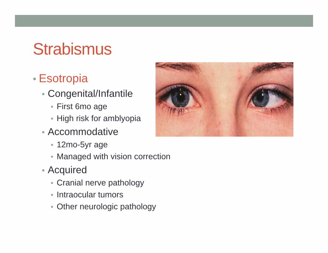
Strabismus
• Esotropia• Congenital/Infantile
• First 6mo age• High risk for amblyopia
• Accommodative• 12mo-5yr age• Managed with vision correction
• Acquired• Cranial nerve pathology• Intraocular tumors• Other neurologic pathology
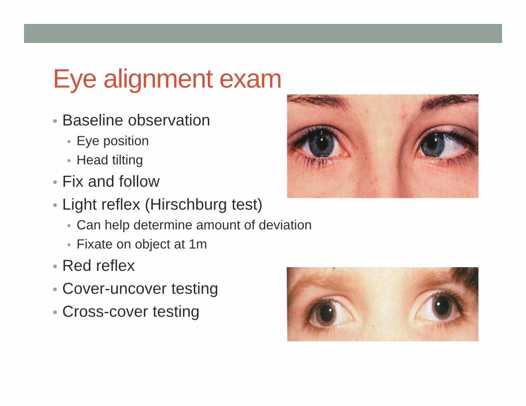
Eye alignment exam• Baseline observation
• Eye position• Head tilting
• Fix and follow• Light reflex (Hirschburg test)
• Can help determine amount of deviation• Fixate on object at 1m
• Red reflex• Cover-uncover testing• Cross-cover testing

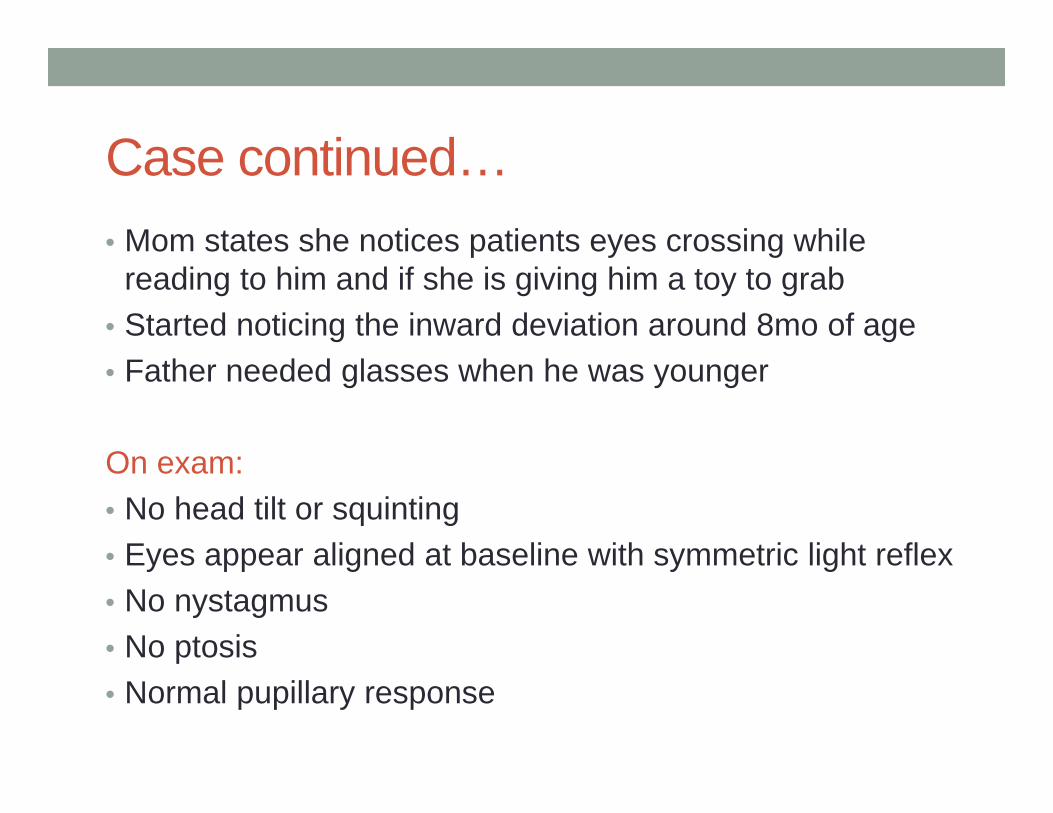
Case continued…• Mom states she notices patients eyes crossing while
reading to him and if she is giving him a toy to grab• Started noticing the inward deviation around 8mo of age• Father needed glasses when he was younger
On exam: • No head tilt or squinting• Eyes appear aligned at baseline with symmetric light reflex• No nystagmus• No ptosis• Normal pupillary response

QUESTION: Differential diagnosis for this patient is less likely to include the following
• A) Infantile esotropia• B) Pseudoesotropia• C) Accommodative esotropia• D) CN VI palsy
Website: b.socrative.com Room Name: TPSEYE
Accomodative esotropia• Presents from infancy to early childhood• Progressive
• Initially intermittent• Increases in degree and persistence over time
• Associated with far sightedness• Treated with corrective lenses
• Surgery if not successful with glasses alone
• Overall good prognosis if detected and treated early

Pseudostrabismus
• Typically pseudoesotropia• Secondary to facial structure
• Wide nasal bridge• Epicanthal folds
• May exist with true strabismus

Additional case:• 12yo female admitted to inpatient service due to
complaints of HA, double vision
• Admitted for workup• Exam significant for:
• Lateral gaze (CN VI) palsy• Papilledema
• MRI normal• LP with elevated opening pressures
• DX: Pseudotumor cerebri

Suppression
• Ability of child to cortically “turn off” deviated eye• Leads to amblyopia if not addressed• Complaint of diplopia presents in older children
• Typically after 6-7 years of age• Mature visual system cannot suppress double image• Development of diplopia implies acquired strabismus
Indications for referral• Suspected congenital esotropia• Presence of intermittent or constant exotropia at 6mo• Associated with other abnormal findings
• Abnormal red reflex• Nystagmus• Abnormal eyelid position
• Any child >6mo with or suspected deviation

������
eye “ick”…or “gunk”
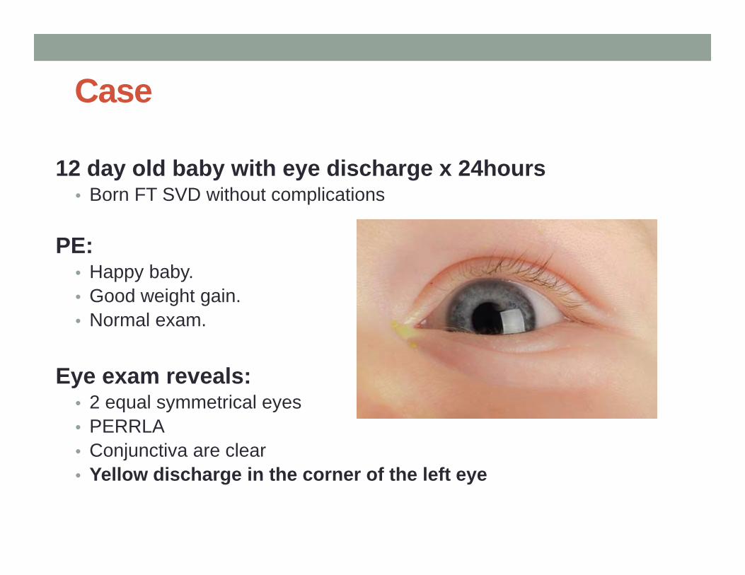
Case
12 day old baby with eye discharge x 24hours• Born FT SVD without complications
PE:• Happy baby. • Good weight gain. • Normal exam.
Eye exam reveals:• 2 equal symmetrical eyes• PERRLA• Conjunctiva are clear• Yellow discharge in the corner of the left eye

QUESTION: What is the most common cause of persistent discharge or tearing in children <3mo of age:
• A) ocular infection• B) allergy• C) glaucoma• D) lacrimal duct obstruction
Website: b.socrative.com Room Name: TPSEYE
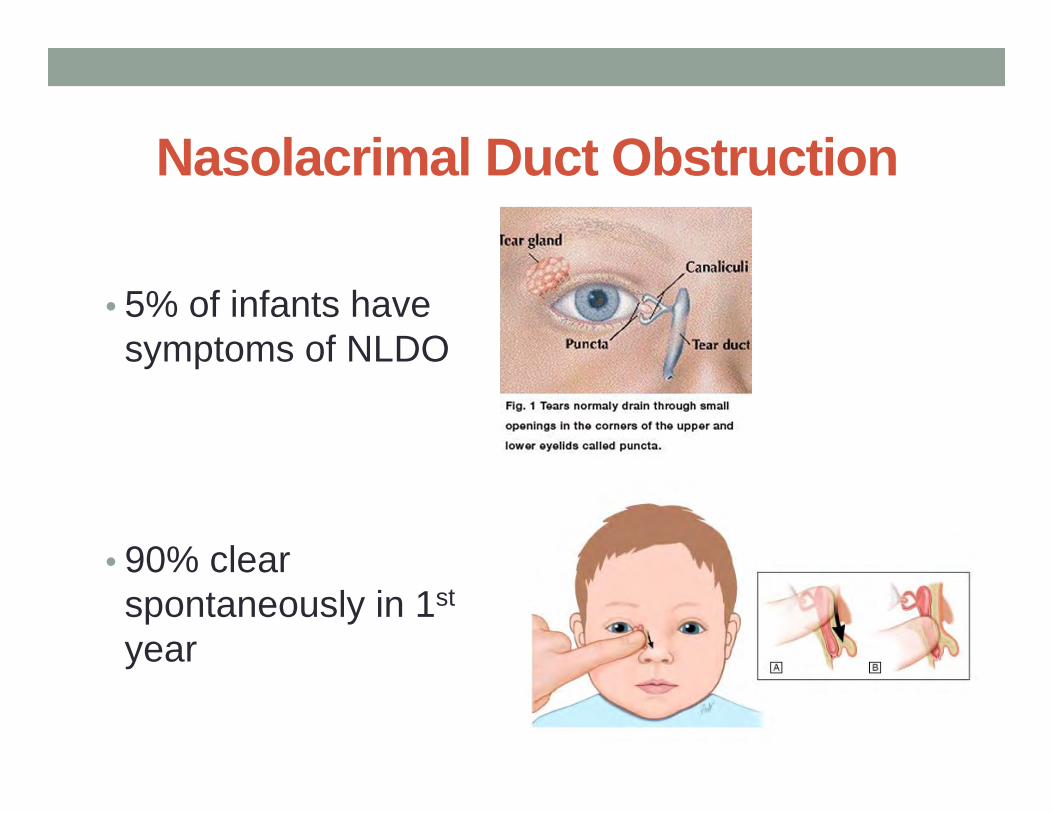
Nasolacrimal Duct Obstruction
• 5% of infants have symptoms of NLDO
• 90% clear spontaneously in 1st
year

Acute Dacryocystitis
• Infection of the Naso Lacrimal System
• Erythema• Swelling• Warmth• Tenderness of the lacrimal sac• +/- purulent discharge

Case
You are about to walk out of the room, but you reflect…this is a 2 week old. Anything else to consider?

Case
Mom and dad seem happily married and both attended the child’s visit today.
Maternal hx: • No h/o STI’s.• H/o UTI during 3rd trimester that was treated.• No symptoms thereafter.
Family history: • Nothing.
Recommendation:• Consider grabbing a few swabs• Return if things get worse
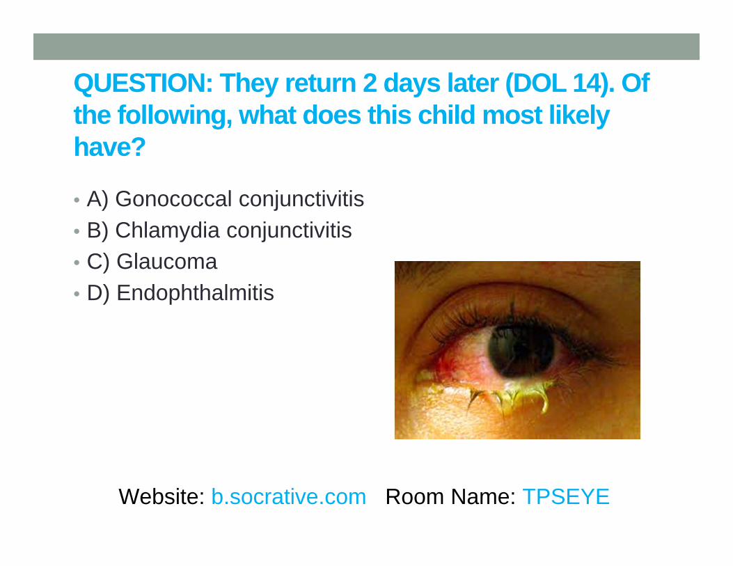
QUESTION: They return 2 days later (DOL 14). Of the following, what does this child most likely have?
• A) Gonococcal conjunctivitis• B) Chlamydia conjunctivitis• C) Glaucoma• D) Endophthalmitis
Website: b.socrative.com Room Name: TPSEYE
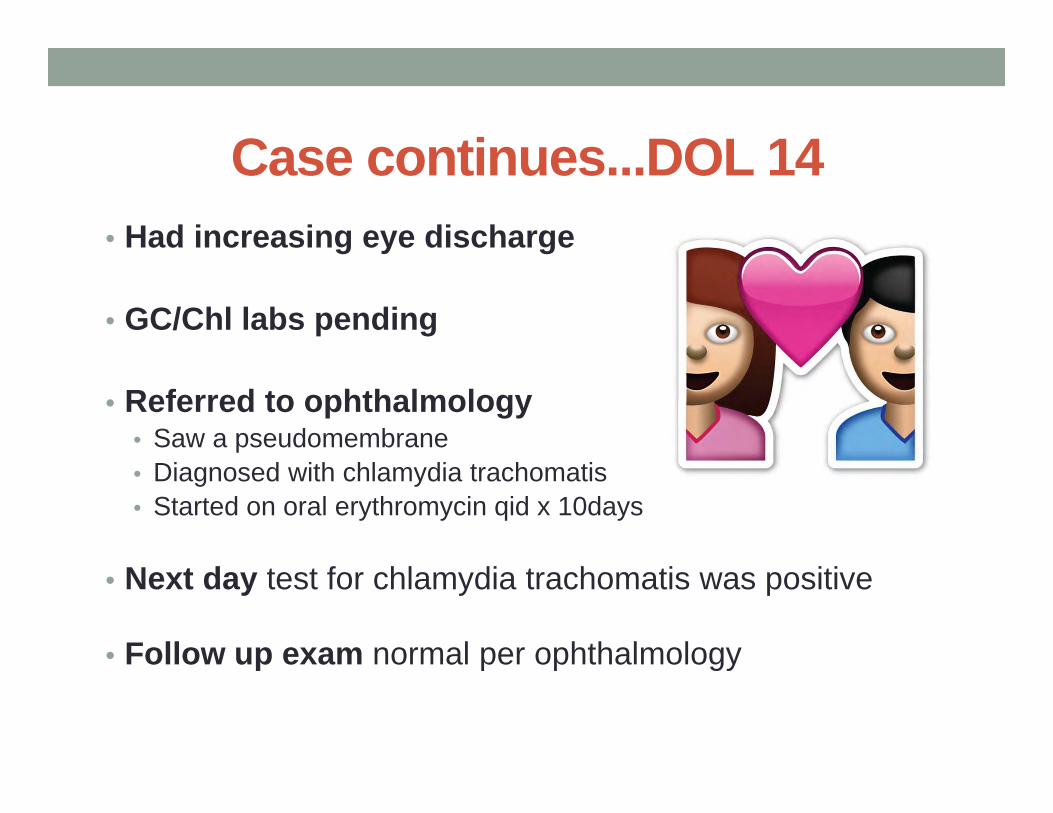
Case continues...DOL 14• Had increasing eye discharge
• GC/Chl labs pending
• Referred to ophthalmology• Saw a pseudomembrane• Diagnosed with chlamydia trachomatis• Started on oral erythromycin qid x 10days
• Next day test for chlamydia trachomatis was positive
• Follow up exam normal per ophthalmology
Neonatal conjunctivitis➢Aseptic : Chemical (DOL 1-2)
➢Bacterial:• Chlamydia (most common) (DOL 5-14)• GC (most dangerous) (DOL 2-5)• Other bacteria: staph/strep/gram negatives
➢Viral• Herpes (<1%) (DOL 1-35)

clarification on conjunctiva…
• The front skin of the eye.
• Extends from under the eyelid to over the white part of the eye.

Chemical conjunctivitis
• Chemical conjunctivitis
• Seen ~24-48 hours of life• Mildly red eye(s)• Some swelling of the lids• Self limiting• Less common now because we use erythromycin drops

GC ophthalmia• Presents: 2-5 days after birth, but can be later
• Findings: • purulent conjunctivitis• profuse exudate and • swelling of the eyelids

Chlamydia Conjunctivitis
• Presents:• 5-14 days after birth• Typically bilaterally• Mild to moderate
conjunctivitis
• Finding:• Mild swelling with watery
discharge→ mucopurulent→ marked swelling w/chemosis→ bloody discharge

CASE
Optho revealed: Pseudomembrane
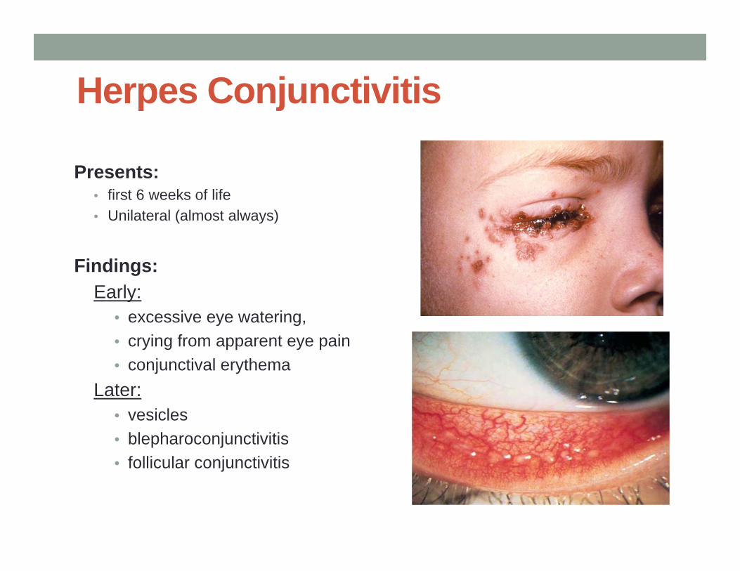
Herpes Conjunctivitis
Presents:• first 6 weeks of life• Unilateral (almost always)
Findings:Early:
• excessive eye watering, • crying from apparent eye pain• conjunctival erythema
Later:• vesicles• blepharoconjunctivitis • follicular conjunctivitis

When to refer…• Can potentially confirm diagnosis!• Help monitor for poor outcomes and resolution of
disease.

Case: Alternate endingQUESTION: 4 week old infant presents with persistent eye discharge/tearing. Previous eye cultures are all negative. On PE, patient possibly has a dull red reflex. Which finding would NOT support your working diagnosis?
• a)Enlarged cornea• b)Smaller than normal cornea• c)Corneal clouding• d)Photophobia
Website: b.socrative.com Room Name: TPSEYE
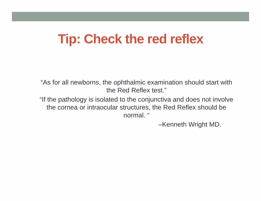
Tip: Check the red reflex
“As for all newborns, the ophthalmic examination should start with the Red Reflex test.”
“If the pathology is isolated to the conjunctiva and does not involve the cornea or intraocular structures, the Red Reflex should be
normal. “–Kenneth Wright MD.

Primary Infantile Glaucoma• Extremely rare, 1/100,000
births• 70% Bilateral
Cause: abnormal fetal development of the angle structures ➢Impaired drainage of
aqueous fluid➢Increased IOP
Most severe consequence• optic nerve damage

Congenital Glaucoma• Tearing• Photophobia• Blepharospasm• Large cornea• Corneal clouding
• Abnormal Red Reflex

Conjunctivitis
Infectious• Viral• Bacterial
NonInfectious• Allergic• Nonallergic
• Trauma (mechanical or chemical)
• Neoplasm• Ocular inflammation• Idiopathic

Conjunctivitis: A Clinical Diagnosis of Exclusion!
(Sangeeta’s) “Rule of 7”:
• No significant pain• No significant loss of vision • No photophobia• No focal pathology of the eye or lids• No focal erythema • No eye idiosyncrasies (PERRLA, EOMI, RR+B)
• No Improvement
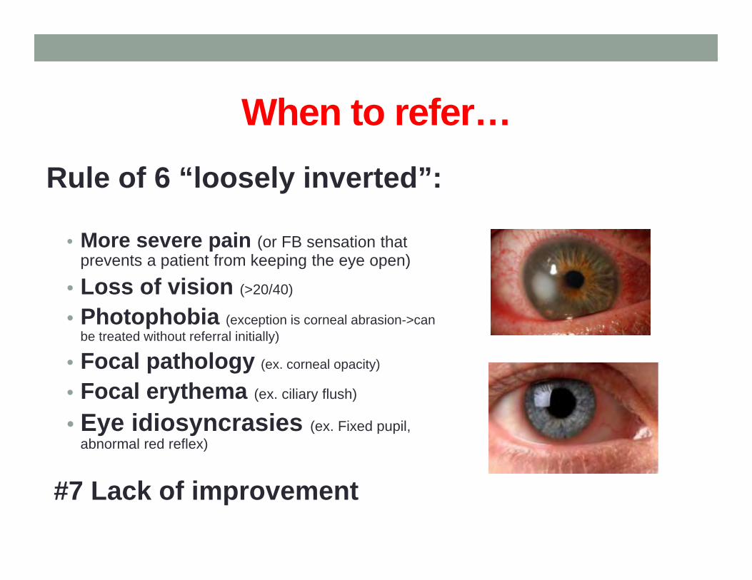
When to refer…Rule of 6 “loosely inverted”:
• More severe pain (or FB sensation that prevents a patient from keeping the eye open)
• Loss of vision (>20/40)
• Photophobia (exception is corneal abrasion->can be treated without referral initially)
• Focal pathology (ex. corneal opacity)
• Focal erythema (ex. ciliary flush)
• Eye idiosyncrasies (ex. Fixed pupil, abnormal red reflex)
#7 Lack of improvement

�������“omg…what’s wrong with my
eye?”

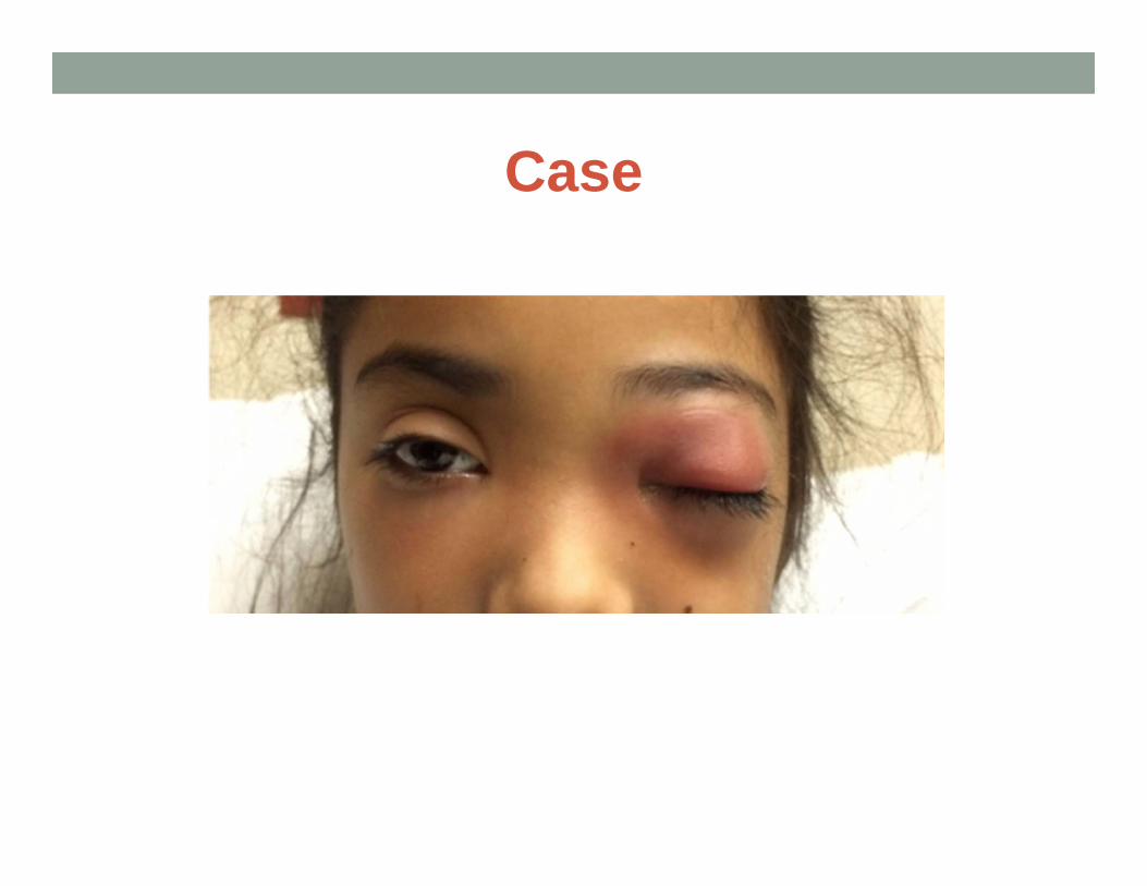
CASE
7yo female presenting with “swelling and redness” of her L eye

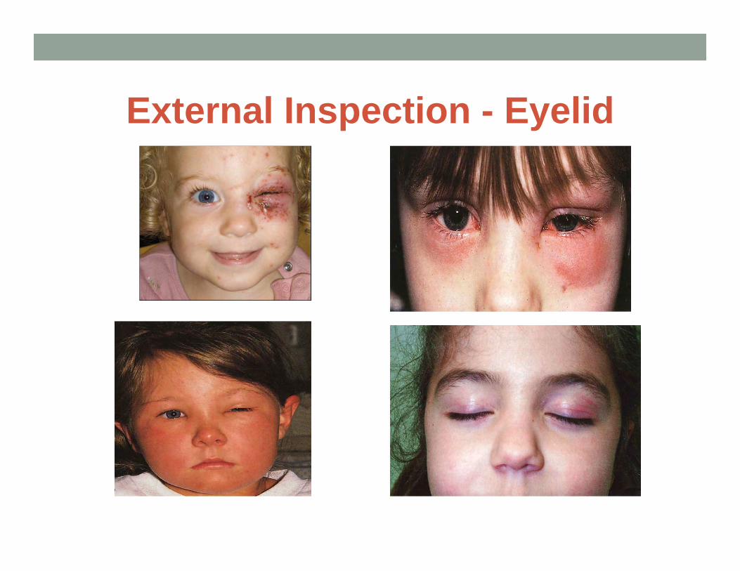
External Inspection - Eyelid • Know normal anatomy for
your patient• Location!
• Edema• Focal
• Hordeolum/Chalazion• Lacrimal pathology
• Generalized (periorbital)• Periorbital cellulitis
• Unilateral vs Bilateral
• Skin changes• Erythema• Ecchymosis

External Inspection - Eyelid
External Inspection - Eyelid
Case

External inspection - Proptosis• Congenital
• Craniofacial abnormalities• Acquired
• Secondary to mass effect• Bilateral
• Graves disease• Unilateral/Bilateral
• Infectious• Tumors• Inflammatory• Vascular
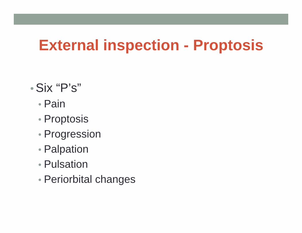
External inspection - Proptosis
• Six “P’s”• Pain• Proptosis• Progression• Palpation• Pulsation• Periorbital changes

External inspection - Proptosis• Exam
• Eyelid symmetry with frontal view
• Axial view• Pseudoproptosis• Direction of
displacement• Visual acuity• Systemic evaluation
• Immediate evaluation

Extraocular movements• Limitation to movement• Neurological
• Intracranial pathology• CN pathology• Neuromuscular
• Mechanical• Entrapment• Obstructive• Congenital
Extraocular Muscle Cranial NerveSR, IR, MR, IO CN III
LR CN VISO CN IV

Extraocular Movements

Increased Orbital Pressure• Limited room for volume expansion• Bony boundaries• Encased structures
• Manifestations• Proptosis• Pain• Limitation in EOM• VA changes

QUESTION: What is the preferred initial imaging modality for this patient?
• A) MRI brain/orbits with IV contrast• B) Orbital ultrasound• C) CT orbits with IV contrast• D) XR sinus
Website: b.socrative.com Room Name: TPSEYE

Case
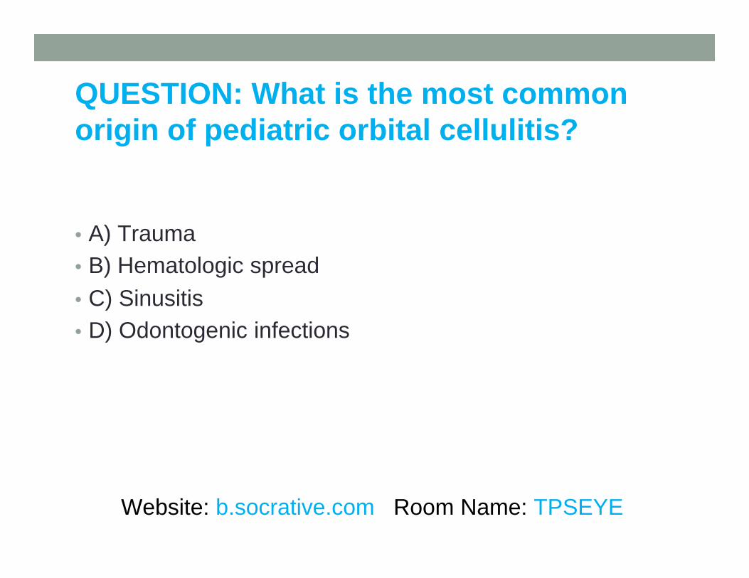
QUESTION: What is the most common origin of pediatric orbital cellulitis?
• A) Trauma• B) Hematologic spread• C) Sinusitis• D) Odontogenic infections
Website: b.socrative.com Room Name: TPSEYE

Orbital Cellulitis• Infection posterior to the orbital septum
• Extension from periorbital structures• 90% of all cases of
orbital cellulitis are caused by paranasal sinusitis• Majority from ethmoid
sinusitis• Dacrocystitis or
odontogenic infections
• Classic sinus organisms: • Haemophilus influenzae• Staphylococcus aureus• Streptococcus species
• S.pneumo• GAS
• Moraxella catarrhalis• Anaerobic species
• Peptostreptococcus• Fusobacterium• Bacteroides

Differential• Infectious
• Bacterial• Fungal
• Idiopathic inflammation• Orbital pseudotumor• Myositis• Sarcoidosis
• Neoplasms• Leukemia• Retinoblastoma• Rhabdomyosarcoma
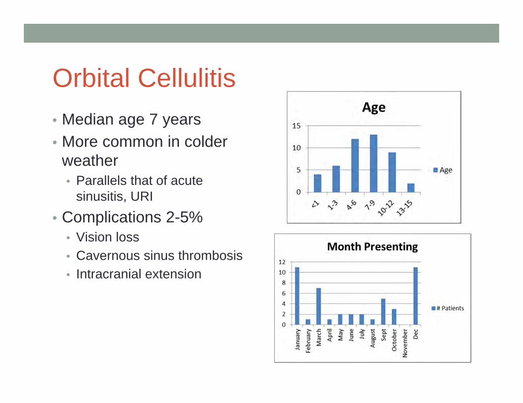
Orbital Cellulitis• Median age 7 years• More common in colder
weather• Parallels that of acute
sinusitis, URI• Complications 2-5%
• Vision loss• Cavernous sinus thrombosis• Intracranial extension

Management
• Admission • Parenteral antibiotics
• Target sinus organisms• MRSA
• Adjunctive therapies• Steroids• Decongestants
• Surgical management• Severe disease

QUESTION: Which of the following increases risk for surgical intervention in a patient with orbital cellulitis?
A) Significant periorbital edema limiting ability to open eyeB) Proptosis of eyeC) Pain with extraocular movementD) Younger age
Website: b.socrative.com Room Name: TPSEYE
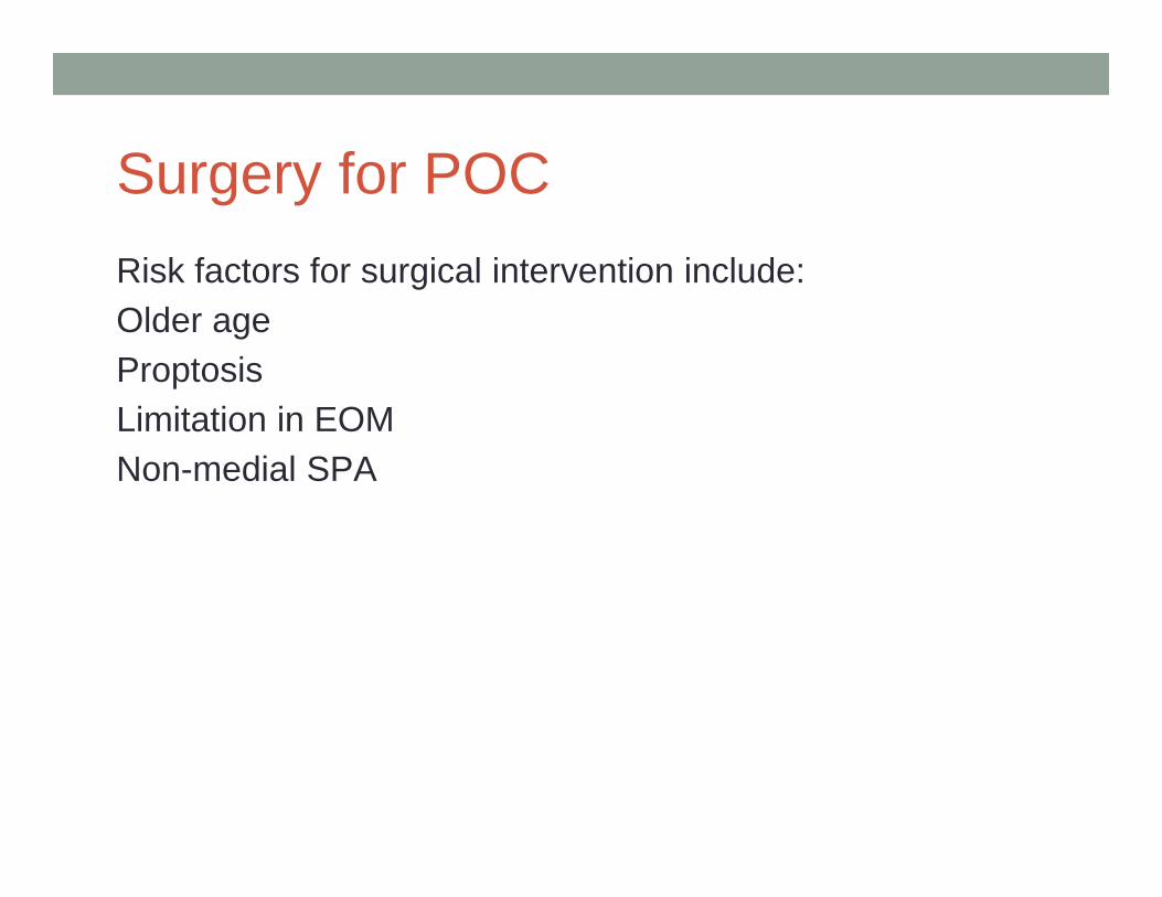
Surgery for POCRisk factors for surgical intervention include:Older ageProptosisLimitation in EOMNon-medial SPA

�������
“can you see that?”
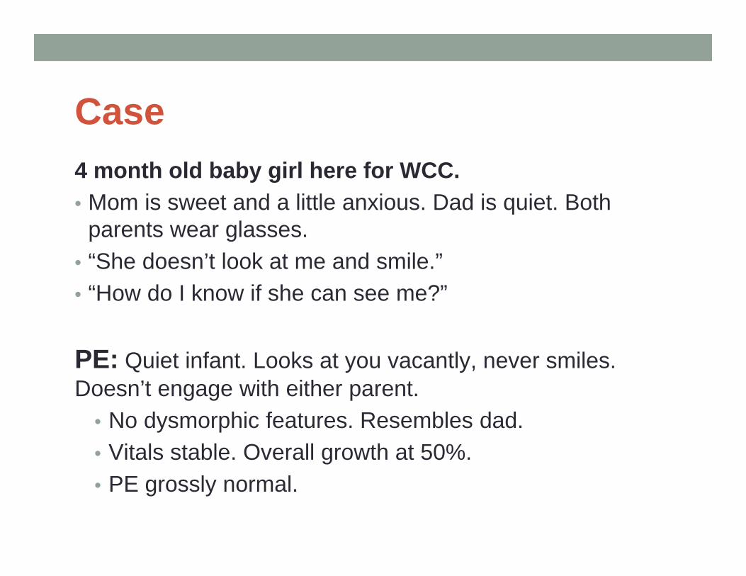
Case4 month old baby girl here for WCC.• Mom is sweet and a little anxious. Dad is quiet. Both
parents wear glasses. • “She doesn’t look at me and smile.”• “How do I know if she can see me?”
PE: Quiet infant. Looks at you vacantly, never smiles. Doesn’t engage with either parent.
• No dysmorphic features. Resembles dad.• Vitals stable. Overall growth at 50%.• PE grossly normal.

VISION• Legal blind adult 20/200
• Newborn 20/400
• (2 months 20/150)• (4 months 20/60)• 3yo 20/40• 4yo 20/30• 5yo 20/20
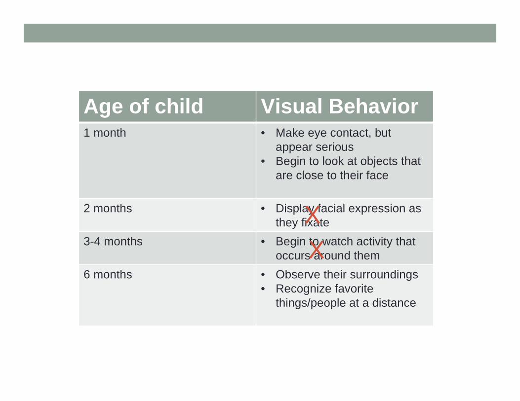
Age of child Visual Behavior1 month • Make eye contact, but
appear serious• Begin to look at objects that
are close to their face
2 months • Display facial expression as they fixate
3-4 months • Begin to watch activity that occurs around them
6 months • Observe their surroundings• Recognize favorite
things/people at a distance
XX

QUESTION: Vision assessment in children younger than 3 years or any nonverbal child is further accomplished by evaluating:
• A) Ability to fix & follow objects• B) Tumbling E eye chart• C) LEA Symbols eye chart• D) Asking the child
Website: b.socrative.com Room Name: TPSEYE
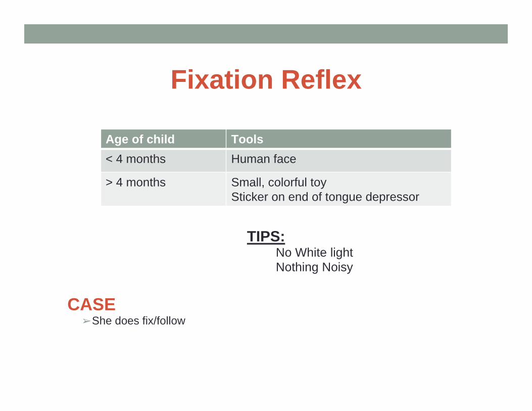
Fixation Reflex
TIPS:No White lightNothing Noisy
CASE➢She does fix/follow
Age of child Tools< 4 months Human face
> 4 months Small, colorful toySticker on end of tongue depressor
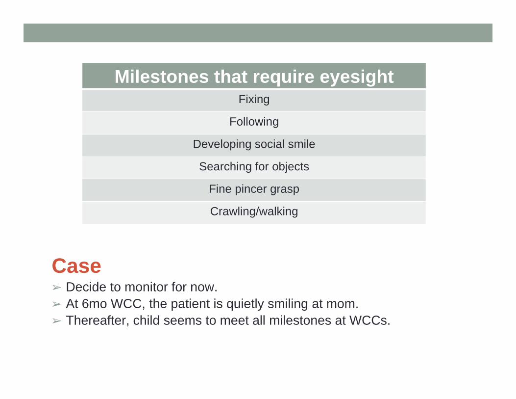
Case➢ Decide to monitor for now. ➢ At 6mo WCC, the patient is quietly smiling at mom. ➢ Thereafter, child seems to meet all milestones at WCCs.
Milestones that require eyesightFixing
Following
Developing social smile
Searching for objects
Fine pincer grasp
Crawling/walking
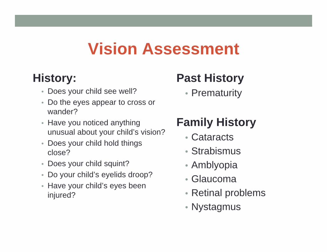
Vision AssessmentHistory:
• Does your child see well?• Do the eyes appear to cross or
wander?• Have you noticed anything
unusual about your child’s vision?• Does your child hold things
close?• Does your child squint?• Do your child’s eyelids droop?• Have your child’s eyes been
injured?
Past History• Prematurity
Family History• Cataracts• Strabismus• Amblyopia• Glaucoma• Retinal problems• Nystagmus
QUESTION: “Dr. Jain, when can you check my little girl’s eyes with an eye chart?”
• A)3• B)3.5• C)4• D)5
Website: b.socrative.com Room Name: TPSEYE
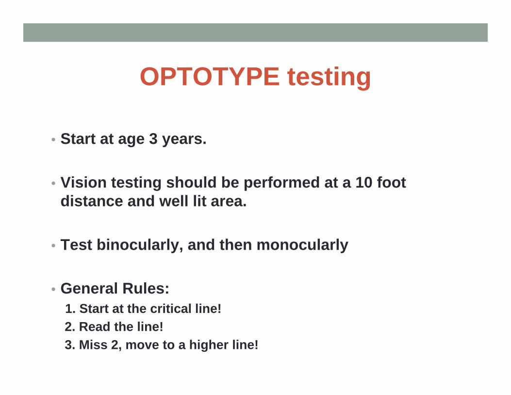
OPTOTYPE testing
• Start at age 3 years.
• Vision testing should be performed at a 10 foot distance and well lit area.
• Test binocularly, and then monocularly
• General Rules: 1. Start at the critical line!2. Read the line!3. Miss 2, move to a higher line!

Tumbling E LEA Symbols

HOTV Snellen

QUESTION: What is the best method for covering the eye that is not being tested?
• A) Patient’s hand• B) Parent’s hand• C) Paddle occluders• D) Occluder patches
Website: b.socrative.com Room Name: TPSEYE

OPTOTYPE testing
• Remember: the child wants to get things right!
Behaviors to watch out for:Skipping symbols
Memorizing charts
Squinting
Peeking
Uncooperative child (especially repeatedly)

Case3yo WCC—the visual acuity testing shows 20/40 vision in both eyes, do you refer to ophtho?
3yo WCC--Would you refer if the Right eye was 20/20 and left eye was 20/40?
Refer if:Worse than 20/50 in a 3yo Worse than 20/40 in a 4yoWorse than 20/30 in a 5+yo
Refer if:2 line difference between eyes even within passing range
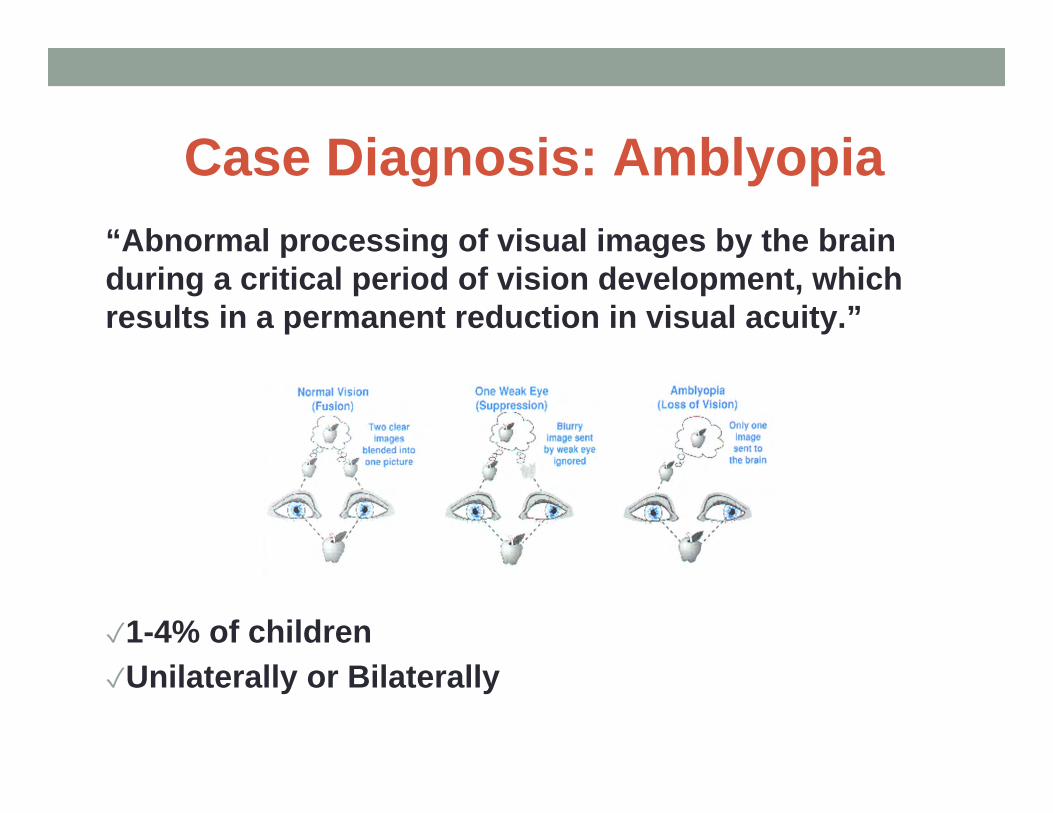
Case Diagnosis: Amblyopia“Abnormal processing of visual images by the brain during a critical period of vision development, which results in a permanent reduction in visual acuity.”
✓1-4% of children✓Unilaterally or Bilaterally

Amblyopia
Causes:• Deprivational• Strabismus• High Refractive Error• Anisometropia
Treatment:• Provide a clear retinal image• Correct ocular dominance

Near Vision testing…see ophtho
• Decline in school performance• Reading difficulty• Eye strain• Headaches• Double vision• Blepharospasm• Blurred or distorted near vision
����� �“Why are things so
blurry?”

CASE 15yo female presenting with “blurry vision” and eye pain

Case follow-up questions
• Prior VA deficits• Monocular vs Binocular “blurriness”• Prior or family history of migraines• Recent trauma• OTC or prescription medication usage• Any other neurological deficits• Significant life stressors/anxiety

Neuro-Ophtho exam
• Cranial nerves• VA• Motility deficits
• Pupillary exam• Nystagmus• Fundoscopic exam
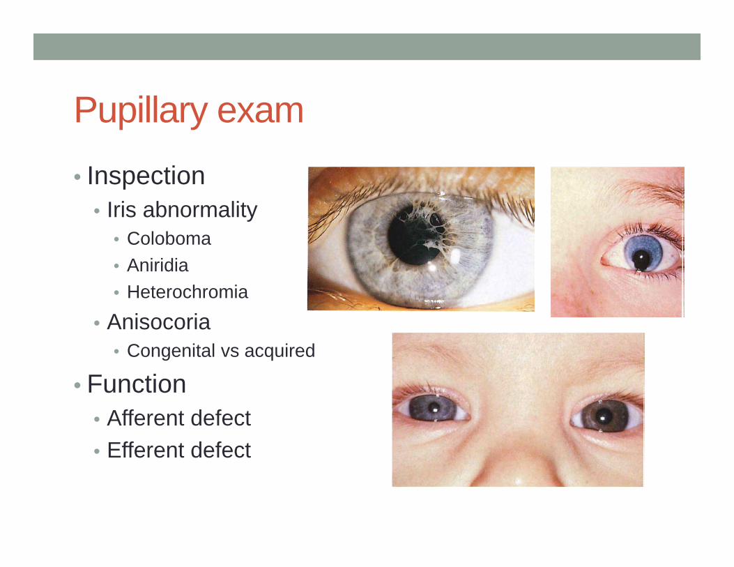
Pupillary exam
• Inspection• Iris abnormality
• Coloboma• Aniridia• Heterochromia
• Anisocoria• Congenital vs acquired
• Function• Afferent defect• Efferent defect

Pupillary light reflex pathway
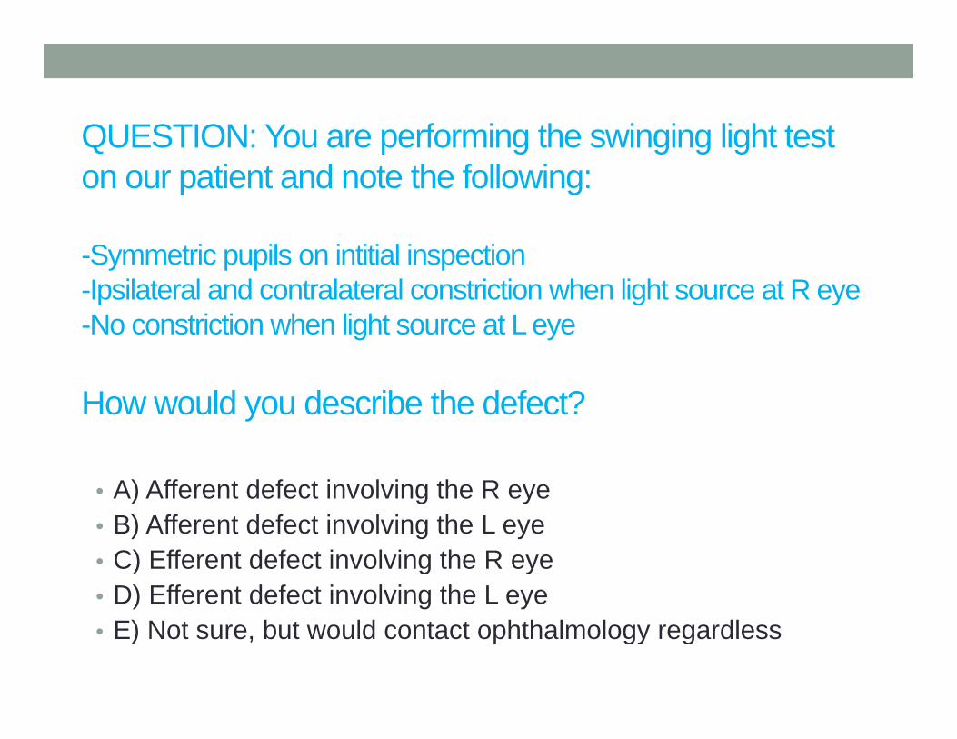
QUESTION: You are performing the swinging light test on our patient and note the following:
-Symmetric pupils on intitial inspection-Ipsilateral and contralateral constriction when light source at R eye-No constriction when light source at L eye
How would you describe the defect?
• A) Afferent defect involving the R eye• B) Afferent defect involving the L eye• C) Efferent defect involving the R eye• D) Efferent defect involving the L eye• E) Not sure, but would contact ophthalmology regardless

Fundoscopic exam• Look for:
• Atrophy• Swelling
• Blurring of disc margin• Elevation of disc• Hemorrhages along optic
nerve• Obscured vessels
Case continued:• First episode of “blurry vision” and HA
• Loss of color vision, only able to appreciate shapes
• Exam significant for:• Relative APD in L eye• Blurring of optic disc margin on fundoscopic exam• No motility defects noted• No other significant neurological findings

Causes of Acute Loss of Vision• Cortical blindness• Psychogenic blindness• Optic neuropathy• Retinal disease• Visual media problems


Optic Neuritis• Demyelination of optic nerve• Can be isolated or associated with other demyelinating
diseases• Neuromyelitis optica (Devic Syndrome)• Multiple sclerosis
• 15% later MS presentation for isolated optic neuritis• Increased risk factors for later MS
• Unilateral symptoms• Recurrent symptoms within a year
• Etiology• Clinical findings• Management

Ptosis
Congenital AcquiredFibrosis of extraocular muscles Horner syndromeHorner syndrome Lid inflammationMyasthenia Mitochondrial myopathiesCN III palsy Myasthenia gravis
CN III palsyTrauma

Abnormal Eye Movements• Nystagmus
• Involuntary, rhythmic ocular oscillation• At least one phase is slow• Variable pathology
• Ocular Flutter• Bursts of rapid horizontal oscillation during fixation• Associated with cerebellar/brainstem disease
• Ocular dysmetria• Over/Undershooting or oscillation on refixation• Associated with cerebellar disease
• Opsoclonus• Nonrhythmic conjugate movements• Neuroblastoma
Nystagmus• Types• Acquired
• Detected by patient• Intracranial pathology
• Congenital• Motor nystagmus
• Bilateral, symmetrical• Can have face turn,
relatively good VA• Sensory
• Due to neonatal blindness• Larger amplitude, poor
fixation• 6-8 weeks

Indications for referral to ophtho• Abnormal red reflex
• Personal history of prematurity or metabolic or genetic disease with ophthalmologic implications.
• Family history of childhood cataract, retinoblastoma, retinal dysplasia, or glaucoma.
• Inability to fix and follow by age three months.
• Abnormal ocular alignment
• Pupillary asymmetry of ≥1 mm in diameter
• Corneal asymmetry
• Unilateral ptosis or other lesions obstructing the visual axis (eg, eyelid hemangioma), which may
cause amblyopia.
• Asymmetry of vision (eye preference) or visual acuity difference of two lines or more between eyes.
• Visual acuity worse than 20/50 in a three-year-old, worse than 20/40 in a four-year-old, or worse
than 20/30 in a child ≥5 years
• Abnormal instrument-based vision screening
• Nystagmus.
• Neurodevelopmental delays

Sources• Coats, David. “Visual development and vision assessment in infants and children,”
UpToDate, Jan 21, 2016.• Cronau, Holly, et al. “Diagnosis and Management of Red Eye in Primary Care”,
AAFP, January 2010, 137-144.• Drutz, Jan. “The pediatric physical examination: HEENT,” UpToDate, Dec 21, 2015.• Heider, Krista, et al. “Cataracts in Children, Congenital and Acquired,” EyeWiki
sponsored by AAO, Dec 2014.• Jacobs, Deborah. “Conjunctivitis”, UpToDate, July 19, 2015• Kaufman, Paul, et al. “Approach to the child with leukocoria”, UpToDate, Feb 12,
2015.• Kaufman, Paul, et al. “Retinoblastoma: Clinical Presentation, evaluation, and
diagnosis,” UpToDate, July 11, 2016.• McCreery, Kathryn, “Cataract in Children”, UpToDate, march 21, 2016• Paysse, Evelyn, “congenital nasolacrimal duct obstruction and dacryocystocele,”
UpToDate, May 27, 2015.• Pina-Garza, J. Clinical Pediatric Neurology, Seventh Edition. 2013. • Rogers, G., Jordan, C. Pediatric Vision Screening. Peds in Review. 2013 (34) 126.• Wright, K. Pediatric Ophthalmology for Primary Care. 2007.• Zitelli, B. Davis, H. Atlas of Pediatric Physical Diagnosis, Fifth Edition. 2007. 713-754.

Images sources• Part 1:
• http://www.cehjournal.org/article/how-to-test-for-the-red-reflex-in-a-child/• http://www.medicinenet.com/image-collection/eye_anatomy_detail_picture/picture.htm• http://www.images.missionforvisionusa.org/anatomy/2007/06/congenital-rubella-cataract.html• http://www.cehjournal.org/article/how-to-test-for-the-red-reflex-in-a-child/• http://www.missionforvisionusa.org/2006/12/what-is-peters-anomaly-of-cornea.html• http://www.oculist.net/downaton502/prof/ebook/duanes/pages/v9/v9c021.html• http://fitsweb.uchc.edu/student/selectives/mMeyer/retinoblastoma.htm• http://www.cmej.org.za/index.php/cmej/rt/printerFriendly/2710/2900
• Part 2: • Zitelli’s Atlas of Pediatric Physical diagnosis• Rogers, G., Jordan, C. Pediatric Vision Screening. Peds in Review. 2013 (34) 126.
• Part 3:• https://www.newkidscenter.com/Newborn-Eye-Discharge.html• http://www.aapos.org/terms/conditions/72• http://dacryocystitis.blogspot.com/• Paysee, Evelyn, “Congenital nasolacrimal duct obstruction,” UpToDate, May 27, 2015• https://en.wikipedia.org/wiki/Conjunctivitis• http://www.poen.net.ar/en/information-for-patients/• https://www.studyblue.com/notes/note/n/exam-3/deck/11202623• https://en.wikipedia.org/wiki/Conjunctivitis• https://www.youtube.com/watch?v=WY_YUu9fe3E• https://en.wikipedia.org/wiki/Conjunctivitis• http://pediatriccare.solutions.aap.org/chapter.aspx?sectionid=56754743&bookid=1017• http://www.herpes.com/Ocular.shtml• https://www.studyblue.com/notes/note/n/3-conjunctiva/deck/8512473• http://www.cyber-sight.org/bins/content_page.asp?cid=2864-3900&lang=1• http://www.slideshare.net/laxmieyeinstitute/my-clouding-cornea• Jacobs, Deborah. “Conjunctivitis”, UpToDate, July 19, 2015.• Jacobs, Deborah. “Conjunctivitis”, UpToDate, July 19, 2015• Jacobs, Deborah. “Conjunctivitis”, UpToDate, July 19, 2015• (Graph)-http://www.aafp.org/afp/2010/0115/p137.html

Image Sources• https://www.eyecenters.com/what-you-should-know-about-corneal-ulcer.html• Ciliary flush--https://quizlet.com/78825852/ophthalmalogy-intro-flash-cards/
• Part 4:• Zitelli’s Atlas of Pediatric Physical diagnosis• Wright, K. Pediatric Ophthalmology for Primary Care. 2007
• Part 5:• https://www.pinterest.com/pin/60587557460294466/• Symbols eye chart-https://www.bernell.com/category/1099• Austin Regional Clinic Tumbling E chart• https://www.good-lite.com/Details.cfm?ProdID=25• Austin Regional Clinic HOTV eye chart• Austin Regional Clinic Snellen eye chart• (Graph of amblyopia) http://health-pictures.com/eye/Amblyopia.htm#.V6KfnDWwRQs
• Part 6• Zitelli’s Atlas of Pediatric Physical diagnosis• https://commons.wikimedia.org/wiki/File:Wiley_Human_Visual_System.gif


![I. MELLÉKLET $/.$/0$=È6,(/ Ë5È6 · chalazion . szaruhártya -EHV]&U GpVHN. szaruhártyaheg . szemhéjviszketés . iridocyclitis . ocularis diszkomfort Általános tünetek, az](https://static.cupdf.com/doc/110x72/60fcb669d36d75315b281ace/i-mellklet-06-56-chalazion-szaruhrtya-ehvu-gpvhn.jpg)