Diagnosis and Management of CutaneousPsoriasis: A Review
C M E1 AMA PRA
Category 1 CreditTM
ANCC1.5 Contact Hours 1.5 Contact Hours
Alisa Brandon, MSc & Medical Student & University of Toronto & Toronto, Ontario, Canada
Asfandyar Mufti, MD & Dermatology Resident & University of Toronto & Toronto, Ontario, Canada
R. Gary Sibbald, DSc (Hons), MD, MEd, BSc, FRCPC (Med Derm), ABIM, FAAD, MAPWCA & Professor & Medicine andPublic Health & University of Toronto & Toronto, Ontario, Canada & Director & International Interprofessional Wound CareCourse and Masters of Science in Community Health (Prevention and Wound Care) & Dalla Lana Faculty of Public Health &University of Toronto & Past President & World Union of Wound Healing Societies & Editor-in-Chief & Advances in Skin andWound Care & Philadelphia, Pennsylvania
The author, faculty, staff, and planners, including spouses/partners (if any), in any position to control the content of this CME activity have disclosed that they have no financial relationshipswith, or financial interests in, any commercial companies pertaining to this educational activity.
To earn CME credit, you must read the CME article and complete the quiz online, answering at least 13 of the 18 questions correctly.
This continuing educational activity will expire for physicians on January 31, 2021, and for nurses on December 4, 2020.
All tests are now online only; take the test at http://cme.lww.com for physicians and www.nursingcenter.com for nurses. Complete CE/CME information is on the last page of this article.
GENERAL PURPOSE:
To provide information about the diagnosis and management of cutaneous psoriasis.
TARGET AUDIENCE:
This continuing education activity is intended for physicians, physician assistants, nurse practitioners, and nurses
with an interest in skin and wound care.
LEARNING OBJECTIVES/OUTCOMES:
After completing this continuing education activity, the provider should be better able to:
1. Describe the epidemiology, pathophysiology, clinical presentation, assessment, and diagnosis of the types and
subtypes of psoriasis.
2. Explain the use of topical treatments, intralesional steroids, phototherapy, conventional systemic treatments,
biologic agents, and pain medicines for psoriasis.
FEBRUARY 2019
C L I N I C A L M A N A G E M E N T
extra
ADVANCES IN SKIN & WOUND CARE & VOL. 32 NO. 2 58 WWW.WOUNDCAREJOURNAL.COMCopyright © 2019 Wolters Kluwer Health, Inc. All rights reserved.
ABSTRACT
Psoriasis is a chronic inflammatory disease that is characterizedby plaque, inverse, guttate, pustular, and erythrodermic variants.This review focuses on the epidemiology, diagnosis, and treatmentof cutaneous psoriasis. Other related topics discussed includeperistomal psoriasis, the Koebner phenomenon, and the relationshipbetween biologic therapy and wound complications.KEYWORDS: biologic therapy, cutaneous psoriasis, erythrodermicpsoriasis, intertriginous psoriasis, Koebner phenomenon, psoriasis,psoriasis vulgaris, pustular psoriasis, topical corticosteroids
ADV SKIN WOUND CARE 2019;32:58–69.
INTRODUCTIONPsoriasis is a chronic inflammatory disease, with a reported
prevalence of 1% to 3% in Europe and the US.1 It may present
at any age, but has a bimodal distribution of first presentation
at between 15 to 20 and 55 to 60 years of age. Younger age at
onset is associated with more severe disease and a family history
affecting more family members.2 In general, approximately 36%
of patients have a family history of psoriasis, and multiple genetic
susceptibility loci have been identified.3,4
Metabolic syndrome (three of five components: central obesity,
high blood pressure, high blood sugar, high serum triglycerides,
and low serum high-density lipoprotein)5 has been associated with
psoriasis. Psoriasis is also associated with chronic obstructive
pulmonary disease, nonalcoholic fatty liver disease, and coronary
artery disease.6–8 Persons with psoriasis may also have a sig-
nificantly decreased quality of life and psychological burden
including anxiety, depression, and suicidal thoughts and behav-
ior.2,9,10 Reviewing this article will facilitate provider knowledge of
psoriasis pathophysiology, diagnosis, and management.
PATHOPHYSIOLOGYPsoriasis is a chronic autoimmune disease with multiple leukocytes
and cytokines interacting to produce the disease process (Figure 1).
The inflammatory cascade of psoriasis begins when antigens in
the skin activate dendritic cells and neutrophils, which release
cytokines including tumor necrosis factor ! (TNF-!), interleukin
23 (IL-23), and IL-12. These cytokines participate in positive feed-
back loops by activating leukocytes, which then release more
cytokines, resulting in continuous inflammation. For example,
IL-23 converts cluster of differentiation 4–positive cells into
T-helper 17 (TH17) cells that release IL-17A; TH17 cells and IL-17A
act to upregulate TNF-!. These cytokines also exert effects on the
skin with IL-17A, IL-20, and IL-22, and TNF-! contributing to
the modified keratinocyte function and the TH17 cells promote
angiogenesis.11,12
CLINICAL PRESENTATIONPsoriasis is a relapsing-remitting disease that often improves with
warmer weather and relapses during stressful life events or in con-
junction with infections. Common presentations include (Figure 2):
1. Plaque psoriasis, with elevated areas of more than 1 cm. This
is the most common subtype and presents with well-demarcated
annular lesions comprising an erythematous base and thick
silvery scale. These lesions are often found on the extensor surfaces
(elbows, knees), scalp, lumbosacral area, and intergluteal cleft.
2. Inverse psoriasis, seen in the body folds. Also called flexural
psoriasis, it is characterized by red shiny lesions devoid of
scale in the inframammary, perineal, and axillary areas.
3. Guttate psoriasis, presenting as teardrop-shaped lesions. Acute
guttate psoriasis is often preceded by a sore throat associated
with group B streptococcal infection, and consists of multiple,
small (2–10 mm) psoriatic lesions, most often on the trunk.
4. Erythrodermic psoriasis, in which 90% or more of the body
is red. This variant consists of complete or almost complete
involvement of the skin and is characterized by gradual
coalescence of plaques caused by infection, drugs, systemic
disease, or withdrawal of corticosteroids.
5. Generalized pustular psoriasis manifests as multiple uniform
sterile pustules on the body and is often accompanied by fever. It
may also be precipitated by withdrawal of systemic corticoste-
roids or infection and represents unstable disease that often
requires hospitalization.
6. Palmoplantar pustulosis psoriasis presents on the hands and
feet as sterile pustules on a base of erythema and scale.2,13
Extracutaneous manifestations of psoriasis include nail abnor-
malities and psoriatic arthritis. About 80% of patients with psoriasis
have nail involvement including pitting (small depressions on
the nail surface), onycholysis (distal nail separation from the nail
bed), subungual hyperkeratosis, and orange-yellow spots beneath
the nail plate (oil spots).2,13
The prevalence of psoriatic arthritis among patients withcutaneous
psoriasis is 30%,14,15 with patients developing arthritis an average
of 12 years after the onset of cutaneous psoriasis.16 There are five
forms of psoriatic arthritis. From most common to least, these
include distal oligoarthritis (inflammation that involves four or
fewer joints), rheumatoid factor–negative polyarthritis, arthritis
mutilans (resorption and shortening of finger bones), sacroiliitis,
and ankylosing spondylitis.13 The most common form of arthritis
affects the distal joints of the digits in an asymmetric pattern. The
soft tissue may become swollen, producing sausage-like digits
called dactylitis.2,13
ASSESSMENT AND DIAGNOSISThe diagnosis of psoriasis is usually clinical. The physical examination
should include an examination of the primary lesion and other
ADVANCES IN SKIN & WOUND CARE & FEBRUARY 201959WWW.WOUNDCAREJOURNAL.COMCopyright © 2019 Wolters Kluwer Health, Inc. All rights reserved.

common areas affected by psoriasis including the scalp. The nails
and joints should be examined for any changes consistent with
psoriasis, and a family history should be taken to further elucidate
the diagnosis.13 Diagnosis can be further supported by the
Auspitz sign or Koebner phenomenon. The Auspitz sign occurs
because an excess of small surface capillaries results in multiple
bleeding points when the silver-gray scale is lifted off.17 The
Koebner phenomenon consists of the appearance of psoriatic
lesions on previously normal skin because of prior trauma;
clinical psoriasis lesions appear after 7 days or more.18 This
phenomena may cause psoriatic lesions to appear around wound
sites, under dressings, and around ostomy sites. Finally, if there is
still doubt about the diagnosis, a simple punch biopsy can be
performed.13
Chronic Plaque PsoriasisClassification of plaque psoriasis severity can guide appropri-
ate treatment. Commonly used tools for classification of
plaque psoriasis include the Psoriasis Area and Severity Index
(PASI), body surface area (BSA), and the Dermatology Life
Quality Index, with a score of more than 10 on each of these
parameters indicating moderate to severe psoriasis. Treatment
may also be guided by the location of the plaques, associated
pruritus, functional and psychosocial limitations, associated
psoriatic arthritis, nail or scalp psoriasis, previously prescribed
treatments, and patient preference.13,19
The PASI score is a formula calculated based on BSA affected,
erythema, thickness of the plaques, and amount of scale, with
each criterion rated on a 0- to 4-point scale. The PASI is often
used to measure response rate, with PASI 90 scores (meaning a
90% reduction in severity from baseline) being a common
target.13,19 The most common method to estimate BSA is with
the patient’s full handprint (including the fingers) equating to 1%
of the total BSA.20
TREATMENT
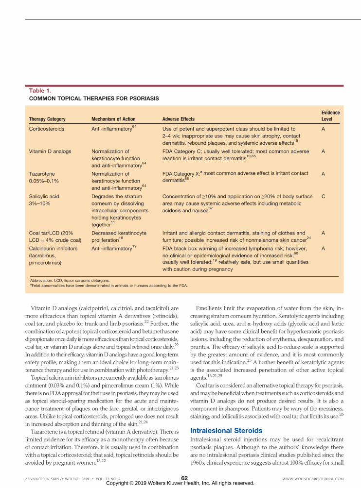
Topical TreatmentMild disease may be effectively treated with topical therapies,
including corticosteroids, vitamin D derivates, retinoids, tar,
Figure 1.
THE BASIC PATHOPHYSIOLOGY OF PSORIASIS
* 2018 Alisa Brandon.
ADVANCES IN SKIN & WOUND CARE & VOL. 32 NO. 2 60 WWW.WOUNDCAREJOURNAL.COMCopyright © 2019 Wolters Kluwer Health, Inc. All rights reserved.

keratolytic agents that break down scale (urea, salicylic acid,
!-hydroxy acid), and emollient moisturizers (Table 1). The choice
of topical agent depends on anatomical area, size and thickness
of the plaque, and whether the agent is being used for initiation
or maintenance therapy.
A combination product of betamethasone dipropionate and
calcipotriol is recommended to initiate treatment on the trunk
or extremities because this preparation is more efficacious than
monotherapy.21 This product is too strong for the face or folds.
When disease control has been established, vitamin D derivates
are recommended for maintenance therapy. Further, thick plaques
(clinical thickness >0.75 mm) respond to keratolytic agents
including salicylic acid or urea, the use of emollients (lubricating
moisturizers), and higher-strength topical corticosteroids (oint-
ment formulation).21
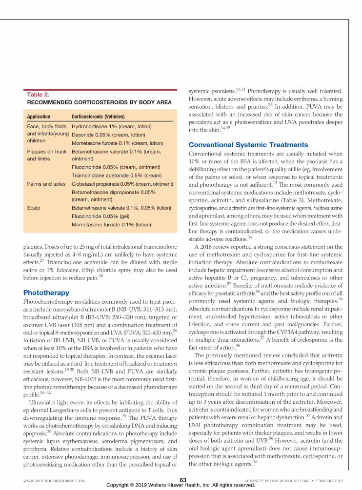
The choice of topical corticosteroid depends on the anatomical
location of the plaque, the thickness of the plaque, and the age of
the patient. For thick plaques on the trunk or limbs, mid- to high-
potency corticosteroids should be used. For infants and young
children,body folds, andthe face, low-tomid-potency corticosteroids
should be used. The palms and soles require high- to very high-
potency corticosteroids (Table 2).13
Figure 2.
SKIN, NAIL, AND SCALP INVOLVEMENT WITH PSORIASIS VULGARIS
ADVANCES IN SKIN & WOUND CARE & FEBRUARY 201961WWW.WOUNDCAREJOURNAL.COMCopyright © 2019 Wolters Kluwer Health, Inc. All rights reserved.
Vitamin D analogs (calcipotriol, calcitriol, and tacalcitol) are
more efficacious than topical vitamin A derivatives (retinoids),
coal tar, and placebo for trunk and limb psoriasis.22 Further, the
combination of a potent topical corticosteroid and betamethasone
dipropionateonce daily is moreefficacious thantopical corticosteroids,
coal tar, or vitamin D analogs alone and topical retinoid once daily.22
In addition to their efficacy, vitamin D analogs have a good long-term
safety profile, making them an ideal choice for long-term main-
tenance therapy and for use in combination with phototherapy.21,23
Topical calcineurin inhibitors are currently available as tacrolimus
ointment (0.03% and 0.1%) and pimecrolimus cream (1%). While
there is no FDA approval for their use in psoriasis, they may be used
as topical steroid-sparing medication for the acute and mainte-
nance treatment of plaques on the face, genital, or intertriginous
areas. Unlike topical corticosteroids, prolonged use does not result
in increased absorption and thinning of the skin.21,24
Tazarotene is a topical retinoid (vitamin A derivative). There is
limited evidence for its efficacy as a monotherapy often because
of contact irritation. Therefore, it is usually used in combination
with a topical corticosteroid; that said, topical retinoids should be
avoided by pregnant women.13,22
Emollients limit the evaporation of water from the skin, in-
creasing stratum corneum hydration. Keratolytic agents including
salicylic acid, urea, and !-hydroxy acids (glycolic acid and lactic
acid) may have some clinical benefit for hyperkeratotic psoriasis
lesions, including the reduction of erythema, desquamation, and
pruritus. The efficacy of salicylic acid to reduce scale is supported
by the greatest amount of evidence, and it is most commonly
used for this indication.25 A further benefit of keratolytic agents
is the associated increased penetration of other active topical
agents.13,21,25
Coal tar is considered an alternative topical therapy for psoriasis,
and may be beneficial when treatments such as corticosteroids and
vitamin D analogs do not produce desired results. It is also a
component in shampoos. Patients may be wary of the messiness,
staining, and folliculitis associated with coal tar that limits its use.26
Intralesional SteroidsIntralesional steroid injections may be used for recalcitrant
psoriasis plaques. Although to the authors’ knowledge there
are no intralesional psoriasis clinical studies published since the
1960s, clinical experience suggests almost 100% efficacy for small
Table 1.
COMMON TOPICAL THERAPIES FOR PSORIASIS
Therapy Category Mechanism of Action Adverse EffectsEvidenceLevel
Corticosteroids Anti-inflammatory64
Use of potent and superpotent class should be limited to
2–4 wk; inappropriate use may cause skin atrophy, contact
dermatitis, rebound plaques, and systemic adverse effects19
A
Vitamin D analogs Normalization of
keratinocyte function
and anti-inflammatory64
FDA Category C; usually well tolerated; most common adverse
reaction is irritant contact dermatitis19,65
A
Tazarotene
0.05%–0.1%
Normalization of
keratinocyte function
and anti-inflammatory64
FDA Category X;a most common adverse effect is irritant contact
dermatitis66A
Salicylic acid
3%–10%
Degrades the stratum
corneum by dissolving
intracellular components
holding keratinocytes
together11
Concentration of Q10% and application on Q20% of body surface
area may cause systemic adverse effects including metabolic
acidosis and nausea67
C
Coal tar/LCD (20%
LCD = 4% crude coal)
Decreased keratinocyte
proliferation19
Irritant and allergic contact dermatitis, staining of clothes and
furniture; possible increased risk of nonmelanoma skin cancer24
A
Calcineurin inhibitors
(tacrolimus,
pimecrolimus)
Anti-inflammatory19
FDA black box warning of increased lymphoma risk; however,
no clinical or epidemiological evidence of increased risk;68
usually well tolerated;19 relatively safe, but use small quantities
with caution during pregnancy
A
Abbreviation: LCD, liquor carbonis detergens.aFetal abnormalities have been demonstrated in animals or humans according to the FDA.
ADVANCES IN SKIN & WOUND CARE & VOL. 32 NO. 2 62 WWW.WOUNDCAREJOURNAL.COMCopyright © 2019 Wolters Kluwer Health, Inc. All rights reserved.
plaques. Doses of up to 25 mg of total intralesional triamcinolone
(usually injected as 4–8 mg/mL) are unlikely to have systemic
effects.27 Triamcinolone acetonide can be diluted with sterile
saline or 1% lidocaine. Ethyl chloride spray may also be used
before injection to reduce pain.28
PhototherapyPhotochemotherapy modalities commonly used to treat psori-
asis include narrowband ultraviolet B (NB-UVB; 311–313 nm),
broadband ultraviolet B (BB-UVB; 280–320 nm), targeted or
excimer UVB laser (308 nm) and a combination treatment of
oral or topical 8-methoxypsoralen and UVA (PUVA; 320–400 nm).29
Initiation of BB-UVB, NB-UVB, or PUVA is usually considered
when at least 10% of the BSA is involved or in patients who have
not responded to topical therapies. In contrast, the excimer laser
may be utilized as a third-line treatment of localized or treatment
resistant lesions.29,30 Both NB-UVB and PUVA are similarly
efficacious; however, NB-UVB is the most commonly used first-
line photo(chemo)therapy because of a decreased photodamage
profile.29–32
Ultraviolet light exerts its effects by inhibiting the ability of
epidermal Langerhans cells to present antigens to T cells, thus
downregulating the immune response.29 The PUVA therapy
works as photochemotherapy by crosslinking DNA and inducing
apoptosis.29 Absolute contraindications to phototherapy include
systemic lupus erythematosus, xeroderma pigmentosum, and
porphyria. Relative contraindications include a history of skin
cancer, extensive photodamage, immunosuppression, and use of
photosensitizing medication other than the prescribed topical or
systemic psoralens.13,31 Phototherapy is usually well tolerated.
However, acute adverse effects may include erythema, a burning
sensation, blisters, and pruritus.33 In addition, PUVA may be
associated with an increased risk of skin cancer because the
psoralens act as a photosensitizer and UVA penetrates deeper
into the skin.34,35
Conventional Systemic TreatmentsConventional systemic treatments are usually initiated when
10% or more of the BSA is affected, when the psoriasis has a
debilitating effect on the patient’s quality of life (eg, involvement
of the palms or soles), or when response to topical treatments
and phototherapy is not sufficient.13 The most commonly used
conventional systemic medications include methotrexate, cyclo-
sporine, acitretin, and sulfasalazine (Table 3). Methotrexate,
cyclosporine, and acitretin are first-line systemic agents. Sulfasalazine
and apremilast, among others, may be used when treatment with
first-line systemic agents does not produce the desired effect, first-
line therapy is contraindicated, or the medication causes unde-
sirable adverse reactions.36
A 2018 review reported a strong consensus statement on the
use of methotrexate and cyclosporine for first-line systemic
induction therapy. Absolute contraindications to methotrexate
include hepatic impairment (excessive alcohol consumption and
active hepatitis B or C), pregnancy, and tuberculosis or other
active infection.37 Benefits of methotrexate include evidence of
efficacy for psoriatic arthritis36 and the best safety profile out of all
commonly used systemic agents and biologic therapies.38
Absolute contraindications to cyclosporine include renal impair-
ment, uncontrolled hypertension, active tuberculosis or other
infection, and some current and past malignancies. Further,
cyclosporine is activated through the CYP3A4 pathway, resulting
in multiple drug interactions.37 A benefit of cyclosporine is the
fast onset of action.36
The previously mentioned review concluded that acitretin
is less efficacious than both methotrexate and cyclosporine for
chronic plaque psoriasis. Further, acitretin has teratogenic po-
tential; therefore, in women of childbearing age, it should be
started on the second to third day of a menstrual period. Con-
traception should be initiated 1 month prior to and continued
up to 3 years after discontinuation of the acitretin. Moreover,
acitretin is contraindicated for women who are breastfeeding and
patients with severe renal or hepatic dysfunction.37 Acitretin and
UVB phototherapy combination treatment may be used,
especially for patients with thicker plaques, and results in lower
doses of both acitretin and UVB.29 However, acitretin (and the
oral biologic agent apremilast) does not cause immunosup-
pression that is associated with methotrexate, cyclosporine, or
the other biologic agents.36
Table 2.
RECOMMENDED CORTICOSTEROIDS BY BODY AREA
Application Corticosteroids (Vehicles)
Face, body folds,
and infants/young
children
Hydrocortisone 1% (cream, lotion)
Desonide 0.05% (cream, lotion)
Mometasone furoate 0.1% (cream, lotion)
Plaques on trunk
and limbs
Betamethasone valerate 0.1% (cream,
ointment)
Fluocinonide 0.05% (cream, ointment)
Triamcinolone acetonide 0.5% (cream)
Palms and soles Clobetasol propionate 0.05% (cream, ointment)
Betamethasone dipropionate 0.05%
(cream, ointment)
Scalp Betamethasone valerate 0.1%, 0.05% (lotion)
Fluocinonide 0.05% (gel)
Mometasone furoate 0.1% (lotion)
ADVANCES IN SKIN & WOUND CARE & FEBRUARY 201963WWW.WOUNDCAREJOURNAL.COMCopyright © 2019 Wolters Kluwer Health, Inc. All rights reserved.

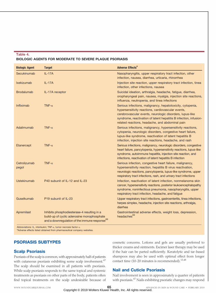
Biologic AgentsOver the past decade, several biologics have been licensed in
the US and Canada as safe and effective treatments for moderate
to severe plaque psoriasis (Table 4). Currently anti–IL-17 agents
(secukinumab brodalumab, ixekizumab), anti–IL-23 inhibitors
(guselkumab), and anti–IL-12-23 inhibitors (ustekinumab) are
commonly used agents for plaque psoriasis and psoriatic arthritis.
The anti-TNF agents (adalimumab, etanercept, infliximab,
certolizumab pegol) were first on the market and may still be
used for psoriatic arthritis, but newer agents have replaced the
TNF inhibitors for increased efficacy in plaque psoriasis.
The choice of biologic is often difficult for the provider and
the patient.39–41 Patients requiring systemic treatment may be
offered biologics based on criteria including the failure of two
or more conventional systemic treatments. The efficacy, safety,
and ease of administration of each particular agent should be
balanced against the coverage provided by the patient’s healthcare
insurance.13
Interleukin 17A plays a critical role in the pathophysiology of
psoriasis, and it is the target of many newly developed biologic
agents, including secukinumab, ixekizumab, and brodalumab.
Unlike secukinumab and ixekizumab, which bind to IL-17A
itself, brodalumab targets the IL-17A receptor on keratinocytes
and immune cells, providing a more direct therapeutic target.42
Infliximab, adalimumab, certolizumab pegol, and etanercept target
TNF-!.43 Ustekinumab prevents IL-12 and IL-23 from stimulating
receptor complexes by binding to the p40 subunit common to both
cytokines.44 In contrast, guselkumab targets only target IL-23 by
binding to its p19 subunit.45
A 2017 Cochrane review (prior to the marketing of some of the
newer agents) concluded that ustekinumab, infliximab, and certo-
lizumab have the best combination of efficacy and safety when
prescribed for plaque psoriasis. Ixekizumab had the highest
efficacy in terms of reaching PASI 90, whereas certolizumab had
the lowest relative risk of serious adverse events.38
There are several safety considerations that are associated with
biologic agents. All patients starting biologics should be given
the opportunity to take part in long-term saferty registries. Live
vaccines should be avoided in patients on biologic therapies and
infants (up to 6 months of age) born to mothers taking biologic
therapy beyond 16 weeks’ gestation. Special care should be taken
when prescribing biologics to patients with a history of cancer,
particularly in the past 5 years, and patients starting biologic therapy
should be tested for infection with hepatitis B and C, human im-
munodeficiency virus, varicella-zoster antibody, and latent tuber-
culosis (preferably with an interferon + release assay).19,46
Table 3.
TRADITIONAL SYSTEMIC MEDICATIONS FOR PSORIASIS
Medication Mechanism of Action Benefits Common Dosing Adverse EffectsEvidenceLevel
Methotrexate Inhibits production of
proliferating cells such
as lymphocytes35
Some evidence for
psoriatic arthritis35 and
best safety profile of
systemic agents37
Once weekly
7.5–25 mg + 1–5
mg folic acid daily
on other days35
FDA pregnancy category X;a
hepatotoxicity, bone marrow
suppression, pulmonary toxicity35
A
Cyclosporine Inhibits immune
response through
inhibition of
calcineurin35
Fast acting35
3–5 mg/kg daily35
Renal toxicity, hypertension, limit
continuous long-term use35
A
Acitretin Modulates epidermal
differentiation and has
anti-inflammatory
effects35
Evidence for generalized
pustular and erythrodermic
psoriasis and patients
who are HIV-positive35
10–50 mg/d35 with
meals
FDA pregnancy category X,a
hypertriglyceridemia, dryness of
oral and nasal mucosa, brittle
nails, alopecia, hepatotoxicity35
A
Sulfasalazine Exact mechanism
unclear; has anti-
inflammatory effects35
Low adverse effects
profile; some evidence
for psoriatic arthritis11,35
1–4 g daily35
Rash, nausea35
A
Abbreviation: HIV, human immunodeficiency virus.aFetal abnormalities have been demonstrated in animals or humans according to FDAbNo FDA approval in the UScNo FDA pregnancy category because not approved for use in the US
ADVANCES IN SKIN & WOUND CARE & VOL. 32 NO. 2 64 WWW.WOUNDCAREJOURNAL.COMCopyright © 2019 Wolters Kluwer Health, Inc. All rights reserved.
PSORIASIS SUBTYPES
Scalp PsoriasisPsoriasis of the scalp is common, with approximately half of patients
with cutaneous psoriasis exhibiting some scalp involvement.47
The scalp should be examined in all patients with psoriasis.
While scalp psoriasis responds to the same topical and systemic
treatments as psoriasis on other parts of the body, patients often
find topical treatments on the scalp undesirable because of
cosmetic concerns. Lotions and gels are usually preferred to
thicker creams and ointments. Excimer laser therapy may be used
if the hair can be parted sufficiently. Keratolytic and tar-based
shampoos may also be used with optimal effect from longer
contact time (10-20 minutes is recommended).13,48
Nail and Cuticle PsoriasisNail involvement is seen in approximately a quarter of patients
with psoriasis.47 Nails exhibiting psoriatic changes may respond
Table 4.
BIOLOGIC AGENTS FOR MODERATE TO SEVERE PLAQUE PSORIASIS
Biologic Agent Target Adverse Effectsa
Secukinumab IL-17A Nasopharyngitis, upper respiratory tract infection, other
infection, nausea, diarrhea, urticaria, rhinorrhea
Ixekizumab IL-17A Injection site reaction, upper respiratory tract infection, tinea
infection, other infections, nausea
Brodalumab IL-17A receptor Suicidal ideation, arthralgia, headache, fatigue, diarrhea,
oropharyngeal pain, nausea, myalgia, injection site reactions,
influenza, neutropenia, and tinea infections
Infliximab TNF-! Serious infections, malignancy, hepatotoxicity, cytopenia,
hypersensitivity reactions, cardiovascular events,
cerebrovascular events, neurologic disorders, lupus-like
syndrome, reactivation of latent hepatitis B infection, infusion-
related reactions, headache, and abdominal pain
Adalimumab TNF-! Serious infections, malignancy, hypersensitivity reactions,
cytopenia, neurologic disorders, congestive heart failure,
lupus-like syndrome, reactivation of latent hepatitis B
infection, injection site reactions, headache, and rash
Etanercept TNF-! Serious infections, malignancy, neurologic disorders, congestive
heart failure, pancytopenia, hypersensitivity reactions, lupus-like
syndrome, autoimmune hepatitis, injection site reaction, and
infections, reactivation of latent hepatitis B infection
Cetrolizumab
pegol
TNF-! Serious infection, congestive heart failure, malignancy,
hypersensitivity reaction, hepatitis B virus reactivation,
neurologic reactions, pancytopenia, lupus-like syndrome, upper
respiratory tract infections, rash, and urinary tract infections
Ustekinumab P40 subunit of IL-12 and IL-23 Infection, reactivation of latent infection, nonmelanoma skin
cancer, hypersensitivity reactions, posterior leukoencephalopathy
syndrome, noninfectious pneumonia, nasopharyngitis, upper
respiratory tract infection, headache, and fatigue
Guselkumab P19 subunit of IL-23 Upper respiratory tract infections, gastroenteritis, tinea infections,
herpes simplex, headache, injection site reactions, arthralgia,
diarrhea
Apremilast Inhibits phosphodiesterase-4 resulting in a
build-up of cyclic adenosine monophosphate
and a downregulation of the immune response69
Gastrointestinal adverse effects, weight loss, depression,
headaches69
Abbreviations: IL, interleukin; TNF-!, tumor necrosis factor !.aAdverse effects listed obtained from pharmaceutical company websites.
ADVANCES IN SKIN & WOUND CARE & FEBRUARY 201965WWW.WOUNDCAREJOURNAL.COMCopyright © 2019 Wolters Kluwer Health, Inc. All rights reserved.

to systemic therapies used to control psoriasis on other body sites.
To specifically target cuticle psoriasis, providers should consider
high-potency topical corticosteroids alone or in combination with
calcipotriol around the nail fold. In selected cases, intralesional
steroid injections can supplement treatment, but they may be very
painful.48
Intertriginous/Inverse PsoriasisIntertriginous psoriasis can occur at the point of contact of any
two skin folds; the inguinal fold, axilla, and external genitalia
are the most common sites. This site is characterized by well-
demarcated shiny red plaques with minimal to no scale. Because
of maceration of the skin, superinfection may occur. Any
erythematous eruption in the folds can be called intertrigo, with a
differential diagnosis that includes inflammatory conditions
(psoriasis, lichen planus), fungal infection (dermatophytes, yeast),
bacterial infections including erythrasma, and irritant and allergic
contact dermatitis.49,50 Intertriginous psoriasis can be distinguished
from these entities based on a history of psoriasis combined with
a physical examination that reveals the characteristic uniform
pink-to-red involvement. Other criteria for differentiation include:
& microscopic potassium hydroxide examination and fungal
cultures to rule out yeast or dermatophyte fungus;
& woods light examination (black light with coral fluorescence
characteristic of erythrasma);
& bacterial swab examined microscopically with culture to identify
organisms responsible for cellulitis/impetigo; and
& skin biopsy if necessary to rule out lichen planus, contact dermatitis,
and others.13,50
Psoriatic intertrigo is often treated with low-potency cortico-
steroids (often combined with an antiyeast agent to prevent
secondary colonization) continuously for 2 to 4 weeks and then
once to twice weekly for maintenance therapy. Alternately, topical
calcineurin inhibitors (pimecrolimus cream, tacrolimus ointment)
can be used as a steroid-sparing agent or when the skin is thin or
fragile. There is little evidence for the use of conventional systemic
therapies or biologics.
Erythrodermic PsoriasisErythrodermic psoriasis is life-threatening and affects 75% or
greater BSA with accompanying erythema, scaling, and occa-
sionally pustules. It may be associated with systemic symptoms
including fever, chills, fatigue, malaise, peripheral edema, heart
failure, renal failure, electrolyte imbalances, difficulties with ther-
moregulation, and superinfection. Erythroderma can be induced
by both the introduction and abrupt withdrawal of medications,
overuse of topical corticosteroids, stress, and alcohol use, among
other causes.51 The differential diagnosis includes severe atopic
dermatitis, drug eruptions, cutaneous lymphoma, and an uncom-
mon psoriasis-like keratinizing disorder (pityriasis rubra pilaris)
with a characteristic yellow hyperkeratosis of the palms and
soles.51
Management involves the evaluation and treatment of systemic
abnormalities and superinfection, as well as systemic medications.
Hospitalization may be necessary. First-line therapies include
methotrexate, acitretin, cyclosporine, and infliximab because of
their rapid onset of action. Systemic corticosteroids should be
generally avoided because they can be a trigger for erythrodermic
psoriasis (but may be the only choice in pregnancy or breastfeeding
mothers), and phototherapy is contraindicated as these patients
are often photosensitive.51 Mild- to moderate-potency topical corti-
costeroids are often safely used in combination with systemic
treatments.
Pustular PsoriasisPustular psoriasis presents as erythematous skin covered with
2- to 3-mm pustules and has generalized (acute, subacute) and
localized forms (palmoplantar pustulosis and acrodermatitis
continua of Hallopeau). Generalized pustular psoriasis may be
accompanied by systemic symptoms including fever, malaise,
arthralgias, peripheral edema, conjunctivitis, iritis, uveitis, oral
pustulosis, and cheilitis, among others. Both generalized and
localized pustular psoriasis may be induced by initiation and
withdrawal of systemic and topical medications, infection, the H1N1
vaccine, sunburn, stress, and hormone changes, among others.52
The differential diagnosis of generalized pustular psoriasis includes
pustular drug eruption, dermatitis with secondary infection,
immunoglobulin A pemphigus, and pustular tinea corporis. A
definitive diagnosis can be made based on history, physical
examination, and, if the diagnosis is still unclear, punch biopsy of
an intact pustule. First-line treatments for generalized pustular
psoriasis include acitretin, cyclosporine, and methotrexate, with
infliximab reserved for severe cases. Second-line treatments include
adalimumab, etanercept, and PUVA. Topical adjunctive treatments
including corticosteroids, calcipotriene, tacrolimus, and pime-
crolimus may be used in conjunction with systemic treatments.52
Localized pustular psoriasis has a debilitating impact on quality
of life and is often resistant to treatment. For localized pustular
psoriasis without psoriatic arthritis, the recommended first-line
therapy is potent or ultrapotent corticosteroids with occlusion and,
if applicable, smoking cessation.53
Guttate PsoriasisGuttate psoriasis is characterized by an acute eruption of drop-
shaped red scaling papules and plaques. It may occur 2 to 3 weeks
after a streptococcal infection in children and young adults. The
differential diagnosis includes pityriasis rosacea, tinea corporis,
secondary syphilis, pityriasis lichenoides chronica, nummular
ADVANCES IN SKIN & WOUND CARE & VOL. 32 NO. 2 66 WWW.WOUNDCAREJOURNAL.COMCopyright © 2019 Wolters Kluwer Health, Inc. All rights reserved.

dermatitis, and drug eruptions. These entities can be dif-
ferentiated from guttate psoriasis based on history and physical
examination with skin biopsy when necessary.54
To the authors’ knowledge, the last review of treatments specific
to guttate psoriasis was published in 2001. Investigations of treat-
ments specific to guttate psoriasis are limited. There is no evidence
to suggest any efficacy for antibiotics. Poor-quality evidence
suggests that tonsillectomy may be efficacious in some patients.
The most commonly used treatments include phototherapy,
topical corticosteroids, vitamin D analogs, and tar. The natural
history of guttate psoriasis is varied; it may clear on its own, turn
into chronic plaque psoriasis, or remit and reappear.54
ASSESSMENT AND MANAGEMENT OF PAININ PSORIASISPsoriasis is often associated with burning and stinging pain,
especially with increased inflammation and the formation of
fissures within plaques.55 Pain control can be achieved through
improved control of psoriasis with systemic and topical medications
as well as analgesics. The World Health Organization’s Pain
Ladder is a frequently used tool for choosing analgesics for the
nociceptive pain component (gnawing, aching, tender, throbbing).
The Pain Ladder suggests starting with acetaminophen and non-
steroidal anti-inflammatory drugs and then, if necessary, adding
mild opioids (codeine, tramadol, or hydromorphone).55,56 For the
neuropathic component (burning, stinging, shooting, stabbing),
oral agents including pregabalin, gabapentin, nortriptyline, and
carbamazepine can provide symptom control. Providers must
address the psychological component of the pain, and any psy-
chiatric comorbidities should be diagnosed and treated.55
PSORIASIS AND WOUNDSWound care specialists may be called to treat peristomal psoriasis.
A recent study reported a prevalence of peristomal psoriasis of 5%
in a general stoma clinic.57 While the majority of these patients
(70%) had widespread cutaneous psoriasis, 3% had psoriasis only
in the peristomal region, and 27% only had subtle signs of psoriatic
disease outside the peristomal area.57 Recent recommendations
from a team of dermatologists who manage peristomal psoriasis
have been reported.57 Simply covering the peristomal psoriasis
with a thick hydrocolloid dressing may be effective in some cases.
Occlusion and topical corticosteroid lotion, gels, and foam vehicles
are also used; providers should avoid greasier creams and oint-
ments because they prevent the adhesion of the ostomy appliance.
Topical products should be left to dry before the stoma appliance is
reattached. In resistant cases, intralesional steroid injections using
triamcinolone 2.5 to 8 mg/mL using 1 to 3 mL can often control the
disease. Minimizing peristomal trauma is imperative during appli-
cation of topical treatments. To avoid Koebner phenomena, flat
appliances are preferred to convex alternatives. Conventional
systemic treatments, phototherapy (with the stoma protected),
and biologics are also used.57
A topic of some debate is whether to recommend continuation
or interruption of biologic therapy for patients with psoriasis who
are undergoing surgery. The 2017 British Association of
Dermatologists guideline recommends stopping biologic therapy
for either three to five half-lives of the drug or the length of one
treatment cycle, whichever is longer, prior to surgery.46 However,
two recent studies of patients with psoriasis managed by biologic
therapy documented that 66% to 74% continued treatment
through the perioperative period, with a greater number of patients
suspending therapy for more extensive cardiothoracic and
orthopedic surgery. Both studies reported no increased risk of
wound complications or infections with continued biologic
therapy. However, these results were based on 77 and 131 surgical
procedures, respectively, and a larger patient population needs to be
studied before making definitive alternate recommendations.58,59
In general, psoriasis does not appear to have a negative effect
on wound healing.60,61 However, the Koebner phenomenon
may result in patients developing psoriatic lesions over surgery
sites62 and tattoos,63 among other injuries. This phenomenon is
more common in patients with unstable, undertreated disease.
Both the epidermis and the dermis must be involved for clinical
disease to occur.62 Lesions typically arise 10 to 20 days after
trauma but have been known to arise anywhere from 3 days to
2 years postinjury.18,62
CONCLUSIONSIn conclusion, psoriasis can be differentiated into plaque, inverse,
erythrodermic, pustular, and guttate forms. In addition to cutaneous
manifestations, nail, scalp, joint, and systemic abnormalities may be
present. Therefore, patients presenting with a suspected diagnosis
of psoriasis should undergo a thorough history and physical
examination including the scalp, nails, and joints.
Optimal management depends on the form of psoriasis, severity,
location, and patient preference. Psoriasis affecting less than 10% of
the patient’s BSA and without debilitating effect on the patient may
be treated with topical therapies. A BSA involvement of more than
10%, debilitating impact on life, or suboptimal response with topical
therapies should prompt consideration of phototherapy or con-
ventional systemic therapy. Biologic therapy is usually reserved
for patients with 10% or more of the body involved, a PASI score
greater than 10, and a Dermatology Quality of life Index score
greater than 10. These patients have usually failed or have a
contraindication to two or more conventional systemic therapies
and/or photochemotherapy.
Currently, psoriasis treatment comprises an effective toolkit
of therapeutic options. There are even more new agents on the
ADVANCES IN SKIN & WOUND CARE & FEBRUARY 201967WWW.WOUNDCAREJOURNAL.COMCopyright © 2019 Wolters Kluwer Health, Inc. All rights reserved.
horizon, each with the potential to give patients with psoriasis
improved quality of life and control of key aspects of psoriasis.
PRACTICE PEARLS
REFERENCES1. Parisi R, Symmons DP, Griffiths CE, Ashcroft DM. Global epidemiology of psoriasis: a
systematic review of incidence and prevalence. J Invest Dermatol 2013;133(2):377-85.
2. Langley RGB, Krueger GG, Griffiths CEM. Psoriasis: epidemiology, clinical features, and
quality of life. Ann Rheum Dis 2005;64(Suppl 2):ii18-ii23.
3. Farber EM, Bright RD, Nall ML. Psoriasis: a questionnaire survey of 2,144 patients. Arch
Dermatol 1968;98(3):248-59.
4. Dand N, Mucha S, Tsoi LC, et al. Exome-wide association study reveals novel psoriasis
susceptibility locus at TNFSF15 and rare protective alleles in genes contributing to type
I IFN signalling. Hum Mol Genet 2017;26(21):4301-13.
5. Rodrıguez-Zuniga MJM. Systematic review and meta-analysis of the association between
psoriasis and metabolic syndrome. J Am Acad Dermatol 2017;77(4):657-66.e8.
6. Li X, Kong L, Li F, et al. Association between psoriasis and chronic obstructive pulmonary
disease: a systematic review and meta-analysis. PloS One 2015;10(12):e0145221.
7. Candia R, Ruiz A, Torres-Robles R, Chavez-Tapia N, Mendez-Sanchez N, Arrese M. Risk of
non-alcoholic fatty liver disease in patients with psoriasis: a systematic review and meta-analysis.
J Eur Acad Dermatol Venereol 2015;29(4):656-62.
8. Horreau C, Pouplard C, Brenaut E, et al. Cardiovascular morbidity and mortality in psoriasis
and psoriatic arthritis: a systematic literature review. J Eur Acad Dermatol Venereol 2013;
27 Suppl 3:12-29.
9. Singh S, Taylor C, Kornmehl H, Armstrong AW. Psoriasis and suicidality: a systematic
review and meta-analysis. J Am Acad Dermatol 2017;77(3):425-40.e2.
10. Dowlatshahi EA, Wakkee M, Arends LR, Nijsten T. The prevalence and odds of depressive
symptoms and clinical depression in psoriasis patients: a systematic review and meta-analysis.
J Invest Dermatol 2014;134(6):1542–51.
11. Nestle FO, Kaplan DH, Barker J. Psoriasis. N Engl J Med 2009;361(5):496-509.
12. Gooderham MJ, Papp KA, Lynde CW. Shifting the focus: the primary role of IL-23 in
psoriasis and other inflammatory disorders. J Eur Acad Dermatol Venereol 2018;32(7):
1111-9.
13. Ladizinski B, Lee KC, Wilmer E, Alavi A, Mistry N, Sibbald RG. A review of the clinical
variants and the management of psoriasis. Adv Skin Wound Care 2013;26(6):271-84.
14. Zachariae H. Prevalence of joint disease in patients with psoriasis. Am J Clin Dermatol
2003;4(7):441-7.
15. Brockbank JE, Schentag C, Rosen C, Gladman DD. Psoriatic arthritis (PSA) is common
among patients with psoriasis and family medicine clinic attendees. Arthritis Rheum 2001;
44(9):S94.
16. Gottlieb A, Korman NJ, Gordon KB, et al. Guidelines of care for the management of
psoriasis and psoriatic arthritis: section 2. Psoriatic arthritis: overview and guidelines of care
for treatment with an emphasis on the biologics. J Am Acad Dermatol 2008;58(5):851-64.17. Mehta S, Singal A, Singh N, Bhattacharya SN. A study of clinicohistopathological correlation
in patients of psoriasis and psoriasiform dermatitis. Indian J Dermatol Venereol Leprol 2009;
75(1):100.18. Weiss G. The Koebner phenomenon: review of the literature. J Eur Acad Dermatol Venereol
2002;16(3):241-8.19. Dauden E, Puig L, Ferrandiz C, Sanchez-Carazo JL, Hernanz-Hermosa JM, the Spanish
Psoriasis Group of the Spanish Academy of Dermatology and Venereology. Consensus
document on the evaluation and treatment of moderate-to-severe psoriasis: Psoriasis
Group of the Spanish Academy of Dermatology and Venereology. J Eur Acad Dermatol
Venereol 2016;30:1-18.20. Thomas CL, Finlay AY. The ‘handprint’ approximates to 1% of the total body surface
area whereas the ‘palm minus the fingers’ does not. Br J Dermatol 2007;157(5):1080-1.21. Chiricozzi A, Pimpinelli N, Ricceri F, et al. Treatment of psoriasis with topical agents:
recommendations from a Tuscany Consensus. Dermatol Ther 2017;30(6):e12549.22. Samarasekera EJ, Sawyer L, Wonderling D, Tucker R, Smith CH. Topical therapies for
the treatment of plaque psoriasis: systematic review and network meta-analyses. Br J
Dermatol 2013;168(5):954-67.
23. Takahashi H, Tsuji H, Ishida-Yamamoto A, Iizuka H. Comparison of clinical effects of
psoriasis treatment regimens among calcipotriol alone, narrowband ultraviolet B phototherapy
alone, combination of calcipotriol and narrowband ultraviolet B phototherapy once a week, and
combination of calcipotriol and narrowband ultraviolet B phototherapy more than twice a week.
J Dermatol 2013;40(6):424-7.
24. Wang C, Lin A. Efficacy of topical calcineurin inhibitors in psoriasis. J Cutan Med Surg
2014;18(1):8-14.
25. Jacobi A, Mayer A, Augustin M. Keratolytics and emollients and their role in the therapy
of psoriasis: a systematic review. Dermatol Ther 2015;5(1):1-18.
26. Roelofzen JH, Aben KK, van der Valk PG, van Houtum JL, van de Kerkhof PC, Kiemeney
LA. Coal tar in dermatology. J Dermatol Treat 2007;18(6):329-34.
27. McGugan AD, Shuster S, Bottoms E. Adrenal suppression from intradermal triamcinolone. J
Invest Dermatol 1963;40(6):271-2.
28. Richards RN. Update on intralesional steroid: focus on dermatoses. J Cutan Med Surg
2010;14(1):19-23.
29. Mehta D, Lim HW. Ultraviolet B phototherapy for psoriasis: review of practical guidelines.
Am J Clin Dermatol 2016;17(2):125-33.
30. Matos TR, Ling TC, Sheth V. Ultraviolet B radiation therapy for psoriasis: pursuing the
optimal regime. Clin Dermatol 2016;34(5):587-93.
31. Menter A, Korman NJ, Elmets CA, et al. Guidelines of care for the management of psoriasis
and psoriatic arthritis: section 5. Guidelines of care for the treatment of psoriasis with phototherapy
and photochemotherapy. J Am Acad Dermatol 2010;62(1):114-35.
32. Pathirana D, Ormerod AD, Saiag P, et al. European S3-guidelines on the systemic treatment
of psoriasis vulgaris. J Eur Acad Dermatol Venereol 2009;23 Suppl 2:1-70.33. George SL. Adverse effects with PUVA and UVB phototherapy. J Dermatol Treat 2001;12(2):101-5.34. Archier E, Devaux S, Castela E, et al. Carcinogenic risks of psoralen UV-A therapy and
narrowband UV-B therapy in chronic plaque psoriasis: a systematic literature review. J
Eur Acad Dermatol Venereol 2012;26 Suppl 3:22-31.35. Stern RS. The risk of squamous cell and basal cell cancer associated with psoralen and
ultraviolet A therapy: a 30-year prospective study. J Am Acad Dermatol 2012;66(4):553-62.36. Menter A, Korman NJ, Elmets CA, et al. Guidelines of care for the management of psoriasis
and psoriatic arthritis: section 4. Guidelines of care for the management and treatment of
psoriasis with traditional systemic agents. J Am Acad Dermatol 2009;61(3):451-85.37. Nast A, Amelunxen L, Augustin M, et al. S3 Guideline for the treatment of psoriasis vulgaris,
updateVshort version part 1Vsystemic treatment. J Dtsch Dermatol Ges 2018;16(5):645-69.38. Sbidian E, Chaimani A, Garcia-Doval I, et al. Systemic pharmacological treatments for
chronic plaque psoriasis: a network meta-analysis. Cochrane Database Syst Rev 2017;
12:CD011535.39. Jabbar-Lopez ZK, Yiu ZZ, Ward V, et al. Quantitative evaluation of biologic therapy options
for psoriasis: a systematic review and network meta-analysis. J Invest Dermatol 2017;137(8):
1646-54.40. Papp KA, Reich K, Paul C, et al. A prospective phase III, randomized, double-blind, placebo-controlled
study of brodalumab in patients with moderate-to-severe plaque psoriasis. Br J Dermatol
2016;175(2):273-86.41. Sofen H, Smith S, Matheson RT, et al. Guselkumab (an IL-23–specific mAb) demonstrates
clinical and molecular response in patients with moderate-to-severe psoriasis. J Allergy Clin
Immunol 2014;133(4):1032-40.
& Psoriasis can manifest in plaque, inverse, guttate, pustular,
or erythrodermic forms.
& Fully examine the skin, nails, scalp, and joints to document
the extent of the disease.
& Patients with less than 10% BSA affected are usually treated
with topical treatments: corticosteroids and vitamin D analogs
are first-line therapies.
& For patients with moderate to severe disease (Q10 BSA or
debilitating disease), consider phototherapy and conventional
systemic treatments.
& Biologic therapies are usually reserved for patients who have
failed or have contraindications to two or more conventional
systemic therapies and with residual psoriasis impairing their
quality of life.
ADVANCES IN SKIN & WOUND CARE & VOL. 32 NO. 2 68 WWW.WOUNDCAREJOURNAL.COMCopyright © 2019 Wolters Kluwer Health, Inc. All rights reserved.

42. Sawyer L, Fotheringham I, Wright E, Yasmeen N, Gibbons C, Holmen Møller A. The comparative
efficacy of brodalumab in patients with moderate-to-severe psoriasis: a systematic literature
review and network meta-analysis. J Dermatol Treat 2018;29(6):557-68.
43. Nesbitt A, Fossati G, Bergin M. Mechanism of action of certolizumab pegol (CDP870): in vitro
comparisonwithotheranti–tumornecrosis factor! agents. InflammBowelDis 2007;13(11):1323-32.
44. Benson JM, Peritt D, Scallon BJ, et al. Discovery and mechanism of ustekinumab: a
human monoclonal antibody targeting interleukin-12 and interleukin-23 for treatment
of immune-mediated disorders. MAbs 2011;3(6):535-45.
45. Guselkumab (Tremfya) for psoriasis. JAMA 2017;318(24):2487-88.
46. Smith CH, Jabbar-Lopez ZK, Yiu ZZ, et al. British Association of Dermatologists guidelines
for biologic therapy for psoriasis 2017. Br J Dermatol 2017;177(3):628-36.
47. Merola JF, Li T, Li W-Q, Cho E, Qureshi AA. Prevalence of psoriasis phenotypes among
men and women in the USA. Clin Exp Dermatol 2016;41(5):486-9.
48. Merola JF, Qureshi A, Husni ME. Underdiagnosed and undertreated psoriasis: nuances
of treating psoriasis affecting the scalp, face, intertriginous areas, genitals, hands, feet,
and nails. Dermatol Ther 2018;31(3):e12589.
49. Lisi P. Differential diagnosis of psoriasis. Reumatismo 2007;59(1s):56-60.
50. Syed ZU, Khachemoune A. Inverse psoriasis: case presentation and review. J Clin Dermatol
2011;12(2):143-6.
51. Rosenbach M, Hsu S, Korman NJ, et al. Treatment of erythrodermic psoriasis: from the
medical board of the National Psoriasis Foundation. J Am Acad Dermatol 2010;62(4):
655-62.
52. Hoegler KM, John AM, Handler MZ, Schwartz RA. Generalized pustular psoriasis: a review
and update on treatment. J Eur Acad Dermatol Venereol 2018;32(10):1645-51.
53. Sevrain M, Richard MA, Barnetche T, et al. Treatment for palmoplantar pustular psoriasis:
systematic literature review, evidence-based recommendations and expert opinion. J Eur
Acad Dermatol Venereol 2014;28:13-16.
54. Chalmers RJG, O’Sullivan T, Owen CM, Griffiths CEM. A systematic review of treatments for
guttate psoriasis. Br J Dermatol 2001;145(6):891-4.
55. Pithadia D, Reynolds K, Lee E, Wu J. Psoriasis-associated cutaneous pain: etiology, assessment,
impact, and management. J Dermatolog Treat 2018:1-21.
56. Vargas-Schaffer G. Is the WHO analgesic ladder still valid? Twenty-four years of experience.
Can Fam Physician 2010;56(6):514-7.
57. Marshall C, Woodmansey S, Lyon CC. Peristomal psoriasis. Clin Exp Dermatol 2017;42(3):282-6.
58. Bakkour W, Purssell H, Chinoy H, Griffiths CEM, Warren RB. The risk of post-operative
complications in psoriasis and psoriatic arthritis patients on biologic therapy undergoing
surgical procedures. J Eur Acad Dermatol Venereol 2016;30(1):86-91.
59. Fabiano A, De Simone C, Gisondi P, et al. Management of patients with psoriasis treated
with biological drugs needing a surgical treatment. Drug Dev Res 2014;75 Suppl 1:S24-6.
60. Morhenn VB, Nelson TE, Gruol DL. The rate of wound healing is increased in psoriasis.
J Dermatol Sci 2013;72(2):87-92.
61. Young PM, Parsi KK, Schupp CW, Armstrong AW. Psoriasis and wound healing outcomes: a
retrospective cohort study examining wound complications and antibiotic use. Dermatol
Online J 2017;23(11).
62. Ganguly AK, Laghimsetty S, Bhagyalakshmi N. Koebner phenomenon triggered by external
dacryocystorhinostomy scar in a patient with psoriasis: a case report and literature review.
Ophthal Plast Reconstr Surg 2018;34(2):e52-e53.
63. Kluger N, Esteve E, Fouere S, Dupuis-Fourdan F, Jegou M-H, Levy-Rameau C. Tattooing and
psoriasis: a case series and review of the literature. Int J Dermatol 2017;56(8):822-7.
64. Norris DA. Mechanisms of action of topical therapies and the rationale for combination
therapy. J Am Acad Dermatol 2005;53(1):S17-S25.
65. Lebwohl M, Ali S. Treatment of psoriasis. Part 1. Topical therapy and phototherapy. J
Am Acad Dermatol 2001;45(4):487-502.
66. Menter A, Korman NJ, Elmets CA, et al. Guidelines of care for the management of psoriasis
and psoriatic arthritis. Section 3. Guidelines of care for the management and treatment of
psoriasis with topical therapies. J Am Acad Dermatol 2009;60(4):643-59.
67. Fluhr JW, Cavallotti C, Berardesca E. Emollients, moisturizers, and keratolytic agents in
psoriasis. Clin Dermatol 2008;26(4):380-6.
68. Siegfried EC, Jaworski JC, Hebert AA. Topical calcineurin inhibitors and lymphoma risk:
evidence update with implications for daily practice. Am J Clin Dermatol 2013;14(3):163-78.
69. Keating GM. Apremilast: a review in psoriasis and psoriatic arthritis. Drugs 2017;77(4):
459-72.
For more than 139 additional continuing education articles related to Skin and Wound Care topics,go to NursingCenter.com/CE.
CONTINUING MEDICAL EDUCATION INFORMATION FOR PHYSICIANSLippincott Continuing Medical Education Institute, Inc., is accredited by the Accreditation
Council for Continuing Medical Education to provide continuing medical education
for physicians.
Lippincott Continuing Medical Education Institute, Inc., designates this journal-based CME activity
for a maximum of 1 AMA PRA Category 1 CreditTM. Physicians should claim only the credit
commensurate with the extent of their participation in the activity.
PROVIDER ACCREDITATION INFORMATION FOR NURSESLippincott Professional Development will award 1.5 contact hours including 1.5 Pharmacology
credits for this continuing nursing education activity.
LPD is accredited as a provider of continuing nursing education by the American Nurses Credentialing
Center’s Commission on Accreditation.
This activity is also provider approved by the California Board of Registered Nursing, Provider
Number CEP 11749 for 1.5 contact hours. LWW is also an approved provider by the District of
Columbia, Georgia, and Florida CE Broker #50-1223.
OTHER HEALTH PROFESSIONALSThis activity provides ANCC credit for nurses and AMA PRA Category 1 CreditTM for MDs and
DOs only. All other healthcare professionals participating in this activity will receive a certificate
of participation that may be useful to your individual profession’s CE requirements.
CONTINUING EDUCATION INSTRUCTIONS
&Read the article beginning on page 58. For nurses who wish to take the test for CNE contact
hours, visit http://nursing.ceconnection.com. For physicians who wish to take the test for CME
credit, visit http://cme.lww.com. Under the Journal option, select Advances in Skin and Wound Care
and click on the title of the CE activity.
&You will need to register your personal CE Planner account before taking online tests. Your planner
will keep track of all your Lippincott Professional Development online CE activities for you.
& There is only one correct answer for each question. A passing score for this test is 13 correct
answers. If you pass, you can print your certificate of earned contact hours or credit and access
the answer key. Nurses who fail have the option of taking the testagainatnoadditional cost. Only the
first entry sent by physicians will be accepted for credit.
Registration Deadline: January 31, 2021 (physicians); December 4, 2020 (nurses).
PAYMENT
& The registration fee for this CE activity is $17.95 for nurses; $22.00 for physicians.
ADVANCES IN SKIN & WOUND CARE & FEBRUARY 201969WWW.WOUNDCAREJOURNAL.COMCopyright © 2019 Wolters Kluwer Health, Inc. All rights reserved.


![A case of simultaneous acute generalized exanthematous ...AGEP is Generalized Pustular Psoriasis (GPP) [8] “GPP shares many of the clinical and immunological findings and the presentation](https://static.cupdf.com/doc/110x72/5fddc90e28884f1c641049be/a-case-of-simultaneous-acute-generalized-exanthematous-agep-is-generalized-pustular.jpg)