


Deficiency in Clonogenic Endometrial MesenchymalStem Cells in Obese Women with Reproductive Failure –a Pilot StudyKeisuke Murakami1,2, Harish Bhandari1, Emma S. Lucas1, Satoru Takeda2, Caroline E. Gargett3,
Siobhan Quenby1, Jan J. Brosens1, Bee K. Tan1,4*
1 Division of Reproductive Health, Clinical Science Research Laboratories, Warwick Medical School, University of Warwick, Coventry, United Kingdom, 2 Department of
Obstetrics and Gynecology, Juntendo University Faculty of Medicine, Tokyo, Japan, 3 The Ritchie Centre, Monash Institute of Medical Research and Department of
Obstetrics and Gynecology, Monash University, Clayton, Australia, 4 Department of Obstetrics and Gynecology, Birmingham Heartlands Hospital, Heart of England NHS
Foundation Trust, Birmingham, United Kingdom
Abstract
Objectives: The mechanisms of obesity associated reproductive complications remain poorly understood. Endometrialmesenchymal stem-cells are critical for cyclic renewal and uterine function. Recently, W5C5+ cells, with high clonogenicity,capable of producing endometrial stroma in vivo, have been described. We sought to investigate the abundance andcloning efficiency of W5C5+ and W5C52 endometrial cells in relation to Body Mass Index, age and reproductive outcome.
Design: W5C5+ and W5C52 cells were purified from mid-luteal endometrial biopsies (n = 54) by magnetic bead separationand subjected to in vitro colony-forming assays.
Results: First trimester pregnancy losses were significantly higher in obese subjects (n = 12) compared to overweight(n = 20) and subjects with normal Body Mass Index (n = 22) (P,0.05, P,0.01, respectively). W5C5+ cells (%) were significantlylower in obese subjects compared to subjects with normal Body Mass Index (P,0.05). W5C5+ cloning efficiency wassignificantly lower in obese subjects compared to overweight and subjects with normal Body Mass Index (P,0.05,respectively). W5C52 cloning efficiency was significantly lower in obese subjects compared to subjects with normal BodyMass Index (P,0.05). Body Mass Index was significantly negatively correlated with W5C5+ cloning efficiency and W5C52
cloning efficiency (P,0.01, respectively), and positively correlated with first trimester loss (P,0.01). We found no significantresults with age (P.0.05).
Conclusions: Our observations suggest that the regenerative capacity and plasticity of the endometrium of obese women issuboptimal, which in turn may account for the increased risk of reproductive complications associated with obesity.
Citation: Murakami K, Bhandari H, Lucas ES, Takeda S, Gargett CE, et al. (2013) Deficiency in Clonogenic Endometrial Mesenchymal Stem Cells in Obese Womenwith Reproductive Failure – a Pilot Study. PLoS ONE 8(12): e82582. doi:10.1371/journal.pone.0082582
Editor: Harpal Singh Randeva, University of Warwick – Medical School, United Kingdom
Received October 18, 2013; Accepted November 5, 2013; Published December 10, 2013
Copyright: � 2013 Murakami et al. This is an open-access article distributed under the terms of the Creative Commons Attribution License, which permitsunrestricted use, distribution, and reproduction in any medium, provided the original author and source are credited.
Funding: This work was supported by the Biomedical Research Unit in Reproductive Health, a joint initiative between the University Hospitals Coventry andWarwickshire NHS Trust and Warwick Medical School. Dr Caroline Gargett also received funding from The Australian National Health and Medical Research Councilfor a Senior Research Fellowship 1042298 (CEG) and the Victorian Government’s Operational Infrastructure Support Program. The funders had no role in studydesign, data collection and analysis, decision to publish, or preparation of the manuscript.
Competing Interests: The authors have declared that no competing interests exist.
* E-mail: [email protected]
Introduction
The pandemic of obesity is one of today’s most blatantly visible
global public health problem. Obesity is associated with adverse
metabolic as well as reproductive outcomes such as miscarriage
and infertility [1–3]. However, the mechanisms of obesity
associated reproductive complications remain poorly understood.
Consequently, there are no effective interventions that are
available in the prevention and treatment of obesity associated
reproductive disorders. We put forward a novel hypothesis that a
suboptimal uterine environment in obese women at time of
embryo implantation predisposes to reproductive failure.
A striking feature of the human endometrium, shared with only
a handful of other mammalian species, is spontaneous decidua-
lization of the stromal compartment during the mid-luteal phase of
each cycle, a process also responsible for the menstrual shedding of
the endometrium in the absence of pregnancy. Decidualization is
characterised by transformation of endometrial stromal fibroblast
into specialized secretory decidual cells, a process indispensable for
embryo implantation [4]. Perturbations in decidualization can
have negative effects on trophoblast invasion, placental develop-
ment and, ultimately, maternal and fetal well-being [5]. An
inevitable consequence of spontaneous decidualization followed by
menstruation is the requirement for cyclic regeneration of the
endometrium. The human endometrium exhibits remarkable
regenerative capacity [6]. The endometrium is rich in mesenchy-
mal stem-like cells (eMSCs), which are immuno-privileged
compared to other types of stem-like cells, rendering them a
PLOS ONE | www.plosone.org 1 December 2013 | Volume 8 | Issue 12 | e82582

promising resource for cell-based therapies [7–9]. They are a
resident cell population, although there is some evidence of active
recruitment of stem-like cells to hypoxic, proteolytic and
inflammatory stimuli associated with cyclic menstruation or
pregnancy [6,10,11]. This process of constant renewal bestows
plasticity on the endometrium, enabling it to adapt to reproductive
failure and a changing environment. Recently, Masuda et al.
identified W5C5 as a novel single marker for purifying eMSCs that
self-renew, have high clonogenicity, are multipotent (differentiate
into adipogenic, osteogenic, chondrogenic and myogenic cell
lineages) and are capable of producing endometrial stroma
(mesodermal tissue) in vivo [12].
We sought to study the relationship between the abundance and
cloning efficiency (CE) of W5C5+ and W5C52 endometrial cells
with body mass index (BMI), age and reproductive outcome.
Materials and Methods
EthicsThe study was approved by the NHS National Research Ethics
- Hammersmith and Queen Charlotte’s & Chelsea Research
Ethics Committee (1997/5065). The study has also been approved
by the University Hospitals Coventry and Warwickshire Research
and Development department, and research sponsorship for this
study has been transferred from Imperial College, London to the
University of Warwick, and written informed consent was
obtained from all participants, in accordance with the guidelines
in The Declaration of Helsinki 2000.
Patient selection and endometrial samplingA total of 54 Caucasian subjects were recruited consecutively
from the Implantation Clinic, a dedicated research clinic at
University Hospital Coventry and Warwickshire for women with
recurrent pregnancy loss or recurrent in vitro fertilization treatment
failure. Weight and height measurements were performed and
BMI was calculated in all subjects. The World Health Organiza-
tion classification of BMI, normal BMI (, 25), overweight (BMI:
25.0–29.9) and obese (BMI $ 30) was used. All endometrial
biopsies were timed and histologically dated between 7 to 10 days
after the pre-ovulatory luteinizing hormone surge. Samples were
obtained using a Wallach EndocellTM sampler (Wallach, Trum-
bull, USA) under ultrasound guidance, starting from the uterine
fundus and moving downwards to the internal cervical ostium.
Preparation of single cell suspensions of humanendometrial stromal cells
Endometrial samples were collected as described above and
single cell suspensions of human endometrial stromal cells
(HESCs) were isolated using a protocol that was a modification
of the method described [12.13]. Briefly, samples were washed in
DMEM/F-12 medium (Invitrogen, Paisley, UK), finely minced
and enzymatically digested with collagenase (0.5 mg/ml) (Sigma-
Aldrich, Gillingham, UK) and deoxyribonuclease type I (0.1 mg/
ml) (Roche, Burgess Hill, UK) for 1 hour at 37uC. The dissociated
cells were filtered through a sterile 40 um cell strainer (Fisher
Scientific, Loughborough, UK). Most of the stromal cells and
blood cells, present as a single cell suspension, passed through the
Table 1. Demographics, W5C5+ cells (%), W5C52 cells (%),W5C5+ CE and W5C52 CE.
Variable All Subjects BMI , 25.0BMI 25.0-29.9 BMI $ 30.0
Age (years) 35.260.7 34.561.0 36.261.2 34.761.7
BMI (kg/m2) 26.760.7 22.560.3 26.660.4 34.561.5
Live Births 0.660.2 0.460.2 0.660.2 0.960.5
First Trimester Loss 3.360.4 1.960.4 3.260.5 5.960.1
W5C5+ cells (%) 7.060.5 8.160.9 7.060.8 4.960.9
W5C52 cells (%) 93.060.5 91.960.9 93.060.8 95.160.9
Cloning Efficiency
W5C5+ (%) 2.460.3 3.460.6 2.260.4 1.060.3
W5C52 (%) 0.760.2 1.260.4 0.560.2 0.260.1
Data are mean 6 SEM. CE = Cloning Efficiency.All Subjects (n = 54); BMI , 25.0 (n = 22); BMI 25.0-29.9 (n = 20); BMI $ 30.0(n = 12).doi:10.1371/journal.pone.0082582.t001
Figure 1. Cloning Efficiency (CE) of endometrial mesenchymalstem cells. (A) W5C5+ CE was significantly greater than W5C52 CE.Data are means 6 SEM. Group comparison by Unpaired t test.****P,0.0001. (B) Relationship between W5C5+ CE and W5C52 CE in allsubjects (n = 54). Pearson correlation coefficient: (R = 0.817, P,0.01).doi:10.1371/journal.pone.0082582.g001
Obesity and Endometrial Stem Cell Deficiency
PLOS ONE | www.plosone.org 2 December 2013 | Volume 8 | Issue 12 | e82582
cell strainer, whereas the undigested fragments, mostly comprising
of glandular clumps, were retained on the strainer. Stromal single
cell suspensions were layered over Ficoll-Paque PLUS (GE
Healthcare, Little Chalfont, UK) and centrifuged to remove
erythrocytes. The medium/Ficoll-Paque PLUS interface, mainly
containing stromal cells, was carefully aspirated, washed with
DMEM/F-12 medium, and then subjected to magnetic bead
separation.
Magnetic beads separationW5C5+ cells are perivascular in their location and are evenly
distributed throughout the endometrium [12]. Magnetic bead
separation was performed according to the manufacture’s
instruction (Miltenyi Biotec, Bisley, UK) on a Wallach EndocellTM
core of approximately 1.5 cm from all study subjects. Briefly, equal
amounts of freshly isolated endometrial stromal cell suspensions
(up to 16106 cells/100 ml of 0.5% BSA in PBS: Magnetic Bead
Buffer) were incubated with phycoerythrin (PE) conjugated Anti-
W5C5 antibody (5 ml/16106 cells) (BioLegend, London, UK) on
ice for 20 minutes. Then cell suspensions (up to 16107 cells/80 ml
of Magnetic Bead Buffer) were incubated with Anti-PE MicroBe-
ads (20 ml/16107 cells) on ice for 20 minutes. Cell suspensions (up
to 16108 cells/500 ul of Magnetic Bead Buffer) were applied onto
the MS columns in a magnetic field followed by washing with
500 ml of Magnetic Bead Buffer three times. The columns were
removed from the magnetic field and W5C5+ cells were flushed
out by firmly pushing the plunger with 1 ml of Magnetic Bead
Buffer. Cell counts were performed after magnetic bead separation
and the percentages of W5C5+ cells and W5C52 cells were
calculated.
In vitro colony-forming assayFreshly isolated W5C5+ and W5C52 cells were seeded at a
clonal density of 50 cells/cm2 (to ensure equal loading onto
fibronectin-coated 60 mm culture dishes and cultured in growth
medium: DMEM/F12 containing 10% dextran-coated charcoal-
treated fetal bovine serum (DCC-FBS), 1% L-glutamine (Invitro-
gen, Paisley, UK), 1% antibiotic-antimycotic solution (Invitrogen,
Paisley, UK), insulin (2 mg/ml) (Sigma-Aldrich, Gillingham, UK),
estradiol (1 nM) (Sigma-Aldrich, Gillingham, UK) and basic
fibroblast growth factor (10 ng/ml) (Merck Millipore, Watford,
UK). The first medium change was after the first 7 days.
Subsequently, media was changed every 3-4 days. Colonies were
monitored microscopically to ensure that they were derived from
single cells. Cultures were terminated at 15 days and stained with
hemotoxylin. Clusters of $ 50 cells were counted and the CE was
determined from the formula: CE (%) = (number of colonies/
number of cells seeded) 6 100.
Statistical AnalysisData were checked for normal distribution using histograms and
the Kolmogorov-Smirnoff test. Data were analysed by Unpaired t
test or ANOVA (post hoc analysis: Tukey’s test), depending on the
number of groups compared. Data are means 6 SEM. Pearson
correlation was used for calculation of associations between
variables. All statistical analyses were performed using GraphPad
Prism 6 (GraphPad Software, Inc., La Jolla, USA) and SPSS
version 21.0 (SPSS, Inc., Chicago, USA). P,0.05 was considered
significant.
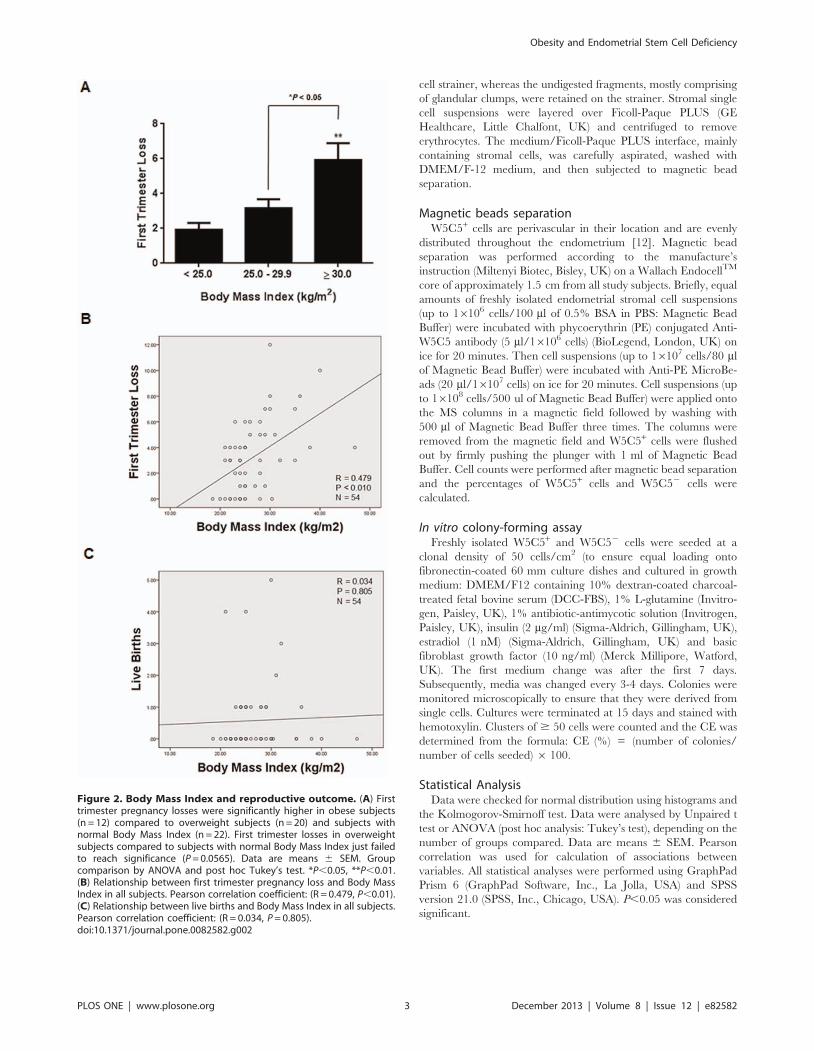
Figure 2. Body Mass Index and reproductive outcome. (A) Firsttrimester pregnancy losses were significantly higher in obese subjects(n = 12) compared to overweight subjects (n = 20) and subjects withnormal Body Mass Index (n = 22). First trimester losses in overweightsubjects compared to subjects with normal Body Mass Index just failedto reach significance (P = 0.0565). Data are means 6 SEM. Groupcomparison by ANOVA and post hoc Tukey’s test. *P,0.05, **P,0.01.(B) Relationship between first trimester pregnancy loss and Body MassIndex in all subjects. Pearson correlation coefficient: (R = 0.479, P,0.01).(C) Relationship between live births and Body Mass Index in all subjects.Pearson correlation coefficient: (R = 0.034, P = 0.805).doi:10.1371/journal.pone.0082582.g002
Obesity and Endometrial Stem Cell Deficiency
PLOS ONE | www.plosone.org 3 December 2013 | Volume 8 | Issue 12 | e82582

Obesity and Endometrial Stem Cell Deficiency
PLOS ONE | www.plosone.org 4 December 2013 | Volume 8 | Issue 12 | e82582
Results
The demographic details, W5C5+ cells (%), W5C52 cells (%),
W5C5+ CE and W5C52 CE of all participating subjects (n = 54)
are presented in Table 1.
W5C5+ cells comprised 7.060.5% (n = 54) of stromal cells in
the endometrium (Table 1). When compared pair-wise to W5C52
CE, the W5C5+ population is on average 6-fold enriched in
clonogenic cells (Figure 1A; ****P,0.0001). Furthermore, W5C5+
CE was significantly positively correlated with W5C52 CE (Figure
1B; **P,0.01).
The number of previous first trimester pregnancy losses was
significantly higher in obese subjects (n = 12) compared to
overweight (n = 20) and subjects with normal BMI (n = 22) (Figure
2A; *P,0.05, **P,0.01, respectively). There was also a trend
towards higher first trimester miscarriage rates in overweight
subjects compared to subjects with normal BMI, although this
failed to reach statistical significance (Figure 2A; P = 0.057).
Moreover, BMI was correlated positively with first trimester loss
(Figure 2B; **P,0.01) but not with live births (Figure 2C;
P.0.05). This is in keeping with the report demonstrating that
obese women are at an increased risk of recurrent pregnancy loss
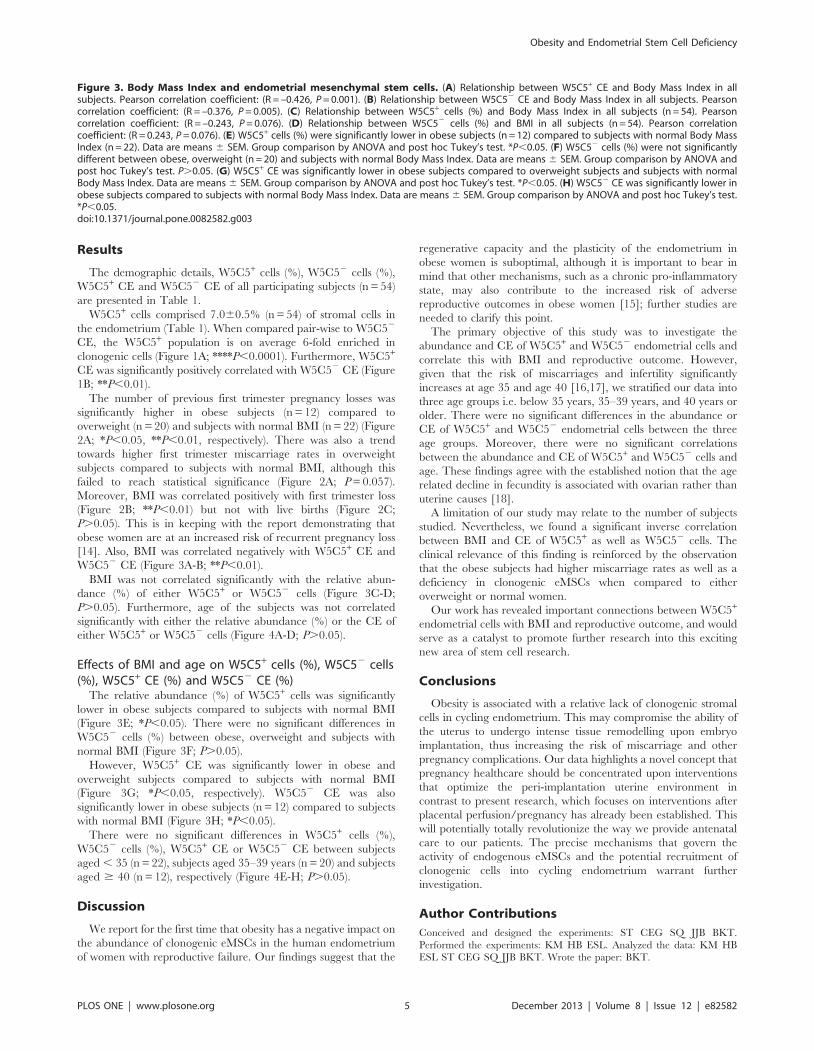
[14]. Also, BMI was correlated negatively with W5C5+ CE and
W5C52 CE (Figure 3A-B; **P,0.01).
BMI was not correlated significantly with the relative abun-
dance (%) of either W5C5+ or W5C52 cells (Figure 3C-D;
P.0.05). Furthermore, age of the subjects was not correlated
significantly with either the relative abundance (%) or the CE of
either W5C5+ or W5C52 cells (Figure 4A-D; P.0.05).
Effects of BMI and age on W5C5+ cells (%), W5C52 cells(%), W5C5+ CE (%) and W5C52 CE (%)
The relative abundance (%) of W5C5+ cells was significantly
lower in obese subjects compared to subjects with normal BMI
(Figure 3E; *P,0.05). There were no significant differences in
W5C52 cells (%) between obese, overweight and subjects with
normal BMI (Figure 3F; P.0.05).
However, W5C5+ CE was significantly lower in obese and
overweight subjects compared to subjects with normal BMI
(Figure 3G; *P,0.05, respectively). W5C52 CE was also
significantly lower in obese subjects (n = 12) compared to subjects
with normal BMI (Figure 3H; *P,0.05).
There were no significant differences in W5C5+ cells (%),
W5C52 cells (%), W5C5+ CE or W5C52 CE between subjects
aged , 35 (n = 22), subjects aged 35–39 years (n = 20) and subjects
aged $ 40 (n = 12), respectively (Figure 4E-H; P.0.05).
Discussion
We report for the first time that obesity has a negative impact on
the abundance of clonogenic eMSCs in the human endometrium
of women with reproductive failure. Our findings suggest that the
regenerative capacity and the plasticity of the endometrium in
obese women is suboptimal, although it is important to bear in
mind that other mechanisms, such as a chronic pro-inflammatory
state, may also contribute to the increased risk of adverse
reproductive outcomes in obese women [15]; further studies are
needed to clarify this point.
The primary objective of this study was to investigate the
abundance and CE of W5C5+ and W5C52 endometrial cells and
correlate this with BMI and reproductive outcome. However,
given that the risk of miscarriages and infertility significantly
increases at age 35 and age 40 [16,17], we stratified our data into
three age groups i.e. below 35 years, 35–39 years, and 40 years or
older. There were no significant differences in the abundance or
CE of W5C5+ and W5C52 endometrial cells between the three
age groups. Moreover, there were no significant correlations
between the abundance and CE of W5C5+ and W5C52 cells and
age. These findings agree with the established notion that the age
related decline in fecundity is associated with ovarian rather than
uterine causes [18].
A limitation of our study may relate to the number of subjects
studied. Nevertheless, we found a significant inverse correlation
between BMI and CE of W5C5+ as well as W5C52 cells. The
clinical relevance of this finding is reinforced by the observation
that the obese subjects had higher miscarriage rates as well as a
deficiency in clonogenic eMSCs when compared to either
overweight or normal women.
Our work has revealed important connections between W5C5+
endometrial cells with BMI and reproductive outcome, and would
serve as a catalyst to promote further research into this exciting
new area of stem cell research.
Conclusions
Obesity is associated with a relative lack of clonogenic stromal
cells in cycling endometrium. This may compromise the ability of
the uterus to undergo intense tissue remodelling upon embryo
implantation, thus increasing the risk of miscarriage and other
pregnancy complications. Our data highlights a novel concept that
pregnancy healthcare should be concentrated upon interventions
that optimize the peri-implantation uterine environment in
contrast to present research, which focuses on interventions after
placental perfusion/pregnancy has already been established. This
will potentially totally revolutionize the way we provide antenatal
care to our patients. The precise mechanisms that govern the
activity of endogenous eMSCs and the potential recruitment of
clonogenic cells into cycling endometrium warrant further
investigation.
Author Contributions
Conceived and designed the experiments: ST CEG SQ JJB BKT.
Performed the experiments: KM HB ESL. Analyzed the data: KM HB
ESL ST CEG SQ JJB BKT. Wrote the paper: BKT.
Figure 3. Body Mass Index and endometrial mesenchymal stem cells. (A) Relationship between W5C5+ CE and Body Mass Index in allsubjects. Pearson correlation coefficient: (R = –0.426, P = 0.001). (B) Relationship between W5C52 CE and Body Mass Index in all subjects. Pearsoncorrelation coefficient: (R = –0.376, P = 0.005). (C) Relationship between W5C5+ cells (%) and Body Mass Index in all subjects (n = 54). Pearsoncorrelation coefficient: (R = –0.243, P = 0.076). (D) Relationship between W5C52 cells (%) and BMI in all subjects (n = 54). Pearson correlationcoefficient: (R = 0.243, P = 0.076). (E) W5C5+ cells (%) were significantly lower in obese subjects (n = 12) compared to subjects with normal Body MassIndex (n = 22). Data are means 6 SEM. Group comparison by ANOVA and post hoc Tukey’s test. *P,0.05. (F) W5C52 cells (%) were not significantlydifferent between obese, overweight (n = 20) and subjects with normal Body Mass Index. Data are means 6 SEM. Group comparison by ANOVA andpost hoc Tukey’s test. P.0.05. (G) W5C5+ CE was significantly lower in obese subjects compared to overweight subjects and subjects with normalBody Mass Index. Data are means 6 SEM. Group comparison by ANOVA and post hoc Tukey’s test. *P,0.05. (H) W5C52 CE was significantly lower inobese subjects compared to subjects with normal Body Mass Index. Data are means 6 SEM. Group comparison by ANOVA and post hoc Tukey’s test.*P,0.05.doi:10.1371/journal.pone.0082582.g003
Obesity and Endometrial Stem Cell Deficiency
PLOS ONE | www.plosone.org 5 December 2013 | Volume 8 | Issue 12 | e82582

Obesity and Endometrial Stem Cell Deficiency
PLOS ONE | www.plosone.org 6 December 2013 | Volume 8 | Issue 12 | e82582

References
1. Eckel RH, Grundy SM, Zimmet PZ (2005) The metabolic syndrome. Lancet
365: 1415–1428.
2. Alanis MC, Goodnight WH, Hill EG, Robinson CJ, Villers MS, et al. (2010)
Maternal super-obesity (body mass index . or = 50) and adverse pregnancy
outcomes. Acta Obstet Gynecol Scand 89: 924–930.
3. Brewer CJ, Balen AH (2010) The adverse effects of obesity on conception and
implantation. Reproduction 140: 347–364.
4. Lam EW, Shah K, Brosens J (2012) The diversity of sex steroid action: the role
of micro-RNAs and FOXO transcription factors in cycling endometrium and
cancer. J Endocrinol 212: 13–25.
5. Burton GJ, Jauniaux E (2011) Oxidative stress. Best Pract Res Clin Obstet
Gynaecol 25: 287–299.
6. Gargett CE, Nguyen HP, Ye L (2012) Endometrial regeneration and
endometrial stem/progenitor cells. Rev Endocr Metab Disord 13: 235–251.
7. Wolff EF, Gao XB, Yao KV, Andrews ZB, Du H, et al. (2011) Endometrial stem
cell transplantation restores dopamine production in a Parkinson’s disease
model. J Cell Mol Med 15: 747–755.
8. Santamaria X, Massasa EE, Feng Y, Wolff E, Taylor HS (2011) Derivation of
insulin producing cells from human endometrial stromal stem cells and use in the
treatment of murine diabetes. Mol Ther 19: 2065–2071.
9. Ulrich D, Muralitharan R, Gargett CE (2013) Toward the use of endometrial
and menstrual blood mesenchymal stem cells for cell-based therapies. Expert
Opin Biol Ther 13: 1387–1400.
10. Du H, Naqvi H, Taylor HS (2012) Ischemia/reperfusion injury promotes and
granulocyte-colony stimulating factor inhibits migration of bone marrow-derivedstem cells to endometrium. Stem Cells Dev 21: 3324–3331.
11. Patterson AL, Zhang L, Arango NA, Teixeira J, Pru JK (2013) Mesenchymal-to-
epithelial transition contributes to endometrial regeneration following naturaland artificial decidualization. Stem Cells Dev 22: 964–974.
12. Masuda H, Anwar SS, Buhring HJ, Rao JR, Gargett CE (2012) A novel markerof human endometrial mesenchymal stem-like cells. Cell Transplant 21: 2201–
2214.
13. Brosens JJ, Hayashi N, White JO (1999) Progesterone receptor regulatesdecidual prolactin expression in differentiating human endometrial stromal cells.
Endocrinology 140: 4809–4820.14. Lo W, Rai R, Hameed A, Brailsford SR, Al-Ghamdi AA, et al. (2012) The effect
of body mass index on the outcome of pregnancy in women with recurrentmiscarriage. J Family Community Med 19: 167–171.
15. Denison FC, Roberts KA, Barr SM, Norman JE (2010) Obesity, pregnancy,
inflammation, and vascular function. Reproduction 140: 373–385.16. Nybo Andersen AM, Wohlfahrt J, Christens P, Olsen J, Melbye M (2000)
Maternal age and fetal loss: population based register linkage study. BMJ 320:1708–1712.
17. Dunson DB, Baird DD, Colombo B (2004) Increased infertility with age in men
and women. Obstet Gynecol 103: 51–56.18. Abdalla HI, Burton G, Kirkland A, Johnson MR, Leonard T, et al. (1993) Age,
pregnancy and miscarriage: uterine versus ovarian factors. Hum Reprod 8:1512–1517.
Figure 4. Age and endometrial mesenchymal stem cells. (A) Relationship between W5C5+ cells (%) W5C5+ CE and age in all subjects. Pearsoncorrelation coefficient: (R = 0.043, P = 0.760). (B) Relationship between W5C52 cells (%) and age in all subjects. Pearson correlation coefficient: (R = –0.043, P = 0.760). (C) Relationship between W5C5+ CE (%) and age in all subjects (n = 54). Pearson correlation coefficient: (R = –0.147, P = 0.288). (D)Relationship between W5C52 CE (%) and age in all subjects (n = 54). Pearson correlation coefficient: (R = –0.085, P = 0.543). (E) W5C5+ cells (%) werenot significantly different between subjects aged , 35 (n = 22), subjects aged 35–39 years (n = 20) and subjects aged $ 40 (n = 12). Data are means 6SEM. Group comparison by ANOVA and post hoc Tukey’s test. P.0.05. (F) W5C52 cells (%) were not significantly different between subjects aged ,35, subjects aged 35–39 years and subjects aged $ 40. Data are means 6 SEM. Group comparison by ANOVA and post hoc Tukey’s test. P.0.05. (G)W5C5+ CE was not significantly different between subjects aged , 35, subjects aged 35–39 years and subjects aged $ 40. Data are means 6 SEM.Group comparison by ANOVA and post hoc Tukey’s test. P.0.05. (H) W5C52 CE was not significantly different between subjects aged , 35, subjectsaged 35–39 years and subjects aged $ 40. Data are means 6 SEM. Group comparison by ANOVA and post hoc Tukey’s test. P.0.05.doi:10.1371/journal.pone.0082582.g004
Obesity and Endometrial Stem Cell Deficiency
PLOS ONE | www.plosone.org 7 December 2013 | Volume 8 | Issue 12 | e82582


![New Method to Quantitate Clonogenic Tumor Cells in the ......[CANCER RESEARCH 43, 5451-5455, November 1983] New Method to Quantitate Clonogenic Tumor Cells in the Blood Circulation](https://static.cupdf.com/doc/110x72/6068d20ce566193e3e18220a/new-method-to-quantitate-clonogenic-tumor-cells-in-the-cancer-research.jpg)


