DAAM Is Required for Thin Filament Formation andSarcomerogenesis during Muscle Development inDrosophilaImre Molnar1, Ede Migh1, Szilard Szikora1, Tibor Kalmar1, Attila G. Vegh2, Ferenc Deak3, Szilvia Barko4,
Beata Bugyi4, Zacharias Orfanos5, Janos Kovacs6, Gabor Juhasz6, Gyorgy Varo2, Miklos Nyitrai4,7,
John Sparrow5, Jozsef Mihaly1*
1 Institute of Genetics, Biological Research Centre HAS, Szeged, Hungary, 2 Institute of Biophysics, Biological Research Centre HAS, Szeged, Hungary, 3 Institute of
Biochemistry, Biological Research Centre HAS, Szeged, Hungary, 4 University of Pecs, Department of Biophysics, Pecs, Hungary, 5 Department of Biology, University of
York, York, United Kingdom, 6 Department of Anatomy, Cell and Developmental Biology, Eotvos Lorand University, Budapest, Hungary, 7 Hungarian Academy of Sciences,
Office for Subsidized Research Units, Budapest, Hungary
Abstract
During muscle development, myosin and actin containing filaments assemble into the highly organized sarcomericstructure critical for muscle function. Although sarcomerogenesis clearly involves the de novo formation of actin filaments,this process remained poorly understood. Here we show that mouse and Drosophila members of the DAAM formin familyare sarcomere-associated actin assembly factors enriched at the Z-disc and M-band. Analysis of dDAAM mutants revealed apivotal role in myofibrillogenesis of larval somatic muscles, indirect flight muscles and the heart. We found that loss ofdDAAM function results in multiple defects in sarcomere development including thin and thick filament disorganization, Z-disc and M-band formation, and a near complete absence of the myofibrillar lattice. Collectively, our data suggest thatdDAAM is required for the initial assembly of thin filaments, and subsequently it promotes filament elongation byassembling short actin polymers that anneal to the pointed end of the growing filaments, and by antagonizing the cappingprotein Tropomodulin.
Citation: Molnar I, Migh E, Szikora S, Kalmar T, Vegh AG, et al. (2014) DAAM Is Required for Thin Filament Formation and Sarcomerogenesis during MuscleDevelopment in Drosophila. PLoS Genet 10(2): e1004166. doi:10.1371/journal.pgen.1004166
Editor: Norbert Perrimon, Harvard Medical School, Howard Hughes Medical Institute, United States of America
Received August 15, 2013; Accepted December 23, 2013; Published February 27, 2014
Copyright: � 2014 Molnar et al. This is an open-access article distributed under the terms of the Creative Commons Attribution License, which permitsunrestricted use, distribution, and reproduction in any medium, provided the original author and source are credited.
Funding: This work was supported by the Hungarian Scientific Research Foundation (OTKA grants K82039 and K109330 to JM, PD83648 and K109689 to BB,OTKA NN107776 to MN). IM was a recipient of a studentship from the Hungarian Academy of Sciences. BB is a Bolyai Fellow of the Hungarian Academy ofSciences. The funders had no role in study design, data collection and analysis, decision to publish, or preparation of the manuscript.
Competing Interests: The authors have declared that no competing interests exist.
* E-mail: [email protected]
Introduction
Striated muscles contain cylindrical structures, myofibrils,
composed of repeating elements called sarcomeres, the basic
contractile units of muscle. A sarcomere, defined as the region
between neighboring Z-discs, contains two filament systems, the
actin-containing thin filaments and the myosin II-containing thick
filaments, and their associated proteins. The thin filaments are
anchored into the Z-disc where they are cross-linked by dimeric a-
actinin and a number of other proteins [1]. These filaments extend
in both directions from the Z-disc into neighboring sarcomeres.
They consist of a filamentous actin (F-actin) core decorated with the
regulatory proteins Tropomyosin (TM) and Troponin. Interdigi-
tated with thin filaments are the bipolar thick filaments, composed
largely of myosin molecules, that are at the middle of the sarcomere
and crosslinked by the M-band proteins. Whereas the structural
properties of these macromolecular complexes have been deter-
mined in detail in recent decades, much less is known about the in
vivo assembly of the filaments and Z-discs to form the very regular
sarcomeric structures [2]. In particular, the initial assembly of thin
filaments and the regulation of actin dynamics during myofibril
formation and maintenance remains poorly understood.
Owing to the regular assembly of actin monomers (G-actin) into
F-actin, these filaments display a polarized morphology and
dynamics with barbed (+) and pointed (2) ends. In vivo filament
growth likely occurs only at the barbed end, whereas the pointed
end is favored for depolymerization [3]. New actin filament
formation critically requires a nucleation step, during which a few
actin monomers combine to form a nucleation seed, prior to
elongation. As nucleation is not favored kinetically, and sponta-
neous in vivo nucleation would lead to anarchic filament assembly,
this step is promoted by nucleation factors. Nucleation factors
described so far include the Arp 2/3 complex, formins, Spire,
Cordon-bleu and Leimodin (Lmod) [4,5]. Although actin nucle-
ation factors have been extensively studied in many different
model systems, the essential nucleation factors in developing
muscles have not been clearly identified. Lmod and the
mammalian formin Fhod3 have both been implicated in actin
assembly in vertebrate striated muscles [6,7] but subsequent work
concluded that they are unlikely to contribute to actin nucleation
during the initial stages of myofibril assembly [8,9,10,11]. In fruit
flies, the genome harbors no clear Lmod ortholog, and genetic
analysis of the Drosophila Fhod ortholog, Fhos, and other members
PLOS Genetics | www.plosgenetics.org 1 February 2014 | Volume 10 | Issue 2 | e1004166
of the formin family, such as Diaphanous, Cappuccino or Form3,
revealed no clear role in muscle development [12,13,14,15].
Regulation of thin filament elongation and length, thought to be
controlled by elongation factors and capping proteins, are also
important aspects of actin dynamics in muscles. Elongation factors,
such as Ena/VASP proteins or the barbed end binding formins
that also function as nucleation factors, promote filament growth,
whereas capping protein binding blocks polymerization. In
contrast to non-muscle cells where thin filament growth is
restricted to the barbed ends, sarcomeric actin filaments elongate
from their pointed ends [16]. In each half sarcomere the thin
filaments are aligned with the same polarity and their barbed ends
are within the Z-discs, where they are capped by CapZ, whereas
their pointed ends are capped by Tropomodulin (Tmod). So far
Tmod, TM, Lmod and the Sarcomere Length Short (SALS)
proteins have all been implicated in thin filament length
regulation. Of these, Tmod binding causes thin filament shorten-
ing; conversely, loss of Tmod function causes lengthening of actin
filaments [16,17]. TM enhances Tmod binding affinity, whereas
Lmod and SALS seem to antagonize the capping activity of Tmod
and promote filament elongation from their pointed ends [9,18].
Surprisingly, instead of promoting elongation, SALS appears to
inhibit filament elongation in vitro. These results together with the
observation that no protein was yet isolated which would catalyze
F-actin assembly at the pointed end, mean that the mechanism
which enables muscle thin filaments to elongate from their pointed
ends remains mysterious.
Here we show that the Drosophila formin DAAM (Dishevelled
associated activator of morphogenesis) plays an important role in
sarcomerogenesis. The absence of dDAAM reduces larval motility,
causes a flightless phenotype and complex defects in sarcomere
organization. The latter include shorter and thinner sarcomeres
with reduced thin filament levels and an absence of both Z-disc
and M-band organization. Our protein localization studies
revealed that, despite being a barbed end binding protein in
non-muscle cells, dDAAM is highly enriched near the thin
filament pointed ends both in Drosophila and mouse muscle cells.
We propose that members of the DAAM family of formins are
very good candidates for the long sought-after muscle actin/thin
filament nucleators.
Results
dDAAM mutations affect flight ability and IFMdevelopment
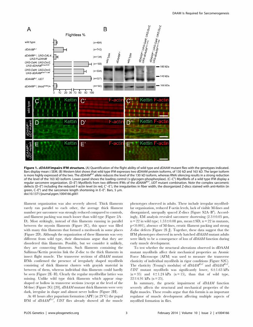
In studies of the Drosophila formin DAAM, we noticed that
,16% of adults homozygous for the viable, hypomorphic
dDAAMEx1 allele were flightless (16.265.3%, mean6SEM, n =
740, p = 0.02). As dDAAM null alleles are homozygous lethal, we
used two dDAAM specific RNAi lines (KK102786 from VDRC and
T129M constructed in our laboratory, targeting two non-
overlapping parts of the mRNA) to verify the flight effect. In the
presence of UAS-Dicer2 and an IFM (indirect flight muscle) specific
driver (UH3-Gal4) [19], both RNAi lines produced strong flightless
phenotypes (RNAiVDRC: 94.765.3%, mean6SEM, n = 103, p,
0.001; RNAiT129M: 87.163.9%, mean6SEM, n = 334, p = 0.002)
(Figure 1A). RNAi silencing in a dDAAM mutant background
(dDAAMEx1, UH3-Gal4; UAS-Dicer2; UAS-dDAAMRNAi-T129M,
subsequently referred to as dDAAMEx1, UDT) caused nearly all
males to be flightless (98.961.1%, mean6SEM, n = 327, p,
0.001) (Figure 1A). The strength of the flightless phenotypes
correlates with the partial reduction of dDAAM protein levels in
dDAAMEx1 IFM and its near absence in IFM from the RNAi
genotypes (Figure 1B). The flightless phenotype exhibited by
dDAAMEx1 mutants could be rescued by muscle-specific expression
of the dDAAM protein (4.162.9%, mean6SEM, n = 134,
p = 0.043) (Figure 1A).
In wild type or UH3-Gal4; UAS-Dicer2 flies (used as parental
control), the IFM displayed, as visualized by phalloidin (labels F-
actin) and anti-Kettin (a Z-disc marker) staining, its typical regular
sarcomeric organization (Figure 1C–C0), with the sarcomere
length of 3.1960.04 mm (mean6SD, n = 63) found in young
adults. In contrast, the IFM of dDAAM mutant flies showed
significant structural alterations (Figure 1D–E0). The IFM of
flightless dDAAMEx1 mutants looked largely normal, but about
25% of the myofibrils were thinner (1.4260.32 mm, mean6SD,
n = 50, p,0.001) than wild type (1.7260.11 mm, mean6SD,
n = 150) and part of the sarcomeres exhibited a reduced length
(down to 2.5960.13 mm, mean6SD, n = 73, p,0.001) (Figure
S1A). In contrast, IFM from the dDAAMEx1, UDT mutant
combination showed gross alterations in IFM fiber morphology
(Figure S3A,B). The myofibrils were thinner than in wild type
(1.1860.3 mm, mean6SD, n = 64, p,0.001) and their organiza-
tion was irregular (Figure 1D–E0). Mutant IFMs exhibited reduced
F-actin staining (Figure 1D–E0) without significant alterations in
the amount of G-actin (Figure S1F). Additionally, phalloidin
staining suggested that many of the thin filaments were of unequal
length, and similar to dDAAMEx1 mutants, shorter sarcomeres
(1.9760.28 mm, mean6SD, n = 62, p,0.001) could often be
detected. M-lines could hardly be identified by Myosin immuno-
staining (Figure S1B–C0), while the Z-discs displayed a highly
irregular and delocalized pattern compared to wild type
(Figure 1D–E0). Thus, loss of dDAAM function impairs IFM
structure from overall muscle shape to myofibrillar and sarcomeric
organization.
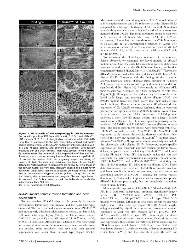
Electron microscopy (EM) of the IFM of dDAAMEx1, UDT
mutants (Figure 2) confirmed and extended all the major
myofibrillar defects seen in the confocal images. Notably, in
longitudinal sections (Figure 2A–D) we revealed irregularly
shaped, thin myofibrils with frayed edges, strong Z-disc defects,
absence of M-lines and shorter sarcomeres. The thick and thin
Author Summary
Sarcomeres, the smallest contractile units of muscle, areformed by two major filament systems, the myosincontaining thick and the actin containing thin filaments.Although it is well established that sarcomerogenesisinvolves the formation of novel actin filaments, so far itremained largely unclear how these filaments form. In thisstudy, we show that the Drosophila and mouse membersof the DAAM formin subfamily are sarcomere associatedactin assembly factors. Genetic analysis revealed thatdDAAM plays an essential role in thin filament formationand sarcomere organization. In addition, we demonstratethat mDaam1 is an early determinant of myofibrillogen-esis. Our data suggest that besides a role at the barbedend of the thin filaments, dDAAM also functions at thepointed end where it antagonizes the capping proteinTropomodulin. Based on these observations, we proposethat DAAM family formins are very good candidates forbeing the long sought-after muscle actin nucleators, thatalso promote filament elongation by assembling shortactin polymers that anneal to the Z-disc anchored growingfilament. Given that cardiomyopathies, muscular dystro-phies and the cardiovascular disease related heart muscledegenerations belong to the major health problemsworldwide, understanding the mechanism of how musclesnormally form is of immense biomedical relevance.
DAAM Is Required for Sarcomerogenesis
PLOS Genetics | www.plosgenetics.org 2 February 2014 | Volume 10 | Issue 2 | e1004166
filament organization was also severely altered. Thick filaments
rarely ran parallel to each other, the average thick filament
number per sarcomere was strongly reduced compared to controls,
and filament packing was much looser than wild type (Figure 2A–
D). Most strikingly, instead of thin filaments running in parallel
between the myosin filaments (Figure 2C), this space was filled
with many thin filaments that formed a meshwork in some places
(Figure 2D). Although the organization of these filaments was very
different from wild type, their dimensions argue that they are
disordered thin filaments. Possibly, but we consider it unlikely,
they are connecting filaments. Such filaments containing the
Sallimus/Kettin proteins link the Z-disc to the thick filaments in
insect flight muscle. The transverse sections of dDAAM mutant
IFMs confirmed the presence of irregularly shaped myofibrils
consisting of thick filament clusters with grayish material in
between of them, whereas individual thin filaments could hardly
be seen (Figure 2E–H). Clearly the regular myofibrillar lattice was
missing. Unlike wild type thick filaments which appear ring-
shaped or hollow in transverse sections (except at the level of the
M-line) (Figure 2G) [20], dDAAM mutant thick filaments were very
dark, irregular in shape and almost never hollow (Figure 2H).
At 48 hours after puparium formation (APF) (at 29uC) the pupal
IFM of dDAAMEx1, UDT flies already showed all the muscle
phenotypes observed in adults. These include irregular myofibril-
lar organization, reduced F-actin levels, lack of visible M-lines and
disorganized, unequally spaced Z-discs (Figure S2A–B0). Accord-
ingly, EM analysis revealed sarcomere shortening (2.360.05 mm,
n = 22 in wild type; 1.5360.08 mm, mean6SD, n = 22 in mutants,
p,0.001), absence of M-lines, erratic filament packing and strong
Z-disc defects (Figure 2I–J). Together, these data suggest that the
IFM phenotypes observed in newly hatched dDAAM mutant adults
were likely to be a consequence of loss of dDAAM function during
early muscle development.
To test whether the structural alterations observed in dDAAM
mutant myofibrils affect their mechanical properties an Atomic
Force Microscope (AFM) was used to measure the transverse
elasticity of individual myofibrils in rigor conditions (Figure S2C).
The elasticity (Young’s modulus) of dDAAMEx1 and dDAAMEx1,
UDT mutant myofibrils was significantly lower, 661.63 kPa
(n = 35) and 461.24 kPa (n = 15), than that of wild type,
2264.91 kPa (n = 25).
In summary, the genetic impairment of dDAAM function
severely affects the structural and mechanical properties of the
flight muscles. These results argue that this formin is an important
regulator of muscle development affecting multiple aspects of
myofibril formation in flies.
Figure 1. dDAAM impairs IFM structure. (A) Quantification of the flight ability of wild type and dDAAM mutant flies with the genotypes indicated.Bars display mean6SEM. (B) Western blot shows that wild type IFM expresses two dDAAM protein isoforms, of 130 kD and 163 kD. The larger isoformis more highly expressed of the two. The dDAAMEx1 allele reduces the level of the 130 kD isoform, whereas RNAi silencing results in a strong reductionof the level of the 163 kD isoform. Lower panel shows the loading control (a-glycogen-phosphorylase). (C–C0) Myofibrils of a wild type IFM display aregular sarcomere organization. (D–E0) Myofibrils from two different IFMs of the dDAAMEx1, UDT mutant combination. Note the complex sarcomericdefects (D–E0) including the reduced F-actin level (in red, C9–E9), the irregularities in fiber width, the disorganized Z-discs stained with anti-Kettin (ingreen, C–E0) and the sarcomere length shortening in E–E0. Bars, 5 mm.doi:10.1371/journal.pgen.1004166.g001
DAAM Is Required for Sarcomerogenesis
PLOS Genetics | www.plosgenetics.org 3 February 2014 | Volume 10 | Issue 2 | e1004166
dDAAM impairs somatic muscle formation and heartdevelopment
To ask whether dDAAM plays a role generally in muscle
development, larval body wall muscles and the heart tube were
examined. The body size and somatic musculature of dDAAMEx68
null mutant early third instar (L3) appeared normal, but late in L3,
100 hours after eggs laying (AEL), the larvae were shorter
(2.0860.31 mm; n = 30) than wild type (3.2460.25 mm; n = 30;
p,0.001; Figure 3E,I). Although gross alterations were not evident
in the overall structure of the musculature, mutant muscles were
also smaller, some myofibers were split and their general
organization was looser than in wild type (Figure 3A–D).
Measurements of the ventral longitudinal 3 (VL3) muscle showed
a 53% length reduction and 38% reduction in width (Figure 3K,L)
compared to wild type. Shortening of VL3 in dDAAM mutants
arises both by sarcomere shortening and a reduction in sarcomere
numbers (Figure 3M,N). The mean sarcomere length of wild type
VL3 muscles at 100 hours AEL was 6.261.6 mm (n = 477
sarcomeres; 12 muscles), but was decreased in dDAAM mutants
to 3.860.7 mm (n = 241 sarcomeres; 8 muscles; p,0.001). The
serial sarcomere number of VL3 was also decreased in dDAAM
mutants (30.162.1; n = 8) compared to wild type (39.764.3;
n = 12; p,0.001).
To investigate the physiological relevance of the muscular
defects observed, we examined the larval motility of dDAAM
mutant larvae. Until the early L3 stages there were no differences
between the wild type and the dDAAM mutant larvae, possibly due
to maternally derived dDAAM (in ,10% of dDAAMEx68 larvae the
dDAAM protein could still be clearly detected at 100 hours AEL,
Figure S3E,F). Consistent with the findings of the structural
analysis, kinematic studies of linear larval crawling at 72 hours
AEL showed that velocities of wild type and mutant larvae did not
significantly differ (Figure 3F). Subsequently at 100 hours AEL
their velocity was decreased by ,60% compared to wild type
(Figure 3F,J). Although, we observed a strong correlation between
larval body length and crawling velocity (Figure 3G,H), the
dDAAM mutant larvae are much slower than their reduced size
would indicate. Rescue experiments with DMef2-Gal4 driven
expression of UAS-DAAM constructs confirmed that the observed
phenotypes are specific to loss of dDAAM function. Western blot
analysis revealed that the IFM expresses two dDAAM protein
isoforms, a short (130 kD) minor isoform and a long (163 kD)
major isoform (Figure 1B). These correspond respectively to the
predicted DAAM-PB and DAAM-PD proteins (Flybase annota-
tion). The rescue experiments (above) were performed with UAS-
DAAM-PB as well as with UAS-DAAM-PD. UAS-DAAM-PB
expression partly rescued the velocity decrease and almost fully
rescued the body and muscle size of dDAAMEx68 mutant larvae,
whereas UAS-DAAM-PD expression almost completely rescued all
the phenotypic traits (Figure 3I–N). Moreover, muscle-specific
expression of these constructs not only rescued the larval muscle
defects, but partly rescued the lethality of dDAAMEx68 to adulthood
(3% for PB and 6.1% for PD). Importantly, unlike the wild type
constructs, the actin polymerization incompetent mutant forms,
UAS-DAAM-PBI732A and UAS-DAAM-PDI1042A mimicking the
Bni1 I1431A mutation [21], failed to rescue (Figure 3I–N). These
data demonstrate that the effect of dDAAM on muscle structure
and larval motility is muscle autonomous, and that the actin-
assembling activity of dDAAM is essential for normal muscle
development. Additionally, it appears that the two muscle-specific
dDAAM isoforms play largely, but not completely, redundant
roles in larval muscle.
Muscle-specific expression of UAS-DAAM-PB and UAS-DAAM-
PD, in a wild type background, produced significantly longer
larvae (PB: 4.2660.15 mm, n = 10, p,0.001; PD:
4.2460.19 mm, n = 10, p,0.001) than wild type. Their VL3
muscles were longer, although in both cases sarcomere size was
slightly shorter than wild type (Figure 3I,K,M). Muscle length-
ening occurred by significantly increasing sarcomere number
compared to wild type (PB: 5662.8, n = 14, p,0.001; PD:
5462.5, n = 12, p,0.001) (Figure 3N). Interestingly, the afore-
mentioned structural aspects were almost identical in larvae
overexpressing either isoform. Nevertheless, larvae expressing the
PB isoform were much faster (,55% faster, n = 10) than wild
type larvae (Figure 3J), while the velocity of larvae expressing PD
(,5% faster, n = 10) and the controls (Figure 3J) were not
Figure 2. EM analysis of IFM morphology in dDAAM mutants.Electronmicrographs of IFM from wild type (A, C, E, G, I) and dDAAMEx1;UDT mutants (B, D, F, H, J). Longitudinal sections of adult IFM (A–D)show that, as compared to the wild type, highly ordered and tightlypacked sarcomeres (A, C), the dDAAM mutant myofibrils (B, D) display Z-disc and M-band defects, and shortened sarcomeres with looselyorganized thin and thick filaments. Transverse sections of wild type (E,G) muscles reveal the hexagonal lattice organization of thin and thickfilaments, which is almost entirely lost in dDAAM mutant myofibrils (F,H). Instead, the mutant fibrils are irregularly shaped, consisting ofclusters of thick filaments, and individual thin filaments are hardlydetectable. Note: wild type thick filaments are hollow (G), while those ofthe dDAAM mutant are very dark, irregularly shaped and almost neverhollow (H). Longitudinal sections of pupal IFM (48 hours APF) (I, J) showthat, as compared to wild type (I), mutants (J) have strong Z-disc and M-line defects, shorter sarcomeres and irregular filament organisation.Arrows mark the Z-discs, asterisks mark the M-bands, m labels themitochondria. Bars, 500 nm.doi:10.1371/journal.pgen.1004166.g002
DAAM Is Required for Sarcomerogenesis
PLOS Genetics | www.plosgenetics.org 4 February 2014 | Volume 10 | Issue 2 | e1004166

significantly different. Lengths of PB and PD overexpressing
larvae were indistinguishable but PB larvae had significantly
wider VL3 muscles compared to PD larvae. Thus increasing
dDAAM isoform levels is sufficient to enhance the number of
sarcomeres initiated, but efficient sarcomere elongation may
require cooperation of both isoforms and regulation of their
ratio.
Larval heart tube size was also reduced in dDAAM mutants
compared to wild type (,40% reduction in diameter). In 100 hour
old wild type larvae the maximum heart diameter was
100.3367.39 mm; n = 9 whereas in dDAAM mutants 60.446
6.18 mm; n = 9, p,0.001 and they displayed reduced F-actin levels
(Figure S3C,D). Many mutant myofibrils appeared thinner than in
wild type and often deviated from the normal orientation (Figure
S3D). These observations strongly suggest that the formin
dDAAM may be a crucial regulator of muscle development in
Drosophila with an effect in every muscle type and developmental
stage examined.
Figure 3. Structural and functional analysis of the larval body wall muscles. Wild type (A, C) and dDAAMEx68 null mutant (B, D) larval bodywall muscles stained with phalloidin. Mutant muscles are smaller, some myofibers are split (arrow on D) and the overall muscle pattern is looser thanin wild type. The relationship of larval age and length (E), and of larval age and velocity (F) in wt (wild type; black line) and dDAAMEx68 (grey line)larvae. The relationship of larval length and velocity of wt (G) and dDAAMEx68 mutant (H) larvae. Quantification of larval length (I), crawling velocity (J),VL3 muscle length (K), width (L), mean sarcomere length (M) and serial sarcomere number (N) in larvae 100 hours AEL with the following genotypes:wt (wild type), Ex68 (dDAAMEx68), Ex68PB (dDAAMEx68; DMef2-Gal4; UAS-dDAAM-PB), Ex68PD (dDAAMEx68; DMef2-Gal4; UAS-dDAAM-PD), Ex68PB*(dDAAMEx68; DMef2-Gal4; UAS-dDAAM-PBI732A), Ex68PD* (dDAAMEx68; DMef2-Gal4; UAS-dDAAM-PDI732A), UASPB (DMef2-Gal4; UAS-dDAAM-PB) andUASPD (DMef2-Gal4; UAS-dDAAM-PD). Bars represent mean values with respective SDs in I–N. Statistical significance: * 0.05.p,0.001; ** p#0.001.Wild type and rescue data were compared to dDAAMEx68 data, unless otherwise indicated in the text. Bars, 100 mm (A–D).doi:10.1371/journal.pgen.1004166.g003
DAAM Is Required for Sarcomerogenesis
PLOS Genetics | www.plosgenetics.org 5 February 2014 | Volume 10 | Issue 2 | e1004166
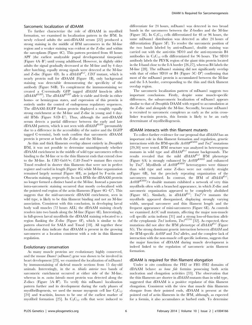
Sarcomeric localization of dDAAMTo further characterize the role of dDAAM in myofibril
formation, we examined its localization pattern in the IFM. In
newly eclosed adults the anti-dDAAM serum [22] produced a
strong staining in the middle of IFM sarcomeres in the M-line
region and a weaker staining was evident at the Z-disc and within
the sarcoplasm (Figure 4C). This pattern persisted from 48 hours
APF (the earliest analyzable pupal developmental timepoint)
(Figure 4A–B9) until young adulthood. However, in slightly older
adults the signal gradually decreased at the M-line and by 4 days
after hatching, equally strong signals were detected at the M-line
and Z-disc (Figure 4D). In a dDAAMEx1, UDT mutant, which is
nearly protein null for dDAAM (Figure 1B), only background
staining was detectable demonstrating the specificity of the
antibody (Figure S4B). To complement the immunostaining we
created a C-terminally GFP tagged dDAAM knock-in allele
(dDAAMEGFP). The dDAAMEGFP allele is viable and fertile in either
homo- or hemizygous states, and expression of this protein is
entirely under the control of endogenous regulatory sequences.
The dDAAM::EGFP fusion protein displayed a roughly equally
strong enrichment at the M-line and Z-disc in young and 4 day-
old IFMs (Figure S1D–E9). Thus, although the anti-dDAAM
serum detects a partial difference between the early and late
dDAAM pattern, which is not seen with dDAAMEGFP (presumably
due to a difference in the accessibility of the native and the EGFP
tagged C-termini), both tools confirm that sarcomeric dDAAM
protein is present at both the Z-disc and the M-line.
As thin and thick filaments overlap almost entirely in Drosophila
IFM, it was not possible to determine unambiguously whether
dDAAM enrichment in the middle of wild type sarcomeres reflects
binding to the M-line or to the thin filament ends that extend close
to the M-line. In UH3-Gal4/+; UAS-Tmod/+ mutant flies excess
Tmod resulted in shorter thin filaments that were not in perfect
register and varied in length (Figure 4G) while M-line organization
remained largely normal (Figure 4H), as judged by F-actin and
Obscurin staining, respectively. In such IFMs the dDAAM protein
no longer formed a distinct band at the M-line. Instead a punctate
intra-sarcomeric staining occurred that mostly co-localized with
the pointed end region of the actin filaments (Figure 4G–G0). This
suggests that the mid-sarcomeric dDAAM enrichment, seen in
wild type, is likely to be thin filament binding and not an M-line
association. Consistent with this conclusion, in developing larval
body wall muscles (72 hours AEL) the dDAAM staining clearly
resolves into two bands along the M-line (Figure 4E). Interestingly,
in full-grown larval myofibrils the dDAAM staining relocated to a
region flanking the Z-disc (Figure 4F), which is similar to the
pattern observed for SALS and Tmod [18]. All together these
localization data indicate that dDAAM is present in the growing
sarcomeres at a location consistent with a role in thin filament
regulation.
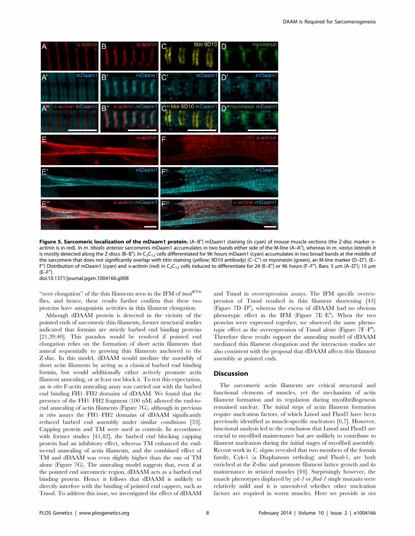
Evolutionary conservationAs many muscle proteins are evolutionary highly conserved,
and the mouse Daam1 (mDaam1) gene was shown to be involved in
heart development [23], we examined the localization of mDaam1
by immunostaining of skeletal muscle sections from 15 day-old
animals. Interestingly, in the m. tibialis anterior two bands of
sarcomeric enrichment occurred at either side of the M-line,
whereas in m. vastus lateralis most protein was detected along the
Z-discs (Figure 5A–B0). To verify this mDaam1 localization
pattern further and its development during the early phases of
myofibrillogenesis, we used the mouse myogenic cell line C2C12
[24] and a-actinin, known to be one of the earliest marker of
myofibril formation [25]. In C2C12 cells that were induced to
differentiate for 24 hours, mDaam1 was detected in two broad
bands in the sarcomeres between the Z-bodies and the M-line
(Figure 5E). In C2C12 cells differentiated for 48 or 96 hours, the
same mDaam1 distribution was detected as after 24 hours of
differentiation (Figure 5F). To resolve the sarcomeric position of
the two bands labeled by anti-mDaam1, double staining was
carried out with the anti-titin 9D10 and the anti-myomesin B4
antibodies in C2C12 cells differentiated for 96 hours. The 9D10
antibody labels the PEVK region of the giant titin protein located
in the I-band close to the I-A border [26,27], whereas B4 labels the
M-line [28]. The mDaam1 staining did not significantly overlap
with that of either 9D10 or B4 (Figure 5C–D0) confirming that
most of the mDaam1 protein is accumulated between the M-line
and the I-A border, corresponding to the thin and thick filament
overlap region.
The sarcomeric localization pattern of mDaam1 suggests two
important conclusions. Firstly, despite some muscle-specific
differences, the subsarcomeric localization of mDaam1 appears
similar to that of Drosophila DAAM with regard to accumulation at
the Z-disc and alongside the M-line. Secondly, because mDaam1
is recruited to sarcomeric complexes as early as the actin cross-
linker a-actinin protein, this formin is likely to be an early
determinant of myofibrillogenesis.
dDAAM interacts with thin filament mutantsTo collect further evidence for our proposal that dDAAM has an
important role in thin filament formation and regulation, genetic
interactions with the IFM-specific Act88FKM88 and Tm23 mutations
[29,30] were tested. IFM structure was analyzed in heterozygous
mutants in wild type and dDAAMEx1 mutant backgrounds. The
results revealed that the mild dDAAMEx1 IFM phenotype
(Figure 6A) is strongly enhanced by Act88FKM88 and enhanced
by Tm23. Myofibrils of Act88FKM88 heterozygotes were thinner
than wild type and some Z-discs were not entirely straight
(Figure 6B), but the precisely repeating organization of the
sarcomeres remained. In contrast, the IFM of dDAAMEx1;
Act88FKM88/+ double mutants exhibited a network of very thin
myofibrils often with a branched appearance, in which Z-disc and
sarcomeric organization appeared to be completely abolished
(Figure 6C). Similarly, in dDAAMEx1; Tm23/+ mutants the
myofibrils appeared disorganized, displaying strongly varying
width, unequal sarcomere and thin filament length and the
frequent appearance of mini-sarcomeres (Figure 6E). As controls
we examined Act5C null mutants, affecting the major non-muscle
cell specific actin isoform [31] and a strong loss-of-function allele
of the cytoplasmic Tm1 isoform, Tm102299 [32]. As expected, these
mutations did not alter the IFM phenotype of dDAAMEx1 (Figure
S5). The strong dominant genetic interaction between dDAAM and
the IFM-specific Act88F and Tm2 alleles, and the complete lack of
interaction with the non-muscle cell specific isoforms, suggests that
the major function of dDAAM during muscle development is
indeed linked to the regulation of sarcomeric actin filament
formation.
dDAAM is required for thin filament elongationUnder in vitro conditions the FH2 or FH1–FH2 domains of
dDAAM behave as bona fide formins possessing both actin
nucleation and elongation activities [33]. The observation that
the thin filaments are shorter in dDAAM mutants than in wild type,
suggested that dDAAM is a positive regulator of thin filament
elongation. Consistent with the view that muscle thin filaments
elongate from their pointed ends, dDAAM is present at the
pointed end of actin filaments in the IFM, although, as expected
for a formin, it also accumulates at barbed ends. To determine
DAAM Is Required for Sarcomerogenesis
PLOS Genetics | www.plosgenetics.org 6 February 2014 | Volume 10 | Issue 2 | e1004166
whether dDAAM is functionally important for pointed end
elongation we investigated genetic interactions of dDAAM with
mutations affecting the pointed end regulator proteins SALS and
Tmod. SALS promotes filament elongation in vivo [18], whereas
Tmod binding is thought to prevent elongation [16]. The presence
of salsf07849/+ in a dDAAMEx1 mutant background had no obvious
phenotypic effect. In contrast, the tmod00848 mutation entirely
suppressed the weak flightless phenotype of dDAAMEx1 (4.960.5%,
mean6SEM, n = 160, p = 0.027) (Figure 1A) suggesting that
dDAAM and Tmod may act antagonistically during thin filament
growth.
To investigate the dDAAM/Tmod interaction in more detail
we first examined the IFM-specific RNAi silencing of tmod, and we
found that in most myofibrils it severely disrupted myofibrillogen-
esis (Figure 7A). However, approximately 10% of the myofibrils
had almost normal looking Z-discs allowing us to determine that
these sarcomeres were shorter (2.6260.11 mm; n = 26; mean6SD;
p,0.001) than wild type. Phalloidin staining revealed the presence
of thin filaments in the mid-sarcomeric region (Figure 7B) and
impaired M-lines are evident by EM analysis (Figure 7H). The
strong effect on myofibrillogenesis is in accordance with previous
reports that Tmod1 in mouse and Unc-94 (tmd-1) in C. elegans are
required for myofibril assembly [34,35,36,37]. The decreased
sarcomere length was unexpected as the inhibition of Tmod
function increases sarcomere length in cultured cardiomyocytes
[38] or in Drosophila primary muscles [18]. We noted however, that
although sarcomere length of UH3-Gal4; UAS-tmodRNAi flight
muscles was reduced, some of the thin filaments clearly failed to
terminate in the H-zone of these mutant sarcomeres (Figure 7H).
Therefore, individual filament length can be longer than in wild
type, which would be consistent with the known function of Tmod
in filament length regulation. To study whether the tmodRNAi
phenotype is sensitive to dDAAM protein level, tmod silencing was
carried out in a dDAAMEx1 mutant background. Most (,80%)
myofibrils displayed a striated pattern with distinct M-lines and
somewhat aberrant Z-discs, and nearly normal sarcomere length
(2.860.13 mm; n = 30; mean6SD; p,0.001) (Figure 7C). This
phenotype suggests that the reduced dDAAM levels suppress the
Figure 4. Sarcomeric localization of the dDAAM protein in the IFM and the larval body wall muscles. dDAAM staining of the IFMmyofibrils of wild type pupae 48 hours (A, A9) and 72 hours APF (B, B9), freshly eclosed adult (C, C9) and 4 day-old adult (D, D9). dDAAM accumulatesat the M-line (arrowheads), at the Z-disc (arrow) and in the sarcoplasm (asterisk). Note: accumulation at Z-disc is weak in pupae and young adults (A–C), but in 4 day-old adults staining is equally strong at the M-line and the Z-disc (D). In developing larval body wall muscles (72 hours AEL) dDAAMstaining resolves into two bands along the M-line (E, E9). In fully matured larval body wall muscles dDAAM staining relocates to a region flanking theZ-disc (F, F9). Arrowheads mark the M-line in E; arrows mark the Z-disc, asterisk marks the sarcoplasm in F. (G–H0) Excess Tmod in UH3-Gal4/+; UAS-Tmod/+ flies leads to shorter thin filaments that are not in perfect register and vary in length as judged by F-actin staining (G, H). In these IFMsdDAAM protein displays a punctate distribution (arrowheads in G9) most of which colocalizes with the pointed end region of the thin filaments (G0).The M-line in these mutant muscles remains nearly intact as judged by Obscurin staining (H9). Phalloidin staining is in red (C9–H0), Kettin (C9–H0) andsls-GFP (A9, B9) as Z-disc markers are in green, anti-dDAAM (A–F9, G9, G0) and anti-Obscurin (H, H9) are in cyan. Bars, 5 mm.doi:10.1371/journal.pgen.1004166.g004
DAAM Is Required for Sarcomerogenesis
PLOS Genetics | www.plosgenetics.org 7 February 2014 | Volume 10 | Issue 2 | e1004166
‘‘over elongation’’ of the thin filaments seen in the IFM of tmodRNAi
flies, and hence, these results further confirm that these two
proteins have antagonistic activities in thin filament elongation.
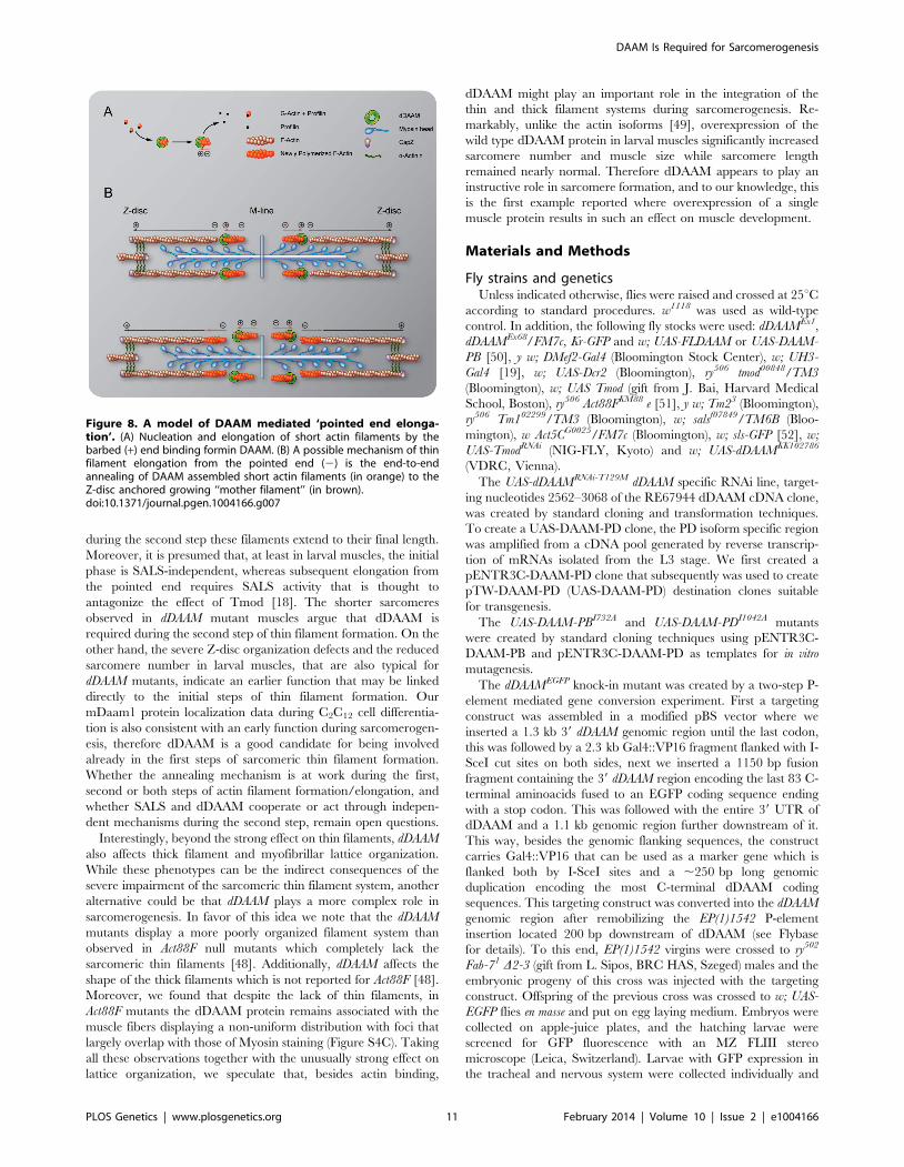
Although dDAAM protein is detected in the vicinity of the
pointed ends of sarcomeric thin filaments, former structural studies
indicated that formins are strictly barbed end binding proteins
[21,39,40]. This paradox would be resolved if pointed end
elongation relies on the formation of short actin filaments that
anneal sequentially to growing thin filaments anchored to the
Z-disc. In this model, dDAAM would mediate the assembly of
short actin filaments by acting as a classical barbed end binding
formin, but would additionally either actively promote actin
filament annealing, or at least not block it. To test this expectation,
an in vitro F-actin annealing assay was carried out with the barbed
end binding FH1–FH2 domains of dDAAM. We found that the
presence of the FH1–FH2 fragment (100 nM) allowed the end-to-
end annealing of actin filaments (Figure 7G), although in previous
in vitro assays the FH1–FH2 domains of dDAAM significantly
reduced barbed end assembly under similar conditions [33].
Capping protein and TM were used as controls. In accordance
with former studies [41,42], the barbed end blocking capping
protein had an inhibitory effect, whereas TM enhanced the end-
to-end annealing of actin filaments, and the combined effect of
TM and dDAAM was even slightly higher than the one of TM
alone (Figure 7G). The annealing model suggests that, even if at
the pointed end sarcomeric region, dDAAM acts as a barbed end
binding protein. Hence it follows that dDAAM is unlikely to
directly interfere with the binding of pointed end cappers, such as
Tmod. To address this issue, we investigated the effect of dDAAM
and Tmod in overexpression assays. The IFM specific overex-
pression of Tmod resulted in thin filament shortening [43]
(Figure 7D–D0), whereas the excess of dDAAM had no obvious
phenotypic effect in the IFM (Figure 7E–E0). When the two
proteins were expressed together, we observed the same pheno-
typic effect as the overexpression of Tmod alone (Figure 7F–F0).
Therefore these results support the annealing model of dDAAM
mediated thin filament elongation and the interaction studies are
also consistent with the proposal that dDAAM affects thin filament
assembly at pointed ends.
Discussion
The sarcomeric actin filaments are critical structural and
functional elements of muscles, yet the mechanism of actin
filament formation and its regulation during myofibrillogenesis
remained unclear. The initial steps of actin filament formation
require nucleation factors, of which Lmod and Fhod3 have been
previously identified as muscle-specific nucleators [6,7]. However,
functional analysis led to the conclusion that Lmod and Fhod3 are
crucial to myofibril maintenance but are unlikely to contribute to
filament nucleation during the initial stages of myofibril assembly.
Recent work in C. elegans revealed that two members of the formin
family, Cyk-1 (a Diaphanous ortholog) and Fhod-1, are both
enriched at the Z-disc and promote filament lattice growth and its
maintenance in striated muscles [44]. Surprisingly however, the
muscle phenotypes displayed by cyk-1 or fhod-1 single mutants were
relatively mild and it is unresolved whether other nucleation
factors are required in worm muscles. Here we provide in vivo
Figure 5. Sarcomeric localization of the mDaam1 protein. (A–B0) mDaam1 staining (in cyan) of mouse muscle sections (the Z-disc marker a-actinin is in red). In m. tibialis anterior sarcomeres mDaam1 accumulates in two bands either side of the M-line (A–A0), whereas in m. vastus lateralis itis mostly detected along the Z-discs (B–B0). In C2C12 cells differentiated for 96 hours mDaam1 (cyan) accumulates in two broad bands at the middle ofthe sarcomere that does not significantly overlap with titin staining (yellow; 9D10 antibody) (C–C0) or myomesin (green), an M-line marker (D–D0). (E–F0) Distribution of mDaam1 (cyan) and a-actinin (red) in C2C12 cells induced to differentiate for 24 (E–E0) or 96 hours (F–F0). Bars: 5 mm (A–D0); 15 mm(E–F0).doi:10.1371/journal.pgen.1004166.g008
DAAM Is Required for Sarcomerogenesis
PLOS Genetics | www.plosgenetics.org 8 February 2014 | Volume 10 | Issue 2 | e1004166

evidence that DAAM, another formin family member, is
important for sarcomeric thin filament formation. We have found
that dDAAM is required for thin filament elongation and that the
actin-assembling activity of dDAAM is indispensable for formation
of functional muscles. In addition, we have shown that in
differentiating C2C12 cells the mouse Daam1 ortholog is incorpo-
rated into sarcomeric complexes at least as early as a-actinin. Thus
DAAM family formins are strong candidates for being involved in
the initial assembly of myofibrillar actin filaments. Interestingly,
although the F-actin content of dDAAM mutant muscles is
reduced, some filaments still form. Notably however, the dDAAM
mutants available for muscle studies are not protein null. This
prevents us from determining whether an additional nucleation
factor, such as Dia or Fhos, is involved or that residual dDAAM
activity is sufficient to promote some level of F-actin formation.
Nevertheless, our results demonstrate that dDAAM is a develop-
mentally important sarcomere-associated actin assembly factor in
Drosophila. Remarkably, expression of the vertebrate DAAM
orthologs are known to be abundant in developing somites and
heart [23,45], and genetic analysis of mDaam1 indicated a role in
sarcomere organization in cardiomyocytes [23]. Overall this
suggests that the regulation of sarcomeric actin filament formation
is an evolutionary conserved DAAM function.
Our studies revealed that in the IFM the dDAAM protein is
mostly enriched at either end of the thin filaments, the expected
positions for proteins affecting thin filament assembly. We
formerly showed that in vitro dDAAM behaves as a bona fide
formin, possessing all the major properties reported for other
formin family members [33]. Here we propose that at Z-discs
dDAAM may regulate G-actin incorporation with the well
described barbed end processive capping mechanism of formins.
Given that the sarcomeric dDAAM expression in the IFM,
including the Z-disc accumulation, is maintained during adult-
hood, it appears likely that dDAAM also contributes to the
maintenance of normal muscle structure and function. Besides the
Z-disc enrichment, dDAAM also accumulates at the pointed end
region of the thin filaments. Since dDAAM promotes thin filament
formation and acts antagonistically to the F-actin pointed end
capping protein, Tmod, the simplest interpretation of these data is
to assume that dDAAM is involved in filament elongation from the
pointed end. This is in good accordance with the evidence that in
cardiac myocytes and in Drosophila primary cultures actin dynamics
predominate at the pointed ends [17,18], yet the presence at the
pointed ends is unexpected for a formin, a barbed end binding
protein. Because available structural studies exclude the possibility
that a formin directly binds to the pointed end, dDAAM might be
recruited to the pointed end by binding to a different protein than
actin, or our findings indicate the presence of F-actin barbed ends
in the vicinity of the pointed end of the thin filaments. Although
we cannot strictly exclude the first possibility, at present the
functional importance of such an association is unclear. Therefore
we favor the second alternative that has interesting mechanistic
implications. If barbed ends indeed exist in the region of the
pointed ends, then pointed end elongation could be achieved
through the end-to-end annealing of short actin filaments to the
Z-disc anchored growing ‘‘mother filament’’ (Figure 8). Such a
mechanism, demonstrated in vitro, would allow rapid filament
elongation at the pointed ends. Our data are compatible with the
model in which dDAAM promotes the formation of these short
filaments by acting as an F-actin barbed end binding processive
capper that also allows filament annealing. An important question
is how long these short filaments are? In this regard, it is interesting
to note that during contractile ring formation in fission yeast the
formin Cdc12p was shown to nucleate short actin filaments that
anneal to each other in the presence of TM [42], and consistently,
TM increased the annealing process by ,2 fold in our in vitro
assay. As TM is a major myofibrillar protein, and the IFM-specific
Tm2 mutation dominantly enhanced the thin filament defects of
dDAAMEx1, we propose that the length of the filaments involved in
the annealing process is unlikely to be shorter, but could be equal
to an F-actin fragment covered by one TM dimer which is about
37–38 nm or 14 actin monomers. Whereas the ability to anneal
end-to-end is an intrinsic property of actin filaments, a better
understanding of this mechanism during myofibril formation
awaits future studies, most importantly the visualization of the
short protofilaments. Nonetheless, it is remarkable that the formin
Fhod3, implicated in myofibril maintenance and maturation
Figure 6. dDAAM interacts with thin filament mutants. IFMmyofibrils from (A) dDAAMEx1, (B) Act88FKM88/+, (C) dDAAMEx1;Act88FKM88/+, (D) Tm23/+ and (E) dDAAMEx1; Tm23/+ flies (actin in red,Kettin in green in all panels). Note: sarcomere organization in dDAAMEx1
(A) is nearly wild type; likewise Act88F (B) and Tm2 (D) heterozygotesdisplay a largely regular myofibril and Z-disc organization. Myofibrils ofthe dDAAMEx1; Act88FKM88/+ (C) and dDAAMEx1; Tm23/+ (E) genotypesare extremely disorganized compared to the controls. Bars, 5 mm.doi:10.1371/journal.pgen.1004166.g005
DAAM Is Required for Sarcomerogenesis
PLOS Genetics | www.plosgenetics.org 9 February 2014 | Volume 10 | Issue 2 | e1004166

[10,46], also displays an accumulation in the pointed end region
[7,47] and might regulate actin assembly with a similar
mechanism as dDAAM.
Previously presented models of thin filament growth in
Drosophila proposed a two-step mechanism [18,43]. According
to this view, during the first step short filaments are assembled, and
Figure 7. The interaction of dDAAM and tmod. Upon silencing of tmod myofibrils get severely disrupted (A–A0), though ,10% of them show amilder effect with regular Z-disc arrangement but missing H-zones (B–B0). In dDAAMEx1, UH3-Gal4; UAS-tmodRNAi muscles most myofibrils have anearly wild type sarcomeric organization with regularly spaced Z-discs and M-lines, and almost normal sarcomere length (C–C0). (D–D0) In UH3-Gal4;UAS-Tmod IFMs the sarcomeric thin filaments often appear to be shorter than wild type as judged by phalloidin staining, whereas myofibrils of UH3-Gal4; UAS-FLDAAM muscle look wild type (E–E0). Simultaneous overexpression of FLDAAM and Tmod results in the same effect as the expression ofTmod alone (F–F0; compare to D–D0). Kettin in green, actin in red in A–F0. (G) An end-to-end actin annealing assay, dark grey: 0 minute control,average filament length in the presence of 1 mM F-actin (F-actin), light gray: average filament length after 60 minutes incubation, in the presence of1 mM F-actin (F-actin), 1 mM F-actin+ 10 nM capping protein (F-actin+CP), 1 mM F-actin+100 nM DAAM-FH1-FH2 (F-actin+DAAM), 1 mM F-actin+1 mMskeletal tropomyosin (F-actin+TM), 1 mM F-actin+100 nM DAAM-FH1-FH2+1 mM skeletal tropomyosin (F-actin+DAAM+TM). Bars represent meanvalues with respective SEMs. (H) Electronmicrograph of a tmodRNAi IFM. Black arrowheads mark the borders of the mid-sarcomeric region where theM-line structures are not evident but thin filaments appear to cross this area. White arrows on the inset, corresponding to the dashed area, mark thinfilaments that fail to terminate in the H-zone. Bars: 5 mm (A–F0); 500 nm (H) 100 nm (H, inset).doi:10.1371/journal.pgen.1004166.g006
DAAM Is Required for Sarcomerogenesis
PLOS Genetics | www.plosgenetics.org 10 February 2014 | Volume 10 | Issue 2 | e1004166
during the second step these filaments extend to their final length.
Moreover, it is presumed that, at least in larval muscles, the initial
phase is SALS-independent, whereas subsequent elongation from
the pointed end requires SALS activity that is thought to
antagonize the effect of Tmod [18]. The shorter sarcomeres
observed in dDAAM mutant muscles argue that dDAAM is
required during the second step of thin filament formation. On the
other hand, the severe Z-disc organization defects and the reduced
sarcomere number in larval muscles, that are also typical for
dDAAM mutants, indicate an earlier function that may be linked
directly to the initial steps of thin filament formation. Our
mDaam1 protein localization data during C2C12 cell differentia-
tion is also consistent with an early function during sarcomerogen-
esis, therefore dDAAM is a good candidate for being involved
already in the first steps of sarcomeric thin filament formation.
Whether the annealing mechanism is at work during the first,
second or both steps of actin filament formation/elongation, and
whether SALS and dDAAM cooperate or act through indepen-
dent mechanisms during the second step, remain open questions.
Interestingly, beyond the strong effect on thin filaments, dDAAM
also affects thick filament and myofibrillar lattice organization.
While these phenotypes can be the indirect consequences of the
severe impairment of the sarcomeric thin filament system, another
alternative could be that dDAAM plays a more complex role in
sarcomerogenesis. In favor of this idea we note that the dDAAM
mutants display a more poorly organized filament system than
observed in Act88F null mutants which completely lack the
sarcomeric thin filaments [48]. Additionally, dDAAM affects the
shape of the thick filaments which is not reported for Act88F [48].
Moreover, we found that despite the lack of thin filaments, in
Act88F mutants the dDAAM protein remains associated with the
muscle fibers displaying a non-uniform distribution with foci that
largely overlap with those of Myosin staining (Figure S4C). Taking
all these observations together with the unusually strong effect on
lattice organization, we speculate that, besides actin binding,
dDAAM might play an important role in the integration of the
thin and thick filament systems during sarcomerogenesis. Re-
markably, unlike the actin isoforms [49], overexpression of the
wild type dDAAM protein in larval muscles significantly increased
sarcomere number and muscle size while sarcomere length
remained nearly normal. Therefore dDAAM appears to play an
instructive role in sarcomere formation, and to our knowledge, this
is the first example reported where overexpression of a single
muscle protein results in such an effect on muscle development.
Materials and Methods
Fly strains and geneticsUnless indicated otherwise, flies were raised and crossed at 25uC
according to standard procedures. w1118 was used as wild-type
control. In addition, the following fly stocks were used: dDAAMEx1,
dDAAMEx68/FM7c, Kr-GFP and w; UAS-FLDAAM or UAS-DAAM-
PB [50], y w; DMef2-Gal4 (Bloomington Stock Center), w; UH3-
Gal4 [19], w; UAS-Dcr2 (Bloomington), ry506 tmod00848/TM3
(Bloomington), w; UAS Tmod (gift from J. Bai, Harvard Medical
School, Boston), ry506 Act88FKM88 e [51], y w; Tm23 (Bloomington),
ry506 Tm102299/TM3 (Bloomington), w; salsf07849/TM6B (Bloo-
mington), w Act5CG0025/FM7c (Bloomington), w; sls-GFP [52], w;
UAS-TmodRNAi (NIG-FLY, Kyoto) and w; UAS-dDAAMKK102786
(VDRC, Vienna).
The UAS-dDAAMRNAi-T129M dDAAM specific RNAi line, target-
ing nucleotides 2562–3068 of the RE67944 dDAAM cDNA clone,
was created by standard cloning and transformation techniques.
To create a UAS-DAAM-PD clone, the PD isoform specific region
was amplified from a cDNA pool generated by reverse transcrip-
tion of mRNAs isolated from the L3 stage. We first created a
pENTR3C-DAAM-PD clone that subsequently was used to create
pTW-DAAM-PD (UAS-DAAM-PD) destination clones suitable
for transgenesis.
The UAS-DAAM-PBI732A and UAS-DAAM-PDI1042A mutants
were created by standard cloning techniques using pENTR3C-
DAAM-PB and pENTR3C-DAAM-PD as templates for in vitro
mutagenesis.
The dDAAMEGFP knock-in mutant was created by a two-step P-
element mediated gene conversion experiment. First a targeting
construct was assembled in a modified pBS vector where we
inserted a 1.3 kb 39 dDAAM genomic region until the last codon,
this was followed by a 2.3 kb Gal4::VP16 fragment flanked with I-
SceI cut sites on both sides, next we inserted a 1150 bp fusion
fragment containing the 39 dDAAM region encoding the last 83 C-
terminal aminoacids fused to an EGFP coding sequence ending
with a stop codon. This was followed with the entire 39 UTR of
dDAAM and a 1.1 kb genomic region further downstream of it.
This way, besides the genomic flanking sequences, the construct
carries Gal4::VP16 that can be used as a marker gene which is
flanked both by I-SceI sites and a ,250 bp long genomic
duplication encoding the most C-terminal dDAAM coding
sequences. This targeting construct was converted into the dDAAM
genomic region after remobilizing the EP(1)1542 P-element
insertion located 200 bp downstream of dDAAM (see Flybase
for details). To this end, EP(1)1542 virgins were crossed to ry502
Fab-71 D2-3 (gift from L. Sipos, BRC HAS, Szeged) males and the
embryonic progeny of this cross was injected with the targeting
construct. Offspring of the previous cross was crossed to w; UAS-
EGFP flies en masse and put on egg laying medium. Embryos were
collected on apple-juice plates, and the hatching larvae were
screened for GFP fluorescence with an MZ FLIII stereo
microscope (Leica, Switzerland). Larvae with GFP expression in
the tracheal and nervous system were collected individually and
Figure 8. A model of DAAM mediated ‘pointed end elonga-tion’. (A) Nucleation and elongation of short actin filaments by thebarbed (+) end binding formin DAAM. (B) A possible mechanism of thinfilament elongation from the pointed end (2) is the end-to-endannealing of DAAM assembled short actin filaments (in orange) to theZ-disc anchored growing ‘‘mother filament’’ (in brown).doi:10.1371/journal.pgen.1004166.g007
DAAM Is Required for Sarcomerogenesis
PLOS Genetics | www.plosgenetics.org 11 February 2014 | Volume 10 | Issue 2 | e1004166
used to set up stocks. Conversion events were confirmed by PCR
and sequencing. Once the Gal4::VP16 containing construct has
been successfully converted into the dDAAM gene, in a subsequent
round of crosses I-SceI was used to induce DNA double strand
breaks that could eventually be repaired through homologous
recombination between the ,250 bp duplicated dDAAM regions.
This event, confirmed by PCR and sequencing, led to the removal
of all non Drosophila sequences with the exception of EGFP, and
resulted in a C-terminally GFP-tagged dDAAM allele. dDAAMEGFP
is fully viable and fertile in hemi- or homozygous state indicating
that the presence of EGFP does not significantly alter dDAAM
function.
ImmunohistochemistryNewly eclosed adult IFMs were dissected from bisected half
thoraces in 4% paraformaldehyde (PF), incubated for 15 minutes,
then washed with relaxing solution (6 mM MgCl2, 5 mM EGTA,
5 mM ATP, 90 mM potassium propionate, 20 mM NaPi,
pH 7.0). The muscles were permeabilized overnight in Triton-
X/glycerol solution (50% v/v glycerol, 0.5% Triton X-100,
20 mM NaPi, 2 mM MgCl2, 1 mM EGTA, 5 mM DTT, pH 7.0)
at 4uC, and washed in PBS supplemented with 0.5% Triton X-100
(PBT), then labeled with primary and secondary antibodies. For
pupal IFM preparations, timed pupae were removed from their
puparia and pinned by the head on dry Sylgard (Dow Corning),
dorsal side down, and then submerged into 4% PF in PBS. After
dissection along the ventral midline, unattached material was
flushed gently away using a syringe to expose the IFMs. These
were detached and incubated in fixative (4% PF in PBS) for a
further 15 minutes, then transferred back to relaxing solution. For
larval heart tube dissections larvae were cut along the ventral
midline in relaxing solution. Then fixed with 4% PF in PBS [53].
Fat bodies and other organs were removed, then labeled with
primary and secondary antibodies. For developmental staging,
white pre-pupae with everted spiracles were removed into fresh
vials at 25uC and harvested at required time-points. Adult flies
were selected as ‘newly eclosed’ between 0 and 8 hr post-eclosion.
Primary antibodies, listed below, were applied overnight at 4uC.
Muscles and heart tubes were washed three times in PBST,
secondary antibodies were applied for 3 hr, then samples were
rinsed three times again in PBST. The following primary
antibodies were used: rat monoclonal anti-Kettin (MAC 155,
1:200; Abcam); rat monoclonal anti-Myosin (MAC 147, 1:200;
Abcam), rabbit polyclonal anti-GFP (1:1000; Sigma) and rabbit
polyclonal anti-dDAAM (1:1000) [22]. For secondary antibodies
we used the appropriate Alexa-488, Alexa-546 and Alexa-647
(1:600), actin was stained with Rhodamine-Phalloidin (1:100) (all
from Life Technologies). Samples were mounted in PBS:glycerol
(1:4). Confocal images were captured on an Olympus FV1000
LSM microscope, images were edited with ImageJ (NIH) and
Olympus FW10-ASW (version1.7a.).
Larval length measurements and crawling assaysAged larvae were collected and rinsed with tap water, and then
gently placed onto agar plates. The plates were placed under an
Olympus SZX12 dissecting microscope equipped with an Olym-
pus C7070 digital camera. Total illumination was applied, images
were acquired at 25 Hz. The recording environment (tempera-
ture, humidity, illumination) was stationary. Twenty seconds of
movie was recorded with DScaler (The DScaler Project Team) for
each larva. During this period of time most wild-type larvae
moved out from the field of view. ImageJ (NIH) software was used
to analyze the image sequences. Persistent forward movements
were selected to characterize larval crawling velocity and larval
length. Larval length was calculated as the average of the
minimum and the maximum head to tail distance for each larva.
Maximum intensity projections were used to generate larval
tracks. To calculate larval crawling velocity, the lengths of these
tracks were divided by the time. Tracked larvae were dissected,
stained and subjected to muscle measurements made on VL3
(ventral longitudinal 3) muscles. Muscle length was measured
manually as the major axis of the VL3 muscles; muscle width was
measured as the minor axis of the VL3 muscles. Sarcomere
number and sarcomere length were measured from gray scale
intensity plots across phalloidin stained sarcomeres, sarcomere size
being the distance between adjacent peaks.
Flight testsFlight tests were carried out with three day old flies [54]. Flies
were released inside a perspex box illuminated from above, and
scored for the ability to fly up, horizontally or down. Flies falling
into the third category (down) were counted as flightless.
Tissue culture and mouse musclesThe mouse myogenic cell line, C2C12 (ATCC), was maintained
in growth medium (DMEM supplemented with 10% FBS;
GIBCO/Life Technologies). Cells were initially plated into 100-
mm-diameter dishes (Greiner) at a density of 104/cm2. When
cultures reached ,80% confluence they were subcultured onto
sterile glass coverslips in 35-mm-diameter dishes. Cultures were
kept in growth medium until they reached 60% confluence and
subsequently were switched to differentiation medium (DMEM
containing 2% horse serum; GIBCO/Life Technologies). This
medium was replaced every day, and samples were processed for
immunostaining at selected time points. Cells were fixed in 4%
formaldehyde in PBS for 10 minutes, and permeabilized in PBS+0.1% Triton-X100 for 3 minutes before staining. Primary
antibodies were applied for 1 hr RT, and after 365 minutes
washing in PBS, cells were incubated with secondary antibodies
for another 1 hr. After washing three times for 5 minutes in PBS,
samples were mounted in PBS:glycerin (1:4).
For sections of m. tibialis anterior and m. vastus lateralis, C57Bl/6
adult male mice were sacrificed by cervical dislocation. Leg muscle
was dissected, embedded in Tissue-Tek O.C.T. compound
(Sakura Finetek) and snap-frozen in isopentane cooled by liquid
nitrogen. 10 mm cryosections were fixed in prechilled acetone and
kept at 280uC.
For mammalian muscle and C2C12 staining the following
antibodies were used: rabbit polyclonal anti-mDaam1 (1:2000;
Sigma), rabbit polyclonal anti-mDaam1 (1:200; Abnova), mouse
monoclonal anti-a-actinin (1:80; Sigma), mouse monoclonal anti-
titin 9D10 (1:20; DSHB) and mouse monoclonal anti-myomesin
(B4, 1:1; DSHB). For secondary antibodies we used the
appropriate Alexa-488, Alexa-546 and Alexa-647 (1:600; Life
Technologies). Images were taken and analyzed in a similar ways
as flight muscles described above.
Electron microscopyMuscles were dissected and fixed in 3.2% paraformaldehyde,
0.5% glutaraldehyde, 1% sucrose, 0.028% CaCl2 in 0.1 N sodium
cacodylate (pH 7.4) overnight at 4uC, and washed 26overnight in
0.1 N sodium cacodylate (pH 7.4) at 4uC. Samples were postfixed
in 0.5% osmium-tetroxide for 1 hr at room temperature, and
embedded into Durcupan (Fluka) by following the manufacturer’s
recommendations. 70–80 nm ultrathin sections were prepared
from 2–3 animals per genotype, stained in Reynold’s lead citrate,
and evaluated using a JEM-1011 electron microscope (JEOL)
equipped with Morada camera and iTEM software (Olympus).
DAAM Is Required for Sarcomerogenesis
PLOS Genetics | www.plosgenetics.org 12 February 2014 | Volume 10 | Issue 2 | e1004166

AFM and force measurementsIFM muscle fibers, falling apart for individual myofibrils upon
preparation, were mounted on a poly-L-lysine coated glass surface
and measured in phosphate buffered saline. To identify the target
points at which to perform individual force measurements, a rough
and low resolution scan was taken (not shown). Experiments were
carried out with Asylum MFP-3D head and controller (Asylum
Research, Santa Barbara, CA). The driver program was written in
IGOR Pro software (version 5.04, Wavemetrics, Lake Oswego,
OR). Rectangular, gold coated, silicon nitride cantilevers were
used, with a nominal spring constant of 30 pN/nm and a V
shaped tip with radius of curvature of roughly 30 nm (Bio-Lever,
BL-RC150 VB-C1, Olympus Optical Co. Ltd). The measure-
ments were performed in contact mode in liquid, with the vertical
piezo working in a closed loop. Constant speed of 0.6 mm/s (scan
rate 0.1 Hz) and total load was kept below 1 nN during
experiments.
Simple force curves were measured by lowering the probe until
a desired deflection is reached and pulling it back. To calculate the
sample’s elasticity the contact region of the lowering part from
force curves has been used. By subtracting a reference curve,
recorded on a hard surface from those measured on the object of
interest, the sample’s force vs. indentation curve can be obtained,
which provides the Young’s modulus of the measured sample [55].
Several points were examined recording multiple force curves at
each selected place; the average and standard deviation of which
was calculated.
Protein gel electrophoresis and western blot analysisAdult IFM samples were dissected as described above. Tissues
were immediately placed in ice-cold RIPA lysis buffer and kept
overnight. SDS-PAGE and Western blot analyses were carried out
according to standard protocols. Membranes were stained with
rabbit anti-dDaam (1:1000), and rabbit anti-glycogen phosphor-
ylase (1:20000) (gift from A. Udvardy, BRC HAS, Szeged) used as
a loading control. Secondary antibody was a-rabbit-HRPO
(1:10000; Sigma). For chemiluminescent detection we used a
Millipore Immobilon kit.
Actin filament annealing testsTo measure the annealing of actin filaments fluorescence
microscopy assays were performed. Actin filaments (10 mM, F-
actin) were polymerized for 2 hr at room temperature in 4 mM
Tris-HCl (pH 7.0), 0.1 mM CaCl2, 0.2 mM ATP, 0.5 mM DTT,
1 mM EGTA, 1 mM MgCl2 and 50 mM KCl (F-buffer). The F-
actin solution was then diluted to 1 mM using F-buffer in the
absence or presence of actin-binding proteins (100 nM capping
protein or 100 nM dDAAM FH1-FH2 or 1 mM skeletal muscle
TM or 100 nM dDAAM FH1-FH2 and 1 mM skeletal muscle
TM). The samples were incubated overnight. For investigation of
the annealing, Alexa-488-phalloidin labeled samples were sheared
five times through a 26 gauge needle. Samples were diluted 100
fold into microscopy buffer (F-buffer supplemented with
50 mMDTT, 5 mM DABCO and 0.5% (w/v) methylcellulose) 0
and 60 minutes after shearing and processed for microscopy
observations. Single actin filaments were observed with an
Olympus IX81 inverted fluorescence microscope using a 1006objective (NA1.4) and a CCD camera (Orca ERG Hamamatsu).
The length of the actin filaments was measured and analyzed with
ImageJ. Under each condition 3–4 independent measurements
were performed and 300–600 filaments were analyzed. Statistical
analysis was carried out using Microsoft Excel or Microcal Origin
6.0.
StatisticsExcel (Microsoft) was used to collect and organise data.
Statistical analysis was carried out using Prism 5 (GraphPad
Software Inc.) and/or SigmaPlot 12 (Systat Software Inc.).
Normality of the data was verified by Shapiro-Wilk test. Pairwise
comparisons were made using the Student’s t test or the Mann-
Whitney U test according to the normality, p,0.05 was
considered as statistically significant.
Supporting Information
Figure S1 Impaired adult IFM structure in dDAAM mutants.
(A–A0) IFM myofibrils of a flightless dDAAMEx1 mutant looks
largely normal, although some of the sarcomeres show reduced
lengths (2.5 mm instead of 3.2 mm; Kettin in green, actin in red).
(B–C0) Myofibrils of wild type (B–B0) and dDAAMEx1, UDT
mutants (C–C0) stained for Myosin (green) and actin (red). Note
the severely impaired Myosin and M-line organization, and the
strong reduction of F-actin level in IFM of the dDAAM mutant (C).
In newly eclosed (D, D9) and 4 day-old (E, E9) dDAAMEGFP adults
anti-GFP staining is evident at the Z-disc (arrows) and M-band
(arrowhead). (F) Coomassie staining shows no significant difference
in the amount of G-actin in wild type and dDAAMEx1, UDT
mutants. Bars, 5 mm.
(TIF)
Figure S2 dDAAM impairs pupal IFM structure and the
mechanical properties of muscles. Myofibrils from a wild type (A–
A0) and dDAAMEx1, UDT mutant (B–B0) pupal IFM (48 hours APF,
29uC) stained for actin (in red) and Kettin (in green). The mutant
IFM shows Z-disc and M-line organization defects. (C) Quantifica-
tion of the transverse elasticity of wild type and dDAAM mutant
myofibrils measured by Atomic Force Microscopy. To characterize
the mechanical properties of the myofibrils, their transverse elasticity
(Young’s modulus) was calculated. The average curve is fitted with a
second order polynomial (C). The elasticity of dDAAMEx1 and
dDAAMEx1, UDT (RNAi) mutant fibers is significantly lower,
661.63 kPa (n = 35) and 461.24 kPa (n = 15), respectively, than
the one of wild type, 2264.91 kPa (n = 25). Bars, 2 mm.
(TIF)
Figure S3 dDAAM affects IFM fiber morphology and heart tube
development. (A–B) IFM structure of a wild type (A) and
dDAAMEx1, UDT mutant (B) as seen under confocal microscope.
In these sagittal sections of thoraces rhodamine-phalloidin was
used to visualize the muscle F-actin. Note that mutant dorsolongi-
tudinal muscle (DLM) fibers are shorter (arrows) and thinner than
in wild type, and some of the muscles appear degenerated. (C–D)
Phalloidin staining of a wild type (C) and dDAAMEx68 mutant (D)
larval heart tube to visualize F-actin (in green). Compared to wild
type, the dDAAMEx68 mutant has reduced F-actin levels, and heart
tube diameter is smaller. In addition, many of the mutant
myofibrils appear thinner than their wild type counterparts and
often deviate from the typical wild type orientation. (E–F) A
developing wild type (E) larval body wall muscle at 72 hours AEL
clearly expresses and accumulates the dDAAM protein (in cyan) in
its myofibrils. A similar but weaker dDAAM expression pattern
can still be detected in ,10% of dDAAMEx68 mutant larvae (F)
even at 100 hours AEL. Kettin (in green) labels the Z-discs in E-F.
Bars: 100 mm (A–B); 40 mm (C–D); 5 mm (E–F).
(TIF)
Figure S4 Sarcomeric localization of the dDAAM protein in wild
type and mutant IFMs. Myofibrils of wild type (A–A0), dDAAMEx1,
UDT (B–B0) and Act88F null mutant (C–C0) IFM from young adults
stained for dDAAM (cyan, A–C0), actin (red, A9–B0) and Myosin
DAAM Is Required for Sarcomerogenesis
PLOS Genetics | www.plosgenetics.org 13 February 2014 | Volume 10 | Issue 2 | e1004166

(green, C9 and C0). Staining of wild type IFM reveals dDAAM
accumulation at M-line and Z-disc, and in the sarcoplasm (A–A0).
In contrast, in a dDAAMEx1, UDT mutant IFM only a weak
background staining is evident (compare A to B). In Act88F null
mutants, which completely lack sarcomeric thin filaments, dDAAM
protein remains associated with muscle fibers and displays a partial
colocalization with myosin (C–C0). Bars, 5 mm.
(TIF)
Figure S5 dDAAM shows no interaction with the non-muscle cell
specific isoforms of actin and tropomyosin. Adult IFM myofibrils
showing dDAAMEx1 (A), Act5CG0025/+ (B), dDAAMEx1;
Act5CG0025/+ (C), Tm102299/+ (D) and dDAAMEx1; Tm102299/+(E) mutants stained for Kettin (green) and actin (red). Note that all
mutant myofibrils look nearly wild type. Bars, 5 mm.
(TIF)
Acknowledgments
We thank Andor Udvardy, Jianwu Bai, Belinda Bullard, Sanford Bernstein,
Developmental Studies Hybridoma Bank (DSHB), Bloomington and
Kyoto Stock Centers and VDRC for fly stocks and reagents. We are
grateful to Rita Gombos and Laszlo Sipos for technical advices, and to
Anna Rehak, Aniko Berente and Velkeyne Ildiko Krausz for technical
assistance.
Author Contributions
Conceived and designed the experiments: JM JS MN GV. Performed the
experiments: IM EM SS TK ZO AGV SB FD JK GJ. Analyzed the data:
IM EM SS TK ZO AGV SB BB FD JK GJ JS JM. Wrote the paper: JM
JS. Prepared the figures: IM SS JM.
References
1. Luther PK (2009) The vertebrate muscle Z-disc: sarcomere anchor for structure
and signalling. J Muscle Res Cell Motil 30: 171–185.
2. Sparrow JC, Schock F (2009) The initial steps of myofibril assembly: integrinspave the way. Nat Rev Mol Cell Biol 10: 293–298.
3. Pollard TD, Blanchoin L, Mullins RD (2000) Molecular mechanisms controlling
actin filament dynamics in nonmuscle cells. Annu Rev Biophys Biomol Struct29: 545–576.
4. Campellone KG, Welch MD (2010) A nucleator arms race: cellular control of
actin assembly. Nat Rev Mol Cell Biol 11: 237–251.
5. Chesarone M, Gould CJ, Moseley JB, Goode BL (2009) Displacement of formins
from growing barbed ends by bud14 is critical for actin cable architecture andfunction. Dev Cell 16: 292–302.
6. Chereau D, Boczkowska M, Skwarek-Maruszewska A, Fujiwara I, Hayes DB,
et al. (2008) Leiomodin is an actin filament nucleator in muscle cells. Science320: 239–243.
7. Taniguchi K, Takeya R, Suetsugu S, Kan OM, Narusawa M, et al. (2009)
Mammalian formin fhod3 regulates actin assembly and sarcomere organizationin striated muscles. J Biol Chem 284: 29873–29881.
8. Skwarek-Maruszewska A, Boczkowska M, Zajac AL, Kremneva E, Svitkina T,
et al. (2010) Different localizations and cellular behaviors of leiomodin and
tropomodulin in mature cardiomyocyte sarcomeres. Mol Biol Cell 21: 3352–3361.
9. Tsukada T, Pappas CT, Moroz N, Antin PB, Kostyukova AS, et al. (2010)
Leiomodin-2 is an antagonist of tropomodulin-1 at the pointed end of the thinfilaments in cardiac muscle. J Cell Sci 123: 3136–3145.
10. Iskratsch T, Lange S, Dwyer J, Kho AL, dos Remedios C, et al. (2010) Formin
follows function: a muscle-specific isoform of FHOD3 is regulated by CK2phosphorylation and promotes myofibril maintenance. Journal of Cell Biology
191: 1159–1172.
11. Iskratsch T, Reijntjes S, Dwyer J, Toselli P, Degano IR, et al. (2013) Two
distinct phosphorylation events govern the function of muscle FHOD3. Cell MolLife Sci 70: 893–908.
12. Anhezini L, Saita AP, Costa MS, Ramos RG, Simon CR (2012) Fhos encodes a
Drosophila Formin-like protein participating in autophagic programmed celldeath. Genesis 50: 672–684.
13. Castrillon DH, Wasserman SA (1994) Diaphanous is required for cytokinesis in
Drosophila and shares domains of similarity with the products of the limbdeformity gene. Development 120: 3367–3377.
14. Emmons S, Phan H, Calley J, Chen W, James B, et al. (1995) Cappuccino, a
Drosophila maternal effect gene required for polarity of the egg and embryo, is
related to the vertebrate limb deformity locus. Genes Dev 9: 2482–2494.
15. Tanaka H, Takasu E, Aigaki T, Kato K, Hayashi S, et al. (2004) Formin3 isrequired for assembly of the F-actin structure that mediates tracheal fusion in
Drosophila. Dev Biol 274: 413–425.
16. Littlefield RS, Fowler VM (2008) Thin filament length regulation in striatedmuscle sarcomeres: pointed-end dynamics go beyond a nebulin ruler. Semin Cell
Dev Biol 19: 511–519.
17. Littlefield R, Almenar-Queralt A, Fowler VM (2001) Actin dynamics at pointedends regulates thin filament length in striated muscle. Nat Cell Biol 3: 544–551.
18. Bai J, Hartwig JH, Perrimon N (2007) SALS, a WH2-domain-containing
protein, promotes sarcomeric actin filament elongation from pointed ends
during Drosophila muscle growth. Dev Cell 13: 828–842.
19. Katzemich A, Kreiskother N, Alexandrovich A, Elliott C, Schock F, et al. (2012)The function of the M-line protein obscurin in controlling the symmetry of the
sarcomere in the flight muscle of Drosophila. J Cell Sci 125: 3367–3379.
20. Reedy MC, Beall C (1993) Ultrastructure of developing flight muscle inDrosophila. I. Assembly of myofibrils. Dev Biol 160: 443–465.
21. Xu Y, Moseley JB, Sagot I, Poy F, Pellman D, et al. (2004) Crystal structures of a
Formin Homology-2 domain reveal a tethered dimer architecture. Cell 116:711–723.
22. Matusek T, Djiane A, Jankovics F, Brunner D, Mlodzik M, et al. (2006) The
Drosophila formin DAAM regulates the tracheal cuticle pattern throughorganizing the actin cytoskeleton. Development 133: 957–966.
23. Li DQ, Hallett MA, Zhu WQ, Rubart M, Liu Y, et al. (2011) Dishevelled-associated activator of morphogenesis 1 (Daam1) is required for heart
morphogenesis. Development 138: 303–315.
24. Yaffe D, Saxel O (1977) Serial passaging and differentiation of myogenic cells
isolated from dystrophic mouse muscle. Nature 270: 725–727.
25. Kontrogianni-Konstantopoulos A, Catino DH, Strong JC, Bloch RJ (2006) De
novo myofibrillogenesis in C2C12 cells: evidence for the independent assemblyof M bands and Z disks. Am J Physiol Cell Physiol 290: C626–637.
26. Trombitas K, Greaser M, French G, Granzier H (1998) PEVK extension of
human soleus muscle titin revealed by immunolabeling with the anti-titin
antibody 9D10. J Struct Biol 122: 188–196.
27. Wang SM, Greaser ML, Schultz E, Bulinski JC, Lin JJ, et al. (1988) Studies oncardiac myofibrillogenesis with antibodies to titin, actin, tropomyosin, and
myosin. J Cell Biol 107: 1075–1083.
28. Grove BK, Kurer V, Lehner C, Doetschman TC, Perriard JC, et al. (1984) A
new 185,000-dalton skeletal muscle protein detected by monoclonal antibodies.J Cell Biol 98: 518–524.
29. Karlik CC, Fyrberg EA (1985) An insertion within a variably spliced Drosophilatropomyosin gene blocks accumulation of only one encoded isoform. Cell 41:
57–66.
30. Okamoto H, Hiromi Y, Ishikawa E, Yamada T, Isoda K, et al. (1986) Molecular
characterization of mutant actin genes which induce heat-shock proteins inDrosophila flight muscles. EMBO J 5: 589–596.
31. Fyrberg EA, Mahaffey JW, Bond BJ, Davidson N (1983) Transcripts of the six
Drosophila actin genes accumulate in a stage- and tissue-specific manner. Cell
33: 115–123.
32. Tetzlaff MT, Jackle H, Pankratz MJ (1996) Lack of Drosophila cytoskeletaltropomyosin affects head morphogenesis and the accumulation of oskar mRNA
required for germ cell formation. EMBO J 15: 1247–1254.
33. Barko S, Bugyi B, Carlier MF, Gombos R, Matusek T, et al. (2010)
Characterization of the biochemical properties and biological function of theformin homology domains of Drosophila DAAM. J Biol Chem 285: 13154–
13169.
34. Fritz-Six KL, Cox PR, Fischer RS, Xu B, Gregorio CC, et al. (2003) Aberrant
myofibril assembly in tropomodulin1 null mice leads to aborted heart
development and embryonic lethality. J Cell Biol 163: 1033–1044.
35. McKeown CR, Nowak RB, Moyer J, Sussman MA, Fowler VM (2008)Tropomodulin1 is required in the heart but not the yolk sac for mouse
embryonic development. Circ Res 103: 1241–1248.
36. Stevenson TO, Mercer KB, Cox EA, Szewczyk NJ, Conley CA, et al. (2007)
unc-94 encodes a tropomodulin in Caenorhabditis elegans. J Mol Biol 374: 936–950.
37. Yamashiro S, Cox EA, Baillie DL, Hardin JD, Ono S (2008) Sarcomeric actinorganization is synergistically promoted by tropomodulin, ADF/cofilin, AIP1
and profilin in C. elegans. J Cell Sci 121: 3867–3877.
38. Sussman MA, Baque S, Uhm CS, Daniels MP, Price RL, et al. (1998) Altered
expression of tropomodulin in cardiomyocytes disrupts the sarcomeric structureof myofibrils. Circ Res 82: 94–105.
39. Lu J, Meng W, Poy F, Maiti S, Goode BL, et al. (2007) Structure of the FH2
domain of Daam1: implications for formin regulation of actin assembly. J Mol
Biol 369: 1258–1269.
40. Shimada A, Nyitrai M, Vetter IR, Kuhlmann D, Bugyi B, et al. (2004) The coreFH2 domain of diaphanous-related formins is an elongated actin binding protein
that inhibits polymerization. Mol Cell 13: 511–522.
41. Andrianantoandro E, Blanchoin L, Sept D, McCammon JA, Pollard TD (2001)
Kinetic mechanism of end-to-end annealing of actin filaments. J Mol Biol 312:721–730.
DAAM Is Required for Sarcomerogenesis
PLOS Genetics | www.plosgenetics.org 14 February 2014 | Volume 10 | Issue 2 | e1004166
42. Skau CT, Neidt EM, Kovar DR (2009) Role of tropomyosin in formin-mediated
contractile ring assembly in fission yeast. Mol Biol Cell 20: 2160–2173.43. Mardahl-Dumesnil M, Fowler VM (2001) Thin filaments elongate from their
pointed ends during myofibril assembly in Drosophila indirect flight muscle.
J Cell Biol 155: 1043–1053.44. Mi-Mi L, Votra S, Kemphues K, Bretscher A, Pruyne D (2012) Z-line formins
promote contractile lattice growth and maintenance in striated muscles of C.elegans. J Cell Biol 198: 87–102.
45. Nakaya MA, Habas R, Biris K, Dunty WC, Jr., Kato Y, et al. (2004)
Identification and comparative expression analyses of Daam genes in mouse andXenopus. Gene Expr Patterns 5: 97–105.
46. Kan OM, Takeya R, Abe T, Kitajima N, Nishida M, et al. (2012) Mammalianformin Fhod3 plays an essential role in cardiogenesis by organizing
myofibrillogenesis. Biol Open 1: 889–896.47. Kan-o M, Takeya R, Taniguchi K, Tanoue Y, Tominaga R, et al. (2012)
Expression and subcellular localization of mammalian formin Fhod3 in the
embryonic and adult heart. PLoS One 7: e34765.48. Beall CJ, Sepanski MA, Fyrberg EA (1989) Genetic dissection of Drosophila
myofibril formation: effects of actin and myosin heavy chain null alleles. GenesDev 3: 131–140.
49. Roper K, Mao Y, Brown NH (2005) Contribution of sequence variation in
Drosophila actins to their incorporation into actin-based structures in vivo. J CellSci 118: 3937–3948.
50. Matusek T, Gombos R, Szecsenyi A, Sanchez-Soriano N, Czibula A, et al.
(2008) Formin proteins of the DAAM subfamily play a role during axon growth.J Neurosci 28: 13310–13319.
51. Mogami K, Hotta Y (1981) Isolation of Drosophila flightless mutants whichaffect myofibrillar proteins of indirect flight muscle. Mol Gen Genet 183: 409–
417.
52. Morin X, Daneman R, Zavortink M, Chia W (2001) A protein trap strategy todetect GFP-tagged proteins expressed from their endogenous loci in Drosophila.
Proc Natl Acad Sci U S A 98: 15050–15055.53. Alayari NN, Vogler G, Taghli-Lamallem O, Ocorr K, Bodmer R, et al. (2009)
Fluorescent labeling of Drosophila heart structures. J Vis Exp (32). pii: 1423.54. Cripps RM, Ball E, Stark M, Lawn A, Sparrow JC (1994) Recovery of
dominant, autosomal flightless mutants of Drosophila melanogaster and
identification of a new gene required for normal muscle structure and function.Genetics 137: 151–164.
55. Vinckier A, Semenza G (1998) Measuring elasticity of biological materials byatomic force microscopy. FEBS Lett 430: 12–16.
DAAM Is Required for Sarcomerogenesis
PLOS Genetics | www.plosgenetics.org 15 February 2014 | Volume 10 | Issue 2 | e1004166