Confocal Laser Endomicroscopy for In Vivo Diagnosis ofClostridium difficile Associated Colitis — A Pilot StudyHelmut Neumann1*, Claudia Gunther1, Michael Vieth2, Martin Grauer1, Nadine Wittkopf1, Jonas Mudter1,
Christoph Becker1, Christoph Schoerner3, Raja Atreya1, Markus F. Neurath1
1 Department of Medicine I, University of Erlangen-Nuremberg, Erlangen, Germany, 2 Institute of Pathology, Klinikum Bayreuth, Bayreuth, Germany, 3 Institute of
Microbiology, Immunology and Hygiene, University of Erlangen-Nuremberg, Erlangen, Germany
Abstract
Background: Clostridium difficile infection (CDI) is one of the most dreaded causes of hospital-acquired diarrhea. Mainobjective was to investigate whether confocal laser endomicroscopy (CLE) has the capability for in vivo diagnosis of C.difficile associated histological changes. Second objective was to prove the presence of intramucosal bacteria using CLE.
Methods: 80 patients were prospectively included, 10 patients were diagnosed with CDI based on toxigenic culture. Tovalidate the presence of intramucosal bacteria ex vivo, CLE was performed in pure C. difficile culture; additionallyfluorescence in situ hybridization (FISH) was performed. Finally, CLE with fluorescence labelled oligonucleotide probespecific for C. difficile was performed ex vivo in order to prove the presence of bacteria.
Results: CLE identified CDI-associated histological changes in vivo (sensitivity and accuracy of 88.9% and 96.3%). In addition,intramucosal bacteria were visualized. The presence of these bacteria could be proven by CLE with labeled, specificmolecular C. difficile probe and FISH-technique. Based on comparison between CLE and FISH analyses, sensitivity andspecificity for the presence of intramucosal bacteria were 100%.
Conclusion: CLE has the potential for in vivo diagnosis of CDI associated colitis. In addition, CLE allowed the detection ofintramucosal bacteria in vivo.
Citation: Neumann H, Gunther C, Vieth M, Grauer M, Wittkopf N, et al. (2013) Confocal Laser Endomicroscopy for In Vivo Diagnosis of Clostridium difficileAssociated Colitis — A Pilot Study. PLoS ONE 8(3): e58753. doi:10.1371/journal.pone.0058753
Editor: Markus M. Heimesaat, Charite, Campus Benjamin Franklin, Germany
Received September 26, 2012; Accepted February 6, 2013; Published March 19, 2013
Copyright: � 2013 Neumann et al. This is an open-access article distributed under the terms of the Creative Commons Attribution License, which permitsunrestricted use, distribution, and reproduction in any medium, provided the original author and source are credited.
Funding: The authors have no funding or support to report.
Competing Interests: The authors have declared that no competing interests exist.
* E-mail: [email protected]
Introduction
Clostridium difficile infection has emerged as one of the most
clinically significant causes of hospital-acquired diarrhea and is
associated with significant morbidity and mortality. C. difficile
infection is often accompanied by fever and leukocytosis and
frequently affects older and immunocompromised patients.
Nevertheless, recent data suggest that even young and healthy
persons who have previously not been exposed to a health care
environment or antimicrobial therapy are at risk as well [1,2].
C. difficile can colonize the large bowel and, in the presence of
antibiotic therapy that limits the growth of naturally residing
microorganisms, produce endotoxins and cytotoxins that can
cause severe mucosal damage, resulting in colitis that may have a
pseudomembranous appearance at endoscopy [1]. It has been
estimated that C. difficile causes approximately 25% of the
antibiotic-associated diarrhea (AAD) and most cases of pseudo-
membranous colitis. In the United States, there are about 300,000
cases of C. difficile-associated diarrhea and colitis per year, resulting
in an annual economic burden of more than one billion dollars to
the health care system [2].
Since a new epidemic strain of C. difficile associated with more
severe disease, mortality and frequent relapses has been identified
in 2003, C. difficile infection has becoming increasingly difficult to
control and eradicate [3]. Accordingly, C. difficile infection now
rivals methicillin-resistant Staphylococcus aureus (MRSA) as the most
common cause for hospital-acquired infections in the United
States.
Therefore, rapid and accurate diagnosis of C. difficile infection is
of crucial importance, not only for individual patient management
but also for prevention of nosocomial transmission. Currently,
diagnosis of C. difficile infection is based on patients’ clinical history
and laboratory tests, including toxigenic culture, which still
remains the gold standard for diagnosis [4].
Recently, confocal laser endomicroscopy has been introduced as
a new endoscopic imaging technique enabling real time in vivo
histology of the cellular and subcellular mucosal layer at a
magnification of 1000 fold. Previously it was shown that
endomicroscopy has the capability to facilitate histopathological
diagnosis of different gastrointestinal diseases including Barrett’s
esophagus, celiac disease, microscopic colitis and inflammatory
bowel diseases [5–11].
Our main study objective was to prospectively investigate
whether CLE has the capability for the in vivo diagnosis of C. difficile
associated histological changes. Second objective of our study was
to prove the presence of C. difficile bacteria using CLE.
PLOS ONE | www.plosone.org 1 March 2013 | Volume 8 | Issue 3 | e58753
Patients and Methods
PatientsConsecutive patients, including both, in-patients and out-
patients with diarrhea who underwent colonoscopy for the
evaluation of their symptoms were prospectively included between
October 2009 and September 2010. All patients signed informt
consent to participate in this study after the endoscopist and
attending physician had explained the procedure in detail to them.
Subjects were enrolled if they met the following inclusion criteria:
more than 18 years of age, ability to provide written informt
consent, diarrhea. Patients with one or more of the following
criteria were excluded from the study: inability to provide written
informt consent, severe uncontrolled coagulopathy, impaired renal
function, pregnancy or breast feeding, active gastrointestinal
bleeding, known allergy to fluorescein or acriflavine and residing
in institutions (e.g. prison).
The study was approved by the local ethical committee and
government authorities (IRB approval of the University of
Erlangen-Nuremberg; http://www.ethik.med.uni-erlangen.de/)
and was conducted according to the declaration of Helsinki.
The UMIN Clinical Trials Registry identification number for this
study was NCT01072110. Written informt consent was obtained
from all participants. Clinical data, including patients’ history and
laboratory data were recorded. Final diagnosis of C. difficile
infection was based on toxigenic culture. Two endoscopists and
one experienced gastrointestinal pathologist read the images.
Endoscopists and pathologist were blinded to all microbiological
results.
In vivo confocal laser endomicroscopy in C. difficilepatients
Patients underwent a standard bowel preparation using either
oral sodium phosphate or polyethylene glycol-electrolyte lavage
solution. All endomicroscopy procedures were performed under
conscious sedation with constant monitoring of vital signs using
either integrated-endomicroscopy (iCLE; EC-3870CILK, Pentax,
Tokyo, Japan) or probe-based endomicroscopy (pCLE; Cellvizio,
ColoFlexUHD, Mauna Kea Technologies, Paris, France). Both
systems use an incident 488 nm wavelength laser (blue laser light)
and enable the detection of fluorescence between 205 – 585 nm
wavelengths [9]. Randomization was performed as followed:
Patients were randomized into both groups by using a computer-
aided system. The results of the randomization were kept in sealed
envelopes that were opened just before the endoscopic procedure.
Authors were blinded to diagnosis of C. difficile infection. In every
patient, 10 localisations were analysed. In case of mucosal
abnormalities, like erythema or ulcers, CLE was performed at
these places. In case of macroscopically normal appearing mucosa,
CLE was performed in the rectum (proximal and distal part),
sigmoid colon (proximal and distal part), descending colon
(proximal and distal part), transverse colon (proximal and distal
part), ascending colon und caecum. During CLE in vivo diagnosis
of CDI was either made or excluded. Therefore, 100 lesions were
examined in CDI patients and 700 in control patients which were
not affected with CDI. iCLE images were collected at a frame rate
of 0.8/s at 102461024 pixels or 1.6/s at 10246512 pixels.
Intravenous fluorescein was used in every patient to optimize tissue
contrast. Additionally, in some patients topical acriflavine hydro-
chloride (0.05%; Sigma-Aldrich, Steinheim, Germany) or topical
cresyl violet (0.13%; Alcon Laboratories, Texas, USA) was applied
after intravenous injection of fluorescein using standard spraying
catheters (Olympus, Tokyo, Japan). First, confocal images were
analyzed in real time. Subsequently, images were reviewed offline
to digitally zoom in on details (iCLE), allowing a higher
magnification of the mucosa (approximately 10,000 fold) or by
using the Cellvizio Viewer (Mauna Kea Technologies, Paris,
France) for virtual staining of mucosal structures to enhance tissue
contrast.
Normal mucosa and pathological lesions were evaluated
according to the Mainz confocal pattern classification for iCLE
and according to the modified Miami classification for pCLE
[12,13].
After endomicroscopy, biopsies were taken from macroscopi-
cally normal and altered mucosa and specimens were fixed in 4%
buffered formalin for subsequent histopathological analysis. In
case of macroscopically normal mucosa random biopsies were
performed from all evaluated colon segments (e.g. terminal ileum,
caecum, ascending colon, transverse colon, descending colon,
sigmoid colon, and rectum). In case of macroscopically visible
lesions targeted biopsies were performed from these areas.
In order to reach total concordance between in vivo iCLE-
imaging and biopsy acquisition, mild suctioning was applied to the
mucosa for confocal imaging. Technically, the resulting mucosal
hemorrhage was located 5 mm to the right from the area which
had been evaluated using iCLE. After performing pCLE, the
probe was gently pushed against the mucosal wall, thereby
marking the area. Thus, the exact correlation between in vivo
imaging and biopsy acquisition was determined.
Ex-vivo endomicroscopy of C. difficileFirst, the C. difficile strain was cultured on C. difficile selective
agar and harvested. Subsequently, the pure C. difficile culture was
resuspended in physiological NaCl. Afterwards, a diluted solution
of 0.05% acriflavine hydrochloride or fluorescein sodium was
added and the solution was subsequently imaged using the
integrated confocal laser endomicroscopy system.
In addition, biopsy samples were obtained from two patients
with C. difficile-infection and subsequently incubated with phos-
phate buffered saline (PBS). Afterwards, a 5% solution of
fluorescein-labelled C. difficile specific probe was added and the
biopsy specimens were subsequently imaged using the integrated
confocal laser endomicroscopy system (Five 1, Optiscan, Notting
Hill, Australia).
Endomicroscopy images were collected at a frame rate of 0.8/s
at 102461024 pixels and digitally magnified after image
acquisition at 10,000 fold to further zoom in on details.
Fluorescence in Situ Hybridization (FISH) of C. difficileFISH was performed on biopsies from different areas of the
sigmoid colon of four patients, two control patients and two
patients infected with C. difficile. FISH staining of bacterial rRNA
on glass slides was performed as previously described [14–16]. In
short, FISH hybridization of bacterial rRNA was performed on
3 mm cross sections of formalin fixed paraffin embedded biopsies
on glass slides. Slides were dewaxed and rehydrated by incubating
them for 30 minutes at 60uC and additional 3 cycles of xylol (each
5 minutes), 2 cycles of 100% ethanol (each 3 minutes), 1 cycle of
96% ethanol (3 minutes), 1 cycle of 70% ethanol and 1 cycle of
distilled water (3 minutes). Afterwards, paraffin cross sections on
glass slides were shortly washed with PBS and preincubated using
hybridization buffer containing 20% formamide for 10 minutes.
For the detection of bacterial rRNA, samples were incubated with
47.5 ng of a Cy3-labeled C. difficile specific probe in 50 ml of
hybridization buffer containing 20% formamide for 90 minutes at
46 uC. Finally, the paraffin cross sections were washed with
incubation buffer for 15 minutes at 46uC and afterwards shortly
with PBS. Nuclear counterstaining was performed using Hoechst
CLE in C. difficile Colitis
PLOS ONE | www.plosone.org 2 March 2013 | Volume 8 | Issue 3 | e58753
dye 3342. The C. difficile specific probe (Cd-198 m; 59 CAT CCT
GTA CTG GCT CAC) was previously designed in a study by
Bloedt and coworkers [17]. We also refer to this study regarding
the evaluation of the FISH probe for C. difficile and the
phylogenetical analysis.
Statistical AnalysisThe statistical software program PASW Statistics 18 (SPSS,
Inc., Chicago, USA) was used for all data analysis. Final statistical
analysis for in vivo histology was based on the results of in vivo CLE
and the results of the toxigenic culture based on a per-patient
analysis. The t-test was used for all continues variables to
determine whether differences between any two groups existed.
A two-sided P value ,0.05 was considered to be significant. To
evaluate the impact of image interpretation for diagnosis of CDI
we calculated the positive and negative predictive values. In
addition, the sensitivity, specificity and accuracy of the endomi-
croscopy findings were also calculated. The median in this study is
presented for non-normally distributed variables, and the mean for
normally distributed variables. The range indicated the range
between the minimum and maximum values. Correlation between
endomicroscopy diagnosis and histopathology was determined
using kappa statistics, which assesses agreement beyond chance
among investigators. Therefore, the strength of rater agreement
was categorized according to the definition proposed by Landis
and Koch for kappa values [18]: 0 – 0.20, slight; 0.21 – 0.40, fair;
0.41 – 0.60, moderate; 0.61 – 0.80, substantial; 0.81 – 1.00, almost
perfect.
Results
During the study period, a total of eighty patients with diarrhea
were prospectively included. In ten (4 female, 6 male; mean age
72.5 years, range 37 – 96 years) out of these eighty patients C.
difficile infection (CDI) was diagnosed as the constitutive cause for
diarrhea based on toxigenic culture as the gold standard.
Remaining causes for diarrhea included infectious pathogens
other than CDI and inflammatory bowel diseases.
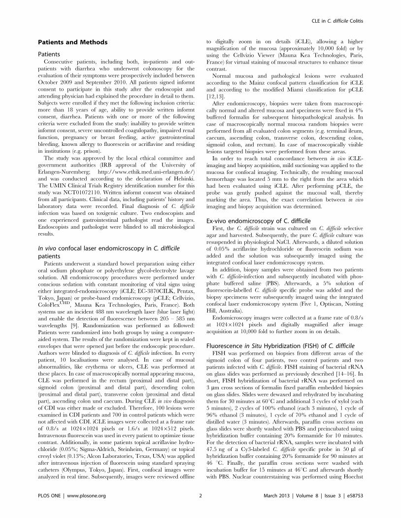
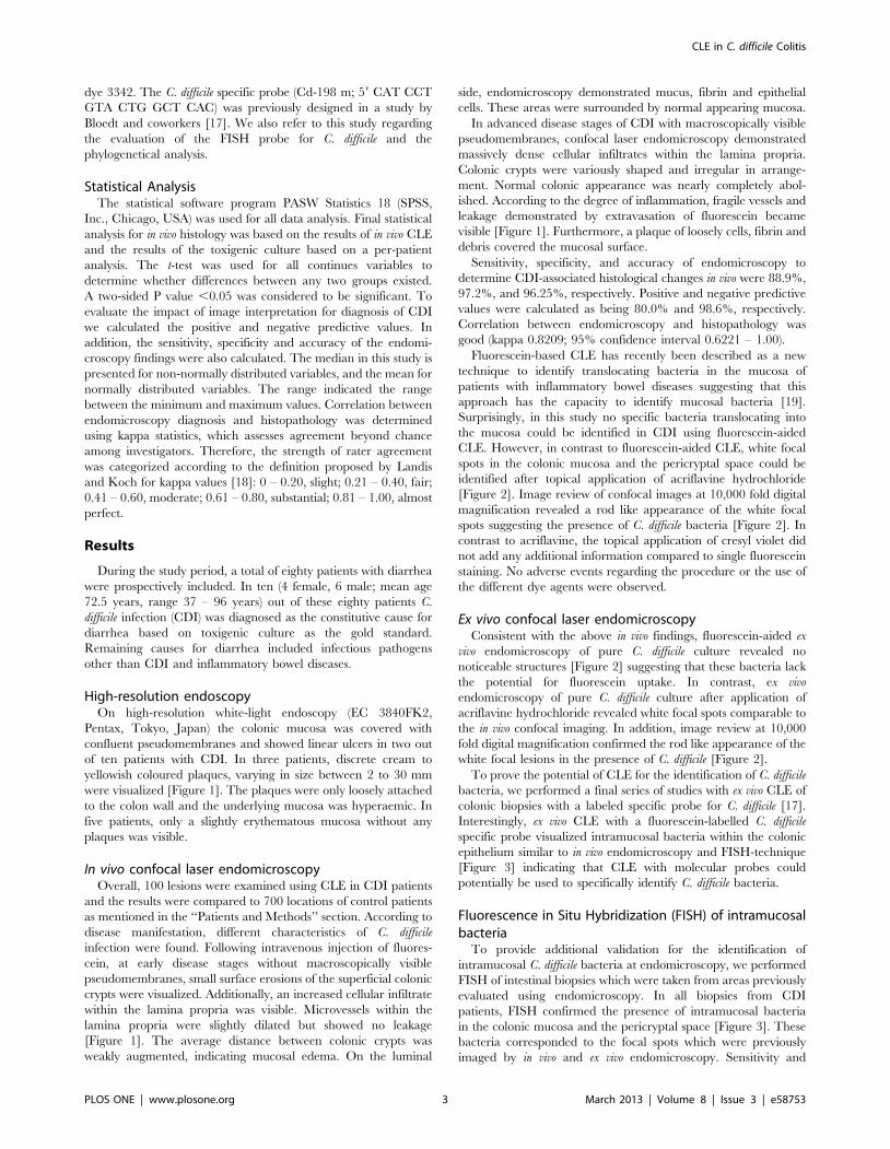
High-resolution endoscopyOn high-resolution white-light endoscopy (EC 3840FK2,
Pentax, Tokyo, Japan) the colonic mucosa was covered with
confluent pseudomembranes and showed linear ulcers in two out
of ten patients with CDI. In three patients, discrete cream to
yellowish coloured plaques, varying in size between 2 to 30 mm
were visualized [Figure 1]. The plaques were only loosely attached
to the colon wall and the underlying mucosa was hyperaemic. In
five patients, only a slightly erythematous mucosa without any
plaques was visible.
In vivo confocal laser endomicroscopyOverall, 100 lesions were examined using CLE in CDI patients
and the results were compared to 700 locations of control patients
as mentioned in the ‘‘Patients and Methods’’ section. According to
disease manifestation, different characteristics of C. difficile
infection were found. Following intravenous injection of fluores-
cein, at early disease stages without macroscopically visible
pseudomembranes, small surface erosions of the superficial colonic
crypts were visualized. Additionally, an increased cellular infiltrate
within the lamina propria was visible. Microvessels within the
lamina propria were slightly dilated but showed no leakage
[Figure 1]. The average distance between colonic crypts was
weakly augmented, indicating mucosal edema. On the luminal
side, endomicroscopy demonstrated mucus, fibrin and epithelial
cells. These areas were surrounded by normal appearing mucosa.
In advanced disease stages of CDI with macroscopically visible
pseudomembranes, confocal laser endomicroscopy demonstrated
massively dense cellular infiltrates within the lamina propria.
Colonic crypts were variously shaped and irregular in arrange-
ment. Normal colonic appearance was nearly completely abol-
ished. According to the degree of inflammation, fragile vessels and
leakage demonstrated by extravasation of fluorescein became
visible [Figure 1]. Furthermore, a plaque of loosely cells, fibrin and
debris covered the mucosal surface.
Sensitivity, specificity, and accuracy of endomicroscopy to
determine CDI-associated histological changes in vivo were 88.9%,
97.2%, and 96.25%, respectively. Positive and negative predictive
values were calculated as being 80.0% and 98.6%, respectively.
Correlation between endomicroscopy and histopathology was
good (kappa 0.8209; 95% confidence interval 0.6221 – 1.00).
Fluorescein-based CLE has recently been described as a new
technique to identify translocating bacteria in the mucosa of
patients with inflammatory bowel diseases suggesting that this
approach has the capacity to identify mucosal bacteria [19].
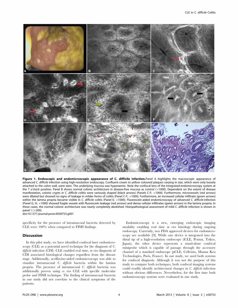
Surprisingly, in this study no specific bacteria translocating into
the mucosa could be identified in CDI using fluorescein-aided
CLE. However, in contrast to fluorescein-aided CLE, white focal
spots in the colonic mucosa and the pericryptal space could be
identified after topical application of acriflavine hydrochloride
[Figure 2]. Image review of confocal images at 10,000 fold digital
magnification revealed a rod like appearance of the white focal
spots suggesting the presence of C. difficile bacteria [Figure 2]. In
contrast to acriflavine, the topical application of cresyl violet did
not add any additional information compared to single fluorescein
staining. No adverse events regarding the procedure or the use of
the different dye agents were observed.
Ex vivo confocal laser endomicroscopyConsistent with the above in vivo findings, fluorescein-aided ex
vivo endomicroscopy of pure C. difficile culture revealed no
noticeable structures [Figure 2] suggesting that these bacteria lack
the potential for fluorescein uptake. In contrast, ex vivo
endomicroscopy of pure C. difficile culture after application of
acriflavine hydrochloride revealed white focal spots comparable to
the in vivo confocal imaging. In addition, image review at 10,000
fold digital magnification confirmed the rod like appearance of the
white focal lesions in the presence of C. difficile [Figure 2].
To prove the potential of CLE for the identification of C. difficile
bacteria, we performed a final series of studies with ex vivo CLE of
colonic biopsies with a labeled specific probe for C. difficile [17].
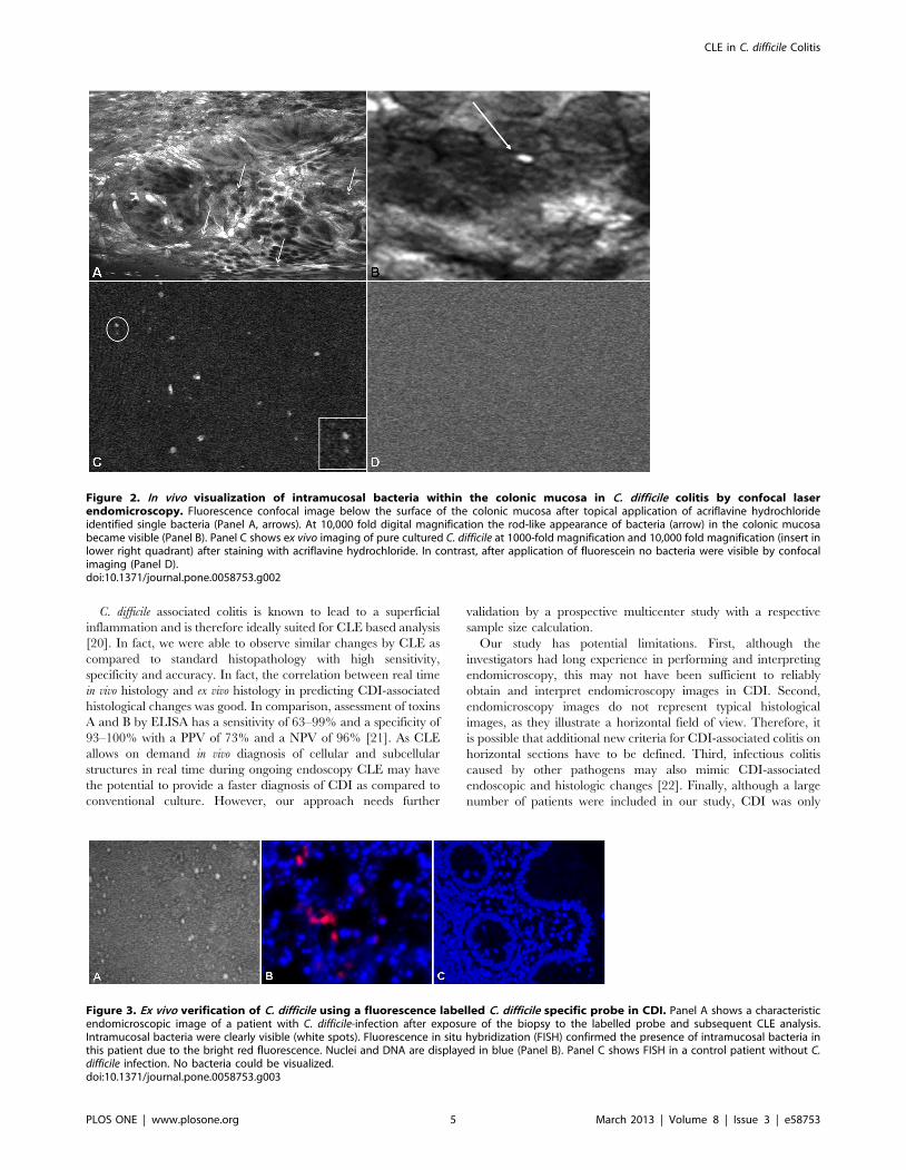
Interestingly, ex vivo CLE with a fluorescein-labelled C. difficile
specific probe visualized intramucosal bacteria within the colonic
epithelium similar to in vivo endomicroscopy and FISH-technique
[Figure 3] indicating that CLE with molecular probes could
potentially be used to specifically identify C. difficile bacteria.
Fluorescence in Situ Hybridization (FISH) of intramucosalbacteria
To provide additional validation for the identification of
intramucosal C. difficile bacteria at endomicroscopy, we performed
FISH of intestinal biopsies which were taken from areas previously
evaluated using endomicroscopy. In all biopsies from CDI
patients, FISH confirmed the presence of intramucosal bacteria
in the colonic mucosa and the pericryptal space [Figure 3]. These
bacteria corresponded to the focal spots which were previously
imaged by in vivo and ex vivo endomicroscopy. Sensitivity and
CLE in C. difficile Colitis
PLOS ONE | www.plosone.org 3 March 2013 | Volume 8 | Issue 3 | e58753
specificity for the presence of intramucosal bacteria detected by
CLE were 100% when compared to FISH findings.
Discussion
In this pilot study, we have identified confocal laser endomicro-
scopy (CLE) as a potential novel technique for the diagnosis of C.
difficile infection (CDI). CLE enabled real time, in vivo diagnosis of
CDI associated histological changes regardless from the disease
stage. Additionally, acriflavine-aided endomicroscopy was able to
visualize intramucosal C. difficile bacteria within the lamina
propria. The presence of intramucosal C. difficile bacteria was
additionally proven using ex vivo CLE with specific molecular
probe and FISH technique. The finding of intramucosal bacteria
in our study did not correlate to the clinical symptoms of the
patients.
Endomicroscopy is a new, emerging endoscopic imaging
modality enabling real time in vivo histology during ongoing
endoscopy. Currently, two FDA approved devices for endomicro-
scopy are available [9]. While one device is integrated into the
distal tip of a high-resolution endoscope (iCLE, Pentax, Tokyo,
Japan), the other device represents a stand-alone confocal
miniprobe which is capable of passage through the accessory
channel of a standard endoscope (pCLE, Cellvizio, Mauna Kea
Technologies, Paris, France). In our study, we used both systems
for confocal diagnosis. Although it was not the purpose of this
study to compare both techniques, both confocal imaging systems
could readily identify architectural changes in C. difficile infection
without obvious differences. Nevertheless, for the first time both
endomicroscopy systems were evaluated in one study.
Figure 1. Endoscopic and endomicroscopic appearance of C. difficile infection.Panel A highlights the macroscopic appearance ofadvanced C. difficile infection using high-resolution endoscopy. Confluent cream to yellow coloured plaques varying in size, which were only looselyattached to the colon wall, were seen. The underlying mucosa was hyperaemic. Note the confocal lens of the integrated-endomicroscopy system atthe 7 o’clock position. Panel B shows normal colonic architecture in disease-free mucosa as control (61000). Dependent on the extent of diseasemanifestation, colonic crypts in C. difficile colitis were variously shaped (black arrows) (Panels C-F; 61000). Furthermore, microvessels (red arrows)were dilated but showed no signs of leakage in milder forms of colitis (Panel C-F; 61000). Furthermore, an increased cellular infiltrate (green arrows)within the lamina propria became visible in C. difficile colitis (Panel E; 61000). Fluorescein-aided endomicroscopy of advanced C. difficile-infection(Panel G, H; 61000) showed fragile vessels with fluorescein leakage (red arrows) and dense cellular infiltrates (green arrows) in the lamina propria. Inthese cases, the normal colonic architecture was nearly completely abolished. Histopathological assessment of mild C. difficile infection is shown inpanel I (6200).doi:10.1371/journal.pone.0058753.g001
CLE in C. difficile Colitis
PLOS ONE | www.plosone.org 4 March 2013 | Volume 8 | Issue 3 | e58753
C. difficile associated colitis is known to lead to a superficial
inflammation and is therefore ideally suited for CLE based analysis
[20]. In fact, we were able to observe similar changes by CLE as
compared to standard histopathology with high sensitivity,
specificity and accuracy. In fact, the correlation between real time
in vivo histology and ex vivo histology in predicting CDI-associated
histological changes was good. In comparison, assessment of toxins
A and B by ELISA has a sensitivity of 63–99% and a specificity of
93–100% with a PPV of 73% and a NPV of 96% [21]. As CLE
allows on demand in vivo diagnosis of cellular and subcellular
structures in real time during ongoing endoscopy CLE may have
the potential to provide a faster diagnosis of CDI as compared to
conventional culture. However, our approach needs further
validation by a prospective multicenter study with a respective
sample size calculation.
Our study has potential limitations. First, although the
investigators had long experience in performing and interpreting
endomicroscopy, this may not have been sufficient to reliably
obtain and interpret endomicroscopy images in CDI. Second,
endomicroscopy images do not represent typical histological
images, as they illustrate a horizontal field of view. Therefore, it
is possible that additional new criteria for CDI-associated colitis on
horizontal sections have to be defined. Third, infectious colitis
caused by other pathogens may also mimic CDI-associated
endoscopic and histologic changes [22]. Finally, although a large
number of patients were included in our study, CDI was only
Figure 2. In vivo visualization of intramucosal bacteria within the colonic mucosa in C. difficile colitis by confocal laserendomicroscopy. Fluorescence confocal image below the surface of the colonic mucosa after topical application of acriflavine hydrochlorideidentified single bacteria (Panel A, arrows). At 10,000 fold digital magnification the rod-like appearance of bacteria (arrow) in the colonic mucosabecame visible (Panel B). Panel C shows ex vivo imaging of pure cultured C. difficile at 1000-fold magnification and 10,000 fold magnification (insert inlower right quadrant) after staining with acriflavine hydrochloride. In contrast, after application of fluorescein no bacteria were visible by confocalimaging (Panel D).doi:10.1371/journal.pone.0058753.g002
Figure 3. Ex vivo verification of C. difficile using a fluorescence labelled C. difficile specific probe in CDI. Panel A shows a characteristicendomicroscopic image of a patient with C. difficile-infection after exposure of the biopsy to the labelled probe and subsequent CLE analysis.Intramucosal bacteria were clearly visible (white spots). Fluorescence in situ hybridization (FISH) confirmed the presence of intramucosal bacteria inthis patient due to the bright red fluorescence. Nuclei and DNA are displayed in blue (Panel B). Panel C shows FISH in a control patient without C.difficile infection. No bacteria could be visualized.doi:10.1371/journal.pone.0058753.g003
CLE in C. difficile Colitis
PLOS ONE | www.plosone.org 5 March 2013 | Volume 8 | Issue 3 | e58753
proven in a subset of patients, thereby potentially affecting
statistical analysis. A future prospective multicenter study with a
respective sample size calculation addressing these points is thus
highly warranted.
Interestingly, CLE allowed the identification of bacteria in the
mucosa of patients with CDI in vivo. Previously, it was shown by
Kiesslich and coworkers that endomicroscopy with topically
applied acriflavine could readily identify Helicobacter pylori infection
in vivo [23]. Very recently, the same group demonstrated that
endomicroscopy was able to identify intramucosal enteric bacteria
in vivo in the colon and ileum of patients with ulcerative colitis and
Crohn’s disease using fluorescein-aided endomicroscopy [19]. It
was shown that intramucosal bacteria were more frequently and
with a wider distribution found in patients with inflammatory
bowel disease than in patients with a normal intestine. However, in
our study no bacteria in the colonic mucosa could be identified by
fluorescein-aided CLE. Importantly, we were only able to visualize
bacteria after application of acriflavine which is a cationic dye that
can be detected by its intrinsic fluorescence and accumulates in the
endosomal/lysosomal compartment of cells [24]. Thus, our
findings suggest that topical administration of acriflavine results
in translocation of the dye in the mucosa via epithelial gaps or
epithelial erosions followed by its uptake in C. difficile bacteria [25].
The fact that these bacteria are negative by fluorescein staining but
positive upon acriflavine use is striking and discriminates this
bacterial strain from other bacteria previously identified in the
colonic mucosa [19]. To our knowledge, this is the first report on a
fluorescein-negative bacterial strain in the colonic mucosa by CLE
analysis. Thus, your finding of staining patterns is very interesting
and may aid in the further classification of different bacteria.
Therefore, we suggest that in the future CLE should be evaluated
to analyze bacterial cultures of various gram-negative and gram-
positive pathogens.
In order to validate the presence of intramucosal C. difficile
bacteria in patients with CDI, we performed several ex vivo studies.
Intramucosal bacteria imaged by endomicroscopy in vivo looked
similar to C. difficile in an ex vivo cell suspension of pure C. difficile
culture when analyzed by CLE. Again, only acriflavine-aided
endomicroscopy was feasible to detect C. difficile by ex vivo CLE,
while fluorescein-aided endomicroscopy revealed no bacteria. This
finding underlines our in vivo results where we could only identify
bacteria after topical application of acriflavine.
High-magnification of C. difficile bacteria, both in and ex vivo
revealed a rod like appearance of these microorganisms. Addi-
tionally, there was a strong concordance between the presence of
intramucosal bacteria identified in vivo and bacteria identified by
FISH in biopsies of areas which were previously investigated using
endomicroscopy. In order to further highlight the potential of CLE
for detection of specific bacteria in CDI, additional ex vivo studies
on endomicroscopy with previously validated specific molecular
probe for C. difficile were performed and the results were compared
to those of the FISH analysis. By using this approach, we could
identify the presence of specific intramucosal C. difficile bacteria
within the colon in CDI. These findings underline the potential of
endomicroscopy for molecular imaging of CDI and specific
detection of C. difficile bacteria. By using ex vivo CLE with the C.
difficile specific probe we have shown that the probe seems to be
able to penetrate the cells without the use of previous fixation. Our
hypothesis is that the mucosal structure of the inflamed tissue
(according to C. difficile colitis) allows penetration of the probe.
This speculation is underlined by recent studies evaluating in vivo
confocal imaging in patients with inflammatory bowel diseases.
These studies suggested that patients with colitis has an increased
number of epithelial gaps in human small and large intestine and
that these gaps must be considered as a component of the intestinal
barrier and may therefore have potential implications for intestinal
barrier dysfunction in human diseases [25,26].
Our findings could have a substantial clinical impact, as
endomicroscopy has the potential to diagnose C. difficile infection
in vivo, thus potentially enabling an accelerated diagnosis and an
improved patient management. Therefore, endomicroscopy might
have the potential to refine our understanding of C. difficile
diagnosis and may help to prevent nosocomial transmission of this
life-threatening disease. Moreover, we were able to describe
intramucosal bacteria in vivo. The clinical significance of our
findings should be elucidated in future prospective studies.
Author Contributions
Conceived and designed the experiments: HN MV MG CG NW CB CS.
Performed the experiments: HN MV MG CG NW CB CS. Analyzed the
data: HN MV CB MFN. Contributed reagents/materials/analysis tools:
HN MV MG CG NW JM CB CS RA MFN. Wrote the paper: HN MV
MG CG NW JM CB CS RA MFN.
References
1. Kelly CP, LaMont JT (2008) Clostridium difficile--more difficult than ever.
N Engl J Med 359: 1932–1940.
2. Centers for Disease Control and Prevention (CDC) (2005) Severe Clostridium
difficile-associated disease in populations previously at low risk. MMWR Morb
Mortal Wkly Rep 54: 1201–1205.
3. Huang H, Weintraub A, Fang H, Nord CE (2009) Antimicrobial resistance in
Clostridium difficile. Int J Antimicrob Agents 34: 516–522.
4. Crobach MJ, Dekkers OM, Wilcox MH, Kuijper EJ (2009) European Society of
Clinical Microbiology and Infectious Diseases (ESCMID): data review and
recommendations for diagnosing Clostridium difficile-infection (CDI). Clin
Microbiol Infect 15: 1053–1566.
5. Kiesslich R, Gossner L, Goetz M, Dahlmann A, Vieth M, et al. (2006) In vivo
histology of Barrett’s esophagus and associated neoplasia by confocal laser
endomicroscopy. Clin Gastroenterol Hepatol 4: 979–987.
6. Leong RW, Nguyen NQ, Meredith CG, Al-Sohaily S, Kukic D, et al. (2008) In
vivo confocal endomicroscopy in the diagnosis and evaluation of celiac disease.
Gastroenterology 135: 1870–1876.
7. Buchner AM, Shahid MW, Heckman MG, Krishna M, Ghabril M, et al. (2010)
Comparison of probe-based confocal laser endomicroscopy with virtual
chromoendoscopy for classification of colon polyps. Gastroenterology 138:
834–842.
8. Neumann H, Grauer M, Vieth M, Neurath MF (2013) In vivo diagnosis of
lymphocytic colitis by confocal laser endomicroscopy. Gut 62: 333–334.
9. Neumann H, Kiesslich R, Wallace MB, Neurath MF (2010) Confocal laser
endomicroscopy: technical advances and clinical applications. Gastroenterology
139: 388–392.
10. Gunther U, Epple HJ, Heller F, Loddenkemper C, Grunbaum M, et al. (2008)
In vivo diagnosis of intestinal spirochaetosis by confocal endomicroscopy. Gut
57: 1331–1333.
11. Neumann H, Vieth M, Atreya R, Grauer M, Siebler J, et al. (2012) Assessment
of Crohn’s disease activity by confocal laser endomicroscopy. Inflamm Bowel
Dis. 18: 2261–2269.
12. Kiesslich R, Burg J, Vieth M, Gnaendiger J, Enders M, et al. (2004) Confocal
laser endoscopy for diagnosing intraepithelial neoplasias and colorectal cancer in
vivo. Gastroenterology 127: 706–713.
13. Wallace M, Lauwers GY, Chen Y, Dekker E, Fockens P, et al. (2011) Miami
classification for probe-based confocal laser endomicroscopy. Endoscopy 43:
882–891.
14. Becker C, Wirtz S, Blessing M, Pirhonen J, Strand D, et al. (2003) Constitutive
p40 promoter activation and IL-23 production in the terminal ileum mediated
by dendritic cells. J Clin Invest 112: 693–706.
15. Amann RI, Ludwig W, Schleifer KH (1995) Phylogenetic identification and in
situ detection of individual microbial cells without cultivation. Microbiol Rev 59:
143–169.
16. Moter A, Gobel UB (2000) Fluorescence in situ hybridization (FISH) for direct
visualization of microorganisms. J Microbiol Methods 41: 85–112.
17. Bloedt K, Riecker M, Poppert S, Wellinghausen N (2009) Evaluation of new
selective culture media and a rapid fluorescence in situ hybridization assay for
CLE in C. difficile Colitis
PLOS ONE | www.plosone.org 6 March 2013 | Volume 8 | Issue 3 | e58753
identification of Clostridium difficile from stool samples. J Med Microbiol 58:
874–877.18. Landis JR, Koch GG (1977) The measurement of observer agreement for
categorical data. Biometrics 33: 159–174.
19. Moussata D, Goetz M, Gloeckner A, Kerner M, Campbell B, et al. (2011)Confocal laser endomicroscopy is a new imaging modality for recognition of
intramucosal bacteria in inflammatory bowel disease in vivo. Gut 60: 26–33.20. Monaghan T, Boswell T, Mahida YR (2008) Recent advances in Clostridium
difficile-associated disease. Gut 57: 850–860.
21. Bartlett JG, Gerding DN (2008) Clinical recognition and diagnosis ofClostridium difficile infection. Clin Infect Dis 46: 12–18.
22. Monkemuller K, Patasiute I, Walther F, Peitz U, Fry LC, et al. (2006)Pseudomembranous colitis due to Salmonella enterica serotype infantis.
Endoscopy 38: 546.
23. Kiesslich R, Goetz M, Burg J, Stolte M, Siegel E, et al. (2005) Diagnosing
Helicobacter pylori in vivo by confocal laser endoscopy. Gastroenterology 128:
2119–2123.
24. Davies JP, Chen FW, Ioannou YA (2000) Transmembrane molecular pump
activity of Niemann-Pick C1 protein. Science 290:2295–2298.
25. Kiesslich R, Goetz M, Angus EM, Hu Q, Guan Y, et al. (2007) Identification of
epithelial gaps in human small and large intestine by confocal endomicroscopy.
Gastroenterology 133: 1769–1778.
26. Liu JJ, Wong K, Thiesen AL, Mah SJ, Dieleman LA, et al. (2011) Increased
epithelial gaps in the small intestines of patients with inflammatory bowel
disease: density matters. Gastrointest Endosc 73: 1174–1180.
CLE in C. difficile Colitis
PLOS ONE | www.plosone.org 7 March 2013 | Volume 8 | Issue 3 | e58753