Comparison of Ventricular Geometry for Two Real-Time 3D Ultrasound Machines with Three-dimensional Level Set
Elsa D. Angelini, Rio Otsuka, Shunishi Homma, Andrew F. Laine
The Heffner Biomedical Imaging LabDepartment of Biomedical Engineering, Columbia University, New York, NY, USA
Average error (1) = 0.86%, (2) = 1%.Gaussian kernel for (3) lead to the smallest error (0.03%).Anisotropic diffusion (3-4) reduces measurement errors for first-order interpolation kernels.
RESULTSRESULTS
Computation Times
Measurements of cylinder diameter were performed manually by a user on B-scan slices for different data processing: (1) Original data (scale=1); (2) Original data (scale=2); (3) Original data (scale=2 with smoothing);(4) Diffused data (thresholds=5, 10 iterations) (scale=1); (5) Diffused data (variable threshold, 10 iterations) (scale=1).
INTRODUCTIONINTRODUCTIONThree-dimensional ultrasound machines based on mat
rix phased-array transducers are gaining predominanc
e for real-time dynamic screening in cardiac and obstet
ric practice.
Comparison of the quantification of cardiac function fro
m two matrix-phased array 3D ultrasound machines:
RT3D machine from Volumetrics Medical Imaging.
Entire cardiac volume is acquired with an array of 64
64 elements and a downsampling factor of 4 between
receive/transmit modes.
Sonos 7500 machine from Philips Medical Systems.
Four cardiac sub-volumes and no downsampling.
DATADATART3D data were acquired by a RT3D Volumetri
cs© machine using acquisition parameters iden
tical to clinical settings.
Phantom object: Two cylinders (diameter = 10
mm) with different signal-to-noise ratios (SNR).
Our experiments focused on a SNR of 2dB.
In-vitro phantom: myocardium muscle sample
in a water tank.
Clinical data: Echocardiographic volume of a h
ealthy volunteer.
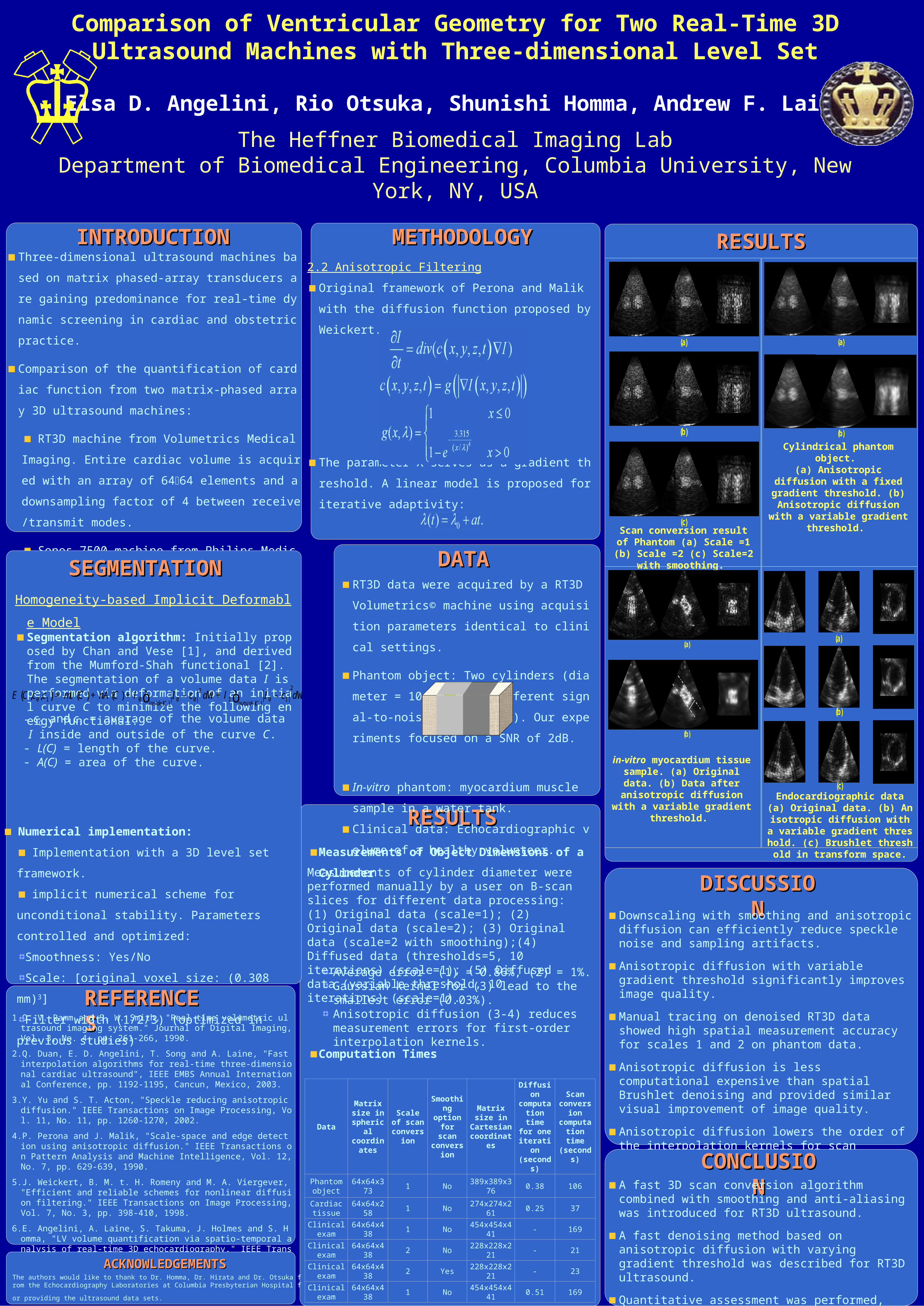
DISCUSSIODISCUSSIONNDownscaling with smoothing and anisotropic diffusion
can efficiently reduce speckle noise and sampling artifacts.
Anisotropic diffusion with variable gradient threshold significantly improves image quality.
Manual tracing on denoised RT3D data showed high spatial measurement accuracy for scales 1 and 2 on phantom data.
Anisotropic diffusion is less computational expensive than spatial Brushlet denoising and provided similar visual improvement of image quality.
Anisotropic diffusion lowers the order of the interpolation kernels for scan conversion enabling optimization of data processing for real-time denoising and visualization.
SEGMENTATIONSEGMENTATIONHomogeneity-based Implicit Deformable Model
Segmentation algorithm: Initially proposed by Chan and Vese [1], and derived from the Mumford-Shah functional [2]. The segmentation of a volume data I is performed via deformation of an initial curve C to minimize the following energy functional:
REFERENCREFERENCESES1. O. V. Ramm and S. W. Smith, "Real time volumetric ultrasound imaging
system." Journal of Digital Imaging, Vol. 3, No. 4, pp. 261-266, 1990.
2. Q. Duan, E. D. Angelini, T. Song and A. Laine, "Fast interpolation algorithms for real-time three-dimensional cardiac ultrasound", IEEE EMBS Annual International Conference, pp. 1192-1195, Cancun, Mexico, 2003.
3. Y. Yu and S. T. Acton, "Speckle reducing anisotropic diffusion." IEEE Transactions on Image Processing, Vol. 11, No. 11, pp. 1260-1270, 2002.
4. P. Perona and J. Malik, "Scale-space and edge detection using anisotropic diffusion." IEEE Transactions on Pattern Analysis and Machine Intelligence, Vol. 12, No. 7, pp. 629-639, 1990.
5. J. Weickert, B. M. t. H. Romeny and M. A. Viergever, "Efficient and reliable schemes for nonlinear diffusion filtering." IEEE Transactions on Image Processing, Vol. 7, No. 3, pp. 398-410, 1998.
6. E. Angelini, A. Laine, S. Takuma, J. Holmes and S. Homma, "LV volume quantification via spatio-temporal analysis of real-time 3D echocardiography." IEEE Transactions on Medical Imaging, Vol. 20, No. 6, pp. 457-469, 2001.
CONCLUSICONCLUSIONONA fast 3D scan conversion algorithm combined with
smoothing and anti-aliasing was introduced for RT3D ultrasound.
A fast denoising method based on anisotropic diffusion with varying gradient threshold was described for RT3D ultrasound.
Quantitative assessment was performed, showing high spatial accuracy of RT3D ultrasound.
(a)
(b)
Cylindrical phantom object. (a) Anisotropic diffusion with a fixed gradient threshold. (b) Anisotropic diffusion with a variable gradient threshold.
(a)
(b)
(c) Scan conversion result of
Phantom (a) Scale =1 (b) Scale =2 (c) Scale=2 with smoothing.
(a)
(b)
(c) Endocardiographic data (a) Original data. (b) Anisotropic diffusion with a variable gradient threshold. (c) Brushlet threshold in tra
nsform space.
(a)
(b)
in-vitro myocardium tissue sample. (a) Original data. (b)
Data after anisotropic diffusion with a variable gradient
threshold.
RESULTSRESULTSMETHODOLOGYMETHODOLOGY2.2 Anisotropic Filtering
Original framework of Perona and Malik with the diffu
sion function proposed by Weickert.
The parameter λ serves as a gradient threshold. A lin
ear model is proposed for iterative adaptivity:
ACKNOWLEDGEMENTSACKNOWLEDGEMENTSThe authors would like to thank to Dr. Homma, Dr. Hirata and Dr. Otsuka from the Echocardiograph
y Laboratories at Columbia Presbyterian Hospital for providing the ultrasound data sets.
Data
Matrix size in
spherical coordinate
s
Scale of scan
conversion
Smoothing option for scan
conversion
Matrix size in Cartesian coordinates
Diffusion computati
on time for one
iteration(seconds)
Scan conversio
n computati
on time(seconds)
Phantom object
64x64x373 1 No 389x389x376 0.38 106
Cardiac tissue
64x64x258 1 No 274x274x261 0.25 37
Clinical exam
64x64x438 1 No 454x454x441 - 169
Clinical exam
64x64x438 2 No 228x228x221 - 21
Clinical exam
64x64x438 2 Yes 228x228x221 - 23
Clinical exam
64x64x438 1 No 454x454x441 0.51 169
Measurements of Object Dimensions of a
Cylinder
( ) ( ) ( )( )( )
220 1 0 0 0 1 0 1, ,
insideC outsideCE C c c L C A C I c d I c dm n l l= + + - W+ - Wò ò
Numerical implementation:
Implementation with a 3D level set framework.
implicit numerical scheme for unconditional
stability. Parameters controlled and optimized:
Smoothness: Yes/No
Scale: [original voxel size: (0.308 mm)3]
Filter width (1/2/3) (optimized in previous studies)
- c0 and c1 = average of the volume data I inside and outside of the curve C.
- L(C) = length of the curve.- A(C) = area of the curve.