Research Archive
Citation for published version:Markus Riessland, et al, ‘Neurocalcin Delta Suppression Protects against Spinal Muscular Atrophy in Humans and across Species by Restoring Impaired Endocytosis’, The American Journal of Human Genetics, Vol. 100 (2): 297-315, February 2017.
DOI:https://doi.org/10.1016/j.ajhg.2017.01.005
Document Version:This is the Accepted Manuscript version. The version in the University of Hertfordshire Research Archive may differ from the final published version.
Copyright and Reuse: © 2017 American Society of Human Genetics.This manuscript version is distributed under the terms of the Creative Commons Attribution licence (http://creativecommons.org/licenses/by/4.0/), which permits unrestricted re-use, distribution, and reproduction in any medium, provided the original work is properly cited.
EnquiriesIf you believe this document infringes copyright, please contact the Research & Scholarly Communications Team at [email protected]

Neurocalcin delta suppression protects against
spinal muscular atrophy in humans and across species
by restoring impaired endocytosis
Markus Riessland,1,2,3,13,14 Anna Kaczmarek,1,2,3,13 Svenja Schneider,1,2,3,13 Kathryn J.
Swoboda,4 Heiko Löhr,5,7 Cathleen Bradler,6,7 Vanessa Grysko,1,2,3 Maria Dimitriadi,8,15
Seyyedmohsen Hosseinibarkooie,1,2,3 Laura Torres-Benito,1,2,3 Miriam Peters,1,2,3 Aaradhita
Upadhyay,1,2,3 Nasim Biglari,1,2,3 Sandra Kröber,1,2,3 Irmgard Hölker,1,2,3 Lutz Garbes,1,2,3
Christian Gilissen,9 Alexander Hoischen,9 Gudrun Nürnberg,7,10 Peter Nürnberg,7,10 Michael
Walter,11 Frank Rigo,12 C. Frank Bennett,12 Min Jeong Kye,1,2 Anne C. Hart,8 Matthias
Hammerschmidt,5,7 Peter Kloppenburg,6,7 and Brunhilde Wirth1,2,3,*
1Institute of Human Genetics, University of Cologne, 50931 Cologne, Germany
2Center for Molecular Medicine Cologne, University of Cologne, 50931 Cologne, Germany
3Institute for Genetics, University of Cologne, 50674 Cologne, Germany
4MassGeneral Hospital for Children, Boston, MA 02115, USA
5Institute for Zoology - Developmental Biology, University of Cologne, 50674 Cologne,
Germany
6Institute for Zoology - Neurophysiology University of Cologne, 50674 Cologne, Germany
7Excellence Cluster on Cellular Stress Responses in Aging Associated Diseases (CECAD),
University of Cologne, 50931 Cologne, Germany
8Department of Neuroscience, Brown University, Providence, Rhode Island, 02912, USA
9Department of Human Genetics, Donders Centre for Neuroscience, Radboud University
Medical Center, 6525 Nijmegen, The Netherlands
10Center for Genomics Cologne, University of Cologne, 50931 Cologne, Germany

2
11Institute of Medical Genetics, University of Tübingen, 72076 Tübingen, Germany
12IONIS Pharmaceuticals, Carlsbad, California, CA 92008, USA
13These authors contributed equally to this work
Present address:
14Laboratory of Molecular and Cellular Neuroscience, The Rockefeller University, New York,
NY 10065, USA
15Department of Biological and Environmental Sciences, University of Hertfordshire, Hatfield,
AL10 9AB, UK
*Correspondence: [email protected]
3
ABSTRACT
Homozygous SMN1 loss causes spinal muscular atrophy (SMA), the most common lethal
genetic childhood motor neuron disease. SMN1 encodes SMN, a ubiquitous housekeeping
protein, which makes the primarily motor neuron-specific phenotype rather unexpected. SMA
individuals harbor low SMN expression from one to six SMN2 copies, which is insufficient to
functionally compensate for SMN1 loss. However, rarely individuals with homozygous absence
of SMN1 and only three to four SMN2 copies are fully asymptomatic, suggesting protection
through genetic modifier(s). Previously, we identified plastin 3 (PLS3) overexpression as an
SMA protective modifier in humans and showed that SMN deficit impairs endocytosis, which
is rescued by elevated PLS3 levels. Here, we identify reduction of the neuronal calcium sensor
Neurocalcin delta (NCALD) as a protective SMA modifier in five asymptomatic SMN1-deleted
individuals carrying only four SMN2 copies. We demonstrate that NCALD is a Ca2+-dependent
negative regulator of endocytosis, as NCALD knockdown improves endocytosis in SMA
models and ameliorates pharmacologically induced endocytosis defects in zebrafish.
Importantly, NCALD knockdown effectively ameliorates SMA-associated pathological defects
across species, including worm, zebrafish and mouse. In conclusion, our study identifies a
previously unknown protective SMA modifier in humans, demonstrates modifier impact in three
different SMA animal models and suggests a potential combinatorial therapeutic strategy to
efficiently treat SMA. Since both protective modifiers restore endocytosis, our results confirm
that endocytosis is a major cellular mechanism perturbed in SMA and emphasize the power of
protective modifiers for understanding disease mechanism and developing therapies.
4
INTRODUCTION
In monogenic disorders, genetic modifiers can influence disease-causing mechanisms
resulting in incomplete penetrance1. Identification of such modifiers is of utmost relevance
since they can uncover regulatory networks and pathological mechanisms, as well as allow
identification of therapeutic pathways. For recessive disorders, full protection through modifiers
is extremely rare, making their identification highly challenging.
Spinal muscular atrophy (SMA), a motor neuron disease, is one of the most common and
devastating autosomal recessive disorders, for which no treatment is available yet. However,
various clinical trials using antisense oligonucleotides (ASOs), small molecules or gene
therapy show highly promising ameliorations2. Most SMA individuals show homozygous
absence of exon 7 of the survival motor neuron 1 (SMN1) [MIM: 600354]3, allowing easy and
efficient genetic testing4. SMN1 encodes SMN, a housekeeping protein involved in snRNP
biogenesis and splicing, microRNA biogenesis, transcription and translation regulation, and
others5-8; full absence of SMN causes embryonic lethality9. Only humans have an almost
identical copy, SMN2 [MIM: 601627], however this produces only ~10% correctly spliced full-
length transcript and protein, due to a single silent mutation affecting an exonic splicing
enhancer and creating a new splice silencer10-12. In SMA individuals, SMN2 is the only source
of SMN, thus its copy number (between 1-6) determines SMA severity13. In type 1 SMA (SMA1
[MIM: 253300]), the severe and most common form (60%), the majority of individuals carry two
SMN2 copies and die within the first two years of life. Most type 2 SMA (SMA2 [MIM: 253550])
affected individuals carry three SMN2 copies and are never able to walk. In type 3 SMA (SMA3
[MIM: 253400]), the mild form, most individuals carry four SMN2 copies and are able to walk,
but often become wheel-chair bound14.
Despite the important housekeeping function of SMN, reduced levels primarily cause spinal
motor neuron (MN) dysfunction in all types of SMA14. Thus, MN loss, impaired maturation and
maintenance of neuromuscular junctions (NMJs), and decreased proprioceptive inputs on MN
soma are hallmarks of SMA15-17. Nonetheless, dramatic reduction of SMN below a certain
threshold, as seen in severely-affected SMA individuals or animal models, compromise almost

5
every organ and many different cellular processes, which is in line with the essential function
of SMN in all cell types18; 19. Therefore, we reasoned that the search for the main cellular
pathway specifically driving MN dysfunction has to be carried out in mildly affected SMA
individuals, in whom only motor neuron function is impaired and moreover, that protective
modifiers identified in these individuals may reveal the critical underlying cellular mechanism.
To do so, we took advantage of very rarely occurring SMA-discordant families, in which
relatives of SMA affected individuals carry a homozygous SMN1 deletion together with three
or four SMN2 copies, but are clinically asymptomatic20-23. In seven of these families, we
previously identified the Ca2+-dependent protein Plastin 3 (PLS3) as a protective modifier24; 25.
PLS3 overexpression (OE) rescues SMA across species and is specifically upregulated in MNs
of asymptomatic individuals produced from induced pluripotent stem cells24; 26-29. Moreover,
PLS3 together with the second modifier found in this study, pointed us towards endocytosis as
the key disturbed cellular mechanism in SMA29.
Here, we report the identification of Neurocalcin delta (NCALD) [MIM: 606722], which encodes
a neuronal Ca2+ sensor protein, as an SMA protective modifier in humans. We show that
NCALD acts as a negative regulator of endocytosis, which is in contrast to PLS3 acting as its
positive regulator. We show Ca2+-dependent interaction of NCALD with clathrin, a protein
essential in endocytic vesicles coating. We demonstrate that low SMN levels reduce voltage-
dependent Ca2+ influx and that NCALD binds clathrin at low Ca2+ levels, thereby acting as a
Ca2+-sensitive inhibitor of endocytosis. Our results, obtained from multiple in vitro and in vivo
systems, show that NCALD suppression reestablishes synaptic function, most likely by
restoring endocytosis. Most importantly, we prove that NCALD knockdown (KD) in various
SMA animal models ameliorates major functional SMA disturbances, such as motor axon
development in zebrafish or MN circuitry and presynaptic function of neuromuscular junction
(NMJ) in mice. Moreover, we introduce a mild SMA mouse model generated by combined low-
dose SMN-ASO treatment and heterozygous Ncald knockout, and show restored motoric
function. Our data support the notion that genetic modifiers reveal additional valuable
treatment options, beyond existing therapies.

6
MATERIAL AND METHODS
Individuals’ DNA, Fibroblast Cell Lines and Lymphoblastoid Cell Lines. Informed written
consent was obtained from each subject or their legal guardians for all biological samples
according to the Declaration of Helsinki. The study has been approved by the Ethical
Committee of University of Cologne (04-138). Human fibroblast and EBV-transformed
lymphoblastoid cell lines (LBs) from SMA individuals, carriers and asymptomatic SMN1-
deleted individuals used in this work are listed in Table S1. DNA was extracted from EDTA
blood samples, primary fibroblast cell lines and LBs using standard protocols. SMN1 and
SMN2 copy number were determined by qRT-PCR or MLPA lysis (MRC Holland) as
described30. For haplotype analysis, polymorphic markers Ag1-CA (D5S1556), C212
(D5F149S1/S2), VS19A (D5S435) and MIT-I105 (D5S351) were analyzed as described31.
SMN2 coding region was sequenced in qRT-PCR products obtained from LBs-isolated RNA
as described32. PLS3 expression was analyzed as described24. All cell lines used were tested
for mycoplasma contamination.
Genome-wide Linkage Analysis. Genome-wide scan was performed in 14 individuals of the
Utah family using Affymetrix GeneChip Human Mapping 10K Array 2.0, which comprises total
10,024 SNPs with a mean intermarker distance of 258kb, equivalent to 0.36cM (Affymetrix).
Parametric linkage analysis was performed by ALLEGRO program33 assuming autosomal
dominant inheritance with full penetrance and 0.0001 disease allele frequency. Haplotypes
were reconstructed with ALLEGRO and presented graphically with HaploPainter34. All data
handling was performed using the graphical user interface ALOHOMORA35.
Transcriptome Analysis. For expression profiling, 400ng total RNA were amplified and
biotinylated using Illumina TotalPrepTMRNA Amplification Kits (Ambion) according to
manufacturer’s protocol. Human HT-12v3 bead arrays (Illumina) were hybridized with 750ng
cRNA for 18h at 58°C according to Illumina Whole-Genome Gene Expression with IntelliHyb
7
SealSystem Manual. Arrays were washed with E1BC buffer, High-Temp Wash Buffer and
100% ethanol, stained with streptavidine-Cy3, and washed with E1BC buffer. Fluorescence
intensities were recorded on BeadArray Reader GX (Illumina). Average signal intensities
without background correction36 were performed with BeadStudio3.1 (Illumina). All data
analysis steps were performed in the statistical environment R (version 2.10-0) with several
bioconductor packages (version 2.6.1). Signal intensities were normalized with VSN (variance
stabilizing and normalization quantification method37) and non-informative probes were
removed based on p-values. Signals were averaged for individual subgroups and a linear
model was designed capturing the influence of the asymptomatic group on gene expression
levels38. Differences between subgroups were extracted as contrasts and analyzed with the
moderated F-test (empirical Bayes method) including a correction step for multiple testing with
5%-FDR-based method39. To attribute significant regulations to individual contrasts, a decision
matrix was generated based on the function “decide tests” within the “limma” package, where
significant up- or downregulations are represented by values of 1 or -1, respectively.
Targeted Resequencing. To identify a potential variant regulating differential NCALD
expression, complete NCALD locus ±1Mb (chr8:101,505,353-104,404,346) was deep-
sequenced from gDNA of family members II-1, III-1, III-4, III-8, and IV-3 at Radboud University
Medical Center Nijmegen using a 5500xl sequencing instrument (Life Technologies). ~3Mb
genomic DNA from chromosome 8 were captured using a 385K NimbleGen SequenceCapture
Array (Roche).
On average, we obtained 2.7Gb of mappable sequence data/individual. Reads were mapped
to the hg19 reference genome with LifeTechnologies BioScope software 1.3. On average, 94%
of bases originated from the target region (mean 544-fold coverage). 99.8% of the targeted
region was covered ≥20 times. Single-nucleotide variants were subsequently high-stringency
called by the DiBayes algorithm. Small insertions and deletions were detected using the Small
IndelTool. Variants were annotated using an in-house analysis pipeline.

8
Animal Models
Zebrafish Experiments. All experiments were performed with the transgenic line tg(mnx1-
GFP)ml2TG 40 and approved by the local animal protection committee (LANUV NRW; reference
number 84-02.04.2012.A251).
Zebrafish Injection and Analysis. Morpholinos (MO) were designed against the translational
start codons of respective genes (Gene Tools, LLC). smn-MO: 5’-
CGACATCTTCTGCACCATTGGC-3’; ncaldb-MO: 5’-GGAGCTTGCTGTTTTGTTTTCCCAT-
3’; control-MO: 5’-CCTCTTACCTCAGTTACAATTTATA-3’. For NCALD mRNA injections,
human NCALD cDNA was cloned into pCS2+ mRNA expression vector and transcribed in vitro
using mMESSAGE mMACHINE SP6 Transcription Kit (Ambion) according to manufacturer’s
protocol. Embryos from TL/EK wildtype and TL/EK-hb9-GFP40 crossings were used to
visualize the MN phenotype. Embryos were injected with the respective dose of MOs or mRNA
in aqueous solution containing 0.05% PhenolRed and 0.05% Rhodamine-Dextran (Sigma). Six
hours after injection embryos were sorted according to homogeneity of the rhodamine
fluorescence signal.
Immunohistochemistry for Motor Axon Quantification. 34hpf zebrafish were manually
dechorionated, fixed in 4% PFA-PBS and permeabilized by collagenase digest of the whole
animal. To visualize the primary motor axons, zebrafish were incubated at 4°C overnight in
PBS-T/1%DMSO/10%FCS containing znp-1 antibody (AB2315626, Hybridoma Bank) and
stained in PBS-T/1%DMSO/10%FCS containing donkey anti-mouse secondary antibody
labelled with AlexaFluor488 (Invitrogen) after all-day washing in PBS-T/1%FCS/1%BSA
(changing solution hourly) and stored in 80% glycerol/20% PBS in the dark at 4°C or embedded
in low-melting agarose microslides for microscopy analysis. The structure of first ten motor
axons posterior to the yolk was analyzed, rated as: 1) normal, 2) truncated (truncation ventral
from midline), 3) severely truncated (shorter than midline), 4) branched I (branching ventral
9
from midline), 5) branched II (branching at midline), or 6) branched III (branching dorsal from
midline).
Western Blot Analysis of Zebrafish. 48hpf dechorionated embryos were gently spinned
down, sacrificed by incubation on ice and lysed in RIPA buffer (Sigma) containing protease
inhibitors (Complete Mini, Roche). The following primary antibodies were used for overnight
incubation: anti-beta-actin (zebrafish) (553399, Anaspec), anti-SMN (MANSMA7, Hybridoma
Bank; 610646, BD Biosciences) and anti-NCALD (12925-1-AP, Proteintech). Signal detection
was performed as described above.
Transmission Electron Microscopy of Zebrafish. 48hpf zebrafish were fixed in 4%PFA for
30min and postfixed in 0.6% glutaraldehyde for another day. Samples were prepared and
embedded in resin as previously described27. The thickness of semi-thin and ultra-thin sections
was 0.5 and 0.1mm, respectively. For immunogold stainings, pre-stained sections were
blocked, incubated with primary antibodies (anti-clathrin, (ab273, Abcam), anti-NCALD),
washed in PBS and stained with gold-labelled secondary antibodies (donkey-anti-mouse 6nm
gold, ab39616, goat-anti-rabbit 20nm gold, ab27237; Abcam). Image acquisition was
performed with TEM CM10 (Philips) microscope, Orius SC200W 1 Gatan camera and the
Digital Micrograph software.
Motor Behaviour Analysis of Zebrafish. 30 zebrafish treated with respective MOs were
placed in 10cm petri dish containing embryo medium. To trigger a swimming response,
zebrafish were stimulated with an electrical impulse (60V; delay: 60ms, duration: 4ms,
frequency: 6pps (SD9 Stimulator)). Swimming behaviour was recorded with 120
frames/second using a high-speed camera (FC-100, Casio). Swimming velocity and distance
were analyzed using LoliTrack software (Loligo Systems).
10
Endocytosis Inhibitor Treatment. Dynasore (dynamin inhibitor) and Pitstop2 (clathrin
inhibitor) (Abcam) were dissolved as stock solutions (50mM) in DMSO. Zebrafish were
dechorionated and incubated with the respective inhibitors in the medium starting at 16hpf at
28°C on a rocking platform (20rpm) until fixed in 4%PBS-PFA at 34hpf. Subsequent zebrafish
immunohistochemistry was performed as described above.
Electrophysiology. 72hpf zebrafish (control, smn-, ncald-, and smn+ncald-morphants) were
anesthetized with 0.02% tricaine (in saline; Sigma) for 1-2min and rinsed with saline containing
(in mM): 134 NaCl, 2.9 KCl, 2.1 CaCl2, 1.2 MgCl2, 10 HEPES, 10 glucose adjusted to pH7.8.
Zebrafish were decapitated and pinned under saline in a Sylgard-coated (Dow Corning)
recording chamber (~3ml volume). Skin was removed using a tungsten pin and forceps;
preparation was incubated in 3M formamide (in saline; Carl Roth) for 2min to prevent muscle
contractions. After rinsing the preparation, the superficial layer of ventral slow muscle cells was
removed by scratching with a tungsten pin to expose deeper fast skeletal muscle cells and
remaining superficial slow muscles were removed with a low resistance pipette (~2 MΩ). The
preparation was continuously superfused with saline at a flow rate of ~2ml/min-1. Experiments
were carried out at ~24°C. Muscle cells were visualized with a fixed-stage upright microscope
(Zeiss Axio Examiner, Zeiss), using a 40x water immersion objective (Zeiss) with infrared-
differential interference contrast and fluorescence optics. Fast muscle cells were identified by
their orientation to the spinal cord and ability to generate action potentials.
Caenorhabditis elegans Experiments. Caenorhabditis elegans strains. LM99 smn-
1(ok355)I/hT2(I;III)41, HA1981 +/hT2(I;III), HA2530 +/hT2(I;III);ncs-1(qa401)X, HA2531 smn-
1(ok355)I/hT2(I;III);ncs-1(qa401)X, HA2599 +/hT2(I;III);uIs72, HA2623 smn-
1(ok355)I/hT2(I;III);uIs72, were maintained at 20°C under standard conditions. +/hT2 strains
used as control for genetic background; RNAi studies were undertaken in a sensitized
background (transgene uIs72) expressing the SID-1 dsRNA channel in neurons42.

11
C. elegans Pharyngeal Pumping. Pharyngeal grinder movement in any axis was scored as
a pumping event. Average pumping rates (±SEM) were combined from at least three
independent trials (n ≥ 25 animals in total). For RNAi knockdown, animals were reared for two
generations (F2) on either control vector L4440 or C44C1.3/ncs-1(RNAi) in HT115. ncs-1 RNAi
clone contains genomic DNA amplified by primers 5’-AAATCGTCTAGCTGTAGTGTCGC-3’
and 5’-TTGTGCTCCCTACACTTTGTTTT-3’ inserted into L4440. Clone was verified by
sequencing.
Mouse Experiments. All mouse experiments were approved by LANUV NRW (reference
number 9.93.2.10.31.07.186 and 84-02.04.2014.A 126). The Taiwanese SMA mice (FVB.Cg-
Tg(SMN2)2Hung Smn1tm1Hung/J, Stock Number:005058) and heterozygous Ncaldko/wt
(Bl6N(Cg)-Ncaldtm1.1(KOMP)Vlcg/J, Stock Number:018575) were purchased from Jackson
Laboratory. The severe SMA [Smnko/ko; SMN2tg/0] mice and the corresponding heterozygous
Smn (HET; [Smnkowt-; SMN2tg/0]) mice were produced as previously described9; 43. The breeding
scheme and genotypes for SMA-Ncaldko/wt and HET-Ncaldko/wt are similar to SMA+ASO-treated
mice (Figure 5A), except that all animals were on congenic C57Bl/6N and untreated.
Primers used for mouse genotyping: mmu SmnKOfw: ATAACACCACCACTCTTACTC; mmu
SmnKOrev1: 5`-AGCCTGAAGAACGAGATCAGC-3`; mmu SmnKOrev2: 5`-
TAGCCGTGATGCCATTGTCA-3`; hsa SMN2fw: 5`-CGAATCACTTGAGGGCAGGAGTTTG-
3`; hsa SMN2rev 5`-AACTGGTGGACATGGCTGTTCATTG-3`; mmu NcaldKOfw: 5`-
CGGTCGCTACCATTAC-3`; mmu NcaldKOrev: 5`-GCATGTGTGACAACAG-3`.
A mild SMA mouse model was produced by suboptimal subcutaneous injection of severe SMA
mice (50% FVB/N: 50% C57BL6/N) on P1 with 30µg of SMN-ASO (IONIS Phamaceuticals)
using a MICROLITER syringe (Hamilton). The SMN-ASO was diluted as previously
described19. SMA-Ncaldko/wt+ASO and HET-Ncaldko/wt+ASO were produced using the breeding
scheme in Figure 5A. Unless stated otherwise, all mouse experiments were performed blinded
for genotype and treatment.
12
Mouse Motoric Tests. Righting reflex test was performed as previously described44. Righting
time scores were evaluated as followed: 0-2s=1; 3-4s=2; 5-6s=3; 7-8s=4; 9-10s=5; ≥11s=6.
Muscle strength was assessed in P73 SMN-ASO injected mixed50 background mice by the
animal’s grasp of a horizontal metal bar mounted to a high-precision force sensor (Grip
strength meter, TSE Systems). Muscle force was recorded in pounds and converted to Newton
[N].
Quantification of Proprioceptive Inputs. Analysis of proprioceptive input on MN soma was
performed as described29. The spinal cord was dissected from euthanized mice and fixed in
4% PFA overnight. The lumbar L4-L5 region was rinsed in PBS, embedded in tissue freezing
medium (Jung) after cryoprotection (first day: 20% sucrose, second day: 30% sucrose) and
sliced into 100µm sections (cryostat, Leica). Samples were permeabilized, blocked in PBS/4%
BSA/1% Triton/PBS for 1h and incubated with anti-CHAT (Choline acetyltransferase , a MN-
specific marker) (AB144P, Millipore) and anti-VGLUT1 (Vesicular glutamate transporter 1 or
SLC17A, an excitatory neurotransmitter used for proprioceptive inputs) (135303, Synaptic
Systems) antibodies overnight. Samples were washed and incubated with secondary
antibodies (donkey anti-rabbit AlexaFluor488, donkey anti-goat AlexaFluor568) and mounted
in Mowiol. Images were taken in Z-stacks of 30-60 slices of 0.3µm interval. Proprioceptive
input numbers on MN and MN soma size were quantified using the ImageJ software.
Quantification of NMJ Size and Maturity. The Transversus abdominis (TVA) muscle was
prepared at the indicated time points, fixed in 4%PFA for 20min and stained with anti-NF-M
(Neurofilament M, NEFM), used as neuronal axon and dendritic marker, (Hybridoma-Bank)),
secondary goat-anti mouse AlexaFluor488 and Bungarotoxin (Invitrogen, labeled with
AlexaFluor555). Surface area of Bungarotoxin-positive post-synapse was measured by
ImageJ with threshold set to the method established by Li. NMJ immaturity index was analyzed
as described previously45: NMJs exhibiting ≥ 3 perforations were evaluated as mature, NMJs
with < 3 perforations as immature.
13
FM1-43 Endocytic Uptake at NMJ under Electrical Stimulation. FM1-43 endocytic uptake
at NMJ under electrical stimulation was undertaken as recently described29. Three animals per
genotype and stimulation set were used. Imaging was performed as described above. All
imaging processes and analyses were performed double-blinded. Images were analyzed with
ImageJ using a macro setting and Li threshold method applied to the postsynaptic terminals
to delineate the area of interest in the presynaptic site.
Microscopy. Unless indicated otherwise, all microscopic experiments were performed with a
fully motorized fluorescence microscope AxioImager M2 (Zeiss) equipped with an ApoTome.
All quantitative measurements were performed using Zen software (Zeiss) and ImageJ and
evaluated with indicated statistical packages.
Primary Motor Neuron Culture. Spinal cords were dissected from E13.5 mouse embryos9.
Neurons were singularized with trypsin (Worthington) and DNAse (Sigma), sieved, plated on
poly-D-lysine/laminin (Sigma) coated coverslips and cultured in neurobasal medium with B27
supplement, 2mM L-glutamine, 1x pen-strep (Invitrogen) containing 50ng/µl BDNF, 50ng/µl
GCNF and 50ng/µl CNTF (Peprotech) at 37°C in a humidified incubator with 5%CO2.
Quantitative RT-PCR. RNA was extracted from cell lines using RNeasy kit (Qiagen). 150ng
RNA was reversely transcribed to cDNA (Quantitect Reverse Transcription Kit, Qiagen). For
NCALD cDNA measurements, 9ng cDNA was used for RT-PCR (LightCycler, Roche). RT-
PCR was performed in triplicates according to manufacturer’s protocol (annealing temperature
68°C, NCALD cDNA primers: 5’-GGAATGCCCAGAGCCCCAGTGT-3’; 5’-
GCCCCAACCCCCGAGTCTTACG-3’). Standard curve-based absolute transcript
quantification was performed using Excel (Microsoft). For statistical evaluation, the Student´s
t-test was applied. For quantitative measurements of SMN and PLS3, previously described
protocols were used24.
14
siRNA-mediated RNA Knockdown. For all siRNA experiments NSC34 (CLU140)32 and
PC1246 cells were transfected with Dharmafect1 (Thermo Scientific) according to
manufacturer´s protocol. siTOX (Dharmacon) and AllStars Negative Control (Qiagen) siRNA
were used as controls. siRNAs sequences: mmu-Smn: 5’-AAGAAGGAAAGTGCTCACATA-3’;
mmu-Ncald 5’-CAGGTGATTCACCCATTATAA-3’; rn-Smn 5’-
CCCGACCTGTGAAGTAGCTAA-3’; rn-Ncald 5’- AGAGACTTCCTAGCAATTTAA-3. After
incubation, cells were harvested for protein isolation or imaging. Every experiment was
performed at least in triplicates.
Transient Overexpression. Human NCALD cDNA was cloned into pcDNA™3.1⁄CT-GFP
TOPO using primers NCALD-FWD 5’-ATGGGGAAACAGAACAGCAAG-3’ and NCALD-REV
5’-GAACTGGCCGGCACTGCTC-3’ (IDT) and manufacturer’s protocol (Invitrogen). To
overexpress human NCALD-GFP, NSC34 cells were transfected with Dharmafect1 according
to manufacturer´s protocol.
Western Blot Analysis. Cells were lysed on ice in RIPA buffer (Sigma) containing protease
inhibitors (Complete Mini, Roche). The following primary antibodies were used: anti-ACTB
(actin, beta), used as control for equal loading (A5316, Sigma), anti-SMN (MANSMA7,
Hybridoma Bank; 610646, BD Biosciences), anti-NCALD (12925-1-AP, Proteintech) and anti-
CLTC (clathrin heavy chain) (C1860, Sigma). Signal was detected with HRP conjugated-
secondary antibodies and Chemiluminescence reagent (Thermo Scientific) according to
manufacturer´s protocol.
NCALD Co-immunoprecipitation. NSC34 cells transiently transfected with pcDNA⁄FLAG-
His-NCALD or control vector were lysed in the following buffer: 50mM Tris/HCl, 5% (w/v)
glycerol, 270mM sucrose, 0.5%(v/v) Tween 20, 0.1%(v/v) β-mercaptoethanol, pH7.5, with
protease inhibitor cocktail (Complete Mini, EDTA-free, Roche). Immunoprecipitations were

15
performed in 1mM EGTA/1mM EDTA or in the presence of 100µM free Ca2+. Cell lysates were
immunoprecipitated with FLAG-M2 affinity beads (Sigma) under gentle agitation overnight at
4°C. Bound proteins were eluted in laemmli buffer (240mM Tris-HCl, pH6.8, 6% SDS, 30%
(v/v) glycerol, 0.06% bromophenol blue (w/v), 16%(v/v) β-mercaptoethanol) and analyzed by
Western blots as described above.
Immunocytochemistry. Cells were cultured on laminin-coated coverslips, washed with PBS,
fixed in 4%PFA/4%sucrose (AppliChem), permeabilized in PBS-T (PBS/0.2%Tween20
(AppliChem)) and blocked in blocking solution (PBS-T/5%BSA (Sigma)/5%FCS (Biochrom)).
Cells were incubated with blocking solution containing primary antibodies (α-HB9, homeobox
9; used as MN-specific marker, (1:100), AB2145209, Hybridoma Bank; α-SV2 (Synaptic
vesicle glycoprotein 2, used as synaptic vesicle marker AB2315387, (SV2-c), Hybridoma Bank;
α-NF- M, AB2314897, (2H3-c), Hybridoma Bank; α- CHAT, AB144P, Millipore; α-Tau (axon-
specific marker), sc-390476, Santa Cruz; α-NCALD) overnight at 4°C. After washing in PBS,
cells were incubated with secondary antibodies labelled with AlexaFluor488, AlexaFluor647 or
AlexaFluor568 (Invitrogen) in PBS, optionally with phalloidin-AlexaFluor568 (Invitrogen). Cells
were washed and mounted on objects slides with Mowiol (Sigma) for imaging.
Endocytosis Assay. Fibroblasts were plated in DMEM (Invitrogen) and starved for 10min in
starvation media (DMEM transparent (HEPES), 2%FKS) prior to fluorescein isothiocyanate
(FITC)-Dextran treatment (5mg/ml, Sigma) for respective time periods at 37°C. Subsequently,
cells were washed with ice-cold PBS and fixed in 4%PFA for 10min. After washing, cells were
stained with phalloidin-AlexaFluor568 and DAPI (Invitrogen) and mounted with Mowiol for
imaging.
Flow Cytometry Analysis. NSC34 cells were transfected with indicated siRNAs for 48h prior
to 6h starvation and incubation with 5mg/ml FITC-Dextran (Sigma) for 20min at 37°C. Cells
were trypsinized (Trypsin, Sigma) on ice and washed with PBS. Uptake of FITC-Dextran was

16
measured with FACS Calibur (BD Biosciences) and analyzed with Cyflogic software (CyFlo
Ltd.). Dead cells were excluded by propidium iodide staining (10µg/ml, Sigma).
Ca2+ Current Recordings in NSC34 and PC12. Whole-cell recordings were performed at
24°C. Electrodes (tip resistance 2.5-3 MΩ) were made of borosilicate glass (0.86mm OD,
1.5mm ID, Science Products) with a temperature-controlled pipette puller (PIP5, HEKA
Elektronik) and filled with solution containing (in mM) 133 CsCl, 1 CaCl2, 2 MgCl2, 10 HEPES
and 10 EGTA, adjusted to pH7.2 and osmolarity of 415mOsm. During experiments, cells were
constantly superfused with saline solution containing (in mM) 84 NaCl, 20 CsCl, 2.5 KCl, 10
CaCl2, 2 MgCl2, 10 HEPES and 30 glucose, adjusted to pH7.3 and osmolarity of 310mOsm.
To isolate Ca2+ currents, a combination of pharmacological blockers and ion substitution were
used. Transient voltage-gated Na+ currents were blocked by tetrodotoxin (10-6M TTX, T-550,
Alomone). 4-Aminopyridine (4AP, 4×10⁻³M, A78403, Sigma) blocked transient K+ currents (IA)
and tetraethylammonium (TEA, 2×10⁻³, Sigma) blocked sustained K+ currents (IK(V)) and Ca2+-
activated K+ currents (IK(Ca)). The pipette solution did not contain potassium. Whole-cell voltage-
clamp recordings were made with EPC10 patch-clamp amplifier (HEKA Elektronik) controlled
by Patchmaster program (V2x53, HEKA-Elektronik). Electrophysiological signals were low-
pass filtered at 2.9kHz (3pole Bessel filter). Data were sampled at 50µs intervals (20kHz). The
offset-potential and capacitance were compensated using ‘automatic mode’ of EPC10 and
liquid-junction potential between intracellular and extracellular solution of 2.5mV (calculated
with Patcher’s PowerTools plug-in) was compensated. Whole-cell capacitance was
determined using EPC10 capacitance compensation (C-slow). To remove uncompensated
leakage and capacitive currents, p/6 protocol was used47. Voltage errors due to series
resistance (RS) were minimized using RS compensation of EPC10 to 70-80% with 100µs time
constant (τ).
Statistical Analysis. If not mentioned otherwise, all statistical analyses were performed using
software programs Excel 2013 (Microsoft), GraphPad Prism 6 (GraphPad Software) and
17
Sigma Plot 11 (Systat Software); ANOVA, Mann-Whitney U-test, Fisher´s exact test or
unpaired two-tailed Student’s t-tests were applied. All data are represented as mean±SEM/SD.
Significance of RNA expression and protein levels was tested using a directional student's t-
test for uncorrelated samples. For experiments performed in C. elegans, Mann-Whitney-U test
was performed. Significance in the differences of mouse behavioral analyses, NMJ and muscle
fiber surface area size, motor axon length, proprioceptive inputs on MNs, NSC34 neurite length
and width of the synaptic cleft was determined by the use of 1-way ANOVA or directional
student's t-test for uncorrelated samples. Survival was analyzed using Kaplan-Meier method
by log rank test.
For all studies using mice, animals numbers were calculated prior to experiments by power
calculation using the G*Power 3.1.7 software (Power=0.8 and alpha-error=0.05). Endpoint
criteria for mouse experiments were defined in animal application prior to experiments. Animal
samples were processed equally and allocated to experimental groups post-analysis. For all
other experiments, sample size was estimated based on the known variability of the assay.
Values of P<0.05 were considered significant. In all cases, three levels of statistical
significance were distinguished: *P<0.05, **P<0.01 and ***P<0.001.
Specific statistical tests, sample size and P-values are indicated in the figure legends.
Statistical Analysis of Electrophysiology. Data were analyzed using Spike2 and statistical
analysis was performed in GraphPad Prism 5.05 (GraphPad Software). All calculated values
are shown as mean±SEM. The EEP frequencies for each cell were measured as mean
frequencies over 30s intervals. Frequencies before and during NMDA application were
compared by a paired t-test for each group. Kruskal-Wallis test followed by Dunns multiple
comparisons was used to compare EPP frequencies in different groups. A significance level
of P<0.05 was accepted for all tests.

18
RESULTS
Identification of NCALD as a Potential SMA Modifier by Genome-Wide Linkage and
Transcriptome-Wide Differential Expression Analysis
In a four-generation Mormon family from Utah, we identified seven individuals carrying
homozygous SMN1 deletions, two affected by type 1 SMA and five fully asymptomatic, except
for increased photosensitivity (Figure 1A) (See Supplemental Information for full clinical
investigation description of Utah family members).
Haplotype analysis of SMA regions showed a co-segregation of three different SMA alleles
(Figure 1A). The two type 1 SMA individuals carried no SMN1 and two SMN2 copies. By
contrast, all five asymptomatic individuals showed homozygous absence of SMN1 and
presence of four SMN2 copies, resembling a genotype associated with type 3 SMA13 (Figure
1A). SMN2 sequencing excluded any further variants affecting expression. In lymphoblastoid
cells (LBs), SMN RNA and protein levels were similar to those in typical type 3 SMA individuals,
thus excluding cis and trans-acting factors regulating SMN2. Increased PLS3 expression was
not found (Figure 1A, GEO: GSE58316). Thus, we concluded that a previously unknown SMA
modifier potentially protects these individuals.
To identify the SMA modifier, we combined linkage with transcriptome-wide differential
expression analysis. Assuming a dominant mode of inheritance, a parametric linkage analysis
with 14 family members revealed eight positive peaks with a maximum LOD score of 1.5
(Figure 1B). In parallel, a transcriptome-wide differential expression analysis with 12 total RNA
samples was performed (GEO: GSE58316) and revealed 17 transcripts significantly
differentially regulated in asymptomatic individuals (Table S2). NCALD was represented by
two independent hybridization probes on the array, both showing a 4-to-5 fold downregulation
in the asymptomatic group versus familial type 1 SMA or an independent type 3 SMA group.
Most importantly, NCALD was the only transcript localized in one of the eight linked regions
19
on chromosome 8q22.3 (between rs28144 and rs958381), making it a highly likely candidate.
Microarray data were confirmed by RT-qPCR and Western blot (Figure 1C and 1D).
To search for the potential genetic mechanisms involved in reduced NCALD expression,
targeted resequencing of ~3 Mb genomic DNA encompassing NCALD in five family members
was carried out. On average 2,723 variants were called per sample. Based on previous
haplotype data, we filtered for heterozygous variants shared between individuals II-1, III-1, III-
4, IV-3, but absent in III-8. This yielded 43 variants (21 previously annotated SNPs), none of
which were in the NCALD coding region. Only the SNP rs147264092 in intron1 with a minor
allele frequency=0.1079 (1000Genome database) was located in NCALD UTR (Table S3).
~600kb upstream of NCALD we identified a 17bp deletion (nt103783522-38, rs150254064;
MAF=0.056 in 1000Genome database) perfectly segregating with the modifier haplotype
(Figure S1) that seemed interesting. The 17 bp deletion is localized adjacent to an H3K27AC
block and a super enhancer (ENCODE), which may influence NCALD expression. We
hypothesize that the combination of both variants acts on NCALD expression (Wirth,
unpublished data). Both variants were further analyzed in 50 SMN1-deleted individuals, who
were chosen because of a discrepant SMA severity according to their SMN2 copy number and
65 controls. The combination of both variants was found in one individual, who unexpectedly
carried only one SMN2 copy. This genotype is regarded as a type 0 SMA with death in utero
or immediately after birth48. In contrast, this individual survived 9 months, suggesting a
potential protection by a genetic modifier, which could be NCALD. No LBs were available to
test expression. The combination of both variants on a haplotype is a very rare event 0.003
(13/5008 haplotypes included in the Phase 3 1000Genome project, see LDlink). Since
homozygous deletions of SMN1 occur with a frequency of 1:6,000 to 1:20,000 depending on
ethnicity49, the combination of homozygous SMN1 deletion and the chromosome 8 modifier
haplotype would statistically occur in less than 1:8,000,000 people. Further work is in progress,
to fully understand the impact of these variants on chromatin structure and NCALD expression.
However, since understanding gene regulation and the interplay between cis and trans-
regulatory elements is extremely challenging and may not yield solid results, we decided to
20
take the direct approach and analyzed the impact of NCALD reduction in four different SMA
animal models: C. elegans, zebrafish, a severe and a mild SMA mouse model.
NCALD is one of 14 neuronal calcium sensor (NCS) proteins in mammals. These proteins are
highly conserved across species and primarily involved in neuronal Ca2+ signaling50; 51. NCALD
encodes a ~22 kDa protein that contains two pairs of EF-hand domains and an N-terminal
myristoyl anchor, which enables switching from cytosolic to membrane-bound forms in a Ca2+-
dependent manner52; 53. A Ca2+-dependent mobility shift of both myristoylated and non-
myristoylated forms was reported54. NCALD is highly abundant in cerebral neurons, spinal
MNs, and in axonal growth cones55. NCALD overexpression inhibits neurite outgrowth56.
NCALD is important in phototransduction57, which may explain photosensitivity in
asymptomatic individuals. Importantly, NCALD interacts with clathrin and actin, both of which
are involved in endocytosis and synaptic vesicle recycling58; 59.
NCALD Knockdown Triggers MN Differentiation and Restores Neurite and Axonal
Growth in SMA
First, we analyzed NCALD expression levels during MN differentiation and maturation in
NSC34 cells treated with retinoic acid (RA)60 to induce differentiation and observed a steady
increase in NCALD amount over time under RA treatment (Figure 2A). siRNA-mediated Ncald
reduction (Figure S2A) induced MN differentiation (indicated by HB9-positive staining) and
triggered neurite outgrowth even without RA treatment (Figure 2B). In contrast, NCALD
overexpression in RA-treated NSC34 cells impaired neurite outgrowth (Figure S2B and S2C).
NCALD is highly abundant in axonal growth cones of spinal MNs55. In addition, we show that
it localizes at the presynaptic terminals of NMJs, suggesting a potential role at the NMJ (Figure
S2D and S2E).
We found that Ncald knockdown in Smn deficient NSC34 cells restored impaired neurite
outgrowth to controls levels (Figure S2E). Similar results were obtained in cultured primary
MNs from SMA (Smnko/ko;SMN2tg/0) versus HET (Smnko/wt;SMN2tg/0) embryos, where reduced
21
axon length of SMA MNs61 was restored by siRNA-mediated Ncald knockdown (Figure 2C).
These findings indicate that reduced NCALD levels counteract the impaired axonal
development of SMN-deficient MNs.
ncald Knockdown Restores Axonal Growth and NMJ Functionality in Zebrafish smn
Morphants
Human NCALD and its ortholog in zebrafish are 98% identical, suggesting important
conserved functions across species. We next investigated the modifying effect of ncald in vivo
in a mnx1:eGFP-expressing zebrafish line40 by MO-mediated knockdown of either smn, ncald
or both together. Consistent with previous results, smn depletion resulted in motor axon-
specific outgrowth defects, such as truncations and ectopic branches24; 62 (Figure 3A).
Knockdown of ncald led to enhanced motor axons branching, whereas double smn+ncald
knockdown fully rescued the truncated motor axon defect associated with Smn deficiency
(Figure 3A, 3C and S3A). Knockdown efficiency was confirmed by Western blot (Figure 3B).
We also found that overexpression of human NCALD mRNA in wildtype zebrafish caused
truncation and branching of motor axons (Figure S3B), resembling the phenotype of smn
morphant zebrafish (Figure 3A) similar to NSC34 cells (Figure S2C).
During NMJ maturation the width of the synaptic cleft is increasing, which is essential in
neurotransmission63. Ultrastructural analysis of the synaptic cleft revealed an impaired NMJ
maturation in smn morphants (Figure 3D and 3E). The width of the synaptic cleft in smn
morphants was significantly smaller than in controls or ncald morphants; double smn+ncald
knockdown significantly restored synaptic maturation, resulting in a cleft width similar to control
embryos (Figure 3D and 3E).
To test the functionality of neuromuscular synapses between caudal primary MNs and ventral
fast muscle cells64, we performed whole-cell patch clamp recordings from muscle cells during
MN stimulation in control (ctrl), smn, ncald, and smn+ncald zebrafish morphants. We recorded
spontaneous endplate potentials at rest (without stimulation) and during MN stimulation by
22
NMDA (N-methyl-D-aspartate, agonist of NMDA receptors) (Figure S3C). In controls, we
recorded at rest small endplate potentials that were primarily not tetrodotoxin (TTX) sensitive
(Figure S3D and S3E) and mostly resembled miniature endplate potentials (mEPPs)65. During
NMDA stimulation, the mEPP frequency did not significantly increase, but large TTX-sensitive
endplate potentials and muscle action potentials were induced by MN spike evoked
transmission. In smn morphants, a significantly lower spontaneous mEPP frequency and only
occasional action potentials during NMDA stimulation were observed (Figure 3F). In the
smn+ncald morphants, the spontaneous mEPP frequency was slightly increased and the
frequency of large NMDA-induced EPP was restored to control levels (Figure 3F and 3G). In
line with the electrophysiological data, swimming velocity after electrical stimulation was
reduced in smn morphants, but rescued in smn+ncald morphants (Figure S3F). Together,
these results show that Ncald knockdown rescues neural circuit function at the NMJs of smn
morphants.
Loss of NCALD Ortholog Suppresses Defects of C. elegans SMA Model
C. elegans lacking the SMN ortholog smn-1, referred to here as Cesmn-1, show
neuromuscular defects, including decreased pharyngeal pumping rate (Figure S4A)26; 41. The
C. elegans ortholog of NCALD is encoded by neuronal calcium sensor-1 (ncs-1)66. Either ncs-
1 knockdown by RNA interference or introduction of the ncs-1(qa401) loss of function allele in
Cesmn-1 animals, significantly ameliorated pumping defects (Figure S4B and S4C),
confirming that NCALD loss ameliorates the SMN loss-of-function-induced neuromuscular
defects across species.
Heterozygous Ncald KO Ameliorates Motor Neuron Development in Severe SMA Mice
We took advantage of an Ncald knockout mouse (Ncaldko/ko) recently generated by the
Knockout Mouse Phenotyping Program at the Jackson Laboratory. Heterozygous Ncaldko/wt
mice are asymptomatic and show >50% reduction of NCALD levels in spinal cord and brain

23
(Figure 4A). Homozygous Ncaldko/ko mice are viable and fertile; however, preliminary reported
data by the International Mouse Phenotype Consortium (IPMC) (online mouse phenotype data
base) and our data revealed behavioral abnormalities, vision defects and metabolic
impairment. In contrast, heterozygous Ncaldko/wt mice showed no gross morphological or
behavioral problems even at 18 months of age. Since asymptomatic individuals show reduced,
but not full loss of NCALD, we used the heterozygous Ncaldko/wt animals for all further
experiments herein.
The Ncaldko/wt allele was bred into a severe SMA mouse model9 on pure C57BL/6N
background. Both SMA and SMA-Ncaldko/wt mice die at a mean age of 13 days and there is no
difference in weight progression at this age (Figure S5A and S5B). Severe SMA mice show
multi-organ failure27; 43; 67 due to very low SMN levels, which could not be rescued by
heterozygous Ncald knockout alone. Nonetheless, we found that other hallmarks of SMA were
improved upon heterozygous Ncald knockout: the size of the NMJs in the Transversus
abdominis muscle (TVA) was increased and the number of proprioceptive inputs on MN soma
was elevated in SMA-Ncaldko/wt versus SMA mice (P10) (Figure 4B and 4C). Moreover, SMA-
Ncaldko/wt mice showed more inputs per MN than SMA mice independent of cell size (Figure
S5C). A comparison of axonal development in cultured primary MNs revealed a large impact
of NCALD reduction on axonal growth and arborization (Figure 4D), confirming our initial
results with siRNA-mediated Ncald knockdown (Figure 2C). Therefore, NCALD reduction
counteracts impaired axonal development and restores NMJ size in SMN-deficient mice, but
is not able to improve survival due to severe multiple organ impairment.
Combinatorial Therapy with a Suboptimal Low-dose SMN-ASO and Reduced Ncald
Expression Ameliorates SMA Pathogenesis in a Severe SMA Mouse Model
In our study, we combined suboptimal low-dose SMN-ASOs with heterozygous Ncald knockout
mice for four reasons: i) asymptomatic individuals carry four SMN2 copies similar to typical
type 3 SMA individuals, but not two SMN2 copies as our severe SMA mouse model or most

24
type 1 SMA individuals; ii) genetic modifiers efficiently protect against SMA only if a sufficient
SMN level is present to suppress inner organ dysfunction29; iii) NCALD expression is mainly
restricted to neuronal tissues, therefore its beneficial effect is directed to MN, but cannot
improve other peripheral organs affected in severe type of SMA, and iv) type 1 SMA
individuals, currently treated with SMN-ASOs, show only a moderate SMN elevation, and may
need additional drugs/molecules supporting MN function. For these reasons, we chose to
establish a mild SMA mouse model that shows no impairment in lifespan or peripheral organs,
but has a prominent motoneuronal phenotype. Since presymptomatic subcutaneous (s.c.)
injection of high dose SMN-ASO in severely-affected SMA mice fully rescues SMA68, and low
dose SMN-ASO in C57BL6/N congenic mice increased survival to only 1 month (intermediate
phenotype)29, we opted for a different strategy to produce a mild SMA phenotype. We crossed
C57BL/6N Ncaldko/wt;Smnko/wt males with FVB/N Smnko/ko;SMNtg/tg females to produce 50%
C57BL/6N:50% FVB/N (mixed50) offspring (Figure 5A). This breeding strategy was already
performed previously and showed increased lifespan and more robustness when compared to
pure C57BL6/N or FVB/N mice27. However, almost as expected, untreated mixed50 SMA and
SMA-Ncaldko/wt mice live 16.5 and 17.0 days, respectively, showing that the modifier alone is
still unable to counteract the massive loss of SMN (Figure 5B). Therefore, mixed50 offspring
were injected s.c. with a single suboptimal dose (30µg) of SMN-ASO on P1. Elevated SMN
levels were obtained in liver, but not in spinal cord or brain (Figure S6A). Survival of SMA+ASO
mice was rescued (Figure 5B), but their motoric abilities were visibly impaired as determined
by righting reflex and grip strength tests (Figure 5C and 5D). This suggests that slightly
elevated SMN levels achieved by systemic SMN-ASO treatment rescued non-neuronal multi-
organ impairment29, but not MN function. In contrast, heterozygous Ncald knockout, in addition
to low dose SMN-ASO treatment, significantly improved motoric abilities (Figure 5C and 5D).
Analysis of NMJs maturation score on P2145 showed that both NMJ size and maturation were
markedly restored by Ncald reduction as compared to SMA+ASO mice (Figure 5E).
Heterogyous Ncald knockout did not rescue tail necrosis and slightly impacted weight
progression in male mice (Figure S6B, S6C and S6D). Our data provide conclusive evidence
25
of the beneficial effect of reduced NCALD on the neuromuscular system and motoric function
in SMA+ASO mice.
Low SMN Decreases Ca2+ Influx in NSC34 and PC12 Cells
Since NCALD is a neuronal Ca2+ sensor, and impaired Ca2+ homeostasis has been reported
in SMA69, we tested if lowering SMN and NCALD levels could modulate voltage-dependent
Ca2+ currents (ICa) in MN-like cells. We performed whole-cell patch-clamp recordings and
ratiometric Ca2+ imaging with fura-2. We recorded ICa of RA-differentiated NSC34 cells that
were treated with siRNAs specific to Smn, Ncald, or Smn+Ncald and analyzed the ICa tail
currents with a series of increasing voltage pulses. In NSC34 cells, Smn depletion significantly
reduced the voltage-dependent Ca2+ influx, which was not restored by additional Ncald
reduction (Figure 6A). Ratiometric Ca2+ imaging with fura-2 revealed a reduced voltage-
dependent Ca2+ influx in SMN-depleted PC12 cells compared to controls (Figure S7A). These
data show that low SMN levels impair Ca2+ influx, which is not restored by NCALD knockdown
and that NCALD depletion rescues synaptic transmission through a different mechanism.
Disturbed Endocytosis and Synaptic Vesicle Recycling is Ameliorated by NCALD
Depletion
We next sought for a common pathway in which both SMA modifiers, NCALD and PLS3, might
operate. Since NCALD binds clathrin directly58 and PLS3 knockout in yeast impairs
endocytosis58; 70, we hypothesized that low SMN levels may impair endocytosis, which in turn
is rescued by reduced NCALD or increased PLS3 levels. Indeed, we recently reported impaired
endocytosis as a disturbed cellular mechanism affected in SMA, which is rescued by elevated
PLS3 levels29. Impaired endocytosis and endocytic trafficking have further been demonstrated
in a C. elegans SMA model71.
Co-immunoprecipitation studies in NSC34 revealed NCALD interaction with clathrin only in the
absence of Ca2+ (Figure 6B) or at low Ca2+ levels (data not shown). TEM analyses after

26
immunogold staining of wild type zebrafish sections showed co-localization of Ncald and
clathrin in the presynaptic sites of NMJs (Figure S7B).
To study the effect of NCALD on endocytosis, we undertook FITC-dextran internalization
assays in various cell culture systems. In primary fibroblast cell lines derived from SMA
individuals, endocytosis rates were strongly reduced compared to controls, but were restored
in fibroblasts of asymptomatic individuals (Figure 6C and S7C). Moreover, Smn knockdown
in NSC34 cells significantly reduced FITC-dextran uptake, which was rescued by concomitant
Ncald knockdown. Ncald knockdown alone increased the rate of endocytosis by 1.3-fold,
demonstrating that low NCALD levels already facilitate endocytosis (Figure S7F).
Moreover, we analyzed endocytic uptake of FM1-43 in mouse NMJs under stimulation at 5 and
20 Hz as described29. FM1-43 uptake was markedly decreased in SMA mice at 5 Hz
stimulation (triggering clathrin-dependent endocytosis), but heterozygous Ncald knockout fully
restored the levels similar to HET mice (Figure 6D and S7D). Heterozygous Ncald knockout
had no impact at 20 Hz stimulation (triggering bulk endocytosis), further strengthening the
specific role of NCALD in the clathrin-dependent endocytosis at the NMJ (Figure S7E).
Lastly, we investigated in vivo the mutual effect of endocytosis and the Smn-Ncald-clathrin
network for SMA using pharmacological inhibition of endocytosis in zebrafish. Using sub-
phenotypical concentrations of either smn MO (2 ng) or a suboptimal dose of Pitstop2 (12.5
µM), an inhibitor of clathrin72, showed almost no axon truncation and branching phenotype as
compared to higher concentrations of smn MO (4 ng, Figure 3A and 3C) or Pitstop2 (25 µM,
Figure S7G). Instead, combination of suboptimal smn MO (2ng) together with suboptimal
Pitstop2 (12.5 µM) resulted in severe motor axons truncation, suggesting a synergistic effect.
Notably, this SMA phenotype was strongly ameliorated by additional Ncald reduction (Figure
6E), (Figure 3A and 3C). Moreover, the treatment with Dynasore (25 µM), an inhibitor of the
endocytosis-driving GTPase dynamin73, either alone or in combination with low smn MO,
resulted in an SMA-like axonal truncation (Figure 6E). These defects were ameliorated by
additional treatment with ncald MO (Figure 6E and S7G). Together, these findings suggest

27
that SMN and clathrin interact genetically to promote endocytosis and MN axonogenesis,
whereas NCALD negatively interferes with an SMN-dependent function of clathrin.
DISCUSSION
Here, we describe NCALD as a genetic SMA modifier in humans. In summary, we show that
1) reduced NCALD levels protects individuals from developing SMA, despite lacking SMN1
and carrying only four SMN2 copies, usually causing type 3 SMA13. Thus, unlike PLS3, which
alleviates SMA pathology upon overexpression24, NCALD reduction acts as a genetic
suppressor of SMA; 2) NCALD is localized at SMA relevant sites including MN soma and
growth cones as well as the presynaptic site of the NMJ. Furthermore, NCALD knockdown is
relevant for MN differentiation and restores neurite and axon outgrowth in MNs or MN-like cells;
3) NCALD has a Ca2+-dependent interaction with clathrin and is thereby able to modulate
endocytosis, and likely vesicle recycling at the motor endplate; 4) NCALD knockdown rescues
neural circuit function of zebrafish smn morphants by restoring axonal outgrowth defects,
endplate potentials and swimming velocity; 5) ncs-1 knockdown in smn-1 deficient C. elegans
restores pumping to normal rates; 6) Heterozygous NCALD knockout in severely or
intermediately affected SMA mice causes clear improvements on the structural level, such as
NMJ size and architecture, MN outgrowth and proprioceptive inputs; 7) Heterozygous NCALD
knockout in mild SMA mice with no lifespan impairment has beneficial effects on NMJ size and
architecture as well as motoric abilities. Finally, 8) across species, the mechanism by which
reduced NCALD level improves SMA pathology is restoration of endocytic function,
strengthening the existing models holding endocytosis as a main impaired cellular mechanism
in SMA.
NCALD Downregulation as a Potential Therapy in Combinatorial Approach
Clinical trials using ASOs to correct SMN2 splicing are highly promising and close to FDA-
approval2. However, for type 1 SMA children with only two SMN2 copies, these approaches
28
are likely insufficient to fully suppress SMA symptoms. It is also unclear, to which extent the
elevation of SMN after disease onset will be able to protect from SMA and whether
combinatorial therapies including SMN-dependent and SMN-independent pathways will be
required to achieve full and long-term rescue74. There is increasing evidence – at least in
mouse models – that systemic SMN elevation is required to fully counteract SMA; systemic
injection of SMN-ASOs or AAV9-SMN led to a robust survival increase in various SMA mouse
models in comparison to a central nervous system (CNS) restricted application68; 75. This is in
line with the observation that additional non-neuronal organs and tissues are impaired in
severe SMA mouse models and partially in type 1 SMA individuals (reviewed in18; 76).
Recently, we have shown that PLS3 overexpression in combination with low SMN elevation
using SMN-ASOs68 increased the survival of severely affected SMA mice from 14 to >250
days29. This might resemble a hypothetic situation in which individuals with type 1 SMA are
treated with a molecule/drug that increases SMN levels acting on the endogenous SMN2
copies in combination with an additional molecule/drug acting on the genetic modifier. In
contrast to PLS3, the effect of NCALD was less pronounced, which is in line with the
observation that asymptomatic individuals protected by reduced NCALD require the presence
of four SMN2 copies, while in case of elevated PLS3 three SMN2 copies are sufficient24; 25. In
addition, the limited effect of NCALD might be due to the restricted expression in neuronal
tissues as compared to PLS3, which is ubiquitously present. Moreover, the broader impact of
PLS3 on F-actin dynamics that influences various cellular processes at NMJ level27; 29 may
further contribute to the more prominent protection.
Nonetheless, a combinatorial therapy that both elevates SMN and decreases NCALD (e.g. by
ASO treatment) may provide a full protection, resulting in asymptomatic individuals. The
advantage of NCALD, in comparison to PLS3, is that suppression of gene function is, in
general, easier to achieved than its activation.
NCALD Suppression Restores Endocytosis and Synaptic Vesicle Recycling in SMA

29
To allow rapid and repeated rounds of neurotransmission at the synaptic endplate, synaptic
vesicle recycling is essential77. In brief, after Ca2+-dependent exocytosis and release of
acetylcholine (ACh) into the synaptic cleft, the synaptic membrane has to be retrieved rapidly
via endocytosis. Then, retrieved vesicular membranes need to be transformed into synaptic
vesicles, which are refilled with ACh. These are eventually transported to the readily releasable
pool near active zones78. Despite the robust fail-safe factor in the motor neuron endplate
potential, disturbances in the presynaptic vesicle cycle can severely impact neurotransmission.
In SMA, impaired neurotransmission, disturbed Ca2+ homeostasis, decreased synaptic vesicle
number, and reduced F-actin caging of reserve pool synaptic vesicles have been reported16;
69; 79; 80. For repeated neurotransmitter release, subsequent endocytosis is important81;
furthermore, endo- and exocytosis are regulated by the Ca2+ dynamics within the presynaptic
terminals82.
We found that low SMN levels cause reduction of voltage-activated Ca2+ influx, in accordance
with recent studies in a zebrafish SMA model and reported mislocalization of calcium channels
in SMA83; 84. However, unlike SMA pathology, Ca2+ influx was not restored by reduced NCALD,
suggesting a different counteraction mechanism. Since NCALD interacts with clathrin and
actin, two major players in endocytosis58; 59, we hypothesized that reduced SMN may disturb
endocytosis and synaptic vesicle recycling, possibly via decreased Ca2+, whereas NCALD
knockdown subsequently compensates for SMN loss. We demonstrate in vitro and in ex vivo
mouse NMJs that NCALD reduction restores impaired clathrin-dependent endocytosis.
Furthermore, chemical endocytosis inhibition in zebrafish caused MN axonogenesis defects
that were reversed upon Ncald suppression. Importantly, NCALD binds clathrin only at low
Ca2+ levels (mimicking unstimulated MNs) but not at high Ca2+ levels (mimicking action
potentials in MNs). For SMA MNs, with low Ca2+ levels even during action potential, we predict
that NCALD constantly binds clathrin, thereby inhibiting/reducing NCALD function in synaptic
vesicle recycling. However, low NCALD levels, as in asymptomatic individuals, may allow free
clathrin to act in endocytosis and synaptic vesicle recycling even at reduced Ca2+ levels
(Figure 7).
30
Implication of NCALD in other Neurodegenerative Disorders
In agreement with this hypothesis, two other proteins connected to endocytosis cause various
forms of SMA. Mutations in UBA1 [MIM: 314370], an E1 Ubiquitin-Activating Enzyme involved
in monoubiquitination which serves as a signal for endocytosis and trafficking of cell surface
proteins, has been associated to X-linked SMA (SMAX2 [MIM: 301830])85-87. BICD2 [MIM:
609797], which when mutated, causes autosomal-dominant lower-extremity-predominant
spinal muscular atrophy-2 (SMALED2 [MIM: 615290]), binds to clathrin heavy chain to promote
its transport and augments synaptic vesicle recycling88-92. These findings provide additional
evidence that disturbances in synaptic vesicle recycling underlie general SMA pathology. Our
findings are further strongly supported by data in C. elegans, in which disturbed endocytic
trafficking at synaptic level has been reported, and it has been suggested that an increased
resistance against infection may explain the high SMA carrier frequency in the population93.
Moreover, reduced NCALD amount might be beneficial for other MN or neurodegenerative
disorders with impaired endocytosis and Ca2+-homeostasis, as was shown for Alzheimer´s (AD
[MIM: 104300]), where NCALD is highly upregulated94, or Parkinson´s (PD [MIM: 168600]),
hereditary spastic paraplegia [MIM: PS303350] and ALS [MIM: PS105400], where impaired
endocytic trafficking was found95. Therefore, it is tempting to speculate that NCALD
downregulation might become an efficient strategy against SMA and other neurodegenerative
diseases.
SUPPLEMENTAL DATA DESCRIPTION
Supplemental Data includes seven figures, three tables and clinical description of Utah family
members.
Accession codes.
Gene Expression Omnibus: all microarray data are available in GSE58316.

31
ACKNOWLEDGMENT
We thank SMA families, Jay Gopalakrishnan for critical reading of the manuscript and CECAD
for help with imaging. This work was supported by grants from the Deutsche
Forschungsgemeinschaft Wi-945/13-1, Wi-945/14-1, RTG 1970 (BW), SMA Europe (MR), EU
FP7 NEUROMICS (BW), CMMC (BW), IGSDHD (AK, SS) AFM-Telethon (LTB) and NIH
PO1NS066888 (ACH).
C. Frank Bennett and Frank Rigo are employees of IONIS Pharmaceuticals. Brunhilde Wirth
and Markus Riessland hold an US PCT/EP2014/066276 entitled “Neurocalcin delta inhibitors
and therapeutic and non-therapeutic uses thereof” with the international publication number
WO/2015/014838 A1.
Web Resources
Statistical environment R, http://www.r-project.org
Bioconductor packages, https://www.bioconductor.org
Patcher’s PowerTools plug-in, http://www.mpibpc.gwdg.de/abtei-
lungen/140/software/index.html (WaveMetrics)
ENCODE, http://genome.ucsc.edu/ENCODE
LDlink, https://dceg.cancer.gov/tools/analysis/ldlink
IMPC, http://www.mousephenotype.org
Clinical Trials, https://clinicaltrials.gov/
Cyflogic, http://www.cyflogic.com
ImageJ, https://imagej.nih.gov/ij/
OMIM, http://www.omim.org
RefSeq, https://www.ncbi.nlm.nih.gov/refseq/
UCSC Genome Browser, http://genome.ucsc.edu
UniProt, http://www.uniprot.org
32
REFERENCES
1. Wirth, B., Garbes, L., and Riessland, M. (2013). How genetic modifiers influence the phenotype of spinal muscular atrophy and suggest future therapeutic approaches. Curr Opin Genet Dev 23, 330-338.
2. Kaczmarek, A., Schneider, S., Wirth, B., and Riessland, M. (2015). Investigational therapies for the treatment of spinal muscular atrophy. Expert Opin Investig Drugs 24, 867-881.
3. Lefebvre, S., Burglen, L., Reboullet, S., Clermont, O., Burlet, P., Viollet, L., Benichou, B., Cruaud, C., Millasseau, P., Zeviani, M., et al. (1995). Identification and characterization of a spinal muscular atrophy-determining gene. Cell 80, 155-165.
4. Wirth, B. (2000). An update of the mutation spectrum of the survival motor neuron gene (SMN1) in autosomal recessive spinal muscular atrophy (SMA). Hum Mutat 15, 228-237.
5. Liu, Q., Fischer, U., Wang, F., and Dreyfuss, G. (1997). The spinal muscular atrophy disease gene product, SMN, and its associated protein SIP1 are in a complex with spliceosomal snRNP proteins. Cell 90, 1013-1021.
6. Pellizzoni, L., Kataoka, N., Charroux, B., and Dreyfuss, G. (1998). A novel function for SMN, the spinal muscular atrophy disease gene product, in pre-mRNA splicing. Cell 95, 615-624.
7. Mourelatos, Z., Dostie, J., Paushkin, S., Sharma, A., Charroux, B., Abel, L., Rappsilber, J., Mann, M., and Dreyfuss, G. (2002). miRNPs: a novel class of ribonucleoproteins containing numerous microRNAs. Genes Dev 16, 720-728.
8. Akten, B., Kye, M.J., Hao le, T., Wertz, M.H., Singh, S., Nie, D., Huang, J., Merianda, T.T., Twiss, J.L., Beattie, C.E., et al. (2011). Interaction of survival of motor neuron (SMN) and HuD proteins with mRNA cpg15 rescues motor neuron axonal deficits. Proc Natl Acad Sci U S A 108, 10337-10342.
9. Hsieh-Li, H.M., Chang, J.G., Jong, Y.J., Wu, M.H., Wang, N.M., Tsai, C.H., and Li, H. (2000). A mouse model for spinal muscular atrophy. Nat Genet 24, 66-70.
10. Lorson, C.L., Hahnen, E., Androphy, E.J., and Wirth, B. (1999). A single nucleotide in the SMN gene regulates splicing and is responsible for spinal muscular atrophy. Proc Natl Acad Sci U S A 96, 6307-6311.
11. Cartegni, L., and Krainer, A.R. (2002). Disruption of an SF2/ASF-dependent exonic splicing enhancer in SMN2 causes spinal muscular atrophy in the absence of SMN1. Nat Genet 30, 377-384.
12. Kashima, T., and Manley, J.L. (2003). A negative element in SMN2 exon 7 inhibits splicing in spinal muscular atrophy. Nat Genet 34, 460-463.
13. Feldkotter, M., Schwarzer, V., Wirth, R., Wienker, T.F., and Wirth, B. (2002). Quantitative analyses of SMN1 and SMN2 based on real-time lightCycler PCR: fast and highly reliable carrier testing and prediction of severity of spinal muscular atrophy. Am J Hum Genet 70, 358-368.
14. Lunn, M.R., and Wang, C.H. (2008). Spinal muscular atrophy. Lancet 371, 2120-2133. 15. Mentis, G.Z., Blivis, D., Liu, W., Drobac, E., Crowder, M.E., Kong, L., Alvarez, F.J., Sumner,
C.J., and O'Donovan, M.J. (2011). Early functional impairment of sensory-motor connectivity in a mouse model of spinal muscular atrophy. Neuron 69, 453-467.
16. Kariya, S., Park, G.H., Maeno-Hikichi, Y., Leykekhman, O., Lutz, C., Arkovitz, M.S., Landmesser, L.T., and Monani, U.R. (2008). Reduced SMN protein impairs maturation of the neuromuscular junctions in mouse models of spinal muscular atrophy. Hum Mol Genet 17, 2552-2569.
17. Monani, U.R., Sendtner, M., Coovert, D.D., Parsons, D.W., Andreassi, C., Le, T.T., Jablonka, S., Schrank, B., Rossol, W., Prior, T.W., et al. (2000). The human centromeric survival motor neuron gene (SMN2) rescues embryonic lethality in Smn(-

33
/-) mice and results in a mouse with spinal muscular atrophy. Hum Mol Genet 9, 333-339.
18. Hamilton, G., and Gillingwater, T.H. (2013). Spinal muscular atrophy: going beyond the motor neuron. Trends Mol Med 19, 40-50.
19. Shababi, M., Feng, Z., Villalon, E., Sibigtroth, C.M., Osman, E.Y., Miller, M.R., Williams-Simon, P.A., Lombardi, A., Sass, T.H., Atkinson, A.K., et al. (2016). Rescue of a Mouse Model of Spinal Muscular Atrophy With Respiratory Distress Type 1 by AAV9-IGHMBP2 Is Dose Dependent. Mol Ther 24, 855-866.
20. Cobben, J.M., van der Steege, G., Grootscholten, P., de Visser, M., Scheffer, H., and Buys, C.H. (1995). Deletions of the survival motor neuron gene in unaffected siblings of patients with spinal muscular atrophy. Am J Hum Genet 57, 805-808.
21. Hahnen, E., Forkert, R., Marke, C., Rudnik-Schoneborn, S., Schonling, J., Zerres, K., and Wirth, B. (1995). Molecular analysis of candidate genes on chromosome 5q13 in autosomal recessive spinal muscular atrophy: evidence of homozygous deletions of the SMN gene in unaffected individuals. Hum Mol Genet 4, 1927-1933.
22. Wang, C.H., Xu, J., Carter, T.A., Ross, B.M., Dominski, M.K., Bellcross, C.A., Penchaszadeh, G.K., Munsat, T.L., and Gilliam, T.C. (1996). Characterization of survival motor neuron (SMNT) gene deletions in asymptomatic carriers of spinal muscular atrophy. Hum Mol Genet 5, 359-365.
23. Prior, T.W., Swoboda, K.J., Scott, H.D., and Hejmanowski, A.Q. (2004). Homozygous SMN1 deletions in unaffected family members and modification of the phenotype by SMN2. Am J Med Genet 130A, 307-310.
24. Oprea, G.E., Krober, S., McWhorter, M.L., Rossoll, W., Muller, S., Krawczak, M., Bassell, G.J., Beattie, C.E., and Wirth, B. (2008). Plastin 3 is a protective modifier of autosomal recessive spinal muscular atrophy. Science 320, 524-527.
25. Heesen, L., Peitz, M., Torres-Benito, L., Holker, I., Hupperich, K., Dobrindt, K., Jungverdorben, J., Ritzenhofen, S., Weykopf, B., Eckert, D., et al. (2016). Plastin 3 is upregulated in iPSC-derived motoneurons from asymptomatic SMN1-deleted individuals. Cell Mol Life Sci 73, 2089-2104.
26. Dimitriadi, M., Sleigh, J.N., Walker, A., Chang, H.C., Sen, A., Kalloo, G., Harris, J., Barsby, T., Walsh, M.B., Satterlee, J.S., et al. (2010). Conserved genes act as modifiers of invertebrate SMN loss of function defects. PLoS Genet 6, e1001172.
27. Ackermann, B., Krober, S., Torres-Benito, L., Borgmann, A., Peters, M., Hosseini Barkooie, S.M., Tejero, R., Jakubik, M., Schreml, J., Milbradt, J., et al. (2013). Plastin 3 ameliorates spinal muscular atrophy via delayed axon pruning and improves neuromuscular junction functionality. Hum Mol Genet 22, 1328-1347.
28. Lotti, F., Imlach, W.L., Saieva, L., Beck, E.S., Hao le, T., Li, D.K., Jiao, W., Mentis, G.Z., Beattie, C.E., McCabe, B.D., et al. (2012). An SMN-Dependent U12 Splicing Event Essential for Motor Circuit Function. Cell 151, 440-454.
29. Hosseinibarkooie, S., Peters, M., Torres-Benito, L., Rastetter, R.H., Hupperich, K., Hoffmann, A., Mendoza-Ferreira, N., Kaczmarek, A., Janzen, E., Milbradt, J., et al. (2016). The Power of Human Protective Modifiers: PLS3 and CORO1C Unravel Impaired Endocytosis in Spinal Muscular Atrophy and Rescue SMA Phenotype. Am J Hum Genet 99, 647-665.
30. Arkblad, E., Tulinius, M., Kroksmark, A.K., Henricsson, M., and Darin, N. (2009). A population-based study of genotypic and phenotypic variability in children with spinal muscular atrophy. Acta Paediatr 98, 865-872.
31. Zerres, K., Wirth, B., and Rudnik-Schoneborn, S. (1997). Spinal muscular atrophy--clinical and genetic correlations. Neuromuscul Disord 7, 202-207.
32. Sun, Y., Grimmler, M., Schwarzer, V., Schoenen, F., Fischer, U., and Wirth, B. (2005). Molecular and functional analysis of intragenic SMN1 mutations in patients with spinal muscular atrophy. Hum Mutat 25, 64-71.
33. Gudbjartsson, D.F., Jonasson, K., Frigge, M.L., and Kong, A. (2000). Allegro, a new computer program for multipoint linkage analysis. Nat Genet 25, 12-13.
34. Thiele, H., and Nurnberg, P. (2005). HaploPainter: a tool for drawing pedigrees with complex haplotypes. Bioinformatics 21, 1730-1732.
34
35. Ruschendorf, F., and Nurnberg, P. (2005). ALOHOMORA: a tool for linkage analysis using 10K SNP array data. Bioinformatics 21, 2123-2125.
36. Dunning, M.J., Barbosa-Morais, N.L., Lynch, A.G., Tavare, S., and Ritchie, M.E. (2008). Statistical issues in the analysis of Illumina data. BMC Bioinformatics 9, 85.
37. Huber, W., von Heydebreck, A., Sultmann, H., Poustka, A., and Vingron, M. (2002). Variance stabilization applied to microarray data calibration and to the quantification of differential expression. Bioinformatics 18 Suppl 1, S96-104.
38. Wettenhall, J.M., and Smyth, G.K. (2004). limmaGUI: a graphical user interface for linear modeling of microarray data. Bioinformatics 20, 3705-3706.
39. Benjamini, Y., and Hochberg, Y. (1995). Controlling the False Discovery Rate: A Practical and Powerful Approach to Multiple Testing. [Royal Statistical Society, Wiley] 57, 289-300.
40. Flanagan-Steet, H., Fox, M.A., Meyer, D., and Sanes, J.R. (2005). Neuromuscular synapses can form in vivo by incorporation of initially aneural postsynaptic specializations. Development 132, 4471-4481.
41. Briese, M., Esmaeili, B., Fraboulet, S., Burt, E.C., Christodoulou, S., Towers, P.R., Davies, K.E., and Sattelle, D.B. (2009). Deletion of smn-1, the Caenorhabditis elegans ortholog of the spinal muscular atrophy gene, results in locomotor dysfunction and reduced lifespan. Hum Mol Genet 18, 97-104.
42. Calixto, A., Chelur, D., Topalidou, I., Chen, X., and Chalfie, M. (2010). Enhanced neuronal RNAi in C. elegans using SID-1. Nat Methods 7, 554-559.
43. Riessland, M., Ackermann, B., Forster, A., Jakubik, M., Hauke, J., Garbes, L., Fritzsche, I., Mende, Y., Blumcke, I., Hahnen, E., et al. (2010). SAHA ameliorates the SMA phenotype in two mouse models for spinal muscular atrophy. Hum Mol Genet 19, 1492-1506.
44. El-Khodor, B.F., Edgar, N., Chen, A., Winberg, M.L., Joyce, C., Brunner, D., Suarez-Farinas, M., and Heyes, M.P. (2008). Identification of a battery of tests for drug candidate evaluation in the SMNDelta7 neonate model of spinal muscular atrophy. Exp Neurol 212, 29-43.
45. Bogdanik, L.P., Osborne, M.A., Davis, C., Martin, W.P., Austin, A., Rigo, F., Bennett, C.F., and Lutz, C.M. (2015). Systemic, postsymptomatic antisense oligonucleotide rescues motor unit maturation delay in a new mouse model for type II/III spinal muscular atrophy. Proc Natl Acad Sci U S A 112, E5863-5872.
46. Greene, L.A., and Tischler, A.S. (1976). Establishment of a noradrenergic clonal line of rat adrenal pheochromocytoma cells which respond to nerve growth factor. Proc Natl Acad Sci U S A 73, 2424-2428.
47. Armstrong, C.M., and Bezanilla, F. (1974). Charge movement associated with the opening and closing of the activation gates of the Na channels. The Journal of general physiology 63, 533-552.
48. Dubowitz, V. (1999). Very severe spinal muscular atrophy (SMA type 0): an expanding clinical phenotype. Europ J Paediatr Neurol 3, 49-51.
49. Sugarman, E.A., Nagan, N., Zhu, H., Akmaev, V.R., Zhou, Z., Rohlfs, E.M., Flynn, K., Hendrickson, B.C., Scholl, T., Sirko-Osadsa, D.A., et al. (2012). Pan-ethnic carrier screening and prenatal diagnosis for spinal muscular atrophy: clinical laboratory analysis of >72,400 specimens. Eur J Hum Genet 20, 27-32.
50. Burgoyne, R.D., and Haynes, L.P. (2012). Understanding the physiological roles of the neuronal calcium sensor proteins. Mol Brain 5, 2.
51. Di Sole, F., Vadnagara, K., Moe, O.W., and Babich, V. (2012). Calcineurin homologous protein: a multifunctional Ca2+-binding protein family. Am J Physiol Renal Physiol 303, F165-179.
52. Ladant, D. (1995). Calcium and membrane binding properties of bovine neurocalcin delta expressed in Escherichia coli. J Biol Chem 270, 3179-3185.
53. Hidaka, H., and Okazaki, K. (1993). Neurocalcin family: a novel calcium-binding protein abundant in bovine central nervous system. Neurosci Res 16, 73-77.

35
54. Viviano, J., Krishnan, A., Wu, H., and Venkataraman, V. (2016). Data on the calcium-induced mobility shift of myristoylated and non-myristoylated forms of neurocalcin delta. Data Brief 7, 630-633.
55. Iino, S., Kobayashi, S., and Hidaka, H. (1998). Neurocalcin-immunopositive nerve terminals in the muscle spindle, Golgi tendon organ and motor endplate. Brain Res 808, 294-299.
56. Yamatani, H., Kawasaki, T., Mita, S., Inagaki, N., and Hirata, T. (2010). Proteomics analysis of the temporal changes in axonal proteins during maturation. Dev Neurobiol 70, 523-537.
57. Venkataraman, V., Duda, T., Ravichandran, S., and Sharma, R.K. (2008). Neurocalcin delta modulation of ROS-GC1, a new model of Ca(2+) signaling. Biochemistry 47, 6590-6601.
58. Ivings, L., Pennington, S.R., Jenkins, R., Weiss, J.L., and Burgoyne, R.D. (2002). Identification of Ca2+-dependent binding partners for the neuronal calcium sensor protein neurocalcin delta: interaction with actin, clathrin and tubulin. Biochem J 363, 599-608.
59. Haucke, V., Neher, E., and Sigrist, S.J. (2011). Protein scaffolds in the coupling of synaptic exocytosis and endocytosis. Nat Rev Neurosci 12, 127-138.
60. Cashman, N.R., Durham, H.D., Blusztajn, J.K., Oda, K., Tabira, T., Shaw, I.T., Dahrouge, S., and Antel, J.P. (1992). Neuroblastoma x spinal cord (NSC) hybrid cell lines resemble developing motor neurons. Dev Dyn 194, 209-221.
61. Rossoll, W., Jablonka, S., Andreassi, C., Kroning, A.K., Karle, K., Monani, U.R., and Sendtner, M. (2003). Smn, the spinal muscular atrophy-determining gene product, modulates axon growth and localization of beta-actin mRNA in growth cones of motoneurons. J Cell Biol 163, 801-812.
62. McWhorter, M.L., Monani, U.R., Burghes, A.H., and Beattie, C.E. (2003). Knockdown of the survival motor neuron (Smn) protein in zebrafish causes defects in motor axon outgrowth and pathfinding. J Cell Biol 162, 919-931.
63. Drapeau, P., Buss, R.R., Ali, D.W., Legendre, P., and Rotundo, R.L. (2001). Limits to the development of fast neuromuscular transmission in zebrafish. J Neurophysiol 86, 2951-2956.
64. Westerfield, M., McMurray, J.V., and Eisen, J.S. (1986). Identified motoneurons and their innervation of axial muscles in the zebrafish. J Neurosci 6, 2267-2277.
65. Fatt, P., and Katz, B. (1951). An analysis of the end-plate potential recorded with an intracellular electrode. J Physiol 115, 320-370.
66. Gomez, M., De Castro, E., Guarin, E., Sasakura, H., Kuhara, A., Mori, I., Bartfai, T., Bargmann, C.I., and Nef, P. (2001). Ca2+ signaling via the neuronal calcium sensor-1 regulates associative learning and memory in C. elegans. Neuron 30, 241-248.
67. Somers, E., Riessland, M., Schreml, J., Wirth, B., Gillingwater, T.H., and Parson, S.H. (2013). Increasing SMN levels using the histone deacetylase inhibitor SAHA ameliorates defects in skeletal muscle microvasculature in a mouse model of severe spinal muscular atrophy. Neurosci Lett 544, 100-104.
68. Hua, Y., Sahashi, K., Rigo, F., Hung, G., Horev, G., Bennett, C.F., and Krainer, A.R. (2011). Peripheral SMN restoration is essential for long-term rescue of a severe spinal muscular atrophy mouse model. Nature 478, 123-126.
69. Ruiz, R., Casanas, J.J., Torres-Benito, L., Cano, R., and Tabares, L. (2010). Altered intracellular Ca2+ homeostasis in nerve terminals of severe spinal muscular atrophy mice. J Neurosci 30, 849-857.
70. Kubler, E., and Riezman, H. (1993). Actin and fimbrin are required for the internalization step of endocytosis in yeast. Embo J 12, 2855-2862.
71. Dimitriadi, M., Derdowski, A., Kalloo, G., Maginnis, M.S., O'Hern, P., Bliska, B., Sorkac, A., Nguyen, K.C., Cook, S.J., Poulogiannis, G., et al. (2016). Decreased function of survival motor neuron protein impairs endocytic pathways. Proc Natl Acad Sci U S A.
72. von Kleist, L., Stahlschmidt, W., Bulut, H., Gromova, K., Puchkov, D., Robertson, M.J., MacGregor, K.A., Tomilin, N., Pechstein, A., Chau, N., et al. (2011). Role of the clathrin

36
terminal domain in regulating coated pit dynamics revealed by small molecule inhibition. Cell 146, 471-484.
73. Macia, E., Ehrlich, M., Massol, R., Boucrot, E., Brunner, C., and Kirchhausen, T. (2006). Dynasore, a cell-permeable inhibitor of dynamin. Dev Cell 10, 839-850.
74. Wirth, B., Barkats, M., Martinat, C., Sendtner, M., and Gillingwater, T.H. (2015). Moving towards treatments for spinal muscular atrophy: hopes and limits. Expert opinion on emerging drugs 20, 353-356.
75. Benkhelifa-Ziyyat, S., Besse, A., Roda, M., Duque, S., Astord, S., Carcenac, R., Marais, T., and Barkats, M. (2013). Intramuscular scAAV9-SMN injection mediates widespread gene delivery to the spinal cord and decreases disease severity in SMA mice. Mol Ther 21, 282-290.
76. Shababi, M., Lorson, C.L., and Rudnik-Schoneborn, S.S. (2014). Spinal muscular atrophy: a motor neuron disorder or a multi-organ disease? J Anat 224, 15-28.
77. Sudhof, T.C. (2004). The synaptic vesicle cycle. Annu Rev Neurosci 27, 509-547. 78. Parsons, R.L., Calupca, M.A., Merriam, L.A., and Prior, C. (1999). Empty synaptic vesicles
recycle and undergo exocytosis at vesamicol-treated motor nerve terminals. J Neurophysiol 81, 2696-2700.
79. Kong, L., Wang, X., Choe, D.W., Polley, M., Burnett, B.G., Bosch-Marce, M., Griffin, J.W., Rich, M.M., and Sumner, C.J. (2009). Impaired synaptic vesicle release and immaturity of neuromuscular junctions in spinal muscular atrophy mice. J Neurosci 29, 842-851.
80. Murray, L.M., Comley, L.H., Thomson, D., Parkinson, N., Talbot, K., and Gillingwater, T.H. (2008). Selective vulnerability of motor neurons and dissociation of pre- and post-synaptic pathology at the neuromuscular junction in mouse models of spinal muscular atrophy. Hum Mol Genet 17, 949-962.
81. Stevens, C.F. (2003). Neurotransmitter release at central synapses. Neuron 40, 381-388. 82. Sudhof, T.C. (2012). Calcium control of neurotransmitter release. Cold Spring Harb
Perspect Biol 4, a011353. 83. Jablonka, S., Beck, M., Lechner, B.D., Mayer, C., and Sendtner, M. (2007). Defective Ca2+
channel clustering in axon terminals disturbs excitability in motoneurons in spinal muscular atrophy. J Cell Biol 179, 139-149.
84. See, K., Yadav, P., Giegerich, M., Cheong, P.S., Graf, M., Vyas, H., Lee, S.G., Mathavan, S., Fischer, U., Sendtner, M., et al. (2014). SMN deficiency alters Nrxn2 expression and splicing in zebrafish and mouse models of spinal muscular atrophy. Hum Mol Genet 23, 1754-1770.
85. Wishart, T.M., Mutsaers, C.A., Riessland, M., Reimer, M.M., Hunter, G., Hannam, M.L., Eaton, S.L., Fuller, H.R., Roche, S.L., Somers, E., et al. (2014). Dysregulation of ubiquitin homeostasis and beta-catenin signaling promote spinal muscular atrophy. J Clin Invest 124, 1821-1834.
86. Mukhopadhyay, D., and Riezman, H. (2007). Proteasome-independent functions of ubiquitin in endocytosis and signaling. Science 315, 201-205.
87. Ramser, J., Ahearn, M.E., Lenski, C., Yariz, K.O., Hellebrand, H., von Rhein, M., Clark, R.D., Schmutzler, R.K., Lichtner, P., Hoffman, E.P., et al. (2008). Rare missense and synonymous variants in UBE1 are associated with X-linked infantile spinal muscular atrophy. Am J Hum Genet 82, 188-193.
88. Li, X., Kuromi, H., Briggs, L., Green, D.B., Rocha, J.J., Sweeney, S.T., and Bullock, S.L. (2010). Bicaudal-D binds clathrin heavy chain to promote its transport and augments synaptic vesicle recycling. Embo J 29, 992-1006.
89. Neveling, K., Martinez-Carrera, L.A., Holker, I., Heister, A., Verrips, A., Hosseini-Barkooie, S.M., Gilissen, C., Vermeer, S., Pennings, M., Meijer, R., et al. (2013). Mutations in BICD2, which Encodes a Golgin and Important Motor Adaptor, Cause Congenital Autosomal-Dominant Spinal Muscular Atrophy. Am J Hum Genet 96, 946-954.
90. Oates, E.C., Rossor, A.M., Hafezparast, M., Gonzalez, M., Speziani, F., Macarthur, D.G., Lek, M., Cottenie, E., Scoto, M., Foley, A.R., et al. (2013). Mutations in BICD2 Cause Dominant Congenital Spinal Muscular Atrophy and Hereditary Spastic Paraplegia. Am J Hum Genet 92, 965-973.
37
91. Peeters, K., Litvinenko, I., Asselbergh, B., Almeida-Souza, L., Chamova, T., Geuens, T., Ydens, E., Zimon, M., Irobi, J., De Vriendt, E., et al. (2013). Molecular Defects in the Motor Adaptor BICD2 Cause Proximal Spinal Muscular Atrophy with Autosomal-Dominant Inheritance. Am J Hum Genet 92, 955-964.
92. Martinez-Carrera, L.A., and Wirth, B. (2015). Dominant spinal muscular atrophy is caused by mutations in BICD2, an important golgin protein. Frontiers in neuroscience 9, 401.
93. Dimitriadi, M., Derdowski, A., Kalloo, G., Maginnis, M.S., O'Hern, P., Bliska, B., Sorkac, A., Nguyen, K.C., Cook, S.J., Poulogiannis, G., et al. (2016). Decreased function of survival motor neuron protein impairs endocytic pathways. Proc Natl Acad Sci U S A 113, E4377-4386.
94. Suszynska-Zajczyk, J., Luczak, M., Marczak, L., and Jakubowski, H. (2014). Hyperhomocysteinemia and Bleomycin Hydrolase Modulate the Expression of Mouse Brain Proteins Involved in Neurodegeneration. J Alzheimers Dis.
95. Schreij, A.M., Fon, E.A., and McPherson, P.S. (2016). Endocytic membrane trafficking and neurodegenerative disease. Cell Mol Life Sci 73, 1529-1545.
96. Burgoyne, R.D., and Morgan, A. (2003). Secretory granule exocytosis. Physiological reviews 83, 581-632.
38
FIGURE LEGENDS
Figure 1. Genome-wide Linkage and Transcriptome Analysis Uncovered NCALD as
Candidate Modifier of SMA.
(A) Pedigree of the Utah family: haplotype analysis of microsatellite markers in the 5q13 SMA
region and SMN1 and SMN2 copies are indicated. Black filled symbols: SMA-affected
individuals, grey filled symbols: asymptomatic SMN1-deleted individuals and symbols with a
dot: SMA carriers. Quantification of PLS3 expression in LBs was done according to24. Note
weak PLS3 have no impact on SMA phenotype24.
(B) Genome-wide linkage analysis identified eight regions with positive LOD scores. Open
arrow marks 8q22.3 region containing NCALD.
(C) Verification of microarray results (Table S2) of NCALD RNA and protein in lymphoblastoid
(LB) cells (NCALD levels are relative to NCALD in SMA patients of Utah family (set to 100%)).
NCALD is represented by two independent probes on the expression array, showing a 4-to-5
fold downregulation in the asymptomatic group versus familial type 1 SMA or an independent
type 3 SMA group. Three independent experiments including all 17 cell lines (asymptomatic,
N = 5; symptomatic, N = 2; independent SMA-III, N = 10) were performed. * P ≤ 0.05.
(D) Expression analysis of NCALD RNA and proteins in fibroblasts (FB) derived from the Utah
family (asymptomatic, N = 5; symptomatic, N = 2). Three independent experiments including
all seven cell lines were performed. ** P ≤ 0.01; *** P ≤ 0.001.
Figure 2. NCALD Downregulation Restores Neurite Outgrowth Defect in SMN-deficient
Neuronal Cells.
(A) Western blot of NSC34 cells treated with 1µM retinoic acid (RA) for 0-120h as a model of
MN differentiation and maturation (n = 3 independent experiments).
(B) Ncald siRNA-treated NSC34 cells show signs of MN differentiation (HB9-positive staining,
marked with white arrows) even in absence of RA (right panel). As positive control, cells were
39
differentiated with RA and treated with control siRNA (middle panel). Negative control was
treated only with control siRNA (left panel). Scale bar, 100 µm.
(C) Primary MNs from SMA or HET murine embryos were fixed at 8 DIV and stained with anti-
neurofilament M (anti NF-M). Quantitative analysis of axon length of MNs. SMA: N = 7, HET:
N = 6, n = 100 per measurement; *** P ≤0.001; dashed line = mean; straight line = median.
Scale bar, 100 µm.
Figure 3. Ncald Reduction Corrects the Phenotype in Smn-deficient Zebrafish
(A) First 10 motor axons posterior to the yolk globule of 34 hpf zebrafish embryos injected with
respective morpholinos (MO). White arrows mark truncated motor axons. Arrowheads mark
extensive branching in ncald or smn+ncald morphants; green = Znp1 staining, for motor axons.
Scale bar, 100 µm.
(B) Western blot of lysates of zebrafish embryos injected with indicated MO.
(C) Quantification of motor axon phenotype. Dashed lines mark the rescue of the truncation
phenotype (**P≤ 0.01). smn+ncald and ncald morphants showed increased branching. n >500
motor axons per MO injection.
(D) TEM images of NMJs of 48 hpf zebrafish embryos injected with respective MO. White
arrows mark synaptic clefts including basal lamina. M = muscle fiber, T = nerve terminal. Scale
bar, 100 nm.
(E) Quantification of synaptic cleft width of MO-injected 48 hpf fish (n = 15 per treatment). **P
≤0.01, dashed line=mean; straight line=median.
(F, G) Whole-cell current clamp recordings EPPs (F) and quantification (G) of mean EPP
frequencies in ventral fast muscle cells of control (n = 12), smn (n = 10), ncald (n = 11) and
smn+ncald (n = 12) morphants under control conditions or NMDA induction. White bar parts
reflect the mEPP frequencies, grey bar parts reflect the frequency of the TTX-sensitive large
EPPs. **P ≤0.01; ***P ≤0.001.
40
Figure 4. Heterozygous Ncald KO Improves Axonal Outgrowth, Proprioceptive Input and
NMJ Size in Severe SMA Mice
(A) Western blot and quantification of NCALD and ACTB (loading control) in spinal cord and
hippocampus of P10-old wt and Ncaldko/wt mice. *P ≤ 0.05.
(B) Representative images and quantification of NMJ area [µm2] in TVA muscle from P10-old
mice stained with antibodies against NF-M and SV2 (green, for presynaptic terminals) and
Bungarotoxin (magenta, for postsynapse). NMJ area was analyzed with ImageJ software (N =
3, n =100-120 NMJs/mouse). ***P ≤ 0.001. Scale bar, 10 µm.
(C) Representative images and quantification of proprioceptive inputs (VGLUT1, green) on MN
soma (CHAT, magenta) in lumbar spinal cord sections from P10-old mice. Mean input number
within 5 µm of MN soma was analyzed (N = 3, n = 100-120 MNs/mouse). ***P ≤ 0.001. Scale
bar, 25 µm. Note, colour code for genotypes is identical to panel D.
(D) Representative merged images of 6 DIV MNs isolated from E13.5 embryos and stained
with DAPI (blue, for DNA) and antibodies against HB9 (green, for MN) and Tau (red, for axon).
The longest axon and axonal branches were quantified with ImageJ (N = 3-5, n = 20-40 axons
per mouse). Scale bar, 25 µm. Each box plot covers values from 25-75% with line at median
and dotted outliers at <5% and >95% CI. For each experiment, image analysis was double-
blinded. n.s. non-significant; *P ≤ 0.05; **P ≤ 0.01; ***P ≤ 0.001.
Figure 5. NCALD Reduction Improves Motoric Function, NMJ Size, and NMJ
Architecture in SMA+ASO Mice
(A) Breeding scheme to produce mixed50 SMA and HET mice. All mixed50 offspring were
injected with 30 µg SMN-ASO at P1.
(B) Kaplan-Meier curves of uninjected mixed50 mice show no differences in survival between
SMA (17 days, N = 7) and SMA-Ncaldko/wt (16.5 days, N = 12). Injection of 30 µg SMN-ASO on
41
P1 increases survival to >180 days for both SMA+ASO (N = 10) and SMA-Ncaldko/wt+ASO (N
= 12) mice.
(C) Righting reflex test shows improvement in SMA-Ncaldko/wt+ASO, but not SMA+ASO mice
during P2-P6 (n ≥12 per genotype). Error bars represent SEM. n.s. non-significant, **P ≤ 0.01,
***P ≤ 0.001.
(D) Grip strength test performance at P73 reveals enhanced strength for SMA-Ncaldko/wt+ASO
mice compared to SMA+ASO mice (N ≥12 per genotype). Error bars indicate SEM. *P ≤0.05,
**P ≤ 0.01, ***P ≤ 0.001.
(E) Representative images of NMJs of ASO-treated mixed50 mice at P21 stained with the
antibody against NF-M (green, for presynaptic terminal) and Bungarotoxin (magenta, for
postsynaptic terminal). Scale bar, 20µm. Box plot shows quantification of NMJ area in µm2 in
TVA muscle which was analyzed and represented as in Figure 4
(F) Bar graph shows percentage of immature NMJs in TVA muscle (mean ± SD). N = 3 mice
per genotype; n = 60-100 NMJs per mouse. n.s. non-significant, *P ≤0.05, **P ≤0.01, ***P ≤
0.001.
Figure 6. Interconnection Between SMN, NCALD, Voltage-dependent Ca2+ Influx,
Endocytosis and SMA
(A) Measurement of I-V relations of Ca2+ tail currents in differentiated NSC34 cells treated with
respective siRNAs and depolarized for 5 ms to 60 mV, in 5 mV increments, at holding potential
-80 mV. Currents were not different between wildtype (N = 7), control siRNA (n = 33) and Ncald
KD (n = 13) and were significantly reduced upon Smn KD (n = 15) and Smn+Ncald KD (n =
12) at current pulses above -35 mV. *P ≤ 0.05; **P ≤ 0.01; ***P ≤ 0.001.
(B) Western blot of co-immunoprecipitation experiment. NSC34 cells were transiently
transfected with FLAG-His-NCALD or control vector. Co-immunoprecipitations with FLAG-M2
affinity beads were performed in the presence or absence of Ca2+. NCALD interacts with
clathrin only in the absence of Ca2+ (addition of EGTA to the cell lysate) but not in the presence.
42
Note the positive clathrin band in the test-CoIP (fourth lane) in the absence but not in the
presence of Ca2+ (last lane). .
(C) Quantification of endocytosis by FITC-dextran uptake in fibroblasts from SMA (N = 10),
controls (N = 3) and asymptomatic individuals (N = 5); n = 50 per cell line and time point. Mean
±SD.*P≤ 0.05, **P ≤ 0.01.
(D) Quantification of FM1-43 intensity at presynaptic terminals in TVA muscles under low
frequency stimulation (5 Hz, 1s). N = 3 per genotype, n ≈100 per mouse. Mean ±SEM. n.s.
non-significant; ***P ≤ 0.001.
(E) Quantification of MN axon phenotype of zebrafish embryos treated with sub-phenotypical
doses of smn MO (2 ng), ncald MO (2 ng) and the endocytosis inhibitors Pitstop2 and
Dynasore, respectively. Dashed lines highlight the synergistic effect of smn MO and Pitstop2
and the effect of Dynasore on axon truncation. Additional ncald MO injection ameliorates the
truncation defect. ***P ≤ 0.001. Motor axons per treatment: Pitstop2: n ≥100, Dynasore: n ≥150.
Figure 7. NCALD Acts as a Ca2+-dependent Regulator of Endocytosis in Synaptic
Vesicle Recycling
Diagrammatic presentation of the mode of action of NCALD in synaptic vesicle recycling in
normal, SMA, and asymptomatic pre-synapse of neuronal cells. From left to right: 1) following
neurotransmitter release, clathrin binds to empty vesicle membrane causing membrane
bending and vesicle formation. High concentration of local Ca2+ which is present after vesicle
release96 causes NCALD conformational change and thereby a release of clathrin so that it
can perform its function. NCALD may fine-tune recycling speed and help to coordinate proper
clathrin coating. 2) In SMA, voltage dependent Ca2+ influx is reduced, decreasing NCALD-
clathrin dissociation, thus inhibiting clathrin coating of vesicles. In our model NCALD regulates
(increases) the Ca2+ dependence of clathrin function. 3) When NCALD level is reduced the
Ca2+ dependence is reduced too and even at relative low intracellular Ca2+ levels clathrin can
mediate endocytosis.

43

*
120
100
80
60
40
20
0.
*****
A
B C DRNA-LBs
Protein-LBs
RNA-FBsProtein-FBs
asympto-
matic
SMA unrel.
SMA
Utah family
Rela
tive N
CA
LD
am
ount [%
]
Rela
tive N
CA
LD
am
ount [%
]
asympto-
matic
SMA
Utah family
128
104/106/108
170/172/174
196
140
98/112
176/186
196
136
104/110
176/178
194
138
104
178
196
-
Markers
VS19A
AG1-CA
C212
I105
SMN1 copies
SMN2 copies
PLS3 expression
128
104/106/108
170/172/174
196
138
104
178
196
1
3
No
138
104
176
196
138
104
178
196
1
1
No
138
104
176
196
128
104/106/108
170/172/174
196
0
4
No
138
104
176
196
138
104
178
196
1
1
No
138
(106)/106
(178)/178
194
128
104/106/108
170/172/174
196
1
4
No
138
104
176
196
128
104/106/108
170/172/174
196
0
4
No
138
(106)/106
(178)/178
194
128
104/106/108
170/172/174
196
1
4
No
138
104
176196
128
104/106/108
170/172/174
196
0
4
No
134
104/108
172/174
196
138
104
176
196
1
2
No
126
104
180
204
138
104
176
196
0
2
No
126
104
180
204
128
104/106/108
170/172/174
196
0
4
No
126
104
180
204
128
104/106/108
170/172/174
196
0
4
Weak
126
104
180
204
138
104
176
196
0
2
ND
138
96/106
164/168
196
134
104/108
172/174
196
1
3
No
134
104/108
172/174
196
126
104
180
204
1
3
Yes
126
104
176/178
194
126
104
180204
1
2
No
Unaffected
Carrier, 1 SMN1 copy
Affected, 0 SMN1 copies
Asymptomatic, 0 SMN1 copies
138
96/106
164/168
196
134
104/108
172/174
196
LOD
LO
D s
core
3
2
1
0
-1
-2
-30 500 1000 1500 2000 2500 3000 3500
cM
138
(106)/106
(178)/178
194
138
104
176
196
I-1 I-2
II-1 II-2 II-3 II-4
III-1 III-2 III-3III- III-4 III-5 III-6 III-7 III-8 III-9
IV-1 IV-2 IV-3 IV-4 IV-5 IV-6
*
120
100
80
60
40
20
0.
C DRNA-LBs
Protein-LBs
asympto-
matic
SMA unrel.
SMA
Utah family
Rela
tive N
CA
LD
am
ount [%
]
Rela
tive N
CA
LD
am
ount [%
]
160
140
120
100
80
60
40
20
0
180 140
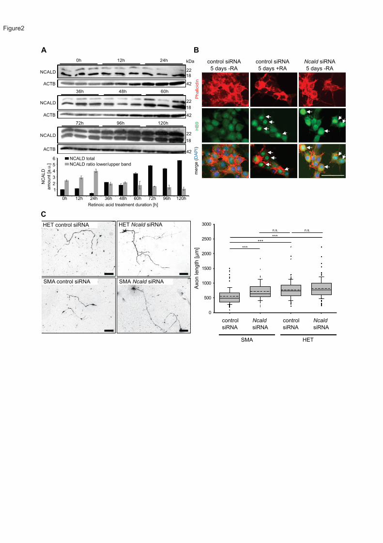
A B
Phallo
idin
HB
9m
erg
e (
DA
PI)
control siRNA
5 days -RA
control siRNA
5 days +RA
Ncald siRNA
5 days -RA
0h
NCALD
12h 24h
36h 48h 60h
72h 96h 120h
ACTB
NCALD
NCALD
kDa
18
18
18
22
22
22
42
42
42
ACTB
NCALD total
NC
ALD
am
ount [a
.u.]
Retinoic acid treatment duration [h]
6
5
4
3
2
1
0h 12h 24h 36h 48h 60h 72h 96h 120h
NCALD ratio lower/upper band
ACTB
0
500
1000
1500
2000
2500
3000
n.s.
***
***
n.s.
C
Ncald
siRNA
control
siRNA
control
siRNA
Ncald
siRNA
SMA HET
Axon leng
th [µ
m]
HET control siRNA HET Ncald siRNA
SMA Ncald siRNASMA control siRNA
***
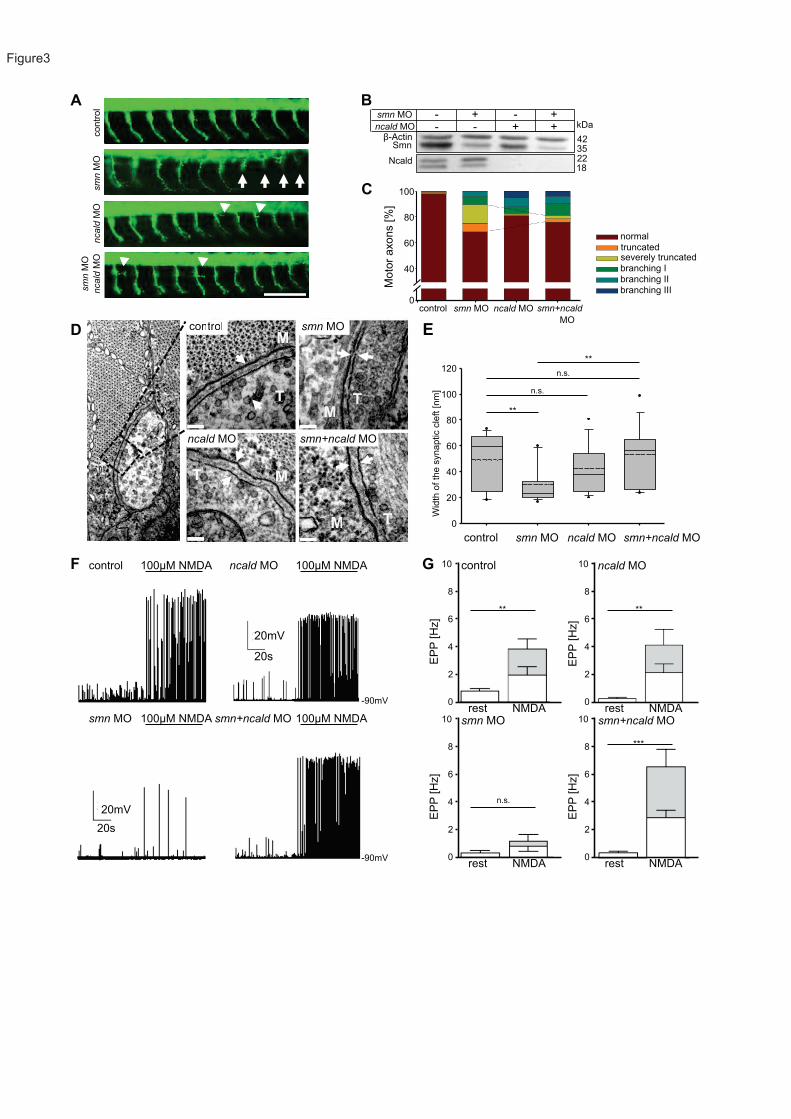
contr
ol
smn
MO
20 s
ncald
MO
smn
MO
ncald
MO
smn MO
ncald MO-ActinSmn
Ncald
100
40
60
80
0
normal
truncatedseverely truncated
branching I
branching II
branching III
control smn MOM
TT
MT
MMT
M T
ncald MO smn+ncald MO
- + - +- +- +
A B
control ncald MO
smn MO smn+ncald MO
100µM NMDA 100µM NMDA
100µM NMDA 100µM NMDA
C
D E
G control ncald MO
smn MO smn+ncald MOrest NMDA
**
**
n.s.
n.s.
control ncald MOsmn MO smn+ncald MO
** **
n.s.
***
20mV
20s
20mV
20s
Wid
th o
f th
e s
ynaptic c
left [nm
]
120
100
80
60
40
20
0
Moto
r axons [%
]
EP
P [H
z]
kDa
42352218
0
2
4
6
8
10
EP
P [H
z]
EP
P [H
z]
EP
P [H
z]
rest NMDA
rest NMDArest NMDA
-90mV
-90mV
0
2
4
6
8
10
0
2
4
6
8
10
0
2
4
6
8
10
control smn MO ncald MO smn+ncald
MO
F
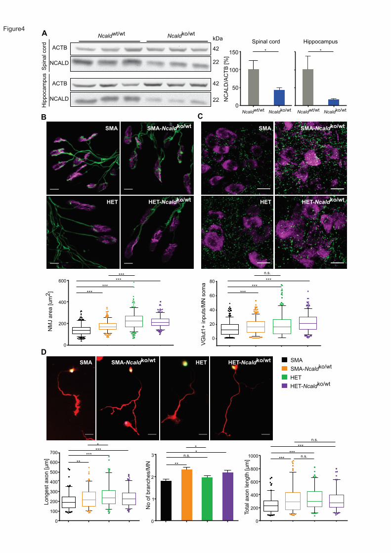
A
C
D
B
SMA-Ncaldko/wt
SMA
HET-Ncaldko/wt
E
ACTBS
pin
al cord
No o
f bra
nch
es/M
N
0
3
2
1
*
**
n.s.
*
600
200
400
0
1000
800
Tota
l axon leng
th [µ
m]
***
***
***
n.s.
n.s.
Hip
poca
mpus
Ncaldwt/wt
Ncaldko/wt
NCALD
kDa
42
42
22
22
Spinal cord Hippocampus
150
100
Ncaldwt/wt
Ncaldko/wt
50
0
NC
ALD
/AC
TB
[%
]
* *N
MJ a
rea [um
2]
0
200
400
600
VG
lut1
+ inpu
ts/M
N s
om
a
0
20
40
80
60
***
n.s.
***
***
******
***
***
HET-Ncaldko/wt
HET
Longest axon [µ
m]
700
600
500
300
200
100
400
0
***
**
***
*
ACTB
NCALD
Ncaldwt/wt
Ncaldko/wt
SMA-Ncaldko/wt
SMA
HET-Ncaldko/wt
HET
SMA
SMA-Ncaldko/wt
HET
HET-Ncaldko/wt
sHET sSMA
SMA-Ncaldko/wt
SMA HET-Ncaldko/wt
HET

BA
Smnko/wt; Ncaldko/wt
XSmn
ko/ko; SMN2tg/tg
25% SMA+ASO: Smnko/ko; SMN2tg/0
25% SMA-Ncaldko/wt+ASO: Smnko/ko; SMN2tg/0; Ncaldko/wt
25% HET+ASO: Smnko/wt; SMN2tg/0
25% HET-Ncaldko/wt+ASO: Smnko/wt; SMN2tg/0; Ncaldko/wt
C57BL/6N FVB/N
mixed50 + 30 µg SMN-ASO P1
SMA+ASO
SMA-Ncaldko/wt+ASO
HET+ASO
HET-Ncaldko/wt+ASO
NM
J im
matu
rity
[%
]
E
SMA+ASO
HET+ASO
0
200
400
600
800
1000
NM
J a
rea [µ
m2] ***
***
n.s.
SMA+ASO
SMA-Ncaldko/wt+ASO
SMA
SMA-Ncaldko/wt
100
50
0
0 50 100 150 200
C
Tim
e s
core
[a.u
.]
Age [d]
2 4 6 8 10 12
1
2
3
4
5
*
***
**
*
*
*****
**
n.s.
Age [d]
Surv
ival [%
]
D
Grip s
treng
th [N
]
SMA+ASO
SMA-Ncaldko/wt+ASO
HET+ASO
HET-Ncaldko/wt+ASO
WT
**
*** n.s.
**
0
0.5
1.0
1.5
2.0
2.5
0
10
20
30
40
50
SMA-Ncaldko/wt
+ASO
HET-Ncaldko/wt
+ASO
SMA+ASO
SMA-Ncaldko/wt+ASO
HET+ASO
HET-Ncaldko/wt+ASO
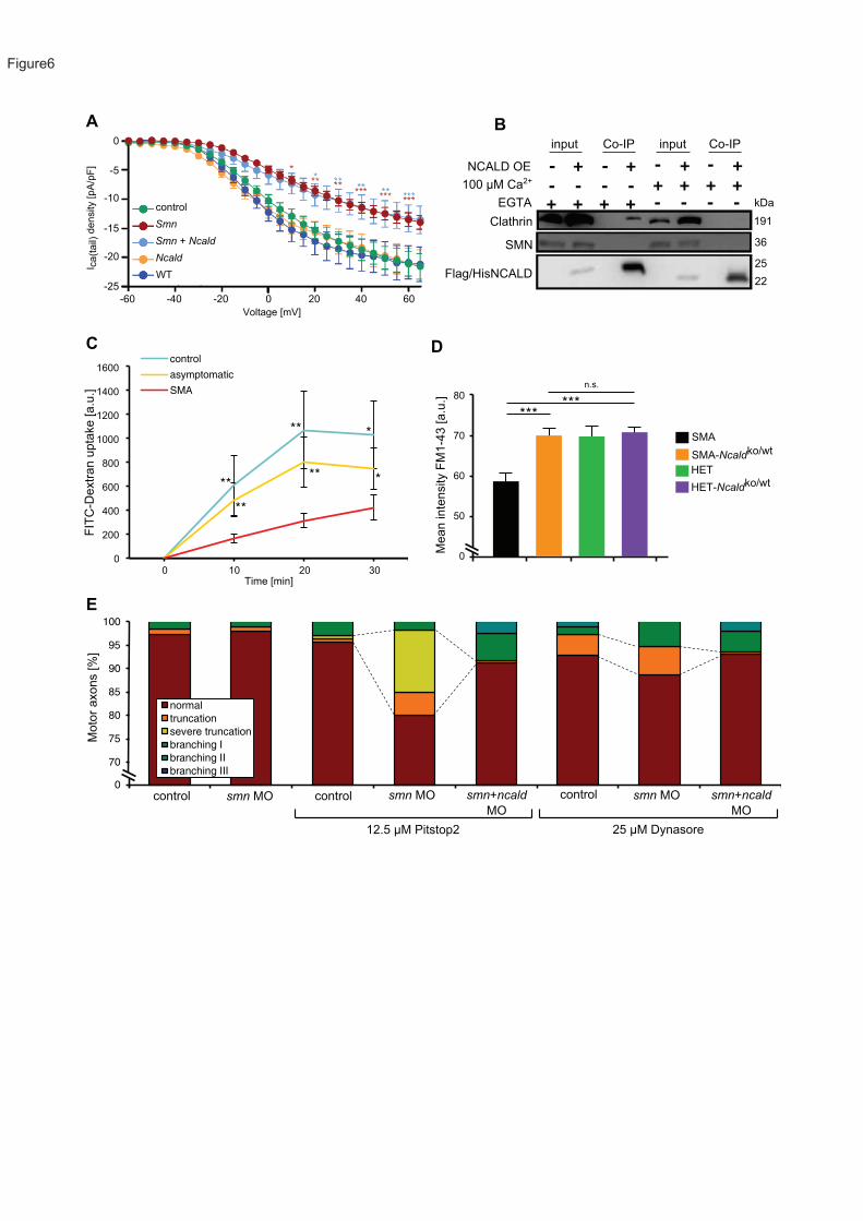
+ + + +- -- -
70
75
80
85
90
95
100
control smn MO control smn MO smn MO+ncald MO control smn MO smn MO +ncaldMO
normal
truncation
severe truncation
branching I
branching II
branching III
-5
0
-10
-15
-20
-25
-60 -40 -20 0 20 40 60
Voltage [mV]
I ca(t
ail)
density [pA
/pF
]
A B
+ + + +
+ + + +
- - - -
- - - -
191
kDa
36
25
22
FIT
C-D
extr
an u
pta
ke [a.u
.]
control
asymptomatic
SMA
**
** ***
** *
1600
1400
1200
1000
800
600
400
200
00 10 20 30
Time [min]
C D
0
Mean in
tensity F
M1-4
3 [a.u
.]
50
60
70
80
SMA
SMA-Ncaldko/wt
HET
HET-Ncaldko/wt
******
n.s.
E
Moto
r axo
ns [%
]
0smn MO smn MO smn MO smn+ncald
MO
smn+ncald
MO
control control control
Smn + Ncald
Smn
control
WT
Ncald
12.5 µM Pitstop2 25 µM Dynasore
100 µM Ca2+
EGTA
Clathrin
SMN
Flag/HisNCALD
NCALD OE
input inputCo-IP Co-IP
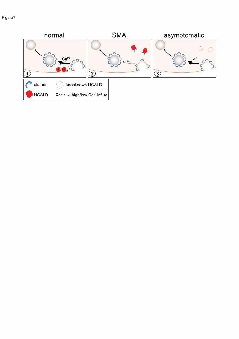
clathrin
NCALD
knockdown NCALD
1 2 3
Ca2+/ Ca2+ high/low Ca2+ influx
normal SMA asymptomatic
Ca2+Ca2+
Ca2+Ca2+