Cell mechanotransduction: cytoskeleton and related signaling pathways
M. Hughes-Fulford1-3 and J. Boonstra4
Hughes-Fulford Laboratory, Department of Veteran’s Affairs1, Northern California Institute for Research and Education2, University of California San Francisco3, Mail
code-151F, 4150 Clement St., San Francisco, California USA 94121 Cellular Architecture and Dynamics, Institute of Biomembranes, University of
Utrecht, Padualaan 8, 3584 CH Utrecht, The Netherlands4
Abstract
Mechanical stimuli regulate a variety of cell physiological functions including gene induction, protein synthesis, proliferation and/or differentiation; understanding mechanotransduction at the cellular level is key to understanding basic biology. Here on Earth, signal transduction affects a wide array of receptors and ligands that signal induction of gene expression. The most common signaling pathways include receptor tyrosine kinase (RTK), G-Protein coupled receptors (GPCR) and extracellular matrix components (integrins). The cytoskeleton functions to maintain cell shape and to move cellular components, separate chromosomes during mitosis and provides sensing networks for mechanotransduction. Mechanotransduction is the process of translating mechanical force on a cell into a biological response. Over the last few decades, mechanotransduction has been shown to occur via extracellular matrix, integrins, cytoskeleton signals, GTPases, adenylate cyclase, PLC and MAP kinases (MAPK), all of which play significant roles in early mechanical signaling. During the last decades a wide variety of space flight experiments have demonstrated that gravity has profound effects on whole organisms, organs and tissues, resulting for example in bone and muscle resorption as well as in the occurrence of cardiovascular malfunctioning, immuno-suppression and many other aspects of clinical medicine. Interestingly, the virtual absence of gravity also has profound effects on the cellular and molecular level, including changes in cell morphology, collapse of the actin cytoskeleton, modification of gene expression, changes in signal transduction cascades and even changes in the polymerization of tubulin. The effects of mechanical stress (e.g. gravity) or lack of stress (microgravity) on cell and molecular properties is discussed with an emphasis on the involvement of signal transduction cascades of RTK, integrins and FasR as well as their role in cytoskeleton perception of gravity in mammalian cells.
Introduction
Mechanical forces have been known for long time to influence cell behaviour.
The mechanism by which mechanical forces are translated by cells into a biological
response has been described as mechanotransduction. During the last decades a wide
variety of studies have demonstrated that mechanotransduction involves the
1

components of the extracellular matrix and several plasma membrane associated
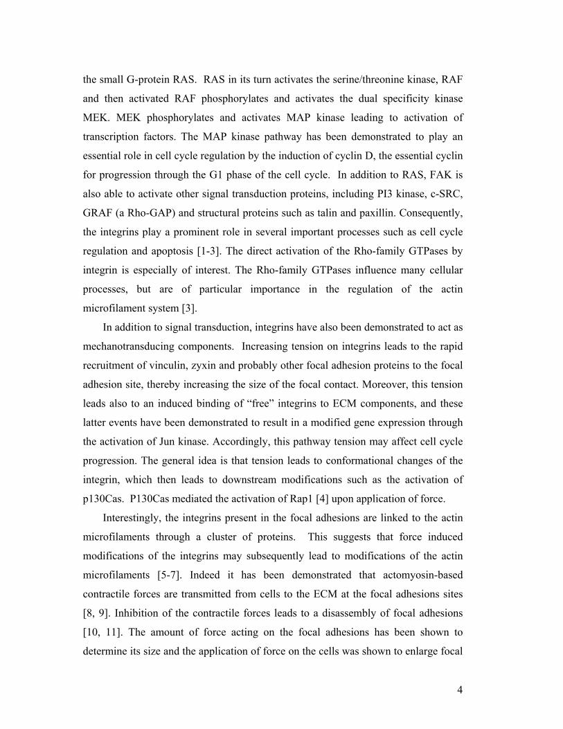
proteins (Fig.1). These proteins play a central role in the transmission of a
mechanical force to a biological response; the most central proteins in this process
include the integrins and cadherins. Subsequently the cytoskeleton has also been
demonstrated to play an important role in transmission of the signals inside the cells.
The eukaryotic cytoskeleton is composed of three basic types of filaments and their
associated proteins. Cytoskeletal filaments are interconnected and their functions are
coordinated by hundreds of associated cytoskeletal accessory proteins. The
cytoskeleton has been demonstrated to be involved in cell adhesion through integrins
Figure 1: Major signaling pathways and transcription factors in cells.
2

In addition, the cytoskeleton appears to be involved in signal transduction
cascades induced by growth factors. Altogether the interactions between integrins,
cadherins, growth factor receptors, signal transduction molecules and the
cytoskeleton constitute a network through which mechanical forces influence gene
expression (Fig. 1). In this contribution we will briefly describe the effects of
mechanical stress (e.g. gravity) or lack of stress (microgravity) on cell and molecular
properties with emphasis on the involvement of signal transduction cascades induced
by receptor tyrosine kinases and extracellular matrix, as well as their role in
cytoskeleton perception of gravity in mammalian cells.
Role of integrins in mechanotransduction
The cytoskeleton not only functions to maintain cell shape, it is also important in
the movement of cellular components, segregation of chromosomes during mitosis
and in forming a sensing network for mechano-transduction. The eukaryotic
cytoskeleton (CSK) is composed of three basic types of filaments; actin
microfilaments, intermediate filaments and microtubules. CSKs are interconnected
and their functions are coordinated by associated cytoskeletal accessory proteins
including integrins. The binding of these proteins with cooperative groups to
cytoskeletal filaments is dynamic and causes rapid polymerization and
depolymerization of filaments. Integrins comprise a large family of transmembrane
glycoproteins that bind to extracellular matrix components at the extracellular side of
the plasma membrane and to the cytoskeleton at the cytoplasmic side. Integrins are
heterodimers having a α and a β subunit. Each subunit has a large extracellular
domain, a single transmembrane domain and a relatively small cytoplasmic domain.
Integrins usually reside in complexes in the cell membrane, called focal adhesion
complexes. The focal adhesion complexes also constitute the end points of the actin
stress fibers. In addition, to being involved in cell attachment to the ECM, integrins
have been demonstrated to be able to directly activate several intracellular signal
transduction cascades. One of the best-known cascades is the MAP kinase pathway.
Upon binding of integrin to the ECM, the focal adhesion kinase (FAK) is
phosphorylated and activated. FAK is a tyrosine kinase and activates subsequently
3
the small G-protein RAS. RAS in its turn activates the serine/threonine kinase, RAF
and then activated RAF phosphorylates and activates the dual specificity kinase
MEK. MEK phosphorylates and activates MAP kinase leading to activation of
transcription factors. The MAP kinase pathway has been demonstrated to play an
essential role in cell cycle regulation by the induction of cyclin D, the essential cyclin
for progression through the G1 phase of the cell cycle. In addition to RAS, FAK is
also able to activate other signal transduction proteins, including PI3 kinase, c-SRC,
GRAF (a Rho-GAP) and structural proteins such as talin and paxillin. Consequently,
the integrins play a prominent role in several important processes such as cell cycle
regulation and apoptosis [1-3]. The direct activation of the Rho-family GTPases by
integrin is especially of interest. The Rho-family GTPases influence many cellular
processes, but are of particular importance in the regulation of the actin
microfilament system [3].
In addition to signal transduction, integrins have also been demonstrated to act as
mechanotransducing components. Increasing tension on integrins leads to the rapid
recruitment of vinculin, zyxin and probably other focal adhesion proteins to the focal
adhesion site, thereby increasing the size of the focal contact. Moreover, this tension
leads also to an induced binding of “free” integrins to ECM components, and these
latter events have been demonstrated to result in a modified gene expression through
the activation of Jun kinase. Accordingly, this pathway tension may affect cell cycle
progression. The general idea is that tension leads to conformational changes of the
integrin, which then leads to downstream modifications such as the activation of
p130Cas. P130Cas mediated the activation of Rap1 [4] upon application of force.
Interestingly, the integrins present in the focal adhesions are linked to the actin
microfilaments through a cluster of proteins. This suggests that force induced
modifications of the integrins may subsequently lead to modifications of the actin
microfilaments [5-7]. Indeed it has been demonstrated that actomyosin-based
contractile forces are transmitted from cells to the ECM at the focal adhesions sites
[8, 9]. Inhibition of the contractile forces leads to a disassembly of focal adhesions
[10, 11]. The amount of force acting on the focal adhesions has been shown to
determine its size and the application of force on the cells was shown to enlarge focal
4

adhesions complexes [11, 12]. Furthermore, it has been demonstrated that mechanical
forces induce an accumulation of F-actin at the focal adhesions in a zyxin-dependent
manner, involving the strengthening of the ECM-integrin-actin linkage [9, 13, 14].
Actin microfilaments have also been demonstrated to regulate integrins.
Treatment of cells with cytochalasin D to cap actin filaments inhibits cell adhesion.
In other cells it was demonstrated by Bennett et al. that inhibition of actin
polymerization resulted in an induction of ligand binding to integrins [15]. The
platelet cytoskeleton regulates the affinity of the integrin α1β3 for fibrinogen [15].
Activation of Cdc42 and Rac is associated with the formation of focal complexes in
fibroblasts [16, 17] and inhibition of Rho results in a decrease of integrin-mediated
aggregation of leukocytes and platelets [18].
In conclusion, mechanical force accelerates integrin activation, both through
extracellular and intracellular rearrangements, which induce protein recruitment
leading to integrin clustering [19]. These observations suggest that the ECM-
integrin-actin complex in the focal adhesion complexes may also constitute a gravity
sensitive component. Indeed, it has been demonstrated that exposure of the
epidermoid human A431 cells to real and simulated microgravity conditions leads to
a rapid (within minutes) rounding of the cells [20]. Similar results were obtained in
fibroblasts incubated in a random positioning machine [Moes et al. unpublished
observations].
Role of cadherins in mechanotransduction
Cadherins are transmembrane glycoproteins playing an important role in cell-cell
adhesion. The extracellular domains are responsible for adhesive recognition due to
their interaction with the extracellular domains of cadherins of neighboring cells. The
cytosolic domains of cadherins interact with a wide variety of proteins including actin
microfilaments and intermediate filaments. One of the best known cadherin-
associated proteins concerns α-catenin. The cadherin-catenin complexes associate to
actin filaments to form the adherens junctions and the association with intermediate
filaments result in the formation of desmosomes. The role and biochemistry of both
cadherins and catenins have been described recently in several review papers [21-24].
5

Cadherins have been demonstrated to be involved in mechanotransduction,
particularly in specialized systems such as the inner ear hair cells [25]. In addition, it
has been demonstrated in fibroblasts that mechanical forces applied to intercellular
junctions induced intracellular responses mediated by cadherins, suggesting that
cadherins function as intercellular mechanotransducers [26]. Furthermore cadherin
engagement was shown to modulate RhoA signaling and contractility in endothelial
cells [27]. These findings strongly suggest a close cross talk between signal
transduction cascades induced by cadherins and integrins [28, 29]. As described
above, cadherins play a prominent role in cellular junctions and as such are essential
in the establishment of the endothelium. It is well known that the endothelium
responds to mechanical deformations, although the mechanisms by which endothelial
cells recognize mechanical stimuli are not as yet understood. Many potential
mechano-sensing systems have been suggested including the cytoskeleton [30], G-
proteins [31] and junction proteins [32, 33].
Involvement of the actin cytoskeleton in growth factor and extracellular matrix
signalling
Actin is an extremely abundant protein in virtually all eukaryotic cells, and is
involved in many cellular functions including migration, endocytosis, intracellular
transport, docking of proteins and mRNA, attachment, signal transduction, membrane
ruffling, neuronal path finding and cytokinesis. Moreover, it largely determines the
cell shape and the position and shape of organelles within the cytoplasm.
The actin family consists of α-, β- and γ-isoforms. The α-isoform is mostly
present in muscle cells whereas the β- and γ-isoforms are present in all cells. Actin is
present in cells in an unassembled, globular form and a polymerized, filamentous
form, called G-actin and F-actin, respectively. The F-actin filaments are composed of
two linear strands of polymerized G-actin wound around each other in a helix. Within
these filaments the actin monomers are oriented in the same direction resulting in
inherent polarity of the filaments resulting in the barbed or plus end and the pointed
or minus end. The barbed ends are characterized by a rapid polymerization and a
slow de-polymerization and the pointed ends exhibit the opposite features. In the
6

cells actin continuously cycles between the polymer and monomer state, a process
called treadmilling.
The actin filaments constitute a highly dynamic network in the cells, the
dynamics being regulated by a large number of actin binding proteins (ABPs) [34,
35]. The ABPs are characterized by their function, such as cross-linking proteins,
actin severing, capping and de-polymerizing proteins, monomer binding proteins,
membrane-associated proteins and actin-regulatory proteins. Several conserved
domains of actin have been identified that act as binding domains for the ABPs,
including the myosin motor domain, the gelsolin homology domain, the calpain
homology (CH) domain, the actin depolymerizing factor/cofilin (ADF/cofilin)
domain and the Wiskott-Aldrich syndrome protein (WASP)-homology domain-2
(WH2) [36-40]. These observations clearly demonstrate that actin metabolism is
Figure 2: Involvement of the actin cytoskeleton in growth factor and
extracellular matrix signalling. The cytoskeleton is directly involved in the signal
transduction of many of the RTK receptors. These signaling pathways includes the
involvement of integrins, ECM, actin stress fibers and cdc42.
7

regulated by a large number of proteins, which on their turn are subject of regulation
as well. This complicated network of actin and the ABPs play an essential role in cell
metabolism and consequently also in cell cycle regulation [41].
The role of actin in ECM-induced signalling is especially apparent from the
structural role of actin in focal adhesions. Actin binds to integrins indirectly through
several proteins like vinculin and α-actinin and disruption of this interaction has large
consequences on the complex structure and function of focal adhesions (Fig.2). In
addition the actin filaments constitute a highly dynamic network, with the dynamics
being regulated by a large number of actin binding proteins, for review see [41]. The
first indications for the relationship between actin and signal transduction were
obtained by studies on the effects of growth factors on cell morphology. For example,
addition of EGF or PDGF cause the formation of membrane ruffles within minutes
after addition of the growth factor [41, 42] this suggests modulation of actin
metabolism through a TRK cascade (Fig. 2). It was demonstrated that exposure of
cells to EGF caused a rapid actin polymerization, the formation of membrane ruffles
and the translocation of several of the down stream signaling molecules to these
newly formed membrane ruffles. This suggests the formation of signaling complexes
at the plasma membrane in the membrane ruffles [43, 44].
Interestingly, treatment of the cells with F-actin disrupting agents like cytochalasin
caused a severe reduction in growth factor induced signaling [45], demonstrating the
mutual interaction between signaling cascades and the actin microfilaments. Finally,
actin has also been reported to be localized in the nucleus [46, 47]. There is evidence
that nuclear actin is involved in chromatin remodeling, transport of proteins and
mRNA transcription. In this latter case, it was demonstrated that actin acts as a
regular component of all RNA polymerases and is probably related to actin-
dependent chromatin remodeling previously reviewed [47-49]. In summary, all these
observations indicate that actin plays a dominant role in cells, not only as a structural
protein, but also as a protein involved in dynamic processes like signal transduction
and transcription.
Taken together, the data shows that actin plays an important role in growth
factor- and in integrin-induced signal transduction. In addition, both signal
8
transduction pathways are interacting as exemplified by the MAP kinase pathway
(Fig. 2). MAPK is recruited to focal adhesions in response to several stimuli such as
integrin activation, activation of v-SRC, activation of PKCε and activation of the
FGF receptor. PDGF and EGF induce cell migration and cause localized cell de-
adhesion requiring MAPK signaling. The effect of growth factors on cell adhesion
required the activation of calpain 2 [50]. The observation that calpain activity was
decreased in FAK-deficient cells are of particular interest [51]. In addition, it was
demonstrated that FAK induces the formation of a complex constituting calpain 2,
FAK and MAP kinase [52]. These data suggest FAK is critical in the integration of
migratory signals from growth factor receptors and integrins via the MAPK pathway
to the calpain proteolytic system, resulting in focal adhesion turnover and cell
migration [53].
Involvement of microtubules in growth factor and extracellular matrix
signalling
Microtubules constitute one of the major components of the cytoskeleton and have
been demonstrated to be involved in cell division by segregation of the chromosomes
during mitosis, intracellular transport and cell morphology [54]. The major
component of microtubules is the heterodimeric protein tubulin. Tubulin
polymerization is dependent upon the GTP/GDP. GTP binding is required for
polymerization, while GTP hydrolysis, most likely through intrinsic tubulin GTPase
activity, results in depolymerization [55]. The functioning of the microtubules
depends largely on the dynamics of polymerization and depolymerization. In addition
to the dynamic behaviour of microtubules, an important role of microtubules in cells
is also realized by the action of motorproteins, like dynein and kinesin, which allow
the transport of cargo’s along the microtubules. Although much effort has been made
to elucidate the cellular mechanisms that underlie microtubule dynamics, the precise
spatial and temporal control of this process is not fully understood yet. However, a
wide variety of signal transduction proteins appear to be associated with
microtubules, suggesting also a role of microtubules in signal transduction. Amongst
others, MAP kinase interacts with microtubules [56] and from these studies it was
9
concluded that microtubules retained MAP kinase in the cytoplasm to regulate
cytoplasmic events. A transcription factor that may be regulated by microtubules is
NFκB. The inhibitor of NFκB, IκB, has been shown to interact with the motorprotein
dynein, and this interaction may sequester NFκB, while on the other hand
depolymerization of microtubules leads to IκB breakdown and consequently to NFκB
activation [57]. Furthermore a close interaction has been demonstrated between
microtubule dynamics and heterotrimeric G proteins [for review see [58]]. These
observations suggest an intimate interaction between microtubules and signal
transduction cascades activated by growth factors and possibly even by the ECM,
which may result in modification of signal transduction by the microtubules.
Microtubules in normal gravity and microgravity
Microtubules have been implicated in cell organization and are required for
separation of chromosomes during mitosis [59-61]. During mitosis, the precise
timing of key cellular processes such as microtubule organizing centers (MTOC), and
cytokinesis is essential for high fidelity chromosome segregation. Temporal
organization of these events is coordinated by a group of proteins collectively termed
cell cycle regulators. Many regulators are kinases or phosphatases that respond to
cellular cues and orchestrate cell cycle progression by altering the phosphorylation
and activity of other downstream regulatory proteins. In recent years studies in yeast
have revealed that many regulators localize near CSKs [62].
Over the past few years, Tabony’s laboratory has shown that microtubule self-
organization in a cell free system is dependent on gravity, suggesting that gravity is
required for normal self-assembly of microtubules in animal cells and that the
microtubule system may be disrupted in microgravity in a living cell [63-65]. Lewis
et al. reported that Jurkat cells flown in space had disrupted microtubules and
increased apoptosis. The increased apoptosis was accompanied with a time dependent
elevation of Fas/APO-1 suggesting an increase in Fas Receptor (FasR) signal
transduction in microgravity. Postflight confocal microscopy of the Jurkat cells
revealed diffuse shortened microtubules extending from poorly defined microtubule
organizing centers (MTOCs) [66, 67]. These observations were confirmed in later
10
microgravity studies with Jurkat and Drosophilia melanogaster (Schneider S-1) cells
that showed cytoskeletal and mitochondrial alterations after exposure to spaceflight
and in insect cells of Drosophila melanogaster (Schneider S-1) after exposure to
conditions created by clinostat rotation [68]. The effects of both treatments were
similar in the different cell types. Fifty percent of the cells displayed effects on the
microtubule network in both cell lines. Under these experimental conditions,
mitochondria clustering and morphological alterations of mitochondrial cristae were
observed to various degrees after 4 and 48 hours of culture. Jurkat cells underwent
cell divisions during exposure to spaceflight but a large number of apoptotic cells
were also observed. Similar results were obtained in Schneider S-1 cells cultured
under clinostat rotation. Both cell lines displayed mitochondrial abnormalities and
mitochondria clustering toward one side of the cells which could be interpreted to be
the result of microtubule disruption and failure of mitochondria transport along
microtubules. Studies by Meloni et al. have also noted altered CSK and motility in J-
111 monocytes during exposure to altered gravity in a Random Positioning Machine
(RPM) [69].
Ground-based experiments revealed a similar enhancement of the spontaneous
and evoked lamellar protrusive activity when the cells were kept at 2g hypergravity
for at least 6 min. This gravity response was independent of the direction of the
acceleration vector in respect to the cells [70]. Exposure of the cells to "simulated
weightlessness" (clinorotation) had no obvious influence on this type of lamellar
actin cytoskeleton dynamics. A 20 min exposure of the cells to simulated
weightlessness or to changing gravity (6 to 31 parabolas) - but not to 2g
(hypergravity, centrifugation) - resulted in an altered arrangement of microtubules
indicated by bending, turning, and loop formation. A similar altered arrangement was
shown by microtubules, which had polymerized into lamellipodia after release from a
taxol block at simulated weightlessness (clinorotation) or during changing gravity (5
parabolas). Data suggest that in human SH-SY5Y neuroblastoma cells, microgravity
affects the dynamics and spatial arrangement of microtubules but has no influence on
the Rac-controlled lamellar actin cytoskeleton dynamics and cell spreading. The
latter, however, seems to be promoted at hypergravity [70].
11

The actin cytoskeleton in growth factors and extracellular matrix signalling in
microgravity
Early experiments in sounding rockets under real microgravity conditions
demonstrated not only a rapid cell rounding, but also modified actin polymerization
[20]. The changed actin polymerization may represent the basis of other gravity-
induced changes as well. It has been demonstrated, under both real and simulated
microgravity conditions, that EGF-induced expression of the early genes c-fos and c-
jun was severely inhibited. Interestingly, the inhibition was also observed if c-fos and
c-jun expression were induced under microgravity conditions by the phorbol ester
(PMA), but no effect was observed by c-fos and c-jun induction by the Ca-ionophore
A23187 or the cyclic AMP inducing forskolin [71, 72]. Changes in cytoskeleton
were also noted by Guignandon et al. [73] when they examined cells in parabolic
flight microgravity and found cytoplasmic retraction and membrane ruffling in
ROS/17/2.8 cells. Increased PGE2 was found in flight medium accompanied by
significant flight-induced changes that included a decrease in cell area and irregular
shape in some cells. These observations demonstrate clearly the specificity of the
effect of microgravity on signal transduction. Notably, both EGF and PMA are
known to stimulate protein kinase C (PKC) activity and therefore PKC may represent
a downstream microgravity sensitive target in the cells. PKC activity has been also
related to actin dynamics. During the past decades numerous studies demonstrated
that microgravity conditions result in dramatic changes in the actin cytoskeleton as
reviewed by Crawford-Young [74]. To date, the cytoskeleton appears to play an
essential role in gravity sensing of the cells and the actin microfilament system also
plays an essential role in growth factor and integrin-induced signal transduction,
consequently causing changes in cell proliferation, differentiation and apoptosis.
Microgravity (10-3-10-9g) includes other variables characteristic of orbital phase
of spaceflight which include: launch effects, altered electromagnetic fields, pressure
changes, changed content of cabin atmospheric gases, mechanical vibrations from
motors and crew activities, cosmic radiation and absence of sedimentation-induced
convection [75, 76]. Because of these conditions, many of the recent spaceflight
12

experiments have included onboard 1g samples to control the effects of these
spaceflight conditions. Alterations in cytoskeleton actin, intermediate filaments and
microtubules have been noted when there is a significant load reduction on the cell in
microgravity [20, 73, 77-80]. Since multiple investigators have observed actin and
microtubule cytoskeletal modifications in microgravity, this suggests a common root
cause in the microgravity environment which alters cell architecture. Since the cell
cycle is dependent on the cytoskeleton, alterations in cytoskeletal structure can block
cell growth either in G1 (F-actin microfilament collapse), or in G2/M (inhibition of
microtubule polymerization during G2/M-phase). It is then possible that microgravity
may inhibit growth in either G1, or G2/M phases of the cell cycle.
The absence of mechanical stress (microgravity) can cause change in cell shape
and signal transduction when exposed to as little as 20 seconds of microgravity in
parabolic flight [73, 77]. When quiescent osteoblasts are activated by sera under
microgravity conditions there is a 60% reduction in growth (p<0.001) when
compared to ground controls. Moreover, a collapse of the osteoblast actin
cytoskeleton and loss of focal adhesions have been noted after several days in
microgravity. The changes seen in the cytoskeleton are probably not due to
alterations in fibronectin message or protein synthesis since no differences have been
noted in microgravity [81]. The altered ability of cells to respond to stimuli like
growth factors and sera suggests that there is a major alteration in anabolic signal
transduction under microgravity conditions, most probably through the growth factor
receptors and/or the RTK pathways that are connected to the cytoskeleton. The fact
that several investigators have noted that changes in specific gene expression are
associated with microgravity exposure [71, 72, 78, 81-90] reinforces the concept that
microgravity is interfering with signal transduction from the cell membrane receptors
to internal signaling pathways.
Studies on STS-56/IML-2 examined sera activation of quiescent osteoblast-like
cells in orbit and demonstrated that microgravity caused a decrease in cell
proliferation within days of exposure to microgravity. In the 1g flight cells, the sera
activated cells had activated Rho activity as evidenced by stress fiber formation. The
collapse of the actin cytoskeleton [78] and the elongation of the nuclear shape [78,
13
89] of osteoblast-like cells were noted in spaceflight while glucose metabolism per
cell was unchanged [78]. The expression of cox-2 mRNA was not induced by sera in
microgravity, but paradoxically, media PGE2 content 24 hours after activation was
significantly increased in flight in both the static (µg) and 1g onboard controls [78,
88, 89, 91]. In normal cells and tissues, the presence of PGE2 causes an induction of
the cox-2 message. This lack of induction of the cox-2 message in the presence of
elevated levels of PGE2 suggests a malfunction of the PGE2 feed-forward-regulation.
This lack of feedback in microgravity may be caused by reduced signaling at the
level of the GCPR signal cascade.
In-flight studies by Stein et al. in astronauts have demonstrated a reduction in
sera levels of PGE2 in flight [92, 93]. Normally in the human, PGE2 is cleared by the
kidneys within seconds, and must be made continually to maintain high sera/urine
levels. In contrast, in the isolated osteoblasts the PGE2 is degraded by the enzyme 15-
hydroxyprostaglandin dehydrogenase; it is therefore possible that the activity of the
degrading enzyme or alterations in the degradation process may be inhibited in the
isolated cell in microgravity allowing for higher levels of PGE2.
It was demonstrated that exposure of cells to EGF caused a rapid actin
polymerization, the formation of membrane ruffles and the translocation of several of
the down stream signaling molecules to these newly formed membrane ruffles,
suggesting the formation of signaling complexes at the plasma membrane in the
membrane ruffles [43, 44]. These initial observations of changes in EGF and PDGF
signaling were followed by studies in which it was demonstrated that a wide variety
of signal transduction proteins associated with actin, amongst these the EGF receptor
[94], PI3 kinase, and phospholipase C (PLC) [43, 94].
Fibroblast Growth Factor-2 (FGF-2) is the ligand for another actin associated
RTK receptor, FGFR. SRC kinase activity has a crucial role in the regulation of
FGFR1 signaling dynamics. Following receptor activation by ligand binding,
activated SRC is colocalized with activated FGFR1 at the plasma membrane. This
localization requires both active SRC and FGFR1 receptor tyrosine kinases, which
are inter-dependent. Src-mediated transport and subsequent activation of FGFR1
require both RhoB endosomes and an intact actin cytoskeleton for full activity [95].
14

RTK receptors like FGFR are implicated in bone cell growth and bone cells
synthesize the FGF-2 growth factor endogenously. Normal bone remodeling is
characterized by a series of cellular events, cell proliferation, sequential activation
and up regulation of osteoblast-characteristic genes, and matrix mineralization.
Figure 3: FGF-2 signal transduction FGF-2 causes induction of several pathways
including p38, Ras/MAPK, SRC, PKA (exact mechanism unknown) and PKC
through its interaction with the FGFR1, FGFR-2 and FGFR-3 receptors in bone.
These events are tightly controlled and coordinated by a number of regulatory
molecules, such as growth factors (GFs), and their downstream transcription factors
ensuring normal growth and development of the skeleton [96]. As seen in Fig. 3,
fibroblast growth factors (FGFs) have important regulatory functions in bone
formation [97-99]. FGFs belong to a gene family currently comprised of 23
members in mammal evolution. They are secreted peptides with molecular size of
approximately 20–35 kDa and expressed in many different types of tissues during
various stages of development. In addition to their mitogenic effects, FGFs are
15

involved in diverse biological processes, including cell motility [100], and migration
[101]. FGF signaling is triggered by the binding of FGFs to their high affinity
receptors, fibroblast growth factor receptors (FgfRs), followed by dimerization and
auto-/trans-phosphorylation of FgfRs [102, 103].
The phosphorylated FgfR kinases selectively activate intracellular signaling
intermediates, eliciting specific cellular responses. Four FgfRs (FgfR1– FgfR4) have
been identified to date. Among them, the isoforms FgfR1a, FgfR1b, FgfR2 and
FgfR3 are the major receptor isoforms expressed in bone. FGF-2 activates FgfR1 (b
and c), FgfR2 (c), and FgfR3 (c) receptors. Of these receptors, 1c, 2c, and 3c result
in mitogenic responses to FGF-2 in osteoblasts [104]. FgfR knockout mice have
helped elucidate the roles of the individual receptors in skeletal development. The
FgfR1 and FgfR2 genes appear to regulate formation and elongation of the limbs in
the developing embryo [105-107]. During the development of the mouse skull, FgfR2
is expressed only in proliferating osteoblasts. Once these cells start differentiating,
FgfR2 is downregulated and FgfR1 is upregulated [108]. Furthermore, disruption of
the FgfR2 gene results in increased osteoblast differentiation, suggesting its role in
the switch between proliferation and differentiation [109].
When osteoblast-like cells and bone cells are put under increased mechanical
stress in a centrifuge (max. of 12g), they synthesize fgf-2 message and protein which
in turns stimulates bone growth [110]. When stem cells from Fgf2-/- mice (Fgf-2
knockout mice) are subjected to stress of 120 μstrain by centrifugation, no Fgf-2 is
synthesized and bone cells do not grow in response to stress [99]. Mechanical stress
promotes Fgf-2 mediated growth via both PKA and a MAPK pathways [99] .
A recent report discovered a lack of fgf-2 mRNA and protein synthesis in
osteoblast-like cells grown in microgravity [89]. The lowering of Fgf-2 content was
associated with a significant change in nuclear shape of the µg flight cells. The cells
under 1g-flight environment had normal nuclear cell shapes. Since nuclear shape is
maintained by the nuclear lamins this might implicate a change in conformation of
two intermediate filaments; nuclear Lamin A and nuclear Lamin C under
microgravity conditions. Since Fgf-2 growth factor increases in cox-2 mRNA
through the FGF-2/RTK pathway, the lowered synthesis of cox-2 message may be
16

associated with lowered RTK activity as was seen in the EGF experiments mentioned
earlier in this chapter. Fgf-2 mRNA levels and cox-2 mRNA levels return to normal
values in the 1g flight controls [89]. Lack of signaling from the RTK class is
suggested by both results from A432 and MC3T3-E1 and preosteoblast stem cell
experiments. At this time it is unknown why RTK cascades are affected by µg, but
this phenomenon is under investigation by several laboratories.
There are other downstream pathways from the cell surface receptors that have
been shown to be affected by the absence of gravity during flight or in simulated
microgravity, they include PKC [111-115] and Protein Kinase A (PKA) [116, 117].
The Hughes-Fulford Lab had previously reported that PKA and PKC are key early
regulators in T-cell activation. In other studies of human T-cells grown on the RPM,
there was a significant loss of CREB message (PKA pathway). In addition, there was
a loss in NFκB and ten other key regulators in the T-cells grown in the RPM. The
group analyzed differential gene expression to find gravity-dependent genes and
pathways (n=3) independent samples for each condition using Affymetrix full
genome gene arrays. There was an inhibited induction of 91 genes in the simulated
freefall environment of the RPM. Altered induction of the ten genes regulated by key
signaling pathways was verified using real-time RT-PCR [116]. It was discovered
that impaired induction of early genes were regulated primarily by transcription
factors NF-κB, CREB, ELK, AP-1, and STAT in the altered gravity environment.
Since the majority of the genes were regulated by NF-κB, CREB, ELK and AP-1, the
pathways that regulated these transcription factors were studied on the RPM.
Boonyaratanakornkit et al. found that the PKA pathway was down-regulated in
simulated μg using the RPM. In contrast, PI3K, PKC, and its upstream regulator
pLAT were not significantly down-regulated by vectorless gravity [116]. Earlier
studies demonstrated that PKA was an essential part of early T-cell activation since
inhibition of that pathway inhibited production of IL-2 and IL-2Ra, two key steps in
T-cell activation [117]. Since NF-κB, AP-1, and CREB are all regulated by PKA and
are transcription factors predicted by microarray analysis to be involved in the altered
gene expression in vectorless gravity, the data suggest that PKA may be a key early
player in the loss of T-cell activation in altered gravity [116]. The same changes in
17

NFκB and CREB were recently discovered in the Leukin studies flown on an
experiment in the International Space Station, ISS. Human T-cells were activated in
spaceflight with and without gravity. This preliminary data suggests a similar
mechanism of downregulation both in the RPM and in true microgravity (manuscript
in preparation)
A considerable amount of experimental evidence support the fact that changes in
mechanotransduction in microgravity occur at the level of RTK signal transduction
[20, 73, 77-80, 89]. It is possible that the disruption of the actin CSK in microgravity
renders the receptor inactive, or that alterations in the cell membrane itself alters the
activity of the RTK receptor response to its growth factor ligand. In a similar way, a
blunting of the self-organization of the microtubules in microgravity and hence
altering the structure of the MTOC could alter cellular processes related to response
in much the same way as disruption of the kinases at the growth factor receptors. As
opportunities to conduct spaceflight experiments using modern technology become
available, the exact molecular causes of change in cell function, mechanotransduction
and downstream signaling in microgravity will become understood.
Bibliography: 1. Assoian, R. K., and Klein, E. A. 2008, Trends Cell Biol 18, 347-352 2. Assoian, R. K., and Yung, Y. 2008, Cell Cycle 7, 24-27 3. Juliano, R. L., Reddig, P., Alahari, S., Edin, M., Howe, A., and Aplin, A. 2004, Biochem Soc Trans 32, 443-446 4. Sawada, Y., Tamada, M., Dubin-Thaler, B. J., Cherniavskaya, O., Sakai, R., Tanaka, S., and Sheetz, M. P. 2006, Cell 127, 1015-1026 5. Vicente-Manzanares, M., Choi, C. K., and Horwitz, A. R. 2009, J Cell Sci 122, 199-206 6. Vicente-Manzanares, M., Sancho, D., Yanez-Mo, M., and Sanchez-Madrid, F. 2002, Int Rev Cytol 216, 233-289 7. Zaidel-Bar, R., Milo, R., Kam, Z., and Geiger, B. 2007, J Cell Sci 120, 137-148 8. Balaban, N. Q., Schwarz, U. S., Riveline, D., Goichberg, P., Tzur, G., Sabanay, I., Mahalu, D., Safran, S., Bershadsky, A., Addadi, L., and Geiger, B. 2001, Nat Cell Biol 3, 466-472 9. Hirata, H., Tatsumi, H., and Sokabe, M. 2008, J Cell Sci 121, 2795-2804 10. Chrzanowska-Wodnicka, M., and Burridge, K. 1996, J Cell Biol 133, 1403-1415
18

11. Riveline, D., Zamir, E., Balaban, N. Q., Schwarz, U. S., Ishizaki, T., Narumiya, S., Kam, Z., Geiger, B., and Bershadsky, A. D. 2001, J Cell Biol 153, 1175-1186 12. Galbraith, C. G., Yamada, K. M., and Sheetz, M. P. 2002, J Cell Biol 159, 695-705 13. Glogauer, M., Arora, P., Chou, D., Janmey, P. A., Downey, G. P., and McCulloch, C. A. 1998, J Biol Chem 273, 1689-1698 14. Glogauer, M., Arora, P., Yao, G., Sokholov, I., Ferrier, J., and McCulloch, C. A. 1997, J Cell Sci 110 (Pt 1), 11-21 15. Bennett, J. S., Zigmond, S., Vilaire, G., Cunningham, M. E., and Bednar, B. 1999, J Biol Chem 274, 25301-25307 16. Hall, A. 1998, Science 279, 509-514. 17. Hall, A. 1998, Science 280, 2074-2075. 18. Calderwood, D. A., Shattil, S. J., and Ginsberg, M. H. 2000, J Biol Chem 275, 22607-22610 19. Puklin-Faucher, E., and Sheetz, M. P. 2009, J Cell Sci 122, 179-186 20. Rijken, P. J., Hage, W. J., van Bergen en Henegouwen, P. M., Verkleij, A. J., and Boonstra, J. 1991, J Cell Sci 100, 491-499. 21. Daugherty, R. L., and Gottardi, C. J. 2007, Physiology (Bethesda) 22, 303-309 22. Delva, E., and Kowalczyk, A. P. 2008, Traffic 23. Jeanes, A., Gottardi, C. J., and Yap, A. S. 2008, Oncogene 27, 6920-6929 24. Scott, J. A., and Yap, A. S. 2006, J Cell Sci 119, 4599-4605 25. Holme, R. H., and Steel, K. P. 2001, Trends Mol Med 7, 138 26. Ko, K. S., Arora, P. D., and McCulloch, C. A. 2001, J Biol Chem 276, 35967-35977 27. Nelson, C. M., Pirone, D. M., Tan, J. L., and Chen, C. S. 2004, Mol Biol Cell 15, 2943-2953 28. Chen, H., Lee, M., Lee, J., An, W. G., Choi, H. J., Kim, S. H., and Koh, K. 2008, Talanta 75, 99-103 29. Schwartz, M. A., and DeSimone, D. W. 2008, Curr Opin Cell Biol 20, 551-556 30. Chen, C. S., and Ingber, D. E. 1999, Osteoarthritis Cartilage 7, 81-94. 31. Gudi, S., Huvar, I., White, C. R., McKnight, N. L., Dusserre, N., Boss, G. R., and Frangos, J. A. 2003, Arterioscler Thromb Vasc Biol 23, 994-1000 32. Shay-Salit, A., Shushy, M., Wolfovitz, E., Yahav, H., Breviario, F., Dejana, E., and Resnick, N. 2002, Proc Natl Acad Sci U S A 99, 9462-9467 33. Shyy, J. Y., and Chien, S. 2002, Circ Res 91, 769-775 34. Dominguez, R. 2004, Trends Biochem Sci 29, 572-578 35. dos Remedios, C. G., Chhabra, D., Kekic, M., Dedova, I. V., Tsubakihara, M., Berry, D. A., and Nosworthy, N. J. 2003, Physiol Rev 83, 433-473 36. Gimona, M., Djinovic-Carugo, K., Kranewitter, W. J., and Winder, S. J. 2002, FEBS Lett 513, 98-106 37. Lappalainen, P., Kessels, M. M., Cope, M. J., and Drubin, D. G. 1998, Mol Biol Cell 9, 1951-1959 38. McGough, A. M., Staiger, C. J., Min, J. K., and Simonetti, K. D. 2003, FEBS Lett 552, 75-81 39. Paunola, E., Mattila, P. K., and Lappalainen, P. 2002, FEBS Lett 513, 92-97 40. Sellers, J. R. 2000, Biochim Biophys Acta 1496, 3-22
19

41. Boonstra, J., and Moes, M. J. 2005, Crit Rev Eukaryot Gene Expr 15, 255-276 42. Moes, M., Rodius, S., Coleman, S. J., Monkley, S. J., Goormaghtigh, E., Tremuth, L., Kox, C., van der Holst, P. P., Critchley, D. R., and Kieffer, N. 2007, J Biol Chem 282, 17280-17288 43. Diakonova, M., Payrastre, B., van Velzen, A. G., Hage, W. J., van Bergen en Henegouwen, P. M., Boonstra, J., Cremers, F. F., and Humbel, B. M. 1995, J Cell Sci 108, 2499-2509. 44. Payrastre, B., van Bergen en Henegouwen, P. M., Breton, M., den Hartigh, J. C., Plantavid, M., Verkleij, A. J., and Boonstra, J. 1991, J Cell Biol 115, 121-128 45. Margadant, C., van Opstal, A., and Boonstra, J. 2007, J Cell Sci 120, 66-76 46. McDonald, D., Carrero, G., Andrin, C., de Vries, G., and Hendzel, M. J. 2006, J Cell Biol 172, 541-552 47. Percipalle, P., Fomproix, N., Cavellan, E., Voit, R., Reimer, G., Kruger, T., Thyberg, J., Scheer, U., Grummt, I., and Farrants, A. K. 2006, EMBO Rep 7, 525-530 48. Grummt, I. 2006, Curr Opin Genet Dev 16, 191-196 49. Schleicher, M., and Jockusch, B. M. 2008, Histochem Cell Biol 129, 695-704 50. Glading, A., Bodnar, R. J., Reynolds, I. J., Shiraha, H., Satish, L., Potter, D. A., Blair, H. C., and Wells, A. 2004, Mol Cell Biol 24, 2499-2512 51. Cuevas, B. D., Uhlik, M. T., Garrington, T. P., and Johnson, G. L. 2005, Oncogene 24, 801-809 52. Carragher, N. O., Westhoff, M. A., Fincham, V. J., Schaller, M. D., and Frame, M. C. 2003, Curr Biol 13, 1442-1450 53. Carragher, N. O., and Frame, M. C. 2004, Trends Cell Biol 14, 241-249 54. Gelfand, V. I., and Bershadsky, A. D. 1991, Annu Rev Cell Biol 7, 93-116 55. Carlier, M. F., Didry, D., Simon, C., and Pantaloni, D. 1989, Biochemistry 28, 1783-1791 56. Reszka, A. A., Bulinski, J. C., Krebs, E. G., and Fischer, E. H. 1997, Mol Biol Cell 8, 1219-1232 57. Rosette, C., and Karin, M. 1995, J Cell Biol 128, 1111-1119 58. Roychowdhury, S., and Rasenick, M. M. 2008, Febs J 275, 4654-4663 59. Telzer, B. R., and Rosenbaum, J. L. 1979, J Cell Biol 81, 484-497 60. Stock, C., Launay, J. F., Grenier, J. F., and Bauduin, H. 1978, Lab Invest 38, 157-164 61. Schloss, J. A., Milsted, A., and Goldman, R. D. 1977, J Cell Biol 74, 794-815 62. Cuschieri, L., Nguyen, T., and Vogel, J. 2007, Cell Cycle 6, 2788-2794 63. Papaseit, C., Pochon, N., and Tabony, J. 2000, Proc Natl Acad Sci U S A 97, 8364-8368. 64. Tabony, J., and Job, D. 1992, Proc Natl Acad Sci U S A 89, 6948-6952. 65. Tabony, J., Glade, N., Papaseit, C., and Demongeot, J. 2002, J Gravit Physiol 9, P245-248 66. Cubano, L. A., and Lewis, M. L. 2000, Exp Gerontol 35, 389-400. 67. Lewis, M. L., and Hughes-Fulford, M. 2000, J Cell Biochem 77, 127-134. 68. Schatten, H., Lewis, M. L., and Chakrabarti, A. 2001, Acta Astronaut 49, 399-418. 69. Meloni, M. A., Galleri, G., Pippia, P., and Cogoli-Greuter, M. 2006, Protoplasma 229, 243-249
20
70. Rosner, H., Wassermann, T., Moller, W., and Hanke, W. 2006, Protoplasma 229, 225-234 71. de Groot, R. P., Rijken, P. J., Boonstra, J., Verkleij, A. J., de Laat, S. W., and Kruijer, W. 1991, Aviat Space Environ Med 62, 37-40 72. de Groot, R. P., Rijken, P. J., den Hertog, J., Boonstra, J., Verkleij, A. J., de Laat, S. W., and Kruijer, W. 1990, J Cell Sci 97, 33-38. 73. Guignandon, A., Vico, L., Alexandre, C., and Lafage-Proust, M. H. 1995, Cell Struct Funct 20, 369-375. 74. Crawford-Young, S. J. 2006, Int J Dev Biol 50, 183-191 75. Hughes-Fulford, M. 2004, J Gravit Physiol 11, 105-109 76. Van Loon, J. J. 2007, Vol., Germany, Weinheim 77. Guignandon, A., Usson, Y., Laroche, N., Vico, L., Alexandre, C., and Lafage-Proust, M. H. 1996, J Gravit Physiol 3, 78-79 78. Hughes-Fulford, M., and Lewis, M. L. 1996, Exp Cell Res 224, 103-109. 79. Lewis, M. L., Reynolds, J. L., Cubano, L. A., Hatton, J. P., Lawless, B. D., and Piepmeier, E. H. 1998, Faseb J 12, 1007-1018. 80. Rijken, P. J., de Groot, R. P., Briegleb, W., Kruijer, W., Verkleij, A. J., Boonstra, J., and de Laat, S. W. 1991, Aviat Space Environ Med 62, 32-36. 81. Hughes-Fulford, M., and Gilbertson, V. 1999, Faseb J 13, S121-127. 82. Boonstra, J. 1999, Faseb J 13, S35-42. 83. Carmeliet, G., and Bouillon, R. 1999, Faseb J 13, S129-134. 84. Carmeliet, G., Nys, G., Stockmans, I., and Bouillon, R. 1998, Bone 22, 139S-143S. 85. Hammond, T. G., Benes, E., O'Reilly, K. C., Wolf, D. A., Linnehan, R. M., Taher, A., Kaysen, J. H., Allen, P. L., and Goodwin, T. J. 2000, Physiol Genomics 3, 163-173. 86. Hammond, T. G., Lewis, F. C., Goodwin, T. J., Linnehan, R. M., Wolf, D. A., Hire, K. P., Campbell, W. C., Benes, E., O'Reilly, K. C., Globus, R. K., and Kaysen, J. H. 1999, Nat Med 5, 359. 87. Hughes-Fulford M., R. T., J Fitzgerald, K Gasuad and V Gilbertson 1999, Vol. (European Space Agency SP; 1222), ESA, Noordwijk, The Netherlands 88. Hughes-Fulford, M. 2001, J Gravit Physiol 8, 1-4. 89. Hughes-Fulford, M., Rodenacker, K., and Jutting, U. 2006, J Cell Biochem 99, 435-449 90. Lewis, M. L., Cubano, L. A., Zhao, B., Dinh, H. K., Pabalan, J. G., Piepmeier, E. H., and Bowman, P. D. 2001, Faseb J 15, 1783-1785. 91. Hughes-Fulford, M., Tjandrawinata, R., Fitzgerald, J., Gasuad, K., and Gilbertson, V. 1998, Gravit Space Biol Bull 11, 51-60. 92. Stein, T. P. 1997, Adv Exp Med Biol 407, 443-449 93. Stein, T. P., Schluter, M. D., and Moldawer, L. L. 1999, Am J Physiol 276, E155-162 94. den Hartigh, J. C., van Bergen en Henegouwen, P. M., Verkleij, A. J., and Boonstra, J. 1992, J Cell Biol 119, 349-355 95. Sandilands, E., Akbarzadeh, S., Vecchione, A., McEwan, D. G., Frame, M. C., and Heath, J. K. 2007, EMBO Rep 8, 1162-1169
21

22
96. Lieberman, J. R., Daluiski, A., and Einhorn, T. A. 2002, J Bone Joint Surg Am 84-A, 1032-1044 97. Itoh, N., and Ornitz, D. M. 2004, Trends Genet 20, 563-569 98. Nakagawa, N., Yasuda, H., Yano, K., Mochizuki, S., Kobayashi, N., Fujimoto, H., Shima, N., Morinaga, T., Chikazu, D., Kawaguchi, H., and Higashio, K. 1999, Biochem Biophys Res Commun 265, 158-163. 99. Li, C. F., and Hughes-Fulford, M. 2006, J Bone Miner Res 21, 946-955 100. Ding, Q., Gladson, C. L., Guidry, C. R., Santoro, S. A., Dickeson, S. K., Shin, J. T., and Thompson, J. A. 2000, Growth Factors 18, 93-107 101. Corti, S., Salani, S., Del Bo, R., Sironi, M., Strazzer, S., D'Angelo, M. G., Comi, G. P., Bresolin, N., and Scarlato, G. 2001, Exp Cell Res 268, 36-44 102. Bellot, F., Crumley, G., Kaplow, J. M., Schlessinger, J., Jaye, M., and Dionne, C. A. 1991, Embo J 10, 2849-2854 103. Crumley, G., Bellot, F., Kaplow, J. M., Schlessinger, J., Jaye, M., and Dionne, C. A. 1991, Oncogene 6, 2255-2262 104. Ornitz, D. M., Xu, J., Colvin, J. S., McEwen, D. G., MacArthur, C. A., Coulier, F., Gao, G., and Goldfarb, M. 1996, J Biol Chem 271, 15292-15297. 105. Arnaud, E., Touriol, C., Boutonnet, C., Gensac, M. C., Vagner, S., Prats, H., and Prats, A. C. 1999, Mol Cell Biol 19, 505-514. 106. Xu, X., Li, C., Takahashi, K., Slavkin, H. C., Shum, L., and Deng, C. X. 1999, Dev Biol 208, 293-306. 107. Xu, X., Weinstein, M., Li, C., and Deng, C. 1999, Cell Tissue Res 296, 33-43. 108. Iseki, S., Wilkie, A. O., and Morriss-Kay, G. M. 1999, Development 126, 5611-5620. 109. Lomri, A., Lemonnier, J., Hott, M., de Parseval, N., Lajeunie, E., Munnich, A., Renier, D., and Marie, P. J. 1998, J Clin Invest 101, 1310-1317. 110. Hatton, J. P., Pooran, M., Li, C. F., Luzzio, C., and Hughes-Fulford, M. 2003, J Bone Miner Res 18, 58-66. 111. Sundaresan, A., Risin, D., and Pellis, N. R. 2004, J Appl Physiol 96, 2028-2033 112. Schmitt, D. A., Hatton, J. P., Emond, C., Chaput, D., Paris, H., Levade, T., Cazenave, J. P., and Schaffar, L. 1996, Faseb J 10, 1627-1634 113. Hatton, J. P., Gaubert, F., Lewis, M. L., Darsel, Y., Ohlmann, P., Cazenave, J. P., and Schmitt, D. 1999, Faseb J 13 Suppl, S23-33 114. Hatton, J. P., Gaubert, F., Cazenave, J. P., and Schmitt, D. 2002, J Cell Biochem 87, 39-50 115. Cogoli, A. 1997, Gravit Space Biol Bull 10, 5-16 116. Boonyaratanakornkit, J. B., Cogoli, A., Li, C. F., Schopper, T., Pippia, P., Galleri, G., Meloni, M. A., and Hughes-Fulford, M. 2005, Faseb J 19, 2020-2022 117. Hughes-Fulford, M., Sugano, E., Schopper, T., Li, C. F., Boonyaratanakornkit, J. B., and Cogoli, A. 2005, Cell Signal 17, 1111-1124