E J C S U P P L E M E N T S x x x ( 2 0 1 4 ) x x x – x x x
.sc ienced i rec t .com
Avai lab le a t wwwScienceDirect
journal homepage: www.ejcancer .com
Cardiovascular disease after cancer therapy
http://dx.doi.org/10.1016/j.ejcsup.2014.03.0021359-6349/� 2014 Published by Elsevier Ltd.This is an open access article under the CC BY-NC-ND license (http://creativecommons.org/licenses/by-nc-nd/3.0/).
* Corresponding author.E-mail address: [email protected] (B.M.P. Aleman).
Please cite this article in press as: Aleman BMP et al. Cardiovascular disease after cancer therapy. EJC Supplements (2014), http://d
10.1016/j.ejcsup.2014.03.002
Berthe M.P. Aleman a,*, Elizabeth C. Moser b, Janine Nuver c, Thomas M. Suter d,Maja V. Maraldo e, Lena Specht e, Conny Vrieling f, Sarah C. Darby g
a Department of Radiation Oncology, The Netherlands Cancer Institute, Amsterdam, The Netherlandsb Department of Radiotherapy and Breast Unit, Champalimaud Foundation, Lisbon, Portugalc Department of Medical Oncology, University Medical Center Groningen, Groningen, The Netherlandsd Department of Cardiology, Bern University Hospital, Bern, Switzerlande Department of Oncology and Haematology, Rigshospitalet, University of Copenhagen, Denmarkf Department of Radiotherapy, Clinique des Grangettes, Geneva, Switzerlandg Clinical Trial Service Unit, University of Oxford, Oxford, United Kingdom
A R T I C L E I N F O A B S T R A C T
Article history:
Received 26 March 2014
Accepted 26 March 2014
Available online xxxx
Keywords:
Cardiovascular
Disease
Therapy
Cancer
Oncology
Improvements in treatment and earlier diagnosis have both contributed to increased sur-
vival for many cancer patients. Unfortunately, many treatments carry a risk of late effects
including cardiovascular diseases (CVDs), possibly leading to significant morbidity and
mortality. In this paper we describe current knowledge of the cardiotoxicity arising from
cancer treatments, outline gaps in knowledge, and indicate directions for future research
and guideline development, as discussed during the 2014 Cancer Survivorship Summit
organised by the European Organisation for Research and Treatment of Cancer (EORTC).
Better knowledge is needed of the late effects of modern systemic treatments and of radio-
therapy to critical structures of the heart, including the effect of both radiation dose and
volume of the heart exposed. Research elucidating the extent to which treatments interact
in causing CVD, and the mechanisms involved, as well as the extent to which treatments
may increase CVD indirectly by increasing cardiovascular risk factors is also important.
Systematic collection of data relating treatment details to late effects is needed, and great
care is needed to obtain valid and generalisable results.
Better knowledge of these cardiac effects will contribute to both primary and secondary
prevention of late complications where exposure to cardiotoxic treatment is unavoidable.
Also surrogate markers would help to identify patients at increased risk of cardiotoxicity.
Evidence-based screening guidelines for CVD following cancer are also needed. Finally, risk
prediction models should be developed to guide primary treatment choice and appropriate
follow up after cancer treatment.
� 2014 Published by Elsevier Ltd. This is an open access article under the CC BY-NC-ND
license (http://creativecommons.org/licenses/by-nc-nd/3.0/).
1. Introduction
Improvement in treatment modalities, including radiotherapy
and systemic therapies, has led to better prognosis for
patients with malignancies [1–3]. Unfortunately, they may
also induce late effects including an increased risk of cardio-
vascular disease (CVD) in long-term survivors [3,4]. In the gen-
eral population CVDs are major causes of morbidity [5] and
x.doi.org/
2 E J C S U P P L E M E N T S x x x ( 2 0 1 4 ) x x x – x x x
mortality, accounting for 30–50% of all deaths in most devel-
oped countries. Because of this high background rate, even
a minor increase in risk of CVD will have an important impact
on morbidity and mortality.
Heart disease following cancer treatment may be the
result of direct cardiovascular damage caused by the treat-
ment itself or of accelerated atherosclerosis due to cancer
treatment-related cardiovascular risk factors [5]. We will
address both aspects. In this paper we will summarise the
discussion regarding current knowledge and future research
goals from a dedicated workshop which took place at the
1st Cancer Survivorship Summit organised by the European
Organisation for Research and Treatment of Cancer (EORTC)
(January 30, 2014, Brussels, Belgium) and focus on treatment
related heart disease in adult cancer survivors.
2. Current knowledge and gaps in knowledge
2.1. Radiotherapy-related cardiotoxicity
2.1.1. Current knowledgeRadiation-related heart disease includes a variety of cardiac
pathologies, such as coronary artery disease, myocardial dys-
function, pericarditis and valvular heart disease [6–8]. Electri-
cal conduction abnormalities have also been reported but
data are less consistent. Radiation-related pericarditis usually
occurs shortly after exposure. Other radiation-related heart
diseases typically present 10–15 years later [4,7]. Radiation-
related ischaemic heart disease (IHD) is generally observed
at a younger age than IHD in the general population [9–11].
The magnitude of the problem depends on both patient
and treatment characteristics (See Tables 1–3). Evidence for
a dose-dependent relationship for radiation-related heart dis-
ease is accumulating [8,12–14]. Exposure of the heart to even a
low radiation dose may lead to an increased risk. A signifi-
cantly increased risk of death from heart disease (with a lin-
ear dose–response relationship) has been observed among the
Japanese atomic bomb survivors during the 40 years after
exposure to a single dose of <4 Gy [15,16]. Patients treated
Table 1 – Excess risks of cardiac mortality after Hodgkin lymph
Stanford Harvard The NeHoppe et al. [82](1997)
Ng et al. [83](2002)
Aleman(2003)
Median age inyears (range)
Not reported 25 (3–50) 25.7 (2–
Interval (years) RR AER RR AER RR
0–5 2 6.4 4.4 6.3 7.65–10 3.6 20.1 2.7 5.3 7.010–15 3.0 20.5 2.5 7.2 4.515–20 5.0 54.2 2.8 13.9 6.8>20 5.6 70.6 4.5 41.1 8.3
Adapted from ‘‘Long-term complications of lymphoma and its treatmen
Between brackets: year of publication.
BNLI, British National Lymphoma Investigation; CCSS, Childhood Cancer
person years.* Death from myocardial infarction only.
Please cite this article in press as: Aleman BMP et al. Cardiovascular dis10.1016/j.ejcsup.2014.03.002
for peptic ulcer with radiotherapy to the stomach had an
increased risk of coronary heart disease which increased with
heart dose (p trend = 0.01)[12]. Increased risks of morbidity
and mortality from CVD have also been observed after treat-
ment for Hodgkin lymphoma [9,17,18]. Reducing the radiation
dose to the heart by shielding a part of the heart (using a sub-
carinal block) reduced the relative risk for cardiac diseases
other than myocardial infarction (MI) [17].
Increased mortality and morbidity from heart disease has
also been reported after radiotherapy for breast cancer (BC),
especially after some of the radiotherapy techniques that
were used in the past [19,20]. Studies on cardiovascular toxic-
ity following radiation for BC frequently compare patients
with left-sided and right-sided BC or compare BC patients
with the general population (taking into account sex, age
and calendar period; see Table 3). Studies comparing patients
who have been irradiated with those who have not have also
been carried out but, unless they are part of a trial in which
patients have been allocated to radiotherapy at random, care
must be taken when interpreting them, as patients selected
for radiotherapy may differ from other patients in terms of
their baseline risks.
In a recent publication 963 women who experienced a
major coronary event after radiotherapy for BC between
1958 and 2001 in Sweden and Denmark were compared with
1205 control women who were also irradiated for BC but did
not have a major coronary event [14]. An increased risk of
major coronary events was observed that started within the
first 5 years after radiotherapy and continued into the third
decade. The major coronary event rate increased linearly with
the mean dose to the heart by 7.4% per Gy (95% confidence
interval, 2.9–14.5; P < 0.001), with no apparent threshold.
Classical risk factors for coronary artery disease also influ-
ence the risk of radiation-related CVDs. Higher risks of devel-
oping CVDs following exposure of the heart to radiation have
been observed in patients with classical risk factors for CVDs
[21]. For example in a large study in 10-year survivors of BC,
smoking and radiotherapy together were associated with an
even more than additive effect on risk of MI [22].
oma therapy over time.
therlands BNLI* CCSSet al. [84] Swerdlow et al. [11]
(2007)Castellino et al. [10](2011)
40) Approximately30 years (all ages)
14 (2–20)
AER RR AER AER
6.1 1.7 4.6 –10.6 2.3 10.9 5.110.7 1.9 8.5 12.328.7 4.1 28.9 12.353.9 3.1 22.2 25
t’’ Ng et al. [85].
Survival Study; RR, relative risk; AER, absolute excess risk per 10,000
ease after cancer therapy. EJC Supplements (2014), http://dx.doi.org/
Table 2 – Excess risks of cardiac morbidity after Hodgkin lymphoma therapy.
University of Florida The Netherlands Princess Margaret Hospital HarvardHull et al. [86] (2003) Aleman et al. [9] (2007) Myrehaug et al. [26] (2008) Galper et al. [87]
(2011)
RR RR AER RR AER RR AER
CABG 1.63 – – – – 3.2 18PTCA – – – – – 1.6 18Valve surgery 8.42 – – – – 9.2 14Pacemaker – – – – – 1.9 9MI/angina pectoris – 3.2 61.7 – – – –CHF – 4.9 25.6 – – – –Cardiac hospitalisation – – – 1.9 35.6 – –
CABG, coronary artery bypass graft; PTCA, percutaneous transluminal coronary angioplasty; MI, myocardial infarction; CHF, Congestive heart
failure; RR, relative risk; AER, absolute excess risk.
Adapted from ‘‘Long-term complications of lymphoma and its treatment’’ Ng et al. [85].
E J C S U P P L E M E N T S x x x ( 2 0 1 4 ) x x x – x x x 3
2.1.2. Gaps in knowledgeAlthough our knowledge concerning radiation-related cardio-
toxicity has improved over the last decades there are still
many open questions.
Effects of specific doses to the whole heart and to specific
cardiac substructures have only been assessed in a few
studies [13,14,23]. With the use of more modern radiother-
apy-techniques, such as intensity modulated radiotherapy,
knowledge about the effects of radiotherapy dose and volume
on critical structures of the heart is increasingly important
[24].
Furthermore, data on the separate and combined effects of
modern radiotherapy and cardiotoxic chemo(immuno)ther-
apy on cardiac disease risk have only been addressed in a
small number of studies generally with limited follow-up
[9,11,25,26].
Potential interactions between treatment and lifestyle
factors (e.g. smoking, hypertension, hypercholesterolemia,
premature menopause) need further study especially since
improved knowledge may lead to intervention possibilities.
Early detection of (sub)clinical cardiac damage may be
important but currently there are no specific guidelines for
screening on radiation-related cardiac diseases. At present,
in order to prevent such diseases, screening can be aimed
only at early detection and at treatment of general risk factors
for CVD.
Furthermore, there are indications of large inter-individual
variation of susceptibility to treatment-related toxicity and of
some variation of genetic susceptibility for radiation-related
CVD [27], but further studies are needed.
Since clinical end-points often do not occur until at least
10–15 years after exposure and intervention before they occur
may be useful, adequate imaging screening tools and surro-
gate markers are needed for treatment-related cardiac
diseases.
Better knowledge concerning risk factors and mechanisms
underlying radiation-related CVDs will contribute to primary
and secondary prevention of long-term treatment complica-
tions in cancer survivors where exposure of the heart to radi-
ation is unavoidable.
Please cite this article in press as: Aleman BMP et al. Cardiovascular dis10.1016/j.ejcsup.2014.03.002
2.2. Cardiotoxicity related to systemic therapy
2.2.1. Current knowledgeThe variety of cardiovascular side-effects from systemic can-
cer therapies is diverse and includes the induction of cardiac
dysfunction, myocardial ischaemia, arrhythmias, thrombo-
embolism, arterial and pulmonary hypertension, peripheral
arterial occlusive disease and pleural effusion (Table 4)[28].
Cardiotoxicity following systemic treatment is typically asso-
ciated with loss of myocardial mass, leading to progressive
cardiac remodelling and dysfunction. Patients experiencing
cardiotoxicity develop heart failure (HF) months to years after
the initial cancer therapy and have a severely impaired car-
diovascular prognosis [29]. Anthracyclines are a well known
example of cardiotoxicity; the pathophysiological mechanism
is complex but involves dose-related myocardial cell death
during cancer treatment and possibly an impairment of
reparatory and homoeostatic mechanisms after the exposure
to chemotherapy [30,31]. Signalling inhibitors -such as anti-
HER2 compounds and angiogenesis inhibitors- were also
found to induce cardiac dysfunction. However, in contrast to
anthracyclines these drugs typically lead to cardiac dysfunc-
tion during cancer treatment with a high potential of revers-
ibility [32]. Furthermore, recent data suggest that patients
exposed to trastuzumab have a low risk of progressive cardiac
disease even when followed for years after the initial cancer
treatment [33]. While cancer drug associated, irreversible car-
diotoxicity has recently been termed Type I cardiotoxicity, the
reversible form of cardiac dysfunction was named Type II dys-
function [28]. Other cancer drug related cardiovascular side-
effects with long-term implications for patients include
BCR/ABL tyrosine kinase inhibitor-induced pulmonary hyper-
tension and peripheral arterial occlusive disease [32,34].
These effects typically occur years into treatment with these
compounds and the course of the disease remains unclear.
2.2.2. Gaps in knowledgeAlthough the cardiotoxic risk of conventional chemothera-
peutics such as anthracyclines has been recognised for more
than 35 years there are still considerable gaps in the
ease after cancer therapy. EJC Supplements (2014), http://dx.doi.org/
Ta
ble
3–
Overv
iew
of
stu
die
so
nri
sko
fca
rdia
cd
isea
sea
fter
RT
reg
imen
sa
pp
lied
du
rin
gth
e1
97
0s
an
d1
98
0s.
Firs
ta
uth
or
Stu
dy
size
Tre
atm
ent
peri
od
RT
regim
en
Meth
od
of
com
pa
riso
nR
R:
inci
den
ceR
R:
mo
rta
lity
Refs
Ru
tqv
ist
(199
0)
54,
617
1970–
1985
�50
%R
TL
vers
us
R-s
ided
tum
ou
rs_
MI:
1.0
9(1
.02–1
.17)
[88]
Ru
tqv
ist
(199
8)
568
01976–
1987
Po
stlu
mp
ect
om
yR
T(1
2%)
vers
us
no
RT
MI:
0.6
(0.4
–1.2
)M
I:0.4
(0.2
–1.1
)[8
9]
Hø
jris
(1999)
308
31982–
1990
Po
stm
ast
ect
om
yR
Tvers
us
no
RT
IHD
:0.8
6(0
.6–1
.3)
IHD
:0.8
4(0
.4–1
.8)
[90]
Pa
sza
t(1
999)
25,
570
1982–
1987
Po
stlu
mp
ect
om
yL
vers
us
R-s
ided
RT
_M
I:2.1
0(1
.11–3
.95)
[91]
Va
llis
(200
2)
212
81982–
1988
Po
stlu
mp
ect
om
yL
vers
us
R-s
ided
RT
MI:
no
dif
fere
nce
MI:
no
dif
fere
nce
[92]
Da
rby
(2003)
89,
407
1970–
1996
�30%
RT
Lvers
us
R-s
ided
tum
ou
rs_
CV
D* :
1.1
0(1
.03–1
.18)
[93]
Gio
rda
no
(2005)
27,
283
1973–
1989
Sev
era
lL
vers
us
R-s
ided
RT
_IH
D�:
1.5
(1.1
9–1
.87)
[94]
Da
rby
(2005)
115
,165
1973–
1901
Sev
era
lL
vers
us
R-s
ided
RT
_C
VD
* :1.4
4(1
.26–1
.65)
[54]
Pa
tt(2
005)
16,
270
1986–
1993
Sev
era
lL
vers
us
R-s
ided
RT
IHD
:1.0
5(0
.94–1
.16)
_[9
5]
EB
CT
CG
(200
5)
32,
800
1961–
1991
Sev
era
lR
Tvers
us
no
RT
_C
VD
:1.2
7(2
p=
0.0
001)
[2]
Ha
rris
(2006)
961
1977–
1994
Po
stlu
mp
ect
om
yL
vers
us
R-s
ided
RT
IHD
:2.7
(1.7
–4.5
)C
VD
:n
od
iffe
ren
ce[2
1]
Ho
on
ing
742
51970–
1986
Sev
era
lR
Tvers
us
no
RT
CV
D:
2.0
7(1
.35–3
.29)
[96]
Ho
on
ing
441
4*
1970–
1986
Sev
era
lR
Tvers
us
no
RT
1970–1
970
MI:
2.7
7(1
.62–4
.75)
1980–1
986
MI:
0.8
7(0
.47–1
.59)
[22]
L,
left
;R
,ri
gh
t;R
T,
rad
ioth
era
py
;R
R,
rela
tive
risk
;M
I,m
yo
card
ial
infa
rcti
on
;C
VD
,ca
rdio
va
scu
lar
dis
ea
se;
IHD
,is
cha
em
ich
ea
rtd
isea
se;
EB
CT
CG
,E
arl
yB
rea
stC
an
cer
Tri
ali
sts
Co
lla
bo
rati
ve
Gro
up
.
Ad
ap
ted
fro
mth
esi
sM
.J.
Ho
on
ing
titl
ed
Ad
vers
eeff
ect
so
ftr
ea
tmen
tin
lon
g-t
erm
surv
ivo
rso
fb
rea
stca
nce
r.*
10-y
ea
rsu
rviv
ors
[22].
�IH
Dm
ort
ali
tya
mo
ng
wo
men
trea
ted
for
bre
ast
can
cer
in1979;
for
wo
men
dia
gn
ose
da
fter
1979
mo
rta
lity
fro
mis
chem
ich
ea
rtd
isea
sed
ecl
ined
by
6%
for
ea
chsu
ccess
ive
yea
ru
nti
l1988
(HR
,0.7
9;
95%
CI:
0.5
2–1
.18).
4 E J C S U P P L E M E N T S x x x ( 2 0 1 4 ) x x x – x x x
Please cite this article in press as: Aleman BMP et al. Cardiovascular dis10.1016/j.ejcsup.2014.03.002
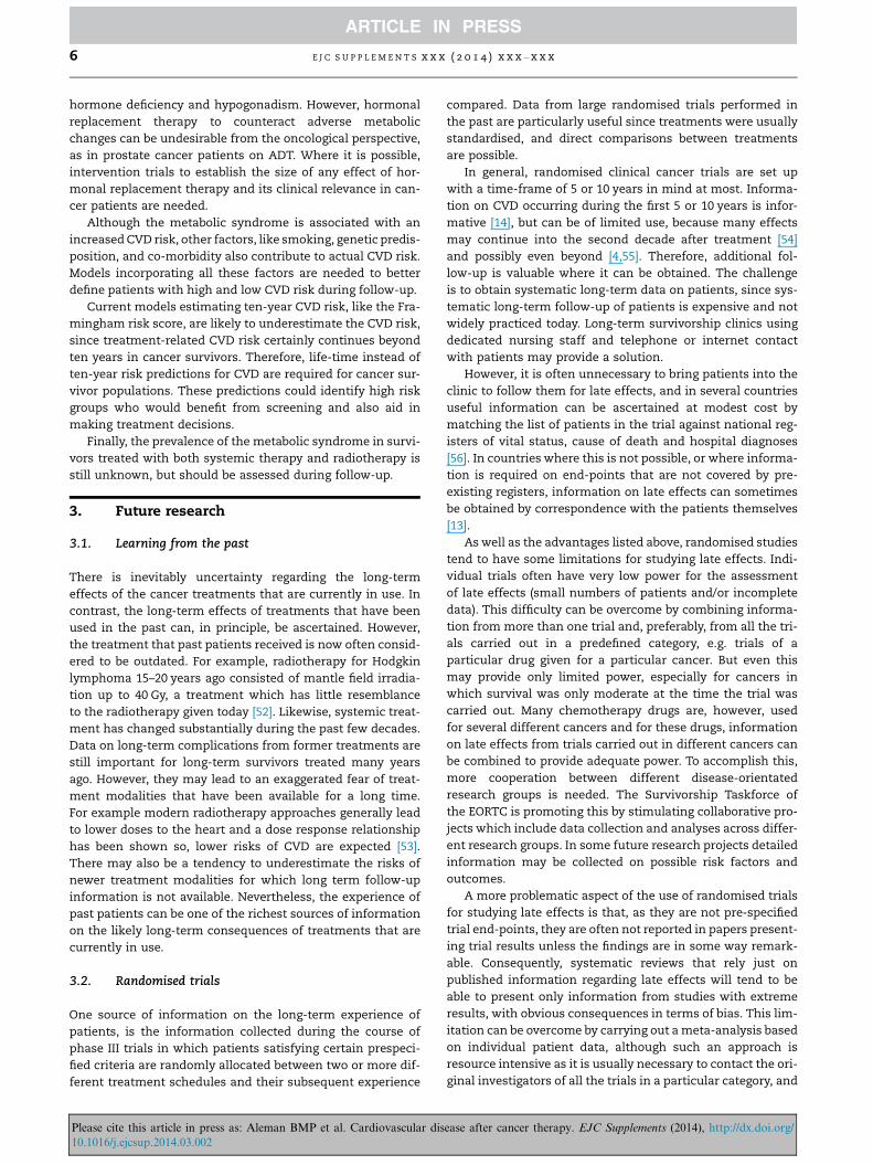
understanding of the mechanisms, individual risk factors and
prevention of this side-effect (See Table 5). The cardiotoxicity
problem is increasing since the introduction of anti-cancer
signalling inhibitors that have the potential of causing cardiac
dysfunction itself or increasing cardiotoxicity of conventional
chemotherapeutics [35]. At present, there is no universally
accepted definition of cardiotoxicity and many of the more
recent clinical trials that investigated potentially ‘cardiotoxic’
cancer drugs did not differentiate between Type I cardiotoxic-
ity and Type II cardiac dysfunction. Furthermore, since the
full spectrum of cardiotoxicity frequently does not become
apparent until months or even years after the initial cancer
treatment, long-term follow-up of patients exposed to poten-
tially cardiotoxic cancer drugs is needed and early surrogate
markers predicting long-term cardiovascular prognosis are
wanted. These predictive markers need to be universally
applicable and detect early myocardial loss by, for example,
measuring high-sensitivity cardiac biomarkers [36]. However,
some of the early work with cardiac biomarkers for the pre-
diction of cardiotoxicity has been challenging because the
dynamics of biomarker release after chemotherapy was
unknown and long-term data confirming the predictive value
are missing [37]. It has become clear that the decrease in left
ventricular ejection fraction measured either by echocardiog-
raphy or nuclear scans is not very sensitive in predicting
which patient eventually develops cardiotoxicity [31]. Newer
imaging techniques such as myocardial strain and strain rate
Doppler echocardiography may be more accurate and sensi-
tive to detect early changes of cardiotoxicity [38]. However,
these methods may have technical limitations particularly
in BC patients after left-sided surgery. Numerous preventive
strategies to mitigate cardiotoxicity particularly of anthracy-
clines have been investigated. They included alterations of
the chemical structure, liposomal encapsulation or co-medi-
cations to reduce iron chelation. Although some of these
strategies appeared successful in reducing cardiotoxicity,
questions of impaired efficacy and possible induction of sec-
ond tumours arose [28]. Global guidelines to treat cardiac dys-
function recommend the early use of renin-angiotensin
inhibitors and beta blockers. Although these drugs have been
tested to attenuate anthracycline-associated cardiotoxicity in
single centre studies, convincing evidence from large ran-
domised multicentre trials are still missing [39]. Finally,
although several risk factors for anthracycline-associated car-
diotoxicity have been identified, individual risk assessment in
patients based on genomics and proteomics is still missing.
The recent preclinical discovery that topoisomerase-IIbeta
may mediate anthracycline-associated cardiotoxicity opens
exciting new options: the opportunity to develop topoisomer-
ase-IIalpha specific anthracyclines that are likely less cardio-
toxic and also the potential to predict patient’s individual risk
for anthracycline-associated cardiotoxicity based on their
individual topoisomerase-IIbeta expression [40].
2.3. Metabolic syndrome
2.3.1. Current knowledgeThe metabolic syndrome is a clustering of metabolic disor-
ders that is associated with a twofold increased risk of CVD
compared to the general population. Key components of the
ease after cancer therapy. EJC Supplements (2014), http://dx.doi.org/
Table 5 – Gaps in knowledge concerning cardiotoxicity related to systemic therapy.
Lack of universally accepted definitions of cardiotoxicity and cardiac dysfunctionDifferentiation between irreversible and reversible cardiac dysfunctionLong-term follow-up data (10–20 years) neededEarly surrogate markers to predict long-term cardiovascular prognosisEarly pharmacological intervention to mitigate cardiotoxicityIndividualised patient risk assessment
Table 4 – Cardiovascular side-effects of selected systemic cancer therapeutics.
Cardiovascular effect Cancer therapy Long-term effect Mechanism
Cardiotoxicity Type I irreversible Anthracyclines Yes Loss of myocardiumCyclophosphamide Rare MyocarditisCisplatin Rare Unknown
Cardiac dysfunctionType II reversible
Anti-HER2 Therapeutics Unlikely, except whencombined with anthracyclines
Mitochondrial dysfunction
Anti-VEGF Therapeutics Unlikely Mitochondrial dysfunction
Myocardial ischaemia Pyrimidine analogues Rare Coronary vasospasmAnti-VEGF therapeutics Rare Arterial thrombosis
Arrhythmia Arsenic trioxide No HERG K+ blockageSelected TKIs HERG K+ blockage
Thromboembolism Cisplatin Rare Endothelial damageAnti-VEGF Therapeutics Endothelial damage
Arterial hypertension Anti-VEGF Therapeutics Unknown Multiple mechanisms
Pulmonary hypertension Selected TKIs Unknown Unknown
Peripheral arterial occlusive disease Selected TKIs Unknown Unknown
Pleural effusion Selected TKIs Unknown Unknown
Abbreviations: HER2, human epidermal growth factor receptor 2; TKI, tyrosine kinase inhibitors; VEGF, vascular endothelial growth factor, HERG
K+, human ether-a-go-go-related gene K+.
E J C S U P P L E M E N T S x x x ( 2 0 1 4 ) x x x – x x x 5
syndrome are decreased insulin sensitivity, hypertension,
overweight and an adverse lipid profile. Unfortunately, differ-
ent classification systems use different criteria to define the
metabolic syndrome, creating heterogeneity in reported prev-
alence between geographical areas and between study popu-
lations [41]. Development of the metabolic syndrome may
contribute to increased CVD risk in prostate cancer patients
using androgen deprivation therapy (ADT), in survivors of tes-
ticular cancer (TC) and of childhood malignancies [42–45].
Following standard cisplatin-based chemotherapy, TC sur-
vivors have an increased prevalence of the metabolic syn-
drome compared with the general male population [46]. The
metabolic syndrome develops early (3–5 years) after treat-
ment and is associated with decreased serum total testoster-
one concentration [47]. Childhood cancer survivors of
haematological malignancies, mainly acute lymphoblastic
leukaemia, and brain tumours have an increased risk of the
metabolic syndrome after cranial, abdominal or total body
irradiation [44,48]. Development of the metabolic syndrome
in these radiotherapy-treated patients is associated with
growth hormone deficiency and hypogonadism. Prostate can-
cer patients may develop an increase in fat mass, a decrease
in lean body mass, an adverse lipid profile, and impaired insu-
lin sensitivity early (within 3–6 months) after start of
Please cite this article in press as: Aleman BMP et al. Cardiovascular dis10.1016/j.ejcsup.2014.03.002
androgen deprivation therapy ADT [49]. These changes differ
from the features of the classically defined metabolic syn-
drome, since ADT induces accumulation of subcutaneous
rather than visceral fat, and an increase rather than a
decrease in high-density lipoprotein cholesterol. Finally, com-
ponents of the metabolic syndrome, including weight gain
and adverse changes in lipid levels, have been reported in
BC survivors [50]. These changes are associated with hor-
monal therapy and with development of early menopause
due to oophorectomy or chemotherapy.
Because of its relatively high prevalence and because of
the effects of the metabolic syndrome in cancer survivors, it
should be addressed in future care plans. Interventional stud-
ies have shown that individuals who make favourable
changes in their lifestyle after cancer diagnosis feel better,
experience less fatigue and may possibly even decrease risk
of cancer recurrence [51].
2.3.2. Gaps in knowledgeInsight into the aetiology of the metabolic syndrome after
cancer treatment might help to identify and treat cancer sur-
vivors with an increased CVD risk.
Development of the metabolic syndrome after cancer ther-
apy is associated with endocrine disorders, mainly growth
ease after cancer therapy. EJC Supplements (2014), http://dx.doi.org/
6 E J C S U P P L E M E N T S x x x ( 2 0 1 4 ) x x x – x x x
hormone deficiency and hypogonadism. However, hormonal
replacement therapy to counteract adverse metabolic
changes can be undesirable from the oncological perspective,
as in prostate cancer patients on ADT. Where it is possible,
intervention trials to establish the size of any effect of hor-
monal replacement therapy and its clinical relevance in can-
cer patients are needed.
Although the metabolic syndrome is associated with an
increased CVD risk, other factors, like smoking, genetic predis-
position, and co-morbidity also contribute to actual CVD risk.
Models incorporating all these factors are needed to better
define patients with high and low CVD risk during follow-up.
Current models estimating ten-year CVD risk, like the Fra-
mingham risk score, are likely to underestimate the CVD risk,
since treatment-related CVD risk certainly continues beyond
ten years in cancer survivors. Therefore, life-time instead of
ten-year risk predictions for CVD are required for cancer sur-
vivor populations. These predictions could identify high risk
groups who would benefit from screening and also aid in
making treatment decisions.
Finally, the prevalence of the metabolic syndrome in survi-
vors treated with both systemic therapy and radiotherapy is
still unknown, but should be assessed during follow-up.
3. Future research
3.1. Learning from the past
There is inevitably uncertainty regarding the long-term
effects of the cancer treatments that are currently in use. In
contrast, the long-term effects of treatments that have been
used in the past can, in principle, be ascertained. However,
the treatment that past patients received is now often consid-
ered to be outdated. For example, radiotherapy for Hodgkin
lymphoma 15–20 years ago consisted of mantle field irradia-
tion up to 40 Gy, a treatment which has little resemblance
to the radiotherapy given today [52]. Likewise, systemic treat-
ment has changed substantially during the past few decades.
Data on long-term complications from former treatments are
still important for long-term survivors treated many years
ago. However, they may lead to an exaggerated fear of treat-
ment modalities that have been available for a long time.
For example modern radiotherapy approaches generally lead
to lower doses to the heart and a dose response relationship
has been shown so, lower risks of CVD are expected [53].
There may also be a tendency to underestimate the risks of
newer treatment modalities for which long term follow-up
information is not available. Nevertheless, the experience of
past patients can be one of the richest sources of information
on the likely long-term consequences of treatments that are
currently in use.
3.2. Randomised trials
One source of information on the long-term experience of
patients, is the information collected during the course of
phase III trials in which patients satisfying certain prespeci-
fied criteria are randomly allocated between two or more dif-
ferent treatment schedules and their subsequent experience
Please cite this article in press as: Aleman BMP et al. Cardiovascular dis10.1016/j.ejcsup.2014.03.002
compared. Data from large randomised trials performed in
the past are particularly useful since treatments were usually
standardised, and direct comparisons between treatments
are possible.
In general, randomised clinical cancer trials are set up
with a time-frame of 5 or 10 years in mind at most. Informa-
tion on CVD occurring during the first 5 or 10 years is infor-
mative [14], but can be of limited use, because many effects
may continue into the second decade after treatment [54]
and possibly even beyond [4,55]. Therefore, additional fol-
low-up is valuable where it can be obtained. The challenge
is to obtain systematic long-term data on patients, since sys-
tematic long-term follow-up of patients is expensive and not
widely practiced today. Long-term survivorship clinics using
dedicated nursing staff and telephone or internet contact
with patients may provide a solution.
However, it is often unnecessary to bring patients into the
clinic to follow them for late effects, and in several countries
useful information can be ascertained at modest cost by
matching the list of patients in the trial against national reg-
isters of vital status, cause of death and hospital diagnoses
[56]. In countries where this is not possible, or where informa-
tion is required on end-points that are not covered by pre-
existing registers, information on late effects can sometimes
be obtained by correspondence with the patients themselves
[13].
As well as the advantages listed above, randomised studies
tend to have some limitations for studying late effects. Indi-
vidual trials often have very low power for the assessment
of late effects (small numbers of patients and/or incomplete
data). This difficulty can be overcome by combining informa-
tion from more than one trial and, preferably, from all the tri-
als carried out in a predefined category, e.g. trials of a
particular drug given for a particular cancer. But even this
may provide only limited power, especially for cancers in
which survival was only moderate at the time the trial was
carried out. Many chemotherapy drugs are, however, used
for several different cancers and for these drugs, information
on late effects from trials carried out in different cancers can
be combined to provide adequate power. To accomplish this,
more cooperation between different disease-orientated
research groups is needed. The Survivorship Taskforce of
the EORTC is promoting this by stimulating collaborative pro-
jects which include data collection and analyses across differ-
ent research groups. In some future research projects detailed
information may be collected on possible risk factors and
outcomes.
A more problematic aspect of the use of randomised trials
for studying late effects is that, as they are not pre-specified
trial end-points, they are often not reported in papers present-
ing trial results unless the findings are in some way remark-
able. Consequently, systematic reviews that rely just on
published information regarding late effects will tend to be
able to present only information from studies with extreme
results, with obvious consequences in terms of bias. This lim-
itation can be overcome by carrying out a meta-analysis based
on individual patient data, although such an approach is
resource intensive as it is usually necessary to contact the ori-
ginal investigators of all the trials in a particular category, and
ease after cancer therapy. EJC Supplements (2014), http://dx.doi.org/
E J C S U P P L E M E N T S x x x ( 2 0 1 4 ) x x x – x x x 7
then collate centrally and check all the data that are forthcom-
ing before it can be analysed.
Another limitation of data arising from randomised trials
is that patients with substantial co-morbidities at the time
of their cancer diagnosis are much less likely to be entered
into randomised trials than patients who are otherwise
healthy. This is likely to be the case even when such patients
are not explicitly excluded in the trial protocol.
A final limitation applies particularly to studies in which
the patients in one arm of the trial have been randomised
to receive potentially cardiotoxic chemotherapy and patients
in the other arm randomised not to receive it. In such trials it
is often the case that the patients in the chemotherapy arm
have their cardiac status more thoroughly assessed –both
during treatment and during follow up- than do patients in
the control arm. This may well introduce considerable bias
into comparisons between the two trial arms for end-points
other than mortality and in such cases meaningful analyses
can only be carried out for cardiac mortality.
3.3. Observational studies
Another approach to obtaining information on the cardiac
side-effects of cancer treatments is to obtain data from
observational studies rather than randomised trials.
Such studies can be informative, especially if they are
population-based as, for example, studies based on large
population-based cancer registries. Care is needed in their
interpretation, however, as patients who are at increased
risk of heart disease at the time when decisions on their
treatment are made, will tend not to be given potentially
cardiotoxic chemotherapy if it can possibly be avoided.
Comparisons of subsequent heart disease rates in patients
with and without such treatment may, therefore, provide
misleading answers [56].
3.4. Achieving relevance for today’s patients
Studies of the long-term cardiac effects of cancer treatments
given in the past may not be immediately relevant to today’s
patients. One reason for this is that medical practice has
changed with time. As mentioned before, modern radiation
approach has significantly reduced normal tissue exposure.
Also in systemic treatment changes have been implemented,
for example, the cardiotoxicity of anthracyclines delivered
today may be lower than previously due to changes in the
way the drug is administered, e.g. by using continuous infu-
sions rather than a bolus or by using a pegylated formulation.
Careful consideration of these issues is therefore needed
when interpreting any studies. A further issue is that baseline
levels of cardiac mortality have decreased substantially over
the last few decades in many countries, and levels of morbid-
ity may have also changed, although such changes are gener-
ally less well documented than changes in mortality. This
may mean that the absolute risk of late cardiac side-effects
in patients treated today may differ from that for past popu-
lations of patients. Proportional increases in the incidence
and mortality arising from the use of particular drugs are,
however, usually reasonably stable across populations with
different baseline rates.
Please cite this article in press as: Aleman BMP et al. Cardiovascular dis10.1016/j.ejcsup.2014.03.002
The experience of the past can also be used to optimise
future treatments as the data generated from patients treated
in the past may provide dose–response information which
can be used in mathematical models and thereby enable us
to predict and compare long-term complications of present
day treatment, thus guiding the choice of treatment in indi-
vidual patients [57].
For newer treatments such as antibodies, small molecules,
and highly conformal radiotherapy, observation time is still
too short for reliable estimation of long-term complications.
Vigilance and a strict safety-monitoring programme, even
after approval of the drug, are essential.
Furthermore, we need uniform definitions of toxicity in
order to allow proper comparison between studies.
3.5. Regulatory issues
For analysis of long-term complications from specific treat-
ments we often need to be able to obtain data from different
registries on particular, identifiable patients. Hence, ethical
issues must be considered and approvals obtained. In some
countries this may be more difficult than in others since there
is a large variation in regulations concerning these issues.
Informed consent from all relevant patients may be virtu-
ally impossible to obtain retrospectively, since a proportion of
patients will have died or their present address may not be
known. Moreover, there may be ethical dilemmas when con-
tacting patients treated many years ago who now consider
themselves to be healthy. In most countries, permission to
omit informed consent is possible in this situation. Another
approach could be to incorporate upfront permission from
patients entered into clinical trials on future outcomes.
3.6. Early detection of cardiac damage
Information on the possible value of measuring early subclin-
ical damage using biomarkers and/or (functional) imaging fol-
lowing cardiotoxic chemotherapy [58–61] and radiation
exposure of the heart [62] is still scarce.
3.6.1. ImagingCardiac imaging techniques such as 2D echocardiography or
MUGA show clinically detectable left-ventricular dysfunction
but earlier subclinical injury cannot be detected with these
imaging modalities. Cardiac magnetic resonance imaging
(MRI) may offer some possibilities in screening since it
enables tissue characterisation and may lead to detection of
diffuse interstitial fibrosis and changes in regional myocardial
function. MRI also has disadvantages such as need for con-
trast, and its high cost and low availability. Newer imaging
techniques, such as contrast and 3D echocardiography are
also under investigation. These imaging modalities are
expensive and should not be unnecessarily repeated. There-
fore, screening guidelines need to be developed and optimal
and cost-effective screening schedules evaluated.
3.6.2. BiomarkersThere are several cardiac biomarkers, such as troponin I (TnI),
troponin T (TnT), B-type natriuretic peptide (BNP), N-terminal
pro-BNP (NT-proBNP) and myeloperoxidase (MPO). There may
ease after cancer therapy. EJC Supplements (2014), http://dx.doi.org/
8 E J C S U P P L E M E N T S x x x ( 2 0 1 4 ) x x x – x x x
be a role for these biomarkers during follow-up to enable
early detection of cardiac toxicity and possibly also during
cancer therapy [63,64]. Circulating cardiac troponin (cTn,
which can be TnI or TnT) is a sensitive and specific biomarker
for detection of myocardial injury. Although most commonly
used to detect myonecrosis in the setting of ischaemia, cTns
are also elevated with other acute and chronic disease pro-
cesses, including HF [65]. An increase in TnI level in patients
undergoing chemotherapy may prove to be a useful means of
detecting cardiotoxicity long before a reduction in left ventric-
ular ejection fraction (LVEF) occurs and could allow for the
selection of high-risk patients who could benefit from preven-
tive treatment, such as treatment with angiotensin-convert-
ing enzyme (ACE) inhibitors and beta-blocking agents. In
patients treated with trastuzumab, an increase in TnI level
could help identifying those patients at risk for cardiotoxicity
and unlikely to recover [66]. So far however, treatment is not
adapted based on troponin levels.
Serum biomarkers may also be useful in helping diagnose
asymptomatic left ventricular dysfunction or HF. Serum bio-
markers such as brain natriuretic peptide (BNP) and N-termi-
nal fragment (NT-proBNP) are most commonly used.
However, there are limitations to BNP or NT-proBNP measure-
ments, since other diseases may also cause abnormal BNP
levels and serum levels also depend on other factors such
as age. In a study analysing eight different biomarkers before
start of treatment and every three months up to 15 months in
patients treated with doxorubicin and trastuzumab, the risk
of cardiotoxicity was especially related to an increase in TnI
and MPO. The combination of both markers offered additive
information about the risk of cardiotoxicity, but independent
validation of these findings is necessary before application to
clinical practice is possible [37]. Another study evaluating 200
patients with anthracycline-induced cardiomyopathy indi-
cated that the percentage of responders to modern HF treat-
ment decreased progressively as the time from the end of
chemotherapy to the start of HF treatment increased [39].
Some biomarkers can also give quantitative information:
the absolute value of these markers shortly after the admin-
istration of chemotherapy may indicate the degree of future
left ventricular dysfunction.
At present there is no clear biomarker-set for CVD risk pre-
diction during or after cancer treatment. Research is on-going
regarding the predictive value and the ideal timing of bio-
marker measurements. Ideally, future research would vali-
date a predictive set of tools to adapt treatment with
respect to toxicity risk. Both biomarkers and imaging need
further exploration in on-going trials. Early imaging, esti-
mated CVD risk and the ability to associate toxicity with
molecular profiling may lead to new recommendations for
monitoring cancer patients during and after chemotherapy.
4. Future guidelines
Although the increased risk of cardiac diseases following can-
cer therapy is well recognised, measures for primary and sec-
ondary prevention are still being developed.
Nowadays, cardiac monitoring is done during systemic
treatment by serial measurements of left ventricular ejection
Please cite this article in press as: Aleman BMP et al. Cardiovascular dis10.1016/j.ejcsup.2014.03.002
fraction (LVEF). A decrease in LVEF reflects myocardial injury
and the first sign of cardiac failure. Often during chemother-
apy this cardiac dysfunction is reversible, and the LVEF
decrease is only present at the moment of stress. By introduc-
tion of medication or rest, permanent dose reduction or ces-
sation of chemotherapy treatment is not always necessary.
What these moments of severe heart stress induce and the
impact on cardiovascular risk during the rest of life are not
well documented.
Recently, some evidence supports the use of other echo-
cardiographic indices and biomarkers for the earlier detection
of cardiac injury before a decrease in LVEF is noticed [67–71].
New imaging tools and markers are sought for earlier detec-
tion of serious cardiovascular morbidity partly to enable pre-
ventive measures, but also to spare low risk patients from
unnecessary monitoring [72,73].
Currently there are no indications that the management of
cancer treatment related cardiac diseases should differ from
that due to other causes. In patients with (subclinical) HF
treatment generally focuses on correcting underlying physio-
logical abnormalities such as increased afterload and
decreased contractility, and frequently includes treatment
with angiotensin-converting enzyme (ACE) inhibitors and/or
beta-blockers [74]. Several guidelines developed for treating
patients with asymptomatic left ventricular dysfunction or
HF (not specifically after cancer treatment) include e.g. beta-
blockers, ACE-inhibitors and diuretics [75].
At this moment, in Europe formal guidelines exist only fol-
lowing antibody treatment (e.g. echocardiograms every three
months) [76]. More data on incidence and reversibility, but
moreover earlier indicators of cardiac damage, might in the
future enable the necessary level of surveillance to be
individualised.
Although little is formalised on cardiac surveillance fol-
lowing cancer treatment, it is clear that hypertension, short-
ness of breath or chronic fatigue need to be taken very
seriously in cancer survivors. The risk of late cancer treat-
ment related cardiotoxicity is often forgotten or underesti-
mated, especially in women. Guidelines and educational
sessions for general practitioners and other caregivers are
needed to explain late cardiac risks and to better anticipate
on late effects after cancer treatment [77,78].
Lifestyle factors such as smoking, obesity and lack of exer-
cise and familial predisposition are important risk factors in
CVD. Only a few studies have addressed whether factors such
as these may modify the risk of treatment-related CVD [51].
Our knowledge of modifying effects of lifestyle and genetic
predisposition on treatment-associated cardiovascular risk is
only beginning to evolve. International collaborative studies
are needed, including large numbers of survivors for whom
not only treatment data but also detailed high-quality data
on medical history, lifestyle, environmental, and occupational
factors are available. The sequence of exposure to treatment
and other risk factors deserves investigation, particularly for
designing interventions where the treatments interact with
modifiable risk factors. International pooling of data already
available and data from new studies is essential to obtain suf-
ficient power for interaction analyses allowing discrimination
between additive, multiplicative and more than multiplicative
effects of treatment and other cancer risk factors. Late effects
ease after cancer therapy. EJC Supplements (2014), http://dx.doi.org/
E J C S U P P L E M E N T S x x x ( 2 0 1 4 ) x x x – x x x 9
research should combine different outcomes like cardiovascu-
lar disease, second malignancies, early menopause, infertility
etc., to obtain more detailed information on host (genetic sen-
sitivity, life style, age etc.) and treatment (dose, type, timing,
interactions etc.) factors. Also the quality of life and psycho-
socio-economic impact should be taken into account, having
possibly a direct (stress) or in-direct (access to care) influence
on morbidity and mortality of various late effects.
Lifestyle interventions, exercise promotion and special
care plans are proposed in cancer survivors. Whether all
patients will benefit from these measures is uncertain. A risk
based strategy is needed and long-term follow-up is essential
[51,79–81].
5. Conclusion
Extensive knowledge has already been gained from the past
concerning cancer treatment related cardiotoxicity, but there
is much more that can be done, although medical practice
continues to evolve and so the resulting data need to be inter-
preted with care. Better knowledge is needed of the late effects
of modern systemic treatments and of radiotherapy on critical
structures of the heart and of possible interactions between
treatment modalities. This knowledge will contribute to a
longer life expectancy and better quality of life of cancer sur-
vivors, with less treatment-related morbidity from other dis-
eases. Finally, prediction models taking into account the full
spectrum of late effects are needed to guide primary treat-
ment choice and appropriate follow up after cancer treatment.
Conflict of interest statement
Maja V. Maraldo were reported by: Berthe M.P. Aleman, Eliza-
beth C. Moser, Conny Vrieling, Sarah C. Darby.
Thomas M. Suter: Participation in a company sponsored
speaker’s bureau: Roche, Robopharmn, Novartis.
Lena Specht: Receipt of grants/research supports: Merck
Serono – Receipt of honoraria or consultation fees: Takeda,
Boehringer Ingelheim, Fresenius biotech – Participation in a
company sponsored speaker’s bureau: Takeda.
R E F E R E N C E
[1] Borchmann P, Eichenauer DA, Engert A. State of the art in thetreatment of Hodgkin lymphoma. Nat Rev Clin Oncol2012;9(8):450–9.
[2] Clarke M, Collins R, Darby S, et al. Effects of radiotherapy andof differences in the extent of surgery for early breast canceron local recurrence and 15-year survival: an overview of therandomised trials. Lancet 2005;366(9503):2087–106.
[3] Darby S, McGale P, Correa C, et al. Effect of radiotherapy afterbreast-conserving surgery on 10-year recurrence and 15-yearbreast cancer death: meta-analysis of individual patient datafor 10,801 women in 17 randomised trials. Lancet2011;378(9804):1707–16.
[4] Hodgson DC. Late effects in the era of modern therapy forHodgkin lymphoma. Hematol Am Soc Hematol Educ Prog2011;2011:323–9.
Please cite this article in press as: Aleman BMP et al. Cardiovascular dis10.1016/j.ejcsup.2014.03.002
[5] de Haas EC, Oosting SF, Lefrandt JD, Wolffenbuttel BH, SleijferDT, Gietema JA. The metabolic syndrome in cancer survivors.Lancet Oncol 2010;11(2):193–203.
[6] Schultz-Hector S, Trott KR. Radiation-induced cardiovasculardiseases: is the epidemiologic evidence compatible with theradiobiologic data? Int J Radiat Oncol Biol Phys2007;67(1):10–8.
[7] Darby SC, Cutter DJ, Boerma M, et al. Radiation-related heartdisease: current knowledge and future prospects. Int J RadiatOncol Biol Phys 2010;76(3):656–65.
[8] Gagliardi G, Constine LS, Moiseenko V, et al. Radiation dose–volume effects in the heart. Int J Radiat Oncol Biol Phys2010;76(3 Suppl):S77–85.
[9] Aleman BM, van den Belt-Dusebout AW, De Bruin ML, et al.Late cardiotoxicity after treatment for Hodgkin lymphoma.Blood 2007;109(5):1878–86.
[10] Castellino SM, Geiger AM, Mertens AC, et al. Morbidity andmortality in long-term survivors of Hodgkin lymphoma: areport from the Childhood Cancer Survivor Study. Blood2011;117(6):1806–16.
[11] Swerdlow AJ, Higgins CD, Smith P, et al. Myocardialinfarction mortality risk after treatment for Hodgkin disease:a collaborative British cohort study. J Natl Cancer Inst2007;99(3):206–14.
[12] Carr ZA, Land CE, Kleinerman RA, et al. Coronary heartdisease after radiotherapy for peptic ulcer disease. Int JRadiat Oncol Biol Phys 2005;61(3):842–50.
[13] Mulrooney DA, Yeazel MW, Kawashima T, et al. Cardiacoutcomes in a cohort of adult survivors of childhood andadolescent cancer: retrospective analysis of the ChildhoodCancer Survivor Study cohort. BMJ 2009;339:b4606.
[14] Darby SC, Ewertz M, McGale P, et al. Risk of ischemic heartdisease in women after radiotherapy for breast cancer. N EnglJ Med 2013;368(11):987–98.
[15] Shimizu Y, Kodama K, Nishi N, et al. Radiation exposure andcirculatory disease risk: Hiroshima and Nagasaki atomicbomb survivor data, 1950–2003. BMJ 2010;340:b5349.
[16] Preston DL, Shimizu Y, Pierce DA, Suyama A, Mabuchi K.Studies of mortality of atomic bomb survivors. Report 13:solid cancer and noncancer disease mortality: 1950–1997.Radiat Res 2003;160(4):381–407.
[17] Hancock SL, Tucker MA, Hoppe RT. Factors affecting latemortality from heart disease after treatment of Hodgkin’sdisease. JAMA 1993;270(16):1949–55.
[18] Heidenreich PA, Schnittger I, Strauss HW, et al. Screening forcoronary artery disease after mediastinal irradiation forHodgkin’s disease. J Clin Oncol 2007;25(1):43–9.
[19] Roychoudhuri R, Robinson D, Putcha V, Cuzick J, Darby S,Moller H. Increased cardiovascular mortality more thanfifteen years after radiotherapy for breast cancer: apopulation-based study. BMC Cancer 2007;7:9.
[20] Cuzick J, Stewart H, Rutqvist L, et al. Cause-specific mortalityin long-term survivors of breast cancer who participated intrials of radiotherapy. J Clin Oncol 1994;12(3):447–53.
[21] Harris EE, Correa C, Hwang WT, et al. Late cardiac mortalityand morbidity in early-stage breast cancer patients afterbreast-conservation treatment. J Clin Oncol 2006(August).
[22] Hooning MJ, Botma A, Aleman BM, et al. Long-term risk ofcardiovascular disease in 10-year survivors of breast cancer. JNatl Cancer Inst 2007;99(5):365–75.
[23] Tukenova M, Guibout C, Oberlin O, et al. Role of cancertreatment in long-term overall and cardiovascular mortalityafter childhood cancer. J Clin Oncol 2010;28(8):1308–15.
[24] Maraldo MV, Brodin NP, Vogelius IR, et al. Risk of developingcardiovascular disease after involved node radiotherapyversus mantle field for Hodgkin lymphoma. Int J Radiat OncolBiol Phys 2012.
ease after cancer therapy. EJC Supplements (2014), http://dx.doi.org/
10 E J C S U P P L E M E N T S x x x ( 2 0 1 4 ) x x x – x x x
[25] Moja L, Tagliabue L, Balduzzi S, et al. Trastuzumabcontaining regimens for early breast cancer. CochraneDatabase Syst Rev 2012;4:CD006243.
[26] Myrehaug S, Pintilie M, Tsang R, et al. Cardiac morbidityfollowing modern treatment for Hodgkin lymphoma: supra-additive cardiotoxicity of doxorubicin and radiation therapy.Leuk Lymphoma 2008;49(8):1486–93.
[27] Hilbers FS, Boekel NB, van den Broek AJ, et al. Geneticvariants in TGFbeta-1 and PAI-1 as possible risk factors forcardiovascular disease after radiotherapy for breast cancer.Radiother Oncol 2012;102(1):115–21.
[28] Suter TM, Ewer MS. Cancer drugs and the heart: importanceand management. Eur Heart J 2013;34(15):1102–11.
[29] Smith LA, Cornelius VR, Plummer CJ, et al. Cardiotoxicity ofanthracycline agents for the treatment of cancer: systematicreview and meta-analysis of randomised controlled trials.BMC Cancer 2010;10:337.
[30] Eschenhagen T, Force T, Ewer MS, et al. Cardiovascular sideeffects of cancer therapies: a position statement from theHeart Failure Association of the European Society ofCardiology. Eur J Heart Fail 2011;13(1):1–10.
[31] Ewer MS, Ali MK, Mackay B, et al. A comparison of cardiacbiopsy grades and ejection fraction estimations in patientsreceiving Adriamycin. J Clin Oncol 1984;2(2):112–7.
[32] de Azambuja AE, Bedard PL, Suter T, Piccart-Gebhart M.Cardiac toxicity with anti-HER-2 therapies: what have welearned so far? Target Oncol 2009;4(2):77–88.
[33] de Azambuja E, Procter M, van Veldhuisen DJ, et al.Trastuzumab-associated cardiac events at 8 years medianfollow-up in the HERA trial (BIG 1-01). JCO 2014 [in press].
[34] Montani D, Bergot E, Gunther S, et al. Pulmonary arterialhypertension in patients treated by dasatinib. Circulation2012;125(17):2128–37.
[35] Harbeck N, Ewer MS, De Laurentiis M, Suter TM, Ewer SM.Cardiovascular complications of conventional and targetedadjuvant breast cancer therapy. Ann Oncol 2011;22(6):1250–8.
[36] Cardinale D, Sandri MT, Martinoni A, et al. Myocardial injuryrevealed by plasma troponin I in breast cancer treated withhigh-dose chemotherapy. Ann Oncol 2002;13(5):710–5.
[37] Ky B, Putt M, Sawaya H, et al. Early increases in multiplebiomarkers predict subsequent cardiotoxicity in patientswith breast cancer treated with Doxorubicin, taxanes, andtrastuzumab. J Am Coll Cardiol 2014;63(8):809–16.
[38] Tassan-Mangina S, Codorean D, Metivier M, et al. TissueDoppler imaging and conventional echocardiography afteranthracycline treatment in adults: early and late alterationsof left ventricular function during a prospective study. Eur JEchocardiogr 2006;7(2):141–6.
[39] Cardinale D, Colombo A, Lamantia G, et al. Anthracycline-induced cardiomyopathy: clinical relevance and response topharmacologic therapy. J Am Coll Cardiol 2010;55(3):213–20.
[40] Zhang S, Liu X, Bawa-Khalfe T, et al. Identification of themolecular basis of doxorubicin-induced cardiotoxicity. NatMed 2012;18(11):1639–42.
[41] Alberti KG, Eckel RH, Grundy SM, et al. Harmonizing themetabolic syndrome: a joint interim statement of theInternational Diabetes Federation Task Force onEpidemiology and Prevention; National Heart, Lung, andBlood Institute; American Heart Association; World HeartFederation; International Atherosclerosis Society; andInternational Association for the Study of Obesity.Circulation 2009;120(16):1640–5.
[42] Mertens AC, Liu Q, Neglia JP, et al. Cause-specific latemortality among 5-year survivors of childhood cancer: theChildhood Cancer Survivor Study. J Natl Cancer Inst2008;100(19):1368–79.
Please cite this article in press as: Aleman BMP et al. Cardiovascular dis10.1016/j.ejcsup.2014.03.002
[43] Keating NL, O’Malley AJ, Smith MR. Diabetes andcardiovascular disease during androgen deprivation therapyfor prostate cancer. J Clin Oncol 2006;24(27):4448–56.
[44] Nottage KA, Ness KK, Li C, Srivastava D, Robison LL, HudsonMM. Metabolic syndrome and cardiovascular risk amonglong-term survivors of acute lymphoblastic leukaemia – fromthe St. Jude Lifetime Cohort. Br J Haematol 2014.
[45] van den Belt-Dusebout AW, Nuver J, de WR, et al. Long-termrisk of cardiovascular disease in 5-year survivors of testicularcancer. J Clin Oncol 2006;24(3):467–75.
[46] de Haas EC, Altena R, Boezen HM, et al. Early development ofthe metabolic syndrome after chemotherapy for testicularcancer. Ann Oncol 2013;24(3):749–55.
[47] Nuver J, Smit AJ, Wolffenbuttel BH, et al. The metabolicsyndrome and disturbances in hormone levels in long-termsurvivors of disseminated testicular cancer. J Clin Oncol2005;23(16):3718–25.
[48] van WM, Neggers SJ, van der Lelij AJ, Pieters R, van denHeuvel-Eibrink MM. The metabolic syndrome in adultsurvivors of childhood cancer, a review. J Pediatr HematolOncol 2010;32(3):171–9.
[49] Collier A, Ghosh S, McGlynn B, Hollins G. Prostate cancer,androgen deprivation therapy, obesity, the metabolicsyndrome, type 2 diabetes, and cardiovascular disease: areview. Am J Clin Oncol 2012;35(5):504–9.
[50] Redig AJ, Munshi HG. Care of the cancer survivor: metabolicsyndrome after hormone-modifying therapy. Am J Med2010;123(1). pp. 87.e1–5.
[51] Ligibel J. Lifestyle factors in cancer survivorship. J Clin Oncol2012;30(30):3697–704.
[52] Specht L, Yahalom J, Illidge T, et al. Modern radiation therapyfor Hodgkin lymphoma: field and dose guidelines from theinternational lymphoma radiation oncology group (ILROG).JCO 2014 [in press].
[53] Maraldo MV, Brodin NP, Aznar MC, et al. Estimated risk ofcardiovascular disease and secondary cancers with modernhighly conformal radiotherapy for early-stage mediastinalHodgkin lymphoma. Ann Oncol 2013;24(8):2113–8.
[54] Darby SC, McGale P, Taylor CW, Peto R. Long-term mortalityfrom heart disease and lung cancer after radiotherapy forearly breast cancer: prospective cohort study of about 300,000women in US SEER cancer registries. Lancet Oncol2005;6(8):557–65.
[55] Travis LB, Fossa SD, Schonfeld SJ, et al. Second cancersamong 40,576 testicular cancer patients: focus on long-termsurvivors. J Natl Cancer Inst 2005;97(18):1354–65.
[56] McGale P, Darby SC. Commentary: a dose–responserelationship for radiation-induced heart disease—currentissues and future prospects. Int J Epidemiol 2008;37(3):518–23.
[57] Brodin NP, Maraldo MV, Aznar MC, et al. Interactive decision-support tool for risk-based radiation therapy plancomparison for hodgkin lymphoma. Int J Radiat Oncol BiolPhys 2014;88(2):433–45.
[58] Daugaard G, Lassen U, Bie P, et al. Natriuretic peptides in themonitoring of anthracycline induced reduction in leftventricular ejection fraction. Eur J Heart Fail 2005;7(1):87–93.
[59] Aggarwal S, Pettersen MD, Bhambhani K, Gurczynski J,Thomas R, L’Ecuyer T. B-type natriuretic peptide as a markerfor cardiac dysfunction in anthracycline-treated children.Pediatr Blood Cancer 2007;49(6):812–6.
[60] Bryant J, Picot J, Baxter L, Levitt G, Sullivan I, Clegg A. Use ofcardiac markers to assess the toxic effects of anthracyclinesgiven to children with cancer: a systematic review. Eur JCancer 2007;43(13):1959–66.
[61] Krawczuk-Rybak M, Dakowicz L, Hryniewicz A, MaksymiukA, Zelazowska-Rutkowska B, Wysocka J. Cardiac function insurvivors of acute lymphoblastic leukaemia and Hodgkin’slymphoma. J Paediatr Child Health 2011;47(7):455–9.
ease after cancer therapy. EJC Supplements (2014), http://dx.doi.org/
E J C S U P P L E M E N T S x x x ( 2 0 1 4 ) x x x – x x x 11
[62] D’Errico MP, Grimaldi L, Petruzzelli MF, et al. N-terminal Pro-B-Type natriuretic peptide plasma levels as a potentialbiomarker for cardiac damage after radiotherapy in patientswith left-sided breast cancer. Int J Radiat Oncol Biol Phys2011.
[63] Cardinale D, Sandri MT. Role of biomarkers in chemotherapy-induced cardiotoxicity. Prog Cardiovasc Dis 2010;53(2):121–9.
[64] Lipshultz SE, Miller TL, Scully RE, et al. Changes in cardiacbiomarkers during doxorubicin treatment of pediatricpatients with high-risk acute lymphoblastic leukemia:associations with long-term echocardiographic outcomes. JClin Oncol 2012;30(10):1042–9.
[65] Newby LK, Rodriguez I, Finkle J, et al. Troponinmeasurements during drug development—considerations formonitoring and management of potential cardiotoxicity: aneducational collaboration among the Cardiac Safety ResearchConsortium, the Duke Clinical Research Institute, and the USFood and Drug Administration. Am Heart J 2011;162(1):64–73.
[66] Cardinale D, Colombo A, Torrisi R, et al. Trastuzumab-induced cardiotoxicity: clinical and prognostic implicationsof troponin I evaluation. J Clin Oncol 2010;28(25):3910–6.
[67] Von Hoff DD, Layard MW, Basa P, et al. Risk factors fordoxorubicin-induced congestive heart failure. Ann InternMed 1979;91(5):710–7.
[68] Slamon D, Eiermann W, Robert N, et al. Adjuvanttrastuzumab in HER2-positive breast cancer. N Engl J Med2011;365(14):1273–83.
[69] Piccart-Gebhart MJ, Procter M, Leyland-Jones B, et al.Trastuzumab after adjuvant chemotherapy in HER2-positivebreast cancer. N Engl J Med 2005;353(16):1659–72.
[70] Romond EH, Perez EA, Bryant J, et al. Trastuzumab plusadjuvant chemotherapy for operable HER2-positive breastcancer. N Engl J Med 2005;353(16):1673–84.
[71] Seidman A, Hudis C, Pierri MK, et al. Cardiac dysfunction inthe trastuzumab clinical trials experience. J Clin Oncol2002;20(5):1215–21.
[72] Slamon DJ, Leyland-Jones B, Shak S, et al. Use ofchemotherapy plus a monoclonal antibody against HER2 formetastatic breast cancer that overexpresses HER2. N Engl JMed 2001;344(11):783–92.
[73] Chien KR. Herceptin and the heart—a molecular modifier ofcardiac failure. N Engl J Med 2006;354(8):789–90.
[74] Barry E, Alvarez JA, Scully RE, Miller TL, Lipshultz SE.Anthracycline-induced cardiotoxicity: course,pathophysiology, prevention and management. Expert OpinPharmacother 2007;8(8):1039–58.
[75] McMurray JJ, Adamopoulos S, Anker SD, et al. ESC Guidelinesfor the diagnosis and treatment of acute and chronic heartfailure 2012: the Task Force for the Diagnosis and Treatmentof Acute and Chronic Heart Failure 2012 of the EuropeanSociety of Cardiology. Developed in collaboration with theHeart Failure Association (HFA) of the ESC. Eur Heart J2012;33(14):1787–847.
[76] Turner JR, Panicker GK, Karnad DR, Cabell CH, Lieberman R,Kothari S. Cardiovascular safety monitoring during oncologydrug development and therapy. Am J Ther 2014.
[77] Witteles RM, Telli M. Underestimating cardiac toxicity incancer trials: lessons learned? J Clin Oncol2012;30(16):1916–8.
[78] Tan-Chiu E, Yothers G, Romond E, et al. Assessment ofcardiac dysfunction in a randomized trial comparingdoxorubicin and cyclophosphamide followed by paclitaxel,with or without trastuzumab as adjuvant therapy in node-positive, human epidermal growth factor receptor 2-overexpressing breast cancer: NSABP B-31. J Clin Oncol2005;23(31):7811–9.
Please cite this article in press as: Aleman BMP et al. Cardiovascular dis10.1016/j.ejcsup.2014.03.002
[79] Forsythe LP, Parry C, Alfano CM, et al. Use of survivorshipcare plans in the United States: associations withsurvivorship care. J Natl Cancer Inst 2013;105(20):1579–87.
[80] Stricker CT, Jacobs LA, Risendal B, et al. Survivorship careplanning after the institute of medicine recommendations:how are we faring? J Cancer Surviv 2011;5(4):358–70.
[81] Hill-Kayser CE, Vachani CC, Hampshire MK, Di LG, Jacobs LA,Metz JM. Impact of internet-based cancer survivorship careplans on health care and lifestyle behaviors. Cancer2013;119(21):3854–60.
[82] Hoppe RT. Hodgkin’s disease: complications of therapy andexcess mortality. Ann Oncol 1997;8(Suppl. 1):115–8.
[83] Ng AK, Bernardo MP, Weller E, et al. Long-term survival andcompeting causes of death in patients with early- stageHodgkin’s disease treated at age 50 or younger. J Clin Oncol2002;20(8):2101–8.
[84] Aleman BM, van den Belt-Dusebout AW, Klokman WJ, van’tVeer MB, Bartelink H, van Leeuwen FE. Long-term cause-specific mortality of patients treated for Hodgkin’s disease. JClin Oncol 2003;21(18):3431–9.
[85] Ng AK, Lacasce A, Travis LB. Long-term complications oflymphoma and its treatment. J Clin Oncol2011;29(14):1885–92.
[86] Hull MC, Morris CG, Pepine CJ, Mendenhall NP. Valvulardysfunction and carotid, subclavian, and coronary arterydisease in survivors of hodgkin lymphoma treated withradiation therapy. JAMA 2003;290(21):2831–7.
[87] Galper SL, Yu JB, Mauch PM, et al. Clinically significantcardiac disease in patients with Hodgkin lymphoma treatedwith mediastinal irradiation. Blood 2011;117(2):412–8.
[88] Rutqvist LE, Johansson H. Mortality by laterality of theprimary tumour among 55,000 breast cancer patients fromthe Swedish Cancer Registry. Br J Cancer 1990;61(6):866–8.
[89] Rutqvist LE, Liedberg A, Hammar N, Dalberg K. Myocardialinfarction among women with early-stage breast cancertreated with conservative surgery and breast irradiation. Int JRadiat Oncol Biol Phys 1998;40(2):359–63.
[90] Hojris I, Overgaard M, Christensen JJ, Overgaard J. Morbidityand mortality of ischaemic heart disease in high-risk breast-cancer patients after adjuvant postmastectomy systemictreatment with or without radiotherapy: analysis of DBCG82b and 82c randomised trials. Radiotherapy Committee ofthe Danish Breast Cancer Cooperative Group. Lancet1999;354(9188):1425–30.
[91] Paszat LF, Mackillop WJ, Groome PA, Schulze K, Holowaty E.Mortality from myocardial infarction followingpostlumpectomy radiotherapy for breast cancer: apopulation-based study in Ontario, Canada. Int J Radiat OncolBiol Phys 1999;43(4):755–62.
[92] Vallis KA, Pintilie M, Chong N, et al. Assessment of coronaryheart disease morbidity and mortality after radiation therapyfor early breast cancer. J Clin Oncol 2002;20(4):1036–42.
[93] Darby S, McGale P, Peto R, Granath F, Hall P, Ekbom A.Mortality from cardiovascular disease more than 10 yearsafter radiotherapy for breast cancer: nationwide cohort studyof 90 000 Swedish women. BMJ 2003;326(7383):256–7.
[94] Giordano SH, Kuo YF, Freeman JL, Buchholz TA, HortobagyiGN, Goodwin JS. Risk of cardiac death after adjuvantradiotherapy for breast cancer. J Natl Cancer Inst2005;97(6):419–24.
[95] Patt DA, Goodwin JS, Kuo YF, et al. Cardiac morbidity ofadjuvant radiotherapy for breast cancer. J Clin Oncol2005;23(30):7475–82.
[96] Hooning MJ, Aleman BM, van Rosmalen AJ, Kuenen MA, KlijnJG, van Leeuwen FE. Cause-specific mortality in long-termsurvivors of breast cancer: a 25-year follow-up study. Int JRadiat Oncol Biol Phys 2006;64(4):1081–91.
ease after cancer therapy. EJC Supplements (2014), http://dx.doi.org/