!TERAPIA!CONSERVATIVA!DELLE!CISTI!OVARICHE!TORTE!NEONATALI:!
RISULTATI!E!FOLLOW4UP!!!!!!!!!!!!!!
Department of Cardiac Thoracic and Vascular Sciences, University of Padova
CARDIAC RESYNCHRONIZATION THERAPY BY MULTIPOINT PACING IMPROVES THE ACUTE
RESPONSE OF LEFT VENTRICULAR MECHANICS AND FLUID DYNAMICS: A THREE-DIMENSIONAL
AND PARTICLE IMAGE VELOCIMETRY ECHOCARDIOGRAPHIC STUDY
M. Siciliano, F. Migliore, D. Muraru, A. Zorzi, S. Cavedon, G. Pedrizzetti, E.
Bertaglia, D. Corrado, S. Iliceto, L. Badano

Disclosures: none
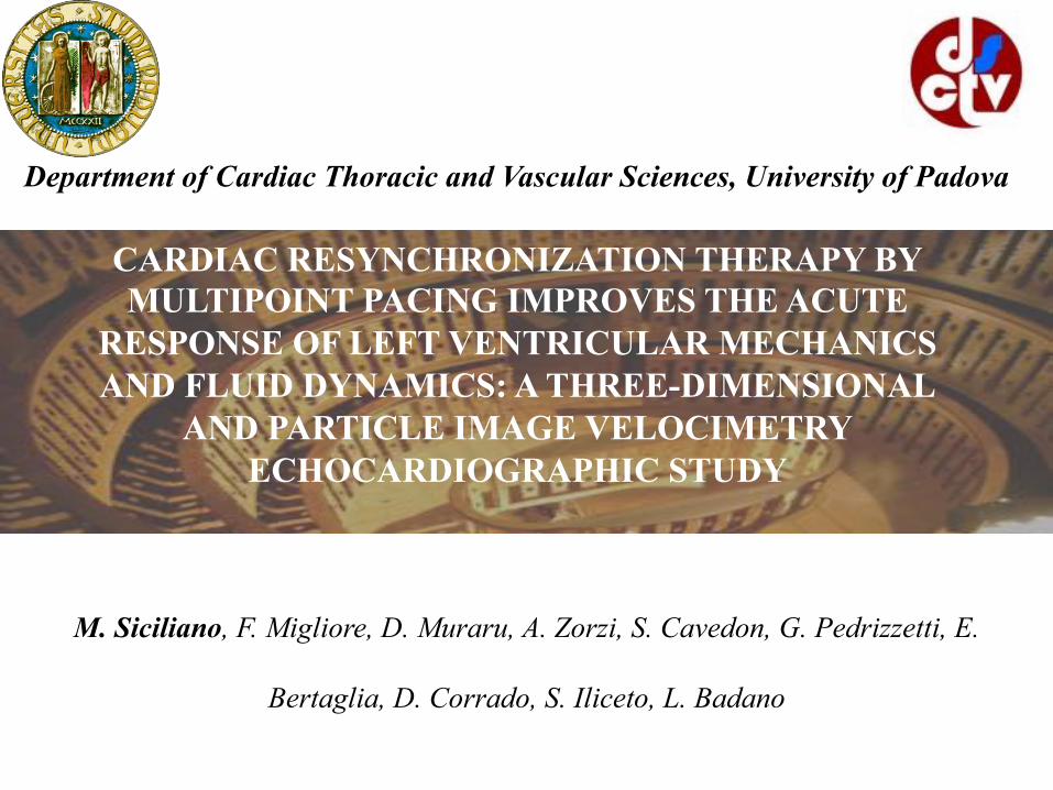
RV LV 130
100 mm/s
Electrical activations Bipolar LV Lead
LAO LAO
RAO
CONVENTIONAL Cardiac Resinchronization Therapy
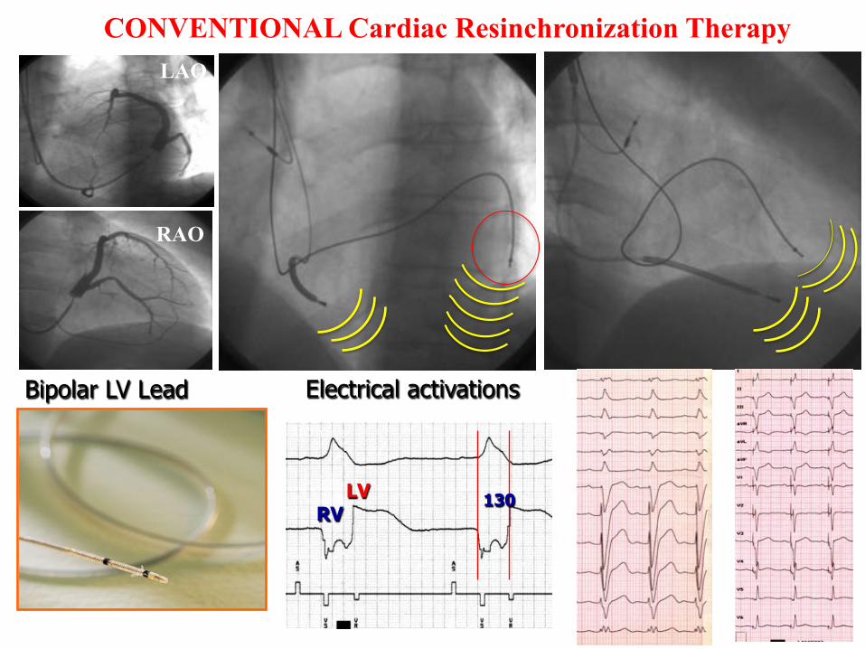
Ypenburg et al. Long-Term Prognosis After Cardiac Resynchronization Therapy Is Related to the Extent of Left Ventricular Reverse Remodeling at Midterm Follow-Up. JACC 2009
Non Responders to Cardiac Resinchronization Therapy The Magnitude of the Problem
43%$
43%!!CRT!pa*ents$are$classified$as$$non4responder$o$negaAve4responder$referred$to$LVESV$a;er$6$months$(N=302)$
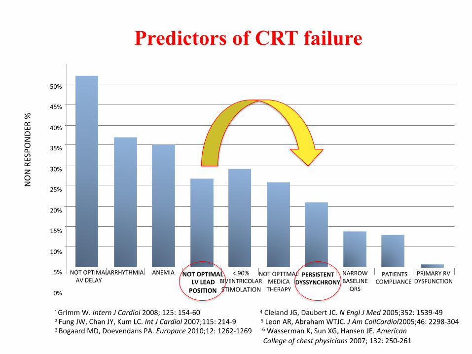
Mullens'W,'et'al.'JACC.'2009;53:7659773.'
$NON$RESPO
NDE
R$%$
NOT$OPTIMAL$$AV$DELAY$
ARRHYTHMIA$ ANEMIA$ NOT!OPTIMAL!!LV!LEAD!!POSITION!
<$90%$BIVENTRICOLAR$STIMOLATION$
$NOT$OPTTMAL$$MEDICA$$THERAPY$
PERSISTENT!!DYSSYNCHRONY!
NARROW$$BASELINE$$
QRS$
$PATIENTS$$COMPLIANCE$
$PRIMARY$RV$$DYSFUNCTION$
50%$
45%$
40%$
35%$
30%$
25%$
20%$
15%$
10%$
5%$
0%$
1"Grimm$W.$Intern'J'Cardiol$2008;$125:$154[60 """""""""""""""""""4"Cleland$JG,$Daubert$JC.$N'Engl'J'Med$2005;352:$1539[49"2"Fung$JW,$Chan$JY,$Kum$LC.$Int'J'Cardiol$2007;115:$214[9 """""""""""5"Leon$AR,$Abraham$WTJC.$J'Am'CollCardiol2005;46:$2298[304$"3"Bogaard$MD,$Doevendans$PA.$Europace$2010;12:$1262[1269$"""""6"Wasserman$K,$Sun$XG,$Hansen$JE.$American''''''''''''''''''''''''''''''''''''''''''''''''''''''''''''''''''''''''''''''''''''''''''''''''''''''''''''''''''''''College'of'chest'physicians$2007;$132:$250[261$""
Predictors of CRT failure

2015
$$$$
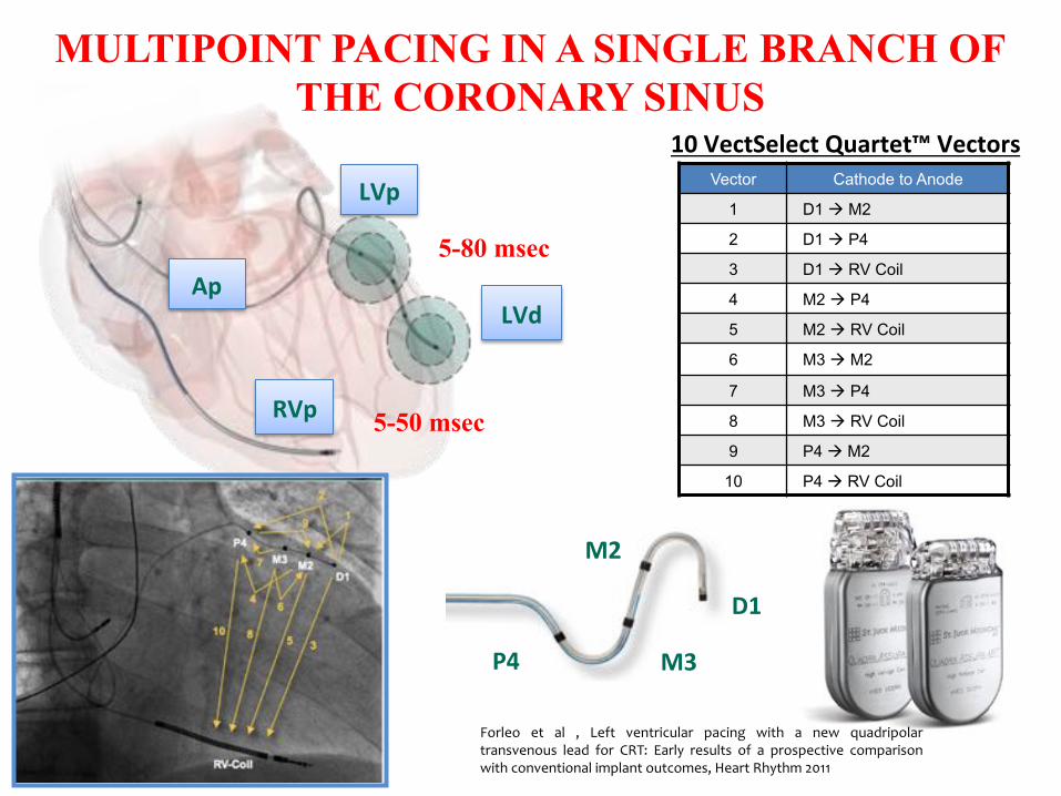
Vector Cathode to Anode
1 D1 ! M2
2 D1 ! P4
3 D1 ! RV Coil
4 M2 ! P4
5 M2 ! RV Coil
6 M3 ! M2
7 M3 ! P4
8 M3 ! RV Coil
9 P4 ! M2
10 P4 ! RV Coil
10!VectSelect!Quartet™!Vectors!
LVp!
LVd!
RVp!
Forleo" et" al" ," Left" ventricular" pacing" with" a" new" quadripolar"transvenous" lead" for" CRT:" Early" results" of" a" prospective" comparison"with"conventional"implant"outcomes,"Heart"Rhythm"2011"
5-80 msec
5-50 msec
Ap!
MULTIPOINT PACING IN A SINGLE BRANCH OF THE CORONARY SINUS
D1!
M2!
M3!P4!
$$
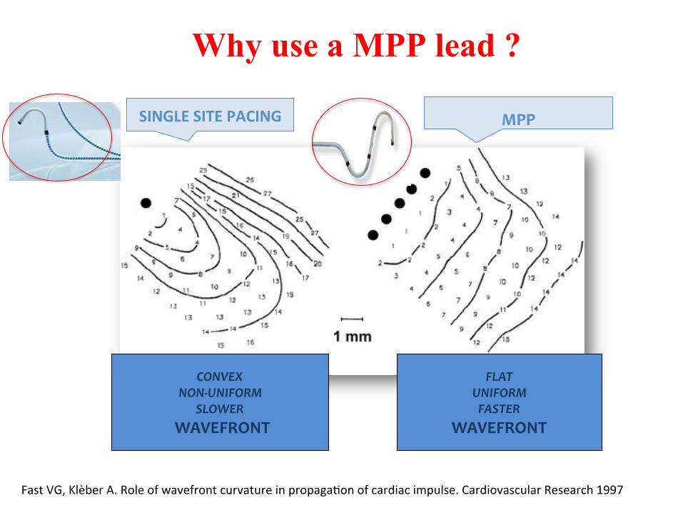
!CONVEX'
NON(UNIFORM'SLOWER'
!WAVEFRONT!!
!!FLAT''
UNIFORM'FASTER'
WAVEFRONT!!
Fast$et$al.,$Cardiovascular$Research$$1997;$33:$258–271$
SINGLE!SITE!PACING! MPP!!
Fast$VG,$Klèber$A.$Role$of$wavefront$curvature$in$propaga*on$of$cardiac$impulse.$Cardiovascular$Research$1997$
Why use a MPP lead ?
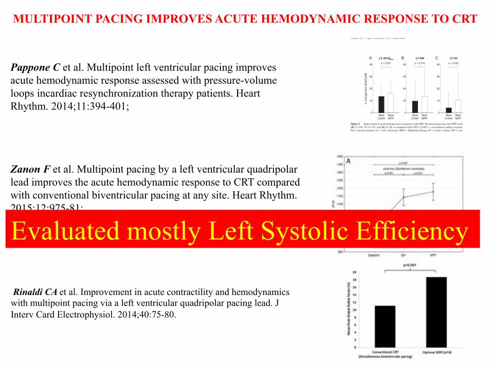
Pappone C et al. Multipoint left ventricular pacing improves acute hemodynamic response assessed with pressure-volume loops incardiac resynchronization therapy patients. Heart Rhythm. 2014;11:394-401;
0
10
20
30
40
0
10
20
30
40
0
20
40
60
80
0
10
20
30
40
BestCONV
p = 0.018
LV dP/dtMax LV SW LV SV LV EF
% c
hang
e fro
m B
ASEL
INE
p < 0.001 p = 0.003 p = 0.003
BestMPP
BestCONV
BestMPP
BestCONV
BestMPP
BestCONV
BestMPP
Figure 2 Improvement in acute hemodynamic parameters with MPP. Biventricular pacing with MPP resulted in significant improvement in (A) LV dP/dtmax,(B) LV SW, (C) LV SV, and (D) LV EF as compared with CONV. CONV ¼ conventional cardiac resynchronization therapy; dP/dt ¼ rate of pressure change;EF ¼ ejection fraction; LV ¼ left ventricular; MPP ¼ MultiPoint Pacing; SV ¼ stroke volume; SW ¼ stroke work.
150 175 200 225 2500
35
70
105
140
LV Volume (mL)
LV P
ress
ure
(mm
Hg)
Best CONV ConfigurationBest MPP Configuration RV Only (DDD)
ischemic etiologylateral LV lead
115 140 165 190 2150
50
100
150
200 non-ischemic etiologylateral LV lead
LV Volume (mL)
LV P
ress
ure
(mm
Hg)
150 180 210 240 2700
45
90
135
180
130 155 180 205 2300
45
90
135
180
LV Volume (mL)
LV P
ress
ure
(mm
Hg)
LV Volume (mL)
LV P
ress
ure
(mm
Hg)
non-ischemic etiologyanterolateral LV lead
non-ischemic etiologylateral LV lead
Best CONV
Best MPP
SW 49.1 % 70.3 %
dP/dt 10.2 % 13.5 %
Best CONV
Best MPP
SW 25.5 % 38.5 %
dP/dt 10.4 % 7.3 %
Best CONV
Best MPP
SW 25.4 % 41.7 %
dP/dt 3.2 % 4.0 %
Best CONV
Best MPP
SW 51.8 % 67.9 %
dP/dt 8.8 % 12.1 %
Figure 1 Representative PV loops. PV loops are shown during RV pacing (dashed gray line), the best CONV configuration (black solid line), and the bestMPP configuration (solid green line) from representative (A) ischemic and (B–D) nonischemic patients. MPP resulted in the expansion of the PV loop in patientswith both etiologies. The embedded tables show the percent increase in LV SW and LV dP/dtmax over BASELINE for the CONV and MPP configurations.CONV ¼ conventional cardiac resynchronization therapy; dP/dt ¼ rate of pressure change; LV ¼ left ventricular; MPP ¼ MultiPoint Pacing; PV ¼ pressure-volume; RV ¼ right ventricular; SW ¼ stroke work.
Heart Rhythm, Vol 11, No 3, March 2014398
Zanon F et al. Multipoint pacing by a left ventricular quadripolar lead improves the acute hemodynamic response to CRT compared with conventional biventricular pacing at any site. Heart Rhythm. 2015;12:975-81;
MULTIPOINT PACING IMPROVES ACUTE HEMODYNAMIC RESPONSE TO CRT
Rinaldi CA et al. Improvement in acute contractility and hemodynamics with multipoint pacing via a left ventricular quadripolar pacing lead. J Interv Card Electrophysiol. 2014;40:75-80.
Evaluated mostly Left Systolic Efficiency

$$Fluid-dynamics Fluid dynamics is a discipline of fluid
mechanics that deals with fluid flow, the natural science of fluid in motion
Intraventricular blood motion is characterized by the formation of vortices, which are fundamental performers in fluid dynamics with a marked, intrinsic
instability that gives rise to rapid accelerations, deviations, and sharp fluctuations of pressure and shear stress.
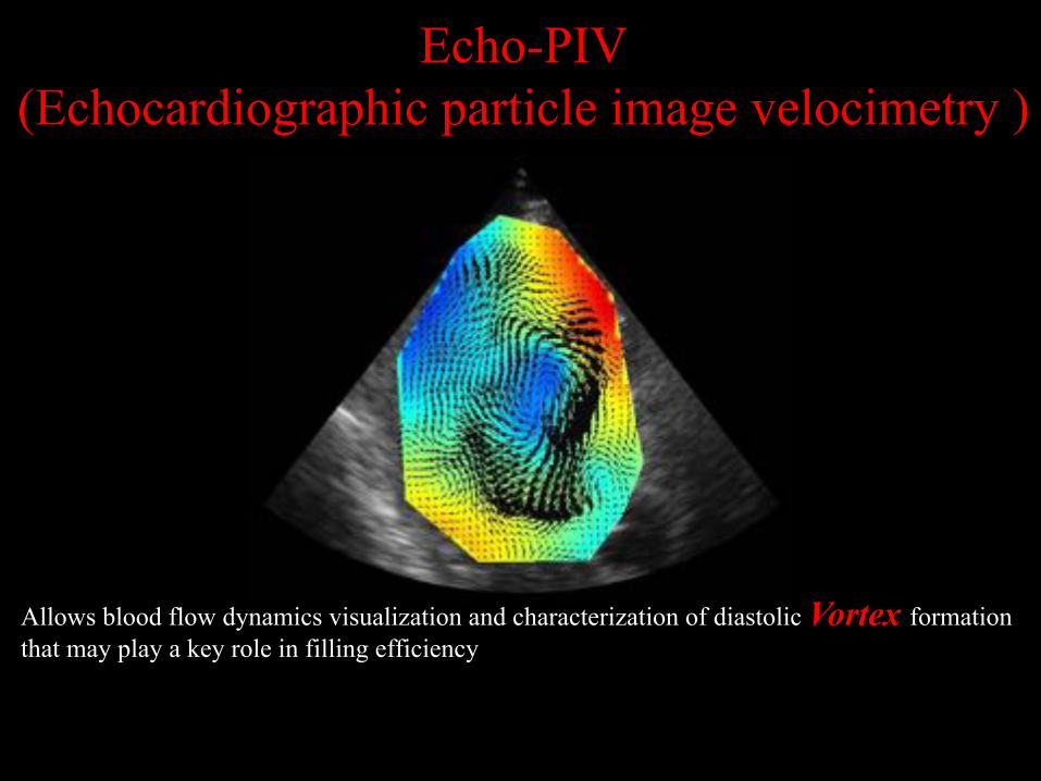
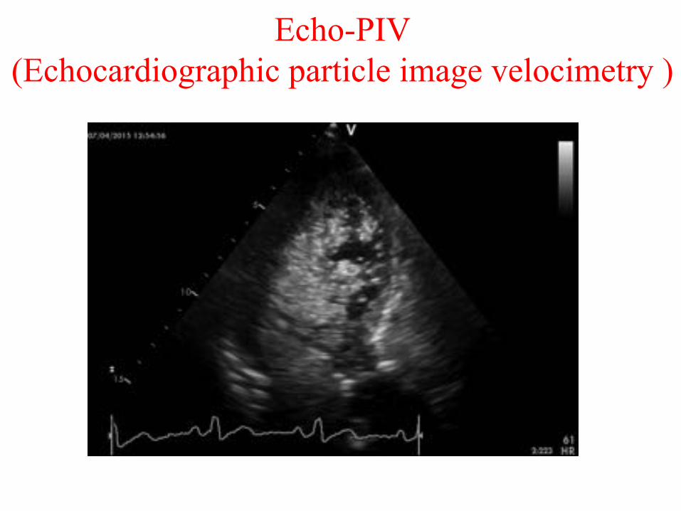
$$Echo-PIV
(Echocardiographic particle image velocimetry )
Allows blood flow dynamics visualization and characterization of diastolic Vortex formation that may play a key role in filling efficiency
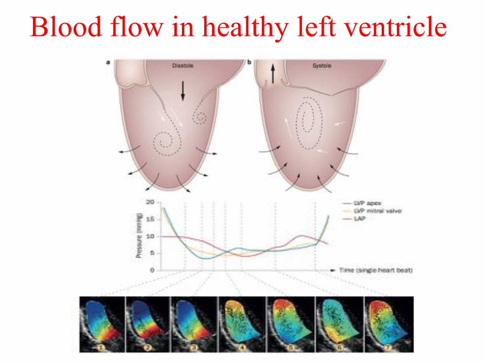
Blood flow in healthy left ventricle
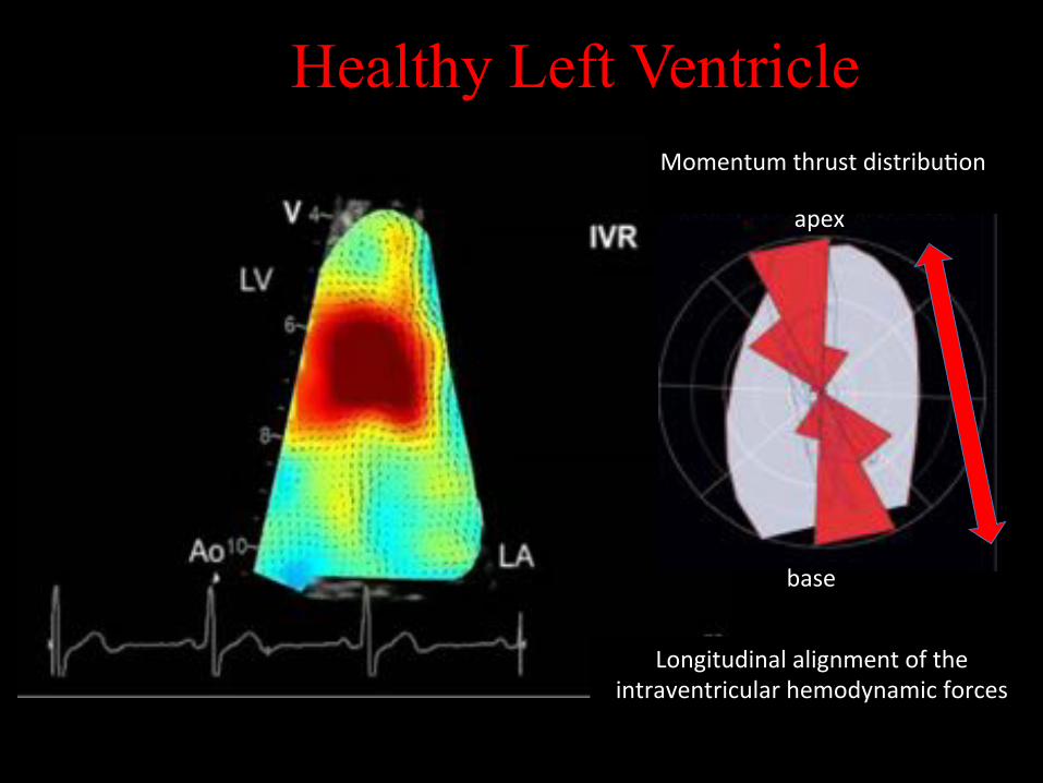
Healthy Left Ventricle
apex$
base$
$Momentum$thrust$distribu*on$$
Longitudinal$alignment$of$the$intraventricular$hemodynamic$forces$$
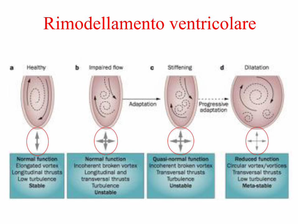
$$Rimodellamento ventricolare
$$$
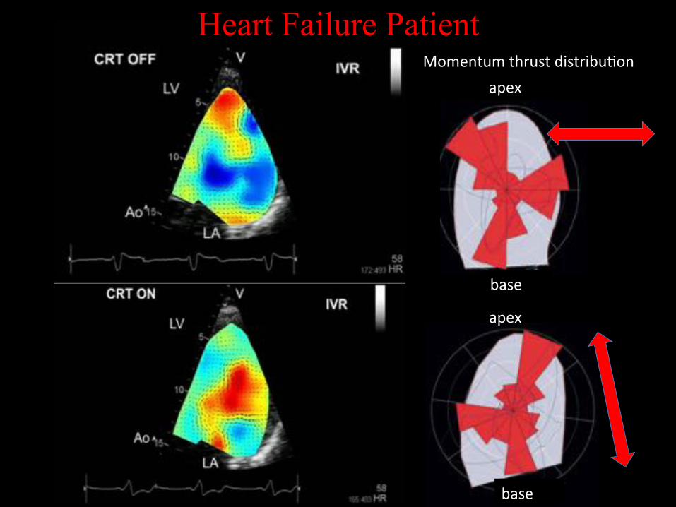
Heart Failure Patient
apex$
base$
Momentum$thrust$distribu*on$
apex$
base$
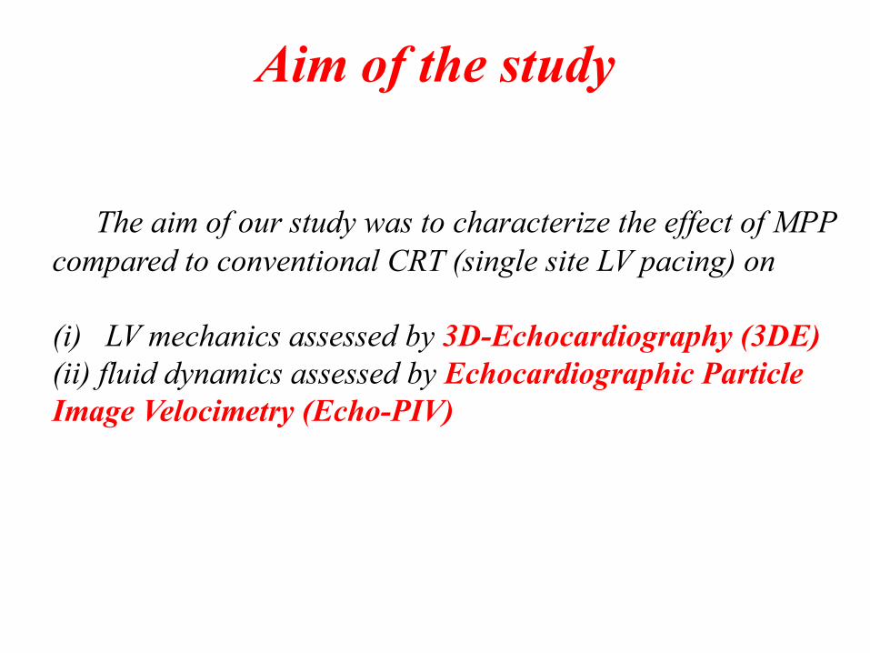
Aim of the study
The aim of our study was to characterize the effect of MPP
compared to conventional CRT (single site LV pacing) on (i) LV mechanics assessed by 3D-Echocardiography (3DE) (ii) fluid dynamics assessed by Echocardiographic Particle Image Velocimetry (Echo-PIV) $
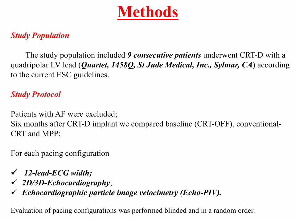
Methods Study Population
The study population included 9 consecutive patients underwent CRT-D with a quadripolar LV lead (Quartet, 1458Q, St Jude Medical, Inc., Sylmar, CA) according to the current ESC guidelines. Study Protocol Patients with AF were excluded; Six months after CRT-D implant we compared baseline (CRT-OFF), conventional-CRT and MPP; For each pacing configuration " 12-lead-ECG width; " 2D/3D-Echocardiography; " Echocardiographic particle image velocimetry (Echo-PIV). Evaluation of pacing configurations was performed blinded and in a random order.
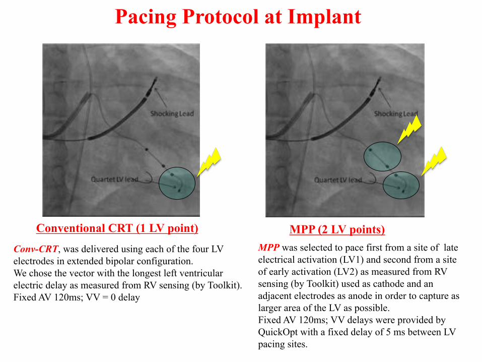
Pacing Protocol at Implant
Conventional CRT (1 LV point) MPP (2 LV points)
MPP was selected to pace first from a site of late electrical activation (LV1) and second from a site of early activation (LV2) as measured from RV sensing (by Toolkit) used as cathode and an adjacent electrodes as anode in order to capture as larger area of the LV as possible. Fixed AV 120ms; VV delays were provided by QuickOpt with a fixed delay of 5 ms between LV pacing sites.
Conv-CRT, was delivered using each of the four LV electrodes in extended bipolar configuration. We chose the vector with the longest left ventricular electric delay as measured from RV sensing (by Toolkit). Fixed AV 120ms; VV = 0 delay
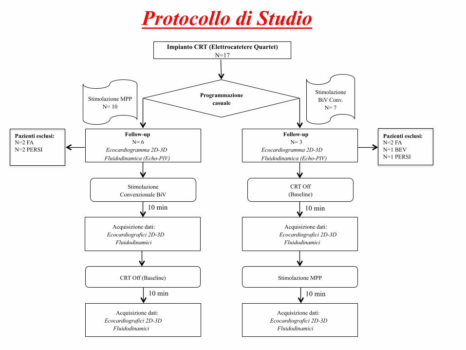
Programmazione casuale
Impianto CRT (Elettrocatetere Quartet) N=17
Follow-up N= 6 Ecocardiogramma 2D-3D
Fluidodinamica (Echo-PIV)
Stimolazione MPP N= 10
Stimolazione Convenzionale BiV
Stimolazione MPP
Follow-up N= 3
Ecocardiogramma 2D-3D Fluidodinamica (Echo-PIV)
CRT Off
(Baseline) $$
Acquisizione dati: Ecocardiografici 2D-3D
Fluidodinamici
Acquisizione dati: Ecocardiografici 2D-3D
Fluidodinamici
CRT Off (Baseline)
Acquisizione dati: Ecocardiografici 2D-3D
Fluidodinamici
Acquisizione dati: Ecocardiografici 2D-3D
Fluidodinamici
Stimolazione BiV Conv.
N= 7
10 min
10 min 10 min
10 min
Protocollo di Studio
Pazienti esclusi: N=2 FA N=2 PERSI
Pazienti esclusi:!N=2 FA N=1 BEV N=1 PERSI
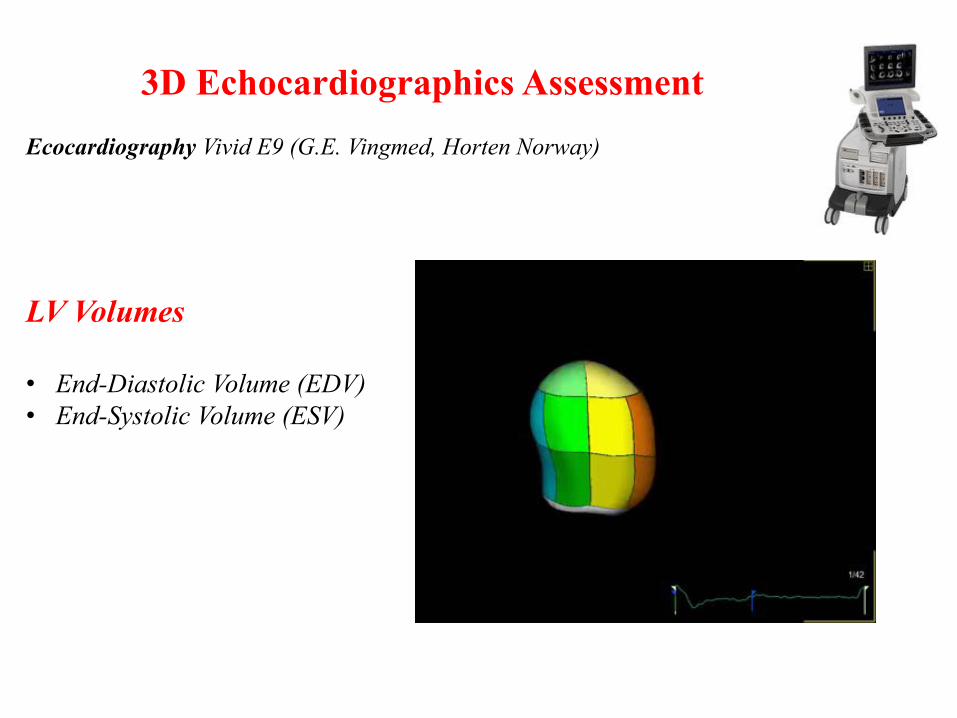
3D Echocardiographics Assessment
Ecocardiography Vivid E9 (G.E. Vingmed, Horten Norway)
LV Volumes • End-Diastolic Volume (EDV) • End-Systolic Volume (ESV)
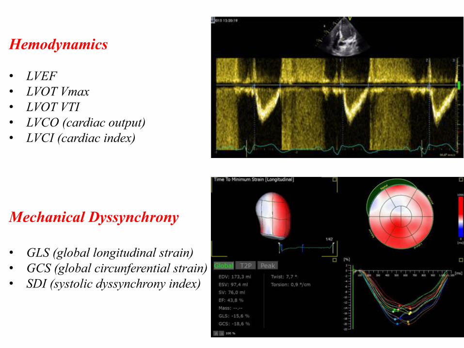
Hemodynamics • LVEF • LVOT Vmax • LVOT VTI • LVCO (cardiac output) • LVCI (cardiac index)
Mechanical Dyssynchrony • GLS (global longitudinal strain) • GCS (global circunferential strain) • SDI (systolic dyssynchrony index)
Echo-PIV (Echocardiographic particle image velocimetry )
$$

$$

$$
Momentum thrust distribution of the intraventricular hemodynamic forces
apex$
base$
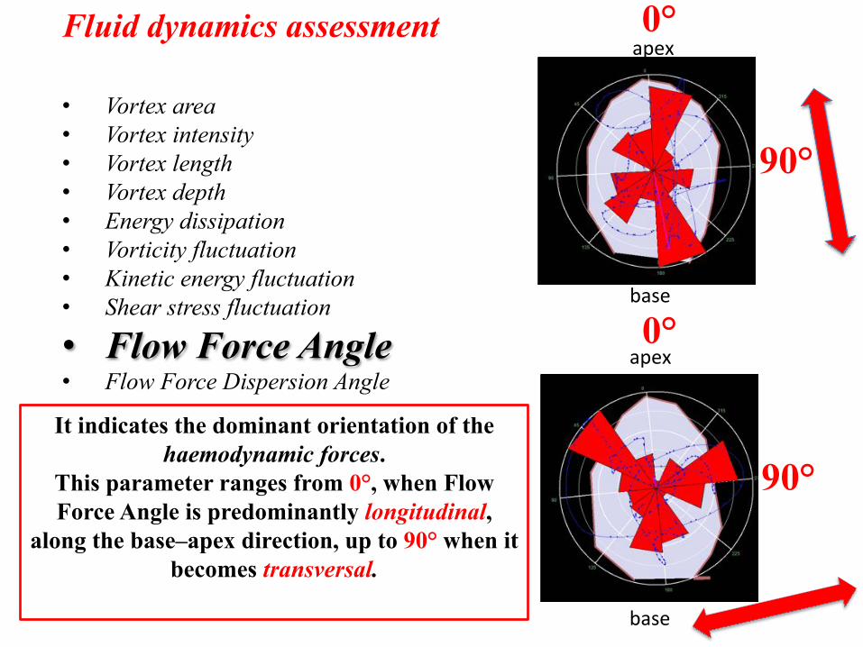
Fluid dynamics assessment • Vortex area • Vortex intensity • Vortex length • Vortex depth • Energy dissipation • Vorticity fluctuation • Kinetic energy fluctuation • Shear stress fluctuation
• Flow Force Angle • Flow Force Dispersion Angle
It indicates the dominant orientation of the haemodynamic forces.
This parameter ranges from 0°, when Flow Force Angle is predominantly longitudinal,
along the base–apex direction, up to 90° when it becomes transversal.
0°
90°
0°
90°
apex$
base$
apex$
base$
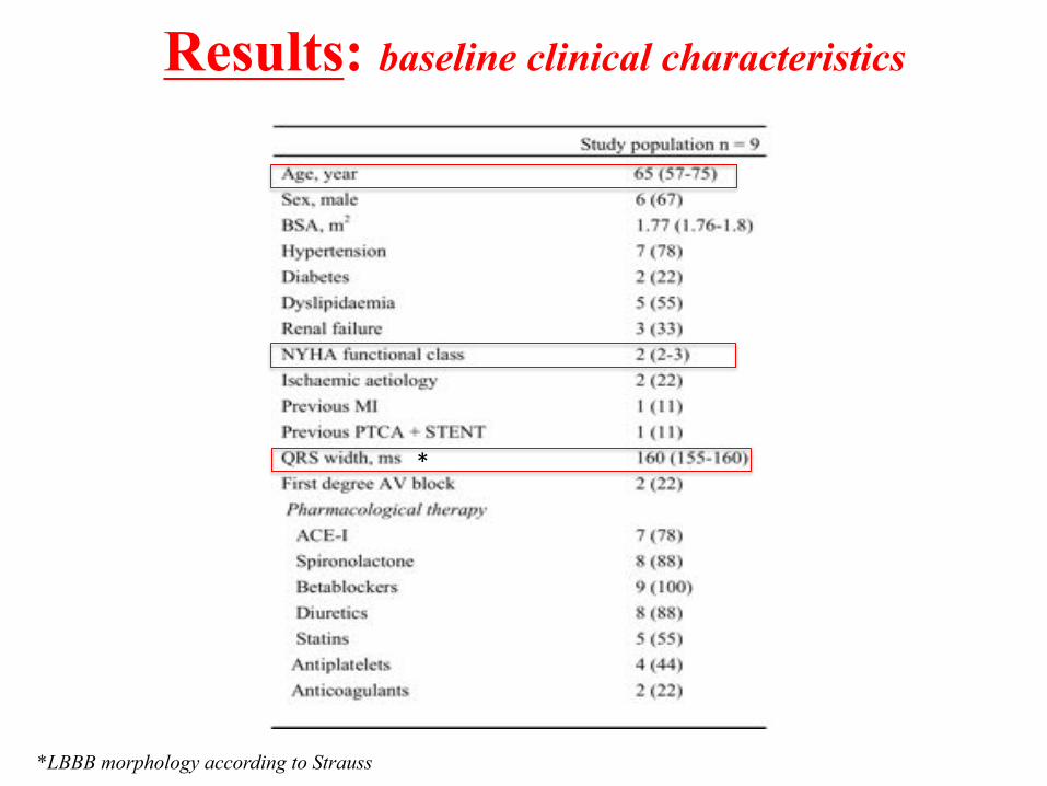
Results: baseline clinical characteristics
$*LBBB morphology according to Strauss
*
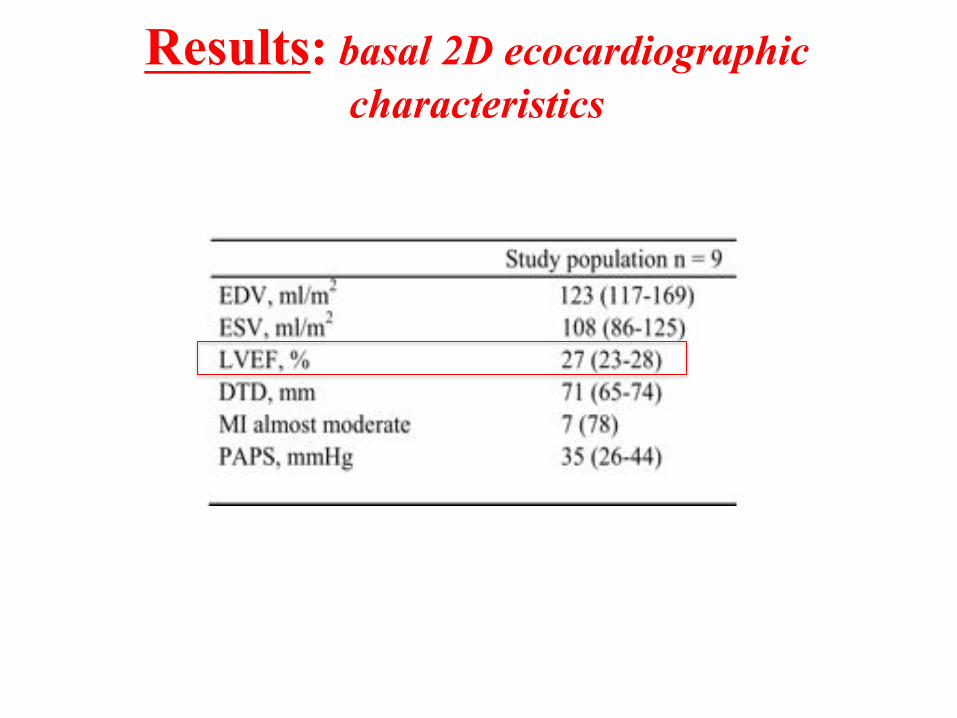
Results: basal 2D ecocardiographic characteristics
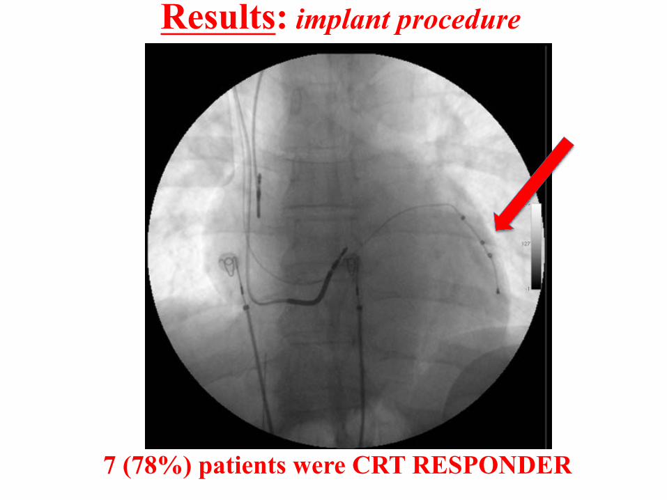
Results: implant procedure
7 (78%) patients were CRT RESPONDER
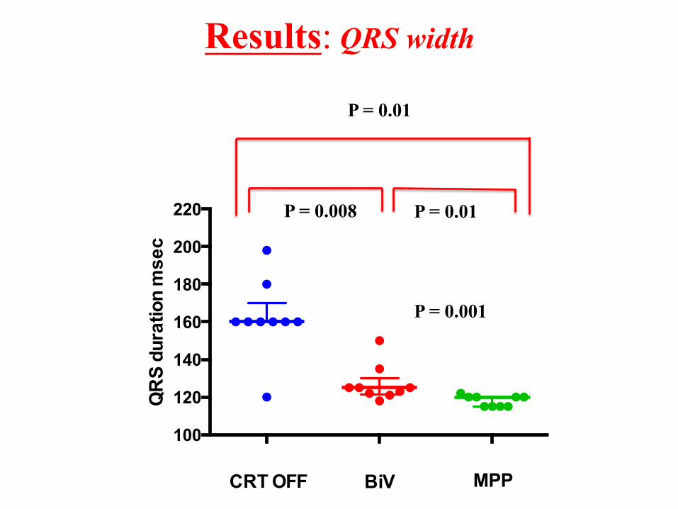
Results: QRS width
100
120
140
160
180
200
220
BiV
QR
S du
ratio
n m
sec
CRT OFF MPP
P = 0.001
P = 0.01
P = 0.008 P = 0.01
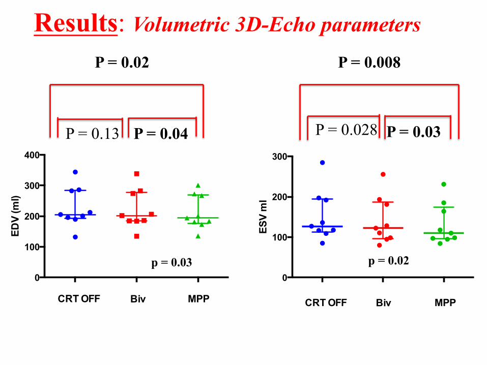
Results: Volumetric 3D-Echo parameters
0
100
200
300
400
EDV
(ml)
CRT OFFBivMPP
CRT OFF Biv MPP
0
100
200
300
Biv
ESV
ml
CRT OFF MPP
p = 0.03
P = 0.008
P = 0.028 P = 0.03
p = 0.02
P = 0.02
P = 0.13 P = 0.04
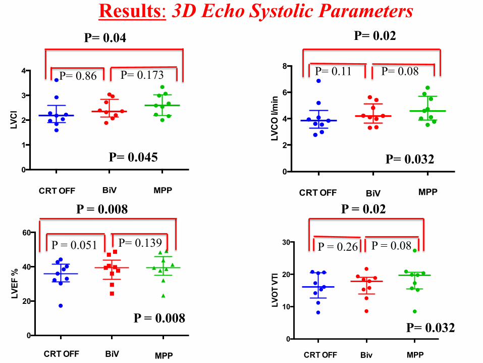
Results: 3D Echo$Systolic Parameters$
0
1
2
3
4
BiV
LVC
I
CRT OFF MPP
0
2
4
6
8
BiV
LVC
O l/m
in
CRT OFF MPP
0
10
20
30
Biv
LVO
T VT
I
CRT OFF MPP
P= 0.032
P = 0.26 P = 0.08
P= 0.032
P= 0.86 P= 0.173
P= 0.04
P= 0.045
P= 0.11 P= 0.08
P= 0.02
P = 0.02
0
20
40
60
BiV
LVEF
%
CRT OFF MPP
P = 0.008
P = 0.008
P = 0.051 P= 0.139
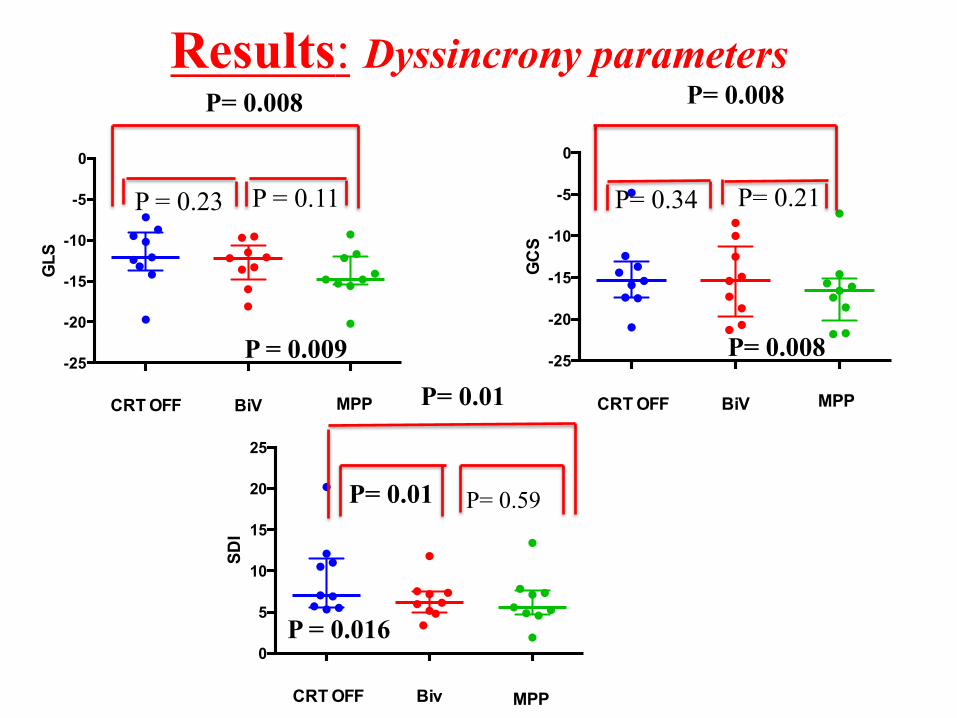
Results: Dyssincrony parameters
0
5
10
15
20
25
Biv
SDI
CRT OFF MPP
P= 0.008
P = 0.23 P = 0.11
P= 0.008
P = 0.016
P= 0.01
P= 0.01 P= 0.59
-25
-20
-15
-10
-5
0
BiV
GLS
CRT OFF MPP
P = 0.009
P = 0.23 P = 0.11
-25
-20
-15
-10
-5
0
BiV
GC
S
CRT OFF MPP
P= 0.34 P= 0.21
P= 0.008
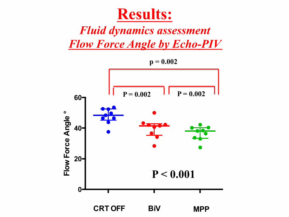
Results: Fluid dynamics assessment
Flow Force Angle by Echo-PIV!
0
20
40
60
BiV
Flow
For
ce A
ngle
°
CRT OFF MPP
p = 0.002
P = 0.002 P = 0.002
P < 0.001
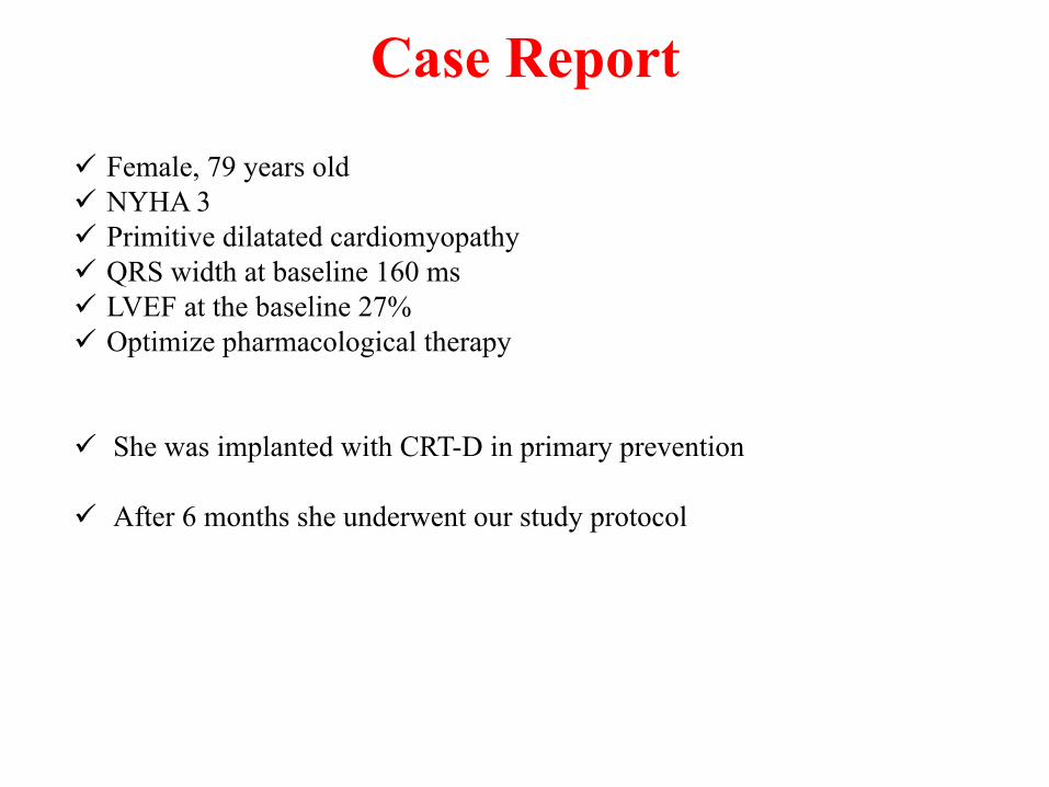
Case Report
" Female, 79 years old " NYHA 3 " Primitive dilatated cardiomyopathy " QRS width at baseline 160 ms " LVEF at the baseline 27% " Optimize pharmacological therapy
" She was implanted with CRT-D in primary prevention " After 6 months she underwent our study protocol $
$$
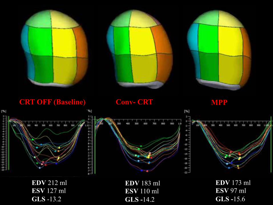
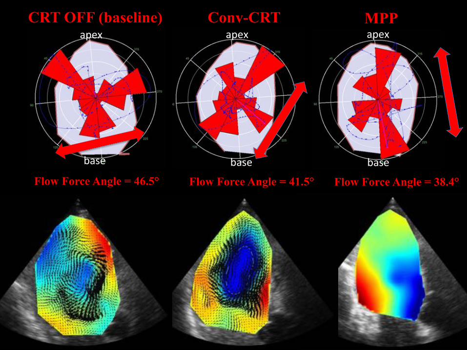
CRT OFF (Baseline) Conv- CRT MPP
EDV 212 ml ESV 127 ml GLS -13.2
EDV 183 ml ESV 110 ml GLS -14.2
EDV 173 ml ESV 97 ml GLS -15.6
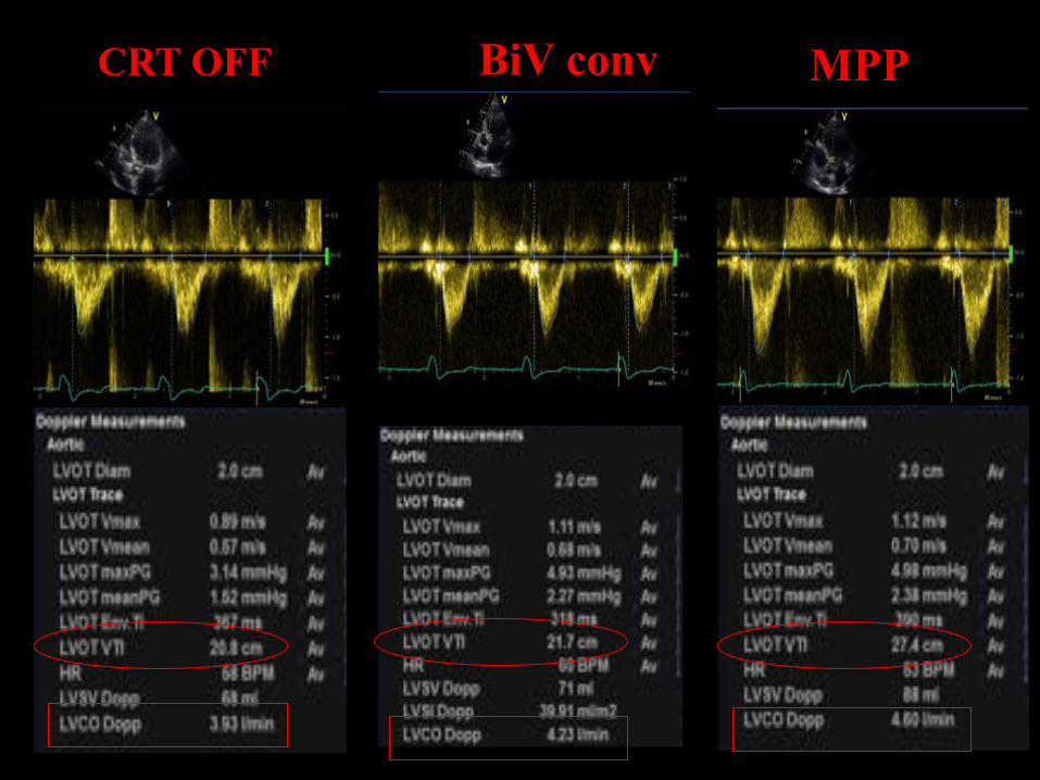
CRT OFF BiV conv MPP
$$
Flow Force Angle = 46.5° Flow Force Angle = 41.5° Flow Force Angle = 38.4°
CRT OFF (baseline) Conv-CRT MPP apex$apex$apex$
base$base$ base$
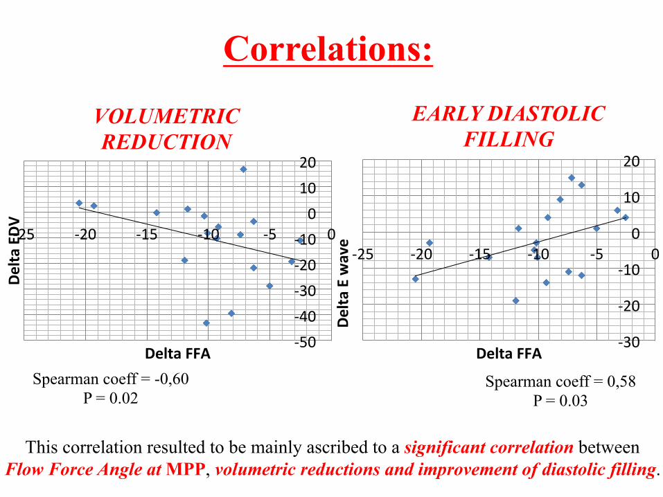
Spearman coeff = -0,60 P = 0.02
[50$[40$[30$[20$[10$0$
10$20$
[25$ [20$ [15$ [10$ [5$ 0$
Delta!EDV!
Delta!FFA!
Correlations:
[30$
[20$
[10$
0$
10$
20$
[25$ [20$ [15$ [10$ [5$ 0$
Delta!E!wave!
Delta!FFA!
Spearman coeff = 0,58 P = 0.03
VOLUMETRIC REDUCTION
EARLY DIASTOLIC FILLING
This correlation resulted to be mainly ascribed to a significant correlation between Flow Force Angle at MPP, volumetric reductions and improvement of diastolic filling.
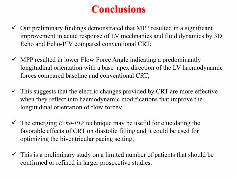
Conclusions
" Our preliminary findings demonstrated that MPP resulted in a significant improvement in acute response of LV mechnanics and fluid dynamics by 3D Echo and Echo-PIV compared conventional CRT;
" MPP resulted in lower Flow Force Angle indicating a predominantly
longitudinal orientation with a base–apex direction of the LV haemodynamic forces compared baseline and conventional CRT;
" This suggests that the electric changes provided by CRT are more effective
when they reflect into haemodynamic modifications that improve the longitudinal orientation of flow forces;
" The emerging Echo-PIV technique may be useful for elucidating the favorable effects of CRT on diastolic filling and it could be used for optimizing the biventricular pacing setting;
" This is a preliminary study on a limited number of patients that should be
confirmed or refined in larger prospective studies.

\$
Thank you…
$$
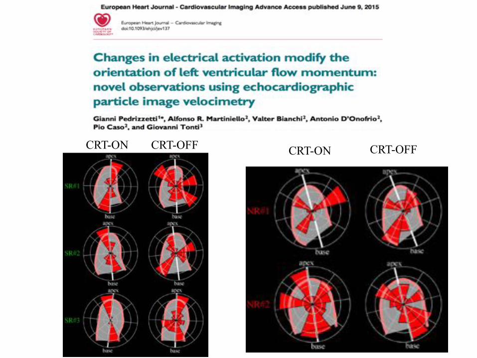
CRT-ON CRT-OFF CRT-ON CRT-OFF


![· LV 01 - LV 02 - 14 - LV LV Of - LV - LV - LV - Skat Foru Out] Profil PM E-Mail Q Pik, Grand? 1272 x) Vorhand ist dran nach passe pa s se. Nach Skatauffiahme:](https://static.cupdf.com/doc/110x72/5e0d1071f8f59d3156471103/lv-01-lv-02-14-lv-lv-of-lv-lv-lv-skat-foru-out-profil-pm-e-mail-q.jpg)


![[Violão 7 Cordas] Bertaglia - Relacao de Acordes 7 Cordas](https://static.cupdf.com/doc/110x72/577cb4e11a28aba7118cc18e/violao-7-cordas-bertaglia-relacao-de-acordes-7-cordas.jpg)