Ca2+-dependent K+ channels from rat olfactory cilia characterizedin planar lipid bilayers
Karen Castilloa,b, Juan Bacigalupoa,b, Daniel Wolffa,*
a Department of Biology, Faculty of Sciences, University of Chile, P.O. Box 653, Santiago, Chileb Millennium Institute for Advanced Studies in Cell Biology and Biotechnology, University of Chile, P.O. Box 653, Santiago, Chile
Abstract Olfactory cilia contain cyclic nucleotide-gated andCa2+-dependent Cl� conductances that underlie excitatorychemotransduction, and a Ca2+-dependent K+ (KCa) conduc-tance, apparently involved in inhibitory transduction. Previoussingle-channel patch-clamp studies on olfactory cilia revealedfour different KCas, with different conductances and kinetics.Here, we further characterized these channels in planar bilayers,where blockers could be properly tested. All four ciliary KCaswere observed: The 16 pS channel, K0.5,Ca = 40 lM and apa-min-sensitive; the 30 and 50 pS channel, K0.5,Ca = 59 lM, clo-trimazole-sensitive and charybdotoxin-insensitive; the 60 pSchannel, clotrimazole-sensitive and charybdotoxin-insensitive;and the 210 pS channel, K0.5,Ca = 63 lM, blocked by charybdo-toxin and iberiotoxin. The presence of the 16 and 210 pS chan-nels was confirmed by immunoblotting.
Keywords: Odor transduction; Olfactory cilia;Calcium-activated potassium channel; Lipid bilayer
1. Introduction
Olfactory receptor neurons (ORNs) have a conspicuous
morphology, which is conserved across most vertebrate spe-
cies. Their cell bodies project a thin, unmyelinated axon to
the olfactory bulb and a single dendrite that reaches the
mucosal epithelial surface, where it swells to form the den-
dritic knob. From the knob emanate a number of cilia, the
organelles where transduction takes place. Indeed, all molec-
ular components involved in transduction reside in the cilia.
Three ionic conductances have been found in the ciliary
membrane: a cyclic nucleotide-gated (CNG) conductance
[1], a Ca2+-dependent Cl� (ClCa) conductance [2] and a
Ca2+-dependent K+ (KCa) conductance [3,4]. Odorants trigger
a cAMP cascade that leads to the opening of cationic CNG
channels, resulting in a depolarizing current. Calcium enters
the cilia through the CNG channel and activates ClCa chan-
nels, allowing an efflux of Cl� that further depolarizes the
neuron, increasing its discharge rate [5]. In contrast, the
*Corresponding author. Fax: +56 2 678 7435.E-mail address: [email protected] (D. Wolff).
KCa conductance appears to have the opposite effect, which
is to hyperpolarize the cell causing a reduction in the firing
rate [3].
The CNG channel has been characterized by whole cell
and single-channel recordings from the dendritic knob [5].
It has been cloned and expressed in heterologous systems
[6,7]. Its unitary conductance ranges from 12 to 55 pS,
depending on the species [5], and it is sensitive to Ca2+ chan-
nel blockers such as l-cis Diltiazem and LY83583 [8]. No
toxins that block this channel have been reported. The ClCachannel, in contrast, is poorly characterized and its primary
sequence remains unknown. The channel unitary conduc-
tance has been estimated by noise analysis to be in the order
of 0.8 pS [9] and it is blocked by niflumic acid, DIDS and
SITS [2,10].
In a recent patch-clamp study [4] conducted in membrane
patches excised from toad olfactory cilia, four KCa channel
types were identified: a high-conductance channel (BKCa;
210 pS), two intermediate conductance channels (IKCa, one
channel of 60, and another one of 30 and 60 pS) and a
small-conductance channel (SKCa, 15 pS). The pharmacolog-
ical profiles of such channels were not investigated, mainly
because charybdotoxin (ChTx) and iberiotoxin (IbTx),
well-known blockers of BKCa channels, and apamin, a toxin
that blocks some SKCa channels, exert their effects from the
extracellular aspect of the membrane. Macroscopic data had
shown that both ChTx and IbTx partially block the odor-
induced KCa current, presumably by affecting the BKCa con-
ductance [[10]; Madrid et al., unpublished results]. The study
of the effects of toxins upon KCa channels is not possible in
membrane patches, because there is no access to their exter-
nal (extracellular) side, located inside the pipette. Outside-
out patches cannot be obtained with the extremely small
patch pipettes required to record from the cilia. To over-
come this difficulty, we used the planar lipid bilayer tech-
nique, where reconstituted channels derived from a ciliary
membrane preparation are accessible to pharmacological
analysis from both sides of the membrane. In this study,
we present results of the biophysical and pharmacological
characterization of the KCa channels from a purified olfac-
tory cilia fraction, incorporated into planar phospholipid
bilayers. Our results confirm the presence of small, interme-
diate and high-conductance KCa channels in olfactory cilia
and support the notion that they might be involved in ver-
tebrate odor-transduction. Also, they would allow to deter-
mine the relative contribution of each KCa channel subtype
to net ciliary odor-triggered KCa inhibitory current.

K. Castillo et al.
2. Materials and methods
2.1. Preparation of a ciliary membrane fraction from rat olfactory
neuronsMembranes were obtained using a modification of method described
by Washburn and co-workers [11], which involves mechanical ruptur-ing, sonication and purification in sucrose gradients. Adult Sprague–Dawley rats were sacrificed with anaesthesia overdose (pentobarbitone60 mg/kg) and nasal olfactory epithelia were surgically removed andsuspended in saline (solution 1) containing (in mM): 145 NaCl, 5KCl, 1.6 K2HPO4, 2 MgSO4, 7.5 DD-glucose, 20 HEPES, pH 7.4, sup-plemented with a cocktail of protease inhibitors containing aprotinin,leupeptin, pepstatin, benzamidin and PMSF. All steps were carried outat 4 �C. The suspension was gently rocked for 20 min and centrifugedfor 10 min at 1500 · g. The supernatant recovered and the pellet wasresuspended in solution 1 supplemented with 1 mM EDTA (solution2) and sonicated using a microprobe at low power for 10 s. The tissuewas allowed to settle and the supernatant was recovered. The pelletwas resuspended in solution 2, sonicated at low power for 30 s andthe supernatant recovered. All the supernatants were pooled togetherand centrifuged at 1500 · g for 10 min. The supernatant was loadedin a discontinuous sucrose gradient formed by a 20% and a 43% su-crose layers and centrifuged at 100 000 · g for 1 h. The band in the gra-dient interface was extracted, diluted 10-fold and centrifuged at100 000 · g for 1 h. The pellet containing the cilia was resuspendedin solution 1 supplemented with 2 mM EGTA and 0.5 mM sucrose, ali-quoted and stored at �70 �C. Protein content was determined with themethod of Hartree [12].
2.2. Western blot analysisPurified olfactory cilia membranes were separated in 8% SDS–
PAGE gels, electrotransferred (Mini-Trans Blot System; Bio-Rad,Hercules, CA) to nitrocellulose membranes (Hybond ECL; Amer-sham-Pharmacia, Piscataway, NJ) at 4 �C and incubated with 5%non-fat milk or 3% BSA in Tris-buffered saline (TBS) with 0.05%Tween 20 (TBS-T) for 1 h at room temperature, to block unspecificsites. Membranes were then incubated 2 h at room temperature, orovernight at 4 �C, with the particular antibody: a polyclonal anti-BKCa
channel recognizing a C-terminus intracellular epitope present in ver-tebrate BKCa channels (Alomone Labs, Jerusalem, Israel), a polyclonalanti-SKCa channel recognizing an intracellular N-terminus sequencepresent in mammals (Alomone Labs, Jerusalem, Israel), a monoclonalanti-voltage-dependent Na+ channel recognizing an intracellular III–IV loop sequence present in vertebrate Na+ channels (Sigma–Aldrich,St. Louis, MO), anti CNG channel recognizing an intracellular C-ter-minus sequence, present in mammals (Alomone Labs, Jerusalem,Israel) or a polyclonal anti-adenylyl cyclase III raised against a peptidemapping at the carboxy-terminus of rat origin adenylyl cyclase III(ACIII, Santa Cruz Biotechnology, Santa Cruz, CA). The membraneswere then developed with a chemiluminescence assay (ECL LumigenPS-3 Detection Reagent; Amersham-Pharmacia, Piscataway, NJ)using horseradish peroxidase-conjugated with the corresponding sec-ondary antibody (anti rabbit for BKCa, SKCa and CNG channels,and anti-mouse for Na+ channel and ACIII).
Fig. 1. Enrichment and purity of the ciliary membrane fraction.(A) Western blots of olfactory cilia (C), deciliated olfactory epithelium(DE), olfactory bulb (OB), whole olfactory epithelium (WE) and brain(B) membrane fractions labelled with anti-ACIII (1:1000) antibodies.Brain membranes were used as negative control. The tracks on theright-hand side correspond to the controls without the primaryantibodies. (B) Western blots of olfactory cilia, deciliated olfactoryepithelium and brain membranes (negative control) labelled with anti-CNG channel antibody (1:600). (C) Western blots of olfactory cilia,deciliated olfactory epithelium, olfactory bulb, and brain (positivecontrol) fractions labelled with anti-voltage-dependent Na+ channelantibodies (1:300). The controls without primary antibodies are shownin the Western blot at the right in A and C. All lanes (A–C) wereloaded with 50 lg protein.
2.3. Single-channel recording in planar phospholipid bilayersBilayers were formed by applying a drop of palmitoyl-oleoyl
phosphatidylethanolamine (POPE) or a mixture of POPE and palmi-toyl-oleoyl phosphatidylcholine (POPC) 4:1 (Avanti Polar Lipids,Birmingham, AL) dissolved in n-decane (20 mg/ml), to a 200 lm diam-eter hole in a teflon cup separating two saline compartments. Channelinsertion occurred spontaneously after touching the bilayer with adroplet of highly purified ciliary membranes. Incorporation of singleionic channels was indicated by the occurrence of discrete current fluc-tuations as a constant voltage difference was applied across the bilayer.After the incorporation of a channel, single KCa channel currents wererecorded for a few minutes (control) in symmetric 100 mM KCl or K-Acetate (KAc), or 100/20 mM KAc, 5 mM HEPES-K+, pH 7.0. FreeCa2+ concentration in the solution was set at the desired values usingvariable amounts of CaCl2 and EGTA. Free Ca2+ concentrations werecalculated with the WinMAXC v2.05 computer program. Single-chan-nel currents were recorded with an 8900 Dagan patch-clamp amplifier(Dagan Corp, Minneapolis, MN). Voltage pulses were applied to thecis compartment and the current was recorded from the trans side,
through Ag/AgCl electrodes connected to the saline compartmentswith 1 M KCl agar bridges [13]. The current was monitored by anoscilloscope, amplified, digitized (VR-10 PCM, lnstrutech Corp, NY)and stored in videotape or in the HDD of a PC, interfaced by a Lab-Master data acquisition board (Scientific Solutions, Solon, OH) andanalyzed with the pClamp 6 software (Axon Instruments, Union City,CA). Experiments were carried out at room temperature (20 ± 2 �C).Chemicals were purchased from Sigma Chemical (St. Louis, MO)and Merck (Darmstadt, Germany), unless otherwise indicated.Toxin stock solutions were prepared by dissolving the lyophilized
compounds (Alomone Labs, Jerusalem, Israel), in bidistilled water atthe following final concentrations: apamin 100 lM, IbTx and ChTX10 lM, aliquoted and stored at �20 �C until used. Clotrimazole(Sigma Chemical, St. Louis, MO) was dissolved in DMSO. Vehicle(water or DMSO) addition controls were carried out. Calcium and tox-ins were added to the solution facing the intracellular side and extracel-lular side of the channel, respectively. KCa channel orientation in thebilayer was determined by applying voltage pulses of opposite polarityor by checking from which side of the chamber Ca2+ increases channelactivity.
3. Results
3.1. Purity of the olfactory cilia membrane fraction
To carry out our study of the ciliary K+ channels in planar
lipid bilayers, it was crucial to obtain a highly purified olfac-
tory cilia membrane preparation. The degree of enrichment
and purity of the preparation in ciliary membranes was as-
sessed by the Western blot method. The enrichment was tested
with antibodies against the ACIII and the CNG channel,
membrane proteins that specifically localize to these organ-
elles. The anti-ACIII antibody strongly labelled band in the cil-
iary and whole cell epithelium fractions, but these bands were
less intense in deciliated olfactory epithelium and olfactory
bulb, and were not observed in the brain fraction, used as neg-
ative control (Fig. 1A). A band in the ciliary fraction was
strongly reactive to the anti-CNG channel antibody, being ab-

K. Castillo et al.
sent in the deciliated epithelium and brain fractions (Fig. 1B).
To test for purity of the ciliary membrane fraction with other
membranes, we used an antibody against the voltage-depen-
dent Na+ channel, since this membrane protein is present in
the cell body and dendrite, but is not expected to be found
in the cilia. Accordingly, the antibody did not label the ciliary
fraction, but it reacted with the deciliated epithelium and a
brain fraction used as positive control (Fig. 1C).
3.2. Single-channel recordings
Fusion of purified olfactory ciliary membranes to planar li-
pid bilayers induced the incorporation of several KCa channels
of small, intermediate and high conductance.
3.3. Small conductance Ca2+-dependent K+ (SKCa) channels
The open probability (P0) of this channel was not apprecia-
bly affected by voltage, but it was dependent on Ca2+ concen-
tration. Fig. 2A illustrates representative unitary currents of a
SKCa channel from the purified ciliary fraction recorded at
60 mV in symmetrical 100 mM KAc and at different free
Ca2+ concentrations. The channel remained almost silent at
5 lM Ca2+, but its P0 increased to 0.3 upon elevating free cal-
Fig. 2. SKCa channel. (A) SKCa channel unitary currents records at three d(B) P0 versus Ca
2+ concentration plot for the SKCa channel at +60 mV. Expen = 3.2; (C) Representative I–V curve, determining a 16 pS slope conductanchannel currents recorded in symmetrical 100 mM KAc, 50 lM Ca2+, at +60Western blotting. The anti-SK3 antibody was used at a 1:600 dilution. Denervas positive and negative controls, respectively. All lanes were loaded with 50
cium to 20 lM, approaching a maximum value of �0.6 at
80 lM Ca2+. The P0 versus Ca2+ concentration curve obtained
with the data of four experiments, determined a K0.5 of
39.8 lM (Fig. 2B). From the I–V relations, a slope conduc-
tance of 16.3 ± 0.7 pS (mean ± S.E.M., n = 6) was determined
for this channel (Fig. 2C). To pharmacologically characterize
this SKCa channel, we tested the effect of apamin, a bee toxin
that blocks SK3, a subfamily of SKCa channels [14]. As illus-
trated in Fig. 2D, the channel is greatly blocked by 22 nM
apamin.
To further assess the presence of the SKCa channel in the rat
olfactory cilia fraction, we carried out a Western blot analysis
using a polyclonal antibody against the SK3 channel. A single
band reacted with the antibody in the ciliary membrane frac-
tion (Fig. 2E) indicating the presence of this SKCa channel sub-
type in the cilia. The band was also present in the deciliated
epithelia and olfactory bulb fractions. Since this SKCa channel
is differentially expressed in denervated and normal skeletal
muscle [15], we used membrane fractions from each muscle
condition as positive and negative controls, respectively. As ex-
pected, the denervated muscle lane exhibited a reactive band
corresponding to SKCa.
ifferent Ca2+ concentrations at +60 mV in symmetrical 100 mM KAc.rimental points were fitted to a Hill equation, with K0.5 = 39.8 lM andce. (D) SKCa channel activity was inhibited by 22 nM apamin. Single-mV. (E) Detection of the SK3 channel in a purified ciliary fraction byated (DM) and innervated (IM) rat skeletal muscle fractions were usedlg protein.
K. Castillo et al.
3.4. Intermediate conductance Ca2+-dependent K+ (IKCa)
channels
In our bilayer study, we identified two KCa channels with
conductances in the intermediate range, referred to as IKCa-1
and IKCa-2.
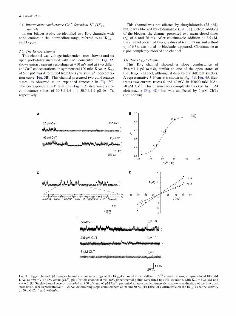
3.5. The IKCa-1 channel
This channel was voltage independent (not shown) and its
open probability increased with Ca2+ concentration. Fig. 3A
shows unitary current recordings at +50 mV and at two differ-
ent Ca2+ concentrations, in symmetrical 100 mM KAc. A K0.5
of 59.5 lMwas determined from the P0 versus Ca2+ concentra-
tion curve (Fig. 3B). This channel presented two conductance
states, as observed at an expanded timescale in Fig. 3C.
The corresponding I–V relations (Fig. 3D) determine slope
conductance values of 30.3 ± 1.4 and 50.5 ± 1.9 pS (n = 7),
respectively.
Fig. 3. IKCa-1 channel. (A) Single-channel current recordings of the IKCa-1KAc, at +50 mV. (B) P0 versus [Ca
2+] plot for this channel at +50 mV. Expen = 6.6. (C) Single-channel currents recorded at +50 mV and 65 lM Ca2+, prestate levels. (D) Representative I–V curve, determining slope conductances ofat 50 lM Ca2+ and +60 mV.
This channel was not affected by charybdotoxin (25 nM),
but it was blocked by clotrimazole (Fig. 3E). Before addition
of the blocker, the channel presented two mean closed times
(sc) of 6 and 26 ms. After clotrimazole addition at 2.5 lM,
the channel presented two sc values of 6 and 37 ms and a third
sc of 6.3 s, attributed to blockade, appeared. Clotrimazole at
6 lM completely blocked the channel.
3.6. The IKCa-2 channel
This KCa channel showed a slope conductance of
59.6 ± 1.4 pS (n = 8), similar to one of the open states of
the IKCa-1 channel, although it displayed a different kinetics.
A representative I–V curve is shown in Fig. 4B. Fig. 4A illus-
trates two current traces 0 and 40 mV, in 100/20 mM KAc,
50 lM Ca2+. This channel was completely blocked by 1 lMclotrimazole (Fig. 4C), but was unaffected by 6 nM ChTx
(not shown).
channel at two different Ca2+ concentrations, in symmetrical 100 mMrimental points were fitted to a Hill equation, with K0.5 = 59.5 lM andsented in an expanded timescale to allow visualization of the two open30 and 50 pS. (E) Effect of clotrimazole on the IKCa-1 channel activity

Fig. 4. IKCa-2 channel. (A) Unitary current recordings of a IKCa-2channel at 0 and +40 mV in asymmetrical 100/20 mM KAc solutionsand 20 lM Ca2+. (B) Representative I–V curve, determining a 60 pSslope conductance. (C) Effect of 1 lM clotrimazole on the channel at50 lM Ca2+.
K. Castillo et al.
3.7. The high-conductance Ca2+-dependent K+ channel (BKCa)
This was the K+ channel that most frequently incorporated
into bilayers and showed the typical biophysical and pharma-
cological characteristics of BKCa channels from other tissues
[14]. Fig. 5A offers unitary current recordings of a BKCa chan-
nel in symmetrical 100 mM KAc, 50 lM Ca2+ and at three
different potentials. Channel P0 showed pronounced voltage-
dependence. Its I–V relationship yielded a slope conductance
of 210.4 ± 5.8 pS (Fig. 5B; n = 12). The Ca2+ dependence of
this ciliary BKCa channel is illustrated in Fig. 5C, where traces
of channel activity at 40 mV and at three different Ca2+ con-
centrations are presented. Fig. 5D displays the channel P0 as
a function of [Ca2+]. The curve shows the characteristic sigmoi-
dal shape, approaching a P0 of 1 above 100 lM Ca2+ and
determines a K0.5 of 63.2 lM. This channel was blocked by
IbTx, a specific inhibitor of BKCa channels, and of ChTx,
blocker of BKCa and other voltage-dependent K+ channels.
The sensitivities to these toxins varied widely among the BKCa
channels studied, although the concentrations that completely
blocked the channel were in the nanomolar range for both tox-
ins. Fig. 5E shows the effects of IbTx on the BKCa channel. It
can be appreciated that the toxin induced long lasting closed
states that increased in duration and frequency as a function
of its concentration. Prior to toxin addition the channel pre-
sented two mean closed times (sc) of 3.0 and 120 ms. After
the addition of 44 nM IbTx, the channel presented two sc val-ues of 3.0 and 180 ms and a third long sc of 3.5 s, correspond-ing to the blocked state, appeared. IbTx at 110 nM completely
blocked the channel. In a few cases, BKCa channels exhibited a
particularly high sensitivity to IbTx, being completely blocked
by 4 nM toxin, in contrast with the 110 nM IbTx needed to
block the channel in Fig. 5E. This BKCa channel was also sen-
sitive to ChTx. As illustrated in Fig. 5F, the addition of 11 nM
ChTx induced long lasting closed states. Before toxin addition,
this channel presented two sc values of 7.0 and 24 ms. Follow-
ing toxin application the channel presented two sc values of 6.0and 25 ms and a third mean closed time of 518 ms, ascribed to
the blocked state, appeared.
The presence of BKCa channels in the olfactory cilia fraction
was also examined by Western blotting, using a polyclonal
anti-BKCa channel antibody. Fig. 5G shows a band near 116
kDa that reacted with the antibody, confirming the presence
of the channel in the olfactory cilia indicated by the electro-
physiological data. This finding is also consistent with previous
results from our laboratory, where the presence of the BKCa
channel in the cilia was demonstrated with immunocytochem-
istry and Western blotting [4]. As expected, the protein was
also found in the deciliated epithelia and brain fractions, used
as positive controls. The extra bands in both ciliary and brain
fractions of the immunoblot, may correspond either to degra-
dation sub-products or to splice variants of the channel.
4. Discussion
Previous electrophysiological studies documented that odor-
ants induce inhibitory responses in isolated vertebrate olfac-
tory neurons, consisting of decreases in the spiking rate
[3,5,16,17]. It was also reported that the inhibitory response
appears to involve the activation of a ChTx-sensitive, hyperpo-
larizing KCa current [10]. This odorant-induced K+ current
was mediated by an increase in cytosolic Ca2+ concentration
due to an odorant-induced apical Ca2+ influx [18]. Evidence
from focal odor stimulation experiments on isolated ORNs
was consistent with the possibility that this odor-triggered
KCa conductance was present in the cilia [18].
The first single KCa channel recordings from the soma and
dendritic knob of mice ORNs were obtained by Maue and
Dionne [19] by means of the patch-clamp technique. The
authors identified a 130 pS voltage-dependent KCa channel
with a fast gating kinetics. Later, a planar lipid bilayer study
of a toad olfactory ciliary membrane fraction suggested the
presence of a 240 pS conductance BKCa channel (in symmet-
rical 200 mM KCl). Its K0.5 for Ca2+ was 46 lM and it was
blockable by 42 nM ChTx [20]. This channel was presum-
ably derived from the cilia, although the purity of the mem-
brane fraction was not assessed. In a recent work, Delgado
et al. [4] obtained single-channel recordings from inside-out
membrane patches excised from toad olfactory cilia. These
authors identified four different KCa channel types, of small
(12 pS), intermediate (one of 29 and 60 pS, and another one
of 60 pS) and one of large conductance (210 pS), all K+-
selective and sensitive to low micromolar Ca2+. However,
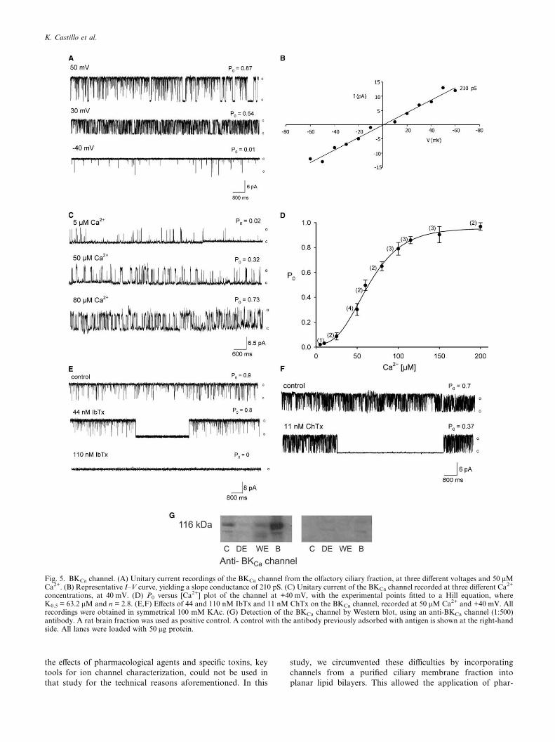
Fig. 5. BKCa channel. (A) Unitary current recordings of the BKCa channel from the olfactory ciliary fraction, at three different voltages and 50 lMCa2+. (B) Representative I–V curve, yielding a slope conductance of 210 pS. (C) Unitary current of the BKCa channel recorded at three different Ca2+
concentrations, at 40 mV. (D) P0 versus [Ca2+] plot of the channel at +40 mV, with the experimental points fitted to a Hill equation, whereK0.5 = 63.2 lM and n = 2.8. (E,F) Effects of 44 and 110 nM IbTx and 11 nM ChTx on the BKCa channel, recorded at 50 lM Ca2+ and +40 mV. Allrecordings were obtained in symmetrical 100 mM KAc. (G) Detection of the BKCa channel by Western blot, using an anti-BKCa channel (1:500)antibody. A rat brain fraction was used as positive control. A control with the antibody previously adsorbed with antigen is shown at the right-handside. All lanes were loaded with 50 lg protein.
K. Castillo et al.
the effects of pharmacological agents and specific toxins, key
tools for ion channel characterization, could not be used in
that study for the technical reasons aforementioned. In this
study, we circumvented these difficulties by incorporating
channels from a purified ciliary membrane fraction into
planar lipid bilayers. This allowed the application of phar-

Table 1Summary of properties of KCa channels from olfactory cilia mem-branes
SKCa IKCa-1 IKCa-2 BKCa
Conductance 16 pS 30 and 50 pS 60 pS 210 pSK0.5 for Ca
2+ 39.8 lM 59.5 lM ND 63.2 lMVoltagedependence
No No No Yes
Blocked by Apamin Clotrimazole Clotrimazole ChTx andIbTx
ND, not determined.
K. Castillo et al.
macological agents and toxins from either side of the
membrane.
Since our study required a highly purified membrane frac-
tion, we optimized a membrane preparation procedure that in-
volved mechanical rupture of the epithelia and a subsequent
series of centrifugation steps of the membrane fraction con-
taining the olfactory cilia [11]. To test the purity of the ciliary
membrane preparation, we conducted Western blot analysis
with antibodies specific to ciliary membrane proteins, such as
ACIII and the CNG channel, and to extra-ciliary membrane
proteins, such as the voltage-dependent Na+ channel. Within
the resolution limit of this method, the ciliary membrane frac-
tion exhibited no signs of contamination by other membranes.
Fusion of purified rat olfactory cilia membranes to planar li-
pid bilayers resulted in the incorporation of small, intermedi-
ate and high conductance KCa channels. From 108 successful
channel incorporations, 55%, corresponded to BKCa channels,
21% to SKCa channels, 15% to IKCa-1 channels and 9% to
IKCa-2 channels. These channels shared close similarities
regarding conductance and gating kinetics to those recorded
with the patch-clamp method in toad olfactory cilia [4]. Con-
sidering that the channels originate from different species, the
evidence is indicative that channels observed with both meth-
ods are virtually identical. A previous study, where odor-trig-
gered whole cell KCa current properties from the same species
were compared, arrived to the same conclusion [21]. Table 1
summarizes the main properties of the KCa channels identified
and characterized in bilayers.
The �16 pS KCa channel identified in the present study was
voltage-independent and apamin-sensitive, indicating that it
belongs to the small-conductance Ca2+-activated K+ channel
family. These K+ channels typically display 9–20 pS conduc-
tances, high K+ selectivity, activation by Ca2+, voltage-inde-
pendence and sensitivity to the bee toxin, apamin [22–24]. At
least three closely related genes, SK1, SK2, and SK3, encode
for SKCa channel subtypes that share a high structural homol-
ogy [23]. The SKCa channel we characterized electrophysiolog-
ically probably corresponds to the SK3 identified in the
olfactory cilia fraction by Western blot analysis, since it is
blocked by apamin in the low nM range [25]. This SKCa chan-
nel displayed a conductance (16 pS) slightly higher than that
recorded in toad by patch clamp (10–14 pS; [4]). However,
both were voltage-independent and presented similar Ca2+-
sensitivities. Interestingly, olfactory cilia SKCa channels dis-
played a much lower Ca2+ sensitivitiy (Kd � 40 lM) than the
SK1, SK2, or SK3 channels reported in the literature
(Kd � 0.3�0.7 lM) [23].
The IKCa channel subfamily exhibits a wide range of unitary
conductances, from 20 to 80 pS, are insensitive to apamine
and some of them are blocked by clotrimazole and ChTx
[14,26–28]. We found two IKCa channels in the bilayers:
IKCa-1, with two open conductance states of 30 and 50 pS,
and IKCa-2, presenting a single 60 pS conductance state. Both
channels closely resemble the IKCa channels recorded by Del-
gado et al. [4] in toad olfactory cilia, regarding unitary conduc-
tances (one of 27 and 58 pS, the other of 60 pS) and kinetics.
IKCa-1 exhibited a K0.5 for Ca2+ virtually identical to that
found by Delgado et al [4]. With regards to IKCa-2, we could
not collect enough data here to obtain its K0.5, however, based
on its similarity to the equivalent IKCa recorded with patch
clamp [4], it seems likely that it has a similar Ca2+-sentitivity.
Furthermore, its CLT-sensitivity supports the view that this
channel belongs to the IKCa family [27].
The 210 pS BKCa channel exhibited the biophysical charac-
teristics of BKCa channels from other tissues [14], and was also
blocked by ChTx and IbTx. Western blotting confirmed the
presence of this channel in the ciliary fraction. It most likely
corresponds to the 210 pS BKCa reported by Delgado et al
[4], due to similarities in conductance, voltage dependence
and calcium sensitivity (K0.5 63 and 69 lM, respectively). It
is also similar to the 240 pS, ChTx-sensitive BKCa channel de-
scribed by Jorquera et al. [20] in the toad olfactory cilia frac-
tion. However, BKCa channels recorded in bilayers [20, and
this work] exhibited a maximal P0 of � 1.0, whereas in excised
patches it reached a maximal value of � 0.4.
One feature of olfactory cilia KCa channels [this work,
4,20,29] is that they present K0.5 in the low micromolar Ca2+
range, which is higher that those generally reported in the lit-
erature for small, intermediate and high conductance KCa
channels [28,30].
These results and those of Delgado et al. [4] confirm the
presence of different KCa channels in olfactory cilia and sup-
port the notion that they might be involved in vertebrate
odor-transduction. Although these findings do not allow deter-
mining the relative contribution of each KCa channel subtype
to the net ciliary odor-triggered KCa current, the knowledge
of their pharmacology represents an important tool for assess-
ing this problem. For example, the finding that the KCa current
was partially blocked by ChTx and IbTx [10, Madrid et al.,
unpublished results] indicates a contribution of BKCa channels
to this current. On the other hand, the recent finding that the
IKCa-1 channel could be activated by odor stimulation in on-
cilium patch-clamp recordings [29], strongly supports the
involvement of this KCa channel in chemotransduction.
Acknowledgments: We are indebted to Ricardo Delgado and CeciliaVergara for useful discussions and comments on an earlier version ofthe manuscript. Supported by MIDEPLAN ICM P99-031-F andFONDECYT 1020994.
References
[1] Nakamura, T. and Gold, G.H. (1987) A cyclic nucleotide-gatedconductance in olfactory receptor cilia. Nature 325, 442–444.
[2] Kleene, S.J. and Gasteland, R.C. (1991) Calcium-activatedchloride conductance in frog olfactory cilia. J. Neurosci. 11,3624–3629.
[3] Morales, B., Ugarte, G., Labarca, P. and Bacigalupo, J. (1994)Inhibitory K+ current activated by odorants in toad olfactoryneurons. Proc. R. Soc. Lond. B. Biol. Sci. 257, 235–242.
[4] Delgado, R., Saavedra, M.V., Schmachtenberg, O., Sierralta, J.and Bacigalupo, J. (2003) Presence of Ca2+-dependent K+
channels in chemosensory cilia support a role in odor transduc-tion. J. Neurophysiol. 90, 2022–2028.
K. Castillo et al.
[5] Schild, D. and Restrepo, D. (1998) Transduction mechanisms invertebrate olfactory receptor cells. Physiol. Rev. 78, 429–466.
[6] Dahllan, R.S., Yau, K.-W., Schrader, K.A. and Reed, R.R.(1990) Primary structure and functional expression of a cyclicnucleotide-activated channel from olfactory neurons. Nature(London) 347, 184–187.
[7] Bradley, J., Li, J., Davidson, N., Lester, H.A. and Zinn, K. (1994)Heteromeric olfactory cyclic nucleotide-gated channels: a subunitthat confers increased sensitivity to cAMP. Proc. Natl. Acad. Sci.USA 91, 8890–8894.
[8] Leinders-Zufall, T., Greer, C.A., Shepherd, G.M. and Zufall, F.F.(1998) Imaging odor-induced calcium transients in single olfac-tory cilia: specificity of activation and role in transduction.J. Neurosci. 18, 5630–5639.
[9] Larsson, H.P., Kleene, S.J. and Lecar, H. (1997) Noise analysis ofion channels in non-space-clamped cables: estimates of channelparameters in olfactory cilia. Biophys. J. 72, 1193–1203.
[10] Morales, B., Labarca, P. and Bacigalupo, J. (1995) Inhibitory K+
current activated by odorants in toad olfactory neurons. FEBSLett. 359, 41–44.
[11] Washburn, K.B., Turne, T.J. and Talamo, B.R. (2002) Compar-ison of mechanical agitation and calcium shock methods forpreparation of a membrane fraction enriched in olfactory cilia.Chem. Senses 27, 635–642.
[12] Hartree, E.F. (1972) Determination of protein: a modification ofthe Lowry method that gives a linear photometric response. Anal.Biochem. 48, 422–427.
[13] Morera, F.J., Wolff, D. and Vergara, C. (2003) External copperinhibits the activity of the large-conductance calcium- andvoltage-sensitive potassium channel from skeletal muscle. J. Mem-brane Biol. 192, 65–72.
[14] Vergara, C., Latorre, R., Marrion, N.V. and Adelman, J.P. (1998)Calcium-activated potassium channels. Curr. Opin. Neurobiol. 8,321–329.
[15] Ramirez, B.U., Behrens, M.I. and Vergara, C. (1996) Neuralcontrol of the expression of a Ca2+-activated K+ channel involvedin the induction of myotonic-like characteristics. Cell Mol.Neurobiol. 16, 39–49.
[16] Dionne, V.E. (1992) Chemosensory responses in isolated receptorneurons from Necturus maculosus. J. Gen. Physiol. 99, 45–433.
[17] Kang, J. and Caprio, J. (1995) In vivo responses of singleolfactory receptor neurons in the channel catfish, Ictaluruspunctatus. J. Neurophysiol. 73, 172–177.
[18] Morales, B., Madrid, R. and Bacigalupo, J. (1997) Calciummediates the activation of the inhibitory current induced by
odorants in toad olfactory receptor neurons. FEBS Lett. 402,259–264.
[19] Maue, R.A. and Dionne, V.E. (1987) Calcium mediates theactivation of the inhibitory current induced by odorants in toadolfactory receptor neurons. J. Gen. Physiol. 90, 95–125.
[20] Jorquera, O., Latorre, R. and Labarca, P. (1995) Ion channelclasses in purified olfactory cilia membranes: planar lipidbilayer studies. Am. J. Physiol. Cell. Physiol. 269, C1235–C1244.
[21] Sanhueza, M., Schmachtenberg, O. and Bacigalupo, J. (2000)Excitation, inhibition and suppression by odors in isolated toadand rat olfactory receptor neurons. Am. J. Physiol. Cell Physiol.279, C31–C39.
[22] Blatz, A.L. and Maglevy, K.L. (1986) Single apamin-blockedCa2+-activated K+ channels of small conductance in cultured ratskeletal muscle. Nature 323, 718–720.
[23] Bond, C.T., Maylie, J. and Adelman, J.P. (1999) Small-conduc-tance calcium-activated potassium channels. Ann. N.Y. Acad.Sci. 868, 370–378.
[24] Stocker, M. (2004) Ca2+-activated K+ channels: Moleculardeterminants and function of the SK family. Nature Rev.Neurosc. 5, 758–770.
[25] Grunnet, M., Jensen, B.S., Olesen, S.P. and Klaerke, D.A. (2001)Apamin interacts with all subtypes of cloned small-conductanceCa2+-activated K+ channels. Pflugers Arch. Euro. J. Physiol. 441,544–550.
[26] Wolff, D., Cecchi, X., Spalvins, A. and Canessa, M. (1988)Charybdotoxin blocks with high affinity the Ca-activated K+channel of Hb A and Hb S red cells: individual differences in thenumber of channels. J. Membrane Biol. 106, 243–252.
[27] Malik-Hall, M., Ganellin, C.R., Galanakis, D. and Jenkinson,D.H. (2000) Compounds that block both intermediate-conductance (IK(Ca)) and small-conductance (SK(Ca))calcium-activated potassium channels. Brit. J. Pharmac. 29,1431–1438.
[28] Maher, A.D. and Kuchel, P.W. (2003) The Gardos channel: areview of the Ca2+-activated K+ channel in human erythrocytes.Internat. J. Biochem. Cell Biol. 35, 1182–1197.
[29] Delgado, R. and Bacigalupo, J. (2004) Cilium-attached andexcised patch-clamp recordings of odourant-activated Ca-depen-dent K channels from chemosensory cilia of olfactory receptorneurons. Europ. J. Neurosci. 20, 2975–2980.
[30] Weiger, T.M., Hermann, A. and Levitan, I.B. (2002) Modulationof calcium-activated potassium channels. J. Comp. Physiol. 188,79–87.


![BBA - Molecular and Cell Biology of Lipids · 30.05.2018 · class A rhodopsin-like G protein couple receptors (GPCRs) [3]. ORs are primarily expressed in the cilia of olfactory](https://static.cupdf.com/doc/110x72/605fea73d38e3965bf5265d4/bba-molecular-and-cell-biology-of-30052018-class-a-rhodopsin-like-g-protein.jpg)