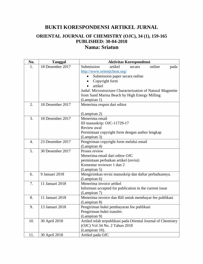
BUKTI KORESPONDENSI ARTIKEL JURNAL
ORIENTAL JOURNAL OF CHEMISTRY (OJC), 34 (1), 159-165
PUBLISHED: 30-04-2018
Nama: Sriatun
No. Tanggal Aktivitas Korespondensi
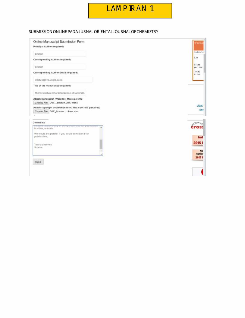
1. 18 Desember 2017 Submission artikel secara online pada
http://www.orientjchem.org/
• Submission paper secara online
• Copyright form
• artikel
Judul: Microstructure Characterization of Natural Magnetite
from Sand Marina Beach by High Energy Milling
(Lampiran 1)
2. 18 Desember 2017 Menerima respon dari editor
(Lampiran 2)
3. 18 Desember 2017 Menerima email
ID manuskrip: OJC-11729-17
Review awal
Permintaan copyright form dengan author lengkap
(Lampiran 3)
4. 23 Desember 2017 Pengiriman copyright form melalui email
(Lampiran 4)
5. 30 Desember 2017 Proses review
Menerima email dari editor OJC
permintaan perbaikan artikel (revisi)
Komentar reviewer 1 dan 2
(Lampiran 5)
6. 9 Januari 2018 Mengirimkan revisi manuskrip dan daftar perbaikannya.
(Lampiran 6)
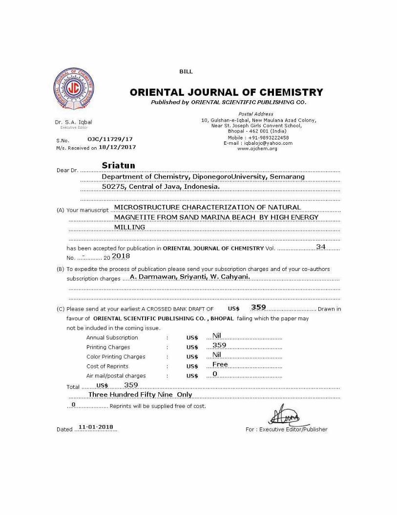
7. 11 Januari 2018 Menerima invoice artikel
Informasi accepted for publication in the current issue
(Lampiran 7)
8. 11 Januari 2018 Menerima invoice dan Bill untuk membayar fee publikasi
(Lampiran 8)
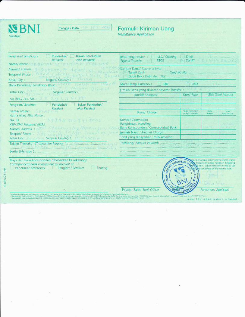
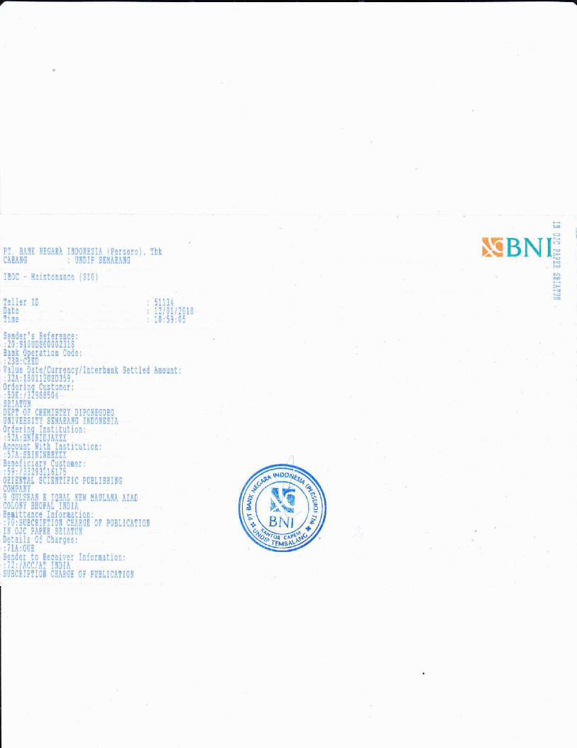
9. 13 Januari 2018 Pengiriman bukti pembayaran fee publikasi
Pengiriman bukti transfer.
(Lampiran 9)
10. 30 April 2018 Artikel telah terpublikasi pada Oriental Journal of Chemistry
(OJC) Vol 34 No. 2 Tahun 2018
(Lampiran 10).
11. 30 April 2018 Artikel pada OJC

(Lampiran 11)
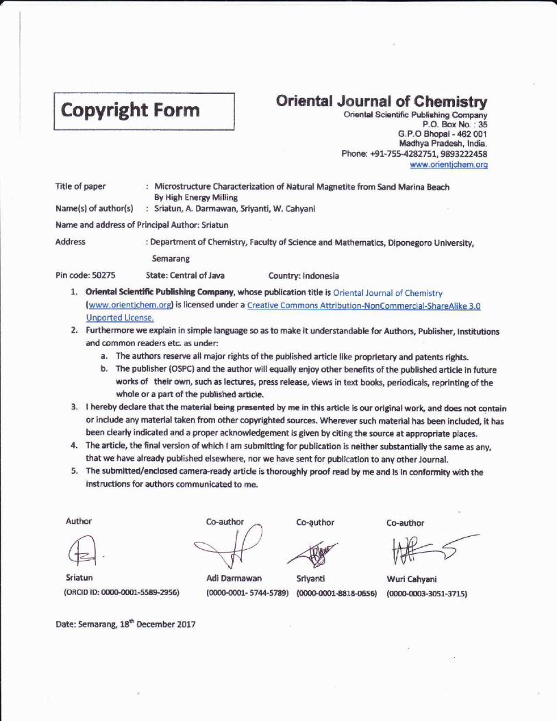
eopyright Formftrir**at lar rlral ,rf latra*ria*nriJi t-Eii,iii r, i.ili i rlEii iri t llEilii-i'i jl
oriental Scientific Publishing CompanyP.O. 8ox .\.!o. : :!5
n ! n !h,1.rl - A?,1 nn4
Madhya Pradesh, lndia.Phone: +91-755-42a27 31. 9893222458
www.orientichem.orq
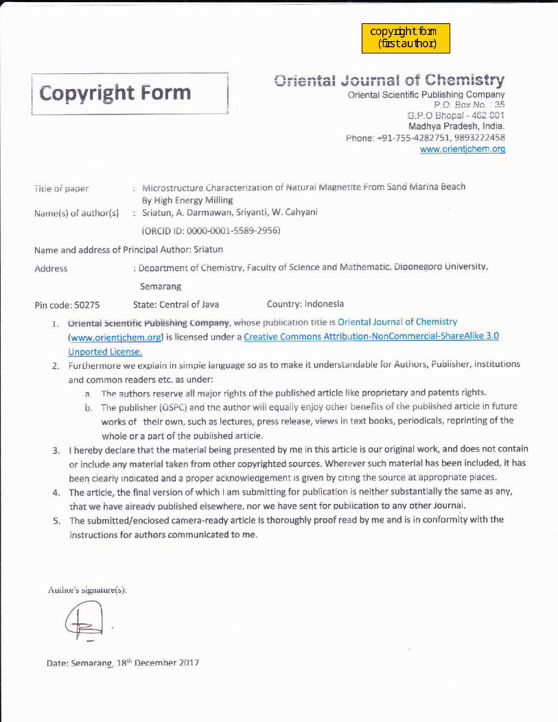
: fuiicrostructure Characterization of ivaiurai Maeneriie From Sand Marina Beach
By High Energy MillingNatne(s) ofauthor(s) : Sriatun, A. Darmawan, Sriyanti, W. Cahyani
iORCiD lD: 0000-1100i-55d9-2Y56)
Name and address of Principal Author: Sriatun
: Depanmeni of chemistrv, Facuiiv of Science and Maihematic Dioonegoro ijniversiiv'
Semarang
Acidress
Pin code: 50275 State: Central of Java Country: lndonesia
1. Oriefitai Scientific pubiashiirg Company, wirose pubircairo.l iati€ $ Oriental Journal of Chemistry
lwww.orientichem.orsl is licensed under a Creative Commons AttributionlNonCommercial-ShareAlike 3.0
Unported License.
2. Furthermore we expiain in simpie ianguage so as io make it unciersiandabie for Auihors, Pubiisher, insiituiions
and common readers etc. as under:
a. The authors reserve all major rights ofthe published article like proprietary and patents rights.
b. The pubiisher (OSPC} anci the authoi wiii equaiiy enjoy otiier benefits of tiie pubiishe,i afticie in future
works of their own, such as lectures, press release, views in text books, periodicals, reprinting of the
whoie or a pari ofthe pubiisheci ariicie.
3. I hereby declare that the material being presented by me in this article is our original work, and does not contain
or include any material taken from other copyrighted sources- Wherever such material has been include4 it has
bcen eieariy inaiieateai anaj a Bioper acknowiecigemcRr is given by eitrng the source at aBBiopfrate piaees.
4. The article, the final version of which I am submitting for publication is neither substantially the same as any,
that we have aireadv pubiisheci eisewhere, nor we have sent for oubiication io anv other iournai.
5. The submitted/enclosed camera-ready article is thoroughly proof read by me and is in conformity with the
instructions for authors communicated to me.
Auttlor s srg[alur9(s.r;
Date: Semarang, 18ii' December 2017
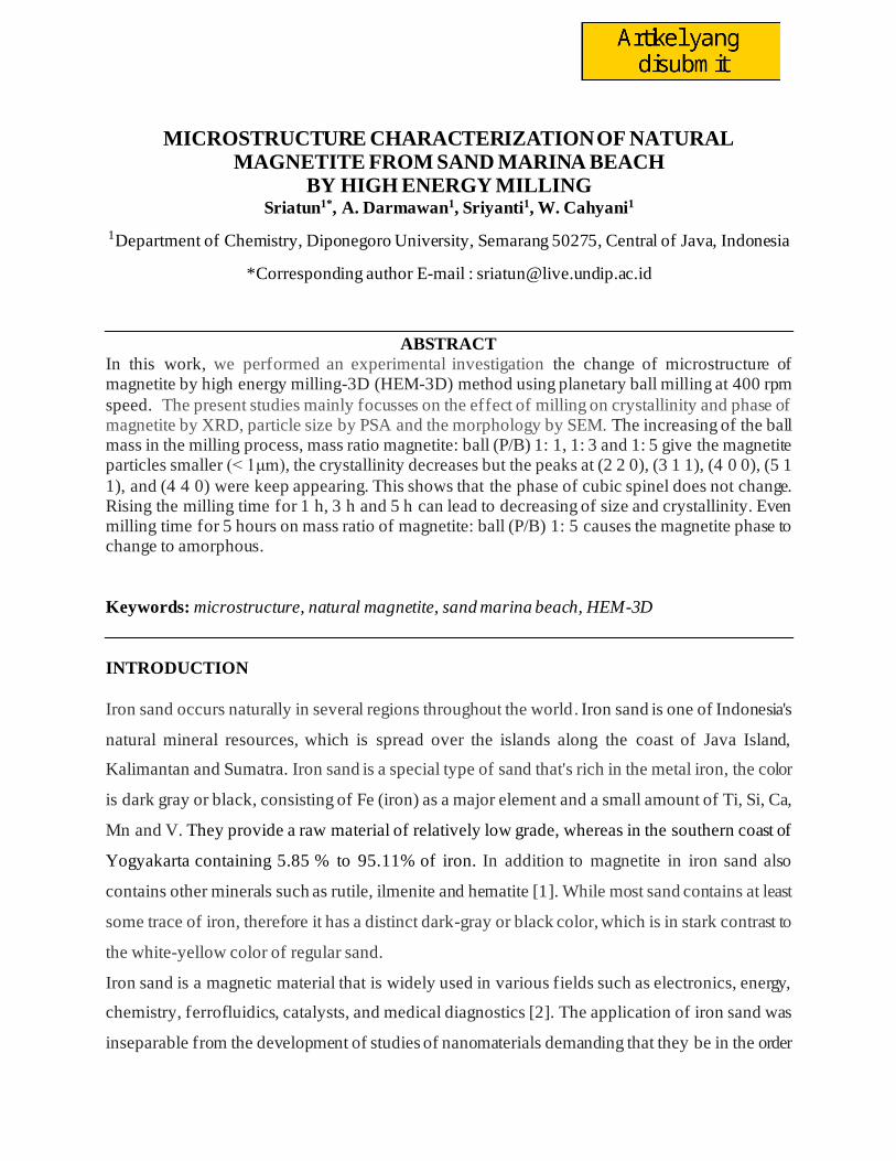
MICROSTRUCTURE CHARACTERIZATION OF NATURAL MAGNETITE FROM SAND MARINA BEACH
BY HIGH ENERGY MILLING Sriatun1*, A. Darmawan1, Sriyanti1, W. Cahyani1
1Department of Chemistry, Diponegoro University, Semarang 50275, Central of Java, Indonesia
*Corresponding author E-mail : [email protected]
ABSTRACT
In this work, we performed an experimental investigation the change of microstructure of magnetite by high energy milling-3D (HEM-3D) method using planetary ball milling at 400 rpm
speed. The present studies mainly focusses on the effect of milling on crystallinity and phase of magnetite by XRD, particle size by PSA and the morphology by SEM. The increasing of the ball mass in the milling process, mass ratio magnetite: ball (P/B) 1: 1, 1: 3 and 1: 5 give the magnetite particles smaller (< 1μm), the crystallinity decreases but the peaks at (2 2 0), (3 1 1), (4 0 0), (5 1
1), and (4 4 0) were keep appearing. This shows that the phase of cubic spinel does not change. Rising the milling time for 1 h, 3 h and 5 h can lead to decreasing of size and crystallinity. Even milling time for 5 hours on mass ratio of magnetite: ball (P/B) 1: 5 causes the magnetite phase to change to amorphous.
Keywords: microstructure, natural magnetite, sand marina beach, HEM-3D
INTRODUCTION
Iron sand occurs naturally in several regions throughout the world. Iron sand is one of Indonesia's
natural mineral resources, which is spread over the islands along the coast of Java Island,
Kalimantan and Sumatra. Iron sand is a special type of sand that's rich in the metal iron, the color
is dark gray or black, consisting of Fe (iron) as a major element and a small amount of Ti, Si, Ca,
Mn and V. They provide a raw material of relatively low grade, whereas in the southern coast of
Yogyakarta containing 5.85 % to 95.11% of iron. In addition to magnetite in iron sand also
contains other minerals such as rutile, ilmenite and hematite [1]. While most sand contains at least
some trace of iron, therefore it has a distinct dark-gray or black color, which is in stark contrast to
the white-yellow color of regular sand.
Iron sand is a magnetic material that is widely used in various fields such as electronics, energy,
chemistry, ferrofluidics, catalysts, and medical diagnostics [2]. The application of iron sand was
inseparable from the development of studies of nanomaterials demanding that they be in the order

of nanometers. Magnetite or Fe3O4 is one of the iron oxide phases which has the greatest magnetic
or ferromagnetic properties among the other phases. Iron oxide has four phases, namely magnetite
(Fe3O4), maghemite (γ-Fe2O3), hematite (α-Fe2O3), and geotite (FeO(OH)). Only magnetite and
maghemite have magnetic properties [3].
Magnetite (Fe3O4) is known as a class of iron oxide compound with a cubic inverse spinel structure
and has face centered cubic close packed oxygen anions and Fe cations occupying interstitial
tetrahedral and octahedral sites [4, 5]. Nano-sized magnetite particles provide many advantages
such as for the separation of magnetic contaminants in water, large of surface area and the ability
to bind through electro-chemical interactions to form sludge. It is also applied to drug delivery and
magnetic resonance technology and others.
For the synthesis of nanosized magnetite particles can be synthesized through various methods
such as mechanical milling [6], sol-gels, direct decomposition [7], co-precipitation [8],
microwave-heating [9] and solvothermal [10, 11]. Mechanical milling method is one way to reduce
the magnetite size is the cheapest and easy. Mechanical milling is defined as the mechanical
breakdown of magnetite into smaller without changing their state of aggregation. The method was
used to increase the surface area and induce defects which is needed for subsequent operations
such as chemical reactions, sorption. Milling also to increase the proportion of regions of high
activity in the surface [12].
Furthermore, this research the small size of magnetite from iron sand was prepared by mechanical
milling method using high energy planetary ball mill. Kinetic energy of the balls depends not only
on its velocity, but also on its mass and how long the collision occurred, due to in this work
investigated the ratio of magnetite and ball mass in the planetary ball mill and the time of impact
during collision.
MATERIALS AND METHODS
Materials
Iron sand was taken from Marina Beach in Semarang.
Instrumentations
Magnet permanent, High energy planetary ball mill-3D, X-ray diffraction (XRD) Rigaku
Multiplex with Cu Kα radiation (λ = 1.54184 Ao) at generator voltage 40 kV and current 40 mA,
Particle Size Analyzer (PSA) Horiba SZ-100, Scanning electron microscope (SEM) JEOL JED
2300.
Procedure
Magnetite preparation
The natural iron sand from Marina Beach Semarang cleaned and washed using aquadest, dried in
oven at 80oC for 24 hours. Natural magnetite was extracted from natural iron sand using permanent
magnet until 12 times. This treatment produces powder material dark gray-black color. Refinement
of magnetite particles carried out by mechanical milling method using High Energy planetary ball
Mill (HEM-E3D) instrument. The milling was done on mass ratio of magnetite: ball (P/B) 1:1, 1:3
and 1:5, speed 400 rpm. Milling of magnetite carried out for 1, 3 and 5 hours. Milled magnetite
dried at 150oC for 1.5 hours. Finally, the microstructure characterization of product was done by
X-ray Diffraction (XRD) to find out the structure of magnetite crystals, PSA to determine the size
of magnetite particle, SEM to know the surface morphology.
RESULTS AND DISCUSSIONS
In this work the change of crystal structure, particle size and morphology of magnetite to be
investigated. The method is high energy milling (HEM) used planetary ball mill. The choice of
this method due to it can reduce the material up to the nano order (nano particle) inside a relatively
short time under conditions atmosphere at room temperature during process milling. This method
using energy collision between the crushing balls and chamber walls are rotated and driven in a
certain way. The change of crystal structure, particle size and morphology of magnetite was studied
on variation the mass ratio magnetite:ball (P/B 1:1, 1:3 and 1:5) and milling time (1, 3 and 5 hours).
Physical changes of magnetite
The process of separation of magnetite compounds from iron sand is done repeatedly, it is intended
that the compound to be obtained has a high purity. The separation process with magnets also uses
a certain distance, the farther the magnet is closer to the iron sands, the less iron oxide attaches.
This makes the sample (magnetite) higher purity and less impurities, although there is still the
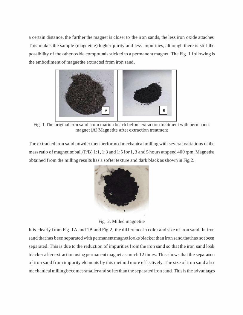
possibility of the other oxide compounds sticked to a permanent magnet. The Fig. 1 following is
the embodiment of magnetite extracted from iron sand.
Fig. 1 The original iron sand from marina beach before extraction treatment with permanent magnet (A) Magnetite after extraction treatment
The extracted iron sand powder then performed mechanical milling with several variations of the
mass ratio of magnetite:ball (P/B) 1:1, 1:3 and 1:5 for 1, 3 and 5 hours at speed 400 rpm. Magnetite
obtained from the milling results has a softer texture and dark black as shown in Fig.2.
Fig. 2. Milled magnetite
It is clearly from Fig. 1A and 1B and Fig 2, the difference in color and size of iron sand. In iron
sand that has been separated with permanent magnet looks blacker than iron sand that has not been
separated. This is due to the reduction of impurities from the iron sand so that the iron sand look
blacker after extraction using permanent magnet as much 12 times. This shows that the separation
of iron sand from impurity elements by this method more eff ectively. The size of iron sand after
mechanical milling becomes smaller and softer than the separated iron sand. This is the advantages
A B
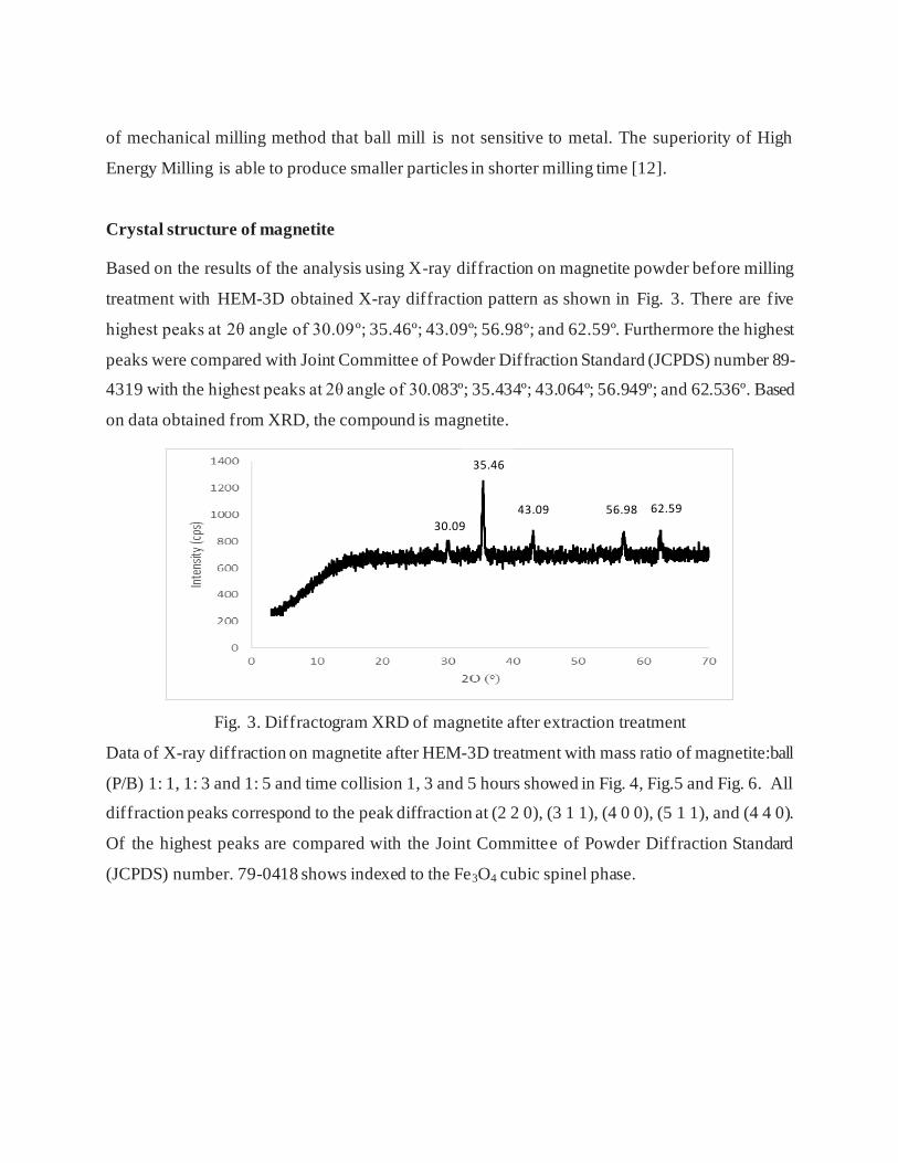
of mechanical milling method that ball mill is not sensitive to metal. The superiority of High
Energy Milling is able to produce smaller particles in shorter milling time [12].
Crystal structure of magnetite
Based on the results of the analysis using X-ray diffraction on magnetite powder before milling
treatment with HEM-3D obtained X-ray diffraction pattern as shown in Fig. 3. There are five
highest peaks at 2θ angle of 30.09º; 35.46º; 43.09º; 56.98º; and 62.59º. Furthermore the highest
peaks were compared with Joint Committee of Powder Diffraction Standard (JCPDS) number 89-
4319 with the highest peaks at 2θ angle of 30.083º; 35.434º; 43.064º; 56.949º; and 62.536º. Based
on data obtained from XRD, the compound is magnetite.
Fig. 3. Diffractogram XRD of magnetite after extraction treatment
Data of X-ray diffraction on magnetite after HEM-3D treatment with mass ratio of magnetite:ball
(P/B) 1: 1, 1: 3 and 1: 5 and time collision 1, 3 and 5 hours showed in Fig. 4, Fig.5 and Fig. 6. All
diffraction peaks correspond to the peak diffraction at (2 2 0), (3 1 1), (4 0 0), (5 1 1), and (4 4 0).
Of the highest peaks are compared with the Joint Committee of Powder Diffraction Standard
(JCPDS) number. 79-0418 shows indexed to the Fe3O4 cubic spinel phase.
30.09
35.46
43.09 56.98 62.59
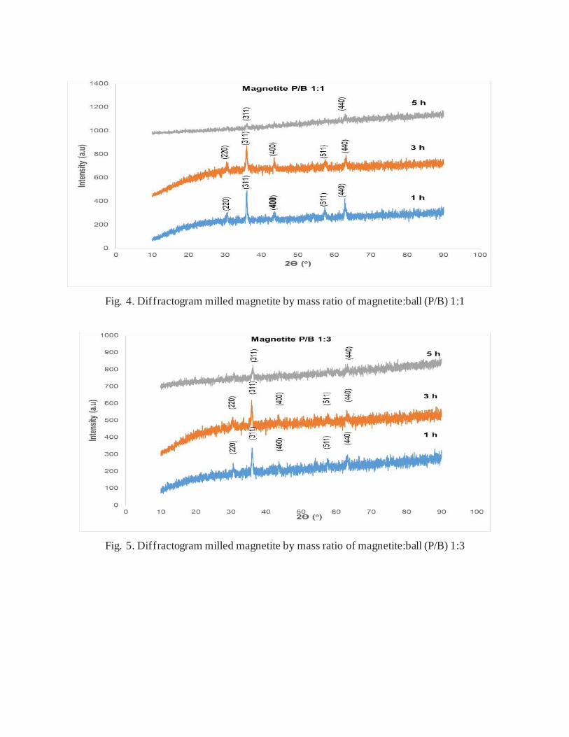
Fig. 4. Diffractogram milled magnetite by mass ratio of magnetite:ball (P/B) 1:1
Fig. 5. Diffractogram milled magnetite by mass ratio of magnetite:ball (P/B) 1:3
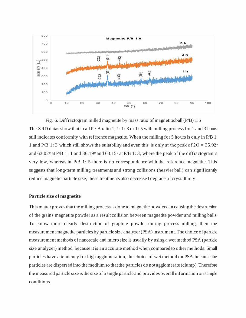
Fig. 6. Diffractogram milled magnetite by mass ratio of magnetite:ball (P/B) 1:5
The XRD datas show that in all P / B ratio 1, 1: 1: 3 or 1: 5 with milling process for 1 and 3 hours
still indicates conformity with reference magnetite. When the milling for 5 hours is only in P/B 1:
1 and P/B 1: 3 which still shows the suitability and even this is only at the peak of 2Ɵ = 35.92o
and 63.02o at P/B 1: 1 and 36.19o and 63.15o at P/B 1: 3, where the peak of the diffractogram is
very low, whereas in P/B 1: 5 there is no correspondence with the reference magnetite. This
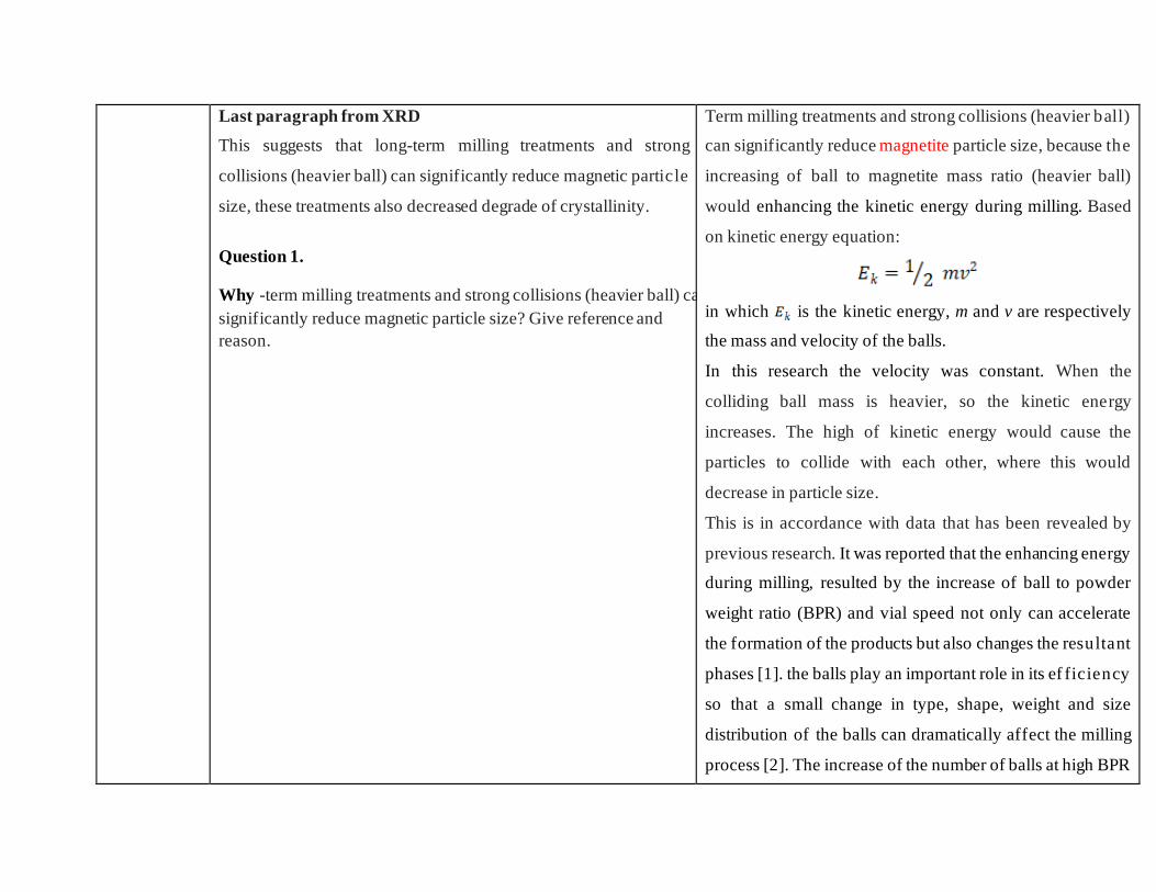
suggests that long-term milling treatments and strong collisions (heavier ball) can significantly
reduce magnetic particle size, these treatments also decreased degrade of crystallinity.
Particle size of magnetite
This matter proves that the milling process is done to magnetite powder can causing the destruction
of the grains magnetite powder as a result collision between magnetite powder and milling balls.
To know more clearly destruction of graphite powder during process milling, then the
measurement magnetite particles by particle size analyzer (PSA) instrument. The choice of particle
measurement methods of nanoscale and micro size is usually by using a wet method PSA (particle
size analyzer) method, because it is an accurate method when compared to other methods. Small
particles have a tendency for high agglomeration, the choice of wet method on PSA because the
particles are dispersed into the medium so that the particles do not agglomerate (clump). Therefore
the measured particle size is the size of a single particle and provides overall information on sample
conditions.
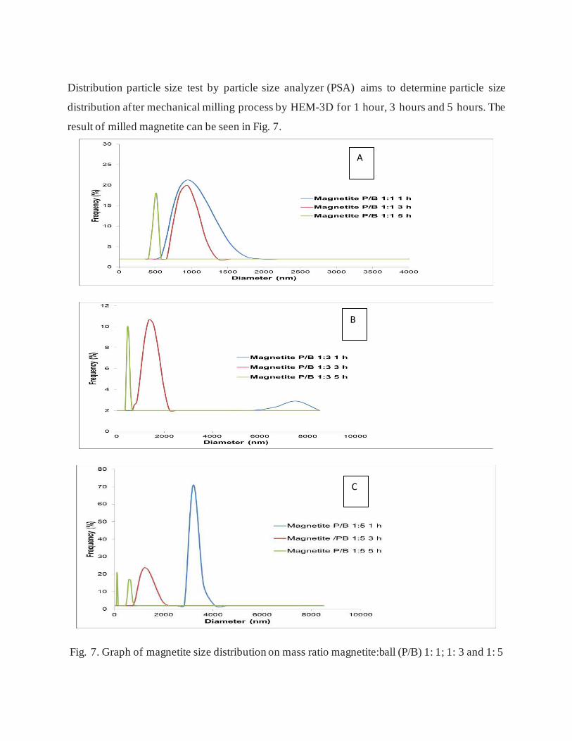
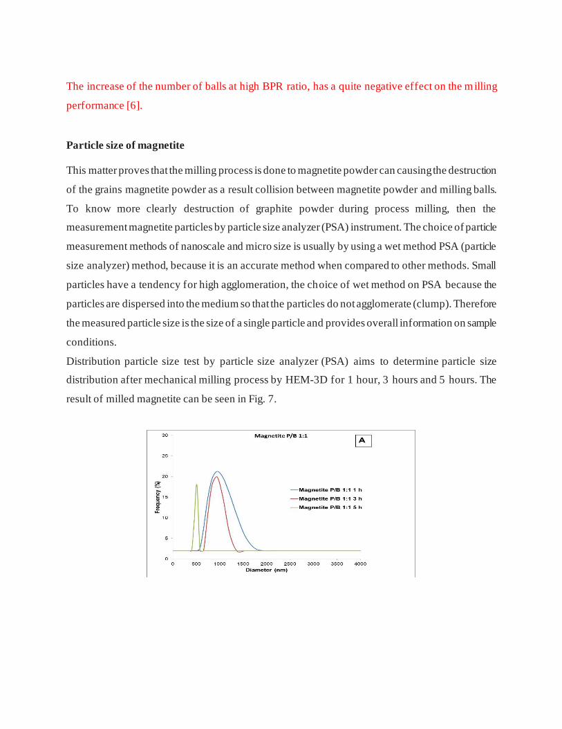
Distribution particle size test by particle size analyzer (PSA) aims to determine particle size
distribution after mechanical milling process by HEM-3D for 1 hour, 3 hours and 5 hours. The
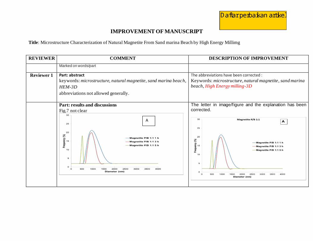
result of milled magnetite can be seen in Fig. 7.
Fig. 7. Graph of magnetite size distribution on mass ratio magnetite:ball (P/B) 1: 1; 1: 3 and 1: 5
A
B
C
In Fig. 7 it is observed that the magnetite/ball mass ratio (P/B) of 1: 1 increase in time causes a
significant reduction in particle size. When for 1 hour milling the size range varies as well as for
3 h, however the milling is performed for 5 hours gives impact to a more homogeneous magnetite
size (the peak is not widened). Significant reduction in size occurred in treatment with a mass ratio
of P/B 1: 3 and 1: 5. This is due to the heavier the ball and the length of time the greater the energy
given to collide with the magnetite particles. Thus the magnetite treatment with HEM (high energy
milling) is effective enough to reduce the size to less than 1000 nm (<1μm).
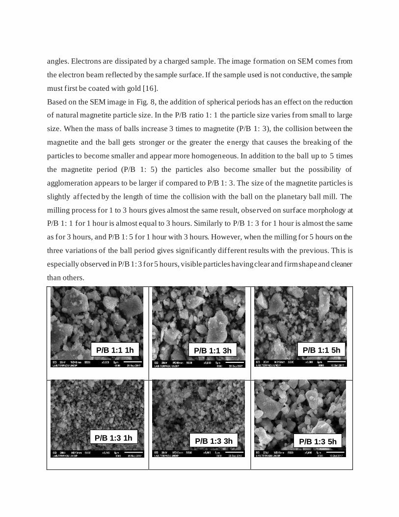
Morphology of magnetite
The surface morphology of a material can be observed using SEM (Scanning electron microscope).
The basic principle of work on SEM is the nature of electron waves, it is diffraction at very small
angles. Electrons are dissipated by a charged sample. The image f ormation on SEM comes from
the electron beam reflected by the sample surface. If the sample used is not conductive, the sample
must first be coated with gold [13].
Based on the SEM image in Fig. 8, the addition of spherical periods has an effect on the reduction
of natural magnetite particle size. In the P/B ratio 1: 1 the particle size varies from small to large
size. When the mass of balls increase 3 times to magnetite (P/B 1: 3), the collision between the
magnetite and the ball gets stronger or the greater the energy that causes the breaking of the
particles to become smaller and appear more homogeneous. In addition to the ball up to 5 times
the magnetite period (P/B 1: 5) the particles also become smaller but the possibility of
agglomeration appears to be larger if compared to P/B 1: 3. The size of the magnetite particles is
slightly affected by the length of time the collision with the ball on the planetary ball mill. The
milling process for 1 to 3 hours gives almost the same result, observed on surface morphology at
P/B 1: 1 for 1 hour is almost equal to 3 hours. Similarly to P/B 1: 3 for 1 hour is almost the same
as for 3 hours, and P/B 1: 5 for 1 hour with 3 hours. However, when the milling for 5 hours on the
three variations of the ball period gives significantly different results with the previous. This is
especially observed in P/B 1: 3 for 5 hours, visible particles having clear and firm shape and cleaner
than others.
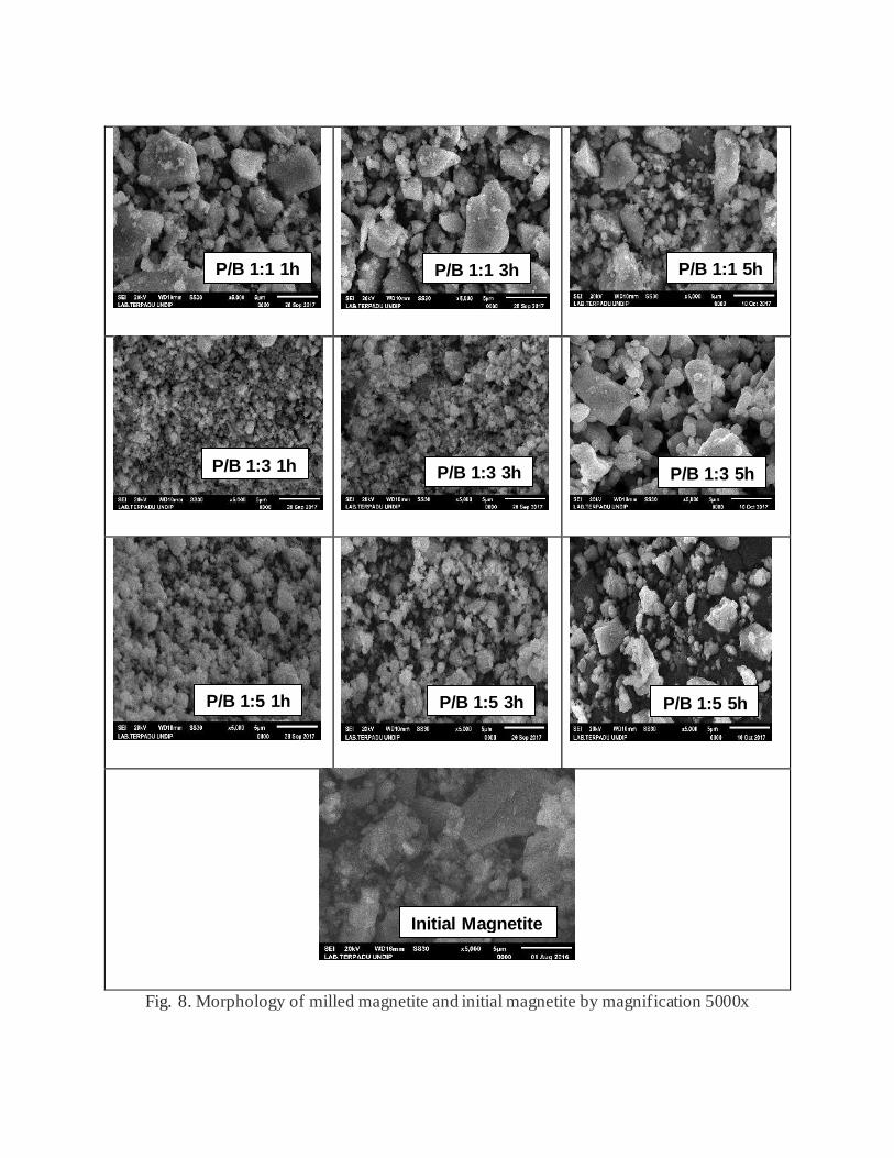
Fig. 8. Morphology of milled magnetite and initial magnetite by magnification 5000x
P/B 1:1 1h P/B 1:1 3h P/B 1:1 5h
P/B 1:3 1h P/B 1:3 3h P/B 1:3 5h
P/B 1:5 1h P/B 1:5 3h P/B 1:5 5h
Initial Magnetite
before treat
CONCLUSION
From the results and discussion can be concluded that the HEM-3D treatment with 400 rpm speed
can reduce particle size and increase the uniformity of shape and magnetite size. The increasing of
the ball mass in the milling process, this means in the mass ratio of magnetite:ball (P/B) 1: 1, 1: 3
and 1: 5 give the magnetite particles smaller, the crystallinity decreases but the phase does not
change. Rising the milling time can lead to decreasing of size and crystallinity. Even milling time
for 5 hours on mass ratio of magnetite:ball (P/B) 1: 5 causes the magnetite phase to change to
amorphous
ACKNOWLEDGEMENT
Sriatun, Adi Darmawan and Sriyanti, gratefully acknowledge financial support from of Besides
APBN DPA SUKPA LPPM Diponegoro University, and Department of Chemistry for the
facilities to carry out this research.
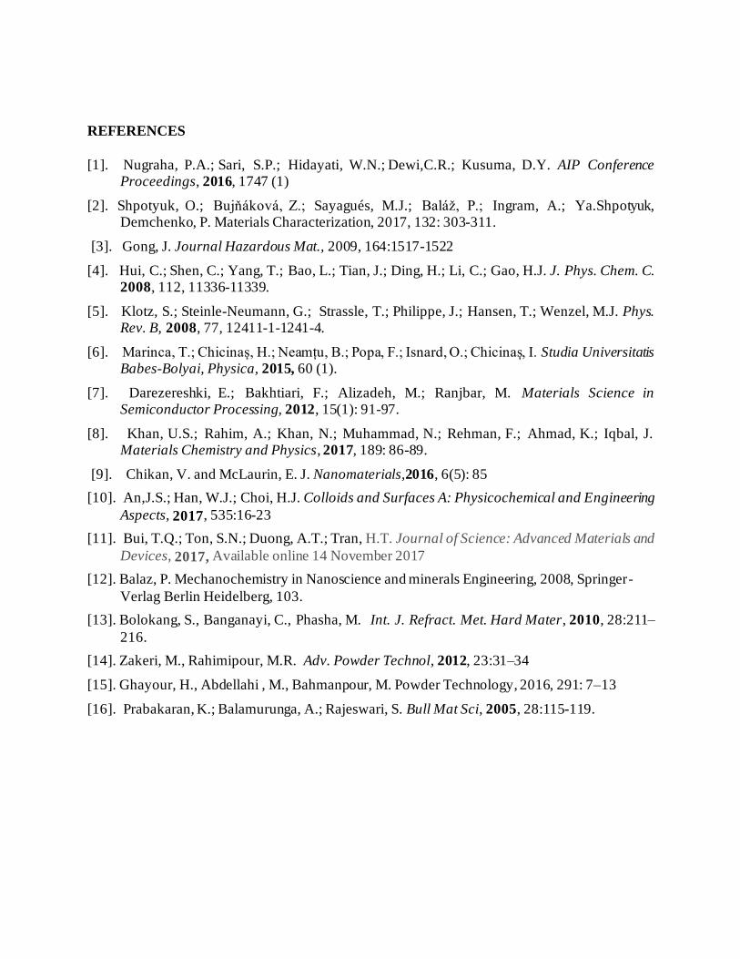
REFERENCES
[1]. Nugraha, P.A.; Sari, S.P.; Hidayati, W.N.; Dewi,C.R.; Kusuma, D.Y. AIP Conference
Proceedings, 2016, 1747 (1)
[2]. Shpotyuk, O.; Bujňáková, Z.; Sayagués, M.J.; Baláž, P.; Ingram, A.; Ya.Shpotyuk,
Demchenko, P. Materials Characterization, 2017, 132: 303-311.
[3]. Gong, J. Journal Hazardous Mat., 2009, 164:1517-1522
[4]. Hui, C.; Shen, C.; Yang, T.; Bao, L.; Tian, J.; Ding, H.; Li, C.; Gao, H.J. J. Phys. Chem. C. 2008, 112, 11336-11339.
[5]. Klotz, S.; Steinle-Neumann, G.; Strassle, T.; Philippe, J.; Hansen, T.; Wenzel, M.J. Phys. Rev. B, 2008, 77, 12411-1-1241-4.
[6]. Marinca, T.; Chicinaș, H.; Neamțu, B.; Popa, F.; Isnard, O.; Chicinaș, I. Studia Universitatis Babes-Bolyai, Physica, 2015, 60 (1).
[7]. Darezereshki, E.; Bakhtiari, F.; Alizadeh, M.; Ranjbar, M. Materials Science in Semiconductor Processing, 2012, 15(1): 91-97.
[8]. Khan, U.S.; Rahim, A.; Khan, N.; Muhammad, N.; Rehman, F.; Ahmad, K.; Iqbal, J. Materials Chemistry and Physics, 2017, 189: 86-89.
[9]. Chikan, V. and McLaurin, E. J. Nanomaterials,2016, 6(5): 85
[10]. An,J.S.; Han, W.J.; Choi, H.J. Colloids and Surfaces A: Physicochemical and Engineering Aspects, 2017, 535:16-23
[11]. Bui, T.Q.; Ton, S.N.; Duong, A.T.; Tran, H.T. Journal of Science: Advanced Materials and Devices, 2017, Available online 14 November 2017
[12]. Balaz, P. Mechanochemistry in Nanoscience and minerals Engineering, 2008, Springer-Verlag Berlin Heidelberg, 103.
[13]. Prabakaran, K.; Balamurunga, A.; Rajeswari, S. Bull Mat Sci, 2005, 28:115-119.

28/10/2020 Confirmation of paper - [email protected] - Diponegoro University Mail
https://mail.google.com/mail/u/1/?tab=wm&ogbl#search/editor%40orientjchem.org/FMfcgxmXKmWgNLggBxfDTBcDrXHvLjNq 1/1
Meet
Hangouts
Compose
More
Inbox 213
Starred
Snoozed
Sent
New meeting
My meetings
Sriatun
No Hangouts contacts
Con�rmation of paper Inbox ×
Dear, Dr. SriatunI thankfully acknowledge the receipt of your valuable research paper entMILLING(OJC-11729-17)We are processing your article for detail review and we will inform you a
Make the initials corrections written as under otherwise the paper may b1) Send the copyright form with the compulsory signature o Please send us your ORCID ID, along with the co-authors orcid
The deadline for receiving your initial corrections is one week.Thanks
Please follow us on all our social media channels below: Facebook: https://www.facebook.com/Oriental-Journal-of-Chemistry-105 Linkedin: https://www.linkedin.com/company/oriental-journal-of-chemistr Twitter: https://twitter.com/Orienjchem

28/10/2020 Copyright form_sriatun and ORCID ID - [email protected] - Diponegoro University Mail
https://mail.google.com/mail/u/1/?tab=wm&ogbl#search/editor%40orientjchem.org/FMfcgxmXKmfZhWSfMfRhMrgmSgqwvmxL 1/1
Meet
Hangouts
Compose
More
Inbox 213
Starred
Snoozed
Sent
New meeting
My meetings
Sriatun
No Hangouts contacts
Copyright form_sriatun and ORCID ID
Sriatun Sriatun <[email protected]>
to editor, Sriatun
Semarang-Indonesia, December 23, 2017
To:
Dr. S.A. Iqbal, Ph.D., FICS, FICC, FIAEM, MNASc
Chief Editor, Oriental Journal of Chemistry
I would greatly appreciate the opportunity to have make an correction atI’ve attached a scanned of copyright form with the signature of all the auAuthor: Sriatun Sriatun (ORCID ID: 0000-0001-5589-2956)Co-author: Adi Darmawan (ORCID ID: 0000-0001-5744-5789) Sriyanti Sriyanti (ORCID ID: 0000-0001-8818-0656) Wuri Cahyani (ORCID ID: 0000-0003-3051-3715)
-- with kind regards,yours sincerely
SriatunDept. of Chemistry, Diponegoro University
Copyright Form
eSriatun
(ORCID lD: 0o(x}{fi}1-558s'2956]
Oriental Journal of ChemistryOrbnhl Sobntific Publbhing Company
P.O. Box l,lo. : 35G.P.O Bhopel -.162 001iladhya Pradesh, hdia.
Phon€: €1-75$4282751, 9893222458www.orientichem.oro
fnb of paper : Micmstrueture Characte.ization of Natural Magnetite from Sand Marlna BeachBy HiSh EnerSy Mllling
Name(s) of author(s) : Sriatun, A. Darmaura4 Srlyanti, W. Cahyani
Name and address of Principal Author: Sriatun
Mdress : Department sf Chemlstry Faculty of Sciience and Mathematics, Dlponegoro Universtw,
Pin code: 50275
Semarang
State: Centrel oflava Country: lndonesia
1. Orientd ScienHfic Publbhlrg Comtcty, whos€ puHication Utle is Oriental Journal of Chemistry(www.orientichem.ord is licensed urdet a creative Commons Attributlon-Noncommercial-shareAlike 3.0
Unported License.
2. Furthemore we explain in slmple language so as to make it understandable for Authors, Publisher, lnsttudonsand common rcaders etc. as under:
a. The authoB reserve all major rights ofthe rublish€d article like proprietary and patents rights.b, The publlsher {OSrc) and the author will equally enJoy other benefits of the published article in future
work of theit own, such as lectures, prcss release, views in text books, pedodicals, reprinfing ofthewhole or a part ot the published ar$cle.
3. I herebry dcdate th3t $e materlal being presented by me in this article h oor odginal work, and does not contalnor include any material taken from other copytighted sources, Wherever such rmterial has been include4 lt hasbeen cleatly indicated and a proper acknowledgerlent is Sirren W citnt the source at appropriate daces.
4' The N?ticle, th€ final version of which I am submittirg for publication is neither substantially the same as any,that we have already gublished elsewhere, nor we have sent for publication to any otherJournal.
5. The submitt€d/€trclo6€d cattet&teady artide is thorol4ftly proof read by me and is ln conformity with theinstructions for authors communicated to me.
Author CGttchor Co-author,n"*\,f)
Y-Adl Darmawan
-rywSriyanti
{(xltosot 881&0656,
Wuri Crhyani
({rodF{m}$s1-371s}
Date: Semarang, 186 December 2017
(qxxHml- 574+57St)

28/10/2020 11729 review report - [email protected] - Diponegoro University Mail
https://mail.google.com/mail/u/1/?tab=wm&ogbl#search/info%40orientjchem.org/FMfcgxmXKwCBdBzNKZrWhWgCqcgXGJWM 1/1
Meet
Hangouts
Compose
More
Inbox 213
Starred
Snoozed
Sent
New meeting
My meetings
Sriatun
No Hangouts contacts
11729 review repo� Inbox ×
Please find attached review report Dr. S. A. Iqbal, Ph. D., FICS, FICC, FIAEM, MNASc.
Chief Editor
Oriental Journal of Chemistry
An ISI, Scopus, SNIP based Journal
www.orientjchem.org
Review Report-OJ…
Thanks a lot. Received, thank you. Thank you for
P.O. BOX No.35, G.P.O. BHOPAL-462001(INDIA) http://www.orientjchem.org
Review Report of Manuscript
Title of the Journal : Oriental Journal of Chemistry
Title of the Manuscript : MICROSTRUCTURE CHARACTERIZATION OF
NATURAL MAGNETITE FROM SAND MARINA BEACH BY HIGH ENERGY MILLING
Ref. No. of Manuscript and : OJC-11729-17
Corresponding Author Name Sriatun
Abstract : (i) Appropriate (ii) Requires modification
(iii) Too Long Requires Brevity (iv) Lacks clarity
Keywords : Sufficient Lacking Require modification
Introduction : Appropriate Not related to the work
Ambiguous Too detailed, requires brevity
Experimental : Incomplete Detailed and clear
(Materials and Methods) Requires improvement Not clearly explained
Tables : Title of table(s) missing Caption not appropriate
Requires correction Tables not in corrected form
Graphs, Figures, : Appropriate Labeling not clear
Structures and Equations Not clear, requires redrawing Not self explanatory
Result and Discussion : Convincing Not Convincing
Not supported by relevant references.
Language and Write-up : Lucid Ambiguous Non-coherent
References : Not according to our format, modify, See example
Ref.(s) Nos. ………………………………….……………..… are incomplete
Ref.(s) Nos. …………………………………….………… require correction
No uniformity maintained Not in chronological order
Don’t use et al.;(write names of all the authors)in references
Copy Right Form Received without signatures Not Received Signature of Coauthors required

Continue to page 2nd
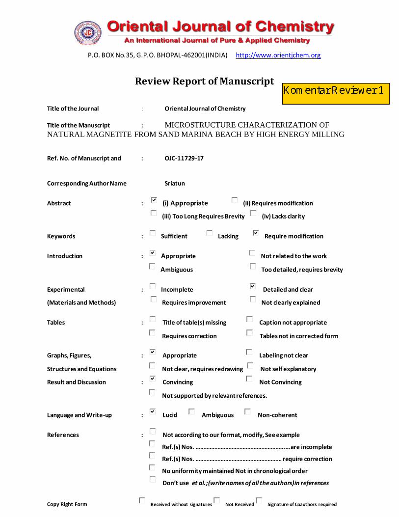
Overall Report in Brief:- keywords: abbreviations not allowed generally.
Figure 7 not clear
Review Decision : The paper is accepted without modification.
: The paper is accepted after minor modification.
: The paper is accepted after major modification.
: Rewrite the paper and send us at your earliest.
: The paper is not acceptable, hence we are reluctantly returning to you.
Reply on this email: [email protected]
Reviewers Name & Address Signature of the Editor
Reviewer 1
P.O. BOX No.35, G.P.O. BHOPAL-462001(INDIA)http://www.orientjchem.org
Review Report of Manuscript
Title of the Journal : Oriental Journal of Chemistry
Title of the Manuscript : MICROSTRUCTURE CHARACTERIZATION OF NATURAL MAGNETITE FROM SAND MARINA BEACH BY HIGH ENERGY MILLING
Ref. No. of Manuscript and : OJC-11729-17 18-12-2017
Corresponding Author Name :Sriatun
Abstract : (i) Appropriate (ii) Requires modification
(iii) Too Long Requires Brevity (iv) Lacks clarity
Keywords : Sufficient Lacking Require modification
Introduction : Appropriate Not related to the work
Ambiguous Too detailed, requires brevity
Experimental : Incomplete Detailed and clear
(Materials and Methods) Requires improvement Not clearly explained
Tables : Title of table(s) missing Caption not appropriate
Requires correction Tables not in corrected form
Graphs, Figures, : Appropriate Labeling not clear
Structures and Equations Not clear, requires redrawing Not self explanatory
Result and Discussion : Convincing Not Convincing
Not supported by relevant references.
Language and Write-up : Lucid Ambiguous Non-coherent
References : Not according to our format, modify, See example
Ref.(s) Nos. ………………………………….……………..… are incomplete
Ref.(s) Nos. …………………………………….………… require correction
No uniformity maintained Not in chronological order
Don’t use et al.;(write names of all the authors)in references
Copy Right Form Received without signatures Not Received Signature of Coauthors required
Continue to page 2nd

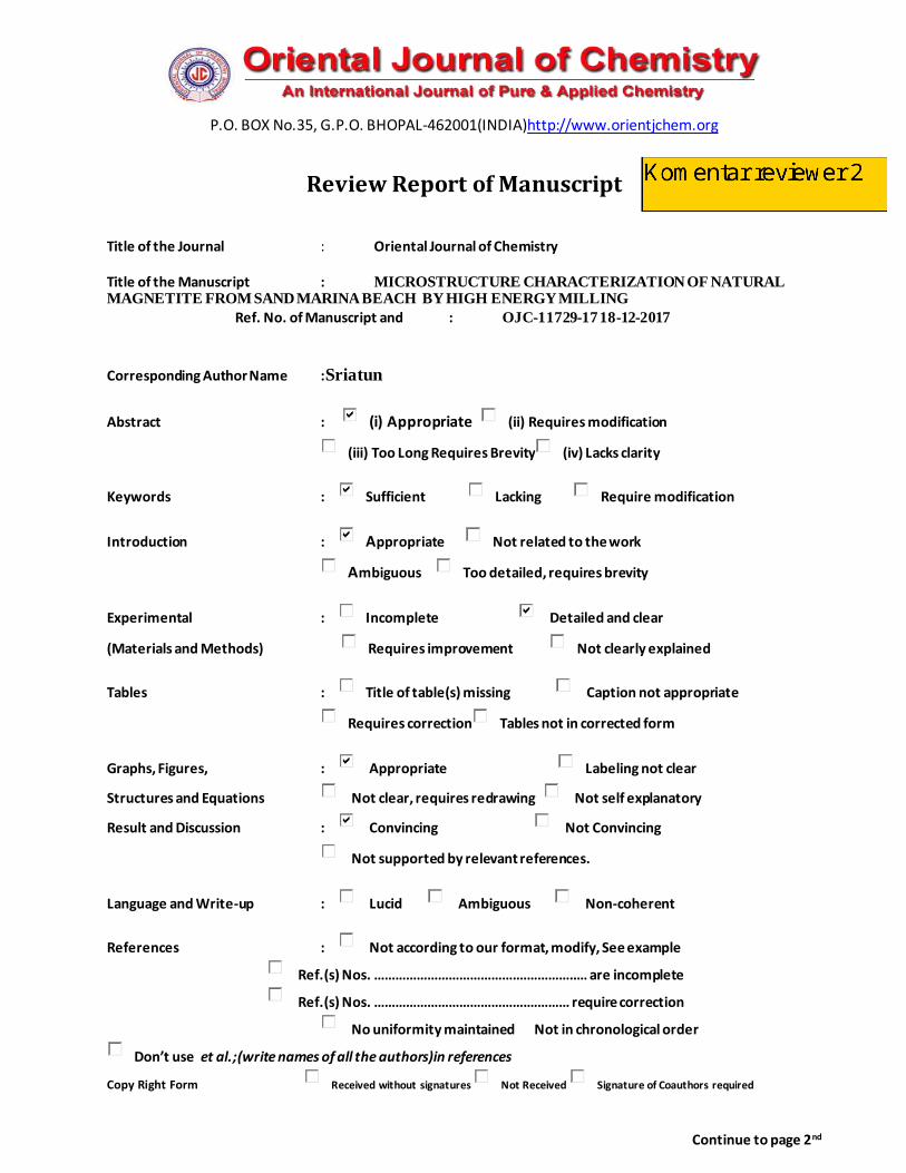
Overall Report in Brief:-
Note: special comments to authors.
Crystal structure of magnetite
Last paragraph from XRD
This suggests that long-term milling treatments and strong collisions (heavier ball) can significantly
reduce magnetic particle size,these treatments also decreased degrade of crystallinity.
1.Why -term milling treatments and strong collisions (heavier ball) can significantly reduce magnetic
particle size? Give reference and reason.
Review Decision : The paper is accepted without modification.
: The paper is accepted after minor modification.
: The paper is accepted after major modification.
: Rewrite the paper and send us at your earliest.
: The paper is not acceptable, hence we are reluctantly returning to you.
Reply on this email: [email protected]
Reviewers Name & Address Signature of the Editor
Reviewer 2

28/10/2020 revised manuscript Sriatun - [email protected] - Diponegoro University Mail
https://mail.google.com/mail/u/1/?tab=wm&ogbl#search/editor%40orientjchem.org/FMfcgxmXLGdDVHmWHvsdMdNCXnBWwBCR 1/1
Meet
Hangouts
Compose
More
Inbox 213
Starred
Snoozed
Sent
New meeting
My meetings
Sriatun
No Hangouts contacts
revised manuscript Sriatun
Sriatun Sriatun <[email protected]>
to editor
Semarang-Indonesia, January, 9, 2018
To:
Dr. S.A. Iqbal, Ph.D., FICS, FICC, FIAEM, MNASc
Chief Editor, Oriental Journal of Chemistry
I apologize for this long time to revise this manuscript. Here I'm sending
Thank youSriatun
Virus-free. www.avast.com
2 Attachments
IMPROVEMENT OF MANUSCRIPT
Title: Microstructure Characterization of Natural Magnetite From Sand marina Beach by High Energy Millimg
REVIEWER COMMENT DESCRIPTION OF IMPROVEMENT
Marked on words/part
Reviewer 1 Part: abstract
keywords: microstructure, natural magnetite, sand marina beach,
HEM-3D
abbreviations not allowed generally.
The abbreviations have been corrected :
Keywords: microstructure, natural magnetite, sand marina beach, High Energy milling-3D
Part: results and discussions
Fig.7 not clear
The letter in image/figure and the explanation has been corrected.
A
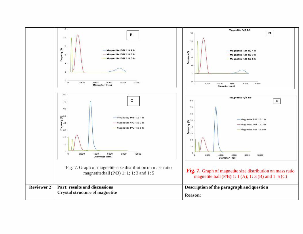
Fig. 7. Graph of magnetite size distribution on mass ratio
magnetite:ball (P/B) 1: 1; 1: 3 and 1: 5
Fig. 7. Graph of magnetite size distribution on mass ratio
magnetite:ball (P/B) 1: 1 (A); 1: 3 (B) and 1: 5 (C)
Reviewer 2 Part: results and discussions
Crystal structure of magnetite
Description of the paragraph and question
Reason:
C
B
Last paragraph from XRD
This suggests that long-term milling treatments and strong
collisions (heavier ball) can significantly reduce magnetic particle
size, these treatments also decreased degrade of crystallinity.
Question 1.
Why -term milling treatments and strong collisions (heavier ball) can
significantly reduce magnetic particle size? Give reference and
reason.
Term milling treatments and strong collisions (heavier ball)
can significantly reduce magnetite particle size, because the
increasing of ball to magnetite mass ratio (heavier ball)
would enhancing the kinetic energy during milling. Based
on kinetic energy equation:
in which is the kinetic energy, m and v are respectively
the mass and velocity of the balls.
In this research the velocity was constant. When the
colliding ball mass is heavier, so the kinetic energy
increases. The high of kinetic energy would cause the
particles to collide with each other, where this would
decrease in particle size.
This is in accordance with data that has been revealed by
previous research. It was reported that the enhancing energy
during milling, resulted by the increase of ball to powder
weight ratio (BPR) and vial speed not only can accelerate
the formation of the products but also changes the resultant
phases [1]. the balls play an important role in its ef ficiency
so that a small change in type, shape, weight and size
distribution of the balls can dramatically affect the milling
process [2]. The increase of the number of balls at high BPR
ratio, has a quite negative effect on the milling performance
[3].
This explanation has been added in discussions of
manuscript.
Refferences:
[1] Bolokang, S., Banganayi, C., Phasha, M. Effect of C and milling parameters on the synthesis of WC powders by mechanical alloying, Int. J. Refract. Met. Hard Mater., 2010, 28:211–216.
[2] Zakeri, M., Rahimipour, M.R. Effect of cup and ball types on alumina–tungsten carbide nanocomposite powder
synthesized by mechanical alloying, Adv. Powder Technol., 2012, 23:31–34
[3] Ghayour, H., Abdellahi, M., Bahmanpour, M. Optimization of the high energy ball-milling: Modeling and parametric study, Powder Technology, 2016, 291:7–13
MICROSTRUCTURE CHARACTERIZATION OF NATURAL MAGNETITE FROM SAND MARINA BEACH
BY HIGH ENERGY MILLING Sriatun1*, A. Darmawan1, Sriyanti1, W. Cahyani1
1Department of Chemistry, Diponegoro University, Semarang 50275, Central of Java, Indonesia
*Corresponding author E-mail : [email protected]
ABSTRACT
In this work, we performed an experimental investigation the change of microstructure of magnetite by high energy milling-3D (HEM-3D) method using planetary ball milling at 400 rpm
speed. The present studies mainly focusses on the effect of milling on crystallinity and phase of magnetite by XRD, particle size by PSA and the morphology by SEM. The increasing of the ball mass in the milling process, mass ratio magnetite: ball (P/B) 1: 1, 1: 3 and 1: 5 give the magnetite particles smaller (< 1μm), the crystallinity decreases but the peaks at (2 2 0), (3 1 1), (4 0 0), (5 1
1), and (4 4 0) were keep appearing. This shows that the phase of cubic spinel does not change. Rising the milling time for 1 h, 3 h and 5 h can lead to decreasing of size and crystallinity. Even milling time for 5 hours on mass ratio of magnetite: ball (P/B) 1: 5 causes the magnetite phase to change to amorphous.
Keywords: microstructure, natural magnetite, sand marina beach, High energy milling-3D
INTRODUCTION
Iron sand occurs naturally in several regions throughout the world. Iron sand is one of Indonesia's
natural mineral resources, which is spread over the islands along the coast of Java Island,
Kalimantan and Sumatra. Iron sand is a special type of sand that's rich in the metal iron, the color
is dark gray or black, consisting of Fe (iron) as a major element and a small amount of Ti, Si, Ca,
Mn and V. They provide a raw material of relatively low grade, whereas in the southern coast of
Yogyakarta containing 5.85 % to 95.11% of iron. In addition to magnetite in iron sand also
contains other minerals such as rutile, ilmenite and hematite [1]. While most sand contains at least
some trace of iron, therefore it has a distinct dark-gray or black color, which is in stark contrast to
the white-yellow color of regular sand.
Iron sand is a magnetic material that is widely used in various fields such as electronics, energy,
chemistry, ferrofluidics, catalysts, and medical diagnostics [2]. The application of iron sand was
inseparable from the development of studies of nanomaterials demanding that they be in the order
of nanometers. Magnetite or Fe3O4 is one of the iron oxide phases which has the greatest magnetic
or ferromagnetic properties among the other phases. Iron oxide has four phases, namely magnetite
(Fe3O4), maghemite (γ-Fe2O3), hematite (α-Fe2O3), and geotite (FeO(OH)). Only magnetite and
maghemite have magnetic properties [3].
Magnetite (Fe3O4) is known as a class of iron oxide compound with a cubic inverse spinel structure
and has face centered cubic close packed oxygen anions and Fe cations occupying interstitial
tetrahedral and octahedral sites [4, 5]. Nano-sized magnetite particles provide many advantages
such as for the separation of magnetic contaminants in water, large of surface area and the ability
to bind through electro-chemical interactions to form sludge. It is also applied to drug delivery and
magnetic resonance technology and others.
For the synthesis of nanosized magnetite particles can be synthesized through various methods
such as mechanical milling [6], sol-gels, direct decomposition [7], co-precipitation [8],
microwave-heating [9] and solvothermal [10, 11]. Mechanical milling method is one way to reduce
the magnetite size is the cheapest and easy. Mechanical milling is defined as the mechanical
breakdown of magnetite into smaller without changing their state of aggregation. The method was
used to increase the surface area and induce defects which is needed for subsequent operations
such as chemical reactions, sorption. Milling also to increase the proportion of regions of high
activity in the surface [12].
Furthermore, this research the small size of magnetite from iron sand was prepared by mechanical
milling method using high energy planetary ball mill. Kinetic energy of the balls depends not only
on its velocity, but also on its mass and how long the collision occurred, due to in this work
investigated the ratio of magnetite and ball mass in the planetary ball mill and the time of impact
during collision.
MATERIALS AND METHODS
Materials
Iron sand was taken from Marina Beach in Semarang.

Instrumentations
Magnet permanent, High energy planetary ball mill-3D, X-ray diffraction (XRD) Rigaku
Multiplex with Cu Kα radiation (λ = 1.54184 Ao) at generator voltage 40 kV and current 40 mA,
Particle Size Analyzer (PSA) Horiba SZ-100, Scanning electron microscope (SEM) JEOL JED
2300.
Procedure
Magnetite preparation
The natural iron sand from Marina Beach Semarang cleaned and washed using aquadest, dried in
oven at 80oC for 24 hours. Natural magnetite was extracted from natural iron sand using permanent
magnet until 12 times. This treatment produces powder material dark gray-black color. Refinement
of magnetite particles carried out by mechanical milling method using High Energy planetary ball
Mill (HEM-E3D) instrument. The milling was done on mass ratio of magnetite: ball (P/B) 1:1, 1:3
and 1:5, speed 400 rpm. Milling of magnetite carried out for 1, 3 and 5 hours. Milled magnetite
dried at 150oC for 1.5 hours. Finally, the microstructure characterization of product was done by
X-ray Diffraction (XRD) to find out the structure of magnetite crystals, PSA to determine the size
of magnetite particle, SEM to know the surface morphology.
RESULTS AND DISCUSSIONS
In this work the change of crystal structure, particle size and morphology of magnetite to be
investigated. The method is high energy milling (HEM) used planetary ball mill. The choice of
this method due to it can reduce the material up to the nano order (nano particle) inside a relatively
short time under conditions atmosphere at room temperature during process milling. This method
using energy collision between the crushing balls and chamber walls are rotated and driven in a
certain way. The change of crystal structure, particle size and morphology of magnetite was studied
on variation the mass ratio magnetite:ball (P/B 1:1, 1:3 and 1:5) and milling time (1, 3 and 5 hours).
Physical changes of magnetite
The process of separation of magnetite compounds from iron sand is done repeatedly, it is intended
that the compound to be obtained has a high purity. The separation process with magnets also uses

a certain distance, the farther the magnet is closer to the iron sands, the less iron oxide attaches.
This makes the sample (magnetite) higher purity and less impurities, although there is still the
possibility of the other oxide compounds sticked to a permanent magnet. The Fig. 1 following is
the embodiment of magnetite extracted from iron sand.
Fig. 1 The original iron sand from marina beach before extraction treatment with permanent magnet (A) Magnetite after extraction treatment
The extracted iron sand powder then performed mechanical milling with several variations of the
mass ratio of magnetite:ball (P/B) 1:1, 1:3 and 1:5 for 1, 3 and 5 hours at speed 400 rpm. Magnetite
obtained from the milling results has a softer texture and dark black as shown in Fig.2.
Fig. 2. Milled magnetite
It is clearly from Fig. 1A and 1B and Fig 2, the difference in color and size of iron sand. In iron
sand that has been separated with permanent magnet looks blacker than iron sand that has not been
separated. This is due to the reduction of impurities from the iron sand so that the iron sand look
blacker after extraction using permanent magnet as much 12 times. This shows that the separation
of iron sand from impurity elements by this method more eff ectively. The size of iron sand after
mechanical milling becomes smaller and softer than the separated iron sand. This is the advantages
A B

of mechanical milling method that ball mill is not sensitive to metal. The superiority of High
Energy Milling is able to produce smaller particles in shorter milling time [12].
Crystal structure of magnetite
Based on the results of the analysis using X-ray diffraction on magnetite powder before milling
treatment with HEM-3D obtained X-ray diffraction pattern as shown in Fig. 3. There are five
highest peaks at 2θ angle of 30.09º; 35.46º; 43.09º; 56.98º; and 62.59º. Furthermore the highest
peaks were compared with Joint Committee of Powder Diffraction Standard (JCPDS) number 89-
4319 with the highest peaks at 2θ angle of 30.083º; 35.434º; 43.064º; 56.949º; and 62.536º. Based
on data obtained from XRD, the compound is magnetite.
Fig. 3. Diffractogram XRD of magnetite after extraction treatment
Data of X-ray diffraction on magnetite after HEM-3D treatment with mass ratio of magnetite:ball
(P/B) 1: 1, 1: 3 and 1: 5 and time collision 1, 3 and 5 hours showed in Fig. 4, Fig.5 and Fig. 6. All
diffraction peaks correspond to the peak diffraction at (2 2 0), (3 1 1), (4 0 0), (5 1 1), and (4 4 0).
Of the highest peaks are compared with the Joint Committee of Powder Diffraction Standard
(JCPDS) number. 79-0418 shows indexed to the Fe3O4 cubic spinel phase.
30.09
35.46
43.09 56.98 62.59
Fig. 4. Diffractogram milled magnetite by mass ratio of magnetite:ball (P/B) 1:1
Fig. 5. Diffractogram milled magnetite by mass ratio of magnetite:ball (P/B) 1:3
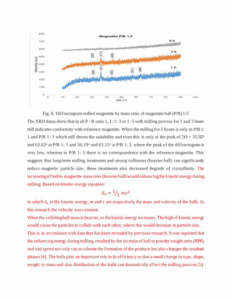
Fig. 6. Diffractogram milled magnetite by mass ratio of magnetite:ball (P/B) 1:5
The XRD datas show that in all P / B ratio 1, 1: 1: 3 or 1: 5 with milling process for 1 and 3 hours
still indicates conformity with reference magnetite. When the milling for 5 hours is only in P/B 1:
1 and P/B 1: 3 which still shows the suitability and even this is only at the peak of 2Ɵ = 35.92o
and 63.02o at P/B 1: 1 and 36.19o and 63.15o at P/B 1: 3, where the peak of the diffractogram is
very low, whereas in P/B 1: 5 there is no correspondence with the reference magnetite. This
suggests that long-term milling treatments and strong collisions (heavier ball) can significantly
reduce magnetic particle size, these treatments also decreased degrade of crystallinity. The
increasing of ball to magnetite mass ratio (heavier ball) would enhancing the k inetic energy during
milling. Based on kinetic energy equation:
𝐸𝑘 = 12⁄ 𝑚𝑣2
in which 𝐸𝑘 is the kinetic energy, m and v are respectively the mass and velocity of the balls. In
this research the velocity was constant.
When the colliding ball mass is heavier, so the kinetic energy increases. The high of kinetic energy
would cause the particles to collide with each other, where this would decrease in particle size.
This is in accordance with data that has been revealed by previous research. It was reported that
the enhancing energy during milling, resulted by the increase of ball to powder weight ratio (BPR)
and vial speed not only can accelerate the formation of the products but also changes the resultant
phases [4]. The balls play an important role in its efficiency so that a small change in type, shape,
weight or mass and size distribution of the balls can dramatically affect the milling process [5].
The increase of the number of balls at high BPR ratio, has a quite negative effect on the milling
performance [6].
Particle size of magnetite
This matter proves that the milling process is done to magnetite powder can causing the destruction
of the grains magnetite powder as a result collision between magnetite powder and milling balls.
To know more clearly destruction of graphite powder during process milling, then the
measurement magnetite particles by particle size analyzer (PSA) instrument. The choice of particle
measurement methods of nanoscale and micro size is usually by using a wet method PSA (particle
size analyzer) method, because it is an accurate method when compared to other methods. Small
particles have a tendency for high agglomeration, the choice of wet method on PSA because the
particles are dispersed into the medium so that the particles do not agglomerate (clump). Therefore
the measured particle size is the size of a single particle and provides overall information on sample
conditions.
Distribution particle size test by particle size analyzer (PSA) aims to determine particle size
distribution after mechanical milling process by HEM-3D for 1 hour, 3 hours and 5 hours. The
result of milled magnetite can be seen in Fig. 7.
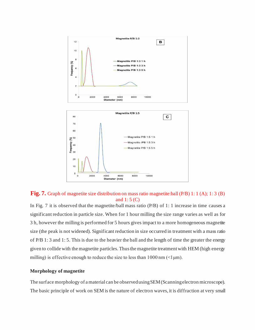
Fig. 7. Graph of magnetite size distribution on mass ratio magnetite:ball (P/B) 1: 1 (A); 1: 3 (B)
and 1: 5 (C) In Fig. 7 it is observed that the magnetite/ball mass ratio (P/B) of 1: 1 increase in time causes a
significant reduction in particle size. When for 1 hour milling the size range varies as well as for
3 h, however the milling is performed for 5 hours gives impact to a more homogeneous magnetite
size (the peak is not widened). Significant reduction in size occurred in treatment with a mass ratio
of P/B 1: 3 and 1: 5. This is due to the heavier the ball and the length of time the greater the energy
given to collide with the magnetite particles. Thus the magnetite treatment with HEM (high energy
milling) is effective enough to reduce the size to less than 1000 nm (<1μm).
Morphology of magnetite
The surface morphology of a material can be observed using SEM (Scanning electron microscope).
The basic principle of work on SEM is the nature of electron waves, it is diffraction at very small
angles. Electrons are dissipated by a charged sample. The image formation on SEM comes from
the electron beam reflected by the sample surface. If the sample used is not conductive, the sample
must first be coated with gold [16].
Based on the SEM image in Fig. 8, the addition of spherical periods has an effect on the reduction
of natural magnetite particle size. In the P/B ratio 1: 1 the particle size varies from small to large
size. When the mass of balls increase 3 times to magnetite (P/B 1: 3), the collision between the
magnetite and the ball gets stronger or the greater the energy that causes the breaking of the
particles to become smaller and appear more homogeneous. In addition to the ball up to 5 times
the magnetite period (P/B 1: 5) the particles also become smaller but the possibility of
agglomeration appears to be larger if compared to P/B 1: 3. The size of the magnetite particles is
slightly affected by the length of time the collision with the ball on the planetary ball mill. The
milling process for 1 to 3 hours gives almost the same result, observed on surface morphology at
P/B 1: 1 for 1 hour is almost equal to 3 hours. Similarly to P/B 1: 3 for 1 hour is almost the same
as for 3 hours, and P/B 1: 5 for 1 hour with 3 hours. However, when the milling for 5 hours on the
three variations of the ball period gives significantly different results with the previous. This is
especially observed in P/B 1: 3 for 5 hours, visible particles having clear and firm shape and cleaner
than others.
P/B 1:1 1h P/B 1:1 3h P/B 1:1 5h
P/B 1:3 1h P/B 1:3 3h P/B 1:3 5h
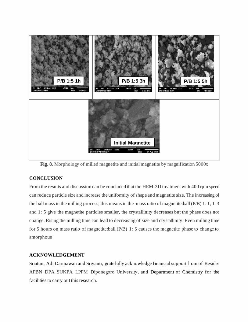
Fig. 8. Morphology of milled magnetite and initial magnetite by magnification 5000x
CONCLUSION
From the results and discussion can be concluded that the HEM-3D treatment with 400 rpm speed
can reduce particle size and increase the uniformity of shape and magnetite size. The increasing of
the ball mass in the milling process, this means in the mass ratio of magnetite:ball (P/B) 1: 1, 1: 3
and 1: 5 give the magnetite particles smaller, the crystallinity decreases but the phase does not
change. Rising the milling time can lead to decreasing of size and crystallinity. Even milling time
for 5 hours on mass ratio of magnetite:ball (P/B) 1: 5 causes the magnetite phase to change to
amorphous
ACKNOWLEDGEMENT
Sriatun, Adi Darmawan and Sriyanti, gratefully acknowledge financial support from of Besides
APBN DPA SUKPA LPPM Diponegoro University, and Department of Chemistry for the
facilities to carry out this research.
P/B 1:5 1h P/B 1:5 3h P/B 1:5 5h
Initial Magnetite
before treat
REFERENCES
[1]. Nugraha, P.A.; Sari, S.P.; Hidayati, W.N.; Dewi,C.R.; Kusuma, D.Y. AIP Conference Proceedings, 2016, 1747 (1)
[2]. Shpotyuk, O.; Bujňáková, Z.; Sayagués, M.J.; Baláž, P.; Ingram, A.; Ya.Shpotyuk, Demchenko, P. Materials Characterization, 2017, 132: 303-311.
[3]. Gong, J. Journal Hazardous Mat., 2009, 164:1517-1522
[4]. Hui, C.; Shen, C.; Yang, T.; Bao, L.; Tian, J.; Ding, H.; Li, C.; Gao, H.J. J. Phys. Chem. C. 2008, 112, 11336-11339.
[5]. Klotz, S.; Steinle-Neumann, G.; Strassle, T.; Philippe, J.; Hansen, T.; Wenzel, M.J. Phys. Rev. B, 2008, 77, 12411-1-1241-4.
[6]. Marinca, T.; Chicinaș, H.; Neamțu, B.; Popa, F.; Isnard, O.; Chicinaș, I. Studia Universitatis Babes-Bolyai, Physica, 2015, 60 (1).
[7]. Darezereshki, E.; Bakhtiari, F.; Alizadeh, M.; Ranjbar, M. Materials Science in Semiconductor Processing, 2012, 15(1): 91-97.
[8]. Khan, U.S.; Rahim, A.; Khan, N.; Muhammad, N.; Rehman, F.; Ahmad, K.; Iqbal, J. Materials Chemistry and Physics, 2017, 189: 86-89.
[9]. Chikan, V. and McLaurin, E. J. Nanomaterials,2016, 6(5): 85
[10]. An,J.S.; Han, W.J.; Choi, H.J. Colloids and Surfaces A: Physicochemical and Engineering
Aspects, 2017, 535:16-23
[11]. Bui, T.Q.; Ton, S.N.; Duong, A.T.; Tran, H.T. Journal of Science: Advanced Materials and
Devices, 2017, Available online 14 November 2017
[12]. Balaz, P. Mechanochemistry in Nanoscience and minerals Engineering, 2008, Springer-
Verlag Berlin Heidelberg, 103.
[13]. Bolokang, S., Banganayi, C., Phasha, M. Int. J. Refract. Met. Hard Mater, 2010, 28:211–
216.
[14]. Zakeri, M., Rahimipour, M.R. Adv. Powder Technol, 2012, 23:31–34
[15]. Ghayour, H., Abdellahi , M., Bahmanpour, M. Powder Technology, 2016, 291: 7–13
[16]. Prabakaran, K.; Balamurunga, A.; Rajeswari, S. Bull Mat Sci, 2005, 28:115-119.

29/10/2020 Invoice of your article (OJC-11729-17) - [email protected] - Diponegoro University Mail
https://mail.google.com/mail/u/1/?tab=wm&ogbl#search/iqbalojc%40yahoo.com/FMfcgxmXLGgVjlgQJKWmZWTTpvdMqTsv 1/1
Meet
Hangouts
Compose
More
Inbox 213
Starred
Snoozed
Sent
New meeting
My meetings
Sriatun
No Hangouts contacts Find someone
Invoice of your a�icle (OJC-11729-17) Inbox
Dr. S.A. Iqbal <[email protected]>
to me
With reference to your article accepted for publication in the current issue
You are urgently requested to pay the charges within a week
Final publication of article will be subjected to “Cross Check” (Ithenticate)
The bank details for the transfer of publication charges areas follows
Account Holder: Oriental Scientific Publishing Company
Address: 9, Gulshan -E-Iqbal,New Maulana Azad Colony , Idgah Hills, Bh
Bank: State Bank of India
Branch: New Market, T.T. Nagar,Bhopal - 462 001, INDIA
Account No.: 33293116175
Swift no. SBI NINBB 268
IFS Code: SBIN0000332
You are requested to please send extra charges for bank deduction so that w

28/10/2020 Invoice of your article (OJC-11729-17) - [email protected] - Diponegoro University Mail
https://mail.google.com/mail/u/1/?tab=wm&ogbl#search/iqbalojc%40yahoo.com/FMfcgxmXLGgVjlgQJKWmZWTTpvdMqTsv 1/1
Meet
Hangouts
Compose
More
Inbox 213
Starred
Snoozed
Sent
New meeting
My meetings
Sriatun
No Hangouts contacts Find someone
Account No.: 33293116175
Swift no. SBI NINBB 268
IFS Code: SBIN0000332
You are requested to please send extra charges for bank deduction so that w
Regards Dr. MeenaIqbal Editor cum
(Remittance in charge)
Looking forward to hear from you soon. Regards Dr. Meena Iqbal Editor
S i t S i t

28/10/2020 Invoice of your article (OJC-11729-17) - [email protected] - Diponegoro University Mail
https://mail.google.com/mail/u/1/?tab=wm&ogbl#search/iqbalojc%40yahoo.com/FMfcgxmXLGgVjlgQJKWmZWTTpvdMqTsv 1/1
Meet
Hangouts
Compose
More
Inbox 213
Starred
Snoozed
Sent
New meeting
My meetings
Sriatun
No Hangouts contacts Find someone
Sriatun Sriatun <[email protected]>
to S.A.
Semarang, January, 13, 2018To:Dr. S.A. Iqbal Chief editor of OJC
Here I send proof of transfer for the payment of publishing the man
Thankyou for the opportunity to contribute as well as feedback from
Best regards
Sriatun
Dept.of Chemistry, Diponegoro University, Semarang, Central of Java, Ind
Proof of payment_…
Reply Forward
t\S BNIValidasi:
Formulir Kiriman UangRe m itta n ce A pp I ic ati o n
Penetinal Beneficiory
Nama/ Norne :
Alamall Address :
Ieleponl Phone :
Kol"l City .
IPenduduk/Resident
. Bukan Penduduk/ lenis pengiriman/ : LLC t CteoringNon Retident type ol fran\ls RTCS
fl oraft i ,
[l SWIFT
Pengirim/ R1mitte( PenduduldResident
Nama/ Nome :
Nama Alias/ A/io5 Nome :
No. lDKrP / tlM / Po\\pon / (ITAS
Alamatt Addre\s :
Ielepo^l Phone :
Negata/ Country :
Sumber Dana/ Source of furd :
'' [:] Tunai/ cosh n cek/Bc No.Debit Ret./ Debir 4(c. No. .................
Mata Uangl Cuffency . ll uso lNeqaral Country '.
Bank Penerima/ Eereficiory Bottk:
Kota/ City :
No Rek./ Acc. No. :
Negan/ Country :
Bukan Pend0duk/Non Resident
KoIal City .
::f:: l:1::i:i' l*i!i' 1"!!")i ' . -:iillil"l:?t l .
f umlah Dana yang djkiJiml Amount T@nsfer '.
Komisi/ C!mmii5ionPer'giifianl Handling
Biaya dari bank koresponden dibebankan ke rekening/Colretpondenl bonk ( harycs ore lor o(@unl ol
Penetima. Be,ieficiory Pengii.n/ Remilt?r Sharing
m€nyetui!i sepenuhnya syarat syarartercant0m pada haamar belakang
Vi^t I l,n.ond bnallt at ept nllthe*d,"",it,"", "" th. -,-* 1",,"
e
Pejabat Bank/ Bonk Officet Pemohonl Applicontti@qttua h.rd|a,n\r
ffi
: 5111{, 111|,1 tlr'1a: 10:59:05
OI P$BI,ICATIC[
0f Charges:
o leceiver lnfcrnation;/AT II{DIAIOT CflAS€S OT PSBI,ICAIIOil
rH
I
I
t\5BN
rrqig"/
I
.t"\ t
q\r?le

29/10/2020 TOC : Oriental Journal of Chemistry
www.orientjchem.org/toc/?vol=34&no=2 1/11
ISSN : 0970 - 020X, ONLINE ISSN : 2231-5039
Search...
Views: 970 PDF Downloads: 1390
Views: 1,118 PDF Downloads: 971
Views: 566 PDF Downloads: 911
Views: 1,162 PDF Downloads: 1002
Views: 561 PDF Downloads: 920
TOC Volume 34 Number 2
A Decade of Development of Ethylidenethiosemicarbazides as Building Blocks for Synthesis of Azoles andAzines (A Review) Sayed M. Riyadh , Shojaa Abed El-Motairi and Anwar A. Deawaly[ HTML Full Text] [ Abstract ] [PDF] [ XML]
DOI : http://dx.doi.org/10.13005/ojc/340201
Methods for the Preparation of Modified Polyorganosiloxanes (A Review)Kostyukovich Alexander Yur’evich , Drozdov Fedor Valer’evich , Vitaly Sergeevich Ivanov , Anton Sergeevich Yegorov andVladimir Viktorovich Men'shikov
[ HTML Full Text] [ Abstract ] [PDF] [ XML]
DOI : http://dx.doi.org/10.13005/ojc/340202
Diethyldithiocarbamate Doped Graphene Quantum Dots Based Metal Complex Nanoparticles byResonance Light Scattering for Green Detection of Lead(II) – (A Review)Chayanee Kaewprom , Prawit Nuengmatcha and Saksit Chanthai[ HTML Full Text] [ Abstract ] [PDF] [ XML]
DOI : http://dx.doi.org/10.13005/ojc/340203
Laccase Biosensor: Green Technique for Quantification of Phenols in Wastewater (A Review)Yashas S. R , Shivakumara B. P, Udayashankara T. H and Krishna B. M.[ HTML Full Text] [ Abstract ] [PDF] [ XML]
DOI : http://dx.doi.org/10.13005/ojc/340204
Nigella Sativa L. Seeds Biomass as A Potential Sorbent of Lead from Aqueous Solutions and WastewatersAbdelhamid Addala , Noureddine Belattar and Maria Elektorowicz[ HTML Full Text] [ Abstract ] [PDF] [ XML]
DOI : http://dx.doi.org/10.13005/ojc/340205
Gas Sensor with Reduced Humidity Response Based on Metal Oxide Nanoparticles Synthesized by SparkDischargeAlexey Vasiliev , Andrey Varfolomeev , Ivan Volkov , Pavel Arsenov , Alexey Efimov , Victor Ivanov , Alexander Pislyakov ,Alexander Lagutin and Thomas Maeder
A New Edition of Web of Science
CNKI Scholar (China National Knowledge Infrastructure)
Journal Archived in:
Life Fellowship Awards in
Chemistry by Researchers Societyof Chemical Sciences, Bhopal,
India.
Membership Form of Researchers
Society of Chemical Sciences Email: [email protected]
Click here for RSCS Details
1,2 1 1
1 2 1 1
1
1 2 1
1 1 2
1,2 1,2 1 1 1 1 2
2 1,3
Home About Us Editorial Board Indexed in Current Issue Coming Issue Archives Submission
Contact Us

29/10/2020 TOC : Oriental Journal of Chemistry
www.orientjchem.org/toc/?vol=34&no=2 2/11
Views: 371 PDF Downloads: 580
Views: 761 PDF Downloads: 655
Views: 495 PDF Downloads: 610
Views: 318 PDF Downloads: 497
Views: 528 PDF Downloads: 631
Views: 472 PDF Downloads: 697
Views: 738 PDF Downloads: 665
[ HTML Full Text] [ Abstract ] [PDF] [ XML]
DOI : http://dx.doi.org/10.13005/ojc/340206
Synthesis and in Vitro Antimalarial Activity of Alkyl Esters Gallate as a Growth Inhibitors of PlasmodiumFalciparum
Ade Arsianti , Hendry Astuti , Fadilah , Daniel Martin Simadibrata , Zoya Marie Adyasa , Daniel Amartya , Anton Bahtiar ,Hiroki Tanimoto and Kiyomi Kakiuchi[ HTML Full Text] [ Abstract ] [PDF] [ XML]
DOI : http://dx.doi.org/10.13005/ojc/340207
Characterization of Jordanian Porcelanite Rock with Reference to the Adsorption Behavior of Lead ionsfrom Aqueous SolutionJumana K. Abu-Hawwas , Khalil. M. Ibrahim and Salem M. Musleh[ HTML Full Text] [ Abstract ] [PDF] [ XML]
DOI : http://dx.doi.org/10.13005/ojc/340208
Investigation of Electrochemical Behaviour of Chromium(VI)-Dithiocarbamate Complexes: Detection ofChromium(VI) in Real Samples
Niranjan Thondavada , Giridhar Chembeti , Gan. G. Redhi and Venkatasubba Naidu Nuthalapati[ HTML Full Text] [ Abstract ] [PDF] [ XML]
DOI : http://dx.doi.org/10.13005/ojc/340209
Development of Antimicrobial Hybrid Materials from Polylactic Acid and Nano-Silver Coated ChitosanNollapan Nootsuwan , Kankavee Sukthavorn , Worawat Wattanathana , Suchada Jongrungruangchok , Chatchai Veranitisagul ,Nattamon Koonsaeng and Apirat Laobuthee[ HTML Full Text] [ Abstract ] [PDF] [ XML]
DOI : http://dx.doi.org/10.13005/ojc/340210
Removal of Lead (ΙΙ) by Lumbang, Aleurites moluccana Activated Carbon CarboxymethylcelluloseComposite Crosslinked with EpichlorohydrinNelson R. Villarante , Ronnette Anne E. Davila and Derick Erl P. Sumalapao[ HTML Full Text] [ Abstract ] [PDF] [ XML]
DOI : http://dx.doi.org/10.13005/ojc/340211
Effect of Alkali Concentration on Mechanical Properties, Microstructure, Zeta Potential and ElectricalConductivity of Thermally Cured Fly-Ash-Blast Furnace Slag Based Blended Geopolymer CompositesKushal Ghosh and Partha Ghosh[ HTML Full Text] [ Abstract ] [PDF] [ XML]
DOI : http://dx.doi.org/10.13005/ojc/340212
1,3 2 1,3 5 5 5 4
6 6
1 2 3
1 2 1 2
1 2 1 3 2
4 1
1 1 2,3

29/10/2020 TOC : Oriental Journal of Chemistry
www.orientjchem.org/toc/?vol=34&no=2 3/11
Views: 605 PDF Downloads: 681
Views: 534 PDF Downloads: 638
Views: 322 PDF Downloads: 486
Views: 572 PDF Downloads: 658
Views: 412 PDF Downloads: 658
Views: 568 PDF Downloads: 590
Views: 360 PDF Downloads: 529
Synthesis and Characteristics of the Magnetic Properties of Fe O –(CTAB-montmorillonite) Composites,based on Variation in Fe /Fe Concentrations
Sri Hilma Siregar , Karna Wijaya , Eko Sri Kunarti and Akhmad Syoufian
[ HTML Full Text] [ Abstract ] [PDF] [ XML]
DOI : http://dx.doi.org/10.13005/ojc/340213
Validation of an Analytical Methodology for the Determination of Chloramphenicol Residues in Honeyusing UPLC-MS/MSSyed Amir Ashraf and Z. R. Azaz Ahmad Azad[ HTML Full Text] [ Abstract ] [PDF] [ XML]
DOI : http://dx.doi.org/10.13005/ojc/340214
Sequential Injection at Valve Mixing (SI-VM) for Determination of Albumin-Creatinine Ratio in Urine
Akhmad Sabarudin[ HTML Full Text] [ Abstract ] [PDF] [ XML]
DOI : http://dx.doi.org/10.13005/ojc/340215
Structural Analysis of Powdered Manganese(II) of 1,10-Phenanthroline (phen) as Ligand andTrifluoroacetate (TFA) as Counter AnionKristian Handoyo Sugiyarto , Cahyorini Kusumawardani, Hari Sutrisno and Muhammad Wahyu Arif Wibowo
[ HTML Full Text] [ Abstract ] [PDF] [ XML]
DOI : http://dx.doi.org/10.13005/ojc/340216
Polyimide Composite Materials Containing Modified Nanostructured Boron Carbide
Elena Aleksandrovna Averina, Anton Sergeyevich Yegorov , Marina Vladimirovna Bogdanovskaya, Alyona Igorevna Wozniak andOlga Anatolevna Zhdanovich
[ HTML Full Text] [ Abstract ] [PDF] [ XML]
DOI : http://dx.doi.org/10.13005/ojc/340217
Synthesis and Characterization of Natural Ca(OH) /KF Superbase Catalyst for Biodiesel Production fromPalm OilI. F. Nurcahyo , Karna Wijaya , Triyono , Arief Budiman and Yun Hin Taufiq-Yap[ HTML Full Text] [ Abstract ] [PDF] [ XML]
DOI : http://dx.doi.org/10.13005/ojc/340218
Electrochemical Preparation of the Ti/Ni-Sb-SnO for Phenol Removal by In-situ Generated OzoneAli Reza Rahmani, Mohammad Taghi Samadi, Mohammad Reza Samarghandi and Ghasem Azarian[ HTML Full Text] [ Abstract ] [PDF] [ XML]
DOI : http://dx.doi.org/10.13005/ojc/340219
3 43+ 2+
1 2 2 2
1 2
2
1,2 1 1 3 4,5
2

29/10/2020 TOC : Oriental Journal of Chemistry
www.orientjchem.org/toc/?vol=34&no=2 4/11
Views: 473 PDF Downloads: 663
Views: 280 PDF Downloads: 476
Views: 363 PDF Downloads: 532
Views: 1,136 PDF Downloads: 762
Views: 585 PDF Downloads: 560
Views: 783 PDF Downloads: 797
Views: 521 PDF Downloads: 559
Novel Organotin(IV) Complexes Derived from Chiral Benzimidazoles: Synthesis, Molecular Structure andSpectral PropertiesA. Akremi , A. Noubigh and M. J. A. Abualreish[ HTML Full Text] [ Abstract ] [PDF] [ XML]
DOI : http://dx.doi.org/10.13005/ojc/340220
Study on the Effect of Deposition Parameters on Characteristics of Electrochemically SynthesizedPolyaniline and Poly O-ToluidineMonimul Huque , Golam Haider , Shah Rokonuzzaman and Aksaruzzaman Nuri[ HTML Full Text] [ Abstract ] [PDF] [ XML]
DOI : http://dx.doi.org/10.13005/ojc/340221
An Efficient Synthesis and In Vitro Antimicrobial Screening of 2-Cyanoimino -4-aryl-6-(1,1'-biphenyl-4-yl)-3,4-dihydro-1H-PyrimidinesSivagami Swaminathan and Ingarsal Namasivayam[ HTML Full Text] [ Abstract ] [PDF] [ XML]
DOI : http://dx.doi.org/10.13005/ojc/340222
An Eco-friendly Approach for Synthesis of Platinum Nanoparticles using Leaf Extracts of JatropaGossypifolia and Jatropa Glandulifera and its Antibacterial ActivityU. Jeyapaul , Mary Jelastin Kala , A. John Bosco , Prakash Piruthiviraj and M. Easuraja[ HTML Full Text] [ Abstract ] [PDF] [ XML]
DOI : http://dx.doi.org/10.13005/ojc/340223
Facile Synthesis of Biologically Derived Fluorescent Carbon Nanoparticles (Fcnps) from an AbundantMarine Alga and its Biological ActivitiesMaria Theresa F. Calangian , Abigail B. Ildefonzo , Vanessa Kate S. Manzano , Genesis Julyus T. Agcaoili , Rey Joseph J. Ganado ,Allan Christopher C. Yago , Eduardo R. Magdaluyo, Jr , Ross D. Vasquez and Francisco C. Franco Jr[ HTML Full Text] [ Abstract ] [PDF] [ XML]
DOI : http://dx.doi.org/10.13005/ojc/340224
Synthesis, Characterization and In Vitro Antitubercular and Antimicrobial Activities of newAminothiophene Schiff Bases and Their Co(II), Ni(II), Cu(II) and Zn(II) Metal ComplexesGanesh More , Sakina Bootwala , Sushma Shenoy , Joyline Mascarenhas and K. Aruna[ HTML Full Text] [ Abstract ] [PDF] [ XML]
DOI : http://dx.doi.org/10.13005/ojc/340225
Seasonal Influences on the Levels of Particulate Cd, Cr and PB in Kuantan River, PahangKamaruzzaman Yunus , Fikriah Faudzi , Mohd. Fuad Miskon and Mohd Mokhlesur Rahman[ HTML Full Text] [ Abstract ] [PDF] [ XML]
DOI : http://dx.doi.org/10.13005/ojc/340226
1 2 3
1 2 2 2
1 2 1 3 4
1 1 1 1 2
3 4 5 2
1 1 2 2 2
1 1 1 2

29/10/2020 TOC : Oriental Journal of Chemistry
www.orientjchem.org/toc/?vol=34&no=2 5/11
Views: 555 PDF Downloads: 558
Views: 339 PDF Downloads: 522
Views: 354 PDF Downloads: 491
Views: 337 PDF Downloads: 571
Views: 426 PDF Downloads: 604
Views: 505 PDF Downloads: 563
Views: 1,060 PDF Downloads: 785
Molecular Docking as A Computational Tool for Analyzing Product Mediated Inhibition for β-GalactosidaseImmobilized on Glutaraldehyde Modified MatricesShakeel Ahmed Ansari , Mohammad Alam Jafri , Rukhsana Satar , Syed Ismail Ahmad and Sandesh Chibber[ HTML Full Text] [ Abstract ] [PDF] [ XML]
DOI : http://dx.doi.org/10.13005/ojc/340227
Synthesis and Biological Evaluation of Certain new Cyclohexane-1-carboxamides as Apoptosis InducersWalaa Hamada Abd-Allah and Mohamed Fathy Elshafie
[ HTML Full Text] [ Abstract ] [PDF] [ XML]
DOI : http://dx.doi.org/10.13005/ojc/340228
Solvent free Cationic Copolymerization of 2-Chloroethyl Vinyl Ether with Styrene Catalyzed by Maghnite-H , a Green CatalystChabani Malika , Meghabar Rachid and Belbachir Mohammed
[ HTML Full Text] [ Abstract ] [PDF] [ XML]
DOI : http://dx.doi.org/10.13005/ojc/340229
Synthesis of High-Sulfur Polymers by Redox Copolymerization of Elemental Sulfur with PyrroleGaukhar Bishimbayeva , Galina Prozorova , Dinara Zhumabayeva , Svetlana Korzhova , Irina Mazyar , Arailym Nalibayeva andUldana Kydyrbayeva[ HTML Full Text] [ Abstract ] [PDF] [ XML]
DOI : http://dx.doi.org/10.13005/ojc/340230
Anticancer Activity of Cu(II), Pd(II) and Zn(II) Complexes of Phosphonate with Glutamine Amino AcidAhmed Imam Hanafy , Zeinhom Mohamad El-Bahy , Marwa Sayed El-Gendy and Omar Makram Ali[ HTML Full Text] [ Abstract ] [PDF] [ XML]
DOI : http://dx.doi.org/10.13005/ojc/340231
Physicochemical Characteristics and Photocatalytic Activity of Silver Nanoparticles-decorated on NaturalHalloysite (An aluminosilicate clay)
I. S. Fatimah and Rivaldo Herianto
[ HTML Full Text] [ Abstract ] [PDF] [ XML]
DOI : http://dx.doi.org/10.13005/ojc/340232
Synthesis and Antibacterial Activity 1-Monolaurin
Febri Odel Nitbani , Jumina , Dwi Siswanta , Eti Nurwening Sholikhah and Dhina Fitriastuti
[ HTML Full Text] [ Abstract ] [PDF] [ XML]
DOI : http://dx.doi.org/10.13005/ojc/340233
1 1 2 3 4
1 2
+1,2 1 1
1 2 1 2 2 1
1
1,2 1,2 3,4 3,5
1 2 3 4 4

29/10/2020 TOC : Oriental Journal of Chemistry
www.orientjchem.org/toc/?vol=34&no=2 6/11
Views: 358 PDF Downloads: 483
Views: 368 PDF Downloads: 530
Views: 619 PDF Downloads: 547
Views: 452 PDF Downloads: 536
Views: 854 PDF Downloads: 665
Views: 343 PDF Downloads: 484
Views: 516 PDF Downloads: 677
Microstructure Characterization of Natural Magnetite from Sand Marina Beach By High Energy MillingSriatun , A. Darmawan, Sriyanti and W. Cahyani[ HTML Full Text] [ Abstract ] [PDF] [ XML]
DOI : http://dx.doi.org/10.13005/ojc/340234
Phytosterol Screening of Skin and Seed Extracts of Wild Grape Ampelocissus Martinii Planch. FruitsWilaiwan Simchuer and Prasong Srihanam[ HTML Full Text] [ Abstract ] [PDF] [ XML]
DOI : http://dx.doi.org/10.13005/ojc/340235
Carbon–Carbon Bond Formation Reaction with Pd/reduced Graphene Oxide CompositePadmakar Anant Kulkarni, Suresh Shamrao Shendage and Ashok Ganuji Awale[ HTML Full Text] [ Abstract ] [PDF] [ XML]
DOI : http://dx.doi.org/10.13005/ojc/340236
Utilization of Natural Zeolite Clipnotilolit-Ca as a Support of ZnO Catalyst for Congo-Red Degradation andCongo-Red Waste Applications with PhotolysisZilfa , Rahmayeni, Yeni Stiadi and Adril[ HTML Full Text] [ Abstract ] [PDF] [ XML]
DOI : http://dx.doi.org/10.13005/ojc/340237
Green Chemiluminescence of Highly Fluorescent Symmetrical Azo-Based Luminol DerivativeSimon Deepa , Sabbasani Rajasekhara Reddy and Kannapiran Rajendrakumar[ HTML Full Text] [ Abstract ] [PDF] [ XML]
DOI : http://dx.doi.org/10.13005/ojc/340238
Investigation of Different Ferrous Concentration Effect on Characteristics of NiFeP Nano Alloy Thin FilmsC. Devi and R. Ashokkumar[ HTML Full Text] [ Abstract ] [PDF] [ XML]
DOI : http://dx.doi.org/10.13005/ojc/340239
Simple Potentiometric Strategy for the Detection of Levofloxacin Hydrochloride and DaclatasvirDihydrochloride in Pure form and Pharmaceutical Preparations
Amira S. Eldin , Mona M. Abdel-Moety , Aliaa S. M. El-Tantawy , Abdalla Shalaby and Magda El-Maamly[ HTML Full Text] [ Abstract ] [PDF] [ XML]
DOI : http://dx.doi.org/10.13005/ojc/340240
Improving Oil Products Quality by Vermiculite SorbentKulash K. Syrmanova , Zhanat B. Kaldybekova , Nurzhan Y. Botabayev , Yersultan T. Botashev , Moldir T. Suleimenova ,Boris Y. Beloborodov and Tatyana V. Rivkina[ HTML Full Text] [ Abstract ] [PDF] [ XML]
1 2 1
1,2 3
1 1 1 2 2
1,2 1 1 1 2
1 1

29/10/2020 TOC : Oriental Journal of Chemistry
www.orientjchem.org/toc/?vol=34&no=2 7/11
Views: 302 PDF Downloads: 486
Views: 409 PDF Downloads: 544
Views: 374 PDF Downloads: 501
Views: 533 PDF Downloads: 489
Views: 528 PDF Downloads: 527
Views: 545 PDF Downloads: 3226
Views: 343 PDF Downloads: 477
DOI : http://dx.doi.org/10.13005/ojc/340241
Chitosan Isolated from Horseshoe Crab Tachypleus Gigas from the Malay Peninsula
Zaleha Kassim , Wan Nurul Akmal Wan Murni , Mohd Razali Md Razak and Sakinah Begum Adam
[ HTML Full Text] [ Abstract ] [PDF] [ XML]
DOI : http://dx.doi.org/10.13005/ojc/340242
Formation Conditions of Hydrocarbon Systems on the Sakhalin Shelf of the Sea of Okhotsk Based on theGeochemical Studies and ModelingV. Yu. Kerimov, G. N. Gordadze, R. N. Mustaev and A. V. Bondarev[ HTML Full Text] [ Abstract ] [PDF] [ XML]
DOI : http://dx.doi.org/10.13005/ojc/340243
Synthesis, Characterization and Swelling study of Poly (Methacrylic acid-co-Maleic acid) HydrogelsSoumia Belkadi , Hayet Bendaikha , Fouad Lebsir and Seghier Ould-Kada
[ HTML Full Text] [ Abstract ] [PDF] [ XML]
DOI : http://dx.doi.org/10.13005/ojc/340244
Synthesis of Co, Mo, Co-Mo and Mo-Co Catalysts, Supported on Mesoporous Silica-Alumina forHydrocracking of α-Cellulose pyrolysis Oil
Muhammad Fajar Marsuki, Wega Trisunaryanti , Iip Izul Falah and Karna Wijaya
[ HTML Full Text] [ Abstract ] [PDF] [ XML]
DOI : http://dx.doi.org/10.13005/ojc/340245
Synthesis and Characterization of all Possible Diastereoisomers of AlvimopanBeeravalli Ramalinga Reddy , Manoj Kumar Dubey , Ch. Venkata Ramana Reddy and Rakeshwar Bandichhor
[ HTML Full Text] [ Abstract ] [PDF] [ XML]
DOI : http://dx.doi.org/10.13005/ojc/340246
Effect of TiO2 For Generation of H2/O2 Gases, Based on Splited water and UV as InisiatorMinto Supeno and Rikson Siburian[ HTML Full Text] [ Abstract ] [PDF] [ XML]
DOI : http://dx.doi.org/10.13005/ojc/340247
Lithium Including Mixed Sodium Inside Graphene Oxide (GO) as Anodic Electrodes for Ion BatteriesSamira Bagheri , Majid Monajjemi , Alireza Ziglari and Afshin Taghva Manesh[ HTML Full Text] [ Abstract ] [PDF] [ XML]
DOI : http://dx.doi.org/10.13005/ojc/340248
1 1 1 2
1 1,2 1 1
1,2 1 2 1
1 2 1 1

29/10/2020 TOC : Oriental Journal of Chemistry
www.orientjchem.org/toc/?vol=34&no=2 8/11
Views: 363 PDF Downloads: 508
Views: 564 PDF Downloads: 604
Views: 583 PDF Downloads: 529
Views: 321 PDF Downloads: 484
Views: 1,685 PDF Downloads: 938
Views: 294 PDF Downloads: 550
Views: 323 PDF Downloads: 500
Ullmann Reaction Optimization Within Bitolyl and Decafluorobiphenyl SynthesisA. V. Kolotaev, A. L. Razinov and D. S. Khachatryan[ HTML Full Text] [ Abstract ] [PDF] [ XML]
DOI : http://dx.doi.org/10.13005/ojc/340249
Titanium dioxide as a Catalyst for Photodegradation of Various Concentrations of Methyl Orangeand Methyl Red dyes using Hg Vapour Lamp with Constant pHA. Kistan , V. Kanchana , L. Sakayasheela , J. Sumathi , A. Premkumar , A. Selvam and Thaminum Ansari A[ HTML Full Text] [ Abstract ] [PDF] [ XML]
DOI : http://dx.doi.org/10.13005/ojc/340250
An Investigation into Transesterification of Waste Cooking OilHala Mohamed Abo-Dief , Ashraf Salah Emam , Khamael Mohammed Abualnaja and Ashraf Talaat Mohamed
[ HTML Full Text] [ Abstract ] [PDF] [ XML]
DOI : http://dx.doi.org/10.13005/ojc/340251
Synthesis and Structural Profile Analysis of the MgO Nanoparticles Produced Through the Sol-Gel MethodFollowed by Annealing ProcessI. Wayan Sutapa , Abdul Wahid Wahab , Paulina Taba and Nursiah La Nafie
[ HTML Full Text] [ Abstract ] [PDF] [ XML]
DOI : http://dx.doi.org/10.13005/ojc/340252
Composite Materials Based on Ultrahigh Molecular Weight Polyethylene and Rare-Earth Element Oxides
Alexey Mikhailovich Nemeryuk, Marina Mikhailovna Lylina, Marina Vladimirovna Bogdanovskaya, Elena Aleksandrovna Averina andAnton Sergeyevich Yegorov
[ HTML Full Text] [ Abstract ] [PDF] [ XML]
DOI : http://dx.doi.org/10.13005/ojc/340253
Removal of Ammonia Nitrogen NH-N and Hexavalent Chromium (VI) From Wastewater Using AgriculturalWaste Activated Carbon
Iqbal Khalaf Erabee
[ HTML Full Text] [ Abstract ] [PDF] [ XML]
DOI : http://dx.doi.org/10.13005/ojc/340254
Comparative Study on Janus Kinase Enzyme Activity of Pomegranate Leaf Extract and its ActiveComponent Ellagic Acid for AsthmaSarithamol S, V. L. Pushpa and K. B. Manoj[ HTML Full Text] [ Abstract ] [PDF] [ XML]
1,2 3 2 2 2 2 4
1,2 3,4 1 5
1 2 2 2
1,2

29/10/2020 TOC : Oriental Journal of Chemistry
www.orientjchem.org/toc/?vol=34&no=2 9/11
Views: 310 PDF Downloads: 453
Views: 391 PDF Downloads: 522
Views: 459 PDF Downloads: 487
Views: 659 PDF Downloads: 568
Views: 579 PDF Downloads: 620
Views: 416 PDF Downloads: 536
Views: 674 PDF Downloads: 525
DOI : http://dx.doi.org/10.13005/ojc/340255
Resistant in Alkaline Media Core-shell Photocatalyst of Fe(TiO / Al O ) for Degradation of Water PollutantAmir Hossein Haghighaty and Shahram Moradi Dehaghi[ HTML Full Text] [ Abstract ] [PDF] [ XML]
DOI : http://dx.doi.org/10.13005/ojc/340256
Pharmacological Studies of Root, Fruit and Flower of Berberis lyciumMansi Gupta and Ajay Singh[ HTML Full Text] [ Abstract ] [PDF] [ XML]
DOI : http://dx.doi.org/10.13005/ojc/340257
Determination of Physicochemical Properties and Fatty Acid Composition of "Kabate" Larva Oil from TimorIsland
Febri Odel Nitbani , Hermania Em Wogo , Titus Lapailaka and Beta Achromi Nurohmah
[ HTML Full Text] [ Abstract ] [PDF] [ XML]
DOI : http://dx.doi.org/10.13005/ojc/3402058
Mechanical and Thermal Properties of the Waste Low and High Density Polyethylene-Nanoclay CompositesArkan J. Hadi , H. K. AbdulKadir , Ghassan J. Hadi , Kamal Bin Yusoh and S. F. Hasany
[ HTML Full Text] [ Abstract ] [PDF] [ XML]
DOI : http://dx.doi.org/10.13005/ojc/340259
Bioaccumulation of Trace Metals in Selected Vegetable Crops around Tummalapalle Uranium Mine inKadapa District, Andhra PradeshS. Kulavardhana Reddy, C. Sivanandha Reddy and Gopireddy Venkata Subba Reddy[ HTML Full Text] [ Abstract ] [PDF] [ XML]
DOI : http://dx.doi.org/10.13005/ojc/340260
Oxidative Desulfurization of Hydrotreated Gas Oil using Fe O and Pd Loaded over Activated Carbon asCatalysts
Jalil R. Ugal , Rawnaq B. Jima'a , Wessal Metaab Khamis Al-Jubori , Bayader Fadhel Abbas and Nedhal Metaab Al-Jubori
[ HTML Full Text] [ Abstract ] [PDF] [ XML]
DOI : http://dx.doi.org/10.13005/ojc/340261
Stability Indicating UHPLC Method Development and Validation for Estimation of Eltrombopag and itsRelated Impurities in Tablet Dosage formTharlapu Satya Sankarsana Jagan Mohan , Khagga Mukkanti and Hitesh A Jogia[ HTML Full Text] [ Abstract ] [PDF] [ XML]
2 2 3
1 2
1 2 1 2
1 2 3 4 5
2 3
1 2 3 3 2
1,2 4 3

29/10/2020 TOC : Oriental Journal of Chemistry
www.orientjchem.org/toc/?vol=34&no=2 10/11
Views: 1,150 PDF Downloads: 802
Views: 514 PDF Downloads: 568
Views: 285 PDF Downloads: 446
Views: 710 PDF Downloads: 515
Views: 228 PDF Downloads: 480
Views: 1,001 PDF Downloads: 713
Views: 614 PDF Downloads: 614
DOI : http://dx.doi.org/10.13005/ojc/340262
Diagnosis, Structure, and in the Vitro Antimicrobial and Antifungal Evaluation of Some Amino BenzoicAcids -Derived Ligand Schiff Base and Their Mixed Complexes with Cu(II), Hg(II), Mn(II) ,Ni(II) and Co(II)Rehab Kadhem Rahem Al-Shemary , Lekaa Khalid Abdul Karem and Faeza Hasan Ghanim[ HTML Full Text] [ Abstract ] [PDF] [ XML]
DOI : http://dx.doi.org/10.13005/ojc/340263
Identification of Antimastits Componenets in Boerhavia Diffusa as an Inhibitor of Staphalococus aureusMonofunctional Glycosyltransferase, Causing Bovine Mastitis (An Insilico Approach)Sruthy B , M. S. Latha and Sherly P. Anand[ HTML Full Text] [ Abstract ] [PDF] [ XML]
DOI : http://dx.doi.org/10.13005/ojc/340264
Influence of Thermal Stability of Quartz and the Particle Size Distribution of Burden Materials on theProcess of Electrothermal Smelting of Metallurgical SiliconNikolay Nikolayevich Zobnin , Anatoliy Korneyevich Torgovets, Irina Anatolyevna Pikalova, Yuliya Sergeyevna Yussupova andSergey Asifogly Atakishiyev[ HTML Full Text] [ Abstract ] [PDF] [ XML]
DOI : http://dx.doi.org/10.13005/ojc/340265
Silver/oil Nanofluids in Heat Exchanger An Experimental Study on Convective Heat TransferKomeil Hosseini and Hamid Reza Ghorbani[ HTML Full Text] [ Abstract ] [PDF] [ XML]
DOI : http://dx.doi.org/10.13005/ojc/340266
Synthesis of Silver Nanoparticles in the Presence of Polyethylene Glycol and Their ElectrochemicalBehavior at A Graphite Electrode by Cyclic VoltammetryDarya O. Perevezentseva , Elena A. Vaitulevich and Vladimir I. Bimatov[ HTML Full Text] [ Abstract ] [PDF] [ XML]
DOI : http://dx.doi.org/10.13005/ojc/340267
Coconut (Cocos nucifera L.) Lipids: Extraction and CharacterizationDwi Hudiyanti , Muhammad Fuad Al Khafiz and Khairul Anam
[ HTML Full Text] [ Abstract ] [PDF] [ XML]
DOI : http://dx.doi.org/10.13005/ojc/340268
A Ferro-alloy, Calcium Carbide and Zinc Sublimates, Production from the Achisay Deposit Ore (Complextests)Viktor Mikhaylovich Shevko , Gulnara Ergeshovna Karataeva, Alexandra Dmitrievna Badikova, Mustafa Azatovich Tuleev andDaniel Daniarovich Amanov[ HTML Full Text] [ Abstract ] [PDF] [ XML]
1 2 3
1 1 2
1 2 1

29/10/2020 TOC : Oriental Journal of Chemistry
www.orientjchem.org/toc/?vol=34&no=2 11/11
Views: 413 PDF Downloads: 534
Views: 573 PDF Downloads: 603
DOI : http://dx.doi.org/10.13005/ojc/340269
Antioxidant Activity of Alkaloid Compounds from Litsea Cubeba LourAminah Dalimunthe , Poppy Anjelisa Zaitun Hasibuan , Jansen Silalahi , Siti Fatimah Sinaga and Denny Satria[ HTML Full Text] [ Abstract ] [PDF] [ XML]
DOI : http://dx.doi.org/10.13005/ojc/340270
1 1 2 1 3
LinksLinks
Article Processing Charges
Current Issue
Editorial Board
Home
Indexed in
Policies
Aims & Scope
Article Submission Workflow
Join Our Team of Reviewers
Online Submission
Contact UsContact Us
Your Name (required)
Your Email (required)
Phone Number
Comment
Type the above text in box below
SendSend
Which is bigger, 5 or 8?
Google Page RankGoogle Page Rank
LicenseLicense
This work is licensed under a Creative CommonsAttribution 4.0 International License.
© 2020 Oriental Journal of Chemistry Top ↑
29/10/2020 Microstructure Characterization of Natural Magnetite from Sand Marina Beach By High Energy Milling : Oriental Journal of Chemistry
www.orientjchem.org/vol34no2/microstructure-characterization-of-natural-magnetite-from-sand-marina-beach-by-high-energy-milling/ 1/5
ISSN : 0970 - 020X, ONLINE ISSN : 2231-5039
Search...
Volume 34, Number 2
Article Publishing History Article Received on : December 18, 2017
Article Accepted on : January 25, 2018
Article Metrics
Microstructure Characterization of Natural Magnetite fromSand Marina Beach By High Energy Milling
Sriatun , A. Darmawan, Sriyanti and W. Cahyani
Department of Chemistry, Diponegoro University, Semarang 50275, Central of Java, Indonesia.
Corresponding Author E-mail: [email protected]
DOI : http://dx.doi.org/10.13005/ojc/340234
ABSTRACT:In this work, we performed an experimental investigation the change of microstructure of magnetite by high energy milling-3D (HEM-3D)method using planetary ball milling at 400 rpm speed. The present studies mainly focusses on the effect of milling on crystallinity and phaseof magnetite by XRD, particle size by PSA and the morphology by SEM. The increasing of the ball mass in the milling process, mass ratiomagnetite: ball (P/B) 1: 1, 1: 3 and 1: 5 give the magnetite particles smaller (<1μm), the crystallinity decreases but the peaks at (220), (311),(400), (511), and (440) were keep appearing. This shows that the phase of cubic spinel does not change. Rising the milling time for 1 h, 3 hand 5 h can lead to decreasing of size and crystallinity. Even milling time for 5 hours on mass ratio of magnetite: ball (P/B) 1: 5 causes themagnetite phase to change to amorphous.
KEYWORDS:Microstructure; Natural Magnetite; Sand Marina Beach; High Energy Milling-3D
Download this article as:
Copy the following to cite this article: Sriatun S, Darmawan A, Sriyanti S, Cahyani W. Microstructure Characterization of Natural Magnetite fromSand Marina Beach By High Energy Milling. Orient J Chem 2018;34(2).
Copy the following to cite this URL: Sriatun S, Darmawan A, Sriyanti S, Cahyani W. Microstructure Characterization of Natural Magnetite fromSand Marina Beach By High Energy Milling. Orient J Chem 2018;34(2). Available from:http://www.orientjchem.org/?p=44592
Introduction
Iron sand occurs naturally in several regions throughout the world. Iron sand is one of Indonesia’s natural mineral resources, which is spreadover the islands along the coast of Java Island, Kalimantan and Sumatra. Iron sand is a special type of sand that’s rich in the metal iron, thecolor is dark gray or black, consisting of Fe (iron) as a major element and a small amount of Ti, Si, Ca, Mn and V. They provide a raw materialof relatively low grade, whereas in the southern coast of Yogyakarta containing 5.85 % to 95.11% of iron. In addition to magnetite in iron sandalso contains other minerals such as rutile, ilmenite and hematite [1]. While most sand contains at least some trace of iron, therefore it has adistinct dark-gray or black color, which is in stark contrast to the white-yellow color of regular sand. Iron sand is a magnetic material that iswidely used in various fields such as electronics, energy, chemistry, ferrofluidics, catalysts, and medical diagnostics [2]. The application ofiron sand was inseparable from the development of studies of nanomaterials demanding that they be in the order of nanometers. Magnetiteor Fe O is one of the iron oxide phases which has the greatest magnetic or ferromagnetic properties among the other phases. Iron oxidehas four phases, namely magnetite (Fe O ), maghemite (γ-Fe O ), hematite (α-Fe O ), and geotite (FeO(OH)). Only magnetite andmaghemite have magnetic properties [3]. Magnetite (Fe O ) is known as a class of iron oxide compound with a cubic inverse spinel structureand has face centered cubic close packed oxygen anions and Fe cations occupying interstitial tetrahedral and octahedral sites [4, 5]. Nano-sized magnetite particles provide many advantages such as for the separation of magnetic contaminants in water, large of surface area andthe ability to bind through electro-chemical interactions to form sludge. It is also applied to drug delivery and magnetic resonance technology
A New Edition of Web of Science
CNKI Scholar (China National Knowledge Infrastructure)
Journal Archived in:
Life Fellowship Awards in
Chemistry by Researchers Societyof Chemical Sciences, Bhopal,
India.
Membership Form of Researchers
Society of Chemical Sciences Email: [email protected]
Click here for RSCS Details3 4
3 4 2 3 2 3
3 4
Home About Us Editorial Board Indexed in Current Issue Coming Issue Archives Submission
Contact Us
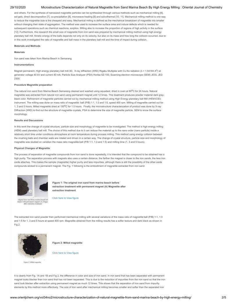
29/10/2020 Microstructure Characterization of Natural Magnetite from Sand Marina Beach By High Energy Milling : Oriental Journal of Chemistry
www.orientjchem.org/vol34no2/microstructure-characterization-of-natural-magnetite-from-sand-marina-beach-by-high-energy-milling/ 2/5
and others. For the synthesis of nanosized magnetite particles can be synthesized through various methods such as mechanical milling [6],sol-gels, direct decomposition [7], co-precipitation [8], microwave-heating [9] and solvothermal [10, 11]. Mechanical milling method is one wayto reduce the magnetite size is the cheapest and easy. Mechanical milling is defined as the mechanical breakdown of magnetite into smallerwithout changing their state of aggregation. The method was used to increase the surface area and induce defects which is needed forsubsequent operations such as chemical reactions, sorption. Milling also to increase the proportion of regions of high activity in the surface[12]. Furthermore, this research the small size of magnetite from iron sand was prepared by mechanical milling method using high energyplanetary ball mill. Kinetic energy of the balls depends not only on its velocity, but also on its mass and how long the collision occurred, due toin this work investigated the ratio of magnetite and ball mass in the planetary ball mill and the time of impact during collision.
Materials and Methods
Materials
Iron sand was taken from Marina Beach in Semarang.
Instrumentations
Magnet permanent, High energy planetary ball mill-3D, X-ray diffraction (XRD) Rigaku Multiplex with Cu Kα radiation (λ = 1.54184 A ) atgenerator voltage 40 kV and current 40 mA, Particle Size Analyzer (PSA) Horiba SZ-100, Scanning electron microscope (SEM) JEOL JED2300.
Procedure Magnetite preparation
The natural iron sand from Marina Beach Semarang cleaned and washed using aquadest, dried in oven at 80 C for 24 hours. Naturalmagnetite was extracted from natural iron sand using permanent magnet until 12 times. This treatment produces powder material dark gray-black color. Refinement of magnetite particles carried out by mechanical milling method using High Energy planetary ball Mill (HEM-E3D)instrument. The milling was done on mass ratio of magnetite: ball (P/B) 1:1, 1:3 and 1:5, speed 400 rpm. Milling of magnetite carried out for1, 3 and 5 hours. Milled magnetite dried at 150 C for 1.5 hours. Finally, the microstructure characterization of product was done by X-rayDiffraction (XRD) to find out the structure of magnetite crystals, PSA to determine the size of magnetite particle, SEM to know the surfacemorphology.
Results and Discussions
In this work the change of crystal structure, particle size and morphology of magnetite to be investigated. The method is high energy milling(HEM) used planetary ball mill. The choice of this method due to it can reduce the material up to the nano order (nano particle) inside arelatively short time under conditions atmosphere at room temperature during process milling. This method using energy collision betweenthe crushing balls and chamber walls are rotated and driven in a certain way. The change of crystal structure, particle size and morphology ofmagnetite was studied on variation the mass ratio magnetite:ball (P/B 1:1, 1:3 and 1:5) and milling time (1, 3 and 5 hours).
Physical Changes of Magnetite
The process of separation of magnetite compounds from iron sand is done repeatedly, it is intended that the compound to be obtained has ahigh purity. The separation process with magnets also uses a certain distance, the farther the magnet is closer to the iron sands, the less ironoxide attaches. This makes the sample (magnetite) higher purity and less impurities, although there is still the possibility of the other oxidecompounds sticked to a permanent magnet. The Fig. 1 following is the embodiment of magnetite extracted from iron sand.
Figure 1: The original iron sand from marina beach beforeextraction treatment with permanent magnet (A) Magnetite afterextraction treatment
Click here to View figure
The extracted iron sand powder then performed mechanical milling with several variations of the mass ratio of magnetite:ball (P/B) 1:1, 1:3and 1:5 for 1, 3 and 5 hours at speed 400 rpm. Magnetite obtained from the milling results has a softer texture and dark black as shown inFig.2.
Figure 2: Milled magnetite
Click here to View figure
It is clearly from Fig. 1A and 1B and Fig 2, the difference in color and size of iron sand. In iron sand that has been separated with permanentmagnet looks blacker than iron sand that has not been separated. This is due to the reduction of impurities from the iron sand so that the ironsand look blacker after extraction using permanent magnet as much 12 times. This shows that the separation of iron sand from impurityelements by this method more effectively. The size of iron sand after mechanical milling becomes smaller and softer than the separated iron
o
o
o
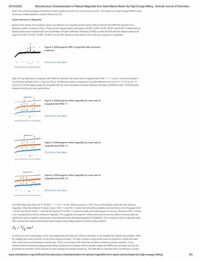
29/10/2020 Microstructure Characterization of Natural Magnetite from Sand Marina Beach By High Energy Milling : Oriental Journal of Chemistry
www.orientjchem.org/vol34no2/microstructure-characterization-of-natural-magnetite-from-sand-marina-beach-by-high-energy-milling/ 3/5
sand. This is the advantages of mechanical milling method that ball mill is not sensitive to metal. The superiority of High Energy Milling is ableto produce smaller particles in shorter milling time [12].
Crystal Structure of Magnetite
Based on the results of the analysis using X-ray diffraction on magnetite powder before milling treatment with HEM-3D obtained X-raydiffraction pattern as shown in Fig. 3. There are five highest peaks at 2θ angle of 30.09º; 35.46º; 43.09º; 56.98º; and 62.59º. Furthermore thehighest peaks were compared with Joint Committee of Powder Diffraction Standard (JCPDS) number 89-4319 with the highest peaks at 2θangle of 30.083º; 35.434º; 43.064º; 56.949º; and 62.536º. Based on data obtained from XRD, the compound is magnetite.
Figure 3: Diffractogram XRD of magnetite after extractiontreatment
Click here to View figure
Data of X-ray diffraction on magnetite after HEM-3D treatment with mass ratio of magnetite:ball (P/B) 1: 1, 1: 3 and 1: 5 and time collision 1,3 and 5 hours showed in Fig. 4, Fig.5 and Fig. 6. All diffraction peaks correspond to the peak diffraction at (2 2 0), (3 1 1), (4 0 0), (5 1 1),and (4 4 0). Of the highest peaks are compared with the Joint Committee of Powder Diffraction Standard (JCPDS) number. 79-0418 showsindexed to the Fe O cubic spinel phase.
Figure 4: Diffractogram milled magnetite by mass ratio ofmagnetite:ball (P/B) 1:1
Click here to View figure
Figure 5: Diffractogram milled magnetite by mass ratio ofmagnetite:ball (P/B) 1:3
Click here to View figure
Figure 6: Diffractogram milled magnetite by mass ratio ofmagnetite:ball (P/B) 1:5
Click here to View figure
The XRD datas show that in all P / B ratio 1, 1: 1: 3 or 1: 5 with milling process for 1 and 3 hours still indicates conformity with referencemagnetite. When the milling for 5 hours is only in P/B 1: 1 and P/B 1: 3 which still shows the suitability and even this is only at the peak of 2Ɵ= 35.92 and 63.02 at P/B 1: 1 and 36.19 and 63.15 at P/B 1: 3, where the peak of the diffractogram is very low, whereas in P/B 1: 5 thereis no correspondence with the reference magnetite. This suggests that long-term milling treatments and strong collisions (heavier ball) cansignificantly reduce magnetic particle size, these treatments also decreased degrade of crystallinity. The increasing of ball to magnetite massratio (heavier ball) would enhancing the kinetic energy during milling. Based on kinetic energy equation:
In which E is the kinetic energy, m and v are respectively the mass and velocity of the balls. In this research the velocity was constant. Whenthe colliding ball mass is heavier, so the kinetic energy increases. The high of kinetic energy would cause the particles to collide with eachother, where this would decrease in particle size. This is in accordance with data that has been revealed by previous research. It wasreported that the enhancing energy during milling, resulted by the increase of ball to powder weight ratio (BPR) and vial speed not only canaccelerate the formation of the products but also changes the resultant phases [4]. The balls play an important role in its efficiency so that a
3 4
o o o o
k
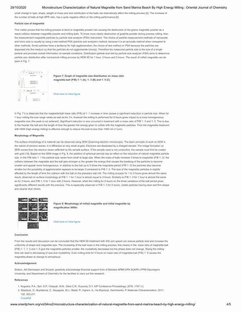
29/10/2020 Microstructure Characterization of Natural Magnetite from Sand Marina Beach By High Energy Milling : Oriental Journal of Chemistry
www.orientjchem.org/vol34no2/microstructure-characterization-of-natural-magnetite-from-sand-marina-beach-by-high-energy-milling/ 4/5
small change in type, shape, weight or mass and size distribution of the balls can dramatically affect the milling process [5]. The increase ofthe number of balls at high BPR ratio, has a quite negative effect on the milling performance [6].
Particle size of magnetite
This matter proves that the milling process is done to magnetite powder can causing the destruction of the grains magnetite powder as aresult collision between magnetite powder and milling balls. To know more clearly destruction of graphite powder during process milling, thenthe measurement magnetite particles by particle size analyzer (PSA) instrument. The choice of particle measurement methods of nanoscaleand micro size is usually by using a wet method PSA (particle size analyzer) method, because it is an accurate method when compared toother methods. Small particles have a tendency for high agglomeration, the choice of wet method on PSA because the particles aredispersed into the medium so that the particles do not agglomerate (clump). Therefore the measured particle size is the size of a singleparticle and provides overall information on sample conditions. Distribution particle size test by particle size analyzer (PSA) aims to determineparticle size distribution after mechanical milling process by HEM-3D for 1 hour, 3 hours and 5 hours. The result of milled magnetite can beseen in Fig. 7.
Figure 7: Graph of magnetite size distribution on mass ratiomagnetite:ball (P/B) 1: 1 (A); 1: 3 (B) and 1: 5 (C)
Click here to View figure
In Fig. 7 it is observed that the magnetite/ball mass ratio (P/B) of 1: 1 increase in time causes a significant reduction in particle size. When for1 hour milling the size range varies as well as for 3 h, however the milling is performed for 5 hours gives impact to a more homogeneousmagnetite size (the peak is not widened). Significant reduction in size occurred in treatment with a mass ratio of P/B 1: 3 and 1: 5. This is dueto the heavier the ball and the length of time the greater the energy given to collide with the magnetite particles. Thus the magnetite treatmentwith HEM (high energy milling) is effective enough to reduce the size to less than 1000 nm (<1μm).
Morphology of Magnetite
The surface morphology of a material can be observed using SEM (Scanning electron microscope). The basic principle of work on SEM isthe nature of electron waves, it is diffraction at very small angles. Electrons are dissipated by a charged sample. The image formation onSEM comes from the electron beam reflected by the sample surface. If the sample used is not conductive, the sample must first be coatedwith gold [16]. Based on the SEM image in Fig. 8, the addition of spherical periods has an effect on the reduction of natural magnetite particlesize. In the P/B ratio 1: 1 the particle size varies from small to large size. When the mass of balls increase 3 times to magnetite (P/B 1: 3), thecollision between the magnetite and the ball gets stronger or the greater the energy that causes the breaking of the particles to becomesmaller and appear more homogeneous. In addition to the ball up to 5 times the magnetite period (P/B 1: 5) the particles also becomesmaller but the possibility of agglomeration appears to be larger if compared to P/B 1: 3. The size of the magnetite particles is slightlyaffected by the length of time the collision with the ball on the planetary ball mill. The milling process for 1 to 3 hours gives almost the sameresult, observed on surface morphology at P/B 1: 1 for 1 hour is almost equal to 3 hours. Similarly to P/B 1: 3 for 1 hour is almost the sameas for 3 hours, and P/B 1: 5 for 1 hour with 3 hours. However, when the milling for 5 hours on the three variations of the ball period givessignificantly different results with the previous. This is especially observed in P/B 1: 3 for 5 hours, visible particles having clear and firm shapeand cleaner than others.
Figure 8: Morphology of milled magnetite and initial magnetite bymagnification 5000x
Click here to View figure
Conclusion
From the results and discussion can be concluded that the HEM-3D treatment with 400 rpm speed can reduce particle size and increase theuniformity of shape and magnetite size. The increasing of the ball mass in the milling process, this means in the mass ratio of magnetite:ball(P/B) 1: 1, 1: 3 and 1: 5 give the magnetite particles smaller, the crystallinity decreases but the phase does not change. Rising the millingtime can lead to decreasing of size and crystallinity. Even milling time for 5 hours on mass ratio of magnetite:ball (P/B) 1: 5 causes themagnetite phase to change to amorphous
Acknowledgement
Sriatun, Adi Darmawan and Sriyanti, gratefully acknowledge financial support from of Besides APBN DPA SUKPA LPPM DiponegoroUniversity, and Department of Chemistry for the facilities to carry out this research.
References
1. Nugraha, P.A.; Sari, S.P.; Hidayati, W.N.; Dewi,C.R.; Kusuma, D.Y. AIP Conference Proceedings, 2016, 1747 (1)2. Shpotyuk, O.; Bujňáková, Z.; Sayagués, M.J.; Baláž, P.; Ingram, A.; Ya.Shpotyuk, Demchenko, P. Materials Characterization, 2017,
132: 303-311. CrossRef

29/10/2020 Microstructure Characterization of Natural Magnetite from Sand Marina Beach By High Energy Milling : Oriental Journal of Chemistry
www.orientjchem.org/vol34no2/microstructure-characterization-of-natural-magnetite-from-sand-marina-beach-by-high-energy-milling/ 5/5
3. Gong, J. Journal Hazardous Mat., 2009, 164:1517-1522 CrossRef
4. Hui, C.; Shen, C.; Yang, T.; Bao, L.; Tian, J.; Ding, H.; Li, C.; Gao, H.J. J. Phys. Chem. C. 2008, 112, 11336-11339. CrossRef
5. Klotz, S.; Steinle-Neumann, G.; Strassle, T.; Philippe, J.; Hansen, T.; Wenzel, M.J. Phys. Rev. B, 2008, 77, 12411-1-1241-4.6. Marinca, T.; Chicinaș, H.; Neamțu, B.; Popa, F.; Isnard, O.; Chicinaș, I. Studia Universitatis Babes-Bolyai, Physica, 2015, 60 (1).7. Darezereshki, E.; Bakhtiari, F.; Alizadeh, M.; Ranjbar, M. Materials Science in Semiconductor Processing, 2012, 15(1): 91-97.
CrossRef8. Khan, U.S.; Rahim, A.; Khan, N.; Muhammad, N.; Rehman, F.; Ahmad, K.; Iqbal, J. Materials Chemistry and Physics, 2017, 189: 86-89.
CrossRef9. Chikan, V. and McLaurin, E. J. Nanomaterials,2016, 6(5): 85
CrossRef10. An,J.S.; Han, W.J.; Choi, H.J. Colloids and Surfaces A: Physicochemical and Engineering Aspects, 2017, 535:16-23
CrossRef11. Bui, T.Q.; Ton, S.N.; Duong, A.T.; Tran, H.T. Journal of Science: Advanced Materials and Devices, 2017, Available online 14 November
201712. Balaz, P. Mechanochemistry in Nanoscience and minerals Engineering, 2008, Springer-Verlag Berlin Heidelberg, 103.13. Bolokang, S., Banganayi, C., Phasha, M. Int. J. Refract. Met. Hard Mater, 2010, 28:211–216.
CrossRef14. Zakeri, M., Rahimipour, M.R. Adv. Powder Technol, 2012, 23:31–34
CrossRef15. Ghayour, H., Abdellahi , M., Bahmanpour, M. Powder Technology, 2016, 291: 7–13
CrossRef16. Prabakaran, K.; Balamurunga, A.; Rajeswari, S. Bull Mat Sci, 2005, 28:115-119.
CrossRef
This work is licensed under a Creative Commons Attribution 4.0 International License.
LinksLinks
Article Processing Charges
Current Issue
Editorial Board
Home
Indexed in
Policies
Aims & Scope
Article Submission Workflow
Join Our Team of Reviewers
Online Submission
Contact UsContact Us
Your Name (required)
Your Email (required)
Phone Number
Comment
Type the above text in box below
SendSend
Which is bigger, 5 or 8?
Google Page RankGoogle Page Rank
LicenseLicense
This work is licensed under a Creative CommonsAttribution 4.0 International License.
© 2020 Oriental Journal of Chemistry Top ↑
ORIENTAL JOURNAL OF CHEMISTRY
www.orientjchem.org
An International Open Free Access, Peer Reviewed Research Journal
ISSN: 0970-020 XCODEN: OJCHEG
2018, Vol. 34, No.(2):Pg. 868-874
This is an Open Access article licensed under a Creative Commons Attribution-NonCommercial-ShareAlike4.0 International License (https://creativecommons.org/licenses/by-nc-sa/4.0/ ), which permits unrestrictedNonCommercial use, distribution and reproduction in any medium, provided the original work is properly cited.
Microstructure Characterization of Natural Magnetite from Sand Marina Beach by High Energy Milling
SRIATUN1*, A. DARMAWAN1, SRIYANTI1 and W. CAHYANI1
1Department of Chemistry, Diponegoro University, Semarang 50275, Central of Java, Indonesia.*Corresponding author E-mail: [email protected]
http://dx.doi.org/10.13005/ojc/340234
(Received: December 18, 2017; Accepted: January 25, 2018)
ABSTRACT
In this work, we performed an experimental investigation the change of microstructure ofmagnetite by high energy milling-3D (HEM-3D) method using planetary ball milling at 400 rpmspeed. The present studies mainly focusses on the effect of milling on crystallinity and phase ofmagnetite by XRD, particle size by PSA and the morphology by SEM. The increasing of the ballmass in the milling process, mass ratio magnetite: ball (P/B) 1: 1, 1: 3 and 1: 5 give the magnetiteparticles smaller (< 1μm), the crystallinity decreases but the peaks at (2 2 0), (3 1 1), (4 0 0),(5 1 1), and (4 4 0) were keep appearing. This shows that the phase of cubic spinel does notchange. Rising the milling time for 1 h, 3 h and 5 h can lead to decreasing of size and crystallinity.Even milling time for 5 h on mass ratio of magnetite: ball (P/B) 1: 5 causes the magnetite phase tochange to amorphous.
Keywords: Microstructure, Natural magnetite, Sand marina beach, High energy milling-3D.
INTRODUCTION
Iron sand occurs naturally in severalregions throughout the world. Iron sand is one ofIndonesia’s natural mineral resources, which isspread over the islands along the coast of JavaIsland, Kalimantan and Sumatra. Iron sand is aspecial type of sand that’s rich in the metal iron, thecolor is dark gray or black, consisting of (Fe) iron asa major element and a small amount of Ti, Si, Ca,
Mn and Vanadium. They provide a raw material ofrelatively low grade, whereas in the southern coastof Yogyakarta containing 5.85 % to 95.11% of iron.
In addition to magnetite in iron sand also containsother minerals such as rutile, ilmenite and hematite1.While most sand contains at least some trace of
iron, therefore it has a distinct dark-gray or blackcolor, which is in stark contrast to the white-yellowcolor of regular sand.
869SRIATUN et al., Orient. J. Chem., Vol. 34(2), 868-874 (2018)
Iron sand is a magnetic material that iswidely used in various fields such as electronics,energy, chemistry, ferrofluidics, catalysts, andmedical diagnostics2. The application of iron sandwas inseparable from the development of studiesof nanomaterials demanding that they be in theorder of nanometers. Magnetite or Fe3O4 is one ofthe iron oxide phases which has the greatestmagnetic or ferromagnetic properties among theother phases. Iron oxide has four phases, namelymagnetite (Fe3O4), maghemite (γ-Fe2O3), hematite(α-Fe2O3), and geotite (FeO(OH)). Only magnetiteand maghemite have magnetic properties3.
Magnetite (Fe3O4) is known as a class ofiron oxide compound with a cubic inverse spinelstructure and has face centered cubic close packedoxygen anions and Fe cations occupying interstitialtetrahedral and octahedral sites4,5. Nano-sizedmagnetite particles provide many advantages suchas for the separation of magnetic contaminants inwater, large of surface area and the ability to bindthrough electro-chemical interactions to form sludge.It is also applied to drug delivery and magneticresonance technology and others.
For the synthesis of nanosized magnetiteparticles can be synthesized through variousmethods such as mechanical milling6,sol-gels, direct decomposition7, co-precipitation8,microwave-heating9 and solvothermal10,11.Mechanical milling method is one way to reducethe magnetite size is the cheapest and easy.Mechanical milling is defined as the mechanicalbreakdown of magnetite into smaller withoutchanging their state of aggregation. The methodwas used to increase the surface area and inducedefects which is needed for subsequent operationssuch as chemical reactions, sorption. Milling alsoto increase the proportion of regions of high activityin the surface12.
Furthermore, this research the small sizeof magnetite from iron sand was prepared bymechanical milling method using high energyplanetary ball mill. Kinetic energy of the ballsdepends not only on its velocity, but also on its massand how long the collision occurred, due to in thiswork investigated the ratio of magnetite and ballmass in the planetary ball mill and the time of impactduring collision.
MATERIALS AND METHODS
MaterialsIron sand was taken from Marina Beach in
Semarang.
InstrumentationsMagnet permanent, High energy planetary
ball mill-3D, X-ray diffraction (XRD) RigakuMultiplex with Cu Ká radiation (λ = 1.54184 Ao) atgenerator voltage 40 kV and current 40 mA, ParticleSize Analyzer (PSA) Horiba SZ-100, Scanningelectron microscope (SEM) JEOL JED 2300.
ProcedureMagnetite preparation
The natural iron sand from Marina BeachSemarang cleaned and washed using aquadest,dried in oven at 80 oC for 24 hours. Naturalmagnetite was extracted from natural iron sandusing permanent magnet until 12 times. Thistreatment produces powder material darkgray-black color. Refinement of magnetite particlescarried out by mechanical milling method using highenergy planetary ball mill (HEM-E3D) instrument.The milling was done on mass ratio of magnetite:ball (P/B) 1:1, 1:3 and 1:5, speed 400 rpm. Milling ofmagnetite carried out for 1, 3 and 5 hours. Milledmagnetite dried at 150 oC for 1.5 hours. Finally, themicrostructure characterization of product was doneby X-ray diffraction (XRD) to find out the structure ofmagnetite crystals, PSA to determine the size ofmagnetite particle, SEM to know the surfacemorphology.
RESULTS AND DISCUSSIONS
In this work the change of crystal structure,particle size and morphology of magnetite wasinvestigated. The method is high energy milling(HEM) used planetary ball mill. The choice of thismethod due to it can reduce the material up to thenano order (nano particle) inside a relatively shorttime under conditions atmosphere at roomtemperature during process milling. This methodusing energy collision between the crushing ballsand chamber walls are rotated and driven in acertain way. The change of crystal structure, particlesize and morphology of magnetite was studied onvariation the mass ratio magnetite:ball (P/B 1:1, 1:3and 1:5) and milling time (1, 3 and 5 hours).
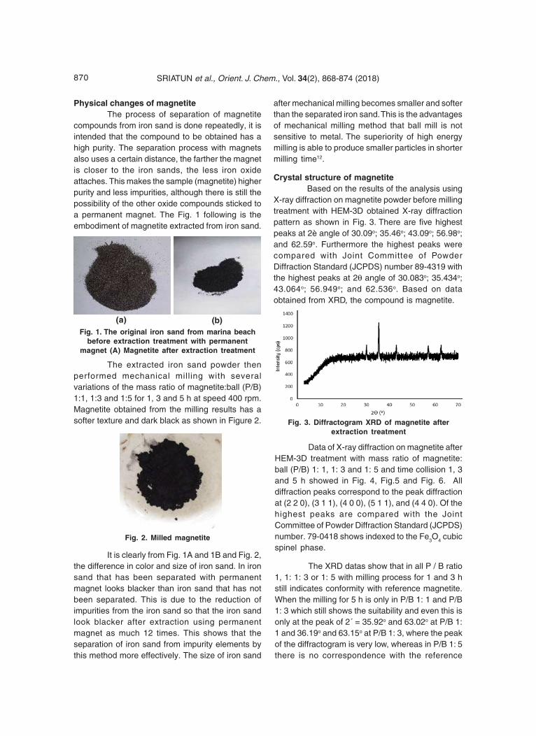
870 SRIATUN et al., Orient. J. Chem., Vol. 34(2), 868-874 (2018)
Physical changes of magnetiteThe process of separation of magnetite
compounds from iron sand is done repeatedly, it isintended that the compound to be obtained has ahigh purity. The separation process with magnetsalso uses a certain distance, the farther the magnetis closer to the iron sands, the less iron oxideattaches. This makes the sample (magnetite) higherpurity and less impurities, although there is still thepossibility of the other oxide compounds sticked toa permanent magnet. The Fig. 1 following is theembodiment of magnetite extracted from iron sand.
after mechanical milling becomes smaller and softerthan the separated iron sand. This is the advantagesof mechanical milling method that ball mill is notsensitive to metal. The superiority of high energymilling is able to produce smaller particles in shortermilling time12.
Crystal structure of magnetiteBased on the results of the analysis using
X-ray diffraction on magnetite powder before millingtreatment with HEM-3D obtained X-ray diffractionpattern as shown in Fig. 3. There are five highestpeaks at 2è angle of 30.09o; 35.46o; 43.09o; 56.98o;and 62.59o. Furthermore the highest peaks werecompared with Joint Committee of PowderDiffraction Standard (JCPDS) number 89-4319 withthe highest peaks at 2θ angle of 30.083o; 35.434o;43.064o; 56.949o; and 62.536o. Based on dataobtained from XRD, the compound is magnetite.
Fig. 1. The original iron sand from marina beachbefore extraction treatment with permanent
magnet (A) Magnetite after extraction treatment
(a) (b)
The extracted iron sand powder thenperformed mechanical milling with severalvariations of the mass ratio of magnetite:ball (P/B)1:1, 1:3 and 1:5 for 1, 3 and 5 h at speed 400 rpm.Magnetite obtained from the milling results has asofter texture and dark black as shown in Figure 2.
Fig. 2. Milled magnetite
It is clearly from Fig. 1A and 1B and Fig. 2,the difference in color and size of iron sand. In ironsand that has been separated with permanentmagnet looks blacker than iron sand that has notbeen separated. This is due to the reduction ofimpurities from the iron sand so that the iron sandlook blacker after extraction using permanentmagnet as much 12 times. This shows that theseparation of iron sand from impurity elements bythis method more effectively. The size of iron sand
Data of X-ray diffraction on magnetite afterHEM-3D treatment with mass ratio of magnetite:ball (P/B) 1: 1, 1: 3 and 1: 5 and time collision 1, 3and 5 h showed in Fig. 4, Fig.5 and Fig. 6. Alldiffraction peaks correspond to the peak diffractionat (2 2 0), (3 1 1), (4 0 0), (5 1 1), and (4 4 0). Of thehighest peaks are compared with the JointCommittee of Powder Diffraction Standard (JCPDS)number. 79-0418 shows indexed to the Fe3O4 cubicspinel phase.
The XRD datas show that in all P / B ratio1, 1: 1: 3 or 1: 5 with milling process for 1 and 3 hstill indicates conformity with reference magnetite.When the milling for 5 h is only in P/B 1: 1 and P/B1: 3 which still shows the suitability and even this isonly at the peak of 2´ = 35.92o and 63.02o at P/B 1:1 and 36.19o and 63.15o at P/B 1: 3, where the peakof the diffractogram is very low, whereas in P/B 1: 5there is no correspondence with the reference
Fig. 3. Diffractogram XRD of magnetite afterextraction treatment
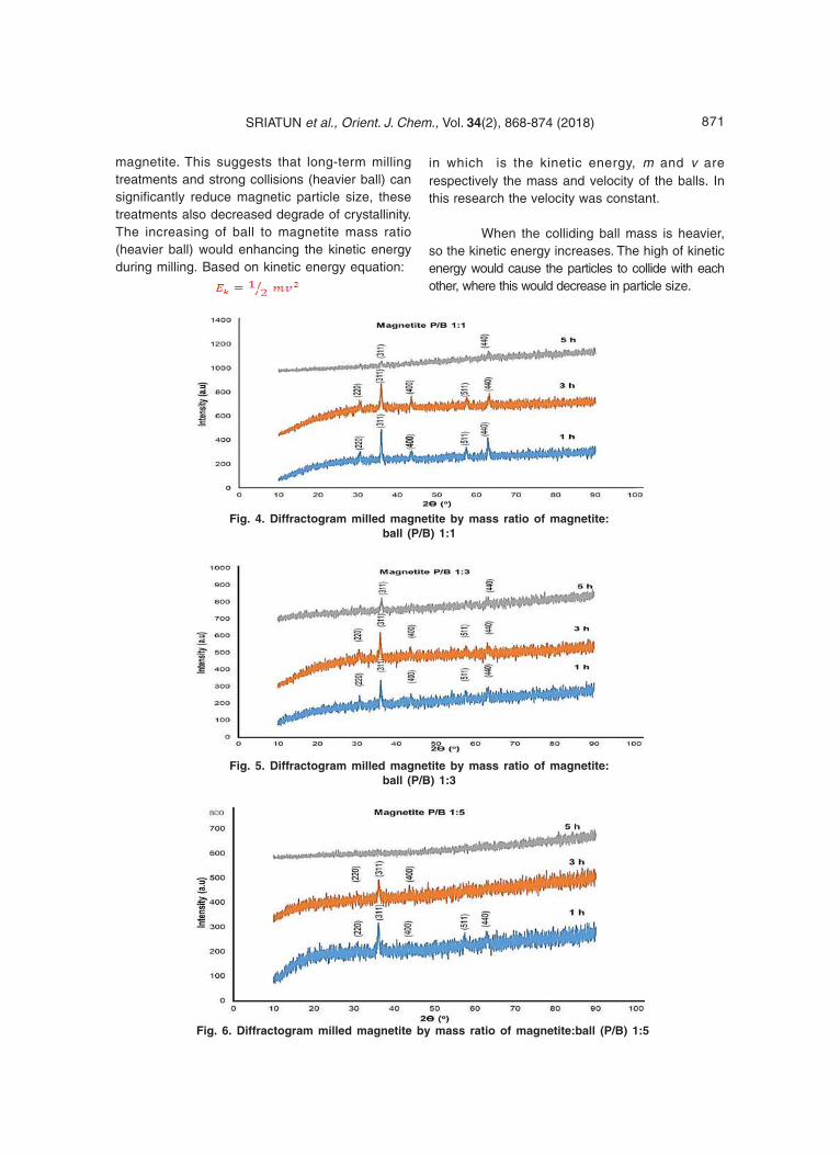
871SRIATUN et al., Orient. J. Chem., Vol. 34(2), 868-874 (2018)
magnetite. This suggests that long-term millingtreatments and strong collisions (heavier ball) cansignificantly reduce magnetic particle size, thesetreatments also decreased degrade of crystallinity.The increasing of ball to magnetite mass ratio(heavier ball) would enhancing the kinetic energyduring milling. Based on kinetic energy equation:
in which is the kinetic energy, m and v arerespectively the mass and velocity of the balls. Inthis research the velocity was constant.
When the colliding ball mass is heavier,so the kinetic energy increases. The high of kineticenergy would cause the particles to collide with eachother, where this would decrease in particle size.
Fig. 5. Diffractogram milled magnetite by mass ratio of magnetite:ball (P/B) 1:3
Fig. 4. Diffractogram milled magnetite by mass ratio of magnetite:ball (P/B) 1:1
Fig. 6. Diffractogram milled magnetite by mass ratio of magnetite:ball (P/B) 1:5
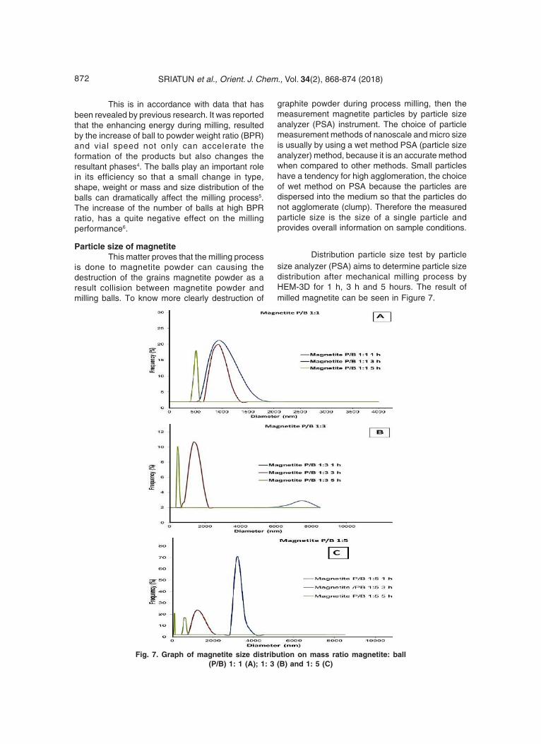
872 SRIATUN et al., Orient. J. Chem., Vol. 34(2), 868-874 (2018)
This is in accordance with data that hasbeen revealed by previous research. It was reportedthat the enhancing energy during milling, resultedby the increase of ball to powder weight ratio (BPR)and vial speed not only can accelerate theformation of the products but also changes theresultant phases4. The balls play an important rolein its efficiency so that a small change in type,shape, weight or mass and size distribution of theballs can dramatically affect the milling process5.The increase of the number of balls at high BPRratio, has a quite negative effect on the millingperformance6.
Particle size of magnetiteThis matter proves that the milling process
is done to magnetite powder can causing thedestruction of the grains magnetite powder as aresult collision between magnetite powder andmilling balls. To know more clearly destruction of
graphite powder during process milling, then themeasurement magnetite particles by particle sizeanalyzer (PSA) instrument. The choice of particlemeasurement methods of nanoscale and micro sizeis usually by using a wet method PSA (particle sizeanalyzer) method, because it is an accurate methodwhen compared to other methods. Small particleshave a tendency for high agglomeration, the choiceof wet method on PSA because the particles aredispersed into the medium so that the particles donot agglomerate (clump). Therefore the measuredparticle size is the size of a single particle andprovides overall information on sample conditions.
Distribution particle size test by particlesize analyzer (PSA) aims to determine particle sizedistribution after mechanical milling process byHEM-3D for 1 h, 3 h and 5 hours. The result ofmilled magnetite can be seen in Figure 7.
Fig. 7. Graph of magnetite size distribution on mass ratio magnetite: ball(P/B) 1: 1 (A); 1: 3 (B) and 1: 5 (C)
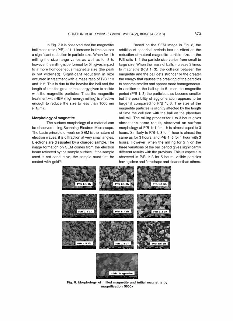
873SRIATUN et al., Orient. J. Chem., Vol. 34(2), 868-874 (2018)
In Fig. 7 it is observed that the magnetite/ball mass ratio (P/B) of 1: 1 increase in time causesa significant reduction in particle size. When for 1 hmilling the size range varies as well as for 3 h,however the milling is performed for 5 h gives impactto a more homogeneous magnetite size (the peakis not widened). Significant reduction in sizeoccurred in treatment with a mass ratio of P/B 1: 3and 1: 5. This is due to the heavier the ball and thelength of time the greater the energy given to collidewith the magnetite particles. Thus the magnetitetreatment with HEM (high energy milling) is effectiveenough to reduce the size to less than 1000 nm(<1μm).
Morphology of magnetiteThe surface morphology of a material can
be observed using Scanning Electron Microscope.The basic principle of work on SEM is the nature ofelectron waves, it is diffraction at very small angles.Electrons are dissipated by a charged sample. Theimage formation on SEM comes from the electronbeam reflected by the sample surface. If the sampleused is not conductive, the sample must first becoated with gold16.
Based on the SEM image in Fig. 8, theaddition of spherical periods has an effect on thereduction of natural magnetite particle size. In theP/B ratio 1: 1 the particle size varies from small tolarge size. When the mass of balls increase 3 timesto magnetite (P/B 1: 3), the collision between themagnetite and the ball gets stronger or the greaterthe energy that causes the breaking of the particlesto become smaller and appear more homogeneous.In addition to the ball up to 5 times the magnetiteperiod (P/B 1: 5) the particles also become smallerbut the possibility of agglomeration appears to belarger if compared to P/B 1: 3. The size of themagnetite particles is slightly affected by the lengthof time the collision with the ball on the planetaryball mill. The milling process for 1 to 3 hours givesalmost the same result, observed on surfacemorphology at P/B 1: 1 for 1 h is almost equal to 3hours. Similarly to P/B 1: 3 for 1 hour is almost thesame as for 3 hours, and P/B 1: 5 for 1 hour with 3hours. However, when the milling for 5 h on thethree variations of the ball period gives significantlydifferent results with the previous. This is especiallyobserved in P/B 1: 3 for 5 hours, visible particleshaving clear and firm shape and cleaner than others.
P/B 1:1 1h P/B 1:1 3h P/B 1:1 5h
P/B 1:3 1h P/B 1:3 3h P/B 1:3 5h
P/B 1:5 1h P/B 1:5 3h P/B 1:5 5h
Initial Magnetite
Fig. 8. Morphology of milled magnetite and initial magnetite bymagnification 5000x
874 SRIATUN et al., Orient. J. Chem., Vol. 34(2), 868-874 (2018)
CONCLUSION
From the results and discussion can beconcluded that the HEM-3D treatment with 400 rpmspeed can reduce particle size and increase theuniformity of shape and magnetite size. Theincreasing of the ball mass in the milling process,this means in the mass ratio of magnetite:ball (P/B)1: 1, 1: 3 and 1: 5 give the magnetite particles smaller,the crystallinity decreases but the phase does notchange. Rising the milling time can lead todecreasing of size and crystallinity. Even milling
time for 5 h on mass ratio of magnetite: ball (P/B) 1:5 causes the magnetite phase to change to
amorphous
ACKNOWLEDGEMENT
Sriatun, Adi Darmawan and Sriyanti,gratefully acknowledge financial support from of
Besides APBN DPA SUKPA LPPM DiponegoroUniversity, and Department of Chemistry for thefacilities to carry out this research.
REFERENCES
1. Nugraha, P.A.; Sari, S.P.; Hidayati, W.N.;Dewi,C.R.; Kusuma, D.Y. AIP ConferenceProceedings., 2016, 1, 17-47
2. Shpotyuk, O.; Bujòáková, Z.; Sayagués, M.J.;Balá•, P.; Ingram, A.; Ya.Shpotyuk,Demchenko, P. Materials Characterization.,2017, 132, 303-311.
3. Gong, J. Journal Hazardous Mat., 2009,164,1517-1522.
4. Hui, C.; Shen, C.; Yang, T.; Bao, L.; Tian, J.;Ding, H.; Li, C.; Gao, H.J. J. Phys. Chem. C.,2008, 112, 11336-11339.
5. Klotz, S.; Steinle-Neumann, G.; Strassle, T.;Philippe, J.; Hansen, T.; Wenzel, M.J. Phys.Rev. B., 2008, 77, 12411-1-1241-4.
6. Marinca, T.; Chicinaˆ, H.; Neamˆu, B.; Popa,F.; Isnard, O.; Chicinaˆ, I. Studia UniversitatisBabes-Bolyai, Physica., 2015, 1, 60.
7. Darezereshki, E.; Bakhtiari, F.; Alizadeh, M.;Ranjbar, M. Materials Science in SemiconductorProcessing, 2012, 15(1), 91-97.
8. Khan, U.S.; Rahim, A.; Khan, N.; Muhammad,
N.; Rehman, F.; Ahmad, K.; Iqbal, J. MaterialsChemistry and Physics., 2017, 189, 86-89.
9. Chikan, V. and McLaurin, E. J. Nanomaterials.,2016, 6(5), 85.
10. An,J.S.; Han, W.J.; Choi, H.J. Colloids andSurfaces A: Physicochemical andEngineering Aspects., 2017, 535, 16-23.
11. Bui, T.Q.; Ton, S.N.; Duong, A.T.; Tran, H.T. Journalof Science: Advanced Materials and Devices,Available online 14 November 2017.
12. Balaz, P.; Mechanochemistry in Nanoscienceand minerals Engineering, Springer-VerlagBerlin Heidelberg., 2008, 103,
13. Bolokang, S., Banganayi, C., Phasha, M. Int. J.Refract. Met. Hard Mater., 2010, 28, 211–216.
14. Zakeri, M., Rahimipour, M.R. Adv. PowderTechnol., 2012, 23, 31–34.
15. Ghayour, H., Abdellahi , M., Bahmanpour,M. Powder Technology., 2016, 291, 7–13.
16. Prabakaran, K.; Balamurunga, A.; Rajeswari,
S. Bull Mat Sci., 2005, 28, 115-119.