Bone Morphogenic Protein-7 Inhibits Progression ofChronic Renal Fibrosis Associated with two GeneticMouse Models
Michael Zeisberg, Cindy Bottiglio, Navin Kumar, Yohei Maeshima, Frank Strutz*, Gerhard A.
Müller* and Raghu Kalluri
Center for Matrix Biology, Department of Medicine, Beth Israel Deaconess Medical Center and
Harvard Medical School, Boston, MA 02215, USA, *Department of Nephrology and
Rheumatology, Georg-August University Medical Center, Robert-Koch-Str. 40, 37075
Göttingen, Germany
Running title: BMP 7 inhibits renal fibrosis
Correspondence should be addressed to:Dr. Raghu Kalluri
Associate Professor of Medicine
Harvard Medical School
Director, Center for Matrix Biology
Department of Medicine, Dana 514
Beth Israel Deaconess Medical Center
330 Brookline Avenue
Boston, MA 02215Tel#617-667-0445Fax#617-667-2562e-Mail:[email protected]
Copyright (c) 2003 by the American Physiological Society.
Articles in PresS. Am J Physiol Renal Physiol (August 12, 2003). 10.1152/ajprenal.00191.2002
American Journal of Physiology � Renal Physiology F-00191-2002 BMP-7 inhibits renal fibrosisZeisberg/Bottiglio/Kumar/Maeshima/Strutz/Muller/Kalluri
1
Abstract
Tubulointerstitial fibrosis is a hallmark feature of chronic renal injury. Specific therapies to
control the progression of renal fibrosis towards end-stage renal failure are still limited. Previous
studies have demonstrated that expression of endogenous BMP-7 is reduced in the kidneys of
several inducible mouse models of acute and chronic renal disease, and that administration of
exogenous recombinant human bone morphogenic protein-7 (rhBMP-7) has a beneficial effect
on kidney function. Here we report that treatment with rhBMP-7 leads to improved renal
function, histology and survival in a3(IV) collagen deficient mice and in MRL/MpJlpr/lpr lupus mice,
two different genetic models for chronic renal injury and fibrosis. Such therapeutic benefit is
also associated with a significant decrease in the expression of pro-fibrotic molecules, such as
collagen I and fibronectin, in renal fibroblasts. Additionally, rhBMP-7 induces the expression of
active matrix metalloproteinase-2 (MMP-2), which is potentially important for the removal of
fibrotic matrix. Collectively these studies provide further evidence for rhBMP-7 as an important
bone protein with protective function against renal pathology.
Keywords: Bone Morphogenic Protein-7, Tubulointerstitial Fibrosis, Fibroblasts, OsteogenicProtein-1, Matrix Metalloproteinases
American Journal of Physiology � Renal Physiology F-00191-2002 BMP-7 inhibits renal fibrosisZeisberg/Bottiglio/Kumar/Maeshima/Strutz/Muller/Kalluri
2
Despite significant advances in understanding the mechanistic pathways mediating the
progression of chronic renal disease towards end-stage renal failure (ESRF), options for
effective pharmacotherapy are still very limited (7, 18, 25, 37). In progressive chronic renal
disease, a self-preserving mechanism dominates independent of the underlying disease, further
validating the notion for existence of a potential common pathway leading to ESRF (13, 39).
Progressive renal disease is almost invariantly characterized by the triad of glomerular injury,
tubulointerstitial fibrosis and tubular atrophy (4, 16).
Bone Morphogenic Protein-7 (syn. Osteogenic Protein-1/ OP-1) is a member of the TGF-
b superfamily that plays a crucial role in renal development (12, 28, 34). Expression of BMP-7
correlates with condensation of the metanephric mesenchyme to generate epithelium, which
eventually leads to formation of tubules and glomeruli (28, 36, 40, 50, 51). In the adult mouse
kidney, BMP-7 is expressed in the collecting duct, thick ascending limb, distal convoluted
tubule, and podocytes within glomeruli (17). Acute renal injury associated with tubular necrosis
leads to reduction in the expression of tubular BMP-7, and after recovery of tubular and
glomerular damage BMP-7 expression is restored (42, 49). Administration of exogenous
recombinant human BMP-7 (rhBMP-7) restores normal tubule architecture in rat models of
ischemic acute renal injury and it also restores tubular homeostasis in a mouse model of
nephrotoxic serum nephritis (42, 49, 56). Furthermore, administration of exogenous rhBMP-7
inhibits progression of chronic renal disease in a rat model of unilateral urethral obstruction
(UUO) and a rat model of streptozotocin (STZ) induced diabetic nephropathy (21, 24). Here we
report that rhBMP-7 exhibits inhibitory effects on chronic renal disease and fibrosis in
MRL/MpJlpr/lpr lupus mice and in a3(IV) collagen deficient (Col4A3-/-) mice. We further report that
rhBMP-7 exerts anti-fibrogenic effects on renal fibroblasts, both in the in vitro and in vivo
settings. We also demonstrate that treatment of mice with rhBMP-7 over a four-month period
American Journal of Physiology � Renal Physiology F-00191-2002 BMP-7 inhibits renal fibrosisZeisberg/Bottiglio/Kumar/Maeshima/Strutz/Muller/Kalluri
3
does not induce significant side effects and is non-toxic. Collectively, these results suggest that
rhBMP-7 is a potential candidate for the treatment of chronic renal failure.
Methods
Materials. For in vivo studies, recombinant human BMP-7 homodimer, non-covalently
attached to prodomain protein (referred to as soluble rhBMP-7) was used (Curis, Inc.,
Cambridge, MA) (21). In cell culture experiments, rhBMP-7, dissolved in 24 mM Na acetate [pH
4.5] containing 1% mannitol, was used (Curis, Inc. Cambridge, MA) (17). Details regarding
these recombinant proteins have been published previously (17, 21).
Mice: Female normal mice on an MRL/MpJ background and autoimmune disease-prone
MRL/MpJlpr/lpr mice were purchased from the Jackson Laboratories (Bar Harbor, ME) and
maintained in the BIDMC animal facility (11). Col4A3-/- mice on a mixed background of C57BL/6
and 129/Sv were maintained at the BIDMC animal facility. Homozygous deletion of Col4A3-/-
was confirmed by PCR as described previously (30). The Col4A3-/- mice were either obtained
from Jackson Laboratories (Bar Harbor, ME) or were kindly provided by Dr. Jeffrey Miner (St.
Louis, MO). Mice were fed standard mouse chow and water ad libitum. All mouse studies
followed the standards approved by the institutional animal care committee.
Experimental design. Normal mice on an MRL/MpJ background (n=24) were treated
with vehicle buffer alone or received rhBMP-7 at 30 mg/kg, 100 mg/kg and 300 mg/kg for 16
weeks to assess long term toxicology of rhBMP-7. MRL/MpJlpr/lpr mice (lupus nephritis mice)
were treated for 12 weeks, starting treatment at the age of four weeks and the study was
terminated at the age of 16 weeks. Mice were divided into a control group (n=6 each) that
American Journal of Physiology � Renal Physiology F-00191-2002 BMP-7 inhibits renal fibrosisZeisberg/Bottiglio/Kumar/Maeshima/Strutz/Muller/Kalluri
4
received vehicle buffer alone and three experimental groups (n=6 each) that received rhBMP-7
at 30 mg/kg, 100 mg/kg and 300 mg/kg. Due to the different kinetics of disease progression,
COL4A3-/- mice were treated for 8 weeks and the study was terminated at the age of 14 weeks.
Col4A3-/- mice (n=14) were divided into a control group (n=7) that received vehicle buffer alone
and a treatment group that received 300 mg/kg rhBMP-7. rhBMP-7 was administered
intraperitoneally (i.p.) three times a week. Serum was obtained every two weeks from baseline
to death on all mice. Urine was collected every two weeks in metabolic cages.
Histologic Assessment of renal injury. Tissue from kidneys, liver, heart, spleen and
brain was fixed in 4% paraformaldehyde and embedded in paraffin. Sections were stained with
hematoxylin and eosin (HE), Masson�s Trichrome Stain (MTC), as well as with periodic acid-
Schiffs stain (PAS). Extent of renal injury was estimated by morphometric assessment of the
tubulointerstitial injury and glomerular damage. The relative interstitial volume was evaluated by
morphometric analysis using a 10 mm2 graticule fitted into the microscope. Five randomly
selected cortical areas, which included glomeruli, were evaluated for each animal (5, 45).
Tubules were evaluated for their widened lumen and thickened basement membranes to
estimate percentage of atrophic tubules (21). 100 glomeruli in each kidney were evaluated for
crescent formation, the crescentic index reflects the percentage of crescentic glomeruli(47).
Glomerulosclerosis was scored for severity (0-4, 0=normal, 4=maximum severity) as described
by Raij (38). Nuclei in 50 glomeruli of similar size in each kidney were counted to estimate
glomerular cellularity (29).
Immunohistochemistry. Kidney tissue samples were frozen in liquid nitrogen and
processed by indirect immunofluorescence technique as previously described (45). Frozen
American Journal of Physiology � Renal Physiology F-00191-2002 BMP-7 inhibits renal fibrosisZeisberg/Bottiglio/Kumar/Maeshima/Strutz/Muller/Kalluri
5
sections were stained with the primary antibody, raised against type I collagen (Southern
Biotechnology Associates Inc., Birmingham, AL), MMP-2 (Chemicon, Temecula, CA), MMP-9
(Chemicon) or BMP-7 (Curis, Inc.). After washing with Tris-buffered saline, sections were
incubated with FITC- and Rhodamine-conjugated secondary antibodies (Sigma, St. Lois, MO).
Deposition of IgG was determined by using FITC-conjugated anti-mouse IgG antibody (Sigma,
St. Lois, MO). Negative controls were performed by substituting the primary antibody with an
irrelevant pre-immune serum.
Cell Culture: Human interstitial fibroblasts (TK173) were maintained in culture as
previously described (27, 32). For stimulation of fibroblasts, media were replaced with serum-
free Iscove�s modified Dulbecco�s medium (IMDM) (45).
Direct-enzyme-linked immunoabsorbent assay: We performed ELISAs for estimation
of type I collagen and fibronectin as described previously (43). Quiescent fibroblasts (8x10³
cells/ well) were stimulated with rhBMP-7 (1.0, 10.0 and 100 ng/ml) in IMDM medium that was
supplemented with 50 mg/ml ascorbic acid and aminopropionitrile (Sigma, St. Louis) for 48 h.
Supernatants were analyzed in Maxisorp� (Nunc) plates using primary antibodies raised
against type I collagen (Southern Biotechnologies, Birmingham, AL), and fibronectin (Sigma, St.
Louis). All assays were performed in triplicate and repeated at least 3 times. The working range
was 10 to 1000ng/ml collagen type I and 0.1 to 10 mg/ml for fibronectin.
Zymography: Zymography was performed as described previously (57). Briefly, cells
(1x10⁵ per well) were plated in 6-well plates and grown for 6 h in DMEM medium containing
10% FCS. The medium was replaced with IMDM or with IMDM containing 1, 10, or 100 ng/ml
American Journal of Physiology � Renal Physiology F-00191-2002 BMP-7 inhibits renal fibrosisZeisberg/Bottiglio/Kumar/Maeshima/Strutz/Muller/Kalluri
6
rhBMP-7. After incubation, medium was removed, cells were counted and the volume of
medium was normalized to cell counts. Electrophoresis was performed using 20 ml of medium
per lane in 10 % gelatin zymogram ready-cast gels (Bio Rad, Hercules, CA). Gels were washed
twice for 10 min at room temperature in renaturing buffer (2.5% v/v Triton X-100) and then
incubated for 18 h at 37 °C in development buffer (50 mm Tris, 200mm NaCl, 5 mm CaCl2, 0,02
% v/v Brij-35). Bands were visualized by staining the gel with Coomassie blue.
Statistical analysis. All values are expressed as mean + SEM unless specified.
Analysis of variance (ANOVA) was used to determine statistical differences between groups
using Sigma-Stat� software (Jandel Scientific, San Rafael, CA). Further analysis was carried
out using t test with Bonferroni correction to identify significant differences. A level of p<0.05
was considered statistically significant.
Results
Long-term treatment with rhBMP-7 does not induce significant side effects. rhBMP-7 is
currently used in the USA and Europe as an injectable drug to enhance bone formation when
applied locally to fractured areas. Hence, we performed initial four-month safety and toxicology
studies with rhBMP-7 administered intraperitoneally in normal mice (MRL/MpJ). After four
months of rhBMP-7 administration at three different doses, provided three times weekly, the
normal MRL/MpJ mice were sacrificed and various different tissues and body fluids were
examined for any toxic side effects. Histopathology from brain, heart, lung, liver, spleen, kidney
and muscle revealed insignificant toxicity. Ectopic bone formation at the injection site was not
observed. Only four out of 24 mice displayed protein casts in few medullary collecting ducts.

American Journal of Physiology � Renal Physiology F-00191-2002 BMP-7 inhibits renal fibrosisZeisberg/Bottiglio/Kumar/Maeshima/Strutz/Muller/Kalluri
7
One mouse from the 4-week/300mg/kg group also showed mild symptoms of hepatitis. Two
mice displayed mildly dilated lateral ventricles, a lesion that is usually detected as an incidental
finding in many mouse studies (Table 1).
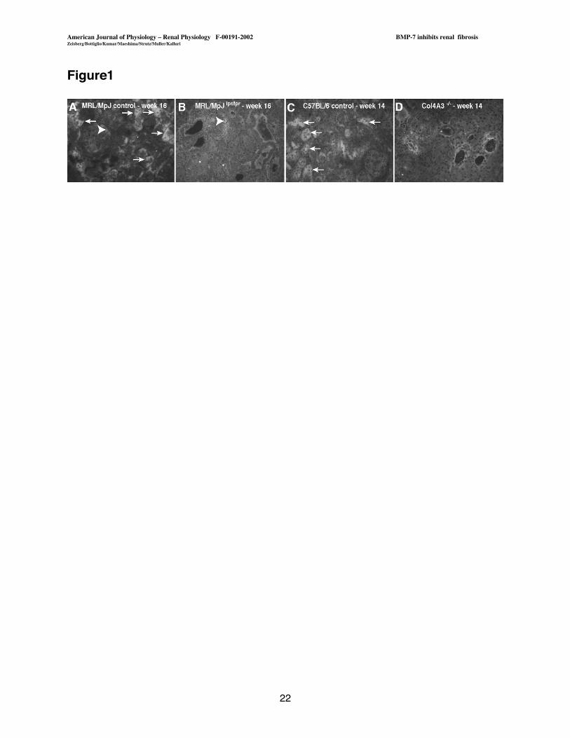
Decrease of tubular endogenous BMP-7 in progression of renal disease in MRL/MpJlpr/lpr
and in Col4A3-/- mice. Recent studies suggested that renal injury was associated with a
decrease of endogenous BMP-7 expression and that administration of exogenous rhBMP-7
served to restore renal function and morphology (21, 52, 54, 56). In the present study we
attempted to explore the potential of rhBMP-7 to prevent progression of renal disease in genetic
models of renal disease. We first assessed endogenous BMP-7 in the normal and diseased
kidneys of these mice. Due to a mutation in the Fas gene MRL/MpJlpr/lpr mice develop a lupus
like disease with progressive renal fibrosis (23). While kidneys from MRL/MpJ control mice
displayed BMP-7 in tubular cells and in glomeruli, confirming previous observations, BMP-7 was
notably decreased in injured kidneys from MRL/MpJlpr/lpr mice (17, 54) (Figure 1 A,B).
Additionally, we used mice that lack the collagen type IV a3 gene (Col4A3-/-) as a second model
for progressive renal disease. Col4A3-/- mice develop progressive renal disease, which leads to
death between week 12 and 16 (1, 30). While C57BL/6 control mice displayed tubular and
glomerular BMP-7 localization, progression of renal disease in Col4A3-/- mice was associated
with a decrease in endogenous BMP-7 expression (Figure 1 C,D). We thus speculated that
supplementation of exogenous rhBMP-7 could improve renal pathology and function in both
these genetic mouse models for chronic renal disease.
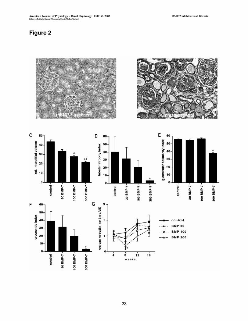
rhBMP-7 ameliorates progression of chronic renal disease in MRL/MpJlpr/lpr mice.
To evaluate a potential role for rhBMP-7 in the treatment of chronic renal disease, we first

American Journal of Physiology � Renal Physiology F-00191-2002 BMP-7 inhibits renal fibrosisZeisberg/Bottiglio/Kumar/Maeshima/Strutz/Muller/Kalluri
8
utilized MRL/MpJlpr/lpr lupus nephritis mice (11). We initiated i.p. injection of rh BMP-7 at 4 weeks
of age. At this age, very early signs of disease could be demonstrated in these mice. After
termination of the study at 16 weeks of age, rhBMP-7 treated mice displayed reduced relative
interstitial volume as well as a reduced number of atrophic tubular structures when compared to
untreated control (Figure 2 A-D). Animals that were treated with 300 mg/kg rhBMP-7 also
displayed reduced glomerular crescents, markedly reduced glomerulosclerosis, and reduced
glomerular hypercellularity (Figure 2 E, F). Serum creatinine levels were significantly reduced in
mice that were treated with 300 mg/kg rhBMP-7 after four weeks of treatment, whereas by the
end of study results failed to reach significance due to wide variance in the treatment groups
(Figure 2G).
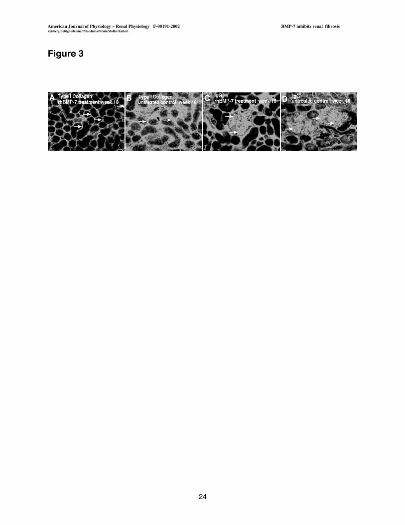
These findings also correlated with reduced interstitial staining for type I collagen in the
treated mice, compared to control mice (Figure 3 A, B). Localization for IgG in glomeruli did not
show substantial difference between untreated and treated mice (Figure 3 C, D).
Treatment with rhBMP-7 inhibits progression of renal disease in Col4A3-/- mice.
We next attempted to investigate, whether administration of exogenous rhBMP-7 had a similar
role on the progression of renal disease in Col4A3-/- mice. Treatment with rhBMP-7 was initiated
after six weeks of age until the study was terminated at the age of 14 weeks (30). In this study,
we treated mice with 300 mg/kg rhBMP-7, which was established by our previous study as the
optimum dosage, and was also confirmed by other groups (21, 52). Treatment with 300 mg/kg
rhBMP-7 led to reduction of tubulointerstitial fibrosis as determined by reduced relative cortical
interstitial volume and significantly reduced tubular atrophy (Figure 4 A-C). Reduction of renal
damage led to decreased renal pathology-related mortality rate in the rhBMP-7 treated group
(Figure 4C). Five out of seven untreated control mice died due to renal failure before week 14,
American Journal of Physiology � Renal Physiology F-00191-2002 BMP-7 inhibits renal fibrosisZeisberg/Bottiglio/Kumar/Maeshima/Strutz/Muller/Kalluri
9
whereas none of the treated mice died due to uremia and kidney disease. Treated mice lived up
to week 26. Renal function, as assessed by serum creatinine (Figure 4D), BUN (Figure E) and
urine protein (Figure 4F), revealed significant improvement in BMP-7 treated mice as compared
to untreated Col4A3-/- mice.
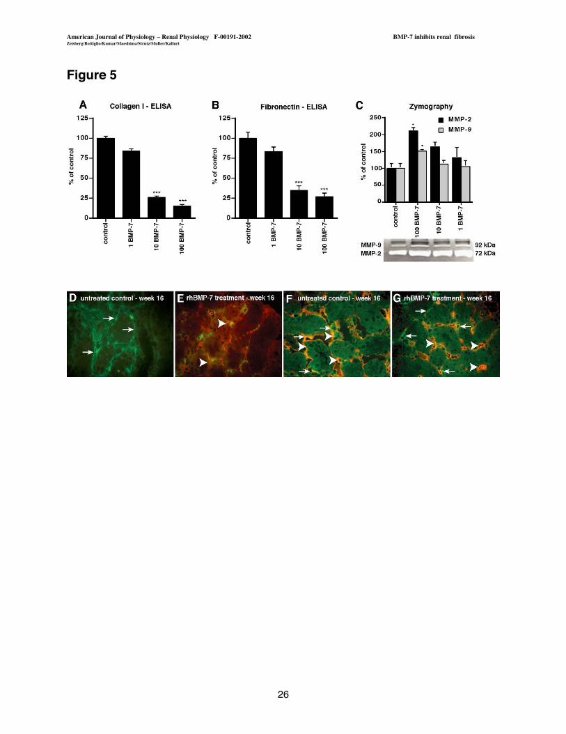
rhBMP-7 has anti-fibrogenic effects on renal fibroblasts. We next attempted to
evaluate the effect of rhBMP-7 on interstitial fibroblasts in order to gain further insights into the
anti-fibrotic action of rhBMP-7. Interstitial fibroblasts play a central role in the progression of
chronic renal disease as the main mediators of ECM deposition, the characteristic feature of
fibrosis (13, 39). We utilized a human interstitial fibroblast cell line (TK173), which displays an
activated fibroblast-like state in vitro, to evaluate the potential of rhBMP-7 to inhibit pro-fibrotic
contributions of these cells (27, 32). Synthesis of type I collagen and fibronectin are considered
as key features of activated fibroblasts and we show here that administration of rhBMP-7
decreases synthesis of collagen type I and fibronectin in the TK173 cells in a dose dependent
manner, without significant alteration in the proliferative capacity of these cells (Figure 5A,B)
(13, 39). In addition to increased synthesis, a decrease in ECM-degradation is considered
important for shifting the balance of ECM-homeostasis towards enhanced matrix deposition
during fibrogenesis (13, 44). Human interstitial fibroblasts secrete substantial levels of matrix
metalloproteinases, MMP-2 and MMP-9, which possess the capacity to cleave basement
membranes and interstitial ECM constituents (8, 10, 26, 48). Treatment of TK 173 cells with
rhBMP-7 led to up-regulation of MMP-2 and also of MMP-9 (Figure 5 C). In the Mrl/MpJlpr/lpr
mice, upregulation of MMP-2 by the interstitial fibroblasts by administration of rhBMP-7 was also
observed (Figure 5 D, E). While MMP-2 was absent in the interstitial areas containing abundant
myofibroblasts, MMP-2 was markedly increased in the interstitium of rhBMP-7 treated
Mrl/MpJlpr/lpr mice (Figure 5 D, E). In the rhBMP-7 treated kidneys, MMP-2 mainly co-localized
American Journal of Physiology � Renal Physiology F-00191-2002 BMP-7 inhibits renal fibrosisZeisberg/Bottiglio/Kumar/Maeshima/Strutz/Muller/Kalluri
10
with a-smooth muscle actin (a-SMA) expressing activated myofibroblasts, further suggesting a
potential of rhBMP-7 to induce MMP-2 in the interstitial fibroblasts (Figure 5 D, E). MMP-9 was
significantly present in the interstitium of untreated control kidneys and thus further increase of
MMP-9 could not be demonstrated following treatment with BMP-7 (Figure 5 F, G) .
Furthermore, MMP-9 in the interstitium rarely co-localized with a-SMA, suggesting that rhBMP-7
in interstitial fibroblasts activates mainly MMP-2 in the injured kidneys (Figure 5 F, G).
Discussion
Progression of chronic renal disease towards ESRF still represents one of the biggest
problems in nephrology, as it leads to an increasing number of patients who require long-term
renal replacement therapy, such as dialysis or kidney transplant (9, 35). While ACE-inhibitors
are currently the most promising therapeutic agents to inhibit progression of renal fibrosis in
clinical use, specific therapeutic options are still not available (2, 7, 15, 25, 55).
In this regard, several recent studies have demonstrated a beneficial role for
administration of exogenous rhBMP-7 in different animal models of chronic renal injury (21, 31,
52, 56). BMP-7 is a member of the TGF-b superfamily and has an important function during
kidney development (20). It is associated with condensation of the metanephric mesenchyme,
leading to the formation of tubules and glomeruli (20, 41). In the adult kidney, BMP-7 expression
can be detected in tubular epithelial cells and in podocytes (17, 54). Endogenous BMP-7
expression significantly decreases during acute renal injury and administration of exogenous
rhBMP-7 accelerates the repair of the injured kidney, suggesting that BMP-7 plays a role in the
maintenance kidney homeostasis (42, 49). Similarly, BMP-7 expression is decreased in several
induced animal models of chronic renal injury, and administration of exogenous rhBMP-7 in
these models inhibits progression or enables recovery of chronic renal injury (21, 52, 54, 56).

American Journal of Physiology � Renal Physiology F-00191-2002 BMP-7 inhibits renal fibrosisZeisberg/Bottiglio/Kumar/Maeshima/Strutz/Muller/Kalluri
11
In this regard, in the present study we establish for the first time a role of rhBMP-7 as a
therapeutic in two genetic mouse models, which mimic long-term chronic renal disease. In
MRL/MpJlpr/lpr mice, which develop lupus nephritis-like renal disease associated with significant
tubulointerstitial fibrosis after three months, treatment with rhBMP-7 inhibits progression of renal
disease in a dose dependent manner. In mice deficient in the a3 chain of type IV collagen,
ESRF associated with interstitial fibrosis after 14 weeks is observed. rhBMP-7 prevents renal
fibrosis and renal related mortality in these mice and increases their survival by 12 weeks. Thus,
we demonstrate the anti-fibrotic effect of rhBMP-7 in two long-term mouse models for chronic
renal fibrosis, providing further evidence for the use of rhBMP-7 as a therapeutic agent for
chronic renal injury.
Previous studies have suggested that rhBMP-7 exerts its anti-fibrotic effect mainly on
tubular epithelial cells, where it inhibits the release of pro-inflammatory chemokines (17). It is
also shown to reverse epithelial to mesenchymal transition (EMT), while acting as an antagonist
of transforming growth factor b1 (TGF-b1)-induced E-cadherin downregulation (56). In chronic
renal fibrosis, activated interstitial fibroblasts, which can either derive from resident interstitial
fibroblasts, from tubular epithelial cells via EMT or from bone marrow-derived mesenchymal
precursor cells, are considered the main pathogenic mediators of renal disease (13, 19, 22, 39).
Renal fibrosis is characterized by an excessive deposition of interstitial ECM, which results in
destruction of kidney architecture and impairment of renal function. Activated interstitial
fibroblasts are the main mediators of enhanced ECM synthesis (3, 6, 14, 16, 33). In the present
study we provide evidence that rhBMP-7 functions (in addition to its effects on tubular epithelial
cells) by inhibiting pro-fibrotic contributions of activated interstitial fibroblasts. We hypothesize
that up-regulation of MMP-2 by rhBMP-7 reflects increased ECM degradation and thus
potentially a decrease in scar tissue in the renal interstitium (46, 58). TGF-b1, the main pro-
American Journal of Physiology � Renal Physiology F-00191-2002 BMP-7 inhibits renal fibrosisZeisberg/Bottiglio/Kumar/Maeshima/Strutz/Muller/Kalluri
12
fibrotic growth factor involved in renal fibrogenesis, mediates all these three pro-fibrotic features,
which are inhibited by rhBMP-7 in our studies, in renal fibroblasts (6, 46). Previous studies have
also demonstrated the potential of rhBMP-7 to counteract TGF-b1 action in tubular epithelial
and mesangial cells (53, 56). Our research coupled with these previous important studies
suggest that rhBMP-7 functions on tubular epithelial cells, mesangial cells and fibroblasts to
restore the health of kidney tissue. These studies provide us further with confidence that
rhBMP-7 should be tested in human clinical trials involving kidney disease patients.
American Journal of Physiology � Renal Physiology F-00191-2002 BMP-7 inhibits renal fibrosisZeisberg/Bottiglio/Kumar/Maeshima/Strutz/Muller/Kalluri
13
Acknowledgements
This study was supported by a sponsored research project from Curis, Inc.; in part by
NIH grants DK 51711 and DK 55001, American Society of Nephrology Carl Gottschalk-
research award, grants from the Deutsche Forschungsgemeinschaft DFG Mü523/7-1 (to G.M.)
and DFG ZE5231/1 (to M.Z.).
American Journal of Physiology � Renal Physiology F-00191-2002 BMP-7 inhibits renal fibrosisZeisberg/Bottiglio/Kumar/Maeshima/Strutz/Muller/Kalluri
14
Abbreviations
BMP, Bone Morphogenic Protein; DMEM, Dulbecco�s Modified Eagle�s Medium; ECM,
extracellular matrix; EMT, epithelial-to-mesenchymal transition; ESRF. End Stage Renal Failure;
MMP, Matrix Metalloproteinase; PAS, Periodic Acid Schiff; STZ, streptozotocin; TGF,
Transforming Growth Factor; UUO, unilateral urethral obstruction.
American Journal of Physiology � Renal Physiology F-00191-2002 BMP-7 inhibits renal fibrosisZeisberg/Bottiglio/Kumar/Maeshima/Strutz/Muller/Kalluri
15
Figures
Figure 1. Immunofluorescence Staining. Frozen kidney sections were stained with
antibodies to BMP-7 and the staining was visualized using FITC-conjugated secondary
antibodies. (A) Normal kidney from Mrl/MpJ control mice displayed tubular staining (arrows) and
glomerular staining (arrowhead). (B) BMP-7 was decreased in kidneys from 16-week old
Mrl/MpJ lpr/lpr mice with interstitial fibrosis. (C) BMP-7 was present in kidneys from control
C57BL/6 mice. (D) In kidneys from 14-week old Col4A3-/- mice, severe interstitial fibrosis was
associated with decreased BMP-7 staining. Original magnification x200.
Figure 2. Treatment of MRL/MpJlpr/lpr mice with rhBMP-7 ameliorates progression of
chronic renal disease. (A, B) Light microscopy. The pictures display representative areas of
PAS-stained kidney sections from MRL/MpJlpr/lpr mice at the age of 16 weeks. Histology of
animals that were treated with 300 mg/kg rhBMP-7 revealed a drastic delay of disease
progression (A) compared to control animals (B). (C-F) Morphometric analysis. Kidney
sections were analyzed for the relative interstitial volume, tubular atrophy, glomerular cellularity
and crescent formation. The graphs summarize average results of each group. (C) Treatment
with rhBMP-7 resulted in a significantly decreased relative interstitial volume compared to
untreated mice. (48.4% +8.9). (D) Occurrence of atrophic tubules was significantly decreased in
the 300 mg/kg treated group (8.5% +7.3 of control). (E, F). Glomerular injury, as determined by
glomerular cellularity (68.5% +1.3) and formation of glomerular crescents (2.8% +0.35 of
control) was decreased in the 300 mg/kg treated group. (G) Serum creatinine levels. The graph
summarizes serum creatinine levels between week 4 and week 16. **=p<0.001, *=p<0.05.
Original magnification x200.

American Journal of Physiology � Renal Physiology F-00191-2002 BMP-7 inhibits renal fibrosisZeisberg/Bottiglio/Kumar/Maeshima/Strutz/Muller/Kalluri
16
Figure 3. Immunofluorescence Staining. Frozen kidney sections, which were obtained from
MRL/MpJlpr/lpr mice at the age of 16 weeks, were stained with antibodies for type I collagen and
IgG. The pictures display representative sections from untreated control mice and from mice
that were treated with 300 mg/kg rhBMP-7. (A, B) Interstitial staining for type I collagen (arrows)
was substantially decreased in rhBMP-7 treated MRL/MpJlpr/lpr mice compared to untreated
control (C, D). Staining for IgG deposition (arrows) did not reveal significant differences between
the two groups. Original magnification x200.
Figure 4. Treatment of Col4A3-/- mice with rhBMP-7 results in decrease of renal disease
and reduction of renal-related mortality. Col4A3-/- mice were treated with 300 mg/kg rhBMP-
7, control mice were injected with vehicle buffer alone. Urine and blood was obtained every two
weeks, the study was terminated at 14 weeks of age. (A, B) Light microscopy. Histology of
kidney specimens from Col4A3-/- mice revealed markedly improved appearing tissue in rhBMP-7
treated group, as compared to substantial tubulointerstitial fibrosis in untreated control Col4A3-/-
mice (C). Relative interstitial volume and tubular atrophy index were determined by
morphometric analysis. The graph summarizes average values of each group. Treatment with
300 mg/kg of rhBMP-7 resulted in decreased relative interstitial volume (23% +3.3 of control)
and tubular atrophy (7.7% +4.5 of control). Whereas 71% of untreated mice died before week
14 due to renal failure, no renal related deaths occurred in the BMP-7 treated group. (D, E, F)
Renal function parameters. Serum-creatinine, BUN and urine protein were determined every
other week. The graphs summarize results, which were obtained during the study. (***:
P<0.0001, *: P<0.05). Original magnification x200.

American Journal of Physiology � Renal Physiology F-00191-2002 BMP-7 inhibits renal fibrosisZeisberg/Bottiglio/Kumar/Maeshima/Strutz/Muller/Kalluri
17
Figure 5. BMP-7 has anti-fibrogenic properties on renal interstitial fibroblasts. (A)
Collagen ELISA. (A-C). TK 173 human interstitial fibroblasts were subjected to rhBMP-7 for 48
h. Tissue culture supernatants were evaluated for secretion of type I collagen and fibronectin by
ELISA and for release of MMPs by zymography. The graphs summarize three independent
experiments. (A) Collagen ELISA. rhBMP-7 significantly decreased rhBMP-7 secretion of type
I collagen into tissue culture supernatants (15.2% + 1.9 compared to control at 100 ng rhBMP-7/
ml). (B) Fibronectin ELISA. After 48 h, fibronectin was significantly decreased in tissue culture
supernatants of interstitial fibroblasts that were exposed to rhBMP-7 (27.1% + 5.1 compared to
control at 100 ng/ml). (C) Zymography. Interstitial fibroblasts were treated with BMP-7 for 48 h
and the tissue culture supernatants were analyzed by zymography for MMP-2 and MMP-9. The
picture displays a representative gel. Densitometric analysis revealed a significant increase of
MMP-2 (210.5% +10.27 compared to untreated control) and MMP-9 (151.2% +4.3) after
treatment with 100 ng/ml rhBMP-7 (D, E). MMP-2/ a-SMA double staining. Frozen kidney
section from rhBMP-7 treated and control MRL/MpJlpr/lpr mice were analyzed after termination of
the study at the age of 16 weeks. In immunohistochemistry double staining experiments for a-
SMA (FITC-green) and MMP-2 (Rhodamine-red), MMP-2 was absent in myofibroblasts after 16
weeks in untreated MRL/MpJlpr/lpr mice (arrows). Administration of exogenous rhBMP-7 resulted
in increased interstitial MMP-2 staining (arrowheads). (F, G). MMP-9/ a-SMA double staining.
Immunohistochemistry double staining experiments for a-SMA (FITC-green, arrows) and MMP-
9 (Rhodamine-red, arrowheads). Substantial staining for MMP-9 was present in untreated
MRL/MpJlpr/lpr mice, treatment with rhBMP-7 resulted in insignificant changes. Original
magnification x400. ***=p<0.0001, *=p<0.05.
American Journal of Physiology � Renal Physiology F-00191-2002 BMP-7 inhibits renal fibrosisZeisberg/Bottiglio/Kumar/Maeshima/Strutz/Muller/Kalluri
18
References
1.Andrews KL, Mudd JL, Li C, and Miner JH. Quantitative trait loci influence renal diseaseprogression in a mouse model of Alport syndrome. Am J Pathol 160: 721-730, 2002.2.Aros C and Remuzzi G. The renin-angiotensin system in progression, remission andregression of chronic nephropathies. J Hypertens 20 Suppl 3: S45-53, 2002.3.Badid C, Vincent M, Fouque D, Laville M, and Desmouliere A. Myofibroblast: a prognosticmarker and target cell in progressive renal disease. Ren Fail 23: 543-549, 2001.4.Becker GJ and Hewitson TD. The role of tubulointerstitial injury in chronic renal failure. CurrOpin Nephrol Hypertens 9: 133-138, 2000.5.Bohle A, Christ H, Grund KE, and Mackensen S. The role of the interstitium of the renalcortex in renal disease. Contrib Nephrol 16: 109-114, 1979.6.Border WA and Noble NA. Targeting TGF-beta for treatment of disease. Nat Med 1: 1000-1001., 1995.7.Brenner BM. Remission of renal disease: recounting the challenge, acquiring the goal. J ClinInvest 110: 1753-1758, 2002.8.Brinckerhoff CE and Matrisian LM. Matrix metalloproteinases: a tail of a frog that became aprince. Nat Rev Mol Cell Biol 3: 207-214, 2002.9.Bruns FJ, Seddon P, Saul M, and Zeidel ML. The cost of caring for end-stage kidneydisease patients: an analysis based on hospital financial transaction records. J Am Soc Nephrol9: 884-890., 1998.10.Chen EI, Kridel SJ, Howard EW, Li W, Godzik A, and Smith JW. A unique substraterecognition profile for matrix metalloproteinase-2. J Biol Chem 277: 4485-4491, 2002.11.Cohen PL and Eisenberg RA. Lpr and gld: single gene models of systemic autoimmunityand lymphoproliferative disease. Annu Rev Immunol 9: 243-269, 1991.12.Dudley AT, Lyons KM, and Robertson EJ. A requirement for bone morphogenetic protein-7 during development of the mammalian kidney and eye. Genes Dev 9: 2795-2807., 1995.13.Eddy AA. Molecular insights into renal interstitial fibrosis [editorial]. J Am Soc Nephrol 7:2495-2508, 1996.14.El Nahas A. Mechanisms of experimental and clinical renal scarring. In: Oxford Textbook ofClinical Nephrology (Second Edition ed.), edited by Davison A, Cameron J, Grunfeld J, Kerr D,Ritz E and Winearls C. Oxford: Oxford University Press, 1998, p. 1749-1788.15.Fogo AB. Editorial Review: The potential for regression of renal scarring. Curr Opin NephrolHypertens 12: 223-225, 2003.16.Fogo AB. Pathology of progressive nephropathies. Curr Opin Nephrol Hypertens 9: 241-246., 2000.17.Gould SE, Day M, Jones SS, and Dorai H. BMP-7 regulates chemokine, cytokine, andhemodynamic gene expression in proximal tubule cells. Kidney Int 61: 51-60, 2002.18.Hebert LA, Wilmer WA, Falkenhain ME, Ladson-Wofford SE, Nahman NS, and RovinBH. Renoprotection: One or many therapies? Kidney Int 59: 1211-1226., 2001.19.Herzlinger D. Renal interstitial fibrosis: remembrance of things past? J Clin Invest 110: 305-306, 2002.20.Hogan BL. Bone morphogenetic proteins in development. Curr Opin Genet Dev 6: 432-438.,1996.21.Hruska KA, Guo G, Wozniak M, Martin D, Miller S, Liapis H, Loveday K, Klahr S,Sampath TK, and Morrissey J. Osteogenic protein-1 prevents renal fibrogenesis associatedwith ureteral obstruction. Am J Physiol Renal Physiol 279: F130-143., 2000.

American Journal of Physiology � Renal Physiology F-00191-2002 BMP-7 inhibits renal fibrosisZeisberg/Bottiglio/Kumar/Maeshima/Strutz/Muller/Kalluri
19
22.Iwano M, Plieth D, Danoff TM, Xue C, Okada H, and Neilson EG. Evidence that fibroblastsderive from epithelium during tissue fibrosis. J Clin Invest 110: 341-350., 2002.23.Kelley VE and Roths JB. Interaction of mutant lpr gene with background strain influencesrenal disease. Clin Immunol Immunopathol 37: 220-229., 1985.24.Klahr S, Morrissey J, Hruska K, Wang S, and Chen Q. New approaches to delay theprogression of chronic renal failure. Kidney Int Suppl: 23-26., 2002.25.Komers R and Anderson S. Optimal strategies for preventing progression of renal disease:should angiotensin converting enzyme inhibitors and angiotensin receptor blockers be usedtogether? Curr Hypertens Rep 2: 465-472, 2000.26.Kridel SJ, Chen E, Kotra LP, Howard EW, Mobashery S, and Smith JW. Substratehydrolysis by matrix metalloproteinase-9. J Biol Chem 276: 20572-20578, 2001.27.Lonnemann G, Shapiro L, Engler-Blum G, Muller GA, Koch KM, and Dinarello CA.Cytokines in human renal interstitial fibrosis. I. Interleukin-1 is a paracrine growth factor forcultured fibrosis-derived kidney fibroblasts. Kidney Int 47: 837-844., 1995.28.Luo G, Hofmann C, Bronckers AL, Sohocki M, Bradley A, and Karsenty G. BMP-7 is aninducer of nephrogenesis, and is also required for eye development and skeletal patterning.Genes Dev 9: 2808-2820., 1995.29.Maeshima Y, Kashihara N, Sugiyama H, Makino H, and Ota Z. Antisense oligonucleotidesto proliferating cell nuclear antigen and Ki- 67 inhibit human mesangial cell proliferation. J AmSoc Nephrol 7: 2219-2229., 1996.30.Miner JH and Sanes JR. Molecular and functional defects in kidneys of mice lackingcollagen alpha 3(IV): implications for Alport syndrome. J Cell Biol 135: 1403-1413., 1996.31.Morrissey J, Hruska K, Guo G, Wang S, Chen Q, and Klahr S. Bone morphogeneticprotein-7 improves renal fibrosis and accelerates the return of renal function. J Am Soc Nephrol13 Suppl 1: S14-21., 2002.32.Muller GA, Frank J, Rodemann HP, and Engler-Blum G. Human renal fibroblast cell lines(tFKIF and tNKF) are new tools to investigate pathophysiologic mechanisms of renal interstitialfibrosis. Exp Nephrol 3: 127-133., 1995.33.Murphy M, McMahon R, Lappin DW, and Brady HR. Gremlins: is this what renalfibrogenesis has come to? Exp Nephrol 10: 241-244, 2002.34.Ozkaynak E, Rueger DC, Drier EA, Corbett C, Ridge RJ, Sampath TK, and OppermannH. OP-1 cDNA encodes an osteogenic protein in the TGF-beta family. Embo J 9: 2085-2093.,1990.35. Pastan S and Bailey J. Dialysis therapy. N Engl J Med 338: 1428-1437., 1998.36.Pavlova A, Stuart RO, Pohl M, and Nigam SK. Evolution of gene expression patterns in amodel of branching morphogenesis. Am J Physiol 277: F650-663., 1999.37.Peters H, Border WA, and Noble NA. Targeting TGF-beta overexpression: maximizing theantifibrotic actions of angiotensin II blockade in anti-Thy1 glomerulonephritis. Nephrol DialTransplant 14: 22-23., 1999.38.Raij L, Azar S, and Keane W. Mesangial immune injury, hypertension, and progressiveglomerular damage in Dahl rats. Kidney Int 26: 137-143., 1984.39.Remuzzi G and Bertani T. Pathophysiology of progressive nephropathies. N Engl J Med339: 1448-1456., 1998.40.Sakurai H and Nigam SK. In vitro branching tubulogenesis: implications for developmentaland cystic disorders, nephron number, renal repair, and nephron engineering. Kidney Int 54: 14-26., 1998.41.Schedl A and Hastie ND. Cross-talk in kidney development. Curr Opin Genet Dev 10: 543-549., 2000.
American Journal of Physiology � Renal Physiology F-00191-2002 BMP-7 inhibits renal fibrosisZeisberg/Bottiglio/Kumar/Maeshima/Strutz/Muller/Kalluri
20
42.Simon M, Maresh JG, Harris SE, Hernandez JD, Arar M, Olson MS, and Abboud HE.Expression of bone morphogenetic protein-7 mRNA in normal and ischemic adult rat kidney. AmJ Physiol 276: F382-389., 1999.43.Strutz F, Heeg M, Kochsiek T, Siemers G, Zeisberg M, and Muller GA. Effects ofpentoxifylline, pentifylline and gamma-interferon on proliferation, differentiation, and matrixsynthesis of human renal fibroblasts. Nephrol Dial Transplant 15: 1535-1546., 2000.44.Strutz F and Muller GA. Interstitial pathomechanisms underlying progressivetubulointerstitial damage. Kidney Blood Press Res 22: 71-80, 1999.45.Strutz F, Zeisberg M, Hemmerlein B, Sattler B, Hummel K, Becker V, and Muller GA.Basic fibroblast growth factor expression is increased in human renal fibrogenesis and maymediate autocrine fibroblast proliferation. Kidney Int 57: 1521-1538., 2000.46.Strutz F, Zeisberg M, Renziehausen A, Raschke B, Becker V, van Kooten C, and MullerG. TGF-beta 1 induces proliferation in human renal fibroblasts via induction of basic fibroblastgrowth factor (FGF-2). Kidney Int 59: 579-592., 2001.47.Timoshanko JR, Kitching AR, Holdsworth SR, and Tipping PG. Interleukin-12 fromintrinsic cells is an effector of renal injury in crescentic glomerulonephritis. J Am Soc Nephrol 12:464-471., 2001.48.Vu TH and Werb Z. Matrix metalloproteinases: effectors of development and normalphysiology. Genes Dev 14: 2123-2133., 2000.49.Vukicevic S, Basic V, Rogic D, Basic N, Shih MS, Shepard A, Jin D, Dattatreyamurty B,Jones W, Dorai H, Ryan S, Griffiths D, Maliakal J, Jelic M, Pastorcic M, Stavljenic A, andSampath TK. Osteogenic protein-1 (bone morphogenetic protein-7) reduces severity of injuryafter ischemic acute renal failure in rat. J Clin Invest 102: 202-214., 1998.50.Vukicevic S, Kopp JB, Luyten FP, and Sampath TK. Induction of nephrogenicmesenchyme by osteogenic protein 1 (bone morphogenetic protein 7). Proc Natl Acad Sci U SA 93: 9021-9026., 1996.51.Vukicevic S, Latin V, Chen P, Batorsky R, Reddi AH, and Sampath TK. Localization ofosteogenic protein-1 (bone morphogenetic protein-7) during human embryonic development:high affinity binding to basement membranes. Biochem Biophys Res Commun 198: 693-700.,1994.52.Wang S, Chen Q, Simon TC, Strebeck F, Chaudhary L, Morrissey J, Liapis H, Klahr S,and Hruska KA. Bone morphogenic protein-7 (BMP-7), a novel therapy for diabeticnephropathy. Kidney Int 63: 2037-2049, 2003.53.Wang S and Hirschberg R. BMP7 antagonizes TGF-beta -dependent fibrogenesis inmesangial cells. Am J Physiol Renal Physiol 284: F1006-1013, 2003.54.Wang SN, Lapage J, and Hirschberg R. Loss of tubular bone morphogenetic protein-7 indiabetic nephropathy. J Am Soc Nephrol 12: 2392-2399., 2001.55.Yu L, Noble NA, and Border WA. Therapeutic strategies to halt renal fibrosis. Curr OpinPharmacol 2: 177-181, 2002.56.Zeisberg M, Hanai J, Sugimoto H, Mammoto T, Charytan D, Strutz F, and Kalluri R.BMP-7 counteracts TGF-beta1-induced epithelial-to-mesenchymal transition and reverseschronic renal injury. Nat Med 9: 964-968, 2003.57.Zeisberg M, Maeshima Y, Mosterman B, and Kalluri R. Renal fibrosis : extracellular matrixmicroenvironment regulates migratory behavior of activated tubular epithelial cells. Am J Pathol160: 2001-2008., 2002.58.Zeisberg M, Strutz F, and Muller GA. Role of fibroblast activation in inducing interstitialfibrosis. J Nephrol 13 Suppl 3: S111-120., 2000.
American Journal of Physiology � Renal Physiology F-00191-2002 BMP-7 inhibits renal fibrosisZeisberg/Bottiglio/Kumar/Maeshima/Strutz/Muller/Kalluri
21
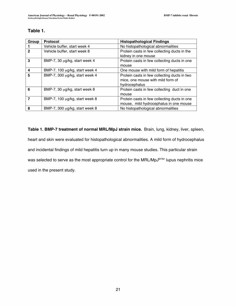
Table 1.
Group Protocol Histopathological Findings1 Vehicle buffer, start week 4 No histopathological abnormalities2 Vehicle buffer, start week 8 Protein casts in few collecting ducts in the
kidney in one mouse3 BMP-7, 30 mg/kg, start week 4 Protein casts in few collecting ducts in one
mouse4 BMP-7, 100 mg/kg, start week 4 One mouse with mild form of hepatitis5 BMP-7, 300 mg/kg, start week 4 Protein casts in few collecting ducts in two
mice, one mouse with mild form ofhydrocephalus
6 BMP-7, 30 mg/kg, start week 8 Protein casts in few collecting duct in onemouse
7 BMP-7, 100 mg/kg, start week 8 Protein casts in few collecting ducts in onemouse, mild hydrocephalus in one mouse
8 BMP-7, 300 mg/kg, start week 8 No histopathological abnormalities
Table 1. BMP-7 treatment of normal MRL/MpJ strain mice. Brain, lung, kidney, liver, spleen,
heart and skin were evaluated for histopathological abnormalities. A mild form of hydrocephalus
and incidental findings of mild hepatitis turn up in many mouse studies. This particular strain
was selected to serve as the most appropriate control for the MRL/MpJlpr/lpr lupus nephritis mice
used in the present study.
American Journal of Physiology � Renal Physiology F-00191-2002 BMP-7 inhibits renal fibrosisZeisberg/Bottiglio/Kumar/Maeshima/Strutz/Muller/Kalluri
22
Figure1
American Journal of Physiology � Renal Physiology F-00191-2002 BMP-7 inhibits renal fibrosisZeisberg/Bottiglio/Kumar/Maeshima/Strutz/Muller/Kalluri
23
Figure 2
American Journal of Physiology � Renal Physiology F-00191-2002 BMP-7 inhibits renal fibrosisZeisberg/Bottiglio/Kumar/Maeshima/Strutz/Muller/Kalluri
24
Figure 3

American Journal of Physiology � Renal Physiology F-00191-2002 BMP-7 inhibits renal fibrosisZeisberg/Bottiglio/Kumar/Maeshima/Strutz/Muller/Kalluri
25
Figure4
American Journal of Physiology � Renal Physiology F-00191-2002 BMP-7 inhibits renal fibrosisZeisberg/Bottiglio/Kumar/Maeshima/Strutz/Muller/Kalluri
26
Figure 5