


Bell Work
What’s the difference between a tendon and a ligament?

The Integumentary System
Unit 1: Levels of Organization
Topic 4: Integument

Skin Structure
Skin is the single largest organ in the human body.Accounts for about 15% of the body weightTHREE distinct layers
Epidermis: Composed of epithelial tissue Dermis: Composed of a combination of connective
tissues Hypodermis: Composed of a combination of
connective tissues

6 Main Functions of the Skin
1. Protection: Physical and solar barrier.1. Keratin protects tissues from heat, chemicals, injury,
& infection.
2. Melanin protects against solar radiation.
2. Vitamin D Synthesis: Vitamin D regulates blood calcium levels – production is stimulated by ultraviolet radiation
3. Extensive Sensory Functions: Detects heat, cold, touch, texture, pleasure, vibration, and tissue injury.

6 Main Functions of the Skin
4. Thermoregulation: Via sweat production & blood flow; preserves homeostasis.
5. Excretion & Absorption: Excretes & absorbs lipid-soluble materials; sweat allows secretion of small amounts of waste materials including salt, carbon dioxide, ammonia, and urea.
6. Reservoir: For 8-10% of adult blood flow
Epidermis
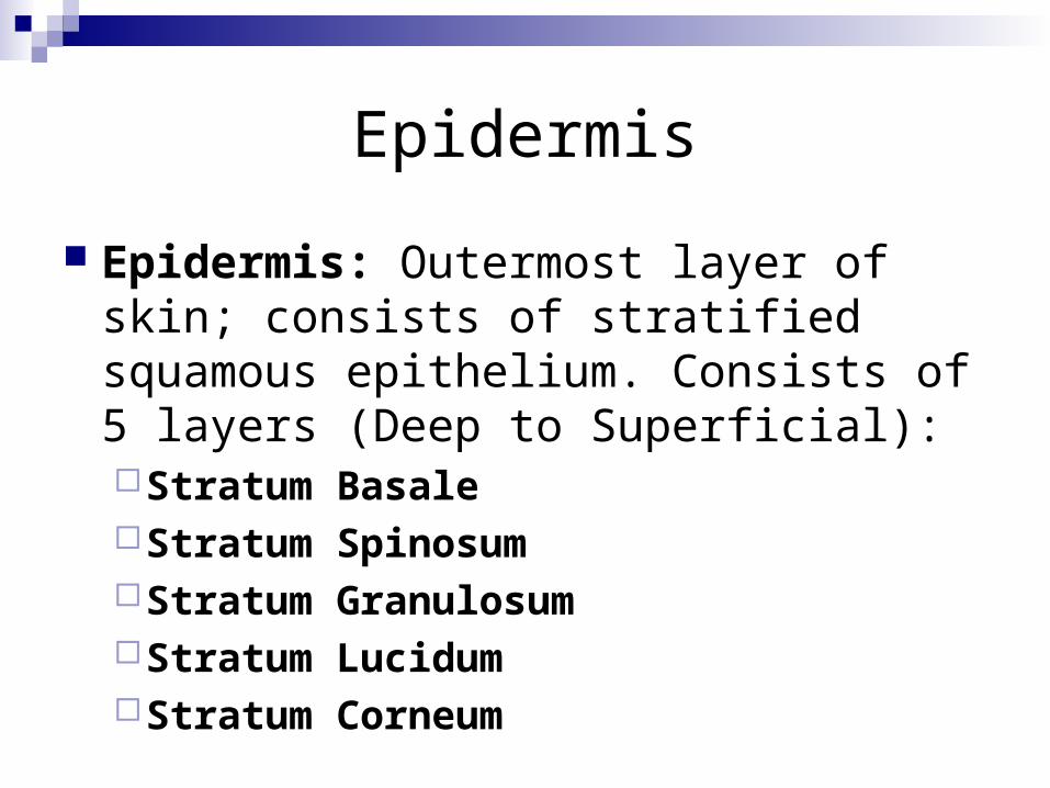
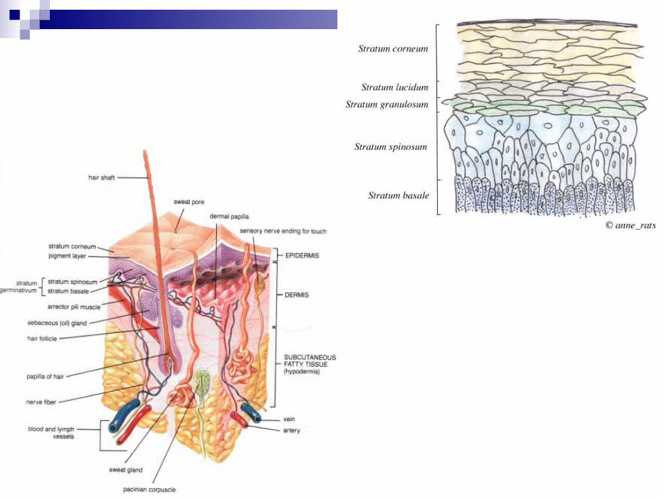
Epidermis: Outermost layer of skin; consists of stratified squamous epithelium. Consists of 5 layers (Deep to Superficial): Stratum BasaleStratum SpinosumStratum GranulosumStratum LucidumStratum Corneum


**Stratum Basale
Stratum Basale: Base layer of single row of cells where mitosis takes place.
Responsible for the development of the following cell types (not on test!) Keratinocytes: Produce protein, keratin (a protective
substance that composes 90% of skin) Melanocytes: Synthesize the pigment melanin, that
absorbs ultraviolet radiation. Merkel Cells: Few in number; sensitive to touch and
produce the skin’s tactile response Langerhans Cells: Derived from red blood cells;
participate in immune response.

Stratum Spinosum
Stratum Spinosum: Consists of 8-10 layers of Keratinocytes – some mitosis still happens here, but mostly provides structure and flexibility.
Composed of Keratinocytes attached to each other via desmosomes.
Contains langerhans cells that aid in the immune system response.

Stratum Granulosum
Stratum Granulosum: The middle layer of 3-5 layers of cells that help form keratin.
Contains lamellar granules that produce a secretion that helps the skin repel water and seal in other fluids.

Stratum Lucidum
Stratum Lucidum: Found only in the thick skin of the palms, fingertips, and soles of the feet
Tends to be translucent.


**Stratum Corneum
Stratum Corneum: The outermost layer of the epidermis.Contains up to 30 layers of the dead, scaly,
keratinized cellsProvides protection.Continuously shed and replaced.


Dermis
Dermis: Composed primarily of connective tissue, mainly collagen, elastic and reticular fibers.
Divides into 2 Categories: Papillary Region: Superficial part that contains the
dermal papillae and comprises the wavy boundary between the epidermis and dermis. Houses capillaries, nerve endings, and Meissner corpuscles (touch sensors). Made up primarily of areolar tissue.
Reticular Region: Deeper region made up of dense irregular connective tissue. Contains many of the glands, nail roots, and root follicles. This is the area affected by stretch marks.


Hypodermis
Hypodermis: Known as the subcutaneous tissue or superficial fascia.The boundary of the dermis is indistinct, but it
is clear that there is more areolar and adipose tissue than in the dermis.
Primarily serves as a storage site for large blood vessels and fat.
Houses Lamellated or Pacinian corpuscles (nerve endings sensitive to pressure).


Skin Color
Skin color is determined by 3 pigments: Melanin: Provides brown, black, tan, and
reddish hues (most common skin pigment)Carotene: Provides yellowish huesHemoglobin: Found in red blood cells;
provides pink and flesh tones.
Skin Disorders
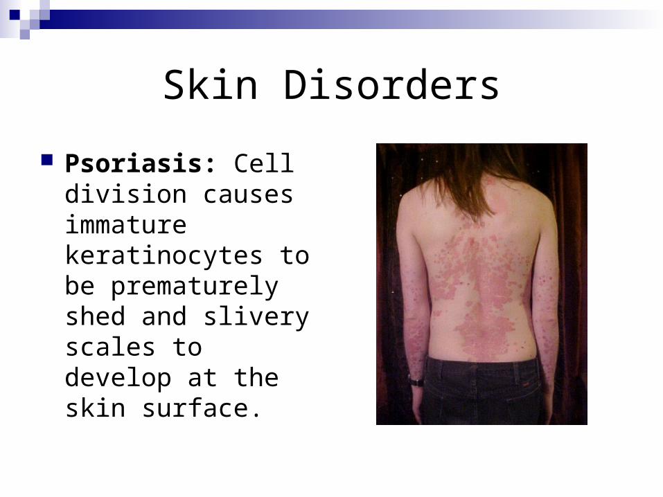
Psoriasis: Cell division causes immature keratinocytes to be prematurely shed and slivery scales to develop at the skin surface.

Skin Disorders
Erythema: Abnormal redness of the skin caused by engorgement of the capillaries in the dermis layer of the skin.
Skin Disorders
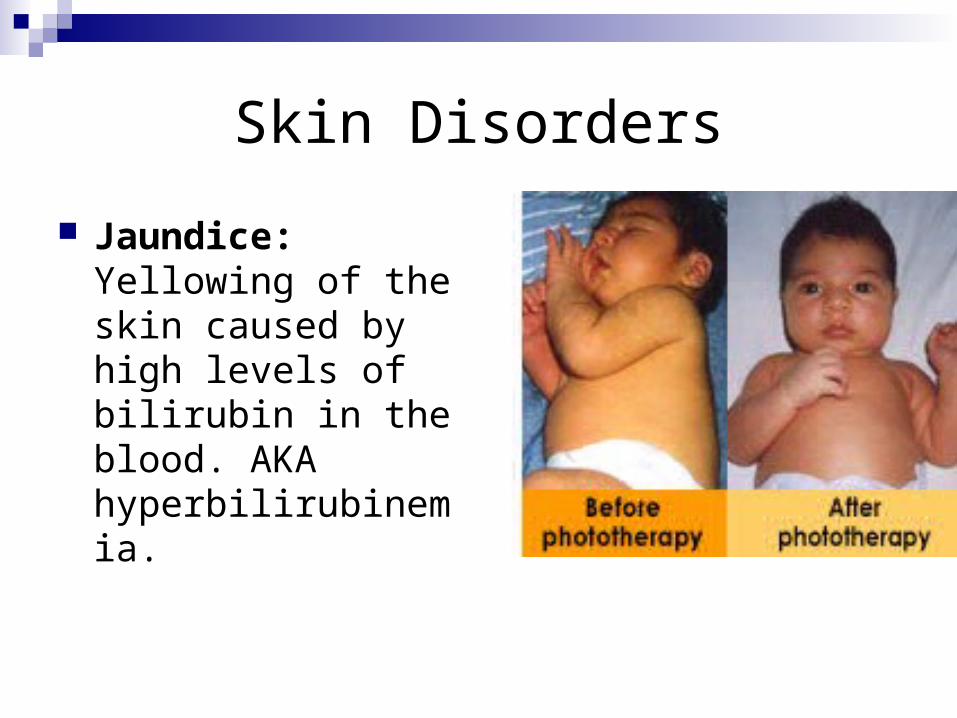
Jaundice: Yellowing of the skin caused by high levels of bilirubin in the blood. AKA hyperbilirubinemia.
Skin Disorders
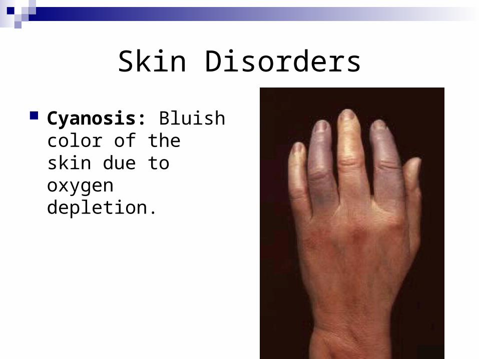
Cyanosis: Bluish color of the skin due to oxygen depletion.

Skin Disorders
Albinism: An inherited condition in which the individual cannot produce melanin.

Skin Accessory Structures
Sebaceous Glands (Oil Glands): Produce oily secretion called sebum to moisturize the skin.
Sudoriferous Glands (Sweat Glands): Produce sweat to excrete wastes and cool the body through evaporation.
Eccrine/Merocrine Glands: Responsible for thermoregulation through watery perspiration to cool the body.

Skin Accessory Structures
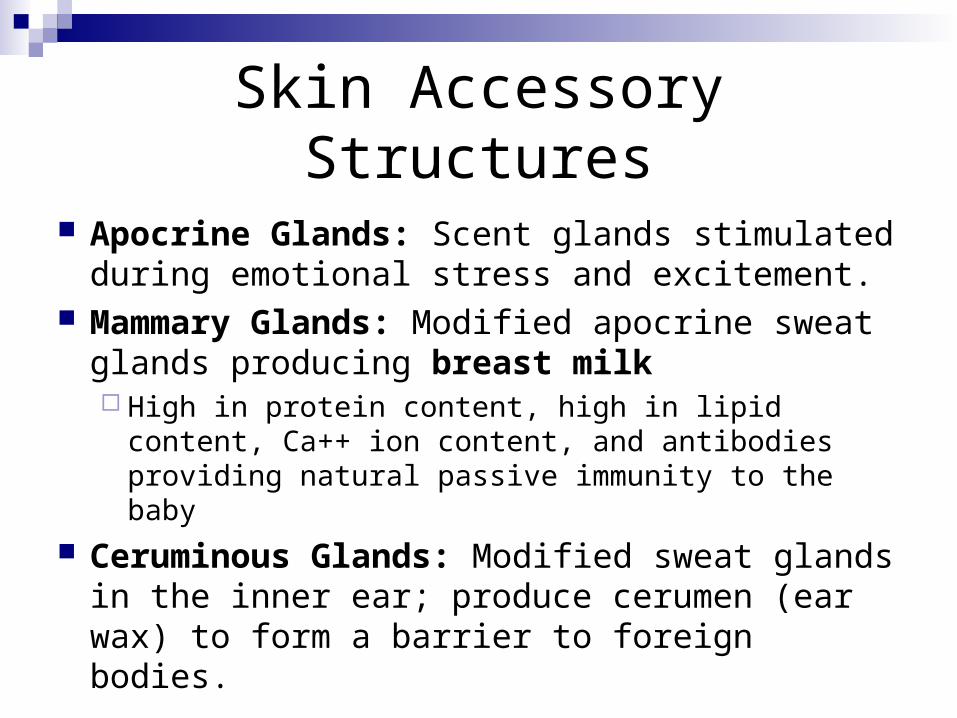
Apocrine Glands: Scent glands stimulated during emotional stress and excitement.
Mammary Glands: Modified apocrine sweat glands producing breast milk High in protein content, high in lipid content, Ca++ ion
content, and antibodies providing natural passive immunity to the baby
Ceruminous Glands: Modified sweat glands in the inner ear; produce cerumen (ear wax) to form a barrier to foreign bodies.

Terminal Hair Structures
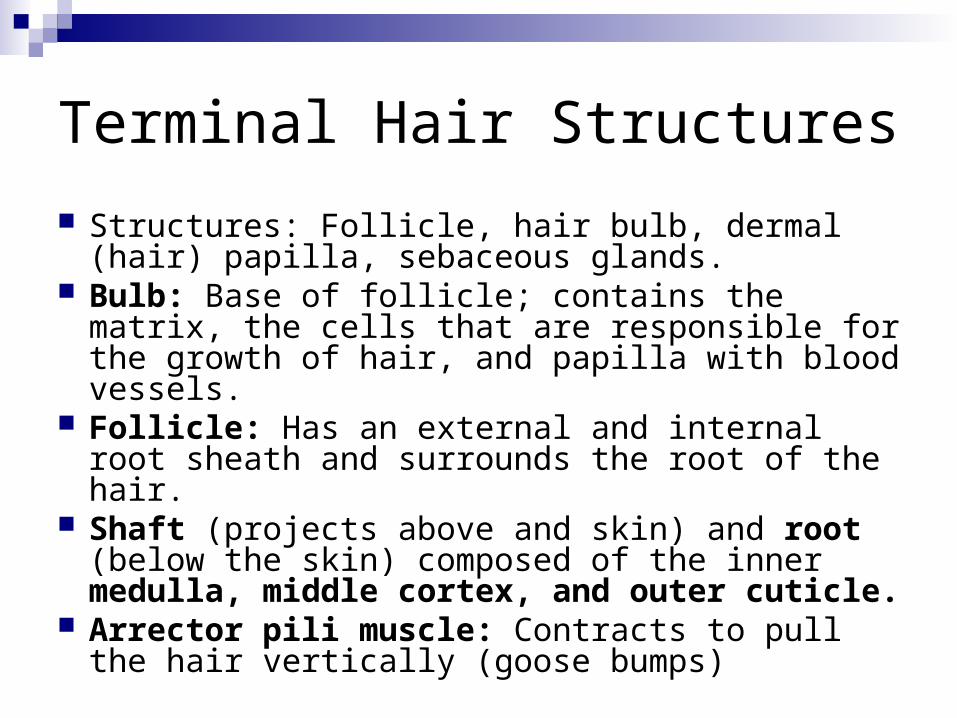
Structures: Follicle, hair bulb, dermal (hair) papilla, sebaceous glands.
Bulb: Base of follicle; contains the matrix, the cells that are responsible for the growth of hair, and papilla with blood vessels.
Follicle: Has an external and internal root sheath and surrounds the root of the hair.
Shaft (projects above and skin) and root (below the skin) composed of the inner medulla, middle cortex, and outer cuticle.
Arrector pili muscle: Contracts to pull the hair vertically (goose bumps)

Nails
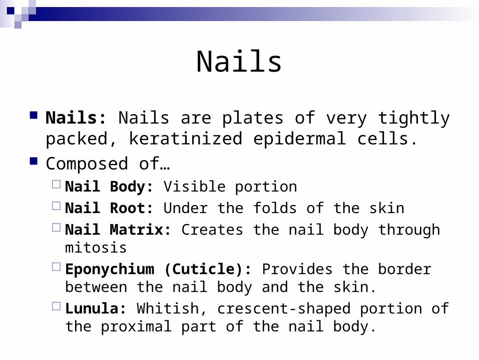
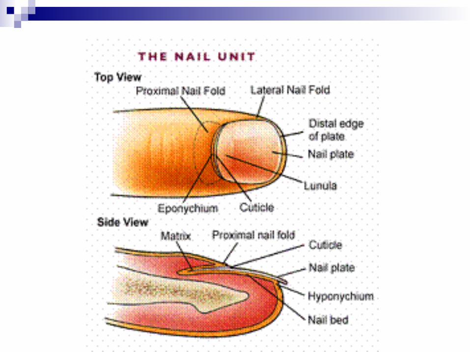
Nails: Nails are plates of very tightly packed, keratinized epidermal cells.
Composed of… Nail Body: Visible portion Nail Root: Under the folds of the skin Nail Matrix: Creates the nail body through mitosis Eponychium (Cuticle): Provides the border between
the nail body and the skin. Lunula: Whitish, crescent-shaped portion of the
proximal part of the nail body.
Skin Cancer
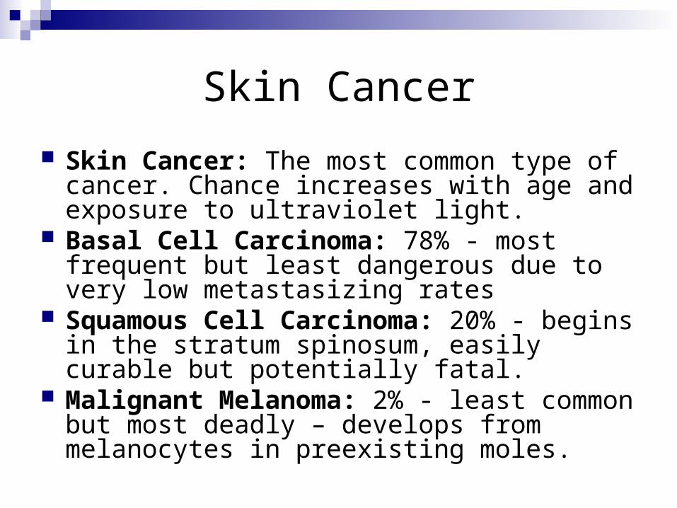
Skin Cancer: The most common type of cancer. Chance increases with age and exposure to ultraviolet light.
Basal Cell Carcinoma: 78% - most frequent but least dangerous due to very low metastasizing rates
Squamous Cell Carcinoma: 20% - begins in the stratum spinosum, easily curable but potentially fatal.
Malignant Melanoma: 2% - least common but most deadly – develops from melanocytes in preexisting moles.

Basal Cell Carcinoma
Squamous Cell Carcinoma
Malignant Melanoma

Burns
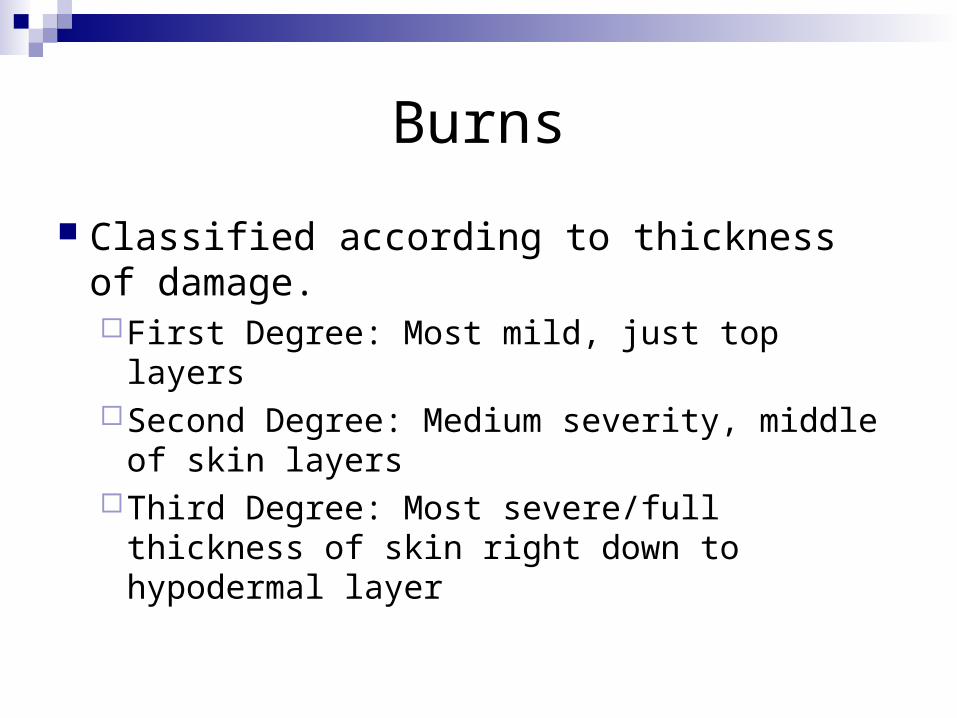
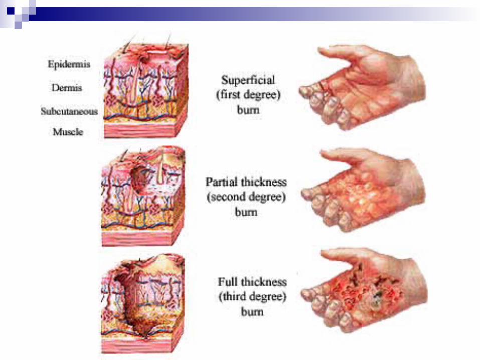
Classified according to thickness of damage.First Degree: Most mild, just top layersSecond Degree: Medium severity, middle of
skin layersThird Degree: Most severe/full thickness of
skin right down to hypodermal layer

First Degree Burn
The least severe type ONLY involves the epidermis Creates a general redness of skin A sunburn is a first-degree burn

Second Degree Burn
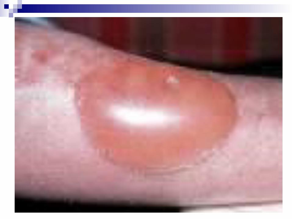
More severe than first-degree, but less severe than third-degree
Involves the epidermis and possibly the dermis
Fluid-filled blisters are a class characteristic
Tend to be very painful Prone to infection

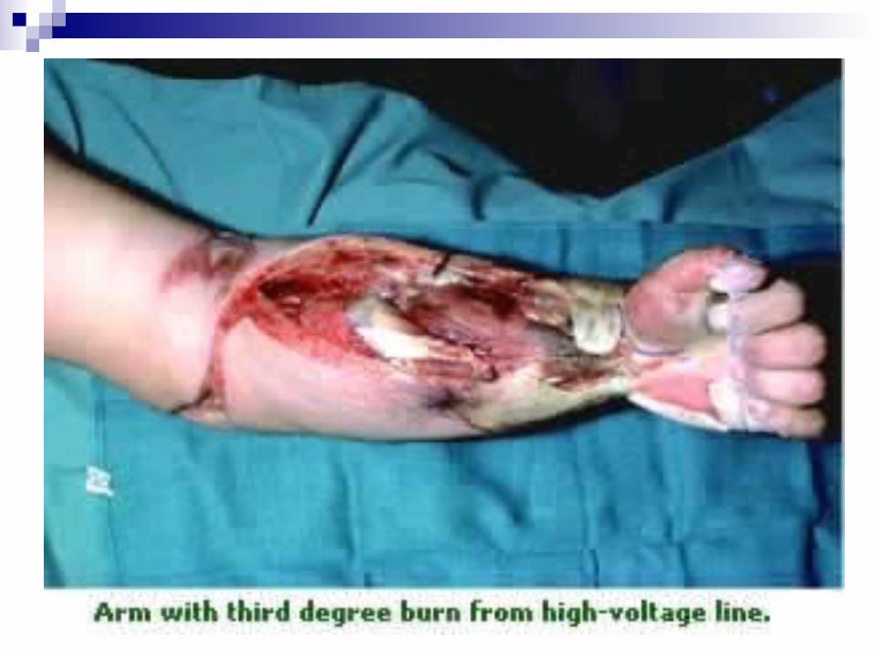
Third Degree Burn
Most severe with loss of function and marked edema
Involves the full thickness of the skin – all three layers
Deadens nerve endings so they are non-painful except near the borders, where they are mixed with second-degree burns
Loss of fluid resulting in shock is a primary concern
Acne
Nearly 85% of people will experience some form of acne during their lifetimes. According to the American Academy of Dermatology (AAD)
Acne is the most common skin disorder in the United States, affecting 40–50 million Americans at one time.

Acne is an inflammatory lesion of the sebaceous glands and the first signs usually show up during puberty when there is an increase in the hormone androgen, which is especially active in stimulating the amount of oil produced by the sebaceous glands in the skin.

This results in the extra production of sebum that combines with dead skin cells and other debris and becomes trapped, creating a plug that blocks the hair follicle.
As the ducts of these glands become plugged, they are frequently infected with bacteria, causing welts, deeper lumps and pimples.
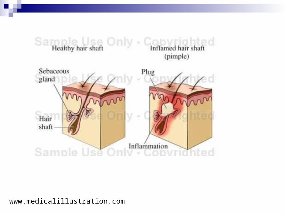
www.medicalillustration.com


![Advances in Tendon and Ligament Tissue Engineering ...tendon and ligament tissue prosthesis material with suit-able mechanical and tissue integrative properties [ , ]. Other materials](https://static.cupdf.com/doc/110x72/60bcf42d35abe3097d0fcf90/advances-in-tendon-and-ligament-tissue-engineering-tendon-and-ligament-tissue.jpg)
