


BCR-ABL1-induced expression of HSPA8 promotes cell survivalin chronic myeloid leukaemia
Edurne San Jose-Eneriz,1 Jose Roman-
Gomez,2 Lucia Cordeu,1 Esteban
Ballestar,3 Leire Garate,1 Enrique J.
Andreu,1 Isabel Isidro,4 Elizabeth
Guruceaga,5 Antonio Jimenez-Velasco,6
Anabel Heiniger,6 Antonio Torres,2
Maria Jose Calasanz,7 Manel Esteller,3
Norma C. Gutierrez,4 Angel Rubio,5
Ignacio Perez-Roger,8 Xabier Agirre1*
and Felipe Prosper1*1Foundation for Applied Medical Research,
Division of Cancer, Area of Cell Therapy and
Haematology Service, Clınica Universitaria,
Universidad de Navarra, Spain, 2Department of
Haematology, Hospital Reina Sofia, Cordoba,
Spain, 3Cancer Epigenetics Laboratory, Molecular
Pathology Programme, Spanish National Cancer
Centre (CNIO), Madrid, Spain, 4Department of
Haematology, Hospital Universitario and Centro
de Investigacion del Cancer (CIC), Universidad de
Salamanca-CSIC, Salamanca, Spain, 5CEIT and
Tecnun, University of Navarra, San Sebastian,
Spain, 6Department of Haematology, Hospital
Carlos Haya, Malaga, Spain, 7Department of
Genetics, School of Sciences, University of
Navarra, Pamplona, Spain, and 8Department of
Chemistry, Biochemistry and Molecular Biology,
University Cardenal Herrera-CEU, Moncada,
Spain
Received 11 December 2007; accepted for
publication 19 March 2008
Correspondence: Dr. Felipe Prosper,
Hematology and Cell Therapy Clınica
Universitaria Avda. Pıo XII 36, Pamplona 31008,
Navarra, Spain. E-mail: [email protected]
*XA and FP are equal senior authors.
Supported in part by grants from Gobierno de
Navarra, Department of Education, Beca Ortiz
de Landazuri 2006, Department of Health-
Gobierno de Navarra, Junta de Andalucia PI-
0004/2007, Fondo de Investigaciones Sanitarias
FIS 03/0661, 06/0003 and RETIC C03/10. This
project was funded through the ‘‘UTE project
CIMA’’.
Summary
In order to determine new signal transduction pathways implicated in
chronic myeloid leukaemia (CML), we performed a gene expression profile
comparison between CD34+ cells from CML patients and healthy donors.
Functional studies were performed using the Mo7e and Mo7e-p210 cell lines.
Expression of CCND1 (Cyclin D1), as well as the chaperone HSPA8, which is
important for regulation of CCND1, were significantly upregulated in CD34+
CML cells. Upregulation of HSPA8 was dependent, at least in part, on STAT5
(signal transducer and activator of transcrition 5)-dependent transcriptional
activation, as demonstrated by chromatin immunoprecipitation. The
presence of HSPA8 in the nuclear protein fraction as well as its binding to
CCND1 suggests that it may contribute to stabilization of the CCND1/CDK4
complex, which, in turn, may participate in proliferation of CML
cells. Treatment of CML cells with the specific HSPA8 inhibitor
15-deoxyspergualin induced inhibition of CML cell viability but did not
induce apoptosis. In conclusion, our studies suggest that STAT5-mediated
activation of HSPA8 induces nuclear translocation and activation of the
CCND1/CDK4 complex leading to increased proliferation of CML cells,
deciphering a new pathway implicated in CML and supporting a potential
role of chaperone inhibitors in the treatment of CML.
Keywords: chronic myeloid leukaemia, signal transduction, HSPA8, CD34+.
research paper
ª 2008 The Authors First published online 5 June 2008Journal Compilation ª 2008 Blackwell Publishing Ltd, British Journal of Haematology, 142, 571–582 doi:10.1111/j.1365-2141.2008.07221.x
Chronic myeloid leukaemia (CML) is a clonal myeloprolif-
erative disorder of the haematopoietic stem cell characterized
by the presence of the Philadelphia chromosome (Ph)
generated by the reciprocal translocation between the long
arms of chromosomes 9 and 22 t(9;22) (q34;q11). This
translocation produces the fusion of the genes BCR (chro-
mosome 22) and ABL1 (chromosome 9), generating the BCR-
ABL1 oncogene that causes the disease (Daley & Baltimore,
1988; Daley et al, 1990; Li et al, 1999). The p210 BCR-ABL1
fusion oncoprotein present in the majority of CML patients,
has a constitutive tyrosine kinase (TK) activity that results in
activation of a number of signal transduction pathways that
contribute to the abnormal regulation of cell cycle, adhesion
and apoptosis observed in CML cells (Horita et al, 2000;
Andreu et al, 2005).
The development of Imatinib Mesylate (previously known
as STI571) has impacted the treatment of CML disease
(Savage & Antman, 2002). Imatinib, a 2-phenylaminopyri-
midine, is a highly specific inhibitor of several tyrosine
kinases, such as BCR-ABL1 (Druker et al, 1996), platelet-
derived growth factor receptor (PDGFR), c-KIT, ARG
(Okuda et al, 2001) or c-fms (macrophage colony-stimulat-
ing factor receptor) (Dewar et al, 2005). Besides its clinical
efficacy, the highly specific inhibition of BCR-ABL1 tyrosine
kinase activity makes this compound a basic tool to study
the mechanism of action of BCR-ABL1 and the biology
of CML.
Recent studies have addressed the genetic abnormalities
associated with expression of the BCR-ABL1 oncogene using
high throughput techniques, such as gene expression micro-
arrays (Jena et al, 2002; Nowicki et al, 2003; Tipping et al,
2003; Hakansson et al, 2004; Janssen et al, 2005; Kronenwett
et al, 2005). Most of these studies have used either cells lines
(Tipping et al, 2003; Hakansson et al, 2004), or mononuclear
cells from patients with CML (Nowicki et al, 2003; Janssen
et al, 2005) and only a recent study focussed on the expression
profile of stem and progenitor CML cells (CD34+ cells)
(Kronenwett et al, 2005). Recent findings suggesting that
Imatinib does not eradicate all leukaemia stem cells, even in
the best responders, and that these cells could remain a
potential source of relapse in chronic phase or advanced phase
of the disease (Goldman & Gordon, 2006)-(Bhatia et al, 2003;
Jorgensen et al, 2006) support the need for new studies aimed
at determining pathways dependent or independent of BCR-
ABL1 that can lead to the development of more effective
therapies.
The current study compared the gene expression profile of
CD34+ cells from CML patients and healthy donors in order
to determine pathways implicated in the pathogenesis of
CML. Based on the results of the microarray analysis, we
defined a new signal transduction pathway implicated in the
abnormal proliferation of CML cells, suggesting that the
chaperone HSPA8 and CCND1 contribute to the abnormal
behaviour of CML cells and represent an interesting target for
new therapies.
Materials and methods
Cell lines and human samples
Human-derived Mo7e (a megakaryoblastic leukaemia cell line
without BCR-ABL1 fusion), Mo7e-p210 cells (Mo7e cells
transfected with p210 isoform of BCR-ABL1) and the chronic
myeloid leukaemia cell lines TCC-S, KU812, BV173 and KYO
were cultured as described (Horita et al, 2000; San Jose-Eneriz
et al, 2006). Peripheral blood (PB) from CML patients at
diagnosis (n = 3) and from healthy donors (n = 2) were
obtained by leucaphaeresis as described (Horita et al, 2000).
Bone marrow samples were obtained from patients with CML
at diagnosis and healthy volunteer donors. Samples were
obtained after informed consent and using guidelines
approved by the Ethics Committee for the Use of Human
Subjects at the University of Navarra. CML patients were 100%
Philadelphia chromosome positive by conventional cytogenetic
analysis. CD34+ cells were enriched using the MACS CD34+
isolation kit (Miltenyi Biotec, Cologne, Germany) and the
AutoMACS selection device as previously described (Horita
et al, 2000; Andreu et al, 2005). After immune selection,
CD34+ cells (purity always above 90%) were cultured in
serum-free media (BIT-9500, Stem Cell Technologies; Van-
couver, Canada) supplemented with 200 pg/ml stem cell factor
(SCF), 50 ng/ml granulocyte colony-stimulating factor
(G-CSF) (both from Amgen; Thousand Oaks, CA, USA),
200 pg/ml granulocyte-macrophage colony-stimulating factor
(GM-CSF; Immunex; Seattle, WA, USA), 1 ng/ml interleukin 6
(IL-6), 50 pg/ml leukaemia inhibitory factor (LIF), 200 pg/ml
macrophage inflammatory protein 1a (MIP1a) (all from R&D
Systems, Minneapolis, MN, USA), 0Æ1 mmol/l 2-mercaptoeth-
anol and penicillin/streptomycin (BioWhitaker, Walkersvill,
MD, USA).
RNA isolation
Total RNA from cells was isolated using the Trizol reagent
(Life Technologies) and purified with the Rneasy� Mini Kit
(Qiagen, Valencia, CA, USA) following the manufacturer’s
instructions. RNA levels, quality and purity were assessed with
the use of the RNA 6000 Nano assay on the Agilent 2100
bioanalyzer (Agilent, Palo Alto, CA, USA). None of the
samples showed RNA degradation or contamination with
genomic DNA.
Oligonucleotide microarray analysis and validation
CD34+ cells from PB of CML patients at diagnosis (n = 3) and
from healthy donors (n = 2) were used for the microarray
analysis. RNA isolation, labelling and hybridization to the HG-
U133 A GeneChip Oligonucleotide Microarray (Affymetrix,
Santa Clara, CA, USA) were performed as previously described
(Gutierrez et al, 2005). All arrays were visually examined for
searching possible irregularities. Data normalization was
E. S. Jose-Eneriz et al
ª 2008 The Authors572 Journal Compilation ª 2008 Blackwell Publishing Ltd, British Journal of Haematology, 142, 571–582

performed with the Affymetrix Microarray Suite software
version 5Æ0 (MAS5Æ0) according to the manufacturer’s protocol
(Affymetrix). All samples had a scaling factor lower than
threefold and a 3¢/5¢ of GAPDH probe set <2Æ5 (Appendix S1).
Unsupervised cluster analysis. Hierarchical clustering based on
the average-linkage method with the centred correlation metric
was carried out using Cluster and Treeview software (Page,
1996) and garban software (Genomic Analysis for Rapid
Biological ANnotation) (Martinez-Cruz et al, 2003).
Supervised analysis. In order to identify genes with statistically
significant changes in expression between both groups, we
used two different algorithms to increase the readability of the
study: SAM (Significant Analysis of Microarrays) (Tusher et al,
2001) and garban. garban analysis was based on genes where
the differences between the two groups using a t-test showed a
P value <0Æ05 or 0Æ01. In the case of SAM, all data were
permuted over 100 cycles by using the two-class (unpaired)
format. Classification of the genes according to the Gene
Ontology and matching of the gene products in the Boehringer
Mannheim chart of Biological Pathways and the Kyoto
Encyclopedia of Genes and Genomes (KEGG) was made
using the garban software.
For validation of the microarray data, semi-quantitative
reverse transcription polymerase chain reaction (RT-PCR)
was used to analyse the expression of some of the genes that
were differentially expressed based on the array information.
All primers were designed with the software Oligo 4Æ0(Molecular Biology Insights, Inc., Cascade, USA). The
primers used, the annealing temperature, cycle number and
the size of PCR products are shown in Supplementary
material Table SI. ABL1 was used as internal control.
Expression of each gene was densitometrically quantified
with the use of Quantity One (Bio-Rad Laboratories Inc.,
Hercules, CA, USA). Values were normalized with ABL1
expression.
In vitro treatment with Imatinib and Deoxyspergualin
Mo7e, Mo7e-p210 cell lines and CD34+ cells were cultured at
a density of 1 · 106 cells/ml and treated with Imatinib at
a concentration of 2 lmol/l (generously provided by
Dr. Elisabeth Buchdunger, Novartis, Basel, Swizterland) for
12 and 24 h (Horita et al, 2000). Deoxyspergualin (DSG)
(generously provided by Nippon Kayaku, Tokyo, Japan), a
HSPA8 inhibitor, was used to treat Mo7e and Mo7e-p210
cells in vitro for up to 72 h (100 lg/ml of DSG). DSG is
modified by polyamine oxidase present in fetal bovine serum
(Tepper et al, 1995), so 1 mmol/l aminoguanidine (Sigma,
St. Louis, MO, USA), an inhibitor of the polyamine oxidase,
was included in the culture. Viability and total cell counts
were determined at various times by trypan blue exclusion.
Proliferation, cell cycle analysis and apoptosis were deter-
mined when indicated.
Cell cycle and apoptosis analysis
For cell cycle analysis, 250 000 cells were cultured at a density
of 1 · 106 cells/ml, washed twice with phosphate-buffered
saline (PBS) and resuspended in 0Æ2% Tween-20 in PBS and
0Æ5 mg/ml Rnase A (Sigma) and incubated for 30 min at 37�C.
Subsequently, cells were stained with 25 lg/ml of propidium
iodide (Sigma) and analysed using a BD FACScan flow
cytometer (Becton Dickinson, San Jose, CA, USA). Apoptosis
was analysed by DNA fragmentation using the QIAmp DNA
Mini Kit (Qiagen, Hilden, Germany). Equal amounts of DNA
were separated on 1% agarose gels containing 0Æ5 lg/ml of
ethidium bromide. The detection of the 85 kDa fragment
of poly(ADP-ribose)polymerase (PARP) that results from
caspase-3 cleavage was also used as a marker for apoptosis.
Quantitative real-time PCR (Q-RT-PCR)
Reverse transcription reactions and Q-RT-PCR were carried
out as described previously (Agirre et al, 2006). We used
Assays-On-Demand (Applied Biosystems, Foster City, CA,
USA) for CCND1 (Hs00765553-m1) and 18S (Hs99999901_s1)
gene expression analysis. Primers (HSPA8-D and HSPA8-R)
and probe (HSPA8-S) for HSPA8 expression analysis were
designed using the program Primer Express 2Æ0 (Applied
Biosystems) and are shown in Supplementary material Table SI.
For the mRNA analysis of each gene, a patron curve was
generated using cDNA obtained from PB or BM of healthy
donors. Data were interpolated in the curve and then the level
of expression of each gene was normalized with the level of
expression of the internal control 18S. Finally, data were
compared among them.
Western blot, immunoprecipitation analysis andsubcellular fractionation
Proteins extracted from cell lines after treatments with
Imatinib or DSG were analysed by polyacrylamide gel electro-
phoresis, and the protein bands were electrophoretically
transferred onto nitrocellulose membranes as described before
(Roman-Gomez et al, 2004). The membranes, after being
blocked, were incubated with primary antibodies against
HSPA8 (Abcam, Cambridge, UK), CCND1 (Calbiochem, San
Diego, CA, USA), CDK4 (BD Pharmingen, San Diego, CA,
USA), PARP (Promega Corp., Madison, WI, USA), b-Actin
(Sigma), b-tubulin (Sigma) or Lamin A (Cell Signaling) and
then with alkaline phosphatase-conjugated secondary antibod-
ies. Bound antibodies were revealed by a chemiluminiscent
reagent (Tropix, Bedford, MA, USA) and detected using
HyperfilmTM enhanced chemilumincescence (Amershan Bio-
sciences, Little Chalfont, Buckinghamshire, England). b-Actin
was used as a loading control.
For immunoprecipitation, 500 lg of protein extracts were
precleared with Sephadex G-10 (Amersham Biosciences,
Uppsala, Sweden) immunoprecipitated with 2 lg of antibody
HSPA8 in CML Pathogenesis
ª 2008 The AuthorsJournal Compilation ª 2008 Blackwell Publishing Ltd, British Journal of Haematology, 142, 571–582 573
anti-HSPA8 (Abcam), anti-CCND1 (Calbiochem) or anti-
CDK4 (BD Pharmingen) and 20 ll of protein A/G (Santa Cruz
Biotechnology, Santa Cruz, CA, USA). Immunoprecipitates
were washed five times with Triton Buffer and then with cold
0Æ5 mol/l LiCl. Subsequently, immunoprecipitates were eluted
with 8 mol/l Urea and 0Æ2 mol/l dithiothreitol, electrophoresed
and transferred to nitrocellulose membranes. Membranes were
incubated with the same primary antibodies used for immu-
noprecipitation as described above. Subcellular fractionation
was performed as previously described (Ishida et al, 2002)
using b-tubulin as cytoplasmatic control and lamin A as
nuclear control.
Bisulphite sequencing
We used bisulphite sequencing technique to analyse and
determine the methylation status of HSPA8 (GeneBank:
NM_006597) and CCND1 (GeneBank: NM_053056) promot-
ers region as described (Agirre et al, 2006; San Jose-Eneriz
et al, 2006). Primers and PCR conditions used for amplifica-
tion of the HSPA8 fragment (Primers HSPA8-BS1, HSPA8-
BS2; 56 CpG dinucleotides) and CCND1 fragment (Primers
CCND1-BS1, CCND1-BS2; 22CpG) are shown in Supplemen-
tary material Table SI.
Chromatin immunoprecipitation (ChIP)
Cell extracts from Mo7e and Mo7e-p210 cell lines with and
without Imatinib treatment were subjected to chromatin
immunoprecipitation in order to assess the interactions
between STAT5 (signal transducer and activator of transcrip-
tion 5) and the HSPA8 and CCND1 promoters. The ChIP assay
was performed as previously described (Ballestar et al, 2003).
Three primer sets were designed in order to include all the
possible STAT5 binding sites in the HSPA8 (HSPA8-1D,
HSPA8-1R, HSPA8-2D, HSPA8-2R, HSPA8-3D and HSPA8-
3R) and CCND1 promoters (CCND1-1D, CCND1-1R,
CCND1-2D, CCND1-2R, CCND1-3D and CCND1-3R) (Sup-
plementary material Table SI). The sensitivity of the PCR
amplification was evaluated on serial dilutions of total DNA
collected after sonication (input fraction). PCR amplifications
were carried out with 36 cycles at 94�C for 30 s, 60�C for 30 s
and 72�C for 30 s. PCR products were run in 1Æ8% agarose gels
and visualized with ethidium bromide.
Immunofluorescence
Mo7e and Mo7e-p210 cells were fixed in paraformaldehyde for
10 min at 4�C. Cytospins were permeabilized with 0Æ1% Triton
in PBS for 15 min and washed three times with PBS. After
blocking non-specific binding sites with 1% bovine serum
albumin (BSA) for 4 h, slides were incubated overnight at 4�C
in a humidified chamber with 150 ll of the corresponding
primary antibody at a concentration of 10 lg/ml diluted in 1%
BSA: anti-HSPA8 (Abcam), anti-CCND1 (Calbiochem) and
anti-b-tubulin (Sigma-Aldrich, Steinheim, Germany). Slides
were then washed three times with 0Æ1% Tween-20 in PBS for
5 min each and incubated for 1 h with fluorescein isothiocy-
anate-conjugated anti-mouse IgM (Sigma-Aldrich) or Alexa
Fluor 488 rabbit anti-mouse IgG (Invitrogen Life Technolo-
gies, Paisley, UK) or Cy3-conjugated donkey anti-mouse IgG
(Jackson ImmunoResearch Labs, West Grove, PA, USA), all
diluted 1:1000 in 1% BSA. Slides were counterstained with
4¢-6-diamino-2-phenylindole in PBS:glycerol (1:1) and obser-
ved under a fluorescence microscope. In the case of double
immunostaining, both primary antibodies were incubated
together.
Results
Expression of CCND1 and HSPA8 is up-regulated inCD34+ CML cells
The comparison between the gene expression profile of CD34+
cells of CML patients and healthy donors showed that 1151
genes were differentially expressed in CML progenitor cells
(P < 0Æ05) (Supplementary material Table SII). Most of these
genes are implicated in metabolism, gene transcription, signal
transduction, transport, developmental processes and cell
proliferation, as expected, based on the known pathways
affected by the expression of the BCR-ABL1 oncogene (Dein-
inger et al, 2000). In vitro treatment of CD34+ CML cells with
Imatinib for 12 and 24 h induced changes in 772 and 730 genes
respectively (Supplementary material Tables SIII and SIV),
although only 113 genes were found to be regulated at both 12
and 24 h. Interestingly, dendrogram analysis clustered CML
untreated samples with samples treated for 12 h with Imatinib
and healthy donors samples with CML samples treated for
24 h with Imatinib, suggesting that treatment with Imatinib
induces a healthy normal gene profile in CD34+ CML cells
after 24 h (Supplementary material Fig S1). Gene expression
changes were confirmed in a small number of genes by semi-
quantitative RT-PCR (Supplementary material Table SV).
As the aim of our study was to determine new signal
transduction pathways implicated in the pathogenesis of the
disease and to search for new therapeutic targets, we focused
on potential candidate genes. Microarray analysis indicated
that expression of CCND1 (Cyclin D1), known to be altered in
different human neoplasias and involved in cell proliferation,
as well as some regulators of the CCND1 activity, such as
HSPA8 (also known as HSP73 or HSC70), were significantly
up-regulated in CD34+ CML cells and were downregulated
after treatment with Imatinib. To confirm these results, we
analysed the mRNA expression of CCND1 and HSPA8 in a
new group of samples from patients with CML (BM CD34+
cells n = 10 and BM mononuclear cells n = 25) as well as CML
cell lines (KYO, KU812, BV173, TCC-S and Mo7e-p210). In
agreement with the results observed in the microarray analysis,
HSPA8 was overexpressed in cell lines, BM CD34+ cells and
BM mononuclear cells of CML patient samples (mean
E. S. Jose-Eneriz et al
ª 2008 The Authors574 Journal Compilation ª 2008 Blackwell Publishing Ltd, British Journal of Haematology, 142, 571–582

expression of HSPA8 in bone marrow of healthy donors and
CML cells was 86Æ36 ± 3Æ26% and 275Æ24 ± 83Æ2% respec-
tively; P = 0Æ02). Similarly, CCND1 was also overexpressed in
cell lines and CML patient samples (mean expression of
CCND1 in bone marrow of healthy donors and CML cells was
66Æ33 ± 34Æ81% and 119Æ32 ± 31Æ1%, respectively; P = 0Æ05).
Protein expression of both HSPA8 and CCND1 was higher in
Mo7e-p210 in comparison with Mo7e, and there was a
significant increase in expression of both proteins in BCR-
ABL1 cells (Fig 1).
BCR-ABL1 mediates regulation of CCND1 and HSPA8expression
To demonstrate that upregulation of CCND1 and HSPA8
proteins was dependent on BCR-ABL1, the Mo7e and Mo7e-
p210 cell lines were treated with Imatinib. As expected,
Imatinib treatment induced inhibition of BCR-ABL1 phos-
phorylation and cell cycle and induced apoptosis of CML cells
(Supplementary material Fig S2). This treatment was also
associated with a decreased expression of CCND1 and HSPA8
protein expression in the Mo7e-p210 cell line but not in
parental Mo7e cell line (Fig 1). Similarly, the upregulated
HSPA8 mRNA level observed in Mo7e-p210 cells was down-
regulated after treatment in vitro with Imatinib in accordance
with the results obtained at the protein level (not shown).
Treatment with Imatinib also induced a significant down-
regulation of CCND1 mRNA expression in Mo7e-p210 cell
lines (not shown).
We next examined the potential mechanisms of BCR-ABL1-
mediated regulation of HSPA8 and CCND1 mRNA levels. As
we have previously demonstrated that abnormal hypomethy-
lation of gene promoters plays a role in the altered expression
of some genes in CML and more so in the progression of the
disease (Roman-Gomez et al, 2005, 2006), we decided to
examine the methylation status of HSPA8 and CCND1
promoters in Mo7e and Mo7e-p210 cell lines. Bisulphite
sequencing analysis did not show changes in the methylation
status of the HSPA8 and CCND1 promoters, indicating that
promoter hypomethylation was not the mechanism of tran-
scriptional regulation of HSPA8 (Supplementary material
Fig S3A) and CCND1 (Supplementary material Fig S3B).
As HSPA8 and CCND1 present a number of STAT5 binding
consensus sequences in the upstream promoter region and we
have previously demonstrated that BCR-ABL1 induces STAT5
phosphorylation and transcriptional activation (Horita et al,
2000), we reasoned that expression of HSPA8 and CCND1
could be regulated at the transcription level as a direct
consequence of STAT5 activation and direct interaction with
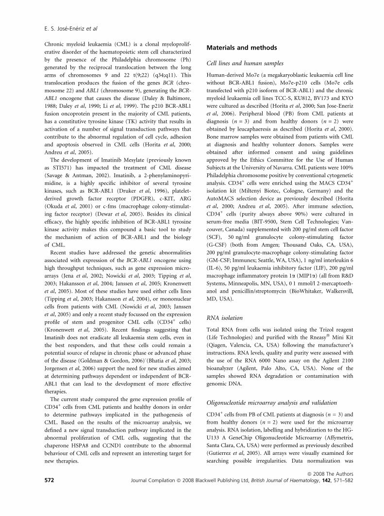
the HSPA8 and CCND1 promoters. ChIP analysis demon-
strated the binding of STAT5 in the HSPA8 promoter region in
Mo7e-p210 but not in the wild-type Mo7e cell line. This
interaction was abrogated after treatment with Imatinib
(Fig 2B). No interaction with STAT5 was observed when we
analysed the promoter of CCND1 by ChIP (Fig 2A). These
results along with the reduced expression of HSPA8 in Mo7e-
p210 after treatment with Imatinib, suggest a transcriptional
regulation of HSPA8 mediated by BCR-ABL1 activation
of STAT5.
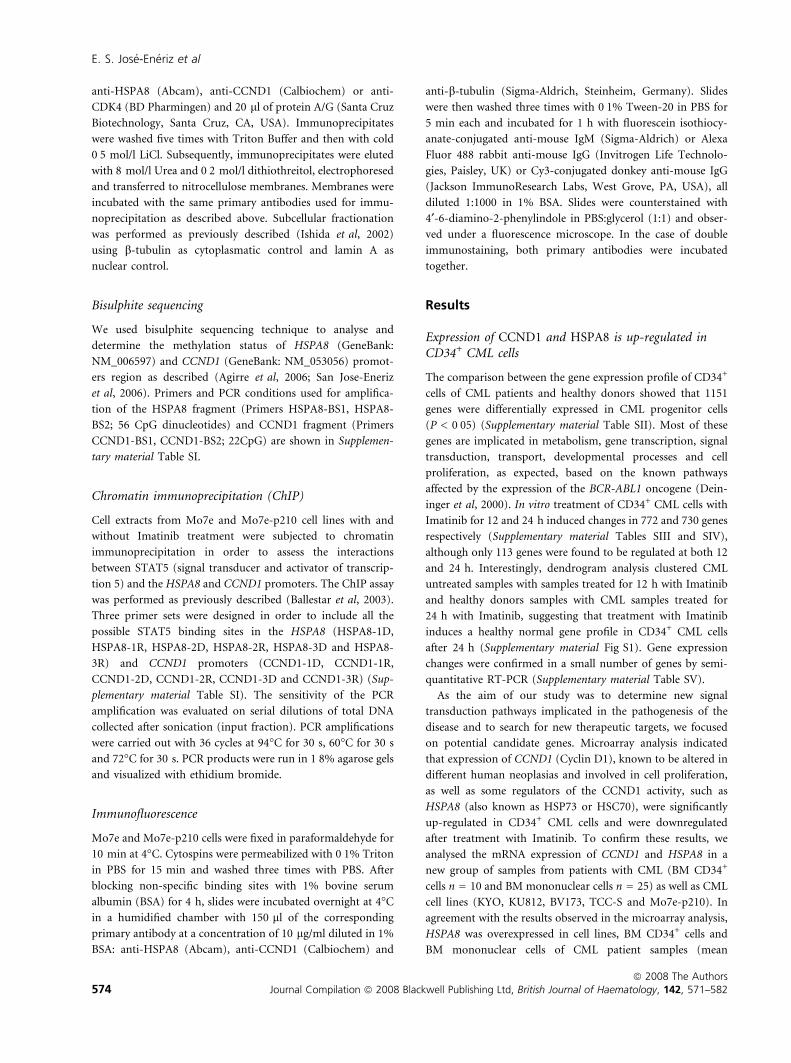
HSPA8 is located into the nucleus and associates withCCND1/CDK4
In order to elucidate the potential role of HSPA8 and CCND1
in the pathogenesis of CML, and based on the fact that HSPA8
is a chaperone that has been associated with CCND1 (Diehl
et al, 2003), we analysed the location of HSPA8 by subcellular
fractionation followed by Western blot of HSPA8 and CCND1
in the Mo7e-p210 cell line. As shown in Fig 3A, HSPA8 and
CCND1 proteins were present in the nuclear fraction in Mo7e-
p210 cells, which was further confirmed by immunofluores-
cence (Fig 3B). Furthermore, the formation of a complex
between HSPA8, CCND1 and CDK4 was demonstrated by
immunoprecipitation with antibodies against HSPA8, CCND1
or CDK4 followed by Western blot analysis, suggesting a
possible role of HSPA8 in the stabilization of the nuclear
complex between HSPA8/CCND1/CDK4 (Fig 3C).
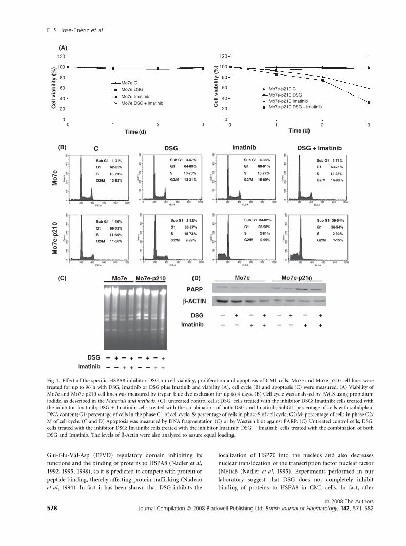
Deoxyspergualin, an inhibitor of HSPA8, inhibitsproliferation and decreases viability of CML cells incombination with Imatinib
Finally, to demonstrate that upregulation of HSPA8 contrib-
utes to abnormal viability of CML cells, Mo7e and Mo7e-p210
were treated with the HSPA8 inhibitor DSG (Nadler et al,
1995; Tepper et al, 1995) alone or in combination with
Imatinib. A dose–response study indicated the optimal dose
of DSG to inhibit proliferation of BCR-ABL1 cells to be
100 lg/ml (Supplementary material Fig S4). As expected,
treatment with DSG, Imatinib or both did not have any effect
Hours (IM)
CCND1
HSPA8
β-ACTIN
Mo7e Mo7e-p210
0 12 24 48 0 12 24 48
CCND1
HSPA8
100 187·5 76·4 81·7 352·1 80·0 21·2 9·0
100 223·9 132·2 114·9 249·4 226·9 199·3 135·3
Fig 1. Effect of Imatinib on HSPA8 and CCND1 protein expression
Mo7e and Mo7e-p210 cells were treated with Imatinib (2 lmol/l) for
up to 48 h and the expression of HSPA8 and CCND1 proteins analysed
by Western blot. The levels of b-actin were also analysed to ensure
equal loading. Expression of each protein was densitometrically
quantified with the use of Multi-Analyst v1Æ1 (Bio-Rad Laboratories
Inc.). Values were normalized with b-actin expression. Protein
expression in Mo7e was considered as 100%. IM: Imatinib. A repre-
sentative example of three different experiments is shown.
HSPA8 in CML Pathogenesis
ª 2008 The AuthorsJournal Compilation ª 2008 Blackwell Publishing Ltd, British Journal of Haematology, 142, 571–582 575
on viability of Mo7e cells. However, viability of Mo7e-p210
cells was significantly reduced after treatment with Imatinib
and DSG (33%) in comparison with Imatinib alone (57%)
(Fig 4A). Although DSG did not induce a decreased viability in
Mo7e-p210 cells, there was a mild reduction in the number of
total cells after treatment with DSG from 3Æ8 · 106 cells/ml
(Mo7e-p210 without treatment) to 3Æ2 · 106 cells/ml (Mo7e-
p210 treated with DSG) (Supplementary material Fig S5). This
suggests that inhibition of HSPA8 could have an effect on
proliferation of CML but does not induce apoptosis or
decrease cell viability. Similar results were obtained when we
analysed cell cycle by FACS where a small decrease in the
percentage of Mo7e-p210 cells in the G2/M phase was
observed with DSG alone (Fig 4B) while an increase in cell
death was observed when DSG and Imatinib were used in
combination (Fig 4B). Treatment with DSG alone did not
induce an increase in cell apoptosis as indicated by DNA
fragmentation and caspase cleavage of PARP (Fig 4C and D).
While treatment with Imatinib induced a downregulation of
HSPA8 and CCND1 protein expression in Mo7e-p210 cells,
the combination of Imatinib and DSG did not have any
additional effect on protein expression, suggesting that DSG
may acts modifying HSPA8 activity (Fig 5).
Discussion
Despite the clinical success obtained with the use of specific
inhibitors of BCR-ABL1 such as Imatinib (Druker et al, 2006),
Dasatinib (Quintas-Cardama et al, 2006) and Nilotinib (Kan-
tarjian et al, 2006) and the high percentage of CML patients
that achieve a complete cytogenetic response or even a
complete molecular response the malignant clone is unlikely
to be eliminated by these treatments (Graham et al, 2002;
Bhatia et al, 2003; Elrick et al, 2005). It has been recently
demonstrated that Imatinib does not inactivate all BCR-ABL-
activated signaling pathways, suggesting that some of these
pathways can be essential for leukaemic progenitor cell survival
(Hu et al, 2006). This implies that persistent malignant
progenitors can be a potential source of relapse in CML
patients and that there is a need to improve our understanding
of the signal transduction pathways implicated in the biology
of CML in order to provide new targets for therapy. The results
of the microarray analysis shown here identified a new
signaling pathway that is altered in CML patients and involved
in cell cycle and cell survival, such as the HSPA8-CCND1
identified in the microarray analysis and confirmed in a larger
number of patients with chronic phase CML at diagnosis in
which expression of HSPA8 and CCND1 was up-regulated
both in CD34+ BM cells (progenitor cells) as well as BM
mononuclear cells.
Two experimental evidences support the relationship
between the BCR-ABL1 kinase activity and the upregulation
of HSPA8 and CCND1: firstly, both proteins were
up-regulated in CML cell lines and patient samples; and
secondly, the treatment with Imatinib significantly reduced the
expression of both proteins. We provide new insights into the
mechanism that mediates the increased expression of HSPA8
and CCND1. In accordance with previous studies from our
group and others demonstrating that BCR-ABL1 phosphory-
lation of STAT5 is one of the mechanisms that contribute to
abnormal regulation of apoptosis in CML (Horita et al, 2000;
Weisberg & Griffin, 2000), our work further suggests that
BCR-ABL1 can also increase cell proliferation by inducing
STAT5-mediated transcriptional upregulation of HSPA8
(Fig 2B). Recent studies in a breast cancer models indicated
that CCND1 is regulated by STAT5 (Joung et al, 2005) which
was not the case in CML (Fig 2A), differences that could
probably be explained based on the different models. It has
been recently published that another Signal Transducer and
Activator of Transcription, STAT3, regulates transcription of
Region 1
Mo
7e
Inp
ut
–Ab
Mo
7e-p
210
Mo
7e-p
210
Imat
inib
(12
h)
Region 3
Region 2
Region 2
Region 1
(A)
(B)
Region 3M
o7e
Inp
ut
–Ab
Mo
7e-p
210
Mo
7e-p
210
Imat
inib
(12
h)
HSPA8
CCND1
Fig 2. Transcriptional regulation of CCND1 and HSPA8 gene CCND1
(A) and HSPA8 (B) chromatin immunoprecipitation assays. Mo7e and
Mo7e-p210 cell lines were cultured with and without Imatinib for
12 h. After fixation, cultures were processed for ChIP assays as
described in the Materials and methods. PCR was performed using
CCND1- and HSPA8-specific primers covering the promoter region
that contains putative STAT5 binding sites. Input: total DNA collected
after sonication (positive control); )Ab: DNA obtained from immu-
noprecipitation performed in the absence of anti-STAT5-specific
antibody (negative control). A representative example of three different
experiments is shown.
E. S. Jose-Eneriz et al
ª 2008 The Authors576 Journal Compilation ª 2008 Blackwell Publishing Ltd, British Journal of Haematology, 142, 571–582
CCND1, so we cannot exclude that activation of STAT3 would
be responsible for the upregulation of CCND1 (Leslie et al,
2006) expression in CML.
HSPA8 and CCND1 are interesting targets for the develop-
ment of new cancer therapies in CML for several reasons:
Up-regulation of CCND1 has been described to play an
important role in a number of tumours such as breast cancer
(Stendahl et al, 2004), colorectal (Kristt et al, 2000), brain
gliomas (Tan et al, 2004), thyroid (Nakashima et al, 2004),
urothelial bladder (Mhawech et al, 2004), oesophageal (Wu
et al, 2004) or endometrial (Moreno-Bueno et al, 2004) cancer
making the CCND1/CDK4 complex an attractive target
(Grillo et al, 2006; Landis et al, 2006; Yu et al, 2006). Further,
the interaction between BCR-ABL1 and CCND1 has been
previously described in advanced phase of the disease with an
increased expression in accelerated phase (de Groot et al, 2000;
Liu et al, 2004).
The involvement of chaperones in regulation of survival and
resistance to apoptosis in different tumours (Jameel et al, 1992;
Chant et al, 1995; Ogata et al, 2000; Becker et al, 2004; Atkins
et al, 2005), including a recently described role of HSP70
overexpression in resistance to Imatinib in CML cells (Pocaly
et al, 2007), has stimulated the development of inhibitors of
heat shock protein (HSP), such as the Hsp90 inhibitor
geldanamycin and its analog 17-AAG (17-allylamino-17-dee-
methoxygeldanamycin) (Guo et al, 2005), two drugs that are
actually undergoing phase I and II clinical trials in different
tumours (Drysdale et al, 2006). On the other hand, chaperone
molecules have been identified, such as leukaemia-associated
antigens (Greiner et al, 2003), providing the ground for
development of an HSP-based vaccine that has shown its
efficacy in several tumour models (Hoos & Levey, 2003).
Similarly, autologous vaccine of leucocyte-derived heat shock
protein 70-peptide complexes (Hsp70PC) and autologous heat
shock protein-peptide vaccine AG-858, based on Hsp 70, have
been used against CML in combination with Imatinib with
immunological and clinical responses (Li et al, 2005). The
Hsp70PC vaccine contains similar amounts of HSP70 and
HSPA8 proteins, suggesting that both proteins could be
possible targets in CML. Besides, HSP70 and HSP90 have
already been related to BCR-ABL1, being the tyrosine kinase a
client protein for these heat shock proteins.
But, could HSPA8 become a therapeutic target? The use of
specific inhibitors of HSPA8, such as DSG, may have
advantages over the use of patient-specific vaccines. DSG
interacts specifically with HSPA8, binding exactly with the
HSPA8
(A)
(B)
(C)
Lamin A
β-Tubulin
Mo7e-p210
C N
CCND1
HSPA8
CCND1
DAPI
DAPI
HSPA8
CCND1 MERGE
MERGE
HSPA8
CCND1
CDK4
IP HSPA8 IP CCND1 IP CDK4
Fig 3. HSPA8 location and HSPA8-CCND1-CDK4 complex formation. (A) Analysis of the HSPA8 and CCND1 cell localization. Subcellular
fractionation was performed using Mo7e-p210 cell line followed by Western blot against HSPA8 and CCND1. b-tubulin was used as cytoplasmatic
control and lamin A as nuclear control. C: cytoplasmatic fraction; N: nuclear fraction. (B) Immunofluorescence staining for HSPA8 (green) and
CCND1 (green) showing nuclear location. DAPI was used for nuclear staining. (C) HSPA8/CCND1/CDK4 complex formation: Cell extracts of
Mo7e-p210 cell line were analysed by immunoprecipitation (IP) with specific antibodies for HSPA8, CCND1 and CDK4 and then subjected to
Western blotting with the same antibodies used for IP. In A, B and C, a representative example of three different experiments is shown.
HSPA8 in CML Pathogenesis
ª 2008 The AuthorsJournal Compilation ª 2008 Blackwell Publishing Ltd, British Journal of Haematology, 142, 571–582 577
Glu-Glu-Val-Asp (EEVD) regulatory domain inhibiting its
functions and the binding of proteins to HSPA8 (Nadler et al,
1992, 1995, 1998), so it is predicted to compete with protein or
peptide binding, thereby affecting protein trafficking (Nadeau
et al, 1994). In fact it has been shown that DSG inhibits the
localization of HSP70 into the nucleus and also decreases
nuclear translocation of the transcription factor nuclear factor
(NF)jB (Nadler et al, 1995). Experiments performed in our
laboratory suggest that DSG does not completely inhibit
binding of proteins to HSPA8 in CML cells. In fact, after
(B)
(C) (D)
(A)M
o7e
Mo
7e-p
210
0
20
40
60
80
100
120
0 1 32Time (d)
Cel
l via
bili
ty (
%)
Mo7e-p210 CMo7e-p210 DSGMo7e-p210 ImatinibMo7e-p210 DSG + Imatinib
0
20
40
60
80
100
120
0 1 2 3
Time (d)
Cel
l via
bili
ty (
%)
Mo7e C
Mo7e DSG
Mo7e Imatinib
Mo7e DSG + Imatinib
C DSG Imatinib DSG + Imatinib
Sub G1 4·10%
G1 69·72%
S 11·64%
G2/M 11·50%
Sub G1 2·92%
G1 68·27%
S 15·73%
G2/M 9·99%
Sub G1 34·02%
G1 58·98%
S 2·81%
G2/M 0·99%
Sub G1 39·54%
G1 56·54%
S 2·92%
G2/M 1·15%
Sub G1 4·01%
G1 62·85%
S 13·79%
G2/M 13·92%
Sub G1 3·71%
G1 63·71%
S 12·28%
G2/M 14·90%
Sub G1 4·38%
G1 60·91%
S 13·27%
G2/M 15·60%
Sub G1 3·47%
G1 64·69%
S 13·73%
G2/M 13·31%
DSG
Imatinib
––
+
– +
– +
+
––
+
– +
– +
+
Mo7e-p210Mo7e
PARP
β-ACTIN
Mo7e-p210Mo7e
DSG
Imatinib
––
+
– +
– +
+
––
+
– +
– +
+
Fig 4. Effect of the specific HSPA8 inhibitor DSG on cell viability, proliferation and apoptosis of CML cells. Mo7e and Mo7e-p210 cell lines were
treated for up to 96 h with DSG, Imatinib or DSG plus Imatinib and viability (A), cell cycle (B) and apoptosis (C) were measured. (A) Viability of
Mo7e and Mo7e-p210 cell lines was measured by trypan blue dye exclusion for up to 4 days. (B) Cell cycle was analysed by FACS using propidium
iodide, as described in the Materials and methods. (C): untreated control cells; DSG: cells treated with the inhibitor DSG; Imatinib: cells treated with
the inhibitor Imatinib; DSG + Imatinib: cells treated with the combination of both DSG and Imatinib; SubG1: percentage of cells with subdiploid
DNA content; G1: percentage of cells in the phase G1 of cell cycle; S: percentage of cells in phase S of cell cycle; G2/M: percentage of cells in phase G2/
M of cell cycle. (C and D) Apoptosis was measured by DNA fragmentation (C) or by Western blot against PARP. (C) Untreated control cells; DSG:
cells treated with the inhibitor DSG; Imatinib: cells treated with the inhibitor Imatinib; DSG + Imatinib: cells treated with the combination of both
DSG and Imatinib. The levels of b-Actin were also analysed to assure equal loading.
E. S. Jose-Eneriz et al
ª 2008 The Authors578 Journal Compilation ª 2008 Blackwell Publishing Ltd, British Journal of Haematology, 142, 571–582

treatment with DSG, CCND1/HSPA8 complexes can be
observed (data not shown) suggesting that DSG may reduce
the amount of complexes by decreasing the efficiency of
interaction or by competition with the interacting proteins. It
is important to stress that DSG by itself was not able to
decrease cell viability of BCR-ABL1 cells. As BCR-ABL1
continues to be active, promoting other survival pathways,
the combination of Imatinib plus DSG was required to
decrease cell viability and to induce apoptosis of CML cells,
indicating an additive effect between both inhibitors. These
results lend support to the potential use of both agents to
inhibit leukaemia progenitors and maybe to overcome the
resistance of the malignant stem and progenitor cells. In any
case, the development of new HSPA8 inhibitors would be
necessary to further prove the benefit of inhibiting HSPA8.
In conclusion, our study supports the role of HSPA8 and
CCND1 in the abnormal proliferation of CML cells and
establishes a new pathway (Fig 6) where BCR-ABL1 induces
the expression of HSPA8 at the level of transcription. HSPA8
then binds to CCND1. This in turn could lead to the
stabilization of CCND1/CDK4 complexes in the nucleus and
to the activation of the cell cycle, as has been demonstrated in
other models (Diehl et al, 2003). This multicomplex is present
in patients with CML and participates in the abnormal
proliferation that characterizes leukaemia cells.
Acknowledgements
We thank Dr. Consuelo del Canizo, Dr. Jose Rifon and
Dr. Javier Perez Calvo for providing the samples of patients
with CML at diagnoses; Dr Kaori Kusama for kindly providing
a specific inhibitor of HSPA8 15-deoxyspergualin (DSG).
References
Agirre, X., Roman-Gomez, J., Jimenez-Velasco, A., Garate, L., Montiel-
Duarte, C., Navarro, G., Vazquez, I., Zalacain, M., Calasanz, M.J.,
Heiniger, A., Torres, A., Minna, J.D. & Prosper, F. (2006) ASPP1, a
common activator of TP53, is inactivated by aberrant methylation of
its promoter in acute lymphoblastic leukaemia. Oncogene, 25, 1862–
1870.
Andreu, E.J., Lledo, E., Poch, E., Ivorra, C., Albero, M.P., Martinez-
Climent, J.A., Montiel-Duarte, C., Rifon, J., Perez-Calvo, J., Arbona,
C., Prosper, F. & Perez-Roger, I. (2005) BCR-ABL induces the
expression of Skp2 through the PI3K pathway to promote p27Kip1
degradation and proliferation of chronic myelogenous leukemia
cells. Cancer Research, 65, 3264–3272.
Atkins, D., Lichtenfels, R. & Seliger, B. (2005) Heat shock proteins in
renal cell carcinomas. Contributions to Nephrology, 148, 35–56.
Ballestar, E., Paz, M.F., Valle, L., Wei, S., Fraga, M.F., Espada, J., Cig-
udosa, J.C., Huang, T.H. & Esteller, M. (2003) Methyl-CpG binding
proteins identify novel sites of epigenetic inactivation in human
cancer. European Molecular Biology Organization, 22, 6335–6345.
Becker, B., Multhoff, G., Farkas, B., Wild, P.J., Landthaler, M., Stolz,
W. & Vogt, T. (2004) Induction of Hsp90 protein expression in
malignant melanomas and melanoma metastases. Experimental
Dermatology, 13, 27–32.
Bhatia, R., Holtz, M., Niu, N., Gray, R., Snyder, D.S., Sawyers, C.L.,
Arber, D.A., Slovak, M.L. & Forman, S.J. (2003) Persistence of
malignant hematopoietic progenitors in chronic myelogenous leu-
kemia patients in complete cytogenetic remission following imatinib
mesylate treatment. Blood, 101, 4701–4707.
Chant, I.D., Rose, P.E. & Morris, A.G. (1995) Analysis of heat-shock
protein expression in myeloid leukaemia cells by flow cytometry.
British Journal of Haematology, 90, 163–168.
Daley, G.Q. & Baltimore, D. (1988) Transformation of an inter-
leukin 3-dependent hematopoietic cell line by the chronic
myelogenous leukemia-specific P210bcr/abl protein. Proceedings of
the National Academy of Sciences. United States of America, 85,
9312–9316.
Daley, G.Q., Van Etten, R.A. & Baltimore, D. (1990) Induction of
chronic myelogenous leukemia in mice by the P210bcr/abl gene of
the Philadelphia chromosome. Science, 247, 824–830.
Deininger, M.W., Vieira, S., Mendiola, R., Schultheis, B., Goldman,
J.M. & Melo, J.V. (2000) BCR-ABL tyrosine kinase activity regulates
HSPA8
HSPA8
CCND1
STAT5P
STAT5P
HSPA8
BCR-ABL1
STAT5 STAT5P
STAT5P
(1)(2)
(3)
NUCLEUS
UpCellCell
proliferationproliferationUp
CDK4
CCND1HSPA8
Fig 6. Potential model representing the participation of HSPA8 in cell
proliferation in CML (see text for details). Phosphorylation of STAT5
by BCR-ABL1 induces dimerization, which allows STAT5 to translo-
cate to the nucleus where it binds to consensus STAT5 binding
sequences of HSPA8 and therefore activates HSPA8 transcription (1),
thus leading to an increase in HSPA8 protein level, which translocates
to the nucleus (2). HSPA8 binding to CCND1 leads to stabilization of
the CCND1/cdk4 complex inducing cell proliferation (3). Upregula-
tion of HSPA8 in CML thus contributes to abnormal cell cycle
proliferation in CML.
HSPA8
β-ACTIN
CCND1
Mo7e-p210
DSGImatinib
––
+
– +
– +
+
Fig 5. Effect of treatment with Imatinib and DSG on HSPA8 and
CCND1 protein expression. Mo7e-p210 cell lines were treated with
Imatinib and DSG as described in the Materials and methods. Cell
extracts were analysed for the expression of HSPA8 and CCND1
proteins by Western Blot. The levels of b-actin were also analysed to
ensure equal loading.
HSPA8 in CML Pathogenesis
ª 2008 The AuthorsJournal Compilation ª 2008 Blackwell Publishing Ltd, British Journal of Haematology, 142, 571–582 579

the expression of multiple genes implicated in the pathogenesis of
chronic myeloid leukemia. Cancer Research, 60, 2049–2055.
Dewar, A.L., Zannettino, A.C., Hughes, T.P. & Lyons, A.B. (2005)
Inhibition of c-fms by imatinib: expanding the spectrum of treat-
ment. Cell Cycle, 4, 851–853.
Diehl, J.A., Yang, W., Rimerman, R.A., Xiao, H. & Emili, A. (2003)
Hsc70 regulates accumulation of cyclin D1 and cyclin D1-dependent
protein kinase. Molecular and Cellular Biology, 23, 1764–1774.
Druker, B.J., Tamura, S., Buchdunger, E., Ohno, S., Segal, G.M.,
Fanning, S., Zimmermann, J. & Lydon, N.B. (1996) Effects of a
selective inhibitor of the Abl tyrosine kinase on the growth of Bcr-
Abl positive cells. Nature Medicine, 2, 561–566.
Druker, B.J., Guilhot, F., O’Brien, S.G., Gathmann, I., Kantarjian, H.,
Gattermann, N., Deininger, M.W., Silver, R.T., Goldman, J.M.,
Stone, R.M., Cervantes, F., Hochhaus, A., Powell, B.L., Gabrilove,
J.L., Rousselot, P., Reiffers, J., Cornelissen, J.J., Hughes, T., Agis, H.,
Fischer, T., Verhoef, G., Shepherd, J., Saglio, G., Gratwohl, A.,
Nielsen, J.L., Radich, J.P., Simonsson, B., Taylor, K., Baccarani, M.,
So, C., Letvak, L. & Larson, R.A. (2006) Five-year follow-up of
patients receiving imatinib for chronic myeloid leukemia. The New
England Journal of Medicine, 355, 2408–2417.
Drysdale, M.J., Brough, P.A., Massey, A., Jensen, M.R. & Schoepfer, J.
(2006) Targeting Hsp90 for the treatment of cancer. Current
Opinion in Drug Discovery and Development, 9, 483–495.
Elrick, L.J., Jorgensen, H.G., Mountford, J.C. & Holyoake, T.L. (2005)
Punish the parent not the progeny. Blood, 105, 1862–1866.
Goldman, J. & Gordon, M. (2006) Why do chronic myelogenous
leukemia stem cells survive allogeneic stem cell transplantation or
imatinib: does it really matter? Leukemia and Lymphoma, 47, 1–7.
Graham, S.M., Jorgensen, H.G., Allan, E., Pearson, C., Alcorn, M.J.,
Richmond, L. & Holyoake, T.L. (2002) Primitive, quiescent, Phila-
delphia-positive stem cells from patients with chronic myeloid
leukemia are insensitive to STI571 in vitro. Blood, 99, 319–325.
Greiner, J., Ringhoffer, M., Taniguchi, M., Hauser, T., Schmitt, A.,
Dohner, H. & Schmitt, M. (2003) Characterization of several leu-
kemia-associated antigens inducing humoral immune responses in
acute and chronic myeloid leukemia. International Journal of Cancer,
106, 224–231.
Grillo, M., Bott, M.J., Khandke, N., McGinnis, J.P., Miranda, M.,
Meyyappan, M., Rosfjord, E.C. & Rabindran, S.K. (2006) Validation
of cyclin D1/CDK4 as an anticancer drug target in MCF-7 breast
cancer cells: Effect of regulated overexpression of cyclin D1 and
siRNA-mediated inhibition of endogenous cyclin D1 and CDK4
expression. Breast Cancer Research and Treatments, 95, 185–194.
de Groot, R.P., Raaijmakers, J.A., Lammers, J.W. & Koenderman, L.
(2000) STAT5-Dependent CyclinD1 and Bcl-xL expression in
Bcr-Abl-transformed cells. Molecular Cell Biology Research
Communications, 3, 299–305.
Guo, F., Rocha, K., Bali, P., Pranpat, M., Fiskus, W., Boyapalle, S.,
Kumaraswamy, S., Balasis, M., Greedy, B., Armitage, E.S., Lawrence,
N. & Bhalla, K. (2005) Abrogation of heat shock protein 70
induction as a strategy to increase antileukemia activity of heat
shock protein 90 inhibitor 17-allylamino-demethoxy geldanamycin.
Cancer Research, 65, 10536–10544.
Gutierrez, N.C., Lopez-Perez, R., Hernandez, J.M., Isidro, I., Gonzalez,
B., Delgado, M., Ferminan, E., Garcia, J.L., Vazquez, L., Gonzalez,
M. & San Miguel, J.F. (2005) Gene expression profile reveals
deregulation of genes with relevant functions in the different sub-
classes of acute myeloid leukemia. Leukemia, 19, 402–409.
Hakansson, P., Segal, D., Lassen, C., Gullberg, U., Morse, H.C., 3rd,
Fioretos, T. & Meltzer, P.S. (2004) Identification of genes differen-
tially regulated by the P210 BCR/ABL1 fusion oncogene using cDNA
microarrays. Experimental Hematology, 32, 476–482.
Hoos, A. & Levey, D.L. (2003) Vaccination with heat shock protein-
peptide complexes: from basic science to clinical applications. Expert
Review of Vaccines, 2, 369–379.
Horita, M., Andreu, E.J., Benito, A., Arbona, C., Sanz, C., Benet, I.,
Prosper, F. & Fernandez-Luna, J.L. (2000) Blockade of the Bcr-Abl
kinase activity induces apoptosis of chronic myelogenous leukemia
cells by suppressing signal transducer and activator of transcription
5-dependent expression of Bcl-xL. Journal of Experimental Medicine,
191, 977–984.
Hu, Y., Swerdlow, S., Duffy, T.M., Weinmann, R., Lee, F.Y. & Li, S.
(2006) Targeting multiple kinase pathways in leukemic progenitors
and stem cells is essential for improved treatment of Ph+ leukemia
in mice. Proceedings of the National Academy of Sciences of the United
States of America, 103, 16870–16875.
Ishida, N., Hayashi, K., Hoshijima, M., Ogawa, T., Koga, S., Miyatake,
Y., Kumegawa, M., Kimura, T. & Takeya, T. (2002) Large scale gene
expression analysis of osteoclastogenesis in vitro and elucidation of
NFAT2 as a key regulator. Journal of Biological Chemistry, 277,
41147–41156.
Jameel, A., Skilton, R.A., Campbell, T.A., Chander, S.K., Coombes,
R.C. & Luqmani, Y.A. (1992) Clinical and biological significance of
HSP89 alpha in human breast cancer. International Journal of
Cancer, 50, 409–415.
Janssen, J.J., Klaver, S.M., Waisfisz, Q., Pasterkamp, G., de Kleijn, D.P.,
Schuurhuis, G.J. & Ossenkoppele, G.J. (2005) Identification of genes
potentially involved in disease transformation of CML. Leukemia,
19, 998–1004.
Jena, N., Deng, M., Sicinska, E., Sicinski, P. & Daley, G.Q. (2002)
Critical role for cyclin D2 in BCR/ABL-induced proliferation of
hematopoietic cells. Cancer Research, 62, 535–541.
Jorgensen, H.G., Copland, M., Allan, E.K., Jiang, X., Eaves, A., Eaves,
C. & Holyoake, T.L. (2006) Intermittent exposure of primitive
quiescent chronic myeloid leukemia cells to granulocyte-colony
stimulating factor in vitro promotes their elimination by imatinib
mesylate. Clinical Cancer Research, 12, 626–633.
Joung, Y.H., Lim, E.J., Lee, M.Y., Park, J.H., Ye, S.K., Park, E.U., Kim,
S.Y., Zhang, Z., Lee, K.J., Park, D.K., Park, T., Moon, W.K. & Yang,
Y.M. (2005) Hypoxia activates the cyclin D1 promoter via the Jak2/
STAT5b pathway in breast cancer cells. Experimental and Molecular
Medicine, 37, 353–364.
Kantarjian, H., Giles, F., Wunderle, L., Bhalla, K., O’Brien, S.,
Wassmann, B., Tanaka, C., Manley, P., Rae, P., Mietlowski, W.,
Bochinski, K., Hochhaus, A., Griffin, J.D., Hoelzer, D., Albitar, M.,
Dugan, M., Cortes, J., Alland, L. & Ottmann, O.G. (2006)
Nilotinib in imatinib-resistant CML and Philadelphia chromo-
some-positive ALL. The New England Journal of Medicine, 354,
2542–2551.
Kristt, D., Turner, I., Koren, R., Ramadan, E. & Gal, R. (2000) Over-
expression of cyclin D1 mRNA in colorectal carcinomas and rela-
tionship to clinicopathological features: an in situ hybridization
analysis. Pathology and Oncology Research, 6, 65–70.
Kronenwett, R., Butterweck, U., Steidl, U., Kliszewski, S., Neumann, F.,
Bork, S., Blanco, E.D., Roes, N., Graf, T., Brors, B., Eils, R.,
Maercker, C., Kobbe, G., Gattermann, N. & Haas, R. (2005) Distinct
molecular phenotype of malignant CD34(+) hematopoietic stem
E. S. Jose-Eneriz et al
ª 2008 The Authors580 Journal Compilation ª 2008 Blackwell Publishing Ltd, British Journal of Haematology, 142, 571–582

and progenitor cells in chronic myelogenous leukemia. Oncogene,
24, 5313–5324.
Landis, M.W., Pawlyk, B.S., Li, T., Sicinski, P. & Hinds, P.W. (2006)
Cyclin D1-dependent kinase activity in murine development and
mammary tumorigenesis. Cancer Cell, 9, 13–22.
Leslie, K., Lang, C., Devgan, G., Azare, J., Berishaj, M., Gerald, W.,
Kim, Y.B., Paz, K., Darnell, J.E., Albanese, C., Sakamaki, T.,
Pestell, R. & Bromberg, J. (2006) Cyclin D1 is transcriptionally
regulated by and required for transformation by activated signal
transducer and activator of transcription 3. Cancer Research, 66,
2544–2552.
Li, S., Ilaria, R.L., Jr, Million, R.P., Daley, G.Q. & Van Etten, R.A.
(1999) The P190, P210, and P230 forms of the BCR/ABL oncogene
induce a similar chronic myeloid leukemia-like syndrome in mice
but have different lymphoid leukemogenic activity. Journal of
Experimental Medicine, 189, 1399–1412.
Li, Z., Qiao, Y., Liu, B., Laska, E.J., Chakravarthi, P., Kulko, J.M., Bona,
R.D., Fang, M., Hegde, U., Moyo, V., Tannenbaum, S.H., Menoret,
A., Gaffney, J., Glynn, L., Runowicz, C.D. & Srivastava, P.K. (2005)
Combination of imatinib mesylate with autologous leukocyte-
derived heat shock protein and chronic myelogenous leukemia.
Clinical Cancer Research, 11, 4460–4468.
Liu, J.H., Yen, C.C., Lin, Y.C., Gau, J.P., Yang, M.H., Chao, T.C.,
Hsiao, L.T., Wang, W.S., Tsai, Y.C. & Chen, P.M. (2004) Overex-
pression of cyclin D1 in accelerated-phase chronic myeloid leuke-
mia. Leukemia and Lymphoma, 45, 2419–2425.
Martinez-Cruz, L.A., Rubio, A., Martinez-Chantar, M.L., Labarga, A.,
Barrio, I., Podhorski, A., Segura, V., Sevilla Campo, J.L., Avila, M.A.
& Mato, J.M. (2003) garban: genomic analysis and rapid biological
annotation of cDNA microarray and proteomic data. Bioinformatics,
19, 2158–2160.
Mhawech, P., Greloz, V., Oppikofer, C., Szalay-Quinodoz, I. & Herr-
mann, F. (2004) Expression of cell cycle proteins in T1a and T1b
urothelial bladder carcinoma and their value in predicting tumor
progression. Cancer, 100, 2367–2375.
Moreno-Bueno, G., Rodriguez-Perales, S., Sanchez-Estevez, C., Mar-
cos, R., Hardisson, D., Cigudosa, J.C. & Palacios, J. (2004) Molecular
alterations associated with cyclin D1 overexpression in endometrial
cancer. International Journal of Cancer, 110, 194–200.
Nadeau, K., Nadler, S.G., Saulnier, M., Tepper, M.A. & Walsh, C.T.
(1994) Quantitation of the interaction of the immunosuppressant
deoxyspergualin and analogs with Hsc70 and Hsp90. Biochemistry,
33, 2561–2567.
Nadler, S.G., Tepper, M.A., Schacter, B. & Mazzucco, C.E. (1992)
Interaction of the immunosuppressant deoxyspergualin with a
member of the Hsp70 family of heat shock proteins. Science, 258,
484–486.
Nadler, S.G., Eversole, A.C., Tepper, M.A. & Cleaveland, J.S. (1995)
Elucidating the mechanism of action of the immunosuppressant 15-
deoxyspergualin. Therapeutic Drug Monitoring, 17, 700–703.
Nadler, S.G., Dischino, D.D., Malacko, A.R., Cleaveland, J.S., Fujihara,
S.M. & Marquardt, H. (1998) Identification of a binding site on
Hsc70 for the immunosuppressant 15-deoxyspergualin. Biochemical
and Biophysical Research Communications, 253, 176–180.
Nakashima, M., Meirmanov, S., Naruke, Y., Kondo, H., Saenko, V.,
Rogounovitch, T., Shimizu-Yoshida, Y., Takamura, N., Namba, H.,
Ito, M., Abrosimov, A., Lushnikov, E., Roumiantsev, P., Tsyb, A.,
Yamashita, S. & Sekine, I. (2004) Cyclin D1 overexpression in thy-
roid tumours from a radio-contaminated area and its correlation
with Pin1 and aberrant beta-catenin expression. Journal of Pathology,
202, 446–455.
Nowicki, M.O., Pawlowski, P., Fischer, T., Hess, G., Pawlowski, T. &
Skorski, T. (2003) Chronic myelogenous leukemia molecular sig-
nature. Oncogene, 22, 3952–3963.
Ogata, M., Naito, Z., Tanaka, S., Moriyama, Y. & Asano, G. (2000)
Overexpression and localization of heat shock proteins mRNA in
pancreatic carcinoma. J Nippon Med Sch, 67, 177–185.
Okuda, K., Weisberg, E., Gilliland, D.G. & Griffin, J.D. (2001) ARG
tyrosine kinase activity is inhibited by STI571. Blood, 97, 2440–2448.
Page, R.D. (1996) TreeView: an application to display phylogenetic
trees on personal computers. Computer Applications in the Bio-
sciences, 12, 357–358.
Pocaly, M., Lagarde, V., Etienne, G., Ribeil, J.A., Claverol, S., Bonneu,
M., Moreau-Gaudry, F., Guyonnet-Duperat, V., Hermine, O., Melo,
J.V., Dupouy, M., Turcq, B., Mahon, F.X. & Pasquet, J.M. (2007)
Overexpression of the heat-shock protein 70 is associated to
imatinib resistance in chronic myeloid leukemia. Leukemia, 21,
93–101.
Quintas-Cardama, A., Kantarjian, H., Jones, D., Nicaise, C., O’Brien,
S., Giles, F., Talpaz, M. & Cortes, J. (2006) Dasatinib (BMS-354825)
is active in Philadelphia chromosome-positive chronic myelogenous
leukemia after imatinib and nilotinib (AMN107) therapy failure.
Blood, 109, 497–499.
Roman-Gomez, J., Jimenez-Velasco, A., Agirre, X., Castillejo, J.A.,
Navarro, G., Barrios, M., Andreu, E.J., Prosper, F., Heiniger, A. &
Torres, A. (2004) Transcriptional silencing of the Dickkopfs-3 (Dkk-
3) gene by CpG hypermethylation in acute lymphoblastic leukaemia.
British Journal of Cancer, 91, 707–713.
Roman-Gomez, J., Jimenez-Velasco, A., Agirre, X., Cervantes, F.,
Sanchez, J., Garate, L., Barrios, M., Castillejo, J.A., Navarro, G.,
Colomer, D., Prosper, F., Heiniger, A. & Torres, A. (2005) Promoter
hypomethylation of the LINE-1 retrotransposable elements activates
sense/antisense transcription and marks the progression of chronic
myeloid leukemia. Oncogene, 24, 7213–7223.
Roman-Gomez, J., Jimenez-Velasco, A., Agirre, X., Castillejo, J.A.,
Navarro, G., Garate, L., Jose-Eneriz, E.S., Cordeu, L., Barrios, M.,
Prosper, F., Heiniger, A. & Torres, A. (2006) Promoter hyperme-
thylation and global hypomethylation are independent epigenetic
events in lymphoid leukemogenesis with opposing effects on clinical
outcome. Leukemia, 20, 1445–1448.
San Jose-Eneriz, E., Agirre, X., Roman-Gomez, J., Cordeu, L., Garate,
L., Jimenez-Velasco, A., Vazquez, I., Calasanz, M.J., Heiniger, A.,
Torres, A. & Prosper, F. (2006) Downregulation of DBC1 expression
in acute lymphoblastic leukaemia is mediated by aberrant methyl-
ation of its promoter. British Journal of Haematology, 134, 137–144.
Savage, D.G. & Antman, K.H. (2002) Imatinib mesylate – a new oral
targeted therapy. The New England Journal of Medicine, 346, 683–
693.
Stendahl, M., Kronblad, A., Ryden, L., Emdin, S., Bengtsson, N.O. &
Landberg, G. (2004) Cyclin D1 overexpression is a negative pre-
dictive factor for tamoxifen response in postmenopausal breast
cancer patients. British Journal of Cancer, 90, 1942–1948.
Tan, P.G., Xing, Z. & Li, Z.Q. (2004) [Expression of cyclin D1 in brain
gliomas and its significance]. Chinese Journal of Cancer, 23, 63–65.
Tepper, M.A., Nadler, S.G., Esselstyn, J.M. & Sterbenz, K.G. (1995)
Deoxyspergualin inhibits kappa light chain expression in 70Z/3 pre-
B cells by blocking lipopolysaccharide-induced NF-kappa B activa-
tion. Journal of Immunology, 155, 2427–2436.
HSPA8 in CML Pathogenesis
ª 2008 The AuthorsJournal Compilation ª 2008 Blackwell Publishing Ltd, British Journal of Haematology, 142, 571–582 581

Tipping, A.J., Deininger, M.W., Goldman, J.M. & Melo, J.V. (2003)
Comparative gene expression profile of chronic myeloid leukemia
cells innately resistant to imatinib mesylate. Experimental Hematol-
ogy, 31, 1073–1080.
Tusher, V.G., Tibshirani, R. & Chu, G. (2001) Significance analysis of
microarrays applied to the ionizing radiation response. Proceedings
of the National Academy of Sciences of the United States of America,
98, 5116–5121.
Weisberg, E. & Griffin, J.D. (2000) Mechanism of resistance to the ABL
tyrosine kinase inhibitor STI571 in BCR/ABL-transformed hema-
topoietic cell lines. Blood, 95, 3498–3505.
Wu, M.Y., Zhuang, C.X., Yang, H.X. & Liang, Y.R. (2004) Expression
of Egr-1, c-fos and cyclin D1 in esophageal cancer and its precursors:
An immunohistochemical and in situ hybridization study. World J
Gastroenterol, 10, 476–480.
Yu, Q., Sicinska, E., Geng, Y., Ahnstrom, M., Zagozdzon, A., Kong, Y.,
Gardner, H., Kiyokawa, H., Harris, L.N., Stal, O. & Sicinski, P.
(2006) Requirement for CDK4 kinase function in breast cancer.
Cancer Cell, 9, 23–32.
Supplementary Material
The following supplementary material is available for this
article online:
Appendix S1. Array design.
Figure S1. Dendrogram of healthy donors, CML and CML
samples treated with 12 and 24 h of Imatinib.
Figure S2. Imatinib inhibition of c-abl tyrosine kinase activity
in CML cell lines.
Figure S3. Transcriptional regulation of HSPA8 and CCND1
gene.
Figure S4. Effect of the specific HSPA8 inhibitor DSG on
cell viability, proliferation and apoptosis of CML cells.
Figure S5. Effect of DSG on proliferation of CML cells.
Table SI. Primers and PCRs conditions.
Table SII. Total analysis of genes differently expressed bet-
ween CD34+ cells of CML patients and healthy donors.
Table SIII. Total analysis of genes differently expressed bet-
ween CD34+ cells of CML patients before and after 12 h with
Imatinib.
Table SIV. Total analysis of genes differently expressed bet-
ween CD34+ cells of CML patients before and after 24 h with
Imatinib.
Table SV. Expression of selected genes by RT-PCR in
comparison with microarray data between CD34+ cells of
CML patients and healthy donor samples and between CML
patients samples at baseline and after treatment with Imatinib
for 24 h.
The material is available as part of the online article from:
http://www.blackwell-synergy.com/doi/abs/10.1111/j.1365-
2141.2008.07221.x
(This link will take you to the article abstract).
Please note: Blackwell Publishing is not responsible for the
content or functionality of any supplementary materials sup-
plied by the authors. Any queries (other than missing material)
should be directed to the corresponding author for the article.
E. S. Jose-Eneriz et al
ª 2008 The Authors582 Journal Compilation ª 2008 Blackwell Publishing Ltd, British Journal of Haematology, 142, 571–582


![myeloid leukaemia [ID1225] 1st appraisal committee meeting ...](https://static.cupdf.com/doc/110x72/62674292a1b63c6cab603f27/myeloid-leukaemia-id1225-1st-appraisal-committee-meeting-.jpg)


