Aurora EMS Protocols
Authorized Version of
Denver Metropolitan Prehospital Protocols
Summer 2019

The Aurora EMS Protocols are based on the Denver Metro EMS Protocols. The Denver Metro EMS Protocols are updated twice annually, and the Aurora EMS Protocols will follow this process.
The medical oversight for the Aurora EMS system is performed by the Prehospital Care Consortium (PCC), which is comprised of members from Aurora Fire Rescue, Falck Rocky Mountain, the AFR Medical Director, 3 Assistant Medical Directors, and EMS Managers representing the hospitals in the community. The PCC participates in protocol development and reviews, training, and the Quality Assurance and Quality Improvement for Aurora Fire Rescue.
Dr. Eric Hill – Medical Director Dr. Danny Willner – Asst. Medical Director Dr. Kalen Abbott – Asst. Medical Director Dr. Maria Mandt – Asst. Medical Director
Aurora Fire Rescue would like to thank the members of the PCC for their dedication and expertise to support the Aurora EMS system.

TABLE OF CONTENTS
Table of Contents
General Guidelines (0001-0999)
• 0010 Introduction
• 0020 Confidentiality
• 0030 Consent
• 0040 Physician at the Scene/Medical Direction
• 0050 Field Pronouncement
• 0051 Termination of Resuscitation
• 0060 Advanced Medical Directives
• 0070 Patient Determination: “Patient or No Patient”
• 0080 Patient Non-Transport or Refusal
• 0090 Emergency Department Divert and Advisory
• 0100 Mandatory Reporting of Abuse Patients
• 0110 Free-Standing Emergency Departments as EMS Destination
• 0111 Emergent vs Non-Emergent Transport
• 0112 Trauma Triage Algorithms
• 0113 SALT Triage
• 0114 Multiple Patient Incident
• 0120 Base Contact for Physician Consultation
• 0130 Transportation of the Pediatric Patient
• 0140 911 Response to Request for Interfacility Transport
• 0150 Alternate Disposition of Acutely Intoxicated Patients
• 0160 Alternate Disposition of Behavioral Health Patients
• 0990 Quick Reference for Procedures and Medications Allowed by Protocol
Procedures (1000-1999)
• 1000 Intubation: Oral
• 1010 Intubation: Nasal
• 1020 Percutaneous Cricothyrotomy
• 1040 Pediatric Needle Cricothyrotomy
• 1050 Supraglottic Airway
• 1060 Continuous Positive Airway Pressure (CPAP)
• 1070 Capnography
• 1080 Needle Thoracostomy for Tension Pneumothorax Decompression
• 1090 Synchronized Cardioversion
• 1100 Transcutaneous Cardiac Pacing
• 1110 Intraosseous Catheter Placement

TABLE OF CONTENTS
• 1120 Tourniquet Protocol
• 1130 Restraint Protocol
• 1140 Orogastric Tube Insertion with Advanced Airway
• 1150 TASER® Probe Removal
• 1160 Pain Management
Respiratory Protocols (2000-2999)
• 2000 Obstructed Airway
• 2010 Adult Universal Respiratory Distress
• 2020 Pediatric Universal Respiratory Distress
• 2030 Adult Wheezing
• 2040 Pediatric Wheezing
• 2050 Pediatric Stridor/Croup
• 2060 CHF/Pulmonary Edema
Cardiac Protocols (3000-3999)
• 3000 Universal Pulseless Arrest
• 3010 Universal Pulseless Arrest Considerations
• 3020 Neonatal Resuscitation
• 3030 Post-Resuscitation Care with ROSC
• 3040 Tachyarrhythmia with Poor Perfusion
• 3050 Bradyarrhythmia with Poor Perfusion
• 3060 Chest Pain
• 3070 Cardiac Alert
• 3080 Hypertension
• 3090 Ventricular Assist Devices (VAD)
General Medical Protocols (4000-4999)
• 4000 Medical Shock
• 4010 Universal Altered Mental Status
• 4020 Syncope
• 4030 Stroke
• 4040 Seizure
• 4050 Hypoglycemia
• 4060 Pediatric BRUE (Formerly ALTE)
• 4070 Drug/Alcohol Intoxication
• 4080 Overdose and Acute Poisoning
• 4090 Allergy and Anaphylaxis
• 4100 Non-Traumatic Abdominal Pain/Vomiting
TABLE OF CONTENTS
• 4110 Suspected Carbon Monoxide Exposure
• 4120 Adrenal Insufficiency
• 4130 Epistaxis Management
• 4140 Sepsis
Environmental Protocols (5000-4999)
• 5000 Drowning
• 5010 Hypothermia
• 5020 Hyperthermia
• 5030 Altitude Illness
• 5040 Insect/Arachnid Stings and Bites
• 5050 Snake Bite
Behavioral Protocols (6000-6999)
• 6000 Psychiatric/Behavioral Patient
• 6010 Agitated/Combative Patient
• 6020 Transport of the Handcuffed Patient
Obstetric Protocols (7000-7999)
• 7000 Childbirth
• 7010 Obstetrical Complications
Trauma Protocols (8000-8999)
• 8000 General Trauma Care
• 8010 Special Trauma Scenarios: Sexual Assault and Abuse/Neglect
• 8020 Trauma in Pregnancy
• 8030 Traumatic Pulseless Arrest
• 8040 Traumatic Shock
• 8050 Amputations
• 8060 Head Trauma
• 8070 Face and Neck Trauma
• 8080 Spinal Trauma
• 8090 Spinal Precautions
• 8100 Suspected Spinal Injury with Protective Athletic Equipment in Place
• 8110 Chest Trauma
• 8120 Abdominal Trauma
• 8130 Burns
Medication Protocols (9000-9999)
• 9000 Medication Administration Guidelines
• 9005 Acetaminophen
TABLE OF CONTENTS
• 9010 Adenosine
• 9020 Albuterol Sulfate
• 9030 Amiodarone
• 9040 Antiemetics
• 9050 Aspirin
• 9060 Atropine Sulfate
• 9070 Benzodiazepines
• 9080 Calcium
• 9090 Dextrose
• 9100 Diphenhydramine
• 9115 DuoDote™
• 9120 Epinephrine
• 9130 Glucagon
• 9150 Hemostatic Agents
• 9160 Hydroxocobalamin
• 9165 Ibuprofen
• 9170 Ipratropium Bromide
• 9175 Ketamine
• 9180 Lidocaine 2%
• 9190 Magnesium Sulfate
• 9200 Methylprednisolone
• 9210 Naloxone
• 9220 Nitroglycerin
• 9230 Opioids
• 9240 Oral Glucose
• 9250 Oxygen
• 9260 Phenylephrine
• 9270 Racemic Epinephrine
• 9280 Sodium Bicarbonate
• 9290 Topical Ophthalmic Anesthetics
• 9300 Vasopressor Continuous Infusion – Adult Patients Only
Appendix A
• THREAT BASED CARE ADDENDUM
0010 GENERAL GUIDELINES: INTRODUCTION
TABLE OF CONTENTS
INTRODUCTION
The following protocols have been developed and approved by the Medical Director and Assistant Medical Directors of Aurora Fire Rescue. These protocols are based on the Denver Metro EMS Protocols developed by the Denver Metro EMS Medical Directors Group. These protocols define the standard of care for EMS providers in the City of Aurora, and delineate the expected practice, actions, and procedures to be followed.
No protocol can account for every clinical scenario encountered, and the Medical Directors recognize that in rare circumstances deviation from these protocols may be necessary and in a patient’s best interest. Variance from protocol should always be done with the patient’s best interest in mind and backed by documented clinical reasoning and judgment. Whenever possible, prior approval by direct verbal order from base station physician is preferred. Additionally, all variance from protocol should be documented and submitted for review by the agency’s Medical Director in a timely fashion.
The protocols are presented in an algorithm format. An algorithm is intended to reflect real-life decision points visually. An algorithm has certain limitations, and not every clinical scenario can be represented. Although the algorithm implies a specific sequence of actions, it may often be necessary to provide care out of sequence from that described in the algorithm if dictated by clinical needs. An algorithm provides decision-making support, but need not be rigidly adhered to and is no substitute for sound clinical judgment.
In order to keep protocols as uncluttered as possible, and to limit inconsistencies, individual drug dosing has not been included in the algorithms. It is expected the EMTs will be familiar with standard drug doses. Drug dosages are included with the medications section of the protocols as a reference.
If viewing protocol in an electronic version, it will be possible to link directly to a referenced protocol by clicking on the hyperlink, which is underlined.
PROTOCOL KEY
Boxes without any color fill describe actions applicable to all certification levels. Blue filled boxes are for Paramedic level. When applicable, actions requiring Base Contact are identified in the protocol.
Teaching points deemed sufficiently important to be included in the protocol are separated into grey-filled boxes with a double line border.
PROTOCOLS CONSIDERATIONS SPECIFIC TO AGE
For the purposes of these clinical care protocols, pediatric patients are those less than 12 years of age. Infant is defined as less than 1 year of age. Neonate is defined as less than one month of age. Pediatric specific indications will be noted by a purple box. Geriatric patients will be considered greater than 65 years of age. Geriatric specific indications will be indicated by a green box.
TRAINING AND EDUCATION
These protocols define the treatments, procedures, and policies approved by the Medical Director for Aurora Fire Rescue. In Colorado, the scope of practice and acts allowed for EMT, EMT-IV, AEMT, EMT-I and Paramedic certifications are defined by the Colorado Department of Public Health and Environment, Chapter Two - Rules Pertaining to EMS Practice and Medical Director Oversight. These protocols do not supersede Chapter Two allowances unless approved by a waiver from the EMPAC, but in some instances may vary from Chapter Two depending on medical directors’ preference.
The curriculum for initial EMS provider training may not cover some of the treatments, procedures and medications included in these protocols. Therefore, it is the responsibility of the EMS agency and Medical Director to ensure the initial training, verification, and maintenance of these skills falling outside traditional EMS education with all agency providers.
Geriatric Protocol
Pediatric Protocol
• Teaching points
EMT
Paramedic

0020 GENERAL GUIDELINES: CONFIDENTIALITY
TABLE OF CONTENTS
CONFIDENTIALITY
A. The patient-physician relationship, the patient-registered nurse relationship, and the patient-EMT relationship are recognized as privileged. This means that the physician, nurse, or EMT may not testify as to confidential communications unless:
1. The patient consents
2. The disclosure is allowable by law (such as Medical Board or Nursing Board proceedings, or criminal or civil litigation in which the patient's medical condition is in issue)
B. The prehospital provider must keep the patient's medical information confidential. The patient likely has an expectation of privacy, and trusts that personal, medical information will not be disclosed by medical personnel to any person not directly involved in the patient's medical treatment.
1. Exceptions
i. The patient is not entitled to confidentiality of information that does not pertain to the medical treatment, medical condition, or is unnecessary for diagnosis or treatment.
ii. The patient is not entitled to confidentiality for disclosures made publicly.
iii. The patient is not entitled to confidentiality with regard to evidence of a crime.
C. Additional Considerations:
1. Any disclosure of medical information should not be made unless necessary for the treatment, evaluation or diagnosis of the patient.
2. Any disclosures made by any person, medical personnel, the patient, or law enforcement should be treated as limited disclosures and not authorizing further disclosures to any other person.
3. Any discussions of prehospital care by and between the receiving hospital, the crewmembers in attendance, or at in-services or audits which are done strictly for educational or performance improvement purposes, will fall under the “Carol J. Shanaberger Act” Colorado Revised Statutes §25-3.5-901 et seq., provided that all appropriate criteria have been met for the agencies peer protection program. Further disclosures are not authorized.
4. Radio communications should not include disclosure of patient names.
5. This procedure does not preclude or supersede Aurora Fire Rescue’s HIPAA policy and procedures.
6. Any communication from the prehospital setting to the receiving hospital or other facility or care provider should be kept in compliance with HIPAA including all smart technology, SMS messaging, wireless communication or otherwise. No personal identifier information should be transmitted over non-HIPAA compliant secure means.

0030 GENERAL GUIDELINES: CONSENT
TABLE OF CONTENTS
General Principles
A. Consent is a legal concept. Decision Making Capacity (DMC) is a medical concept. B. A person is deemed to have decision-making capacity if he/she:
(Must meet all criteria)
1. Is not clinically intoxicated with alcohol or drugs 2. Understands nature and risk of illness or injury 3. Understands the possible consequences of refusal of care or delay of treatment 4. Given the risks and options, voluntarily refuses treatment or transport 5. Criteria that does NOT meet a Mental Health Hold:
a. Not homicidal or suicidal b. Not gravely disabled or psychotic c. Not a danger to self or others
General Principles: Adults
A. An adult in the State of Colorado is 18 years of age or older. B. Every adult is presumed capable of making medical treatment decisions. This includes the right to make "bad"
decisions that the prehospital provider believes are not in the best interests of the patient. C. A person is deemed to have decision-making capacity if he/she has the ability to provide informed consent, i.e.,
the patient: 1. Understands the nature of the illness/injury or risk of injury/illness. 2. Understands the possible consequences of delaying treatment and/or refusing transport. 3. Not intoxicated with drugs and/or alcohol 4. Given the risks and options, the patient voluntarily refuses or accepts treatment and/or transport.
D. A call to 9-1-1 itself does not prevent a patient from refusing treatment. A patient may refuse medical treatment (IVs, oxygen, medications), but you should try to inform the patient of the need for therapies, offer again, and treat to the extent possible.
E. The odor of alcohol on a patient’s breath does not, by itself, prevent a patient from refusing treatment. F. Implied Consent: An unconscious adult is presumed to consent to treatment for life-threatening
injuries/illnesses. G. Involuntary Consent: a person other than the patient in rare circumstances may authorize Consent. This may
include a court order (guardianship), authorization by a law enforcement officer for prisoners in custody or detention, or for persons under a mental health hold or commitment who are a danger to themselves or others or are gravely disabled.
Procedure: Adults
A. Consent may be inferred by the patient's actions or by express statements. If you are not sure that you have consent, clarify with the patient or CONTACT BASE. This may include consent for treatment decisions or transport/destination decisions.
B. Determining whether or not a patient has decision-making capacity to consent or refuse medical treatment in the prehospital setting can be very difficult. Every effort should be made to determine if the patient has decision-making capacity, as defined above.
C. For patients who do not have decision-making capacity, CONTACT BASE. D. If the patient lacks decision-making capacity and the patient's life or health is in danger, and there is no
reasonable ability to obtain the patient's consent, proceed with transport and treatment of life-threatening injuries/illnesses. If you are not sure how to proceed, CONTACT BASE.
E. For patients who refuse medical treatment, if you are unsure whether or not a situation of involuntary consent applies, CONTACT BASE.
General Principles: Minors
A. A parent, including a parent who is a minor, may consent to medical or emergency treatment of his/her child. There are exceptions:
1. Neither the child nor the parent may refuse medical treatment on religious grounds if the child is in imminent danger as a result of not receiving medical treatment, or when the child is in a life- threatening situation, or when the condition will result in serious handicap or disability.
2. The consent of a parent is not necessary to authorize hospital or emergency health care when an EMT in good faith relies on a minor's consent, if the minor is at least 15 years of age and emancipated or married.
3. Minors may seek treatment for abortion, drug addiction, and venereal disease without consent of parents. Minors > 15 years may seek treatment for mental health.
B. When in doubt, your actions should be guided by what is in the minor's best interests and base contact.
TABLE OF CONTENTS
0030 GENERAL GUIDELINES: CONSENT
Procedure: Minors
A. A parent or legal guardian may provide consent to or refuse treatment in a non- life-threatening situation. B. When the parent is not present to consent or refuse:
1. If a minor has an injury or illness, but not a life-threatening medical emergency, you should attempt to contact the parent(s) or legal guardian. If this cannot be done promptly, transport.
2. If the child does not need transport, they can be left at the scene in the custody of a responsible adult (e.g., teacher, social worker, grandparent). It should only be in very rare circumstances that a child of any age is left at the scene if the parent is not also present.
3. If the minor has a life-threatening injury or illness, transport and treat per protocols. If the parent objects to treatment, CONTACT BASE immediately and treat to the extent allowable, and notify police to respond and assist.

0040 GENERAL GUIDELINES: PHYSICIAN AT THE SCENE/MEDICAL DIRECTION
TABLE OF CONTENTS
Purpose
A. To provide guidelines for prehospital personnel who encounter a physician at the scene of an emergency
General Principles
A. The prehospital provider has a duty to respond to an emergency, initiate treatment, and conduct an assessment of the patient to the extent possible.
B. A physician who voluntarily offers or renders medical assistance at an emergency scene is generally considered a "Good Samaritan." However, once a physician initiates treatment, he/she may feel a physician-patient relationship has been established.
C. Good patient care should be the focus of any interaction between prehospital care providers and the physician.
Procedure
A. See algorithm below and sample note to physician at the scene
Special notes
A. Every situation may be different, based on the physician, the scene, and the condition of the patient.
B. CONTACT BASE when any question(s) arise.

0004 GENERAL GUIDELINES: PHYSICIAN AT THE SCENE/MEDICAL DIRECTION
TABLE OF CONTENTS
NOTE TO PHYSICIANS ON INVOLVEMENT WITH EMS PROVIDERS
THANK YOU FOR OFFERING YOUR ASSISTANCE.
The prehospital personnel at the scene of this emergency operate under standard policies, procedures, and protocols developed by their Medical Director. The drugs carried and procedures allowed are restricted by law and written protocols. After identifying yourself by name as a physician licensed in the State of Colorado and providing identification, you may be asked to assist in one of the following ways:
1. Offer your assistance or suggestions, but the prehospital care providers will remain under the medical control of their base physician, or
2. With the assistance of the prehospital care providers, talk directly to the base physician and offer to direct patient care and accompany the patient to the receiving hospital. Prehospital care providers are required to obtain an order directly from the base physician for this to occur.
THANK YOU FOR OFFERING YOUR ASSISTANCE DURING THIS EMERGENCY.
Medical Director Agency

0004 GENERAL GUIDELINES: PHYSICIAN AT THE SCENE/MEDICAL DIRECTION
TABLE OF CONTENTS
Physician complies
Physician does not relinquish patient care
and continues with care inconsistent with
protocols
EMS arrives on scene
EMT attempts patient care
Provide care per protocol
Prehospital provider identifies self and level of training
Provide general instructions and utilize physician assistance
Shares Physician at the Scene/Medical Direction Note with physician and advise physician of your
responsibility to the patient
Provide care per protocol
PHYSICIAN AT THE SCENE/MEDICAL DIRECTION ALGORITHM
Physician wants to help or is involved in or will not relinquish patient care
Physician reports on patient and relinquishes patient care
Physician requests or performs care inappropriate or inconsistent with protocols
Physician willing to just help out
CONTACT BASE for Medical Consult

0050 GENERAL GUIDELINES: FIELD PRONOUNCEMENT
TABLE OF CONTENTS
Purpose
A. To provide guidelines for resuscitation and field pronouncement of patients in cardiac arrest in the prehospital setting. EMS may transport any patient perceived to be viable, or if scene dynamics or public perception necessitates transport.
General Principles
A. Agency policy determines base contact requirements for patients for whom resuscitation efforts are being withheld.
B. Medical Arrest: 1. EMS providers should try their best to determine a patient’s end-of-life wishes and honor
them. Refer to Advanced Medical Directives protocol for discussion of advanced directives and decision making about appropriateness of performing or withholding resuscitation efforts.
a. Do not attempt resuscitation for patients with a “No CPR” directive based on the patient’s wishes or compelling reasons to withhold resuscitation as covered in Advanced Medical Directives protocol.
b. Do not attempt resuscitation for patients with definite signs of death, such as dependent lividity, rigor mortis, decomposition.
2. For medical arrests, Field Pronouncement can be made under standing order per the Medical Director when patient is pulseless and apneic with definite signs of death (dependent lividity, rigor mortis, decomposition). Documentation in medical record must clearly document all of the criteria used to make the pronouncement.
C. Traumatic Arrest: 1. Do not attempt resuscitation if there is evidence of a non-survivable injury and no sign of life.
Examples of non-survivable injuries include decapitation, evidence of massive head, chest, or abdominal trauma, or massive burn with charring.
2. Blunt trauma: consider field pronouncement if there are no signs of life. Signs of life include spontaneous movement, breathing, presence of a pulse, or reactive pupils.
3. Penetrating trauma: consider field pronouncement if there are no signs of life, and the arrest duration is suspected to be > 10 minutes.
4. For trauma arrests, Field Pronouncement can be made under standing order per the Medical Director when the patient has non-survivable injuries as noted above, or if the criteria for no signs of life are met for blunt and penetrating arrest as noted above. Documentation in the medical record must clearly document all of the exam criteria used to make the pronouncement.
5. Exceptions to the above recommendations to consider field pronouncement include arrests with the following mechanisms/scenarios:
a. Hypothermic arrest b. Drowning w/ hypothermia and submersion < 60 min c. Lightning strike and electrocution d. Avalanche victim e. Pregnant patient with estimated gestational age ≥20 weeks

0051 GENERAL GUIDELINES: TERMINATION OF RESUSCIATION FOR MEDICAL PULSELESS
TABLE OF CONTENTS
ARREST
Purpose
A. To provide guidelines for termination of resuscitation for patients in medical pulseless arrest in the prehospital setting. EMS may transport any patient perceived to be viable, or if scene dynamics or public perception necessitates transport.
General Principles
A. Resuscitate according to Universal Pulseless Arrest Algorithm on scene (unless unsafe) until one of the following endpoints is met:
1. Return of spontaneous circulation (ROSC). 2. No ROSC despite 30 minutes of ALS care or BLS care with an AED. If shockable rhythm still
present, continue resuscitation and transport to closest emergency department. 3. Contact base for TOR at any point if the effort is considered futile despite adequate CPR with
ventilation and no reversible causes have been identified. B. For BLS-only providers, contact base for TOR when all of the following criteria met:
1. No AED shock advised 2. No ROSC 3. Arrest unwitnessed by either EMS or bystanders 4. No bystander CPR before EMS arrival
C. The following patients found pulseless and apneic warrant resuscitation efforts beyond 30 minutes and should be transported:
1. Hypothermic arrest 2. Drowning w/ hypothermia and submersion < 60 min 3. Lightning strike and electrocution 4. Avalanche victim 5. Pregnant patient with estimated gestational age ≥20 weeks
D. Once the patient is pronounced, they become a potential coroner’s case. From that point on the patient should not be moved and no clothing or medical devices (lines, tubes etc.) should be removed or altered pending coroner evaluation.

0060 General Guidelines: Advanced Medical Directives
TABLE OF CONTENTS
General Principles:
1. These guidelines apply to both adult and pediatric patients. 2. It is the intention of this guideline to protect the welfare of patients and to respect the
appropriate exercise of professional judgments made in good faith by EMS personnel. In cases where there is doubt, contact base physician for consult.
3. From Colorado State Statute: Any EMS personnel who in good faith complies with a CPR directive shall not be subject to civil or criminal liability or regulatory sanction for such compliance pursuant to CRS Section 15-18.6-104
4. EMS providers should try their best to determine a patient’s end-of-life wishes and honor them. These wishes may not be written down or documentation may be unavailable. In cases where no documentation exists, consider if compelling reasons to withhold resuscitation exist. Example of compelling reasons to withhold resuscitation may include when written information is not available, yet the situation suggests that the resuscitation effort will be futile, inappropriate, and inhumane and the family, life partner, caregiver, or healthcare agent indicates that the patient would not wish to be resuscitated.
5. Specific examples where resuscitation efforts should be withheld or stopped include: a. A readily available “No CPR” directive based on the patient’s wishes:
i. According to CO State Rules this could include: personally written directive, wallet card, “No CPR” bracelet, Healthcare Agent verbal request, MOST form, or other document or item of information that directs that resuscitation not be attempted. Photocopied, scanned, faxed copies are valid.
b. The resuscitation may be stopped if after a resuscitation effort has been initiated, the EMS practitioner is provided with a Do Not Resuscitate directive or compelling reasons that such an effort should have been withheld.
c. Suspected suicide does not necessarily invalidate an otherwise valid No CPR directive, DNR order, etc. When in doubt, contact base.
6. “Do Not Resuscitate” does not mean “do not care.” A dying patient for whom no resuscitation effort is indicated should still be provided with comfort care which may include the following:
a. Clearing the airway (including stoma) of secretions. b. Provide oxygen using nasal cannula or facemask and other non-invasive measures
to alleviate respiratory distress. c. Pain management. d. Transport to the hospital as needed to manage symptoms with the No CPR directive
in place
Additional Considerations
1. Document the presence of the CPR Directive on the incident report. Describe the patient’s medical history, presence of an advanced directive (if any), or verbal request to withhold resuscitation.
2. Mass casualty incidents are not covered in detail by these guidelines. 3. If the situation appears to be a potential crime scene, EMS providers should disturb the
scene as little as possible and communicate with law enforcement regarding any items that are moved or removed from the scene.
4. Mechanisms for disposition of bodies by means other than EMS providers and vehicles should be prospectively established in each county or locale.
5. In all cases of unattended deaths occurring outside of a medical facility, the coroner should be contacted immediately.

0070 GENERAL GUIDELINES: PATIENT DETERMINATION: “PATIENT OR NO PATIENT”
TABLE OF CONTENTS
Person does not meet definition of a patient, and does not require
PCR or refusal of care
Person is a minor
(Age < 18 yrs)
Yes
No
Person lacks decision-making
capacity (See adjacent)
Yes
No
Acute illness or injury suspected
based on appearance,
MOI, etc
Yes
No
Person has a complaint
resulting in a call for help
Yes
No
3rd party caller
indicates individual is ill,
injured or gravely disabled
Yes
No
For anyone determined to be a patient, vital signs should be obtained every 5 minutes or after the completion of any
intervention
Individual meets definition of a
Patient (PCR Required)
General Guidelines
This protocol is intended to refer to individual patient contacts. In the event of a multiple party incident, such
as a multi-vehicle collision, it is expected that a reasonable effort will be made to identify those parties with acute illness or injuries. Adult patients indicating
that they do not wish assistance for themselves or dependent minors in such a multiple party incident do
not necessarily require documentation as patients.
No protocol can anticipate every scenario and providers must use best judgment. When in doubt as to whether individual is a “patient”, err on the
side of caution and perform a full assessment and documentation
Decision-Making Capacity
(Must meet all criteria)
• Understands nature of illness or injury • Understands consequences of refusal of care • Not intoxicated with drugs or alcohol • No criteria for a Mental Health Hold:
o Not homicidal or suicidal o Not gravely disabled or psychotic o Not a danger to self or others

0080 GENERAL GUIDELINES: PATIENT NON-TRANSPORT OR REFUSAL
TABLE OF CONTENTS
Arrived on scene
Cancelled PTA
EMS Dispatch
Not a patient
(see Patient Determination)
Patient
Transport
Determine if Standing Order Refusal (SOR)
No transport
Standing Order Refusal
No Base Contact required if ALL criteria met:
• 18 and older, or 5 and older if parent/guardian on scene
• Patient has decision- making capacity
Base Contact Required
• < 5 years old • < 18 years old unless
parent/guardian on scene
• If uncertain about patient’s decision- making capacity
A person who has decision-making capacity may refuse examination, treatment and transport
Refer to General Guidelines: Consent for complete decision-making capacity guidelines
A person is deemed to have decision-making capacity if he/she has the ability to provide informed consent, i.e., the patient:
1. Understands the nature of the illness/injury or risk of injury/illness
2. Understands the possible consequences of delaying treatment and/or refusing transport
3. Given the risks and options, the patient voluntarily refuses or accepts treatment and/or transport.
If in doubt about patient decision-making capacity, CONTACT BASE for physician consult.
For potentially intoxicated patients, refer to Drug/Alcohol Intoxication
High Risk Patients
Base contact is strongly recommended whenever, in the clinical judgement of the EMS provider, the patient is at high
risk of deterioration without medical intervention.
Documentation Requirements for Refusal
• Confirm decision-making capacity • EMS assistance offered and declined • Risks of refusal explained to patient • Patient understands risks of refusal • Name of Base Station physician authorizing
refusal of care unless standing order refusal • Signed refusal of care against medical advice
document, if possible • Any minor with any complaint/injury is a patient
and requires a PCR

0090 GENERAL GUIDELINES: EMERGENCY DEPARTMENT DIVERT AND ADVISORY
TABLE OF CONTENTS
Purpose
A. To provide a standard approach to ambulance diversion that is practical for field use B. To facilitate unobstructed access to hospital emergency departments for ambulance patients C. To allow for optimal destination policies in keeping with general EMS principles and Colorado
State Trauma System Rules and Regulations
General Principles
A. EMSystem, an internet-based tracking system, is used to manage diversion in the Denver Metro area
B. The State Trauma Triage Algorithms should be followed C. The only time an ambulance can be diverted from a hospital is when that hospital is posted on
EMSystem as being on official divert (RED) status. D. Overriding factors: the following are appropriate reasons for a Paramedic to override ED Divert
and, therefore, deliver a patient to an emergency department that is on ED divert: 1. Cardiopulmonary arrest 2. Imminent cardiopulmonary arrest 3. Unmanageable airway emergencies 4. Unstable trauma and burn patients transported to Level I and Level II Trauma Centers 5. Patients meeting “Cardiac Alert“ criteria (participating hospitals) 6. Patients meeting “Stroke Alert“ criteria (participating hospitals) 7. Imminent delivery
E. Prehospital personnel should honor advisory categories, when possible, considering patient’s condition, travel time, and weather. Patients with specific problems that fall under an advisory category should be transported to a hospital not on that specific advisory when feasible.
F. There are several categories that are considered advisory (yellow) alert categories. These categories are informational only and should alert field personnel that a hospital listed as being on an advisory alert may not be able to optimally care for a patient that falls under that advisory category.
G. The following are advisory (yellow) categories recognized by the State. Individual facilities may not utilize these categories often, or ever:
1. ICU (Intensive Care Unit) 2. Psych (Psychiatric) 3. OB (Obstetrics) 4. OR (Operating Room)
H. Zone saturation exists when all hospitals within that zone are on ED Divert. I. A Zone Master is the designated hospital within a Zone responsible for determining and tracking
hospital assignments when the zone is saturated. J. When an ambulance is transporting a patient that the Paramedic feels cannot go outside the zone
due to patient acuity or other concerns, the Paramedic should contact the Zone Master and request a destination assignment.
K. In general, patients contacted within a zone should be transported to an appropriate facility within the zone. Patients may be transported out of the primary zone at the Paramedic’s discretion, if it is in the patient’s best interest or if the transport to an appropriate facility is shorter.
L. The zones, hospitals in each zone, Zone Masters, and the Zone Master contact phone numbers are listed on EMSystem.

0100 GENERAL GUIDELINES: MANDATORY REPORTING OF ABUSE PATIENTS
TABLE OF CONTENTS
Purpose
A. To provide guidelines for the reporting of suspected abuse patients.
Definition of Abuse:
A. Any recent act or failure to act on the part of a parent or caretaker which results in death, serious physical or emotional harm, sexual abuse or exploitation OR an act or failure to act which presents an imminent risk of serious harm.
Types of Abuse:
A. Types of maltreatment: 1. neglect (majority of cases) 2. physical abuse 3. sexual abuse 4. emotional abuse 5. exploitation
Role of Mandated Reporter:
A. A mandatory reporter has reasonable cause to know or suspect that someone has been subjected to abuse, neglect, or exploitation. He or she is to immediately report (within 24 hours) the information to local law enforcement or as directed by agency specific guidelines. Report can be given in two ways:
1. Verbal report 2. Written report
B. Mandatory reporters that do not report abuse, neglect, or exploitation can be: 1. Charged with a class 3 misdemeanor 2. Liable for damages proximately caused by failing to report
What to report:
A. The name, address, age, sex, and race of the child, at-risk elder, or at-risk adult with intellectual and developmental disability
B. The name(s) and address(es) of the person(s) responsible for the suspected abuse, neglect, or exploitation—if known
C. A description of the alleged mistreatment and the situation D. The nature and extent of any injuries—if known E. Knowledge of previous cases of known or suspected abuse, neglect, or exploitation of the victim
or others under the person’s care F. The family composition, including any siblings or others in the household G. The name, address and/or contact phone number, and occupation of the person making the
report H. Relation of the person making report to the victim and/or how information was obtained I. Any action taken by the reporting source J. Any other information reporting person feels is important.
Additional Information:
A. An at-risk elder or at-risk adult with intellectual and developmental disability (per Colorado Revised Statutes §18-6.5-102), or child who are suspected to be victims of abuse, neglect, or exploitation, as defined in Colorado Revised Statutes §19-3-304, should be reported in a manner consistent with agency guidelines/procedures within 24 hours.
B. Any “suspected” or known incident of abuse, neglect, or exploitation must be reported. C. Protecting patient confidentiality does not legally justify a failure to report D. There is established immunity for reporters “acting in good faith” E. For children, the Colorado Child Abuse and Neglect Hotline is 844-CO-4-KIDS (844-264-5437)

0110 GENERAL GUIDELINES: FREE-STANDING EMERGENCY DEPARTMENTS AS EMS DESTINATION
TABLE OF CONTENTS
Purpose
A. A freestanding emergency department (FSED) is a facility that is structurally separate and distinct from a hospital and provides emergency care. There are two types of FSEDs:
1. A hospital outpatient department (HOPD), also referred to as an off-site hospital-based or satellite emergency department (ED), these may be either hospital owned or hospital affiliated.
2. The second type of FSED is the independent freestanding emergency centers (IFECs).
B. The number of FSEDs is increasing rapidly with an ever-changing regulatory and health care environment. These facilities have various capability and capacity and the range of accepting ambulance patient is also variable.
C. Hospital-affiliated free-standing emergency departments accepting EMS traffic include:
Centennial Medical Plaza (The Medical Center of Aurora) Saddle Rock ER (The Medical Center of Aurora) Southlands ER (Parker Adventist) SCL Health (South of Southlands Mall)
Recommendations
A. Hemodynamically stable patients may be considered for transport to a hospital-affiliated FSED with the following exceptions:
1. No OB patients > 20 weeks estimated gestational age 2. No trauma patients meeting RETAC trauma center destination guidelines. 3. No alerts (e.g. STEMI, Stroke, Sepsis). 4. No unstable cardiac arrythmias 5. No post-cardiac arrest patients with ROSC unless uncontrolled airway
B. Give consideration to the fact that elderly patients often require hospitalization for conditions such as falls, generalized weakness, dehydration, syncope. These patients should be targeted for full function hospital to avoid secondary transport
C. A psychiatric patient may exceed the capability of the FSED. The facility may not have security available or be able to provide psychiatric evaluation. These patients should be transported to facilities with the capabilities to meet patient’s needs.
D. When time and conditions allow, patients whom pre-hospital providers presume to require inpatient management may be transported to a hospital emergency department to avoid subsequent patient transfers.

0111 EMERGENT VS. NON-EMERGENT TRANSPORT
TABLE OF CONTENTS
Background: 1. Emergent (“lights and sirens”) transport of patients has not been demonstrated to
improve patient outcomes. 2. Emergent patient transports place EMS providers and the public at risk.
Emergent patient transports should be reserved for situations that meet the following two criteria.
1. Patient has injury or illness that requires emergent hospital intervention not immediately
available to the EMS providers.
AND
2. Benefit to the patient of emergent transport outweighs risks to the patient, EMS providers, and the public that are created by emergency transport.
Criteria 1 Patient has injury or illness that may require emergent hospital intervention.
Examples:
1. Airway a. Inability to establish or maintain a patent airway b. Upper airway stridor
2. Breathing a. Severe respiratory distress
3. Circulation a. Cardiac Arrest b. Hemodynamic instability c. Severe, uncontrolled hemorrhage
4. Neurologic a. GCS <8 b. Seizure activity unresponsive to treatment
5. Obstetric a. Complicated Delivery
6. Trauma a. Penetrating/blunt trauma to head, neck, or torso b. Two or more suspected proximal long bone fractures with symptoms of shock or absence of distal pulses after manipulation
Criteria 2 Benefit to the patient of emergent transport outweighs risks to the patient, EMS providers, and the
public that are created by emergency transport.
Notes: • In most situations time saved by emergent transport will not outweigh risks when transport time is short
(< 10 minutes) • Emergent transport should never be used solely to “get the attention” of the receiving facility. • The decision to transport emergent should be made jointly by the primary treating EMS provider (who
must consider patient condition and availability of treatments enroute) and the Emergency vehicle operator (who must consider time of day, anticipated transport time, and road/traffic conditions at time of call).

0112 TRAUMA TRIAGE ALGORITHMS
TABLE OF CONTENTS

0112 TRAUMA TRIAGE ALGORITHMS
TABLE OF CONTENTS

0113 SALT Triage
TABLE OF CONTENTS

0114 MULTIPLE PATIENT INCIDENT
TABLE OF CONTENTS
The Multiple Patient Incident Criteria should be used on all incidents involving more than one patient. If the total number of patients in any one category exceeds the maximum number indicated, the event should be considered a Mass Casualty Incident (MCI).
The Multiple Patient Incident Distribution Worksheet should be utilized by the Transport Officer to keep track of patients. The worksheet can be used as a secondary triage system to help avoid overloading a single trauma centers capabilities. Treatment officer should attempt to disperse the patients to several trauma centers if indicated based on number of patients and severity of injuries.

0120 GENERAL GUIDELINES: BASE CONTACT FOR PHYSICIAN CONSULTATION
TABLE OF CONTENTS
Purpose
A. To explain the Medical Directors' expectations regarding base physician contact.
General Principles
A. The Aurora EMS protocols function as standing order treatment guidelines designed to reflect CDPHE Chapter 2 Rules pertaining to EMS practice and Medical Director oversight. Protocols are to be used as guidelines and cannot account for every patient scenario. Deviation from protocol may at times be justified and in the patient’s best interest. The medical directors place great faith in the training and expertise of our EMS colleagues and therefore wide latitude is granted throughout the protocol.
B. Base contact for physician consultation is not the same as emergency department pre- notification of patient arrival and handoff. Base contact may be used in multiple care scenarios including but not limited to: forewarning of unstable or complicated patients, patient refusal, and medical consultation and discussion.
C. Throughout the protocol patient “BASE CONTACT” is used to signify the need for call in. These algorithm points are set and agreed upon by the medical directors and reflect critical decision points in care where communication with physician support is expected.
Preferred Base Contact Times.
A. The medical director feels strongly that access to medical consultation should be readily available at all times and utilized in the following circumstances:
1. Any time “BASE CONTACT” is required or recommended per protocol. 2. Unusual presentations or patient care situations not covered by set protocol and
outside the scope of practice or comfort level of care by individual prehospital provider.
3. Necessary deviation from protocol deemed to be in the best interest of the patient.
4. For selected patient care refusals as indicated by General Guidelines: Patient Non-Transport or Refusal.
5. During the care of critically ill patient who is not responding to protocol/ algorithmic treatment.
BASE CONTACT
Aurora Fire Rescue and Falck Rocky Mountain can contact a physician at any of these Emergency Departments as a BASE CONTACT physician. EMS crews should receive the consultation or medication order from the facility they are transporting too if it is one of these facilities.
• AIP (University) • Childrens Hospital • TMCA (The Medical Center of Aurora) • CMP (Centennial Medical Plaza) • Saddle Rock ER

0130 GENERAL GUIDELINES: TRANSPORTATION OF THE PEDIATRIC PATIENT
TABLE OF CONTENTS
General Principles:
For the purpose of the protocols, pediatric patients are defined as <12 years of age. The unique anatomy, physiology and developmental needs of children in this age range affect prehospital care. Several specific differences include:
A. Airways are smaller, softer and easier to obstruct or collapse. Actions such as neck hyperflexion, hyperextension, or cricoid pressure may create an upper airway obstruction in a child
B. Respiratory reserves are small, resulting in the possibility of rapid desaturation in the setting of increased demand. One of the earliest signs of physiologic stress in a child may be an unexplained increase in respiratory rate
C. Infants and young children utilize their abdominal musculature to assist with respirations. Tight, abdominally-placed straps used to secure children to spine boards may result in onset of or worsening respiratory distress
D. Circulatory reserves are small. The loss of as little as one unit of blood can produce severe shock in an infant.
E. Fluid overload is not a concern in children. 20 mL/kg boluses are always considered safe as the initial fluid resuscitation.
F. The developmental stage of a child impacts his/her ability to cooperate. The perception and memory of pain is escalated by anxiety. Discuss or forewarn what will be done with any child over 2 years of age. Infants, especially those under 6 months of age, tolerate painful procedures better if allowed to suck on a pacifier (especially if dipped in D25W) during the procedure. Utilize the parent or familiar guardian whenever possible to distract/comfort (tell a story, sing a song, etc.) for all pediatric patients during painful procedures.
G. Vital signs on pediatric should include a blood pressure regardless of age. Providers should, if possible, make at least one attempt at obtaining a blood pressure on every pediatric patient.
Specific Consideration: Transportation safety
Children represent a unique challenge for safe transportation in emergency vehicles. The National Highway Traffic Safety Administration has established guidelines to ensure the safe restraint and positioning of children in emergency vehicles. Children should be restrained during transport. Transport of a child in a restrained adult’s arms is not recommended, but may be considered in special circumstances (i.e. severe croup, newborn). Transportation of children on the side bench seat in the rear compartment is also not recommended. The published goals are to prevent forward motion/ejection of the child, secure the torso, and protect the head, neck and spine in each of the following scenarios:
1. For a child who is not a patient, but requires transport to a facility All reasonable effort should be made to transport children who are not patients in a vehicle other than the ambulance. If transport in a vehicle other than an ambulance is not possible, transport in a size- appropriate child restraint system in the front passenger seat (with air bags off) or rear-facing EMS provider’s seat in the ground ambulance
2. For a child who is injured/ill and whose condition does not require continuous monitoring or interventions Transport child in a size-appropriate child restraint system secured appropriately on a cot (rear- facing) or in an integrated seat in the EMS provider’s seat. Do not use a rear-facing child restraint system in a rear-facing EMS provider’s seat. If no child restraint system is available, secure the child on the cot using three horizontal restraints across the child’s chest, waist and knees and one vertical restraint across each of the child’s shoulders. Remove any bulky clothing on child before restraining. Use blankets to maintain warmth.
3. For a child whose condition requires continuous or intensive monitoring or interventions Transport child in a size-appropriate child restraint secured appropriately on a cot. If no child restraint system is available, secure the child on the cot using three horizontal restraints across the child’s chest, waist and knees and one vertical restraint across each of the child’s shoulders.
4. For a child whose condition requires spinal precautions or lying flat Perform spinal immobilization procedure per protocol. Three points of restraint with shoulder straps is the optimal for the patient. Avoid placing any restraints across the abdomen. Secure the patient, not just the immobilization device to the stretcher. We do not recommend utilizing the child restraint

0130 GENERAL GUIDELINES: TRANSPORTATION OF THE PEDIATRIC PATIENT
TABLE OF CONTENTS
system if spinal immobilization is required, as upright positioning places additional axial load on the patient’s neck and emergent airway intervention is not possible.
5. For a child requiring transport as part of a multiple patient transport (newborn with mother, multiple children, etc.) If possible, transport each as a single patient. When available resources prevent single patient transportation, transport patients using safe, designated space available exercising extreme caution and driving at reduced speeds. For mother and newborn, the newborn should be transported in a rear-facing EMS provider seat using a convertible or integrated child restraint system. Do not use a rear-facing child restraint system in a rear-facing EMS provider’s seat.
Transportation of the child with special health care needs:
Treat the child, not the equipment. Starting with the ABCs still applies to medically complicated or medical technology-assisted children.
A. The parent/guardian of a special needs child is the expert on that child and knows the details of that illness, typical responses, and baseline interactions better than anyone. Utilize and trust his/her knowledge and concerns. This may include vital signs, medication responses, or physical positioning (i.e. of contracted limbs) that may not be typical.
B. Medically complicated children are often given healthcare notes describing their unique medical history and emergency healthcare needs. Ask the parent/guardian for an emergency information sheet, emergency healthcare form, or QR code.
C. Ask the parent/guardian for the “go bag” for medical technology-assisted children. This will contain the child’s spare equipment and supplies that may be needed on scene, during transport or in the hospital
D. Transport the child to their medical “home” hospital whenever possible

0140 GENERAL GUIDELINES: 911 SYSTEM RESPONSE TO REQUEST FOR INTERFACILITY TRANSPORT
TABLE OF CONTENTS
No
Can appropriate facility staff be added to crew to monitor treatment during transport?
911 response to healthcare facility for interfacility transport
Yes
No
Yes
No
Yes
No
Yes
No
Yes Transport
Can out-of-protocol treatment be interrupted or discontinued during transport?
Is there an alternative treatment available within Aurora EMS Protocols?
Can transport be safely delayed until out-of- protocol treatment completed?
Is requested treatment during transport allowed under Aurora EMS Protocols?
This patient should be transported by Critical Care Transport or other appropriate
interfacility transport team
Guidelines:
• The purpose of this protocol is to address the scenario where a 911 response is requested for an interfacility transport and is not intended to supersede existing interfacility transport agency protocols for care.
• Follow existing Aurora EMS Protocols during transport • All reasonable efforts should be made to accommodate sending physician’s destination choice, as specialized care
may have already been arranged at the receiving facility, however, transports must be in compliance with Aurora EMS Protocols.
• Per Colorado 6 CCR 1015-3, Chapter 2 - Rules Pertaining to EMS Practice and Medical Director Oversight, Section 15 - Interfacility Transport, subsection 15.2 “The transporting EMS provider may decline to transport any patient he or she believes requires a level of care beyond his or her capabilities.”

0150 ALTERNATE DISPOSITION OF ACUTELY INTOXICATED PATIENTS
TABLE OF CONTENTS
No Contact EMDR by phone to confirm bed availability.
Does EMDR accept the patient?
All criteria are met for direct transport to EMDR
Transport to appropriate Emergency Department
Patient is acutely into intoxicated with drugs or alcohol and has no acute injury or illness requiring treatment in
an emergency room, and cannot remain on scene. Ref. Drug/Alcohol
Purpose
1. To provide an alternative destination for which to transport acutely intoxicated patients by ambulance.
2. To provide direction and criteria for patients who are eligible to be transported by ambulance to EMDR.
3. To ensure patients who are in need of a higher level of emergency care are transported to an appropriate receiving facility.
General Principles
1. East Metro Detoxification and Recovery Services (EMDR) located at 1290 South Potomac Street, and formerly known as the Arapahoe House, will accept acutely intoxicated patients transported by ambulance when all specified criteria are met and verbal confirmation of the facility’s ability to receive a new patient has been given by phone.
2. Patients who are determined to be clinically intoxicated, but according to the Drug/Alcohol Intoxication protocol (4070), do not require transport to an emergency department, can be transported by ambulance to EMDR if all of the criteria are met and the associated work sheet is complete.
3. Send the completed form to the battalion chief. 4. In the event the patient meets all of the criteria for transport to EMDR, however, the attending
EMS providers determine care in the emergency department is warranted, and/or if during the assessment, the patient’s condition deteriorates, then the patient should be transported to the nearest appropriate emergency department.
No
Yes
No
Yes
Yes
• Complete EMDR checklist form • Transport patient directly to EMDR • Give verbal handoff to EMDR staff and give
them pink copy of EMDR checklist

0150 ALTERNATE DISPOSITION OF ACUTELY INTOXICATED PATIENTS
TABLE OF CONTENTS
All criteria must be a NO for direct transport to EMDR
Vitals: • Systolic BP < 90 or > 180 • Diastolic BP >110 • Pulse < 60 or >130 • Respirations <12 or > 30 • Pulse oximetry < 90% on room air or prescribed oxygen • BGL < 60 or > 250
Assessment:
• Suspected acute illness or injury requiring medical attention • Respiratory difficulty as evidenced by labored breathing or wheezing • Decreased level of consciousness (must respond appropriately to verbal stimuli)
• Aggressive or combative behavior
• Patient is incapacitated due to intoxication (unable to stand from seated position and
walk independently)
• Bizarre behavior not explained by intoxication
History:
• Seizure within the past 48 hours • Untreated GI bleeding in last 24 hours
• Medical Device (Colostomy, Trach, G-Tube, Foley) (Can be taken to detox if they can perform self care of the device and have 48 hours of
supplies if needed)
• Currently on Mental Health Hold

0160 ALTERNATE DISPOSITION OF BEHAVIORAL HEALTH PATIENTS
TABLE OF CONTENTS
Purpose
1. To provide an alternative destination for which to transport patients who are having a behavioral health problem
2. To provide direction and criteria for patients who are eligible to be transported by ambulance to WIC (Aurora Mental Health Walk-In Clinic)
3. To ensure patients who are in need of a higher level of emergency care are transported to an appropriate receiving facility.
General Principles
1. Aurora Mental Health Walk-In Clinic (WIC) located at 2206 Victor Place, will accept patients with acute behavioral health problems transported by ambulance when all specified criteria are met.
2. Patients who are determined to have a psychiatric or behavioral health emergency, but according to the Psych / Behavioral Patient Protocol do not require transport to an emergency department, can be transported by ambulance to WIC if all of the criteria are met and the associated work sheet is complete.
3. Send the completed form to the battalion chief. 4. In the event the patient meets all of the criteria for transport to WIC, however, the attending EMS
providers determine care in the emergency department is warranted, and/or if during the assessment, the patient’s condition deteriorates, then the patient should be transported to the nearest appropriate emergency department.
No
Yes
No
Yes
• Complete WIC checklist form • Transport patient directly to WIC • Give verbal handoff to WIC staff and give them
pink copy of WIC checklist
All criteria are met for direct transport to WIC
Transport to appropriate emergency department
Patient has acute behavioral health emergency and has no acute injury or illness requiring treatment in an
emergency room, and cannot remain on scene. Ref. Psych/Behavioral Protocol
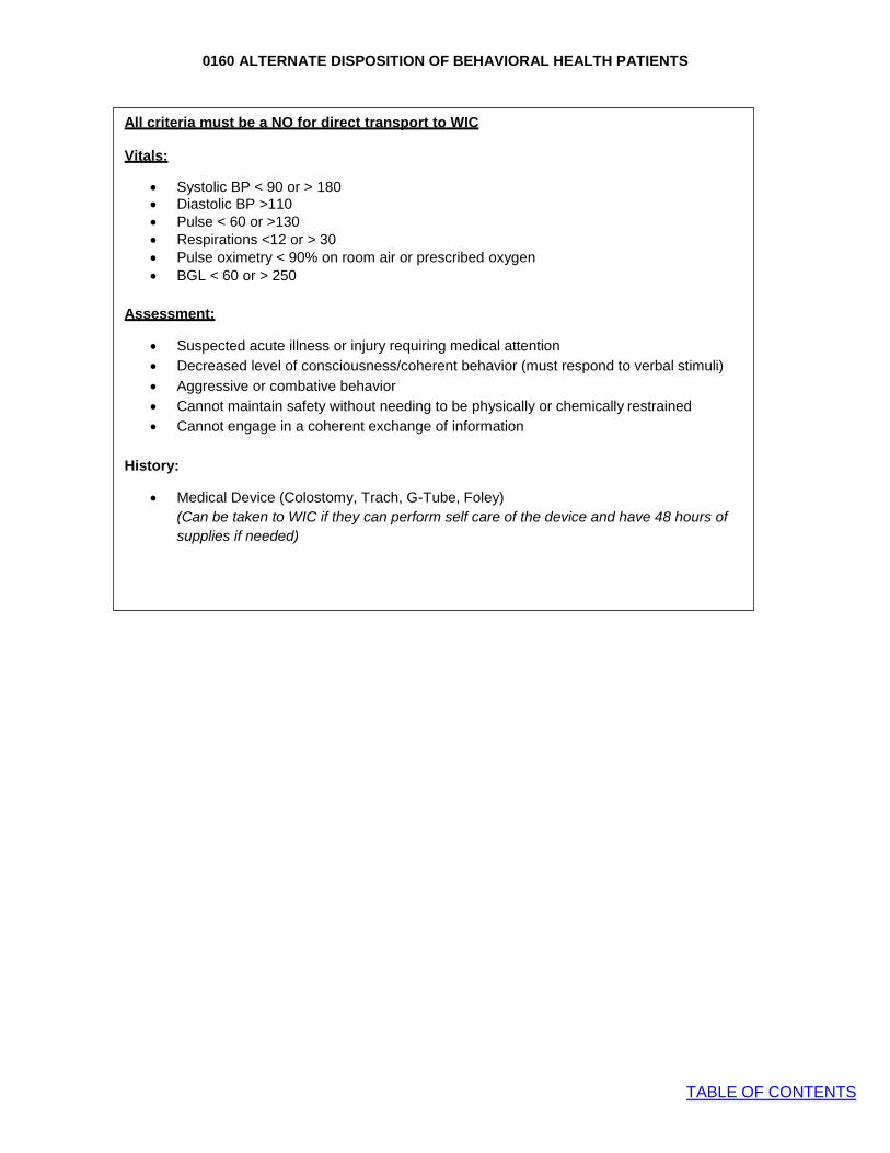
0160 ALTERNATE DISPOSITION OF BEHAVIORAL HEALTH PATIENTS
TABLE OF CONTENTS
All criteria must be a NO for direct transport to WIC
Vitals:
• Systolic BP < 90 or > 180 • Diastolic BP >110 • Pulse < 60 or >130 • Respirations <12 or > 30 • Pulse oximetry < 90% on room air or prescribed oxygen • BGL < 60 or > 250
Assessment:
• Suspected acute illness or injury requiring medical attention • Decreased level of consciousness/coherent behavior (must respond to verbal stimuli)
• Aggressive or combative behavior • Cannot maintain safety without needing to be physically or chemically restrained • Cannot engage in a coherent exchange of information
History:
• Medical Device (Colostomy, Trach, G-Tube, Foley) (Can be taken to WIC if they can perform self care of the device and have 48 hours of
supplies if needed)

0990 QUICK REFERENCE FOR PROCEDURES AND MEDICATIONS ALLOWED BY PROTOCOL
TABLE OF CONTENTS
Abbreviations S = Standing order B = Base contact
Airway Procedures B BIV P Capnography S S S Supraglottic airway S Continuous positive airway pressure (CPAP) S Orotracheal intubation S Nasotracheal intubation S Percutaneous cricothyrotomy S Bougie assisted surgical cricothyrotomy S Pediatric needle cricothyrotomy S Needle thoracostomy for tension pneumothorax decompression S Orogastric tube insertion with advanced airway S
Cardiovascular Procedures B BIV P Tourniquet S S S ECG - Acquire (including 12-lead) S S S ECG - Interpretation (including 12-lead) S Blood glucose monitoring S S S IV – Peripheral S S IV – External jugular S IO
• Rescue or primary vascular access device when peripheral IV access not obtainable in a patient with critical illness
S
• Utilization of IO access for all other patients B Use of established central line (including PICC) for fluid and medication administration (must have appropriate equipment, e.g. Huber needle, and training to access subcutaneous ports)
S
Automated / Semi-automated external defibrillator (AED) S S S Defibrillation – Manual S Valsalva maneuver S Synchronized cardioversion S Transcutaneous cardiac pacing
• Adult S • Pediatric B
Medications B BIV P
Specialized prescription medications to address an acute crisis given the route of administration is within the scope of the provider
B B B
Acetaminophen (Tylenol) S S S Adenosine (Adenocard)
• Adult S • Pediatric B
Albuterol sulfate (MDI and nebulizer) S S S Amiodarone
• Pulseless arrest S • Tachyarrhythmia with poor perfusion B
Antiemetic
• Ondansetron (Zofran) ODT S S S • Ondansetron (Zofran) IV/IO S S
Aspirin S S S Atropine sulfate
• Hemodynamically unstable bradycardia S • Organophosphate poisoning and stable bradycardia B
Benzodiazepines (midazolam)
• Seizure S • Sedation for transcutaneous pacing or cardioversion S • Sedation for severely agitated or combative patient – Adult S • Sedation for severely agitated or combative patient – Pediatric B
Calcium
• Pulseless arrest assumed due to hyperkalemia S • Calcium channel blocker overdose B
Dextrose S S

0990 QUICK REFERENCE FOR PROCEDURES AND MEDICATIONS ALLOWED BY PROTOCOL
TABLE OF CONTENTS
Medications B BIV P Diphenhydramine (Benadryl) S DuoDote™ / Mark I Kits S S S Epinephrine
• Pulseless arrest – IV/IO S • Pediatric bradycardia – IV/IO B • Asthma – IM S • Anaphylaxis– IM S S S • Pediatric severe systemic allergic reaction refractory to IM epinephrine - IV/IO S • Stridor at rest (alternative to racemic epinephrine) S • Epinephrine Auto-injector S S S • Adult hypotension refractory to fluid resuscitation – IV drip S • Adult bradycardia with signs of poor perfusion – IV drip S • Adult severe systemic allergic reaction – IV drip S
Glucagon
• Hypoglycemia S • Calcium channel blocker and β-blocker overdose S
Hemostatic agents S S S Hydroxocobalamin (Cyanokit) S Ibuprofen S S S Ipratropium Bromide (Atrovent) S Lidocaine 2% Solution – Anesthetic for IO needle insertion in adults S Magnesium sulfate
• Torsades de pointes associated with prolonged QT interval S • Refractory severe bronchospasm S • Eclampsia S
Methyprednisolone (Solu-Medrol) S Naloxone (Narcan) S S S Nitroglycerin (Nitrostat, Nitroquick)
• Sublingual, patient assisted B B S • Sublingual, agency supplied S • Nitroglycerin paste S
Opioids
• Adult S • Pediatric (1-12 years) S • Pediatric (<1 year) B
Oral glucose (Glutose, Insta-glucose) S S S Oxygen S S S Phenylephrine (Intranasal)
• Epistaxis S S S • Prior to nasotracheal intubation S
Racemic epinephrine (Vaponephrine) S Sodium bicarbonate
• Pulseless arrest assumed due to hyperkalemia S • Tricyclic antidepressant overdose S
Topical ophthalmic anesthetics S

1000 PROCEDURE PROTOCOL: OROTRACHEAL INTUBATION
TABLE OF CONTENTS
Indications:
• Respiratory failure • Absence of protective airway reflexes • Present or impending complete airway obstruction
Contraindications:
• There are no absolute contraindications. However, in general the primary goals of airway
management are adequate oxygenation and ventilation, and these should be achieved in the least invasive manner possible
o Orotracheal intubation is associated with worse outcomes among pediatric patients and head injured patients when compared to BLS airway maneuvers. Therefore, it is relatively contraindicated in these populations, and BLS airway is preferred unless patient cannot be oxygenated or ventilated by other means.
o Intubation is associated with interruptions in chest compressions during CPR, which is associated with worse patient outcomes. Additionally, intubation itself has not been shown to improve outcomes in cardiac arrest. Intubation should only be performed during pulseless arrest if it does not cause interruptions in chest compressions.
Technique for Video Laryngoscopy:
1. Initiate BLS airway sequence. 2. Start high flow nasal cannula oxygen at 15 lpm 3. Suction airway and pre-oxygenate with BVM ventilations, if possible 4. Check equipment and position patient:
a. If trauma: have assistant hold in-line spinal immobilization in neutral position b. If no trauma, neutral head position is preferred for use with the Airtraq video
laryngoscope c. Prepare the Airtraq device for intubation:
i. Attach the camera device to the appropriate disposable blade. Camera device will automatically begin recording.
ii. Turn the light on the disposable blade. iii. Choose your endotracheal tube and load it into the channel on the blade. No
stylet should be used. Using some KY jelly on the tube can help to pass it easier in the channel
5. Suction out airway before insertion of the blade into the mouth 6. Perform video laryngoscopy
a. Insert the blade into the midline area of mouth and follow the curvature of the tongue until the tip of the blade is in the hypopharynx.
b. Gentle manipulation of the camera should be done until the vocal cords are in view. If you cannot see the cords, try pulling the blade backwards towards yourself.
c. Do no attempt to blindly pass the tube. If you don’t see cords on first attempt, remove blade and ventilate the patient and suction as needed. Clean the tip of the blade if the camera view was obscured.
d. Can repeat attempt at video laryngoscopy 1 time if patient condition permits before moving to secondary airway.
7. Advance ETT through the cords under video visualization. Once placed, hold tube with hand at the mouth and disengage the tube from the channel on the blade.
8. Confirm tracheal location and appropriate depth and secure tube a. Correct tube depth may be estimated as 3 times the internal diameter of tube at teeth
or gums (e.g: 7.0 ETT is positioned at 21 cm at teeth) 9. Confirm and document tracheal location by:
a. ETCO2 waveform capnography b. Presence and symmetry of breath sounds c. Rising SpO2
10. Ventilate with BVM. Assess adequacy of ventilations
Paramedic

1000 PROCEDURE PROTOCOL: OROTRACHEAL INTUBATION
TABLE OF CONTENTS
11. During transport, continually reassess ventilation, oxygenation and tube position with continuous waveform capnography and SpO2
Direct Laryngoscopy
1) Direct laryngoscopy is not the primary intubation technique. It can only be used in the following
circumstances. a) Pediatric patients <12 years old when you are unable to ventilate and oxygenate the patient using
BLS and iGel airway placement. b) Choking patient when you need to use Magill forceps to remove an obstruction. c) Airtraq device mechanical failure
2) Continue BLS airway sequence 3) Start high flow nasal cannula oxygen at 15 lpm 4) Suction airway and pre-oxygenate with BVM ventilations, if possible 5) Check equipment and position patient:
a) If trauma: have assistant hold in-line spinal immobilization in neutral position b) If no trauma, sniffing position or slight cervical hyperextension is preferred
6) Perform laryngoscopy a) To improve laryngeal view, use right hand to manipulate larynx, or have assistant apply backwards,
upwards, rightward pressure (BURP) 7) Place ETT. Confirm tracheal location and appropriate depth and secure tube
a) Correct tube depth may be estimated as 3 times the internal diameter of tube at teeth or gums (e.g: 7.0 ETT is positioned at 21 cm at teeth)
8) Confirm and document tracheal location by: a) ETCO2 waveform capnography b) Presence and symmetry of breath sounds c) Rising SpO2
d) Other means as needed 9) Ventilate with BVM. Assess adequacy of ventilations 10) During transport, continually reassess ventilation, oxygenation and tube position with continuous
waveform capnography and SpO2
Precautions: • Ventilate at age-appropriate rates. Do not hyperventilate
• If the intubated patient deteriorates, think “DOPE”
o Dislodgement o Obstruction o Pneumothorax o Equipment failure (no oxygen)
• Reconfirm and document correct tube position, preferably with waveform capnography, after moving patient and before disconnecting from monitor in ED
• Unsuccessful intubation does not equal failed airway management. Many patients cannot be intubated without paralytics. Abandon further attempts at intubation and use supraglottic airway or BVM ventilations if 2 attempts at intubation unsuccessful.
• After the call, the intubation video must be downloaded from the Airtraq device and attached to the PCR.
• Any intubation performed without a video attached to the PCR must have an atypical incident report sent to Operations Commander

1010 PROCEDURE PROTOCOL: NASOTRACHEAL INTUBATION
TABLE OF CONTENTS
Indications:
• Age 12 years and older spontaneously breathing patient with indication for intubation who cannot tolerate either supine position or laryngoscopy
• Present or impending airway obstruction • Lack of protective airway reflexes
Contraindications:
• Apnea • Severe mid-face trauma
Technique:
1. Initiate BLS airway sequence 2. Suction airway and pre-oxygenate with BVM ventilations, if possible 3. Check equipment, choose correct ETT size (usually 7.0 in adult, limit is size of naris) 4. Position patient with head in midline, neutral position 5. If trauma: cervical collar may be in place, or assistant may hold in-line stabilization in neutral
position 6. If no trauma, patient may be sitting upright 7. Administer phenylephrine nasal drops in each nostril 8. Lubricate ETT with lidocaine jelly or other water-soluble lubricant 9. With gentle steady pressure, advance the tube through the nose to the posterior pharynx. Use the
largest nostril. Abandon procedure if significant resistance is felt 10. Keeping the curve of the tube exactly in midline, continue advancing slowly 11. There will be slight resistance just before entering trachea. Wait for an inspiratory effort before
final passage through cords. Listen for loss of breath sounds 12. Continue advancing tube until air is definitely exchanging through tube, then advance 2 cm more
and inflate cuff 13. Note tube depth and tape securely 14. Confirm and document endotracheal location by:
a. ETCO2, preferably with waveform capnography b. Presence and symmetry of breath sounds c. Rising SpO2
d. Other means as needed 15. Ventilate with BVM. Assess adequacy of ventilations 16. During transport, continually reassess ventilation, oxygenation and tube position with continuous
ETCO2 and SpO2
Precautions:
• Before performing BNTI, consider if patient can be safely ventilated with non-invasive means
such as CPAP or BVM • Use caution in anticoagulated or bleeding disorders given risk of epistaxis. • Ventilate at age-appropriate rates. Do not hyperventilate • If the intubated patient deteriorates, think “DOPE”
o Dislodgement o Obstruction o Pneumothorax o Equipment failure (no oxygen)
• Reconfirm and document correct tube position, preferably with waveform capnography, after moving patient and before disconnecting from monitor in ED
• Blind nasotracheal intubation is a very gentle technique. The secret to success is perfect positioning and patience.
Paramedic
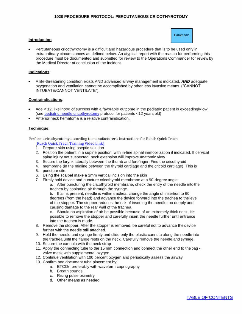
1020 PROCEDURE PROTOCOL: PERCUTANEOUS CRICOTHYROTOMY
TABLE OF CONTENTS
Introduction:
• Percutaneous cricothyrotomy is a difficult and hazardous procedure that is to be used only in extraordinary circumstances as defined below. An atypical report with the reason for performing this procedure must be documented and submitted for review to the Operations Commander for review by the Medical Director at conclusion of the incident.
Indications:
• A life-threatening condition exists AND advanced airway management is indicated, AND adequate
oxygenation and ventilation cannot be accomplished by other less invasive means. (“CANNOT INTUBATE/CANNOT VENTILATE”)
Contraindications:
• Age < 12, likelihood of success with a favorable outcome in the pediatric patient is exceedingly low.
(see pediatric needle cricothyrotomy protocol for patients <12 years old) • Anterior neck hematoma is a relative contraindication.
Technique:
Perform cricothyrotomy according to manufacturer’s instructions for Rusch Quick Trach (Rusch Quick Trach Training Video Link) 1. Prepare skin using aseptic solution 2. Position the patient in a supine position, with in-line spinal immobilization if indicated. If cervical
spine injury not suspected, neck extension will improve anatomic view 3. Secure the larynx laterally between the thumb and forefinger. Find the cricothyroid 4. membrane (in the midline between the thyroid cartilage and the cricoid cartilage). This is 5. puncture site. 6. Using the scalpel make a 3mm vertical incision into the skin 7. Firmly hold device and puncture cricothyroid membrane at a 90-degree angle.
a. After puncturing the cricothyroid membrane, check the entry of the needle into the trachea by aspirating air through the syringe. b. If air is present, needle is within trachea, change the angle of insertion to 60 degrees (from the head) and advance the device forward into the trachea to the level of the stopper. The stopper reduces the risk of inserting the needle too deeply and causing damage to the rear wall of the trachea. c. Should no aspiration of air be possible because of an extremely thick neck, it is possible to remove the stopper and carefully insert the needle further until entrance into the trachea is made.
8. Remove the stopper. After the stopper is removed, be careful not to advance the device further with the needle still attached.
9. Hold the needle and syringe firmly and slide only the plastic cannula along the needle into the trachea until the flange rests on the neck. Carefully remove the needle and syringe.
10. Secure the cannula with the neck strap 11. Apply the connecting tube to the 15 mm connection and connect the other end to the bag -
valve mask with supplemental oxygen. 12. Continue ventilation with 100 percent oxygen and periodically assess the airway 13. Confirm and document tube placement by:
a. ETCO2, preferably with waveform capnography b. Breath sounds c. Rising pulse oximetry d. Other means as needed
Paramedic

1020 PROCEDURE PROTOCOL: PERCUTANEOUS CRICOTHYROTOMY
TABLE OF CONTENTS
14. Ventilate with BVM assessing adequacy of ventilation 15. Observe for subcutaneous air, which may indicate tracheal injury or extra- tracheal tube position 16. Secure tube with tube ties or device 17. Continually reassess ventilation, oxygenation and tube placement
Precautions:
• Success of procedure is dependent on correct identification of cricothyroid membrane • Bleeding will occur, even with correct technique. Straying from the midline is dangerous and likely to
cause hemorrhage
Complications: 1. Respiratory arrest and patient demise due to: a. Severity of patient's airway injury. b. Lack of attention to other potential airway maneuvers. c. Subcutaneous air due to improper tube or catheter positioning, along with positive ventilation. d. Bleeding from superficial neck vessels is very common. Use direct pressure after QuickTrach is in place. e. Perforations of the back wall of the trachea and the esophagus from excessively deep penetration by the QuickTrach. With stopper in place, this should be an extremely rare complication.

1040 PROCEDURE PROTOCOL: PEDIATRIC NEEDLE CRICOTHYROTOMY
TABLE OF CONTENTS
Introduction:
• Needle cricothyrotomy is a difficult and hazardous procedure that is to be used only in extraordinary circumstances as defined below. An atypical report with the reason for performing this procedure must be documented and submitted for review to the Operations Commander for review by the Medical Director at conclusion of the incident.
• Due to the funnel-shaped, rostral, highly compliant larynx of a pediatric patient, cricothyrotomy is an extremely difficult procedure to successfully perform. As such, every effort should be made to effectively oxygenate the patient before attempting needle cricothyrotomy.
• This protocol is considered optional, and may not be adopted by all EMS Medical Directors or by all EMS agencies.
• A standardized, pre-prepared kit is recommended, and can be assembled using common airway equipment. An example is given below. Kit selection may vary and should be approved by the individual agency Medical Director.
• Example of kit: o 14 ga. and 16 ga. catheter over needle o 3 mL syringe o 15 mm endotracheal tube adaptor that fits the 3 mL
syringe used by agency (syringe barrel sizes vary)
Indications:
• A life-threatening condition exists AND adequate oxygenation and ventilation cannot be accomplished by other less invasive means for patients < 12 years old.
Contraindications:
• If patient can be ventilated and oxygenated by less invasive means
Technique:
1. Ensure patent upper airway with placement of an oral airway and nasal airway, unless contraindicated. 2. Open pre-prepared kit, attach angiocath to syringe, and aspirate 1-2 mL of saline into syringe 3. Prepare skin using aseptic solution 4. Insert the IV catheter through the skin and cricothyroid membrane into the trachea. Direct the needle at a
45° angle caudally (toward the feet). When the needle penetrates the trachea a “pop” will be felt. 5. Aspirate with the syringe. If air is retuned easily or bubbles are seen (with saline), the needle is in the
trachea. 6. Advance the catheter over the needle while holding the needle in position, then withdraw needle after
catheter is advanced flush to skin. 7. Remove the plunger and attach the 3 mL syringe to the catheter hub 8. Attach the 15 mm adaptor to the needle hub 9. Oxygenate the patient with bag-valve-mask device using the 15 mm adaptor provide high flow oxygen. 10. Confirm and document catheter placement by:
a. ETCO2 preferably with waveform capnography b. Rising pulse oximetry
11. Do not let go of catheter and be careful not to kink the catheter. There is no reliable way to secure it in place, and it is only a temporizing measure until a definitive airway can be established at the hospital
12. Observe for subcutaneous air, which may indicate tracheal injury or extra- tracheal catheter position 13. Continually reassess oxygenation and catheter position.
Paramedic

1050 PROCEDURE PROTOCOL: SUPRAGLOTTIC AIRWAY
TABLE OF CONTENTS
Indications:
• Rescue airway if unable to intubate a patient in need of airway protection • Primary airway if intubation anticipated to be difficult and rapid airway control
is necessary • Preferred advanced airway in the pediatric patient
Contraindications:
• Intact gag reflex • Caustic ingestion
Technique:
1. Initiate BLS airway sequence 2. Start high flow nasal cannula oxygen at 15 lpm 3. Suction airway as needed and preoxygenate with BVM, if possible 4. For adult patients select proper size i-gel O2 based on IDEAL patient body weight (not what
the pt actually weighs) : a. # 3 Small adult 30-60kg (65-130 lbs) b. # 4 Medium adult 50-90kg (110-200 lbs) c. # 5 Large adult 90 + kg (200 + lbs)
5. For Pediatric patients refer to length based tape and AFR pediatric field guide a. #1 Neonate 2-5 kg b. #1.5 Infant 5-12 kg c. # 2 Small pediatric 10-25 kg d. # 2.5 Large pediatric 25-35 kg
6. Open packaging and remove inner tray, setting the support strap (adult) and packet of lubricant to one side within easy reach. Remove the i-gel O2.
7. Open the packet of lubricant and place a small bolus on the inner side of the main shell of the packaging.
8. Grasp the i-gel O2 along the integral bite block and lubricate the back, sides and front of the cuff with a thin layer of lubricant. (Ensuring any excess is removed prior to insertion.)
9. Grasp the lubricated i-gel O2 firmly along the bite block. The patient should be in the ‘sniffing the morning air’ position with head extended and neck flexed. * Unless suspected spinal trauma.
10. Position the device so that the i-gel O2 cuff outlet is facing towards the chin of the patient. Introduce the leading soft tip into the mouth of the patient towards the hard palate.
11. Glide the device downwards and backwards along the hard palate with a continuous but gentle push until a definitive resistance is felt.
12. Once insertion has been completed, the tip of the airway should be located into the upper esophageal opening, with the cuff located against the laryngeal framework. The incisors should be resting on the bite block.
13. Secure the device underneath the patient’s neck with a head strap (or tape). Take care to ensure there is sufficient tension to hold the i-gel O2 securely in place, but not excessive tension that may cause trauma. Some adjustment of the strap may be needed to ensure optimal positioning.
14. For pediatric patients secure with tape 15. Place extension elbow and Waveform Capnography sensor on the end of i-gel O2 16. Confirm tube placement by auscultation, chest movement and Waveform Capnography.
17. Lubricate and insert appropriate size suction catheter into gastric lumen. a. Size # 5 use 14F b. Size # 2 through # 4 use 12F c. Size # 1.5 use 10F d. Size # 1 not applicable
18. Once the i-gel O2 has been correctly prepared, inserted and secured, positive pressure ventilation can commence.
19. Continuously monitor ETCO2 (preferably waveform capnography), SpO2, vital signs
Paramedic

1050 PROCEDURE PROTOCOL: SUPRAGLOTTIC AIRWAY
TABLE OF CONTENTS
Precautions:
1. Do not remove a properly functioning supraglottic airway in order to attempt intubation 2. Correct sizing of supraglottic airways is critical for correct function 3. Supraglottic airways are safe and effective in pediatric patients, provided the correct size tube
is selected. The age-range for supraglottic airway use is dependent on the specific device being used. Providers should be trained on and familiar with correct size selection for their device.
4. Use with caution in patients with broken teeth, which may lacerate balloon 5. Use with caution in patients with known esophageal disease who are at increased risk of
esophageal injury.

1060 PROCEDURE PROTOCOL: CONTINUOUS POSITIVE AIRWAY PRESSURE (CPAP)
TABLE OF CONTENTS
Indications: • Symptomatic patients with moderate-to-severe respiratory distress as evidenced by
at least two (2) of the following: o Rales (crackles) o Dyspnea with hypoxia (SpO2 less than 90% despite O2) o Dyspnea with inability to speak in full sentences o Accessory muscle use o Respiratory rate greater than 24/minute despite O2
o Diminished tidal volume
Contraindications: • Respiratory or cardiac arrest • Systolic BP less than 90mmHg • Lack of airway protective reflexes • Significant altered level of consciousness such that unable to follow verbal instructions or signal
distress • Vomiting or active upper GI bleed • Suspected pneumothorax • Trauma • Patient size or anatomy prevents adequate mask seal
Technique: 1. Place patient in a seated position and explain the procedure to him or her 2. Assess vital signs (BP, HR, RR, SpO2, and ETCO2) 3. Apply the CPAP mask and secure with provided straps, progressively tightening as tolerated to
minimize air leak 4. Operate CPAP device according to manufacturer specifications 5. Start with the lowest continuous pressure that appears to be effective. Adjust pressure following
manufacturer instructions to achieve the most stable respiratory status utilizing the signs described below as a guide
6. Monitor patient continuously, record vital signs every 5 minutes. 7. Assess patient for improvement as evidenced by the following:
a. Reduced dyspnea b. Reduced verbal impairment, respiratory rate and heart rate c. Increased SpO2
d. Stabilized blood pressure e. Appropriate ETCO2 values and waveforms f. Increased tidal volume
8. Observe for signs of deterioration or failure of response to CPAP: a. Decrease in level of consciousness b. Sustained or increased heart rate, respiratory rate or decreased blood pressure c. Sustained low or decreasing SpO2 readings d. Rising ETCO2 levels or other ETCO2 evidence of ventilatory failure e. Diminished or no improvement in tidal volume
Precautions: • Should patient deteriorate on CPAP:
o Troubleshoot equipment o Consider endotracheal intubation o Assess need for possible chest decompression due to pneumothorax o Assess for possibility of hypotension due to significantly reduced preload from positive
pressure ventilation • In-line nebulized medications may be given during CPAP as indicated and in accordance with
manufacturer guidelines • Some fixed pressure CPAP devices do not have FiO2 adjustment and will only administer up to
30% oxygen. If no improvement in oxygenation with a fixed pressure CPAP device, consider adding supplemental oxygen.
Paramedic

1070 PROCEDURE PROTOCOL: CAPNOGRAPHY
TABLE OF CONTENTS
EMT
Paramedic
Indications: A. MANDATORY: to rule out esophageal intubation and confirm endotracheal tube position in all
intubated patients. B. To identify late endotracheal tube dislodgement C. To monitor ventilation and perfusion in any ill or injured patient
Contraindications:
A. None
Technique: A. In patient with ETT or advanced airway: place ETCO2 detector in-line between airway adaptor
and BVM after airway positioned and secured B. Patients without ETT or advanced airway in place: place ETCO2 cannula on patient. May be
placed under CPAP or NRB facemask C. Assess and document both capnography waveform and ETCO2 value
Precautions:
A. To understand and interpret capnography, remember the 3 determinants of ETCO2: 1. Alveolar ventilation 2. Pulmonary perfusion 3. Metabolism
B. Sudden loss of ETCO2: 1. Tube dislodged 2. Circuit disconnected 3. Cardiac arrest
C. High ETCO2 (> 45) 1. Hypoventilation/CO2 retention
D. Low ETCO2 (< 25) 1. Hyperventilation 2. Low perfusion: shock, PE, sepsis
E. Cardiac Arrest: 1. In low-pulmonary blood flow states, such as cardiac arrest, the primary determinant of
ETCO2 is blood flow, so ETCO2 is a good indicator of quality of CPR 2. If ETCO2 is dropping, change out person doing chest compressions 3. In cardiac arrest, if ETCO2 not > 10 mmHg after 20 minutes of good CPR, this likely
reflects very low CO2 production and is associated with poor outcome 4. Sudden rise in EtCO2 may be an indicator of ROSC

1080 PROCEDURE PROTOCOL: NEEDLE THORACOSTOMY FOR TENSION
TABLE OF CONTENTS
PNEUMOTHORAX DECOMPRESSION
Indications:
A. All of the following clinical indicators must be present: 1. Severe respiratory distress 2. Hypotension and signs of shock 3. Unilateral absent or decreased breath sounds
B. Consider bilateral needle chest decompression in traumatic pulseless arrest if patient is being resuscitated and any trauma to trunk
Technique:
A. Expose entire chest B. Clean skin overlying site with available skin prep C. > 12 year old:
Insert Air Release System (ARS) catheter at 2nd intercostal space at midclavicular line. Remove needle and leave catheter in place.
D. < 12 year old: Use 18g 1 ½ “ angiocath at 2nd intercostal space at midclavicular line. Remove needle and leave catheter in place.
E. Notify receiving hospital of needle decompression attempt
Precautions:
A. Angiocath may become occluded with blood or by soft tissue B. A simple pneumothorax is NOT an indication for needle decompression C. Extra care is needed when performing on a pediatric patient.
Paramedic

1090 PROCEDURE PROTOCOL: SYNCHRONIZED CARDIOVERSION
TABLE OF CONTENTS
Paramedic Unstable tachyarrhythmia with a
pulse
Perform Synchronized
Cardioversion
1st: 0.5 Joules/kg 2+: 1 Joules/kg
1st: 120 Joules 2nd: 150 Joules 3+: 200 Joules
Pediatric Adult
Sedate with benzodiazepine if not contraindicated
Precautions: • If rhythm is AV nodal reentrant tachycardia (AVNRT, historically referred to as “PSVT”) it is
preferred to attempt a trial of adenosine prior to electrical cardioversion, even if signs of poor perfusion are present, due to rapid action of adenosine
• If defibrillator does not discharge in “synch” mode, then deactivate “synch” and reattempt • If sinus rhythm achieved, however briefly, then dysrhythmia resumes immediately, repeated
attempts at cardioversion at higher energies are unlikely to be helpful. First correct hypoxia, hypovolemia, etc. prior to further attempts at cardioversion
• If pulseless, treat according to Universal Pulseless Arrest Algorithm • Chronic atrial fibrillation is rarely a cause of hemodynamic instability, especially if rate is < 150
bpm. First correct hypoxia, hypovolemia, before considering cardioversion of chronic atrial fibrillation, which may be difficult, or impossible and poses risk of stroke
• Sinus tachycardia rarely exceeds 150 bpm in adults or 180 bpm in children and does not require or respond to cardioversion. Treat underlying causes.
• Transient dysrhythmias or ectopy are common immediately following cardioversion and rarely require specific treatment other than supportive care
Check: • O2 via NRB facemask • Functioning IV line • Suction • Advanced airway equipment ready
Continue treatment according to Tachycardia with Poor Perfusion

1100 PROCEDURE PROTOCOL: TRANSCUTANEOUS CARDIAC PACING
TABLE OF CONTENTS
Indications
1. Symptomatic bradyarrhythmias (includes A-V block) not responsive to medical therapy
2. Pacing is rarely indicated in patients under the age of 12 years. CONTACT BASE
Precautions 1. Conscious patient will experience discomfort; consider sedation with benzodiazepine if blood
pressure allows.
Contraindications 1. Pacing is contraindicated in pulseless arrest.
Technique
1. Apply electrodes as per manufacturer specifications: (-) left anterior, (+) left posterior. 2. Turn pacer unit on. 3. Start pacing 4. Select pacing rate at 80 beats per minute (BPM) 5. Initial pacing energy is set at 30 mAmps. 6. Confirm that pacer senses intrinsic cardiac activity by adjusting ECG size. 7. If no initial capture, increase current 10 mAmps every 10-15 seconds until capture or 200 mAmps
(usually captures around 100 mAmps). 8. Check for femoral pulse once there is electrical capture. 9. If no capture occurs with maximum output, discontinue pacing and resume ACLS.
Complications
1. Ventricular fibrillation and ventricular tachycardia are rare complications, but follow appropriate protocols if either occur.
2. Muscle tremors may complicate evaluation of pulses; femoral pulse may be more accurate. 3. Pacing may cause diaphragmatic stimulation and apparent hiccups.
Paramedic

1110 PROCEDURE PROTOCOL: INTRAOSSEUS CATHETER PLACEMENT
TABLE OF CONTENTS
Indications:
1. Rescue or primary vascular access device when peripheral IV access not
obtainable in a patient with critical illness defined as any of the following: A. Cardiopulmonary arrest or impending arrest B. Profound shock with severe hypotension and poor perfusion C. Hypoglycemia with severe symptoms (e.g. unresponsive) and no venous access
2. Utilization of IO access for all other patients requires base station contact
Technique: 1. Site of choice – Proximal tibia. 2 fingerbreadths inferior and 2 fingerbreadths medial to the tibial
tuberosity. 2. Clean skin per agency approved aseptic technique (alcohol, chlorhexidine) 3. Place intraosseous needle perpendicular to the bone.
A. For infants less than 6 months consider manual insertion of needle rather than powered device to avoid puncturing through both sides of the bone.
4. Follow manufacturer’s guidelines specific to the device being used for insertion. 5. Entrance into the bone marrow is indicated by a sudden loss of resistance. 6. Flush line with 10 mL saline. Do not attempt to aspirate marrow
A. IO infusion is very painful. If the patient is conscious, administer lidocaine for pain control before infusing fluids or medications.
7. Secure line A. Even if properly placed, the needle will not be secure. The needle must be secured and the IV
tubing taped. The IO needle should be stabilized at all times. 8. Observe for signs of limb swelling, decreased perfusion to distal extremity that would indicate a
malpositioned IO catheter or other complication. If limb becomes tense or malperfused, disconnect IO tubing immediately and leave IO in place.
9. A person should be assigned to monitor the IO at the scene and en route to the hospital. 10. Do not make more than one IO placement attempt per bone. 11. Do not remove IO needles in the field. 12. Notify hospital staff of all insertion sites/attempts and apply patient wristband included with kit to identify
IO patient.
Complications: 1. Fracture 2. Compartment syndrome 3. Infection
Contraindications:
1. Fracture of target bone 2. Cellulitis (skin infection overlying insertion site) 3. Osteogenesis imperfecta (rare condition predisposing to fractures with minimal trauma) 4. Total knee replacement (hardware will prevent placement) 5. DO NOT USE EZ IO Drill for patients less than 3kg (Shorter than grey color on Broselow)
– Pink EZ IO needle should be placed by hand. 6. DO NOT USE EZ IO Drill for newborns (pts less than 24 hours old)
– Pink EZ IO needle should be placed by hand.
Side Effects and Special Notes: 1. IO placement may be considered prior to peripheral IV attempts in critical patients without identifiable
peripheral veins 2. Some authorities recommend aspiration of marrow fluid or tissue to confirm needle location. This is not
recommended for field procedures, as it increases the risk of plugging the needle.
Paramedic
1110 PROCEDURE PROTOCOL: INTRAOSSEUS CATHETER PLACEMENT
TABLE OF CONTENTS
3. Expect flow rates to be slower than peripheral IVs. Pressure bags may be needed. Any drug or IV fluid may be infused.
4. Some manufacturers recommend the use of lidocaine for the treatment of pain associated with fluid administration. Check with your manufacturer and Medical Director for further guidance

1120 PROCEDURE PROTOCOL: TOURNIQUET PROTOCOL
TABLE OF CONTENTS
Indications
A. A tourniquet should be used for initial control of life threatening hemorrhage.
Precautions
A. In cases of life-threatening bleeding, benefit of tourniquet use outweighs any theoretical risk of limb ischemia.
B. A commercially made tourniquet is the preferred tourniquet. If none is available, a blood pressure cuff inflated to a pressure sufficient to stop bleeding is an acceptable alternative.
Technique
A. First, attempt to control hemorrhage by using direct pressure over bleeding area. B. If a discrete bleeding vessel can be identified, point pressure over bleeding vessel is more
effective than a large bandage and diffuse pressure. C. If unable to control hemorrhage using direct pressure, apply tourniquet according to
manufacturer specifications and using the steps below: 1. Cut away any clothing so that the tourniquet will be clearly visible. NEVER obscure a
tourniquet with clothing or bandages. 2. Apply tourniquet 3-4” proximal to the wound and not across any joints.
a. In tactical situations or if unable to quickly identify the location of the injury, apply the tourniquet as proximal as possible.
3. Tighten tourniquet until bleeding stops. Applying tourniquet too loosely will only increase blood loss by inhibiting venous return.
4. If bleeding is not controlled with the application of a single tourniquet, a 2nd can be applied adjacent to the 1st.
5. Mark the time and date of application on the patient’s skin next to the tourniquet. 6. Keep tourniquet on throughout hospital transport – a correctly applied tourniquet should
only be removed by the receiving hospital. 7. Pain management as needed.
Paramedic
EMT

1130 PROCEDURE PROTOCOL: RESTRAINT PROTOCOL
TABLE OF CONTENTS
Indications: A. Physical restraint of patients is permissible and encouraged if the patient poses
a danger to him/herself or to others. Only reasonable force is allowable, i.e., the minimum amount of force necessary to control the patient and prevent harm to the patient or others. Try alternative methods first (e.g., verbal de-escalation should be used first if the situation allows).
B. Paramedic: Consider pharmacological sedation for agitated patients that require transport and are behaving in a manner that poses a threat to him/herself or others.
1. See Agitated/Combative Patient Protocol: (The term “chemical restraint” is no longer preferred) C. Restraints may be indicated for patients who meet the following criteria:
1. A patient who is significantly impaired (e.g. intoxication, medical illness, injury, psychiatric condition, etc) and lacks decision-making capacity regarding his or her own care.
2. A patient who exhibits violent, combative or uncooperative behavior who does not respond to verbal de-escalation.
3. A patient who is suicidal and considered to be a risk for behavior dangerous to his or herself or to healthcare providers.
4. A patient who is on a mental health hold. Precautions:
A. When appropriate, involve law enforcement B. Restraints shall be used only when necessary to prevent a patient from seriously injuring him/herself or
others (including the EMS providers), and only if safe transportation and treatment of the patient cannot be accomplished without restraints. They may not be used as punishment, or for the convenience of the crew.
C. Any attempt to restrain a patient involves risk to the patient and the prehospital provider. Efforts to restrain a patient should only be done with adequate assistance present.
D. Be sure to evaluate the patient adequately to determine his or her medical condition, mental status and decision-making capacity.
E. Do not use hobble restraints and do not restrain the patient in the prone position or any position that impairs the airway or breathing.
F. Search the patient for weapons. G. Handcuffs are not appropriate medical restraints and should only be placed by law enforcement
personnel. See Transport of Handcuffed Patient Protocol. Technique:
A. Treat the patient with respect. Attempts to verbally reassure or calm the patient should be done prior to the use of restraints. To the extent possible, explain what is being done and why.
B. Have all equipment and personnel ready (restraints, suction, a means to promptly remove restraints). C. Use assistance such that, if possible, 1 rescuer handles each limb and 1 manages the head or supervises
the application of restraints. D. Apply restraints to the extent necessary to allow treatment of, and prevent injury to, the patient. Under-
restraint may place patient and provider at greater risk. E. After application of restraints, check all limbs for circulation. During the time that a patient is in restraints,
continuous attention to the patient’s airway, circulation and vital signs is mandatory. A restrained patient may never be left unattended.
Documentation Document the following in all cases of restraint: A. Description of the facts justifying restraint B. Efforts to de-escalate prior to restraint C. Type of restraints used D. Condition of the patient while restrained,
including reevaluations during transport Complications:
A. Aspiration: continually monitor patient’s airway
E. Condition of the patient at the time of transfer of care to emergency department staff
F. Any injury to patient or to EMS personnel
B. Nerve injury: assess neurovascular status of patient’s limbs during transport C. Complications of medical conditions associated with need for restraint
1. Patients may have underlying trauma, hypoxia, hypoglycemia, hyperthermia, hypothermia, drug ingestion, intoxication or other medical conditions
D. Excited Delirium Syndrome. This is a life-threatening medical emergency. These patients are truly out of control. They will have some or all of the following symptoms: paranoia, disorientation, hyper-aggression, hallucination, tachycardia, increased strength, and hyperthermia.
EMT
Paramedic

1140 PROCEDURE PROTOCOL: OROGASTRIC TUBE INSERTION WITH ADVANCED AIRWAY
TABLE OF CONTENTS
Indications:
• Gastric decompression in the intubated patient • Gastric decompression with placement of supraglottic airway • Intended for situations where time and conditions allow for gastric decompression without
interruption of routine care
Contraindications:
• Known esophageal varices
Technique:
1. Determine length of tube for insertion. Measure from tip of nose, to earlobe, then down to xiphoid process
2. Liberally lubricate the distal end of the orogastric tube 3. Suction airway and pre-oxygenate with BVM ventilations, if possible 4. Insert tube:
a. For orotracheal and nasotracheal intubation, insert tube into patient’s mouth; continue to advance the tube gently until the appropriate distance is reached
b. For supraglottic airway, insert tube through gastric access lumen and continue to advance tube till appropriate distance is reached.
5. Confirm placement by injecting 30cc of air and auscultate for the swish or bubbling of the air over the stomach. Aspirate gastric contents to confirm proper placement.
6. Secure with tape to inserted airway and attach to low continuous suction if indicated
Paramedic

1150 PROCEDURE PROTOCOL: TASER® PROBE REMOVAL
TABLE OF CONTENTS
Indications • Patient with TASER® probe(s) embedded in skin.
Contraindications
• TASER® probe embedded in the eye or genitals. In such cases, transport patient to an emergency department for removal.
Technique
1. Confirm the TASER® has been shut off and the barb cartridge has been disconnected. . 2. Using a pair of shears cut the TASER® wires at the base of the probe. 3. Place one hand on the patient in area where the probe is embedded and stabilize the skin
surrounding the puncture site. Using the other hand (or use pliers) firmly grasp the probe. 4. In one uninterrupted motion, pull the probe out of the puncture site maintaining a 90° angle to the
skin. Avoid twisting or bending the probe. 5. Repeat the process for any additional probes. 6. Once the probes are removed, inspect and assure they have been removed intact. In the event the
probe is not removed intact or there is suspicion of a retained probe, the patient must be transported to the emergency department for evaluation.
7. Cleanse the probe site and surrounding skin with betadine and apply sterile dressing. 8. Advise patient to watch for signs of infection including increased pain at the site, redness swelling
or fever.
EMT
Paramedic

1160 PROCEDURE PROTOCOL: PAIN MANAGEMENT
TABLE OF CONTENTS
Goal of Pain Management
A. Use comfort measure therapies as first line. B. If used, medications should be administered to a point where pain is tolerable.
This point is not necessarily pain free.
Assessment
A. Determine patient’s pain assessment and consider using a pain scale:
1. Pediatric use observational scale (see Pediatric Pain Scales) 2. Adult Self-report scale (Numeric Rating Scale [NRS])
B. Categorize the assessment of pain to mild, moderate, or severe.
1. Overreliance on pain scores may lead to either inadequate pain control in stoic patients, or over sedation in patients reporting high levels of pain. Use subjective and objective findings to evaluate need for and efficacy of pain management.
2. For pediatric patients, pain scale use is recommended. A pain score of 0-3 is mild pain, scores from 4- 6 moderate pain, and 7-10 severe pain.
General Pain Management Technique
Use comfort measure therapies as first line:
• Place patient in position of comfort • Splint/support painful area • Apply ice, if applicable • Consider compression, if applicable
Some conditions are complex and may be harmed by opioid use. It may be better to have physician evaluation prior to opioid use. These conditions include: • Headaches • Chronic abdominal pain
Consider acetaminophen/NSAID Consider acetaminophen/NSAID
Consider titration of opioids until pain tolerable or dosing maximized
For severe pain consider IN administration of opioid if IV not
readily available
Consider IV titration of opioids until pain tolerable or dosing maximized
Contact Base or follow agency
specific guidelines for pain that is not managed with opioids or if there are specific circumstances where
opioids should not be administered
May add acetaminophen/NSAID
• Transport in position of comfort and reassess as indicated
• Consider and prepare for administration of antiemetic if patient develops nausea and/or vomiting after pain medication
Mild pain
Moderate pain
Severe pain
EMT
Paramedic

1160 PROCEDURE PROTOCOL: PAIN MANAGEMENT
TABLE OF CONTENTS
General Information
A. Document assessment or pain scale before and after administration of pain medications. Reassess pain 5 minutes after IV administration.
B. Strongly consider ½ typical dosing in the elderly or frail patient
Pediatric Pain Scales
Recommended Pain Scale for Ages 4-12 Years
Faces Pain Scale – Revised (FPS-R)
0
2
4
6
8
10
This Faces Pain Scale-Revised has been reproduced with permission of the International Association for the Study of Pain® (IASP). The figure may NOT be reproduced for any other purpose without permission.

2000 OBSTRUCTED AIRWAY
TABLE OF CONTENTS
EMT Paramedic
• Perform Heimlich maneuver Yes • For visibly pregnant or obese
patients perform chest thrusts only
No • For infants, 5 chest thrusts then 5 back blows
If obstruction is complete, patient will be mute.
If patient can speak, obstruction is incomplete
Does patient show universal sign of choking?
Able to ventilate or obstruction cleared?
Yes Yes Is obstruction cleared?
No No
• Perform laryngoscopy • If foreign body visualized, use
McGill forceps to remove or consider pushing object into mainstem bronchus with ETT
Able to ventilate or obstruction cleared?
Yes
No
• Perform abdominal thrusts or chest thrusts until obstruction relieved then reattempt ventilations with BVM
• For infants, 5 chest thrusts, then 5 back blows
Consider cricothyrotomy if suspected supraglottic obstruction
and unable to oxygenate with BVM
• Supportive care and rapid transport
• If patient deteriorating or develops worsening distress proceed as for complete obstruction
Once obstruction relieved:
• Position of comfort or left lateral recumbent position
• O2 via NRB 15 Lpm • Monitor ABCs, SpO2, vital signs • Suction PRN and be prepared
for vomiting, which commonly occurs after obstruction relieved
Attempt to determine cause of obstruction
Assess severity of obstruction
Severe or Complete Obstruction (mute, silent cough, severe stridor)
Unconscious Patient
Mild or Moderate Obstruction
Begin chest thrusts Each time airway is opened look in
mouth for FB and if found, remove it
• Open airway with head tilt-chin lift or
jaw thrust if craniofacial trauma • Attempt ventilation with BVM
• Do not interfere with a spontaneously breathing of coughing patient
• Position of comfort • Give high flow oxygen • Suction if needed

2010 ADULT UNIVERSAL RESPIRATORY DISTRESS
TABLE OF CONTENTS
Obstructed Airway protocol
Patent airway?
Is anaphylaxis likely?
Assist ventilations with BVM and
airway adjuncts as needed
Are ventilations adequate for
physiologic state?
Adult Wheezing protocol
Is asthma or COPD likely?
Allergy/Anaphylaxis protocol
• Transport • Provide supportive care • Maximize oxygenation and
ventilation • Contact Base if needed for
consult
CHF/Pulmonary Edema protocol
Is CHF/pulmonary edema likely?
No
Yes
No
Yes
Yes
No
? Yes
No
? Yes
No
EMT
Paramedic
Respiratory Distress
For all patients: While assessing ABCs: give
supplemental O2, monitor vital signs, cardiac rhythm, SpO2 and waveform
capnography
• Consider 12 lead ECG
Mixed picture may exist
• Goal is maximization of
oxygenation and ventilation in all cases
• CPAP may be particularly useful in mixed picture with hypoxia and/or hypoventilation
• Avoid albuterol in suspected pulmonary edema
Consider pulmonary and non-pulmonary causes of
respiratory distress:
• Pulmonary embolism • Pneumonia • Heart attack • Pneumothorax • Sepsis • Metabolic acidosis (e.g.:
DKA) • Anxiety

2020 PEDIATRIC UNIVERSAL RESPIRATORY DISTRESS
TABLE OF CONTENTS
Patent Airway? No
Obstructed Airway protocol
Yes
Age-appropriate ventilation rate in respiratory failure:
Are ventilations adequate for age?
No Assist ventilations at age- appropriate rate with BVM and
airway adjuncts as needed
Yes
Is anaphylaxis likely?
Yes Allergy/Anaphylaxis protocol
Assisted ventilation rates listed do not apply to the patient in cardiac arrest
No
Is there a barky cough and stridor?
Yes Pediatric Stridor/Croup protocol
No
Is there wheezing? Yes
Pediatric Wheezing protocol
No
Characteristics of Stridor:
• High-pitched, harsh sound most often heard on inspiration
• Occurs with upper airway restriction
• Significant restriction may result in biphasic stridor (heard on inspiration and expiration)
Consider pulmonar
• Foreign body • Croup • Pneumonia • Bronchiolitis
y and non-pulmonary causes:
• Pulmonary embolism • Sepsis • Metabolic derangement • Anxiety
EMT Paramedic
Age Breaths/min
Neonate 40 Infants 30 Children 20
For all patients: While assessing ABCs: give
supplemental O2, monitor vital signs, cardiac rhythm, SpO2, and consider waveform capnography
• Provide supportive care • Maximize oxygenation and ventilation • CONTACT BASE if needed for consult
Respiratory Distress

2030 ADULT WHEEZING
TABLE OF CONTENTS
EMT
Paramedic
Adult Respiratory Distress Protocol and prepare for
immediate transport
Presentation suggests Bronchospasm: wheezing, prolonged expiratory phase,
decreased breath sounds, accessory
muscle use, known hx of asthma/COPD
Therapeutic Goals:
• Maximize oxygenation • Decrease work of
breathing • Identify cardiac ischemia
(Obtain 12 lead EKG) • Identify complications,
e.g. pneumothorax
Give oxygen, check SpO2, waveform capnography, & consider IV for severe
Consider pulmonary and non-pulmonary causes of
respiratory distress:
Examples: pulmonary embolism, pneumonia,
pulmonary edema, anaphylaxis, heart attack,
pneumothorax, sepsis, metabolic acidosis (e.g.:
DKA), Anxiety
Yes
Is response to treatment adequate?
No
Yes
No
• Consider IM epinephrine. Indicated only if no
response to neb, CPAP and for pt in severe distress. Contraindicated if any concern for myocardial ischemia or known coronary artery disease.
• Consider IV magnesium
Is response to treatment adequate?
• IV methylprednisolone • Obtain ECG: rule out unstable rhythm, ACS
• Reassess for pneumothorax • Consider CPAP early, especially in COPD • If CPAP contraindicated, ventilate with BVM,
and consider advanced airway
Give nebulized albuterol + ipratropium
EMT may administer either MDI or nebulized albuterol
May give continuous neb for severe respiratory
distress
• Continue monitoring and assessment en route • Be prepared to assist ventilations as needed • Contact base for medical consult as needed
IV methylprednisolone will help resolve acute asthma exacerbation over hours,
without immediate effect. In severe exacerbations, it may
be given prehospital but should not be given for mild attacks responding well to
bronchodilators
IV magnesium may be beneficial in some patients
with severe attacks. It should not be given routinely, rather should be reserved for life- threatening asthma attacks
not responding to conventional therapy
IM epinephrine is only indicated for most severe
attacks deemed life- threatening and not
responding to inhaled bronchodilators. Use extreme caution when administering. Cardiopulmonary monitoring
is mandatory
COPD
• Correct hypoxia: do not withhold maximum oxygen for fear of CO2
retention • Patients with COPD are
older and have comorbidities, including heart disease.
• Wheezing may be a presentation of pulmonary edema, “cardiac asthma”
• Common triggers for COPD exacerbations include: Infection, dysrhythmia (e.g.: atrial fibrillation), myocardial ischemia
• COPD exacerbations are particularly responsive to CPAP, which may help avoid the need for intubation and should be considered early in treatment

2040 PEDIATRIC WHEEZING
TABLE OF CONTENTS
• IM epinephrine if no response to neb and severe distress
• Start IV • IV methylprednisolone • 20mL/kg NS bolus
Consider the cause of wheezing before
initiating specific therapy
Initial best indicator is age. If patient ≤ 2
years old, bronchiolitis is most likely. Age > 2 reactive airways disease is more likely.
Age ≤ 2 years old
Bronchiolitis most common
• Viral illness characterized by fever, copious secretions and respiratory distress typically seen November through April
• Most important interventions are to provide supplemental oxygen and suction secretions adequately
• Bronchodilators and steroids do not work
• Administer oxygen to obtain saturations > 90%
• Nasal suction • Transport in position of comfort • Monitor SpO2, RR, retractions,
mental status
If worsening respiratory distress despite above therapies, re-suction nostrils and assist ventilations with
BVM
BLS airway preferred in pediatrics
Age > 2 years old
Asthma most common
Presentation suggests asthma: wheezing, prolonged expiratory phase, decreased breath sounds, accessory muscle use, known hx of asthma or
albuterol use
EMT may administer either MDI or
nebulized albuterol May give continuous neb for severe
respiratory distress
Give nebulized albuterol + ipratropium
Is response to treatment adequate
No
Severe exacerbation
Yes
Is response to treatment adequate?
Yes
No
• Assess for pneumothorax • Assist ventilations with BVM
BLS airway preferred in pediatrics
• Continue monitoring and assessment en route
• Be prepared to assist ventilations as needed
• Contact Base for medical consult if deterioration
IV methylprednisolone
Will help resolve acute asthma exacerbation over hours, without
immediate effect. In severe exacerbations, it may be given
prehospital but should not be given for mild attacks responding well to
bronchodilators.
IM epinephrine
Is indicated for the most severe attacks deemed life-threatening and
not responding to inhaled bronchodilators.
Although bronchiolitis and
asthma are the most common causes of
wheezing in infants and children, respectively, you
should consider pulmonary and non- pulmonary causes of respiratory distress,
especially if patient not responding as expected to
treatment:
Examples: pneumonia, pulmonary edema,
congenital heart disease, anaphylaxis, pneumothorax, sepsis, metabolic acidosis
(e.g.: DKA, toxic ingestion), foreign body aspiration, and
croup.
• Pediatric Universal Respiratory Distress protocol
• Assess: SpO2, consider waveform capnography, RR, lung sounds, accessory muscle use and mental status
EMT Paramedic

2050 PEDIATRIC STRIDOR/CROUP
TABLE OF CONTENTS
Yes
Give nebulized epinephrine
If signs of poor perfusion AND/OR
hypotension for age, see Medical Shock protocol and begin fluid resuscitation
• Continue monitoring and assessment en route • Contact Base for repeat dose of nebulized
epinephrine and medical consult as needed
Check SpO2, give oxygen as needed
Considerations with Stridor:
• Stridor is a harsh, usually inspiratory sound caused by narrowing or obstruction of the upper airway
• Causes include croup, foreign body aspiration, allergic reactions, trauma, infection, mass
• Epiglottitis is exceedingly rare. May consider in the unimmunized child. Treatment is minimization of agitation. Airway manipulation is best done in the hospital.
Minimize agitation: Transport in position of comfort, interventions only as necessary
Pediatric Universal Respiratory Distress protocol and prepare for immediate
transport
Characteristics of Croup:
• Most common cause of stridor in children
• Child will have stridor, barky cough, and URI symptoms of sudden, often nocturnal onset
• Most often seen in children < 9 years old
• Agitation worsens the stridor and respiratory distress
EMT
Paramedic
No
Are symptoms severe and croup most likely?
• Stridor at rest or biphasic stridor • Severe retractions • SpO < 90% despite O
2 2
• Altered LOC • Cyanosis
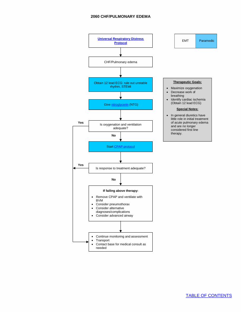
2060 CHF/PULMONARY EDEMA
TABLE OF CONTENTS
EMT Paramedic
CHF/Pulmonary edema
Obtain 12 lead ECG: rule out unstable rhythm, STEMI
Give nitroglycerin (NTG)
Yes Is oxygenation and ventilation
adequate?
No
Start CPAP protocol
Yes Is response to treatment adequate?
No
• Continue monitoring and assessment • Transport • Contact base for medical consult as
needed
Therapeutic Goals:
• Maximize oxygenation • Decrease work of
breathing • Identify cardiac ischemia
(Obtain 12 lead ECG)
Special Notes:
• In general diuretics have little role in initial treatment of acute pulmonary edema and are no longer considered first line therapy.
Universal Respiratory Distress Protocol
If failing above therapy:
• Remove CPAP and ventilate with BVM
• Consider pneumothorax • Consider alternative
diagnoses/complications • Consider advanced airway
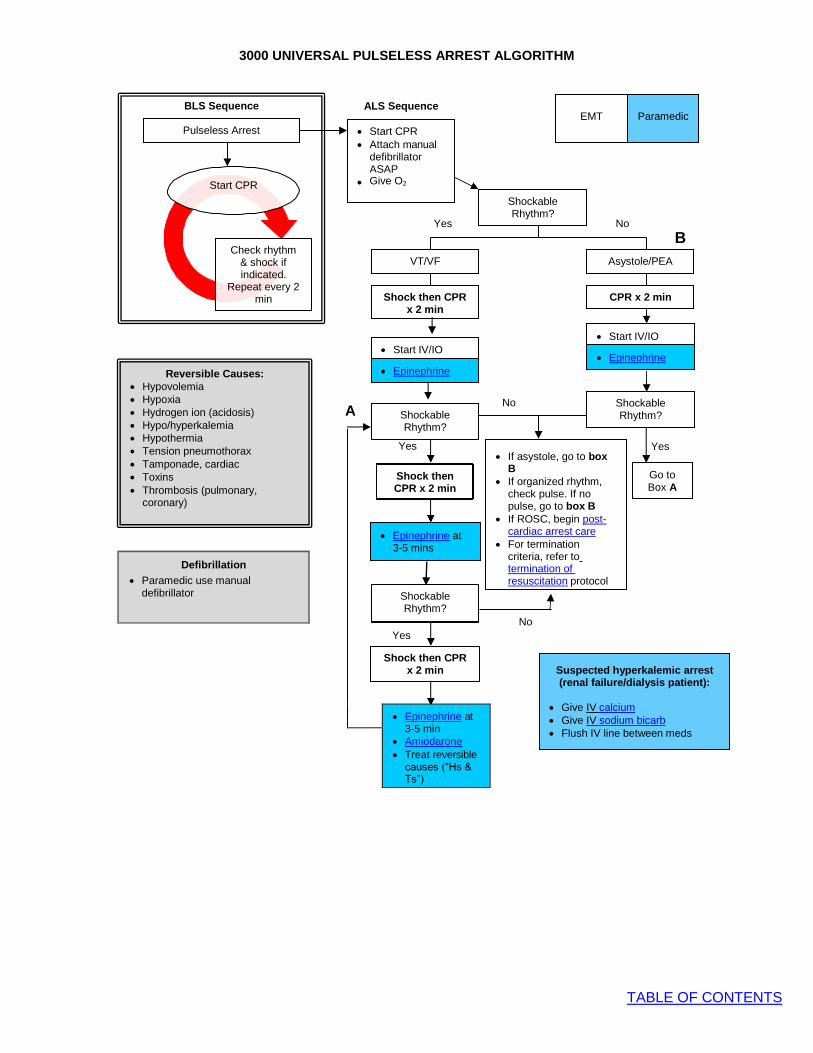
3000 UNIVERSAL PULSELESS ARREST ALGORITHM
TABLE OF CONTENTS
Yes
Go to Box A
No
Shockable Rhythm?
Asystole/PEA VT/VF
Shockable Rhythm?
Shockable Rhythm?
• If asystole, go to box B
• If organized rhythm, check pulse. If no pulse, go to box B
• If ROSC, begin post- cardiac arrest care
• For termination criteria, refer to termination of resuscitation protocol
Shock then CPR x 2 min
Epinephrine at 3-5 min
Shockable Rhythm?
Shock then CPR x 2 min
Yes
A
ALS Sequence
Yes No
B
No No
Yes
• Epinephrine at
3-5 min • Amiodarone • Treat reversible
causes (“Hs & Ts”)
• Start CPR • Attach manual
defibrillator ASAP
• Give O2
Shock then CPR x 2 min
CPR x 2 min
BLS Sequence
Start CPR
Check rhythm & shock if indicated.
Repeat every 2 min
Pulseless Arrest
Reversible Causes: • Hypovolemia • Hypoxia • Hydrogen ion (acidosis) • Hypo/hyperkalemia • Hypothermia • Tension pneumothorax • Tamponade, cardiac • Toxins • Thrombosis (pulmonary,
coronary)
• Epinephrine
• Start IV/IO • Epinephrine
• Start IV/IO
Paramedic
EMT
Suspected hyperkalemic arrest (renal failure/dialysis patient):
• Give IV calcium • Give IV sodium bicarb • Flush IV line between meds
Defibrillation
• Paramedic use manual defibrillator
• Epinephrine at 3-5 mins

3010 UNIVERSAL PULSELESS ARREST CONSIDERATIONS
TABLE OF CONTENTS
PEDIATRIC PATIENT
Compressions
• Follow current PALS guidelines for chest compressions • Minimize interruptions, resume compressions immediately
after shocks, rhythm checks. Check pulses only if organized rhythm
• Push hard ( 1/3 of anteroposterior chest diameter and fast (100-120/min) and allow complete chest recoil
• Assess quality of CPR with continuous waveform capnography
Defibrillation:
• 1st shock 2 J/kg, subsequent shocks 4 J/kg • Paramedic use manual defibrillator
Ventilations
• If no advanced airway, alternate ventilations and compressions in 15:2 ratio
• If advanced airway in place, ventilate continuously at 10 breaths/minute
• Do not over ventilate
Airway
• No intubation for cardiac arrest < 12 years’ old • An appropriately-sized supraglottic airway (e.g. i-gel) may
be placed • If unable to ventilate with i-gel airway, resort to BLS airway
management • Early ventilation is paramount in resuscitation as arrest is
usually respiratory in nature
ROSC
• Pulse and blood pressure • Sustained abrupt rise in ETCO2, typically > 40
ADULT PATIENT
Compressions
• Follow current ACLS guidelines for chest compressions • Minimize interruptions, resume compressions immediately
after shocks, rhythm checks. Check pulses only if organized rhythm
• Push hard and fast and allow complete chest recoil • Assess quality of CPR with continuous waveform
capnography • If ETCO2 < 10, improve quality of compressions • If using automated CPR devices, use manufacturer’s
specifications
Defibrillation
• Biphasic: Zoll X-Series o 1st: 120 Joules o 2nd: 150 Joules o 3+: 200 Joules
Ventilations
• Open the airway, place NPA/OPA, place NRB facemask with O2 at 15 L/min for first 4 minutes of chest compressions, unless hypoxic arrest suspected (e.g.: asphyxiation, overdose, status asthmaticus), In which case begin ventilations immediately.
• Do not over ventilate • If no advanced airway, 30:2 compressions to ventilation
ratio • If advanced airway in place ventilate at rate of 10
breaths/min
Airway
• An advanced airway (ETT, i-gel) may be placed at any time after initial 6 minutes of passive oxygenation, if applicable, or as soon as possible if asphyxial arrest suspected, provided placement does not interrupt compressions
ROSC
• Pulse and blood pressure • Sustained abrupt rise in ETCO2, typically > 40
Regarding where to work arrest and presence of family
members:
• CPR in a moving ambulance or pram is ineffective • In general, work cardiac arrest on scene either to return of
spontaneous circulation (ROSC), or to field pronouncement, unless scene unsafe
• Family presence during resuscitation is preferred by most families, is rarely disruptive, and may help with grieving process for family members. Family presence during resuscitation is recommended, unless disruptive to resuscitation efforts
• Contact base for termination of resuscitation
Pacing
• Pacing is not indicated for asystole and PEA. Instead start chest compressions according to Universal Pulseless Arrest Algorithm.
• Pacing should not be undertaken if it follows unsuccessful defibrillation of VT/VF as it will only interfere with CPR and is not effective
ICD/Pacemaker patients
• If cardiac arrest patient has an implantable cardioverter defibrillator (ICD) or pacemaker: place pacer/defib pads at least 1 inch from device. Biaxillary or anterior posterior pad placement may be used

3020 NEONATAL RESUSCITATION
TABLE OF CONTENTS
Term Gestation? Breathing or crying? Good flex or tone?
No
Yes EMT Paramedic
Warm, clear airway if
necessary, dry, simulate
No HR <100, gasping or apnea
Yes
No
Labored breathing or persistent cyanosis?
Yes
PPV, SpO2 monitoring
Clear airway SpO2 monitoring
Supplementary O2 as needed
HR <100?
No Post resuscitation care
Yes
Take ventilation corrective steps
HR <60? No
Yes
Chest compressions Coordinate w. PPV
100% O2
Take ventilation corrective steps
HR <60 after 60 seconds of No
chest compressions?
Yes
Consider:
• Hypovolemia • Pneumothorax
IV epinephrine
Birth
30 sec
60 sec
Targeted Preductal (Right
Arm) SpO2 After Birth (From 2015 NRP
Guidelines)
• 1 minute: 60%-65% • 3 minutes: 60%-75% • 5 minutes: 80%-85% • 10 minutes: 85%-95%
Neonatal Oxygen
Recommendations (From 2015 NRP Guidelines)
• Begin resuscitation of newborns ≥ 35 weeks gestation with room air. If breathing is labored, supplement with oxygen to the targets listed
• Begin resuscitation of newborns <35 weeks gestation with supplemental oxygen titrated to the targets listed
General Considerations
(From 2015 AHA Guidelines)
• Newborn infants who do not require resuscitation can be identified generally based on 3 questions:
• Term gestation? • Crying or breathing? • Good muscle tone?
• If answer to all 3 questions is “yes” then
baby does not require resuscitation and should be dried skin-to-skin on mother covered to keep warm
• If answer to any of 3 questions is “no then
infant should receive 1 or more of the following 4 categories of intervention in sequence:
• Initial steps in stabilization (warm, clear airway, dry, stimulate)
• Ventilation • Chest compressions • Administration of epinephrine and/or
volume expansion • Initial resuscitation steps should be
completed within 60 seconds as illustrated • The decision to progress beyond initial
steps is based on an assessment of respirations (apnea, gasping, labored, or unlabored breathing) and heart rate (>/< 100 bpm)
Assisting Ventilations
• Assist ventilations at a rate of 40-60 breaths per minute to maintain HR > 100
• Use 2 person BVM when possible
Chest Compressions
• Indicated for HR < 60 despite adequate ventilation w. supplemental O2 for 30 seconds
• 2 thumbs-encircling hands technique preferred
• Allow full chest recoil • Coordinate with ventilations so not
delivered simultaneously • 3:1 ratio for compressions to ventilations
Medications
• Epinephrine is indicated if the newborn’s
heart rate remains less than 60 beats/min after at least 30 seconds of PPV AND another 60 seconds of chest compressions coordinated with PPV using 100% oxygen
Routine Care: • Provide warmth • Clear airway if necessary • Dry • Ongoing evaluation

3030 POST-RESUSCITATION CARE WITH ROSC
TABLE OF CONTENTS
Assess for dysrhythmia
Medical Hypotension/Shock
protocol
Is there hypotension for age
and/or signs of shock?
• Assess for shock and volume status • Peripheral access: IO/IV • Oxygenation/Ventilation
o Secure advanced airway if indicated
o Avoid hyperventilation o Avoid hyper/hypocapnia (EtCO2) o Correct hypoxemia
• Elevate head of bed at 30°
Initiate Cardiac Alert Is STEMI Present?
• Continuous rhythm monitoring and
pulse checks • Focused neuro exam (AVPU/GCS) • Targeted Temperature Management
(TTM) goal 33 - 36 C (91.4 - 96.8
F) o Check patient temperature, if
possible o Avoid fever and provide passive
cooling o Place ice packs to neck, axillae,
groin if needed for fever • Transfer to closest appropriate facility
Yes
No
Yes
No
Yes
No
EMT
Paramedic
ROSC after cardiac arrest
Perform 12 lead EKG
Post-Cardiac Care
• Following ROSC, several simultaneous and stepwise interventions must be performed to optimize care and maximize patient outcome
• Survival and neurologic outcome worsen with fever, hypoxia, hypo/hypercapnia, and hypotension. Post-ROSC care should focus on prevention of these elements
Recurrent dysrhythmia? Treat recurrent dysrhythmia per
appropriate protocol
Return of spontaneous
circulation (ROSC) criteria:
• Pulse and measurable blood pressure
• Increase in ETCO2 on capnography
Document:
• Time of arrest (or time last seen normal)
• Witnessed vs. unwitnessed arrest
• Initial rhythm shockable vs. non-shockable
• Bystander CPR given • Time of ROSC • GCS after ROSC • Initial temperature of patient
after ROSC, if possible

3040 TACHYARRHYTHMIA WITH POOR PERFUSION
TABLE OF CONTENTS
12 lead ECG
Tachyarrhythmia
EMT Paramedic
• Support ABCs • IV access • Give oxygen
• 12 lead ECG
Probable Sinus Tachycardia?
• Adult: rate usually <150
• Children: rate usually <180 • Infants: rate usually < 220
Yes • Search for and treat
underlying cause: e.g. dehydration, fever, hypoxia, hypovolemia, pain
• Consider medical shock
Stable
• Identify Rhythm • Measure QRS width
No
A Is patient stable?
Unstable signs include altered mental status, chest pain, hypotension, signs of shock-rate-related symptoms
uncommon if HR <150 in adults
B Unstable
Immediate synchronized cardioversion
Narrow QRS Adult < 0.12 msec
Pediatric <0.09 msec
Wide QRS Adult > 0.12 msec
Pediatric >0.09 msec
• Repeat 12 lead ECG • Identify rhythm • Contact Base
Regular
• Children who are stable with AVNRT generally remain so and transport is preferred over intervention
• Try Valsalva maneuver • Give adenosine IV if
suspected AV nodal reentrant tachycardia (AVRNT)*
C Irregular
• Atrial fibrillation, flutter, or MAT
• Do not give adenosine
• If becomes unstable go to
box B
Regular • Contact Base for consult
• V Tach (>80%) or SVT with aberrancy
• Contact Base for verbal order for amiodarone unless contraindicated
• If regular and polymorphic
(Torsades de Pointes) consider magnesium
Irregular
• See box C
• Contact Base for consult • Do NOT give adenosine
Converts
• Repeat 12 lead ECG
• Monitor in transport • If recurrent dysrhythmia, go
to box A
Doesn’t Convert
• Contact Base for consult • Monitor in transport • If unstable, go to box B

3050 BRADYARRHYTHMIA WITH POOR PERFUSION
TABLE OF CONTENTS
Are there signs or symptoms of poor perfusion present?
(Altered mental status, chest pain, hypotension, signs of shock)
Adequate perfusion Poor perfusion
Monitor and transport
Consider vasopressor infusion early if pacing and poor perfusion or hypotension persists either due to lack of capture or poor contractility despite capture
Monitor and transport
• Give epinephrine • Consider atropine • If no improvement,
Contact Base to discuss transcutaneous pacing
• Give atropine • Prepare for
transcutaneous pacing
Bradycardia with a pulse Heart rate < 60
EMT Paramedic
• Support ABCs • Cardiac monitor • Give Oxygen • Identify rhythm • Start IV • 12-lead ECG • Initiate transport
Pediatric Considerations:
• Consider any HR <60 in an ill child abnormal regardless of age
• Perform CPR if HR < 60 with poor perfusion despite oxygenation and ventilation
• Administer epinephrine if bradycardia persists despite oxygenation/ventilation and chest compressions
• Atropine should be administered for increased vagal tone or AV block
Reminders:
• If pulseless arrest develops, go to pulseless arrest algorithm
• Search for possible contributing factors: “5 Hs and 5 Ts”
• Symptomatic severe bradycardia is usually related to one of the following: o Ischemia (MI) o Drugs (beta blocker, Calcium channel
blocker) o Electrolytes (hyperkalemia)
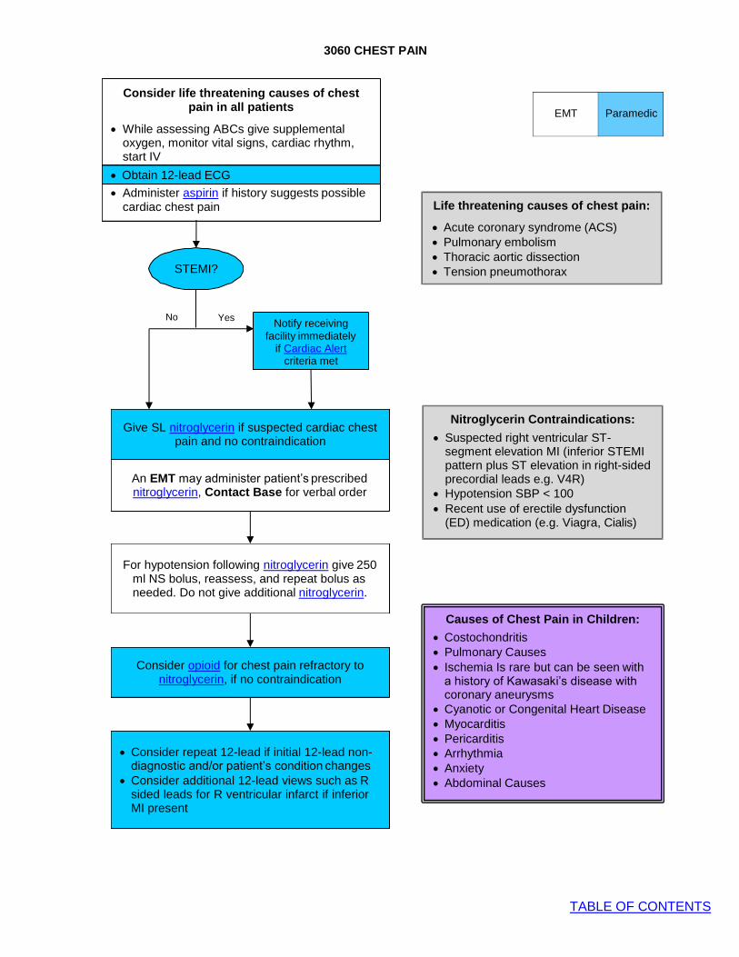
3060 CHEST PAIN
TABLE OF CONTENTS
Obtain 12-lead ECG
Consider life threatening causes of chest
pain in all patients
• While assessing ABCs give supplemental oxygen, monitor vital signs, cardiac rhythm, start IV
• Obtain 12-lead ECG
• Administer aspirin if history suggests possible cardiac chest pain
STEMI?
No Yes Notify receiving
facility immediately if Cardiac Alert
criteria met
Give SL nitroglycerin if suspected cardiac chest
pain and no contraindication
An EMT may administer patient’s prescribed nitroglycerin, Contact Base for verbal order
For hypotension following nitroglycerin give 250 ml NS bolus, reassess, and repeat bolus as needed. Do not give additional nitroglycerin.
Consider opioid for chest pain refractory to nitroglycerin, if no contraindication
• Consider repeat 12-lead if initial 12-lead non- diagnostic and/or patient’s condition changes
• Consider additional 12-lead views such as R sided leads for R ventricular infarct if inferior MI present
Life threatening causes of chest pain:
• Acute coronary syndrome (ACS) • Pulmonary embolism • Thoracic aortic dissection • Tension pneumothorax
Nitroglycerin Contraindications:
• Suspected right ventricular ST- segment elevation MI (inferior STEMI pattern plus ST elevation in right-sided precordial leads e.g. V4R)
• Hypotension SBP < 100 • Recent use of erectile dysfunction
(ED) medication (e.g. Viagra, Cialis)
Causes of Chest Pain in Children:
• Costochondritis • Pulmonary Causes • Ischemia Is rare but can be seen with
a history of Kawasaki’s disease with coronary aneurysms
• Cyanotic or Congenital Heart Disease • Myocarditis • Pericarditis • Arrhythmia • Anxiety • Abdominal Causes
EMT Paramedic

3070 CARDIAC ALERT
TABLE OF CONTENTS
Goal:
• To identify patients with ST-segment elevation myocardial infarction (STEMI) in the prehospital setting and provide advanced receiving hospital notification in order to minimize door-to- balloon times for percutaneous coronary intervention (PCI)
Inclusion Criteria:
• Chest discomfort consistent with ACS
• 12-lead ECG showing ST-segment elevation (STE) at least 1 mm in two or more anatomically contiguous leads
• Age 35 years old or older (If STEMI patient outside age criteria, contact receiving hospital for consult)
Exclusion Criteria:
• Wide complex QRS (paced rhythm, BBB, other) • Symptoms NOT suggestive of ACS (e.g.: asymptomatic patient) • If unsure if patient is appropriate for Cardiac Alert, discuss with receiving hospital MD
Actions:
• Treat according to chest pain protocol en route (cardiac monitor, oxygen, aspirin, nitroglycerin
and opioid as needed for pain control). • Notify receiving hospital ASAP with ETA and request CARDIAC ALERT. Do not delay hospital
notification. If possible, notify ED before leaving scene. • Transmit the EKG to the receiving facility (AIP, Childrens, Parker, TMCA) • Start 2 peripheral IVs – avoid the right wrist or hand if possible in the field to avoid interfering
with cath lab radial access
• Place combo pads on patient due to risk of cardiac arrhythmia
• Rapid transport • If patient does not meet inclusion criteria, or has exclusion criteria, yet clinical scenario and
ECG suggests true STEMI, request medical consult with receiving hospital emergency physician.
Additional Documentation Requirements:
• Time of first patient contact • Time of first ECG
Paramedic
3080 HYPERTENSION
TABLE OF CONTENTS
Intent:
A. Even with extremes of blood pressure, treat the medical emergency associated with hypertension (“treat the patient, not the number”)
1. Treat chest pain, pulmonary edema, or stroke according to standard protocols (pain control will usually improve BP significantly)
B. Do not use medication to treat asymptomatic hypertension C. Do not treat hypertension in acute stroke D. Obtain a 12 lead ECG if patient’s chief complaint is hypertension
Paramedic

3090 VENTRICULAR ASSIST DEVICES
TABLE OF CONTENTS
Assess the patient
Typically, LVAD patients have no discernible pulse. Blood pressure measurement requires manual BP cuff and Doppler which the patient may have. Utilize other parameters for patient assessment: • Level of consciousness
• Respiratory rate and work of breathing
• Signs of perfusion: skin color/temperature, capillary refill (HR >100 is hemodynamically unstable) • Cardiac monitor, SpO2, blood glucose level
Is the patient stable?
STABLE
• Address any medical problems according to protocol
• Transport to University of Colorado Hospital for further treatment, if practical
• Contact VAD Coordinator
UNSTABLE • Determine if VAD is running and
functioning properly • Auscultate chest for whirling sounds • Examine VAD control unit for alarms
VAD RUNNING
• 250 mL bolus • Notify destination of VAD patient
inbound • Consider chest compressions if apneic
with no clinical evidence of perfusion
VAD NOT RUNNING • Consider chest compressions if required • Address VAD alarms/faults • Consider defibrillation if required • Notify destination of VAD patient
inbound
• Initiate ACLS (PALS if patient pre- pubescent) and address underlying dysrhythmia or other problems per protocol
• Initiate ACLS (PALS if patient pre- pubescent)
Key Points
• Unstable VAD patients should be transported to the nearest appropriate facility. University of Colorado Hospital is the only facility in the region that definitively treats VAD patients—and is therefore the preferred destination when patient condition is stable and conditions/operational factors allow transport.
• Contact VAD Coordinator as soon as possible at 24/7 pager # (303) 266-4522. For pediatric patients contact the Children’s Hospital Colorado transplant coordinator pager at (303) 890-3503. Provide patient name, DOB, condition & ETA at destination for consultation and/or if transporting to University of Colorado Hospital. VAD coordinator will call back.
• VAD patient family members are excellent resources to assist with patient history and evaluation/repair of VAD alarms/faults. • It is vital to transport the patient’s back-up batteries and emergency equipment with the patient. • Device specific information for EMS can be found at: https://www.mylvad.com/medical-professionals/ems
EMT Paramedic
Ventricular Assist Device (VAD)
A Ventricular Assist Device (VAD) is a mechanical device used to support circulation in a patient with significant cardiac ventricular dysfunction. The Left Ventricular Assist Device (LVAD) is commonly used to support the left side of the heart and to provide extra cardiac output to the body. This device can be placed short term to bridge patients until they can receive a heart transplant or long term for people who are not candidates for a transplant. LVAD patients can be identified by an electric driveline cable that comes directly out of their abdomen and connects to an external control pack powered by two external batteries they will be wearing with a bag, harness or vest. The patient still has underlying heart function and rhythm that can be assessed and treated as appropriate per protocols.
Common VAD Complications
• CVA • TIA • Arrhythmias • Infections • Sepsis • Obstructions • Pump Failure

4000 MEDICAL SHOCK PROTOCOL
TABLE OF CONTENTS
Pediatric Fluid Administration
• For children <40 kg or not longer than length based tape, hand pull/push fluid with a 60 mL syringe utilizing a 3 way stop cock.
• The treatment of compensated shock requires aggressive fluid replacement of 20 mL/kg up to 3 boluses.
• Goal of therapy is normalization of vital signs within the first hour.
• Hypotension is a late sign in pediatric shock patients. Pediatric Shock
Signs of Compensated Shock Signs of Decompensated Shock
• Normal mental status • Normal systolic blood
pressure • Tachycardia • Prolonged (>2 seconds)
capillary refill • Tachypnea • Cool and pale distal
extremities • Weak peripheral pulse
• Decrease mental status • Weak central pulses • Poor color • Hypotension for age
Hypotension for age and/or signs of poor perfusion
• ABCs • Complete set of vital signs • Full monitoring • O2 via NRB facemask @ 15L/min
• IV/IO access
Consider etiology of shock state
Treat dysrhythmia per appropriate protocol
Administer IV/IO fluids 20 mL/kg up to 1 L; reassess and repeat if needed
If patient at risk for adrenal insufficiency, see Adrenal
Insufficiency protocol
For ongoing hypotension, poor perfusion or pulmonary edema, consider Vasopressor Infusion
Etiologies of Shock
• Dysrhythmia, myocardial ischemia • Sepsis • Hemorrhage • Anaphylaxis • Overdose • Cyanide or carbon monoxide poisoning • Other: PE, MI, tension pneumothorax
Tachycardia for Age
EMT Paramedic
Hyp
Age
otension for Age
Blood Pressure <1 year <70 mmHg
1-10 years <70 + (2 x age in years) >10 years <90 mmHg
Age Heart Rate <1 year >160 bpm
1-2 years >150 bpm 2-5 years >140 bpm 5-12 years >120 bpm >12 years >100 bpm
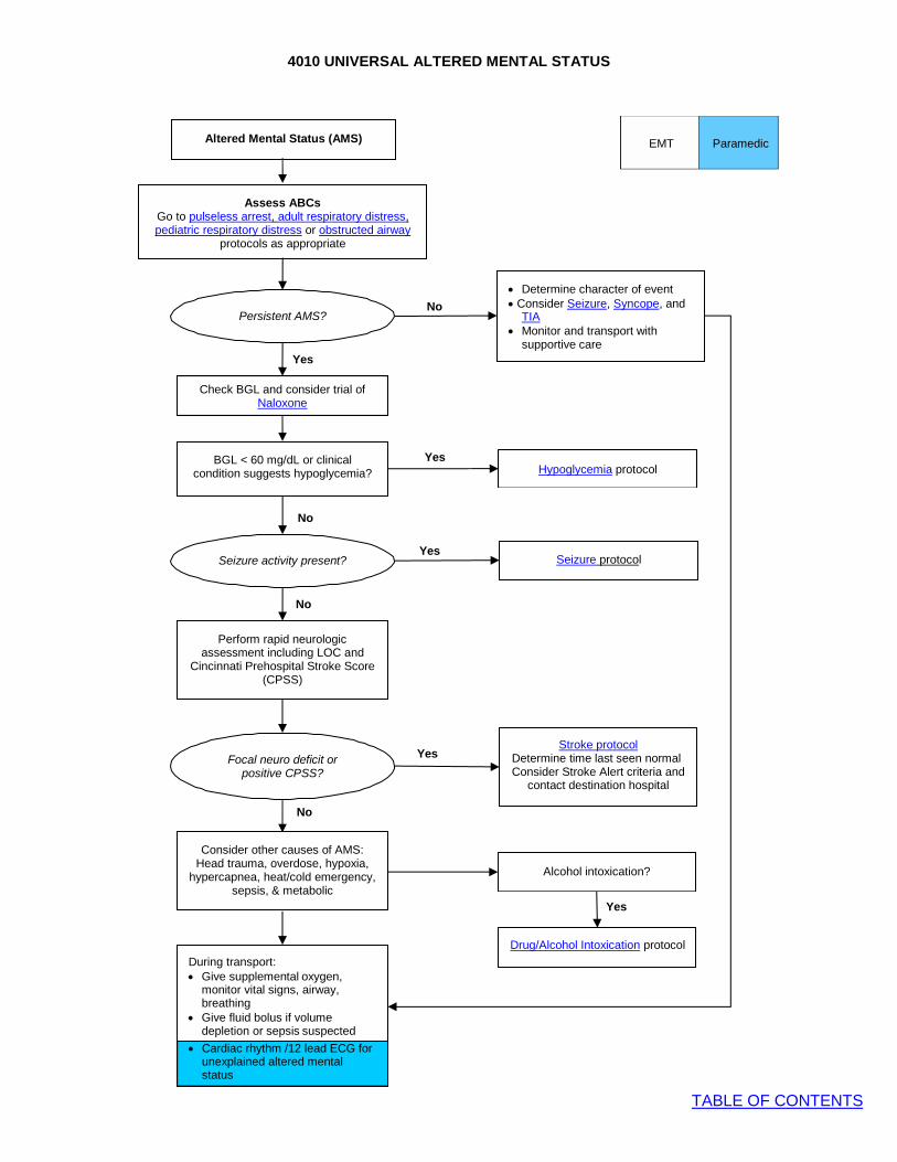
4010 UNIVERSAL ALTERED MENTAL STATUS
TABLE OF CONTENTS
Cardiac rhythm/12 lead ECG
Persistent AMS?
Yes
• Determine character of event
No • Consider Seizure, Syncope, and TIA
• Monitor and transport with supportive care
Check BGL and consider trial of Naloxone
Yes Hypoglycemia protocol
No
Seizure activity present?
Yes
Seizure protocol
No
Focal neuro deficit or
positive CPSS?
Yes
Stroke protocol
Determine time last seen normal Consider Stroke Alert criteria and
contact destination hospital
No
Alcohol intoxication?
Yes
Drug/Alcohol Intoxication protocol
During transport: • Give supplemental oxygen,
monitor vital signs, airway, breathing
• Give fluid bolus if volume depletion or sepsis suspected
• Cardiac rhythm /12 lead ECG for unexplained altered mental status
EMT Paramedic
Altered Mental Status (AMS)
Assess ABCs Go to pulseless arrest, adult respiratory distress, pediatric respiratory distress or obstructed airway
protocols as appropriate
Perform rapid neurologic assessment including LOC and
Cincinnati Prehospital Stroke Score (CPSS)
Consider other causes of AMS: Head trauma, overdose, hypoxia,
hypercapnea, heat/cold emergency, sepsis, & metabolic
BGL < 60 mg/dL or clinical condition suggests hypoglycemia?
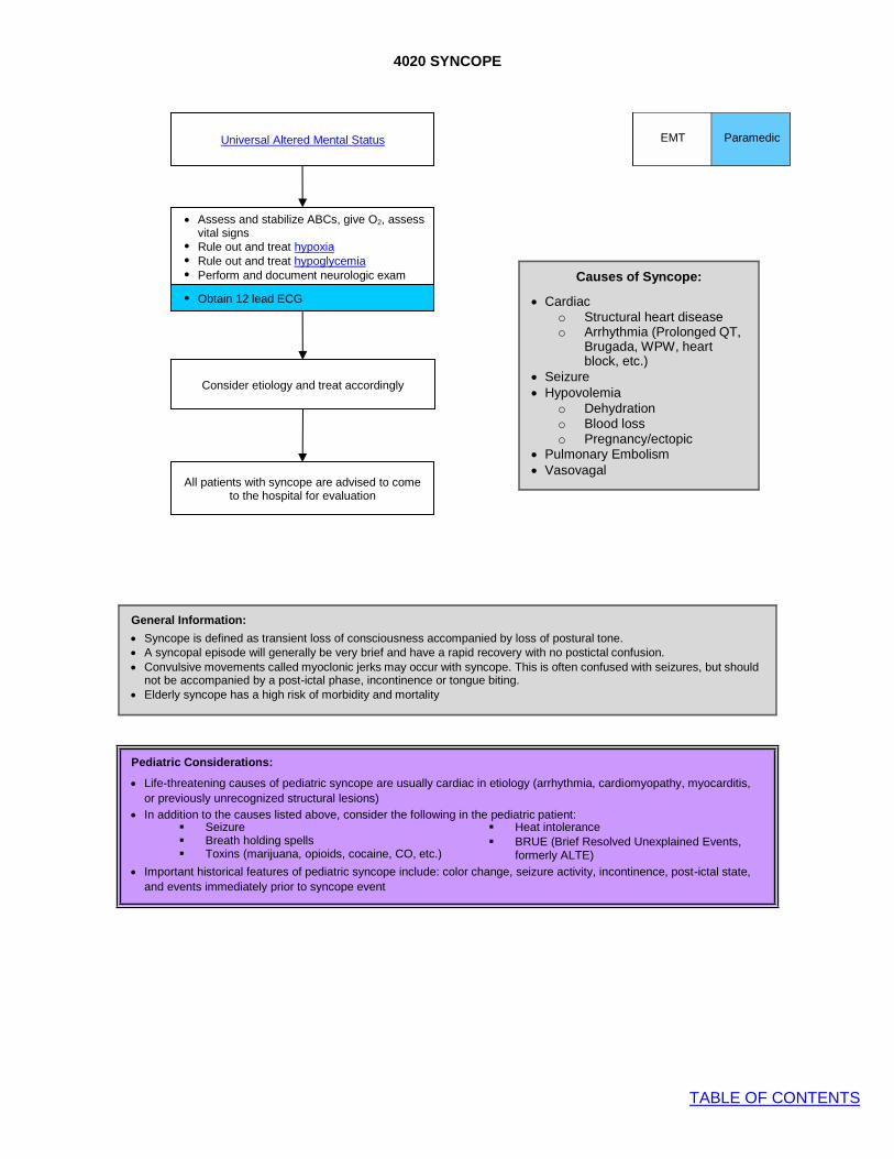
4020 SYNCOPE
TABLE OF CONTENTS
Pediatric Considerations:
• Life-threatening causes of pediatric syncope are usually cardiac in etiology (arrhythmia, cardiomyopathy, myocarditis, or previously unrecognized structural lesions)
• In addition to the causes listed above, consider the following in the pediatric patient:
• Important historical features of pediatric syncope include: color change, seizure activity, incontinence, post-ictal state, and events immediately prior to syncope event
EMT Paramedic
▪ Seizure ▪ Heat intolerance ▪ Breath holding spells ▪ BRUE (Brief Resolved Unexplained Events,
formerly ALTE) ▪ Toxins (marijuana, opioids, cocaine, CO, etc.)
General Information:
• Syncope is defined as transient loss of consciousness accompanied by loss of postural tone. • A syncopal episode will generally be very brief and have a rapid recovery with no postictal confusion. • Convulsive movements called myoclonic jerks may occur with syncope. This is often confused with seizures, but should
not be accompanied by a post-ictal phase, incontinence or tongue biting. • Elderly syncope has a high risk of morbidity and mortality
Universal Altered Mental Status
• Assess and stabilize ABCs, give O2, assess
vital signs • Rule out and treat hypoxia • Rule out and treat hypoglycemia • Perform and document neurologic exam
• Obtain 12 lead ECG
Consider etiology and treat accordingly
All patients with syncope are advised to come to the hospital for evaluation
Causes of Syncope:
• Cardiac o Structural heart disease o Arrhythmia (Prolonged QT,
Brugada, WPW, heart block, etc.)
• Seizure • Hypovolemia
o Dehydration o Blood loss o Pregnancy/ectopic
• Pulmonary Embolism • Vasovagal
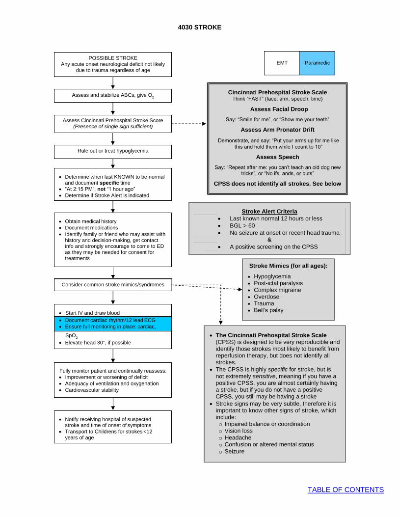
4030 STROKE
TABLE OF CONTENTS
Document cardiac rhythm/12 lead ECG Ensure full monitoring in place: cardiac,
• Obtain medical history • Document medications • Identify family or friend who may assist with
history and decision-making, get contact info and strongly encourage to come to ED as they may be needed for consent for treatments
• Determine when last KNOWN to be normal and document specific time
• “At 2:15 PM”, not “1 hour ago” • Determine if Stroke Alert is indicated
• Notify receiving hospital of suspected stroke and time of onset of symptoms
• Transport to Childrens for strokes <12 years of age
POSSIBLE STROKE Any acute onset neurological deficit not likely
due to trauma regardless of age
Assess and stabilize ABCs, give O2
Assess Cincinnati Prehospital Stroke Score (Presence of single sign sufficient)
Rule out or treat hypoglycemia
EMT Paramedic
Cincinnati Prehospital Stroke Scale Think “FAST” (face, arm, speech, time)
Assess Facial Droop
Say: “Smile for me”, or “Show me your teeth”
Assess Arm Pronator Drift
Demonstrate, and say: “Put your arms up for me like this and hold them while I count to 10”
Assess Speech
Say: “Repeat after me: you can’t teach an old dog new tricks”, or “No ifs, ands, or buts”
CPSS does not identify all strokes. See below
Consider common stroke mimics/syndromes
Fully monitor patient and continually reassess: • Improvement or worsening of deficit • Adequacy of ventilation and oxygenation • Cardiovascular stability
• The Cincinnati Prehospital Stroke Scale (CPSS) is designed to be very reproducible and identify those strokes most likely to benefit from reperfusion therapy, but does not identify all strokes.
• The CPSS is highly specific for stroke, but is not extremely sensitive, meaning if you have a positive CPSS, you are almost certainly having a stroke, but if you do not have a positive CPSS, you still may be having a stroke
• Stroke signs may be very subtle, therefore it is important to know other signs of stroke, which include: o Impaired balance or coordination o Vision loss o Headache o Confusion or altered mental status o Seizure
• Start IV and draw blood • Document cardiac rhythm/12 lead ECG • Ensure full monitoring in place: cardiac,
SpO2
• Elevate head 30°, if possible
Stroke Mimics (for all ages):
• Hypoglycemia • Post-ictal paralysis • Complex migraine • Overdose • Trauma • Bell’s palsy
Stroke Alert Criteria • Last known normal 12 hours or less • BGL > 60 • No seizure at onset or recent head trauma
& • A positive screening on the CPSS

4030 STROKE
TABLE OF CONTENTS
No Yes
No Yes
Does pt. meet Stroke Alert Criteria AND
Pt > 18 years old?
Mobile Stroke Treatment Unit – Activation and Transfer of Care
AFR Dispatched to Incident
Review of CAD notes reveals possible Stroke patient AND
Pt > 18 years old?
Cancel MSTU response Treat per protocol Prepare pt for transport
Treat and transport per protocol 10 minutes after AFD arrival
Cancel MSTU Response Treat and transport per protocol
Pt report made to MSTU team
MSTU Team DOES NOT accept transfer of care
MSTU team ACCEPTS transfer of care
Treat and transport per protocol Assist MSTU team loading pt into MSTU
Pt care transferred to MSTU team
MSTU on scene MSTU NOT on scene
AFD arrival on scene Request 10 minute ticker
Treat per protocol
Treat and transport per protocol
Request dispatch of MSTU via Aurora Comm. Center
Do not request MSTU Response

4040 SEIZURE
TABLE OF CONTENTS
Yes Actively Seizing?
• If seizure brief and self-limited, treatment not necessary
• Rule out hypoglycemia (check blood glucose) • If prolonged (e.g.: > 5 min) or recurrent
seizure, then treat as follows:
No
Yes
No
Yes
Actively seizing after 5 minutes?
• Establish IV access if not already in place
• Repeat benzodiazepine
Universal Seizure Precautions:
• Ensure airway patency, but do not force anything between teeth.
• Give oxygen • Suction as needed • Protect patient from injury • Check pulse immediately after seizure
stops • Keep patient on side
Document:
• Document: Seizure history: onset, time interval, previous seizures, type of seizure
• Obtain medical history: head trauma, diabetes, substance abuse, medications, compliance with anticonvulsants, pregnancy
Pregnancy and Seizure:
• If ≥20 weeks gestational age or up to 6 weeks postpartum administer magnesium sulfate
Actively seizing after 5 minutes?
Give benzodiazepine IN route if no IV
• Transport and monitor ABCs, vital signs, and neurological condition
• Cardiac monitoring if recurrent seizures and/or meds given
• Complete head to toe assessment
• Check pulse and reassess ABC • Give supplemental oxygen
• Support ABCs: • Give oxygen • Rule out or treat hypoglycemia • Universal seizure precautions (see below) • Consider the cause (see below)
EMT Paramedic
No
CONTACT BASE
Consider the Cause of Seizure
• Epilepsy • EtOH withdrawal or intoxication • Hypoglycemia • Stimulant use • Trauma • Intracranial hemorrhage • Overdose (TCA) • Eclampsia • Infection: Meningitis, sepsis • Febrile (age 6 months to 6 years
old)

4050 HYPOGLYCEMIA
TABLE OF CONTENTS
• Glucagon IM • Alternative: If
severe symptoms (coma), consider IO and administer dextrose IO
Symptoms resolved?
Administer dextrose IV & reassess patient
Are you able to establish IV access?
No
Yes
Yes
No
No Yes
Yes
Yes
Monitor and transport or contact base for refusal if
indicated
Considerations for Hyperglycemia:
• In general, treat the patient, not the glucose value. Treat shock if present.
• Consider NS bolus for patients with hyperglycemia and no evidence of fluid overload.
• Pediatric patients with concern for DKA should not exceed 10-20 mL/kg of fluids.
EMT
Paramedic
Check blood glucose level in ANY patient with signs or symptoms consistent with
hypoglycemia
Examples: Altered MS, agitation, focal neurologic deficit,
seizure, weakness, diaphoresis, decreased motor tone, pallor
No
Administer oral glucose. Reassess patient
Can the patient safely tolerate oral glucose?
intact gag reflex, follows verbal
commands
Is BGL < 60? If hypoglycemia still most
likely despite normal reading on glucometer,
administer glucose regardless, while
considering other causes of altered mental status
Regarding refusals after a
hypoglycemic episode:
See Patient Refusal protocol
Transport is always indicated for any of the following patients:
• Pts with unexplained hypoglycemia • Pts taking oral hypoglycemic meds • Pts not taking food by mouth • Pts who do not have competent adult
to monitor
Still symptomatic?
No
Recheck BGL and consider other
causes of altered mental status
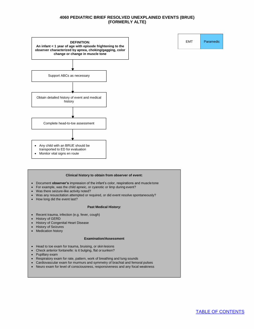
4060 PEDIATRIC BRIEF RESOLVED UNEXPLAINED EVENTS (BRUE)
TABLE OF CONTENTS
EMT
Paramedic
(FORMERLY ALTE)
DEFINITION: An infant < 1 year of age with episode frightening to the
observer characterized by apnea, choking/gagging, color change or change in muscle tone
Support ABCs as necessary
Obtain detailed history of event and medical history
Complete head-to-toe assessment
• Any child with an BRUE should be transported to ED for evaluation
• Monitor vital signs en route
Clinical history to obtain from observer of event:
• Document observer’s impression of the infant’s color, respirations and muscle tone • For example, was the child apneic, or cyanotic or limp during event? • Was there seizure-like activity noted? • Was any resuscitation attempted or required, or did event resolve spontaneously? • How long did the event last?
Past Medical History:
• Recent trauma, infection (e.g. fever, cough) • History of GERD • History of Congenital Heart Disease • History of Seizures • Medication history
Examination/Assessment
• Head to toe exam for trauma, bruising, or skin lesions • Check anterior fontanelle: is it bulging, flat or sunken? • Pupillary exam • Respiratory exam for rate, pattern, work of breathing and lung sounds • Cardiovascular exam for murmurs and symmetry of brachial and femoral pulses • Neuro exam for level of consciousness, responsiveness and any focal weakness
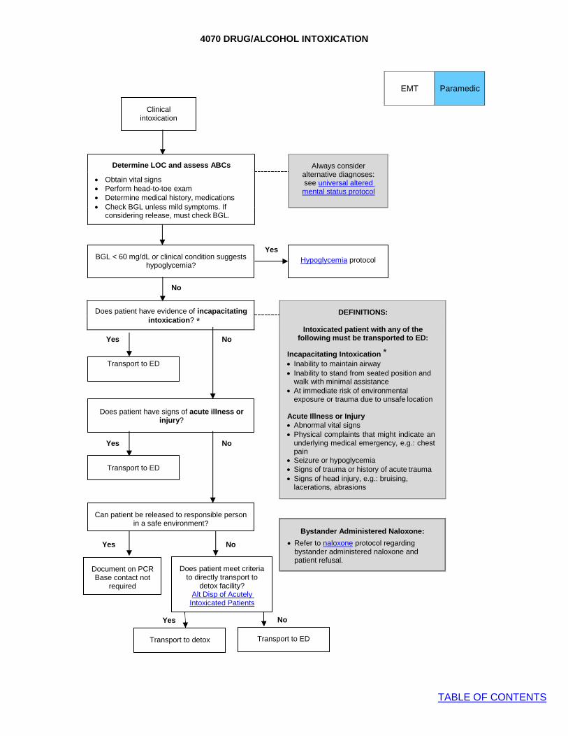
4070 DRUG/ALCOHOL INTOXICATION
TABLE OF CONTENTS
Can patient be released to responsible person in a safe environment?
Determine LOC and assess ABCs
• Obtain vital signs • Perform head-to-toe exam • Determine medical history, medications • Check BGL unless mild symptoms. If
considering release, must check BGL.
Always consider
alternative diagnoses: see universal altered mental status protocol
Yes
No
Does patient have evidence of incapacitating intoxication? *
DEFINITIONS:
Intoxicated patient with any of the
following must be transported to ED:
Incapacitating Intoxication * • Inability to maintain airway • Inability to stand from seated position and
walk with minimal assistance • At immediate risk of environmental
exposure or trauma due to unsafe location
Acute Illness or Injury • Abnormal vital signs • Physical complaints that might indicate an
underlying medical emergency, e.g.: chest pain
• Seizure or hypoglycemia • Signs of trauma or history of acute trauma • Signs of head injury, e.g.: bruising,
lacerations, abrasions
Yes No
Transport to ED
Does patient have signs of acute illness or injury?
Yes No
Transport to ED
Yes No
Yes No
Transport to ED Transport to detox
EMT
Paramedic
Clinical intoxication
Document on PCR Base contact not
required
Does patient meet criteria to directly transport to
detox facility? Alt Disp of Acutely
Intoxicated Patients
Bystander Administered Naloxone:
• Refer to naloxone protocol regarding bystander administered naloxone and patient refusal.
Hypoglycemia protocol BGL < 60 mg/dL or clinical condition suggests
hypoglycemia?

4080 OVERDOSE AND ACUTE POISONING
TABLE OF CONTENTS
PPE and decontaminate when appropriate
ABCs IV, oxygen, monitor
Obtain specific information: • Type of ingestion(s) • What, when and how much ingested? • Bring the poison, container, all medication and other
questionable substances to the ED • Note actions taken by bystanders or patient (e.g.: induced
emesis, “antidotes”, etc) • Supportive Care is key to overdose management
EMT Paramedic
Need for airway management?
Yes Consider Naloxone
No
Hypotension for age?
Yes IV fluid bolus per Medical Shock protocol
No
Altered mental status?
Yes Universal Altered Mental Status protocol
• Check BGL • Consider specific ingestions
No
Specific ingestion?
See Adult or Pediatric Respiratory Distress protocols
Nerve Agent Antidote Kit
Atropine
Pralidoxime
Organophosphate or nerve agent
DUMBELS syndrome
Sodium Bicarbonate for QRS > 100 msec
If intubated, consider hyperventilation to
ETCO2 25-30 mmHg
See Seizure protocol
Tricyclic antidepressant
Wide complex tachycardia,
seizure
Benzodiazepine for severe symptoms
See Agitated/ Combative
Patient protocol
Stimulant
Tachycardia, HTN, agitation,
sweating, psychosis
Fluids per Medical Shock Protocol
Calcium and
Vasopressor Infusion for hypotension
Glucagon
Calcium Channel Blocker
Bradycardia, heart block, hypotension
Fluids per Medical Shock Protocol
Vasopressor Infusion
Glucagon
ß-Blocker
Bradycardia, heart block, hypotension

TABLE OF CONTENTS
Give methylprednisolone
Give diphenhydramine
Consider diphenhydramine if
significant discomfort
EMT Paramedic
4090 ALLERGY AND ANAPHYLAXIS
Generalized or Systemic Reaction Multisystem involvement: skin, mucus
membranes, and gastrointestinal symptoms
Localized Reaction Including isolated tongue, airway
No No
Transport and reassess for
signs of deterioration
Airway involvement? Tongue or uvula swelling, stridor
Yes
Yes
Yes
• For pediatrics, consider IV epinephrine bolus
• Start IV • Give diphenhydramine • Give methylprednisolone
• Monitor ABCs, SpO2, cardiac rhythm
• Reassess for signs of deterioration
• Assess ABCs, give oxygen • If possible, determine likely trigger • Determine PMH, medications, allergies • Classify based on symptom severity and
systems involved • Other specific protocols may apply: e.g.:
obstructed airway, bites & envenomation
Allergic reaction, anaphylaxis or angioedema
Give immediate IM epinephrine & manage airway per Obstructed
Airway Protocol
If persistent signs of severe shock with hypotension not responsive to IM epinephrine and fluid bolus:
• Contact Base • Consider IV epinephrine drip
per vasopressor infusion protocol
Definitions:
• Anaphylaxis: severe allergic reaction that is rapid in onset and potentially life- threatening. Multisystem signs and symptoms are present including skin and mucus membranes.
• Angioedema: deep mucosal edema causing swelling of mucus membranes of upper airway. May accompany hives.
Document:
• History of allergen exposure, prior allergic reaction and severity, medications or treatments administered prior to EMS assessment
• Specific symptoms and signs presented: itching, wheezing, respiratory distress, nausea, weakness, rash, anxiety, swelling of face, lips, tongue, throat, chest tightness, etc.
• Give epinephrine IM, then: • Start IV and give IV bolus
per medical shock protocol
• Give diphenhydramine
• Give methylprednisolone
• Consider addition of albuterol if wheezing
Impending airway obstruction?
Does patient have any of the following signs or symptoms?
• Hypotension • Signs of poor perfusion • Bronchospasm, stridor • Altered mental status

4100 NON-TRAUMATIC ABDOMINAL PAIN/VOMITING
TABLE OF CONTENTS
EMT
Paramedic
Non-traumatic abdominal pain and/or vomiting
• Monitor and transport • Frequent reassessment for
deterioration and response to treatment
Elderly Patients:
• Much more likely to have life- threatening cause of symptoms
• Shock may be occult, with absent tachycardia in setting of severe hypovolemia
Consider antiemetic for vomiting
Consider opioid for severe pain
Cardiac monitor and 12 lead ECG for any of the following:
• Diabetic • Age > 50 • Upper abdominal pain concerning for
ACS • Unstable vital signs in the adult patient
Pediatric Patients:
• Life-threatening causes vary by age. Consider occult or non-accidental trauma, toxic ingestion, button battery ingestion, GI bleed, peritonitis
• For most pediatric patients without signs of shock, no IV is required and pharmacologic pain management should be limited
• Consider IV • If GI bleed, start 2nd IV • Transport in position of comfort
History:
• Onset, location, duration, radiation of pain
• Associated sx: vomiting, bilious emesis, GU sx, hematemesis, coffee ground emesis, melena, rectal bleeding, vaginal bleeding, known or suspected pregnancy, recent trauma
If signs of poor perfusion AND/OR
hypotension for age, see Medical Shock protocol and begin fluid resuscitation
Life-threatening causes:
• Cardiac etiology: MI, ischemia • Vascular etiology: AAA, dissection • GI bleed • Gynecologic etiology: ectopic
pregnancy
• Assess ABCs • Give oxygen • Complete set of vital signs • Consider life-threatening causes

4110 SUSPECTED CARBON MONOXIDE EXPOSURE
TABLE OF CONTENTS
Yes
No
COHb Severity Signs and Symptoms
<15-20%
Mild
Headache, nausea, vomiting, dizziness, blurred vision
21-40% Moderate Confusion, syncope, chest pain, dyspnea, tachycardia, tachypnea, weakness
41-59% Severe Dysrhythmias, hypotension, cardiac ischemia, palpitations, respiratory arrest, pulmonary edema, seizures, coma, cardiac arrest
>60% Fatal Death
EMT
Paramedic
ABCs
SpCO 15% SpCO 5-15% SpCO 0-5%
Measure COHb% (SpCO)
100% FIO2
and transport Contact Base
for consult No further
evaluation of SpCO is needed
General Guidelines:
• Signs and Symptoms of CO
exposure include: Headache, dizziness, coma, altered mentation, seizures, visual changes, chest pain, tachycardia, arrhythmias, dyspnea, N/V, “flu-like illness”
• The absence or low readings of COHb is not a reliable predictor of toxicity of other fire byproducts
• In smoke inhalation victims, consider cyanide treatment with Hydroxocobalamin as per indications
• The fetus of a pregnant woman is at higher risk due to the greater affinity of fetal hemoglobin to CO. With CO exposure, the pregnant woman may be asymptomatic while the fetus may be in distress. In general, pregnant patients exposed to CO should be transported.
100% FIO2
and transport Symptoms of CO
or hypoxia

4120 ADRENAL INSUFFICIENCY PROTOCOL
TABLE OF CONTENTS
EMT
Paramedic
Patient at risk for adrenal insufficiency (Addisonian crisis):
• Identified by family or medical alert bracelet • Chronic steroid use • Congenital Adrenal Hyperplasia • Addison’s disease
• Monitor 12 lead ECG for signs of hyperkalemia
• Continue to monitor for development of hypoglycemia
• Contact base for consult if patient not responding to treatment
Give corticosteroid
All symptomatic patients: • Check blood glucose and treat
hypoglycemia, if present • Start IV and give oxygen • If signs of poor perfusion AND/OR
hypotension for age, see Medical Shock protocol and begin fluid resuscitation
• Chronic corticosteroid use is a common cause for adrenal crisis, carefully assess for steroid use in patients with unexplained shock.
• Administration of steroids are life-saving and necessary for reversing shock or preventing cardiovascular collapse
• Patients at risk for adrenal insufficiency may show signs of shock when under physiologic stress which would not lead to cardiovascular collapse in normal patients. Such triggers may include trauma, dehydration, infection, myocardial ischemia, etc.
• If no corticosteroid is available during transport, notify receiving hospital of need for immediate corticosteroid upon arrival
• Under Chapter 2 Rule: specialized prescription medications to address an acute crisis may be given by all levels with a direct VO, given the route of administration is within the scope of the provider. This applies to giving hydrocortisone for adrenal crisis, for instance, if a patient or family member has this medication available on scene. Contact base for direct verbal order
Assess for signs of acute adrenal crisis: • Pallor, weakness, lethargy • Vomiting, abdominal pain • Hypotension, shock • Congestive heart failure

4130 EPISTAXIS MANAGEMENT
TABLE OF CONTENTS
EMT
Paramedic
Active nosebleed
ABCs
IV access and IV fluid bolus if signs of
hypoperfusion, shock
• Spray both nares with phenylephrine
• Compress nostrils with clamp or fingers, pinching over fleshy part of nose, not bony nasal bridge
• Transport in position of comfort, usually sitting upright
• Tilt head forward
• Have patient blow nose to expel clots
General Guidelines:
• Most nose bleeding is from an anterior source and may be easily controlled.
• Avoid phenylephrine in pts with known CAD.
• Anticoagulation with aspirin, clopidogrel (Plavix), warfarin (Coumadin) will make epistaxis much harder to control. Note if your patient is taking these, or other, anticoagulant medications.
• Posterior epistaxis is a true emergency and may require advanced ED techniques such as balloon tamponade or interventional radiology. Do not delay transport. Be prepared for potential airway issues.
• For patients on home oxygen via nasal cannula, place the cannula in the patient’s mouth while nares are clamped or compressed for nosebleed.

4140 SEPSIS PROTOCOL
TABLE OF CONTENTS
Evaluate and identify potential sepsis – is there suspected or confirmed infection?
• ABCs • Complete set of vital signs • Cardiac monitoring including SpO2 and
waveform capnography • O2 as appropriate
Are there two or more SIRS criteria? No
Yes
Is there evidence of hypoperfusion? (ANY ONE OF THE FOLLOWING):
• Routine Care • IV, O2, monitor
• Hypotension for age
• Altered mental status (excluding simple febrile seizure)
• Delayed capillary refill AND mottling
• Systolic BP < 90 mmHg No
• MAP <65 mmHg • Sustained EtCO2
<25 mmHg
• Consider fluid bolus if sepsis suspected • Transport to closest appropriate hospital • Continue to re-assess vital signs and
perfusion
Yes
• IV fluid bolus @ 30mL/kg (if no sign of
fluid overload) • 2 large bore IV’s • Transport to closest appropriate hospital
NOTIFY HOSPITAL of Prehospital Sepsis Alert
For ongoing hypotension, poor perfusion or pulmonary edema, consider Vasopressor
Infusion (adult patients only)
Evaluate potential SIRS Criteria: • Temp < 36C (96.8F) or > 38C (100.4F) • HR > 90 (or tachycardic for age) • RR > 20 or mechanical ventilation (or
tachypneic for age)
Principles of Sepsis
• Multiple studies demonstrate the benefit of early recognition and treatment of sepsis, including in the prehospital setting.
• Early hospital notification of sepsis may lead to shorter time to IV fluid and IV antibiotics and increase survival.
• Patients with septic shock require aggressive IV fluid resuscitation. Starting dose should be 30mL/kg of IV fluid.
• EtCO2 has been demonstrated to correlate with serum lactate levels and predictive of severity of sepsis. A sustained EtCO2 <25 mmHg may indicate hypoperfusion.
Pediatric Fluid Administration
• For children <40 kg or not longer than length-based tape, hand pull/push fluid with a 60 mL syringe utilizing a 3 way stop cock.
• The treatment of compensated shock requires aggressive fluid replacement, may need to repeat fluid bolus up to 60mL/kg.
• Goal of therapy is normalization of vital signs within the first hour.
• Hypotension is a late sign in pediatric shock patients.
EMT Paramedic
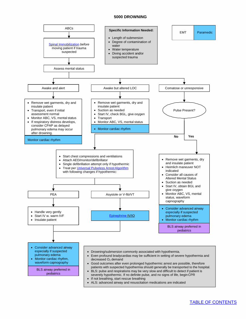
5000 DROWNING
TABLE OF CONTENTS
ABCs
EMT Paramedic
Spinal Immobilization before moving patient if trauma
suspected
Assess mental status
Awake and alert Awake but altered LOC Comatose or unresponsive
• Remove wet garments, dry and insulate patient
• Transport, even if initial assessment normal
• Monitor ABC, VS, mental status • If respiratory distress develops,
consider CPAP as delayed pulmonary edema may occur after drowning.
• Remove wet garments, dry and insulate patient
• Suction as needed • Start IV, check BGL, give oxygen • Transport • Monitor ABC, VS, mental status
• Monitor cardiac rhythm
Pulse Present?
Monitor cardiac rhythm No Yes
• Start chest compressions and ventilations • Attach AED/monitor/defibrillator • Single defibrillation attempt only if hypothermic • Treat per Universal Pulseless Arrest Algorithm
with following changes if hypothermic:
PEA Asystole or V-fib/VT
• Handle very gently • Start IV w. warm IVF • Insulate patient
Epinephrine IV/IO
• Consider advanced airway
especially if suspected pulmonary edema
• Monitor cardiac rhythm
BLS airway preferred in pediatrics
BLS airway preferred in
pediatrics
• Consider advanced airway especially if suspected pulmonary edema
• Monitor cardiac rhythm, waveform capnography
Specific Information Needed:
• Length of submersion • Degree of contamination of
water • Water temperature • Diving accident and/or
suspected trauma
• Drowning/submersion commonly associated with hypothermia. • Even profound bradycardias may be sufficient in setting of severe hypothermia and
decreased O2 demand • Good outcomes after even prolonged hypothermic arrest are possible, therefore
patients with suspected hypothermia should generally be transported to the hospital. • BLS: pulse and respirations may be very slow and difficult to detect if patient is
severely hypothermic. If no definite pulse, and no signs of life, begin CPR • If not breathing, start rescue breathing • ALS: advanced airway and resuscitation medications are indicated
• Remove wet garments, dry and insulate patient
• Heimlich maneuver NOT indicated
• Consider all causes of Altered Mental Status
• Suction as needed • Start IV, obtain BGL and
give oxygen • Monitor ABC, VS, mental
status, waveform capnography
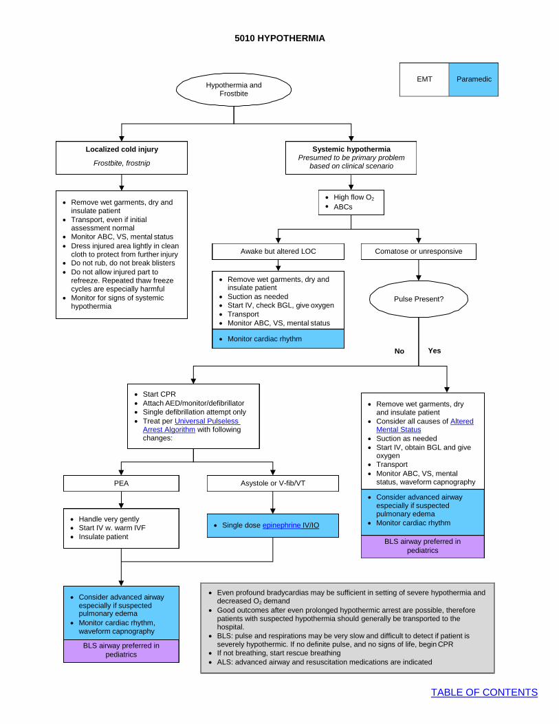
5010 HYPOTHERMIA
TABLE OF CONTENTS
Localized cold injury
Frostbite, frostnip
Hypothermia and Frostbite
EMT Paramedic
Systemic hypothermia
Presumed to be primary problem based on clinical scenario
• High flow O2
• ABCs
Awake but altered LOC Comatose or unresponsive
• Remove wet garments, dry and
insulate patient • Suction as needed • Start IV, check BGL, give oxygen • Transport • Monitor ABC, VS, mental status
• Monitor cardiac rhythm
Pulse Present?
No Yes
PEA
• Start CPR • Attach AED/monitor/defibrillator • Single defibrillation attempt only • Treat per Universal Pulseless
Arrest Algorithm with following changes:
Asystole or V-fib/VT
• Remove wet garments, dry and insulate patient
• Consider all causes of Altered Mental Status
• Suction as needed • Start IV, obtain BGL and give
oxygen • Transport • Monitor ABC, VS, mental
status, waveform capnography
• Consider advanced airway especially if suspected
• Handle very gently • Start IV w. warm IVF • Insulate patient
• Single dose epinephrine IV/IO
pulmonary edema • Monitor cardiac rhythm
BLS airway preferred in
pediatrics
• Consider advanced airway especially if suspected
• Monitor cardiac rhythm,
waveform capnography
BLS airway preferred in pediatrics
pulmonary edema
• Even profound bradycardias may be sufficient in setting of severe hypothermia and decreased O2 demand
• Good outcomes after even prolonged hypothermic arrest are possible, therefore patients with suspected hypothermia should generally be transported to the hospital.
• BLS: pulse and respirations may be very slow and difficult to detect if patient is severely hypothermic. If no definite pulse, and no signs of life, begin CPR
• If not breathing, start rescue breathing • ALS: advanced airway and resuscitation medications are indicated
• Remove wet garments, dry and insulate patient
• Transport, even if initial assessment normal
• Monitor ABC, VS, mental status • Dress injured area lightly in clean
cloth to protect from further injury • Do not rub, do not break blisters • Do not allow injured part to
refreeze. Repeated thaw freeze cycles are especially harmful
• Monitor for signs of systemic hypothermia

5020 HYPERTHERMIA
TABLE OF CONTENTS
Administer IV/IO fluids 20 mL/kg up to 1 L of cool saline; reassess and
repeat if needed
Monitor VS and transport
Heat Stroke
• Very high core body temperature
• Hot, dry skin w. cessation of sweating
• Hypotension
• Altered mental status
• Seizure
• Coma
Rapid transport indicated
Heat Exhaustion
• Elevated body temperature
• Cool, diaphoretic skin
• Generalized weakness
• Anxiety
• Headache
• Tachypnea
• Possible syncope
Heat Cramps
• Normal or slightly elevated body temperature
• Warm, moist skin
• Generalized weakness
• Diffuse muscle cramping
EMT Paramedic
Hyperthermia • Classify by clinical syndrome
• Consider non-environmental causes (see below)
Adequate airway and breathing?
No Yes
Assist ventilations and manage airway
as needed
Administer O2
Administer IV/IO fluids 20 mL/kg up to 1 L of cool saline; reassess and repeat
if needed
• Remove excess clothing • For heat stroke, consider external
cooling measures if prolonged transport
• Treat seizures, cardiac arrhythmias per protocol
• Monitor and transport
Consider other causes of hyperthermia besides environment exposure, including:
• Neurolopeptic malignant syndrome (NMS): patients taking antipsychotic medications
• Sympathomimetic overdose: cocaine,
methamphetamine
• Anticholingergic toxidrome: overdose (“Mad
as a hatter, hot as a hare, blind as a bat, red
as a beet”) common w. ODs on psych meds,
OTC cold medications, Benadryl, Jimson
weed, etc.
• Infection: fever (sepsis)
• Thyrotoxicosis: goiter (enlarged thyroid)

5030 HIGH ALTITUDE ILLNESS
TABLE OF CONTENTS
Symptoms of illness at altitude
• ABCs • IV, oxygen • Cardiac monitor
• Head to toe assessment • Complete history: • Rate of ascent, prior altitude
illness, rapidity of sx onset • Consider non-altitude-
related illness
AMS HAPE HACE
Consider antiemetic for vomiting • O NRB facemask
If signs of poor perfusion AND/OR hypotension for age, see Medical
Shock protocol and begin fluid resuscitation
2
• Consider CPAP • Assist ventilations as needed • Airway management as indicated
• Do NOT give diuretic
• Never assume that symptoms at altitude are necessary due to altitude illness.
• Acute exacerbations of chronic medical illness at altitude are more common that altitude illness.
Acute mountain sickness (AMS): headache, insomnia,
anorexia, nausea, fatigue
High-altitude pulmonary edema (HAPE): dyspnea, cough, headache, nausea, fever
High-altitude cerebral edema (HACE): ataxia, confusion, neuro
deficits, seizure, coma, and headache
EMT Paramedic
• Descent from altitude
• O2 NRB facemask • Assist ventilations as needed • Airway management as indicated • Elevate head of bed
HACE is rare at elevations in Colorado; always consider alternative cause of altered
mental status
Special Notes: • There are no specific factors that accurately predict susceptibility to altitude sickness, but symptoms are worsened by exertion,
dehydration, and alcohol ingestion.
• Acute Mountain Sickness (AMS) can begin to appear at around 6,500 ft above sea level, although most people will tolerate up to
8000 ft without difficulty. Altitude illness should not be suspected below 6,500 ft. AMS is the most frequent type of altitude
sickness encountered. Symptoms often manifest themselves six to ten hours after ascent and generally subside in one to two
days, but they occasionally develop into the more serious conditions.
• High altitude pulmonary edema (HAPE) and cerebral edema (HACE) are the most severe forms of high altitude illness. The rate
of ascent, altitude attained, exertion, and individual susceptibility are contributing factors to the onset and severity of high-altitude
illness
• Mild HAPE may be managed with high-flow oxygen and supportive care, and does not necessarily require descent from altitude.
• More severe forms of HAPE and all forms of HACE require descent

5040 INSECT/ARACHNID STINGS AND BITES PROTOCOL
TABLE OF CONTENTS
EMT Paramedic
Specific Information Needed:
• Timing of bite/sting • Identification of spider, bee, wasp,
other insect, if possible • History of prior allergic reactions to
similar exposures • Treatment prior to EMS eval: e.g.
EpiPen, diphenhydramine, etc
Specific Precautions:
• For all types of bites and stings, the goal of prehospital care is to prevent further envenomation and to treat allergic reactions
• Anaphylactoid reactions may occur upon first exposure to allergen, and do not require prior sensitization • Anaphylactic reactions typically occur abruptly, and rarely > 60 minutes after exposure
Initiate general care for bites and stings
Assess for localized vs. systemic signs and symptoms
and depending on animal involved
Consider opioid for
severe pain (e.g.: black widow spider) and /or diphenhydramine if needed for itching
diphenhydramine if needed for itching
Localized Symptoms:
• Pain, warmth and swelling
• Administer oxygen • Start IV
Treat per allergy & anaphylaxis protocol
Systemic Symptoms:
• Hives, generalized erythema, swelling, angioedema
• Hypotension
• Altered mental status
• Other signs of shock
General Care
• For bees/wasps: o Remove stinger mechanism by
scraping with a straight edge. Do not squeeze venom sac
• For spiders: o Bring in spider if captured or dead
for identification

5050 SNAKE BITE PROTOCOL
TABLE OF CONTENTS
EMT
Paramedic
• Assess ABCs, mental status • Administer oxygen • Start IV • Monitor VS
Specific Precautions:
• The prairie rattlesnake is native to Denver Metro region and is most common venomous snake bite in the region. • Exotic venomous snakes, such as pets or zoo animals, may have different signs and symptoms than those of pit
vipers. In case of exotic snake bite, contact base and consult zoo staff or poison center for direction. • Take a picture of the snake, including images of head and tail. If an adequate photo can be taken, it is not
necessary to bring snake to ED. • Never pick up a presumed-to-be-dead snake by hand. Rather, use a shovel or stick. A dead snake may reflexively bite
and envenomate. • > 25% of snake bites are “dry bites”, without envenomations. • Conversely, initial appearance of bite may be deceiving as to severity of envenomation. • Fang marks are characteristic of pit viper bites (e.g. rattlesnakes). • Jaw prints, without fang marks, are more characteristic of non-venomous species.
Systemic Symptoms:
• Metallic or peculiar taste in mouth
• Hypotension
• Altered mental status
• Widespread bleeding
• Other signs of shock
Localized Symptoms:
• Pain and swelling
• Numbness, tingling to bitten part
• Bruising/eccymoses
Assess for localized vs. systemic signs and symptoms
If there is hypotension for age and/or definite signs of shock, treat per Shock
Protocol
• Transport with bitten part immobilized
• Monitor ABCs and for development of systemic signs/sx
• Complete General Care en route
Opioid for severe pain and if not contraindicated
by hypotension
Be prepared to manage airway if signs of airway
obstruction develop
Opioid for severe pain
Obtain specific information:
• Appearance of snake (rattle, color, thermal pit, elliptical pupils)
• Appearance of wound: location, # of fangs vs. entire jaw imprint
• Timing of bite • Prior 1st aid • To help with identification of snake,
photograph snake, if possible. Include image of head, tail, and any distinctive markings.
• Do not bring snake to ED
Initiate general care for snake bites
General Care:
• Remove patient from proximity to snake • Remove all constricting items from bitten
limb (e.g.: rings, jewelry, watch, etc.) • Immobilize bitten part • Initiate prompt transport • Do NOT use ice, refrigerants, tourniquets,
scalpels or suction devices • Mark margins of erythema and/or edema
with pen or marker and include time measured

6000 PSYCHIATRIC/BEHAVIORAL PATIENT PROTOCOL
TABLE OF CONTENTS
Scene Safety
A. Scene safety and provider safety are a priority. Consider police contact if scene safety is a concern.
B. Refer to restraint protocol as needed, especially as it relates to A.
Specific Information Needed
A. Obtain history of current event; inquire about recent crisis, toxic exposure, drugs, alcohol, emotional trauma, and suicidal or homicidal ideation.
B. Obtain past history; inquire about previous psychiatric and medical problems, medications.
Specific Objective Findings
A. Evaluate general appearance 1. E.g.: Well groomed, disheveled, debilitated, bizarrely dressed
B. Evaluate vital signs. 1. Is a particular toxidrome suggested, e.g.: symphathomimetic?
C. Note medic alert tags, breath odors suggesting intoxication. D. Determine ability to relate to reality.
1. Does the patient know who s/he is, where s/he is, who you are and why you are there? 2. Does the patient appear to be hallucinating or responding to internal stimuli?
E. Note behavior. Consider known predictors of violence: 1. Is the patient male, intoxicated, paranoid or displaying aggressive or threatening behavior or
language? Treatment
A. If patient agitated or combative, see Agitated/Combative Patient Protocol B. Attempt to establish rapport C. Assess ABCs D. Transport to closest appropriate Emergency Department, or the Walk-In Clinic if appropriate per the
Alternate Disposition Of Behavioral Health Patients protocol. E. Be alert for possible elopement F. Consider organic causes of abnormal behavior (trauma, overdose, intoxication, hypoglycemia) G. If patient restraint considered necessary for patient or EMS safety, refer to Restraint Protocol. H. Check blood sugar I. If altered mental status or unstable vital signs:
1. Administer oxygen. 2. Establish venous access. 3. Refer to Universal Altered Mental Status Protocol.
Transporting Patients Who Have a Psychiatric Complaint
A. If a patient has an isolated mental health complaint (e.g. suicidality), and does not have a medical complaint or need specific medical intervention, then that patient may be appropriately transported by law enforcement according to their protocols.
B. If a patient has a psychiatric complaint with associated illness or injury (e.g. overdose, altered mental status, chest pain, etc), then the patient should be transported by EMS
C. Reasonable concern for suicidal or homicidal ideation, or grave disability from psychiatric decompensation, is sufficient to assume that the patient may lack medical decision-making capacity to refuse ambulance transport. Effort should be made to obtain consent for transport from the patient, and to preserve the patient’s dignity throughout the process. However, the patient may be transported over his or her objections and treated under implied consent if patient does not comply.
D. A patient being transported for psychiatric evaluation may be transported to any appropriate receiving emergency department.
E. Accusations of kidnapping or assault of the patient are only theoretical and rarely occur. The Medical Directors feel strongly that the risk of abandonment of a potentially suicidal or otherwise gravely impaired patient is far greater. Be sure to document your reason for taking the patient over their objections, that you believe that you are acting in the patient's best interests, and be sure to consult a BASE PHYSICIAN if there are concerns.
EMT
Paramedic

6000 PSYCHIATRIC/BEHAVIORAL PATIENT PROTOCOL
TABLE OF CONTENTS
Specific Precautions
A. Patients presenting with psychiatric decompensation often have an organic etiology. Be suspicious for hypoglycemia, hypoxia, head injury, intoxication, or toxic ingestion.
B. Providers transporting a patient over his or her objections should reassure the patient. The provider should strongly consider whether the patient may need restraint and/or sedation for safety. Beware of weapons. These patients can become combative.
Transporting Patients on a Mental Health Hold
A. By law, patients detained on a mental health hold may not refuse transport. Similarly, by law, patients on a mental health hold are required to be evaluated by a physician or psychologist and must be transported.
B. Although it is commonly believed that the original copy of the mental health hold (form M-1) is required to accompany the patient, a legible copy of the M-1 is also sufficient if the original cannot be found.
C. The M-1 form documenting the mental health hold should be as complete as possible, including the correct date and time that the patient was detained. The narrative portion should be completed. A signature and license or badge number is also required. Assure that the form is complete before departing.
D. The mental health hold does not need to be started on patients who are intoxicated on drugs and/or alcohol. Nor is it required for patients who are physically incapable of eloping from care, such as those who are intubated, or physically unable.
E. The patient rights form (M-2) does not need to accompany the patient. The receiving facility may complete this form if there are concerns.
F. If possible, seek direction from the sending facility regarding whether the patient may require sedation and restraint. Consider ALS transport if this is the case.
G. Recall that patients who are a danger to self/others or gravely disabled due to mental illness may be transported by EMS without a mental health hold, under implied consent. Crews can consult a BASE PHYSICIAN if any concerns.

6010 AGITATED/COMBATIVE PATIENT PROTOCOL
TABLE OF CONTENTS
Yes
No
Restraint Protocol
Obtain IV access as soon as may be safely
accomplished
Restraints
No transport in hobble or prone position. Do not inhibit patient breathing, ventilations
• Give ketamine • Goal is rapid tranquilization in
order to minimize time struggling
Sedate • Consider cause of
agitation • benzodiazepine
Consider Cause of Agitation:
Both benzodiazepines and butyrophenones (e.g. haloperidol) are acceptable options for agitated
patients. In certain clinical scenarios individual medications
may be preferred
• EtOH (butyrophenone) • Sympathomimetic (benzo) • Psych (butyrophenone) • Head injury (butyrophenone)
EMT
Paramedic
Patient is agitated and a danger to self or others
• Attempt to reasonably address patient concerns
• Assemble personnel
Patient does not respond to verbal de-escalation techniques
• Repeat sedation dose • If still significantly
agitated 5 minutes after 2nd dose sedative, Contact Base
Continue cardiac, SpO2, waveform capnography monitoring and rapid
transport
Start external cooling measures
• Reassess ABCs post sedation • Cardiac, SpO2, and
capnography must be initiated • High flow O2
• Start 2 large bore IVs as soon as may be safely accomplished
• Administer 2 liters NS bolus
• Complete Restraint Protocol
Still significantly agitated?
Does patient have signs of the Excited Delirium Syndrome?
Excited Delirium Syndrome
These patients are truly out of control and have a life-threatening medical emergency they will have
some or all of the following sx:
Paranoia, disorientation, hyper- aggression, hallucination,
tachycardia, increased strength, hyperthermia
Assume the patient has a medical cause of agitation and treat
reversible causes
General Guideline:
Emphasis should be placed on scene safety, appropriate use of restraints and
aggressive treatment of the patient’s agitation.
Still significantly agitated?

6020 TRANSPORT OF THE HANDCUFFED PATIENT
TABLE OF CONTENTS
Purpose:
1. Guideline for transport of patients in handcuffs placed by law enforcement
Guideline:
1. Handcuffs are only to be placed by law enforcement. EMS personnel are not permitted to use handcuffs.
2. Request that law enforcement remain with the patient in the ambulance, if possible. If not possible, request that police ride behind ambulance so as to be readily available to remove handcuffs if needed in an emergency situation to facilitate medical care of the patient.
3. EMS personnel are not responsible for the law enforcement hold on these patients. 4. Handcuffed patients will not be placed in the prone position. 5. Handcuffs may be used with spinal immobilization. Medical priorities should take priority
in the positioning of the handcuffs.

7000 CHILDBIRTH PROTOCOL
TABLE OF CONTENTS
• Allow patient to remain
in position of comfort • Visualize perineum • Determine if there is
time to transport
Delivery not imminent
• Transport in position of comfort, preferably on left side to patient’s requested hospital if time and conditions allow
• Monitor for progression to imminent delivery
Imminent Delivery
Delivery is imminent if there is crowning or bulging of perineum
Emergency Childbirth Procedure • If there is a prolapsed umbilical cord or apparent breech presentation, go to
obstetrical complications protocol and initiate immediate transport • For otherwise uncomplicated delivery: • Position mother supine on flat surface, if possible • Do not attempt to impair or delay delivery • Support and control delivery of head as it emerges • Protect perineum with gentle hand pressure • Check for cord around neck, gently remove from around neck, if present • Suction mouth, then nose of infant as soon as head is delivered • If delivery not progressing, baby is “stuck”, see obstetrical complications
protocol and begin immediate transport • As shoulders emerge, gently guide head and neck downward to deliver anterior
shoulder. Support and gently lift head and neck to deliver posterior shoulder • Rest of infant should deliver with passive participation – get a firm hold on baby • Keep newborn at level of mother’s vagina until cord stops pulsating and is
double clamped
Critical Thinking:
• Normal pregnancy is accompanied by higher heart rates and lower blood pressures
• Shock will be manifested by signs of poor perfusion
• Labor can take 8-12 hours, but as little as 5 minutes if high PARA
• The higher the PARA, the shorter the labor is likely to be
• High risk factors include: no prenatal care, drug use, teenage pregnancy, DM, htn, cardiac disease, prior breech or C section, preeclampsia, twins
• Note color of amniotic fluid for meconium staining
Postpartum Care Mother
• Placenta should deliver in 20-30 minutes. If delivered, collect in plastic bag and bring to hospital. Do not pull cord to facilitate placenta delivery and do not delay transport awaiting placenta delivery
• If the perineum is torn and bleeding, apply direct pressure with sanitary pads
• Postpartum hemorrhage – see obstetrical complications protocol
• Initiate transport once delivery of child is complete and mother can tolerate movement
Postpartum Care Infant
• Suction mouth and nose only if signs of obstruction by secretions
• Respirations should begin within 15 seconds after stimulating reflexes. If not, begin artificial ventilations at 40- 60 breaths/min
• If apneic, cyanotic or HR < 100, begin neonatal resuscitation
• Dry baby and wrap in warm blanket • After umbilical cord stops pulsating, double clamp 6” from
infant abdominal wall and cut between clamps with sterile scalpel. If no sterile cutting instrument available, lay infant on mother’s abdomen and do not cut clamped cord
• Document 1 and 5 minute APGAR scores
ABCs O2 15 liters via NRB
IV access
Overview:
• EMS providers called to a possible prehospital childbirth should determine if there is enough time to transport expectant mother to hospital or if delivery is imminent
• If imminent, stay on scene and immediately prepare to assist with the delivery
EMT
Paramedic
Obtain obstetrical history (see adjacent)
Specific Information Needed:
• Obstetrical history: o Number of pregnancies (gravida) o Live births (PARA) o Expected delivery date o Length of previous labors o Narcotic use in past 4 hours
If suspected imminent
childbirth:

7010 OBSTETRICAL COMPLICATIONS
TABLE OF CONTENTS
EMT
Paramedic
For All Patients with obstetrical complications
• Do not delay: immediate rapid transport • Give high-flow oxygen • Start IV en route if time and conditions allow. Treat signs of shock w. IV fluid boluses per Medical Hypotension/Shock
Protocol
Possible actions for specific complications (below)
• The following actions may not be feasible in every case, nor may every obstetrical complication by anticipated or effectively managed in the field. These should be considered “best advice” for rare, difficult scenarios. In every case, initiate immediate transport to definite care at hospital
Postpartum Hemorrhage
• Massage abdomen (uterine fundus) until firm • Initiate rapid transport • Note type and amount of bleeding • Treat signs of shock with IV fluid boluses
Shoulder Dystocia
• Support baby’s head • Suction oral and nasal passages • DO NOT pull on head • May facilitate delivery by placing mother with
buttocks just off the end of bed, flex her thighs upward and gentle open hand pressure above the pubic bone
• IF infant delivered see childbirth protocol – Postpartum care of infant and mother
Breech Delivery
• Never attempt to pull infant from vagina by legs • IF legs are delivered gently elevate trunk and legs to aid
delivery of head • Head should deliver in 30 seconds. If not, reach 2 fingers
into vagina to locate infant’s mouth. Press vaginal wall away from baby’s mouth to access an airway
• Apply gentle abdominal pressure to uterine fundus • IF infant delivered see childbirth protocol – Postpartum care
of infant and mother
Complications of Late Pregnancy
3rd Trimester Bleeding (6-8 months)
• High flow O2 via NRB, IV access • Suspect placental abruption or placenta previa • Initiate rapid transport • Position patient on left side • Note type and amount of bleeding • IV NS bolus for significant bleeding or shock
Pre-Eclampsia/Eclampsia
• High flow O2 via NRB, IV access • SBP > 140, DBP > 90, peripheral edema,
headache, seizure • Transport position of comfort • Treat seizures with Magnesium Sulfate • See seizure protocol
Prolapsed Umbilical Cord
• Discourage pushing by mother • Position mother in Trendelenberg or supine with hips
elevated • Place gloved hand in mother’s vagina and elevate the
presenting fetal part off of cord until relieved by physician • Feel for cord pulsations • Keep exposed cord moist and warm
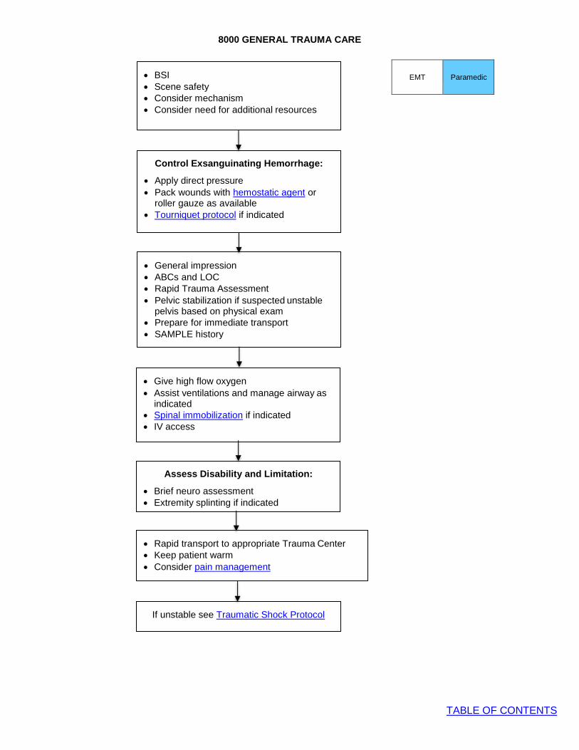
8000 GENERAL TRAUMA CARE
TABLE OF CONTENTS
Assess Disability and Limitation:
• Brief neuro assessment • Extremity splinting if indicated
• Give high flow oxygen • Assist ventilations and manage airway as
indicated • Spinal immobilization if indicated • IV access
EMT
Paramedic
Control Exsanguinating Hemorrhage:
• Apply direct pressure • Pack wounds with hemostatic agent or
roller gauze as available • Tourniquet protocol if indicated
• General impression • ABCs and LOC • Rapid Trauma Assessment • Pelvic stabilization if suspected unstable
pelvis based on physical exam • Prepare for immediate transport • SAMPLE history
• BSI • Scene safety • Consider mechanism • Consider need for additional resources
• Rapid transport to appropriate Trauma Center • Keep patient warm • Consider pain management
If unstable see Traumatic Shock Protocol
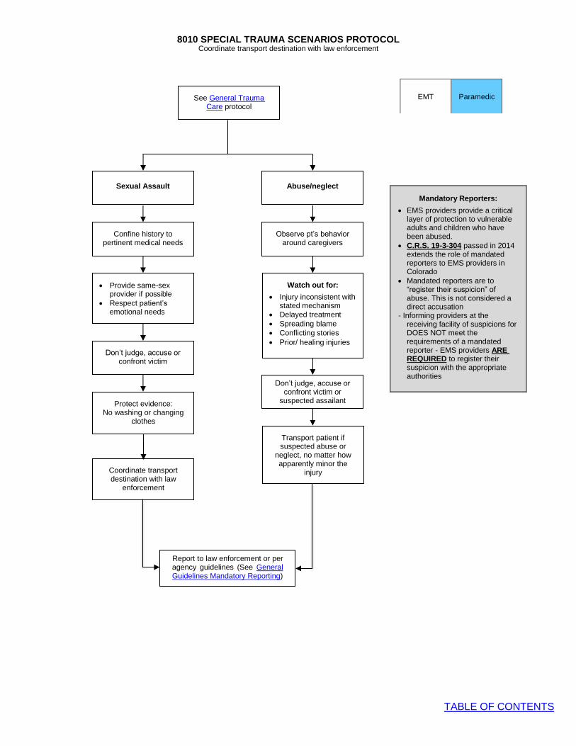
8010 SPECIAL TRAUMA SCENARIOS PROTOCOL
TABLE OF CONTENTS
Transport patient if suspected abuse or
neglect, no matter how apparently minor the
injury
EMT
Paramedic
Coordinate transport destination with law enforcement
See General Trauma Care protocol
Coordinate transport destination with law
enforcement
Report to law enforcement or per agency guidelines (See General Guidelines Mandatory Reporting)
Abuse/neglect
Sexual Assault
Protect evidence: No washing or changing
clothes
Don’t judge, accuse or confront victim or
suspected assailant
Don’t judge, accuse or confront victim
Watch out for:
• Injury inconsistent with stated mechanism
• Delayed treatment • Spreading blame • Conflicting stories • Prior/ healing injuries
• Provide same-sex provider if possible
• Respect patient’s emotional needs
Observe pt’s behavior around caregivers
Confine history to pertinent medical needs
Mandatory Reporters:
• EMS providers provide a critical layer of protection to vulnerable adults and children who have been abused.
• C.R.S. 19-3-304 passed in 2014 extends the role of mandated reporters to EMS providers in Colorado
• Mandated reporters are to “register their suspicion” of abuse. This is not considered a direct accusation
- Informing providers at the receiving facility of suspicions for DOES NOT meet the requirements of a mandated reporter - EMS providers ARE REQUIRED to register their suspicion with the appropriate authorities
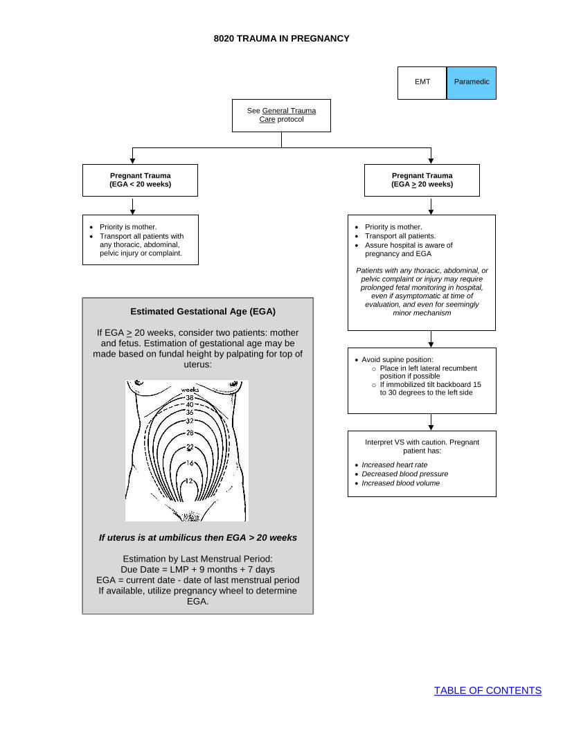
8020 TRAUMA IN PREGNANCY
TABLE OF CONTENTS
Paramedic
Pregnant Trauma (EGA > 20 weeks)
Pregnant Trauma (EGA < 20 weeks)
Estimated Gestational Age (EGA)
If EGA > 20 weeks, consider two patients: mother and fetus. Estimation of gestational age may be
made based on fundal height by palpating for top of uterus:
If uterus is at umbilicus then EGA > 20 weeks
Estimation by Last Menstrual Period: Due Date = LMP + 9 months + 7 days
EGA = current date - date of last menstrual period If available, utilize pregnancy wheel to determine
EGA.
See General Trauma Care protocol
EMT
Interpret VS with caution. Pregnant patient has:
• Increased heart rate
• Decreased blood pressure
• Increased blood volume
• Avoid supine position: o Place in left lateral recumbent
position if possible o If immobilized tilt backboard 15
to 30 degrees to the left side
• Priority is mother. • Transport all patients. • Assure hospital is aware of
pregnancy and EGA
Patients with any thoracic, abdominal, or pelvic complaint or injury may require prolonged fetal monitoring in hospital,
even if asymptomatic at time of evaluation, and even for seemingly
minor mechanism
• Priority is mother. • Transport all patients with
any thoracic, abdominal, pelvic injury or complaint.

8030 TRAUMATIC ARREST
TABLE OF CONTENTS
o Bilateral needle chest decompression if any trauma to trunk
Pull/push for pediatrics
Traumatic Arrest
For non-survivable injury refer to field pronouncement for
traumatic arrest
Consider mechanism of injury. If medical cause of arrest suspected, treat per Universal Pulseless Arrest Algorithm
Blunt (include isolated GSW to head)
Penetrating
If arrest suspected to be >10 minutes, refer to Field Pronouncement for Traumatic Arrest
Signs of Life? (any of the following)
• Spontaneous movement • Pulses • Breathing • Reactive pupils
Yes
No
Refer to Field Pronouncement for Traumatic Arrest
• Rapid transport to appropriate trauma center.
• Identify and treat reversible life threats o Control Exsanguinating Hemorrhage o Advanced airway o Bilateral Needle Chest Decompression if
any trauma to trunk
o 2 IVs preferred IV NS bolus 20 mL/kg up to 1 L (IO if no IV access)
o Hypothermia prevention o Consider pelvic stabilization o Initiate BLS CPR and ventilations at age
appropriate rate
• Pull/push for pediatric fluid administration
Paramedic
EMT
Non-survivable Injuries • Decapitation • Massive burns without signs of life • Evidence of massive blunt head, chest,
abdominal trauma • Decomposition • Dependent lividity or rigor mortis
Exceptions to Traumatic Arrest Protocol: • Hypothermia • Drowning • Pregnant with estimated gestational age
≥20 weeks • Lightning strike or electrocution • Avalanche victim

8040 TRAUMATIC SHOCK
TABLE OF CONTENTS
Treat suspected tension pneumothorax with needle decompression.
Pediatric Fluid Administration
• For children <40 kg or not longer than length-based tape, hand pull/push fluid with a 60 mL syringe utilizing a 3 way stop cock
• Hypotension is a late sign in pediatric shock patients
Pediatric Shock
Hypotension for Age
Tachycardia for Age
Minimum Blood Pressure with TBI
Age MAP (mmHg) Minimum SBP
(mmHg) 0-23 months 50-70 75
2-5 years 60-80 80 6-8 years 65-85 85
9-12 years 70-95 90 >12 years ≥80 ≥110
Signs of Compensated Shock
Signs of Decompensated Shock
• Normal mental status • Normal systolic blood
pressure • Tachycardia • Prolonged (>2 seconds)
capillary refill • Tachypnea • Cool and pale distal
extremities • Weak peripheral pulse
• Decrease mental status • Weak central pulses • Poor color • Hypotension for age
EMT
Paramedic
Complete General Trauma Care
• Correct hypoxia and manage the airway if needed.
• Keep patient warm.
IV Fluid Resuscitation
• Use IV fluid sparingly. • Titrate small boluses of crystalloid to
presence of peripheral pulses. • However, hypotension is particularly
harmful to patients with severe TBI. In patients with TBI, more aggressive fluid resuscitation is justified to maintain a normal blood pressure
• Most pediatric trauma mortality is from TBI, therefore fluid resuscitation to normal BP is recommended
• Use push/pull technique to administer IV fluid boluses in children
For trauma patients with hypotension for age or signs of shock:
• Initiate rapid transport to appropriate trauma center.
• Treat and stabilize in route to hospital.
Shock is defined as impaired tissue perfusion and may be manifested by any of the following:
• Altered mental status • Tachycardia • Poor skin perfusion • Low blood pressure
Traditional signs of shock may be absent early in the process, therefore, maintain a high index of suspicion and be vigilant for subtle signs of poor perfusion
Do not use Trendelenburg’s position routinely to treat hypotension. It is unnecessary and may impair respirations and/or aggravate injuries. Supine position preferred
Age Blood Pressure <1 year <70 mmHg
1-10 years <70 + (2 x age in years) >10 years <90 mmHg
Age Heart Rate
<1 year >160 bpm 1-2 years >150 bpm 2-5 years >140 bpm
5-12 years >120 bpm >12 years >100 bpm
Identify and treat reversible causes of shock:
• Control exsanguinating hemorrhage.
• Treat suspected tension pneumothorax with needle decompression.
• Apply pelvic compression device for suspected unstable pelvic fracture.

8050 AMPUTATIONS
TABLE OF CONTENTS
Partial Amputation
General Trauma Care Protocol
Complete Amputation
Life-threatening
bleeding
EMT
Paramedic
• Cover with moist sterile dressing
• Splint near-amputated part in anatomic position
Apply tourniquet without
delay
Non-Life-threatening
bleeding
• Large bore IV • If hypotensive for age, treat per
Traumatic Shock Protocol • Document neurovascular exam
Control with direct pressure to bleeding area or vessel
• Monitor and transport to appropriate
Trauma Center • Treat other injuries per protocol
Consider pain management
Amputated part:
• Wrap in moist, sterile dressing • Place in sealed plastic bag • Place bag in ice water • Do not freeze part
Stump:
• Cover with moist sterile dressing covered by dry dressing
Partial Amputation:
• Cover with moist sterile dressing • Splint near-amputated part in anatomic
position
Apply tourniquet if bleeding not controlled with direct pressure

8060 HEAD TRAUMA PROTOCOL
TABLE OF CONTENTS
BLS airway preferred in pediatrics
Consider advanced airway if adequate ventilation and oxygenation cannot be achieved with basic airway maneuvers
Support ventilations & maintain ETCO2
35-45 mmHg
Open airway and assist ventilations
Yes
No
EMT
Paramedic
General Trauma Care protocol
Assess for hypotension and/or
signs of shock and treat per Traumatic Shock protocol en route
• Monitor cardiac rhythm
• Correct hypoxia • Treat hypotension • Decrease ICP by elevating head
30 if possible. Use reverse Trendelenburg if spinal precautions needed
• Complete Rapid Trauma Assessment en route to hospital
• Treat other injuries per protocol
Pediatric GCS
(Minimum 3, Maximum 15)
Eyes: 1. Does not open eyes 2. Opens eyes to pain 3. Opens eyes to voice 4. Opens eyes
spontaneously
Verbal: 1. No vocal response 2. Inconsolable, agitated 3. Inconsistently consolable,
moaning. 4. Cries but consolable,
inappropriate interactions. 5. Smiles, oriented to
sounds, follows objects, interacts
Motor: 1. No motor response. 2. Extension to pain. 3. Flexion to pain. 4. Withdrawal from pain 5. Localizes pain. 6. Obeys Commands.
Glasgow Coma Score (GCS)
(Minimum 3, Maximum 15)
Eyes: 1. Does not open eyes 2. Opens eyes to pain 3. Opens eyes to voice 4. Opens eyes
spontaneously
Verbal: 1. No sounds 2. Incomprehensible sounds 3. Inappropriate words 4. Confused, disoriented 5. Oriented
Motor: 1. No movement 2. Extension to painful stimuli 3. Flexion to painful stimuli 4. Withdrawal to painful
stimuli 5. Localizes to painful stimuli 6. Obeys commands
GCS < 8 or comatose?
Monitor: • ABCs, VS, mental status,
ETCO2
• Rapid transport to appropriate trauma center

8070 FACE AND NECK TRAUMA
TABLE OF CONTENTS
General Trauma Care Protocol
• Clear airway • Rapid trauma assessment • Spinal Precautions Protocol • Assess for need for airway management
Laryngeal trauma* Yes
No
Severe airway Bleeding?
No
Yes
Direct pressure if appropriate
Consider advanced airway if adequate ventilation and oxygenation cannot be achieved with basic airway maneuvers
BLS airway preferred in pediatrics
• Complete neuro exam • Assess for subcutaneous air • Cover/protect eyes as indicated • Do not try to block drainage from ears,
nose • Save avulsed teeth in saline-soaked gauze,
do not scrub clean
• Transport ASAP to appropriate Trauma Center
• IV access en route • Treat other injuries per protocol • Suction airway as needed
Consider opioid for pain control as needed
Monitor ABCs, VS, mental status, SpO2, ETCO2
Spinal precautions not routinely indicated for penetrating neck injury
Penetrating injury is very rarely associated
with unstable spinal column
*Suspect laryngeal trauma with:
• Laryngeal tenderness, swelling, bruising
• Voice changes • Respiratory distress • Stridor
Regarding Nasal Intubation:
• Contraindicated in pediatrics • Relatively contraindicated with mid-
face trauma. • Avoid if mid-face grossly unstable
Avoid intubation if patient can be oxygenated by less invasive means
EMT Paramedic

8080 SPINAL TRAUMA
TABLE OF CONTENTS
EMT
Paramedic
Patient with signs of traumatic acute spinal cord injury
General Trauma Care protocol
Consider opioid for pain control
Monitor ABCs, VS, mental status, SpO2, waveform capnography
• Complete patient assessment • Treat other injuries per protocol • Monitor for status changes
If hypotension and/or signs of shock, resuscitate per Traumatic Shock protocol
Spinal Immobilization not routinely indicated for penetrating neck injury
Penetrating injury is very rarely associated
with unstable spinal column
Large bore IV and consider 2nd line
Rapid transport to appropriate Trauma Center
Signs of Spinal Cord Injury:
• Sensory loss, weakness and/or paralysis • Typically bilateral, but may be
asymmetrical • Sensory changes typically have a level,
corresponding to the level of the injury • Numbness, tingling or painful burning in
arms, legs • Central cord syndrome is an incomplete
spinal cord injury and causes painful burning or sensory changed in shoulders and upper extremities bilaterally and spares the lower extremities. It may be subtle
• Full spinal precautions if any neurological signs
and symptoms consistent with a spine injury are present
• Document neuro assessments before and after immobilization

8090 SPINAL PRECAUTIONS PROTOCOL
TABLE OF CONTENTS
Yes Is the patient able to
comfortably lay still and comply with instructions?
Is the patient ambulatory on scene at time of EMS
arrival?
Transport patient in a position of comfort on gurney with
cervical collar
Full spinal immobilization
Place c-collar
on patient and ask them to not move
neck
If NONE of above criteria, and you think patient is not
likely to have a spinal injury, no spinal
precautions required
Is there an objective neurological deficit?
EMT
Paramedic
Does patient have/complain of any of the following:
• Midline C/T/L spine tenderness on palpation
• Neurologic complaints or deficits
• Other injuries which are potentially distracting
• Alteration in mentation or under influence of drugs or EtOH
• Barrier to evaluate for spinal injury (e.g. language or developmental barrier)
Yes No
No Yes
No
Yes
No
Notes:
• Backboards have not been shown to be any benefit for spinal injuries, and may cause harm.
• Backboards/scoops are useful tools for carrying non-ambulatory patients to a gurney. Patients who do not need a backboard should be gently slid off of backboard/scoop onto gurney.
• Self-extrication from a vehicle with assistance is likely better than standard extrication procedures.
• Vacuum mattresses should be used preferentially over a backboard if readily available.
• Use caution and have a higher index of suspicion when assessing for spinal injury in elderly patients, who are at much higher risk and may have minimal symptoms.
• Consider improvised cervical spine immobilization such as towel rolls and tape or a SAM splint if needed to prevent airway compromise or worsening spinal injury if the rigid cervical collar cannot be correctly sized to the patient
• Neurological exam documentation is MANDATORY in patients with potential spinal trauma, including serial exams.
• Cervical collar is not indicated in isolated penetrating trauma
• Full spinal immobilization includes backboard or scoop, and cervical collar
• In pediatrics, do not transport sitting upright or in the car seat if suspected spinal injury is present.
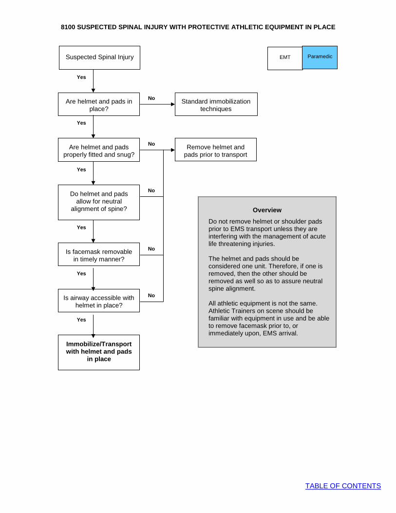
8100 SUSPECTED SPINAL INJURY WITH PROTECTIVE ATHLETIC EQUIPMENT IN PLACE
TABLE OF CONTENTS
No
No
No
Remove helmet and pads prior to transport
Yes
No
Yes
No
Yes
Yes
Yes
Yes
EMT
Suspected Spinal Injury
Do helmet and pads allow for neutral
alignment of spine?
Is facemask removable in timely manner?
Is airway accessible with helmet in place?
Immobilize/Transport with helmet and pads
in place
Paramedic
Overview
Do not remove helmet or shoulder pads prior to EMS transport unless they are interfering with the management of acute life threatening injuries.
The helmet and pads should be considered one unit. Therefore, if one is removed, then the other should be removed as well so as to assure neutral spine alignment.
All athletic equipment is not the same. Athletic Trainers on scene should be familiar with equipment in use and be able to remove facemask prior to, or immediately upon, EMS arrival.
Are helmet and pads properly fitted and snug?
Standard immobilization techniques
Are helmet and pads in place?

8110 CHEST TRAUMA
TABLE OF CONTENTS
nd
2
Are you able to oxygenate and
No
ventilate effectively?
Yes
Consider advanced airway if adequate ventilation and oxygenation cannot be achieved with basic airway maneuvers
Consider tension pneumothorax and
Chest Needle Decompression
Penetrating trauma?
Yes
Rapid transport
& stabilize in route
Apply approved vented occlusive dressing over wounds
No
Large bore IV
consider 2 line
Flail Chest? Yes
Splint with bulky dressing
No
Hypotension for age? Yes
Treat per Traumatic Shock protocol
in route
No
Consider opioid for
pain control
Monitor ABCs, VS, mental status, SpO , waveform
capnography
BLS airway preferred in pediatrics
EMT Paramedic
• General Trauma Care protocol
• Rapid transport to Trauma Center
Tension pneumothorax should be suspected with presence of
the following:
• Unilateral absent breath sounds AND: JVD, hypotension, difficult/unable to ventilate
• Needle decompression is NEVER indicated for simple pneumothorax
Assess need for assisted ventilations

8120 ABDOMINAL TRAUMA
TABLE OF CONTENTS
• General Trauma Care protocol
• Rapid transport to Trauma Center
• IV access
• Consider 2nd
line if MOI significant
Yes Penetrating trauma? Cover wounds, viscera with saline
moistened gauze dressing
No
Do not attempt to repack exposed viscera
Hypotension for age? Resuscitate per Traumatic Shock protocol
No
Consider opioid for pain control
Monitor ABCs, VS, mental status, SpO2, waveform
capnography
EMT Paramedic
Yes
Documentation • MOI • Time of injury • Initial GCS
• Penetrating trauma • Weapon/projectile/trajectory
• Blunt vehicular trauma • Condition of vehicle • Speed • Ejection • Airbag deployment • Restraints, helmets

8130 BURNS
TABLE OF CONTENTS
• General Trauma Care protocol
• Rapid transport to Trauma Center
Stop burning process:
• Remove clothes if not adhered to patient’s skin
• Flood with water only if flames/ smoldering present
Respiratory Distress Yes hoarseness or
stridor?
• O2 NRB 15 Lpm
• Manage airway and assist ventilations as indicated
• Consider CO, CN No
Evaluate degree and body surface are involved
Yes Critical Burn*?
No
• Start 2 large-bore IVs
• Fluids per ABA recommendations (chart below)**
IV NS TKO
• Remove rings, jewelry, constricting items
• Dress burns with dry sterile dressings
• Treat other injuries per protocol
• Cover patient to keep warm
Consider opioid for pain control
Monitor ABCs, VS, mental status, SpO2, waveform capnography
EMT Paramedic
** ABA Recommended Prehospital Fluid Therapy 14 and older 500 mL/hr NS or LR 5 - 13 years 250 mL/hr NS or LR Younger than 5 125 mL/hr D5W, NS or LR If no signs of clinical hypovolemia or shock, large volume of IV fluid not needed. For typical 30 minute prehospital time, give 250 mL bolus for patient age ≥ 14.
Document: • Type and degree of burn(s) • % BSA • Respiratory status including any voice
changes (hoarseness) • Singed nares, soot in mouth • SpO2 • PMH • Confined space (assume CO)
*Critical Burn: • 2º > 30% BSA • 3º > 10% BSA • Respiratory injury, facial burn • Associated injuries, electrical or deep
chemical burns, underlying PMH (cardiac, DM), age < 10 or > 50 yrs.
Types of Burns: • Thermal: remove from environment, put
out fire • Chemical: brush off or dilute chemical.
Consider HAZMAT • Electrical: make sure victim is de-
energized and suspect internal injuries • Assume CO if enclosed space • Consider cyanide poisoning (CN) if
unconscious or pulseless arrest
Designated Regional Burn Centers
Consider direct transport of isolated burns if time and conditions allow
• Children’s Hospital Colorado: Age ≤ 14
• University of Colorado Hospital: Age ≥ 15
• Swedish Medical Center: Any age

TABLE OF CONTENTS
9000 GENERAL GUIDELINES: MEDICATION ADMINISTRATION
Purpose
A. Provide guidance to EMS providers in the principles of administration, delivery, and safety of approved medications
General Principles
A. The appropriate procedure for safe medication administration includes:
1. Verification of the “Six Rights” of medication administration (right patient, right drug, right dose, right route, right time, right documentation)
2. Medication administration cross-check with practice partner verifying the Six Rights prior to drug administration. This should include verbal repeat-back of the order by the practice partner.
3. Obtaining vital signs every 5 minutes or after any intervention.
B. Pediatric medication dosing and equipment size recommendations vary by length and/or weight. As such, an assessment tool such as a length-based tape should be utilized on every pediatric patient to guide medication dosing and equipment size. The risk of dosing error is high in children and the use of volume- based dosing guides have been shown to reduce the rate of error. We recommend the use of a volume- based medication dosing guide for all children based upon age or weight.
C. Optional routes of medication administration are vast, and appropriateness given the clinical situation should be considered. Specific considerations include:
1. Intranasal (IN) administration often results in more rapid resolution or improvement in symptoms compared to IV or intramuscular (IM) administration
2. IM drug absorption and onset of action is often the slowest, as vascular absorption from fat tissue is prolonged
D. Ideally, expired medications should never be utilized for patient care. However, the nation is increasingly faced with the challenge of critical or potentially life-saving medication shortages. As such, the Denver Metro EMS Medical Directors have issued guidelines for the appropriate response to a national medication crisis. Approved medications required for potentially emergent conditions and for which no reasonable substitution is available may be used after the posted expiration date with the following restrictions:
1. Medication should be approved for use by the agency’s EMS Medical Director.
2. Expired medications will be used only after the supply of non-expired medications have been exhausted
3. Standard medication storage, inspection and delivery practices should be maintained
E. EMS agencies should work to establish a system of Just Culture. This is an approach to work place safety that assumes humans, despite their best intentions to do the right thing, will make errors. Change and care improvement does not happen without accurate, honest reporting of error. A report of error should be treated with respect and examination of root cause, and not punitive action

9005 MEDICATIONS
TABLE OF CONTENTS
ACETAMINOPHEN (TYLENOL)
Description Acetaminophen elevates the pain threshold and readjusts hypothalamic temperature-regulatory center.
Onset & Duration • Onset: 20 minutes • Duration: 4 hours
Indications • Mild pain
Contraindications • Known hypersensitivity • Known or suspected chronic liver disease
Adverse Reactions • Acetaminophen has a wide therapeutic window. Recommended maximum therapeutic doses are
less than half the toxic dose. o Single toxic dose in a 70 kg adult is greater than 7 gm. o Single toxic dose in a child is greater than 150 mg/kg. o Chronic supratherapeutic acetaminophen poisoning is possible as many medications contain
acetaminophen.
Drug Interactions • Avoid concomitant administration with other acetaminophen-containing medication, such as many
prescription opioids (e.g. Percocet) or OTC cough and cold medications.
Dosage and Administration Adult:
1000 mg PO
Pediatric: 15 mg/kg PO
Protocol • Pain management
Weight Age Dose
(160 mg/5 mL)
n/a < 6 months BASE CONTACT
5-8kg 6 months - 12 months
2.5ml (80mg)
9-11kg 1-2 years 4ml (128mg)
12-16kg 2-3 years 5ml (160mg)
17-21kg 4-5 years 7.5ml (240mg)
22-27kg 6-8 years 10ml (320mg)
28-33kg 9-10 years 12.5ml (400mg)
34-43kg 11-12 years 15ml (480mg)

9010 MEDICATIONS
TABLE OF CONTENTS
ADENOSINE (ADENOCARD)
Description Adenosine transiently blocks conduction through the AV node thereby terminating reentrant tachycardias involving the AV node. It is the drug of choice for AV nodal reentrant tachycardia (AVNRT, often referred to as “PSVT”). It will not terminate dysrhythmias that do not involve the AV node as a reentrant limb (e.g. atrial fibrillation).
Onset & Duration • Onset: almost immediate • Duration: 10 sec
Indications • Narrow-complex supraventricular tachyarrhythmia after obtaining 12 lead ECG (This may be the
only documented copy of the AVRNT rhythm) • Pediatric administration requires call in for direct verbal order
Contraindications • Any irregular tachycardia. Specifically never administer to an irregular wide-complex tachycardia,
which may be lethal • Heart transplant
Adverse Reactions • Chest pain • Shortness of breath • Diaphoresis • Palpitations • Lightheadedness
Drug Interactions • Methylxanthines (e.g. caffeine) antagonize adenosine, a higher dose may be required • Dipyridamole (persantine) potentiates the effect of adenosine; reduction of adenosine dose may be
required • Carbamazepine may potentiate the AV-nodal blocking effect of adenosine
Dosage and Administration Adult:
12 mg IV bolus, rapidly, followed by a normal saline flush. Additional dose of 12 mg IV bolus, rapidly, followed by a normal saline flush. Contact medical control for further considerations
Pediatric:
Children who are stable with AVNRT generally remain so and transport is preferred over intervention.
CONTACT BASE 0.1 mg/kg IV bolus (max 6 mg), rapidly followed by normal saline flush. Additional dose of 0.2 mg/kg (max 12 mg) rapid IV bolus, followed by normal saline flush.

9010 MEDICATIONS
TABLE OF CONTENTS
Protocol • Tachyarrhythmia with Poor Perfusion
Special Considerations • Reliably causes short lived but very unpleasant chest discomfort. Always warn your patient of this
before giving medication and explain that it will be a very brief sensation • May produce bronchospasm in patients with asthma • Transient asystole and AV blocks are common at the time of cardioversion • Adenosine is not effective in atrial flutter or fibrillation • Adenosine is safe in patients with a history of Wolff-Parkinson-White syndrome if the rhythm is
regular and QRS complex is narrow • A 12-lead EKG should be performed and documented, when available • Adenosine requires continuous EKG monitoring throughout administration

9020 MEDICATIONS
TABLE OF CONTENTS
ALBUTEROL SULFATE (PROVENTIL, VENTOLIN)
Description • Albuterol is a selective ß-2 adrenergic receptor agonist. It is a bronchodilator and positive
chronotrope. • Because of its ß agonist properties, it causes potassium to move across cell membranes inside
cells. This lowers serum potassium concentration and makes albuterol an effective temporizing treatment for unstable patients with hyperkalemia.
Onset & Duration • Onset: 5-15 minutes after inhalation • Duration: 3-4 hours after inhalation
Indications • Bronchospasm • Known or suspected hyperkalemia with ECG changes (i.e.: peaked T waves, QRS widening)
Contraindications • Severe tachycardia is a relative contraindication
Adverse Reactions • Tachycardia • Palpitations • Dysrhythmias
Drug Interactions • Sympathomimetics may exacerbate adverse cardiovascular effects. • ß-blockers may antagonize albuterol.
How Supplied MDI: 90 mcg/metered spray (17-g canister with 200 inhalations) Pre-diluted nebulized solution: 2.5 mg in 3 ml NS (0.083%)
Dosage and Administration
Adult: Single Neb dose Albuterol sulfate solution 0.083% (one unit dose bottle of 3.0 ml), by nebulizer, at a flow rate (6-8 lpm) that will deliver the solution over 5 to 15 minutes. May be repeated twice (total of 3 doses). Continuous Neb dose In more severe cases, place 3 premixed containers of albuterol (2.5 mg/3ml) for a total dose of 7.5 mg in 9 ml, into an oxygen-powered nebulizer and run a continuous neb at 6-8 lpm.
Pediatric: Single Neb dose Albuterol sulfate 0.083% (one unit dose bottle of 3.0 ml), by nebulizer, at a flow rate (6-8 lpm) that will deliver the solution over 5-15 minutes. May be repeated twice during transport (total of 3 doses).
9020 MEDICATIONS
TABLE OF CONTENTS
Protocol • Adult Wheezing • Pediatric Wheezing • Allergy and Anaphylaxis
Special Considerations • Consider inline nebs for patients requiring endotracheal intubation or CPAP. • May precipitate angina pectoris and dysrhythmias • Should be used with caution in patients with suspected or known coronary disease, diabetes
mellitus, hyperthyroidism, prostatic hypertrophy, or seizure disorder • Wheezing associated with anaphylaxis should first be treated with epinephrine IM.
9030 MEDICATIONS
TABLE OF CONTENTS
AMIODARONE (CORDARONE)
Description Class III antiarrhythmic agent which inhibits adrenergic stimulation, decreases AV conduction and sinus node function, and prolongs the action potential and refractory period id myocardial tissue
Indications • Pulseless arrest in patients with shock-refractory or recurrent VF/VT • Wide complex tachycardia not requiring immediate cardioversion due to hemodynamic instability
Precautions • Wide complex irregular tachycardia • Sympathomimetic toxidromes, i.e. cocaine or amphetamine overdose • NOT to be used to treat ventricular escape beats or accelerated idioventricular rhythms
Contraindications
• 2nd or 3rd degree AV block • Cardiogenic shock
Adverse Reactions • Hypotension • Bradycardia
Dosage and Administration Adult:
• Pulseless Arrest (Refractory VT/VF): o 300 mg IV bolus. o Administer additional 150 mg IV bolus in 3-5 minutes if shock refractory or recurrent
VF/VT. • Symptomatic VT and undifferentiated wide complex tachycardia with a pulse:
o CONTACT BASE 150 mg IV bolus infusion over 10 minutes. Pediatric:
• Pulseless Arrest (Refractory VT/VF): o 5mg/kg IV bolus. o CONTACT BASE for additional doses.
Protocol
• Universal Pulseless Arrest Algorithm • Tachycardia with Poor Perfusion
Special Considerations • A 12-lead EKG should be performed and documented, when available. • Amiodarone is preferred to adenosine for treatment of undifferentiated WCT with a pulse.

9040 MEDICATIONS
TABLE OF CONTENTS
ANTIEMETICS: ONDANSETRON (ZOFRAN)
Description • Ondansetron is a selective serotonin 5-HT3 receptor antagonist antiemetic. Ondansetron is the preferred
antiemetic.
Indications • Nausea and vomiting
Contraindications • Ondansetron: No absolute contraindication. Should be used with caution in first trimester of pregnancy and
should be reserved for only those patients with severe dehydration and intractable vomiting
Adverse Effects: • Ondansetron: Very low rate of adverse effects, very well tolerated.
Dosage and Administration
Ondansetron
Adult: 4 mg IV/IM/PO/ODT. May repeat x 1 dose as needed.
Pediatric ≥ 4 years old: 4 mg IV/PO/ODT
Pediatric 6 months to 4 years old: 2 mg IV/PO/ODT
Pediatric < 6 months: BASE CONTACT
Protocol • Abdominal Pain/Vomiting
• Altitude Illness
.

9050 MEDICATIONS
TABLE OF CONTENTS
ASPIRIN (ASA)
Description Aspirin inhibits platelet aggregation and blood clotting and is indicated for treatment of acute coronary syndrome in which platelet aggregation is a major component of the pathophysiology. It is also an analgesic and antipyretic.
Indications • Suspected acute coronary syndrome
Contraindications • Active gastrointestinal bleeding
• Aspirin allergy
How Supplied Chewable tablets 81mg
Dosage and Administration • 324mg PO
Protocol • Chest Pain
Special Considerations • Patients with suspected acute coronary syndrome taking warfarin (Coumadin), clopidogrel
(Plavix) or novel oral anticoagulants may still be given aspirin.
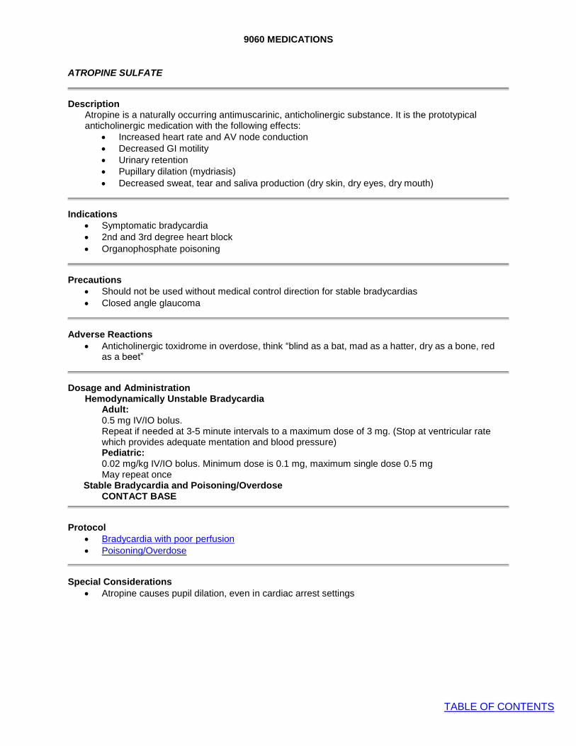
9060 MEDICATIONS
TABLE OF CONTENTS
ATROPINE SULFATE
Description Atropine is a naturally occurring antimuscarinic, anticholinergic substance. It is the prototypical anticholinergic medication with the following effects:
• Increased heart rate and AV node conduction • Decreased GI motility • Urinary retention • Pupillary dilation (mydriasis) • Decreased sweat, tear and saliva production (dry skin, dry eyes, dry mouth)
Indications • Symptomatic bradycardia
• 2nd and 3rd degree heart block
• Organophosphate poisoning
Precautions • Should not be used without medical control direction for stable bradycardias
• Closed angle glaucoma
Adverse Reactions • Anticholinergic toxidrome in overdose, think “blind as a bat, mad as a hatter, dry as a bone, red
as a beet”
Dosage and Administration Hemodynamically Unstable Bradycardia
Adult: 0.5 mg IV/IO bolus. Repeat if needed at 3-5 minute intervals to a maximum dose of 3 mg. (Stop at ventricular rate which provides adequate mentation and blood pressure) Pediatric: 0.02 mg/kg IV/IO bolus. Minimum dose is 0.1 mg, maximum single dose 0.5 mg May repeat once
Stable Bradycardia and Poisoning/Overdose CONTACT BASE
Protocol
• Bradycardia with poor perfusion
• Poisoning/Overdose
Special Considerations • Atropine causes pupil dilation, even in cardiac arrest settings

9070 MEDICATIONS
TABLE OF CONTENTS
BENZODIAZEPINES (MIDAZOLAM)
Description • Benzodiazepines are sedative-hypnotics that act by increasing GABA activity in the brain. GABA
is the major inhibitory neurotransmitter, so increased GABA activity inhibits cellular excitation. Benzodiazepine effects include anticonvulsant, anxiolytic, sedative, amnestic and muscle relaxant properties. Each individual benzodiazepine has unique pharmacokinetics related to its relative lipid or water solubility.
• Selection of specific agent as preferred benzodiazepine is at individual agency Medical Director discretion.
Onset & Duration • Any agent given IV will have the fastest onset of action, typical time of onset 2-3 minutes
• Intranasal administration has slower onset and is less predictable compared to IV administration, however, it may still be preferred if an IV cannot be safely or rapidly obtained. Intranasal route has faster onset compared to intramuscular route.
o Diazepam should not be given intranasally as it is not well absorbed. • IM administration has the slowest time of onset.
Indications • Status epilepticus
• Sedation of the severely agitated/combative patient • Sedation for cardioversion or transcutaneous pacing (TCP)
Contraindications • Hypotension
• Respiratory depression
Adverse Reactions • Respiratory depression, including apnea
• Hypotension
• Consider ½ dosing in the elderly for all benzodiazepines
Dosage and Administration MIDAZOLAM:
Seizure or sedation for cardioversion or transcutaneous pacing:
Adult: IV/IO route: 2 mg
• Dose may be repeated x 1 after 5 minutes if still seizing. Contact Base for more than 2 doses
IN/IM route (intranasal preferred): 5 mg • Dose may be repeated x 1 after 5 minutes if still seizing. Contact Base for more than
2 doses
Pediatric: IV/IO route 0.1 mg/kg
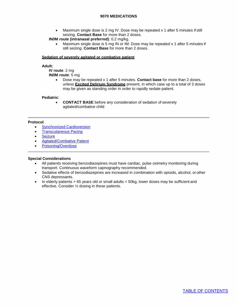
9070 MEDICATIONS
TABLE OF CONTENTS
• Maximum single dose is 2 mg IV. Dose may be repeated x 1 after 5 minutes if still seizing. Contact Base for more than 2 doses.
IN/IM route (intranasal preferred): 0.2 mg/kg. • Maximum single dose is 5 mg IN or IM. Dose may be repeated x 1 after 5 minutes if
still seizing. Contact Base for more than 2 doses.
Sedation of severely agitated or combative patient
Adult: IV route: 2 mg IN/IM route: 5 mg
• Dose may be repeated x 1 after 5 minutes. Contact base for more than 2 doses, unless Excited Delirium Syndrome present, in which case up to a total of 3 doses may be given as standing order in order to rapidly sedate patient.
Pediatric:
• CONTACT BASE before any consideration of sedation of severely agitated/combative child
Protocol • Synchronized Cardioversion
• Transcutaneous Pacing
• Seizure
• Agitated/Combative Patient • Poisoning/Overdose
Special Considerations • All patients receiving benzodiazepines must have cardiac, pulse oximetry monitoring during
transport. Continuous waveform capnography recommended. • Sedative effects of benzodiazepines are increased in combination with opioids, alcohol, or other
CNS depressants. • In elderly patients > 65 years old or small adults < 50kg, lower doses may be sufficient and
effective. Consider ½ dosing in these patients.

9080 MEDICATIONS
TABLE OF CONTENTS
CALCIUM
Description • Cardioprotective agent in hyperkalemia. • Calcium chloride contains 3 times the amount of elemental calcium contained in the same volume of
calcium gluconate. Therefore, 1 g (10 mL) vial of calcium chloride 10% solution contain 273 mg of elemental calcium, whereas 1 g (10 mL) of 10% calcium gluconate contains 90 mg of elemental calcium. For this reason, larger doses of calcium gluconate are required.
• Doses below refer to dose of calcium solution, not elemental calcium.
Indications • Pulseless arrest associated with any of the following clinical conditions:
o Known hyperkalemia o Renal failure with or without hemodialysis history o Calcium channel blocker overdose
• Not indicated for routine treatment of pulseless arrest • Calcium channel blocker overdose with hypotension and bradycardia
Contraindications • Known hypercalcemia
• Suspected digoxin toxicity (i.e. digoxin overdose)
Side Effects/Notes • Extravasation of calcium chloride solution may cause tissue necrosis. • Because of the risk of medication error, if calcium chloride is stocked, consider limiting to 1 amp per
medication kit to avoid accidental overdose. Calcium gluconate solution will require 3 amp supply for equivalent dose.
• Must give in separate line from IV sodium bicarb to prevent precipitation/formation of calcium carbonate.
• In setting of digoxin toxicity, may worsen cardiovascular function.
Dosage and Administration
Calcium Gluconate 10% Solution
Adult: • Pulseless arrest assumed due to hyperkalemia:
o 3 g (30 mL) slow IV/IO push • Calcium channel blocker overdose with hypotension and bradycardia:
o Contact Base for order. 3 g (30 mL) slow IV/IO push. Dose may be repeated every 10 minutes for total of 3 doses
Pediatric: • Pulseless arrest assumed due to hyperkalemia:
o 100mg/kg/dose slow IV/IO push. Max 3gm (30mL) • Calcium channel blocker overdose with hypotension for age and bradycardia:
o Contact Base for order. 60 mg/kg (0.6 mL/kg), not to exceed 1 g slow IV/IO push not to exceed 2 mL/minute, may repeat every 10 minutes for total of 3 doses
Calcium Chloride 10% Solution
Adult: • Pulseless arrest assumed due to hyperkalemia:
o 1 g (10 mL) slow IV/IO push • Calcium channel blocker overdose with hypotension and bradycardia:
o Contact Base for order. 1 g (10 mL) slow IV/IO push. Dose may be repeated every 10 minutes for total of 3 doses
Pediatric: • Pulseless arrest assumed due to hyperkalemia:
o 20mg/kg/dose slow IV/IO push. May repeat in 10 minutes • Calcium channel blocker overdose with hypotension for age and bradycardia:
o Contact Base for order. 20 mg/kg (0.2 mL/kg), not to exceed 1 g slow IV/IO push not to exceed 1 mL/min, may repeat every 10 minutes for total of 3 doses.

9080 MEDICATIONS
TABLE OF CONTENTS
Protocol • Universal Pulseless Arrest • Poisoning/Overdose

9090 MEDICATIONS
TABLE OF CONTENTS
DEXTROSE
Description Glucose is the body's basic fuel and is required for cellular metabolism. A sudden drop in blood sugar level will result in disturbances of normal metabolism, manifested clinically as a decrease in mental status, sweating and tachycardia. Further decreases in blood sugar may result in coma, seizures, and cardiac arrhythmias. Serum glucose is regulated by insulin, which stimulates storage of excess glucose from the blood stream, and glucagon, which mobilizes stored glucose into the blood stream.
Indications • Hypoglycemia
• The unconscious or altered mental status patient with an unknown etiology.
Precautions
• None
Dosage and Administration Adult:
25 gm (250 mL of a 10% solution) IV/IO infusion Alternative: 25 gm (50 mL of a 50% solution) IV/IO bolus
Pediatric:
5 mL/kg of 10% solution (maximum of 250 mL) IV/IO
Protocol • Hypoglycemia
• Universal Altered Mental Status
• Seizures
• Poisoning/Overdose
• Psych/Behavioral
Special Considerations • The risk to the patient with ongoing hypoglycemia is enormous. With profound hypoglycemia and
no IV access consider IO insertion. • Draw blood sample before administration, if possible. • Use glucometer before administration, if possible. • Extravasation may cause tissue necrosis when utilizing concentrations above D12.5; use a large
vein and aspirate occasionally to ensure route patency. • Dextrose can be irritable to the vein and the vein should be flushed after administration.

9100 MEDICATIONS
TABLE OF CONTENTS
DIPHENHYDRAMINE (BENADRYL)
Description Antihistamine for treating histamine-mediated symptoms of allergic reaction. Also anticholinergic and antiparkinsonian effects used for treating dystonic reactions caused by antipsychotic and antiemetic medications (e.g.: haloperidol, droperidol, reglan, compazine, etc).
Indications
• Allergic reaction
• Dystonic medication reactions or akathisia (agitation or restlessness)
Precautions • Asthma or COPD, thickens bronchial secretions
• Narrow-angle glaucoma
Side effects • Drowsiness
• Dilated pupils
• Dry mouth and throat • Flushing
Drug Interactions • CNS depressants and alcohol may have additive effects. • MAO inhibitors may prolong and intensify anticholinergic effects of antihistamines.
Dosage and Administration Adults: 50 mg slow IV/IO/IM Pediatrics: 1 mg/kg slow IV/IO/IM. Max (not to exceed 50 mg)
Protocol • Allergy/Anaphylaxis

9115 MEDICATIONS
TABLE OF CONTENTS
DuoDote™ (NERVE AGENT ANTIDOTE KIT)
Description
Nerve agents can enter the body by inhalation, ingestion, and through skin. These agents are absorbed rapidly and can produce injury or death within minutes. The DuoDote™ Nerve Agent Antidote kit consists of one auto-injector for self and/or buddy administration. One Injector contains 2.1mg atropine and 600mg pralidoxime chloride (2-PAM)
Indications
• Suspected nerve agent exposure accompanied with signs and symptoms of nerve agent poisoning
Injection sites • Outer thigh- mid-lateral thigh (preferred site) • Buttocks- upper lateral quadrant of buttock (gluteal) in thin individuals
Instructions
• Place the auto-injector in the dominate hand. Firmly grasp the center of the auto injector with the green tip (needle end) pointing down.
• With the other hand, pull off the gray safety release. The DuoDote™ auto-injector is now
ready to be administered.

7010 MEDICATIONS
TABLE OF CONTENTS
• The injection site is the mid-outer thigh. The DuoDote™ auto-injector can inject through clothing. However, make sure pockets at the injection site are empty.
• Swing and firmly push the green tip at a 90- degree angle against the mid-outer thigh. Continue to firmly push until you feel the auto injector trigger.
• No more than three (3) sets of antidotes should be administered.
Special Considerations
• Presence of tachycardia is not a reliable indicator of effective treatment due to potential nicotinic effects of nerve agent exposure. The end- point of treatment is clear dry lung sounds.
• Attempt to decontaminate skin and clothing between injections.
Protocol:
• Overdose and Acute Poisoning

9120 MEDICATIONS
TABLE OF CONTENTS
EPINEPHRINE (ADRENALIN)
Description Endogenous catecholamine alpha, beta-1, and beta-2 adrenergic receptor agonist. Causes dose- related increase in heart rate, myocardial contractility and oxygen demand, peripheral vasoconstriction and bronchodilation.
Indications • Pulseless Arrest • Anaphylaxis
• Asthma
• Bradycardia with poor perfusion
Adverse Reactions • Tachycardia and tachydysrhythmia
• Hypertension
• Anxiety
• May precipitate angina pectoris
Drug Interactions • Should not be added to sodium bicarbonate or other alkaloids as epinephrine will be inactivated
at higher pH.
Dosage and Administration Adult:
Pulseless Arrest 1 mg (10 ml of a 1:10,000 solution), IV/IO bolus. Repeat every 3-5 minutes up to maximum of 3 doses. May administer 1 additional dose for recurrent arrest after ROSC or narrow complex PEA. Bradycardia with hypotension and poor perfusion refractory to other interventions Continuous infusion titrated to effect: see Vasopressor infusion Asthma: 0.3 mg (0.3 ml of a 1:1,000 solution) IM. May repeat dose x 1. Systemic allergic reaction: 0.3 mg (0.3 ml of a 1:1,000 solution) IM. May repeat dose x 1. Severe systemic allergic reaction (Anaphylaxis) refractory to IM epinephrine: Continuous infusion titrated to effect: see Vasopressor infusion ALTERNATIVE to racemic epinephrine: (for stridor at rest) 5 mL of 1:1,000 epinephrine via nebulizer x 1
Epinephrine Auto-Injector:
Systemic allergic reaction: Adult: 0.3 mg IM with autoinjector (adult EpiPen, Auvi-Q) Pediatric: 0.15 mg IM with autoinjector (EpiPen Jr., Auvi-Q)
Pediatric:
Pulseless arrest: 0.01 mg/kg IV/IO (0.1 ml/kg of 1:10,000 solution). Subsequent doses repeated every 3-5min: 0.01 mg/kg IV/IO (0.1 ml/kg of 1:10,000 solution) Bradycardia (CONTACT BASE) 0.01 mg/kg (0.1 ml/kg of 1:10,000 solution) IV/IO Asthma 0.01 mg/kg (0.01 ml/kg of 1:1,000 solution) IM Alternative: 0.15 mg (0.15 mL of 1:1,000) for <25 kg and 0.3 mg (0.3 mL of 1:1,000) for >25 kg

9120 MEDICATIONS
TABLE OF CONTENTS
Moderate to Severe Allergic Reactions 0.01 mg/kg (0.01 ml/kg of 1:1,000 solution) IM Alternative: 0.15 mg (0.15 mL of 1:1,000) for <25 kg and 0.3 mg (0.3 mL of 1:1,000) for >25 kg Severe systemic allergic reaction (Anaphylaxis) refractory to IM epi (Contact Base): 0.01 mg/kg (0.1 ml/kg of 1:10,000 solution) IV/IO ALTERNATIVE to racemic epinephrine: (for stridor at rest) 5 mL of 1:1,000 epinephrine via nebulizer x 1
Protocol • Universal Pulseless Arrest Algorithm
• Bradyarrhythmia with poor perfusion
• Neonatal Resuscitation
• Allergy and Anaphylaxis Protocol • Adult Wheezing
• Pediatric Wheezing
• Vasopressor Infusion
Special Considerations • May increase myocardial oxygen demand and angina pectoris. Use with caution in patients with
known or suspected CAD
• Intramuscular injection into the thigh is preferred route and site of administration. Intramuscular injection of epinephrine in the thigh results in higher concentrations of medication versus intramuscular or subcutaneous injection in the upper arm.
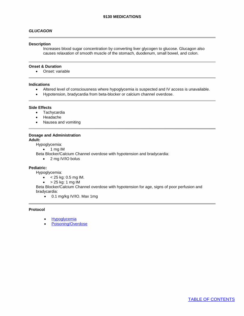
9130 MEDICATIONS
TABLE OF CONTENTS
GLUCAGON
Description Increases blood sugar concentration by converting liver glycogen to glucose. Glucagon also causes relaxation of smooth muscle of the stomach, duodenum, small bowel, and colon.
Onset & Duration • Onset: variable
Indications • Altered level of consciousness where hypoglycemia is suspected and IV access is unavailable. • Hypotension, bradycardia from beta-blocker or calcium channel overdose.
Side Effects • Tachycardia
• Headache
• Nausea and vomiting
Dosage and Administration Adult:
Hypoglycemia: • 1 mg IM
Beta Blocker/Calcium Channel overdose with hypotension and bradycardia: • 2 mg IV/IO bolus
Pediatric:
Hypoglycemia: • < 25 kg: 0.5 mg IM. • > 25 kg: 1 mg IM
Beta Blocker/Calcium Channel overdose with hypotension for age, signs of poor perfusion and bradycardia:
• 0.1 mg/kg IV/IO. Max 1mg
Protocol
• Hypoglycemia • Poisoning/Overdose

9150 MEDICATIONS
TABLE OF CONTENTS
HEMOSTATIC AGENT (QuickClot, Celox, Bloodstop, Actcel, HemCon, ChitoGauze)
Description
QuickClot Combat Gauze is a standard roller or Z-fold gauze impregnated with a clotting agent such as kaolin (a clay containing the active ingredient aluminum silicate) which works on contact with blood to initiate the clotting process (intrinsic pathway) by activating factor XII. This reaction leads to the transformation of factor XII to its’ activated form XIIa, which triggers the clotting cascade.
Mucoadhesive agents such as HemCon, ChitoGauze and Celox utilize a granular chitosan salt derived from the shells of marine arthropods (which are positively charged) to react with and bind to negatively charged red blood cells rapidly forming a cross- linked barrier clot to seal the injured vessels.
Used in conjunction with direct pressure and wound packing these products lead to hemostasis.
Onset and Duration • Onset of action is 3-5 minutes after wound exposure and clotting action remains unless
the dressing and/or the clot is disturbed.
Indications • Active bleeding from open wounds with that cannot be controlled with direct pressure.
Most often involving wounds to the scalp, face, neck, axilla, groin or buttocks.
Contraindications • Not to be used to treat internal bleeding such as intra-abdominal, intra-thoracic or
vaginal bleeding. • Not to be used for minor bleeding that can be controlled by direct pressure.
Precautions • Bleeding control is achieved via combination of direct pressure and hemostatic gauze
packing for a minimum of 3-5 minutes. • Stabilize patient per General Trauma Care Protocol. • If a tourniquet is indicated (refer to Tourniquet Protocol), it should be applied first, before
application of hemostatic agent. • DO NOT USE LOOSE GRANULAR OR POWDERED HEMOSTATIC AGENTS. These
are out date and will produce exothermic reactions that may cause burns and additional tissue damage.
Procedure 1. Manufacturers may have different recommendations on application of their products.
Follow specific manufacturer guidelines for the particular product carried.

9160 MEDICATIONS
TABLE OF CONTENTS
HYDROXOCOBALAMIN (CYANOKIT)
Description
• Cyanide inhibits cytochrome oxidase, thereby arresting cellular respiration and forcing anaerobic metabolism, which leads to lactate production and acidosis and ultimately death. Hydroxocobalamin binds cyanide ions to form cyanocobalamin which is excreted in urine.
Indications
• Adult or pediatric patient with suspected cyanide poisoning from any route, including smoke inhalation in an enclosed space, with any of the following clinical signs:
o Pulseless arrest o Coma/unresponsiveness o Signs of shock
Precautions
• Administer only after basic life support measures have been initiated and always in conjunction with other supportive treatment modalities.
Adverse Reactions • Hypertension
• Allergic reaction/anaphylaxis
Dosage and Administration • Dosing
o Adult dose is 5 gm IV o Pediatric dose is 70 mg/kg up to 5 gm IV
Average Weight
by Group
Grey 4 kg
Pink
6.5 kg
Red
8.5 kg
Purple 10.5 kg
Yellow 13 kg
White
16.5 kg
Blue 21 kg
Orange 26.5 kg
Green 33 kg
Adult
Dose
275mg (11mL)
450mg (18mL)
600mg (24mL)
725mg (29mL)
900mg (36mL)
1150mg (46mL)
1475mg (59mL)
1850mg (74mL)
2300mg (92mL)
5000mg (200mL)
• 5 gm vial instructions:
1. The Cyanokit consists of a 5 gm vial of hydroxocobalamin 2. Reconstitute: Place the vial in an upright position. Add 200 mL
of 0.9% Sodium Chloride Injection* to the vial using the transfer spike. Fill to the line. *0.9% Sodium Chloride Injection is the recommended diluent (diluent not included in the kit). Lactated Ringer’s Solution and 5% Dextrose Injection have also been found to be compatible with hydroxocobalamin.
3. Mix: The vial should be repeatedly inverted or rocked, not shaken, for at least 60 seconds prior to infusion.
4. Infuse Vial: Use vented intravenous tubing, hang and infuse desired dose over 15 minutes.
Special Considerations • It is understood that Cyanokit may not be available to all agencies at all times and therefore is not
considered standard of care. Notify receiving facility if Cyanokit used.
Protocols • Carbon Monoxide Exposure
• Burns

9165 MEDICATIONS
TABLE OF CONTENTS
IBUPROFEN (ADVIL,MOTRIN)
Description Ibuprofen is a non-steroidal anti-inflammatory drug (NSAID) that inhibits synthesis of prostaglandins in body tissues by inhibiting at least 2 cyclo-oxygenase (COX) isoenzymes, COX-1 and COX-2.
Onset & Duration • Onset: 30-60 minutes
• Duration: 6-8 hours
Indications • Mild pain
Contraindications • Aspirin or NSAID allergy
• Peptic ulcer disease
• Chronic kidney disease
• Anticoagulated patient
Adverse Reactions • Allergy/anaphylaxis
• Hives, angioedema, bronchospasm, rash, hypotension, etc.
Drug Interactions • Avoid concomitant administration with other NSAID within past 6 hours.
Dosage and Administration Adult:
600 mg PO
Pediatric: 10 mg/kg PO
Protocol
• Pain management
Weight Age Dose
(100 mg/5 mL)
n/a < 6 months DO NOT GIVE
5-8kg 6 months- 12 months
3 mL (60 mg)
9-11kg 1-2 years 4 mL (80 mg)
12-16kg 2-3 years 5 mL (100 mg)
17-21kg 4-5 years 7.5 mL (150 mg)
22-27kg 6-8 years 10 mL (200 mg)
28-33kg 9-10 years 15 mL (300 mg)
34-43kg 11-12 years 20 mL (400mg)

9170 MEDICATIONS
TABLE OF CONTENTS
IPRATROPIUM BROMIDE (ATROVENT)
Description Ipratropium is an anticholinergic bronchodilator chemically related to atropine.
Onset & Duration • Onset: 5-15 minutes. • Duration: 6-8 hours.
Indications • Bronchospasm
Contraindications • Do not administer to children < 2 years
• Soy or peanut allergy is a contraindication to the use of Atrovent metered dose inhaler, not the nebulized solution, which does not have the allergen contained in propellant.
Adverse Reactions • Palpitations
• Tremors
• Dry mouth
How Supplied Premixed Container: 0.5 mg in 2.5ml NS
Dosage and Administration
Adult Bronchospasm:
Ipratropium (0.5 mg/2.5 ml) along with albuterol in a nebulizer
Child (2 yrs – 12 yrs) Mod and Severe Bronchospasm
Ipratropium (0.5 mg/2.5 ml) along with albuterol in a nebulizer Not indicated for repetitive dose or continuous neb use
Protocol • Adult Wheezing
• Pediatric Wheezing

9175 MEDICATIONS
TABLE OF CONTENTS
KETAMINE
Description Ketamine is a non-competitive NMDA receptor antagonist which produces complex neuroinhibition resulting in dissociative amnestic and analgesic effects.
Onset & Duration • Onset: 1-5 minutes with IM administration
• Duration: 15-30 minutes
Indications
• Adult patient with signs of excited delirium where the safety of the patient and/or providers is of substantial concern
Contraindications
• Relatively contraindicated in penetrating eye trauma
Side Effects • Laryngospasm: very rare adverse reaction causes stridor and respiratory distress. After giving
ketamine: o Prepare to provide ventilatory support including bag-valve-mask ventilation and suction o Apply cardiac monitoring, capnography, and pulse oximetry once sedated o Establish IV/IO access
• Emergence Reaction: presents as anxiety, agitation, dysphoria, hallucinations or nightmares. Can present as the ketamine is wearing off. For severe reactions, consider midazolam.
• Nausea and vomiting: always have suction available after ketamine administration. Administer Zofran as needed.
• Hypersalivation: suction is usually sufficient.
Dosage and Administration
Adult:
• 5mg/kg IM (concentration is 500mg/5cc) • CONTACT BASE if additional dose needed. Additional dose will be ½ of the initial dose.
Special Considerations • Excited delirium is a medical emergency. Expedite a rapid and safe transport once it is safe to do
so. • Once sedated, patient must be placed on cardiac monitor, capnography, and pulse oximetry
during transport. • Apply physical restraints once patient sedated and maintain during transport • All cases of ketamine use will be reviewed by the Medical Director.
Protocol • Agitated/Combative Patient

9180 MEDICATIONS
TABLE OF CONTENTS
LIDOCAINE 2% SOLUTION
Description Local anesthetic for relief of pain during intraosseous fluid administration.
Indications
• Analgesic for intraosseous infusion
Side Effects
• Seizures
• Drowsiness
• Tachycardia
• Bradycardia
• Confusion
• Hypotension
Precautions
• Lidocaine is metabolized in the liver. Elderly patients and those with liver disease or poor liver perfusion secondary to shock or congestive heart failure are more likely to experience side effects
Dosage and Administration Adult: • 50 mg slow IO push
Protocol • Intraosseous Procedure
Special Notes • Seizure from lidocaine toxicity likely to be brief and self-limited. If prolonged, or status epilepticus,
treat per Seizure protocol • Treat dysrhythmias according to specific protocol
Lidocaine Jelly 2%:
• Indication – Anesthetic lubricant for Nasotracheal Intubation • Contraindication – Known history of hypersensitivity to local
anesthetics • Dosage and Administration
o Apply a moderate amount of jelly to the endotracheal tube shortly before use.
o Avoid introducing the jelly into the lumen of the tube o If jelly has dried before insertion, reapply

9190 MEDICATIONS
TABLE OF CONTENTS
MAGNESIUM SULFATE
Description Magnesium sulfate reduces striated muscle contractions and blocks peripheral neuromuscular transmission by reducing acetylcholine release at the myoneural junction. In cardiac patients, it stabilizes the potassium pump, correcting repolarization. It also shortens the Q-T interval in the presence of ventricular arrhythmias due to drug toxicity or electrolyte imbalance. In respiratory patients, it may act as a bronchodilator in acute bronchospasm due to asthma or other bronchospastic diseases. In patients suffering from eclampsia, it controls seizures by blocking neuromuscular transmission and lowers blood pressure as well as decreases cerebral vasospasm.
Indications Antiarrhythmic • Torsade de pointes associated with prolonged QT interval Respiratory • Severe bronchospasm unresponsive to continuous albuterol, ipratropium, and IM epinephrine. Obstetrics • Eclampsia: Pregnancy ≥20 weeks gestational age or up to 6 weeks post-partum with seizures
Precautions • Bradycardia
• Hypotension
• Respiratory depression
Adverse Reactions • Bradycardia
• Hypotension
• Respiratory depression
Dosage and Administration • Torsades de Pointes suspected caused by prolonged QT interval:
o 2 gm, IV bolus. • Refractory Severe Bronchospasm:
o 2 gm, IV bolus, over 2 minutes. • Eclampsia:
o 2 gm, IV bolus slowly o Mix 4 gm, diluted in 50 ml of Normal Saline (0.9 NS), IV drip over 15-30 minutes.
Protocol • Universal Pulseless Arrest Algorithm
• Adult wheezing
• Obstetric Complications
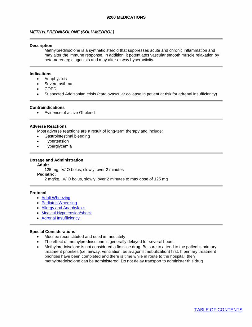
9200 MEDICATIONS
TABLE OF CONTENTS
METHYLPREDNISOLONE (SOLU-MEDROL)
Description Methylprednisolone is a synthetic steroid that suppresses acute and chronic inflammation and may alter the immune response. In addition, it potentiates vascular smooth muscle relaxation by beta-adrenergic agonists and may alter airway hyperactivity.
Indications • Anaphylaxis
• Severe asthma
• COPD
• Suspected Addisonian crisis (cardiovascular collapse in patient at risk for adrenal insufficiency)
Contraindications • Evidence of active GI bleed
Adverse Reactions Most adverse reactions are a result of long-term therapy and include: • Gastrointestinal bleeding
• Hypertension
• Hyperglycemia
Dosage and Administration Adult:
125 mg, IV/IO bolus, slowly, over 2 minutes Pediatric:
2 mg/kg, IV/IO bolus, slowly, over 2 minutes to max dose of 125 mg
Protocol • Adult Wheezing
• Pediatric Wheezing
• Allergy and Anaphylaxis
• Medical Hypotension/shock
• Adrenal Insufficiency
Special Considerations • Must be reconstituted and used immediately
• The effect of methylprednisolone is generally delayed for several hours. • Methylprednisolone is not considered a first line drug. Be sure to attend to the patient’s primary
treatment priorities (i.e. airway, ventilation, beta-agonist nebulization) first. If primary treatment priorities have been completed and there is time while in route to the hospital, then methylprednisolone can be administered. Do not delay transport to administer this drug
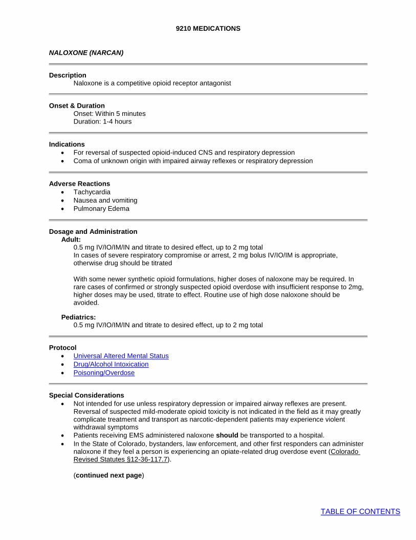
9210 MEDICATIONS
TABLE OF CONTENTS
NALOXONE (NARCAN)
Description Naloxone is a competitive opioid receptor antagonist
Onset & Duration Onset: Within 5 minutes Duration: 1-4 hours
Indications • For reversal of suspected opioid-induced CNS and respiratory depression
• Coma of unknown origin with impaired airway reflexes or respiratory depression
Adverse Reactions • Tachycardia
• Nausea and vomiting
• Pulmonary Edema
Dosage and Administration Adult:
0.5 mg IV/IO/IM/IN and titrate to desired effect, up to 2 mg total In cases of severe respiratory compromise or arrest, 2 mg bolus IV/IO/IM is appropriate, otherwise drug should be titrated
With some newer synthetic opioid formulations, higher doses of naloxone may be required. In rare cases of confirmed or strongly suspected opioid overdose with insufficient response to 2mg, higher doses may be used, titrate to effect. Routine use of high dose naloxone should be avoided.
Pediatrics:
0.5 mg IV/IO/IM/IN and titrate to desired effect, up to 2 mg total
Protocol • Universal Altered Mental Status
• Drug/Alcohol Intoxication
• Poisoning/Overdose
Special Considerations • Not intended for use unless respiratory depression or impaired airway reflexes are present.
Reversal of suspected mild-moderate opioid toxicity is not indicated in the field as it may greatly complicate treatment and transport as narcotic-dependent patients may experience violent withdrawal symptoms
• Patients receiving EMS administered naloxone should be transported to a hospital. • In the State of Colorado, bystanders, law enforcement, and other first responders can administer
naloxone if they feel a person is experiencing an opiate-related drug overdose event (Colorado Revised Statutes §12-36-117.7).
(continued next page)

9210 MEDICATIONS
TABLE OF CONTENTS
• There are significant concomitant inherent risks in patients who have received naloxone, including:
o Recurrent respiratory/CNS depression given short half-life of naloxone o Co-existing intoxication from alcohol or other recreational or prescription drugs o Acetaminophen toxicity from combination opioid/acetaminophen prescriptions o Non-cardiogenic pulmonary edema associated with naloxone use o Acute psychiatric decompensation, overdose, SI/HI or psychosis requiring ED evaluation o Sudden abrupt violent withdrawal symptoms which may limit decision making capacity
• Given the above risks, it is strongly preferred that patients who have received naloxone be transported and evaluated by a physician. However, if the patient clearly has decision-making capacity he/she does have the right to refuse transport. If adamantly refusing, patients must be warned of the multiple risks of refusing transport.
• If the patient is refusing transport contact base. If any concerns or doubts about decision-making capacity exist, err on the side of transport.

9220 MEDICATIONS
TABLE OF CONTENTS
NITROGLYCERIN (NITROSTAT, NITROQUICK, etc)
Description Short-acting peripheral venodilator decreasing cardiac preload and afterload
Onset & Duration Onset: 1-3 min. Duration: 20-30 min.
Indications • Pain or discomfort due to suspected Acute Coronary Syndrome
• Pulmonary edema due to congestive heart failure
Contraindications • Suspected right ventricular ST-segment elevation MI (Inferior STEMI pattern plus ST elevation in
right sided-precordial leads) • Hypotension SBP < 100
• Recent use of erectile dysfunction (ED) medication (e.g. Viagra, Cialis)
Adverse Reactions • Hypotension
• Headache
• Syncope
Dosage and Administration
• Chest Pain: 0.4 mg (1/150 gr) sublingually, every 5 minutes PRN up to a total of 3 doses for persistent CP
• Pulmonary Edema: 0.4 mg (1/150 gr) sublingually, every 5 minutes PRN titrated to symptoms and blood pressure
Protocol • Chest Pain
• CHF/Pulmonary Edema

9230 MEDICATIONS
TABLE OF CONTENTS
OPIOIDS (FENTANYL)
Description Opioid analgesics with desired effects of analgesia, euphoria and sedation as well as undesired effects of respiratory depression and hypotension. A synthetic opioid, fentanyl is 100 times more potent than morphine, and is less likely to cause histamine release.
Indications • Treatment of hemodynamically stable patients with moderate to severe pain due to traumatic or
medical conditions, including cardiac conditions, abdominal pain, back pain, etc.. • Treatment of shivering with Targeted Temperature Management (TTM).
Contraindications • Fentanyl - Hemodynamic instability or shock
• Respiratory depression
Caution/Comments: • Opioids should only be given to hemodynamically stable patients and titrated slowly to effect. • The objective of pain management is not the removal of all pain, but rather, to make the patient’s
pain tolerable enough to allow for adequate assessment, treatment and transport • Respiratory depression, including apnea, may occur suddenly and without warning, and is more
common in children and the elderly. Start with ½ traditional dose in the elderly. • Chest wall rigidity has been reported with rapid administration of fentanyl
Dosage and Administration
FENTANYL: • Adult doses may be rounded to nearest 25 mcg increment • Initial dose in adults typically 100 mcg
• Strongly consider ½ typical dosing in elderly or frail patient • Greater volumes and repeat IN administration are associated with greater drug run off and may
therefore be less effective. If no IV established, administer first dose IN while then attempting to gain access
Adult:
IV/IO route: 1-2 mcg/kg. • Dose may be repeated after 5 minutes and titrated to clinical effect to a maximum
cumulative dose of 3 mcg/kg • Additional dosing requires BASE CONTACT
IN route: 1-2 mcg/kg.
• Dose may be repeated after 10 minutes after initial IN dose to a maximum cumulative dose of 3 mcg/kg. IV route is preferred for repeat dosing.
• Additional dosing requires BASE CONTACT
Pediatric (1-12 years): IV/IO route: 1-2 mcg/kg.
• Dose may be repeated after 5 minutes and titrated to clinical effect to a maximum cumulative dose of 3 mcg/kg.
• Additional dosing requires BASE CONTACT

9230 MEDICATIONS
TABLE OF CONTENTS
IN route: 2 mcg/kg. • Administer a maximum of 1 ml of fluid per nostril • Dose may be repeated after 10 minutes after initial IN dose to a maximum cumulative
dose of 4 mcg/kg. IV route is preferred for repeat dosing.
Pediatric < 1 year: BASE CONTACT
NOTE: IV route is preferred for all opioid administration because of more accurate titration and maximal clinical effect. IO/IN/IM are acceptable alternatives when IV access is not readily available. Repeat doses of IN Fentanyl can be given if IV access cannot be established. Continuous pulse oximetry monitoring is mandatory when any opioid is administered. Frequent evaluation of the patient’s vital signs is also indicated. Emergency resuscitation equipment and naloxone must be immediately available.
Protocol Chest Pain Post Resuscitation Care with ROSC Abdominal Pain Amputations Burns Bites/Stings Snake Bites Face and Neck Trauma Chest Trauma Abdominal Trauma Spinal Trauma Pain Management

9240 MEDICATIONS
TABLE OF CONTENTS
ORAL GLUCOSE (GLUTOSE, INSTA-GLUCOSE)
Description Glucose is the body's basic fuel and is required for cellular metabolism
Indications • Known or suspected hypoglycemia and able to take PO
Contraindications • Inability to swallow or protect airway
• Unable to take PO meds for another reason
Administration All ages: One full tube 15 g buccal.
Protocol • Universal Altered Mental Status
• Hypoglycemia

9250 MEDICATIONS
TABLE OF CONTENTS
OXYGEN
Description Oxygen added to the inspired air increases the amount of oxygen in the blood, and thereby increases the amount delivered to the tissue. Tissue hypoxia causes cell damage and death. Breathing, in most people, is regulated by small changes in the acid-base balance and CO2
levels. It takes relatively large decreases in oxygen concentration to stimulate respiration.
Indications • Suspected hypoxemia or respiratory distress from any cause
• Acute chest or abdominal pain
• Hypotension/shock states from any cause
• Trauma
• Suspected carbon monoxide poisoning
• Obstetrical complications, childbirth
Precautions • If the patient is not breathing adequately, the treatment of choice is assisted ventilation, not just
oxygen. • When pulse oximetry is available, titrate SpO2 to ≥ 90%. This may take some time. • Do not withhold oxygen from a COPD patient out of concerns for loss of hypoxic respiratory drive.
This is never a concern in the prehospital setting with short transport times
Administration
Flow LPM Dosage Indications
Low Flow 1-2 LPM Minor medical / trauma
Moderate Flow 3-9 LPM Moderate medical / trauma
High Flow 10-15 LPM Severe medical / trauma
Special Notes
• Do not use permanently mounted humidifiers. If the patient warrants humidified oxygen, use a single patient use device.
• Adequate oxygenation is assessed clinically and with the SpO2 while adequate ventilation is assessed clinically and with waveform capnography.
OXYGEN FLOW RATES
METHOD FLOW RATE OX YGEN INSPIRED
(approximate) AIR
Room Air 21% Nasal Cannula 1 LPM 24%
2 LPM 28% 6 LPM 44% Simple Face Mask 8 - 10 LPM 40-60% Non-rebreather Mask 10 LPM 90% Bag/Valve/Mask (BVM) Room Air 21%
12 LPM 40% Bag/Valve/Mask with Reservoir 10-15 LPM 90-100% Oxygen-powered breathing device hand-regulated 100%

9260 MEDICATIONS
TABLE OF CONTENTS
PHENYLEPHRINE (INTRANASAL)
Description • Phenylephrine is an alpha adrenergic agonist. When administered intranasally, it causes
vasoconstriction in the nasal mucosa and subsequently decreased bleeding and nasal decongestion.
Indications • Prior to nasotracheal intubation to induce vasoconstriction of the nasal mucosa
• Nosebleed (epistaxis).
Precautions • Avoid administration into the eyes, which will dilate pupil.
Dosage and Administration • Instill two drops of 1% solution, or 2 sprays, in the nostril prior to attempting nasotracheal
intubation. • For patients with active nosebleed, first have patient blow nose to expel clots. Then, administer 2
sprays into affected naris(es).
Protocol • Nasotracheal intubation
• Epistaxis
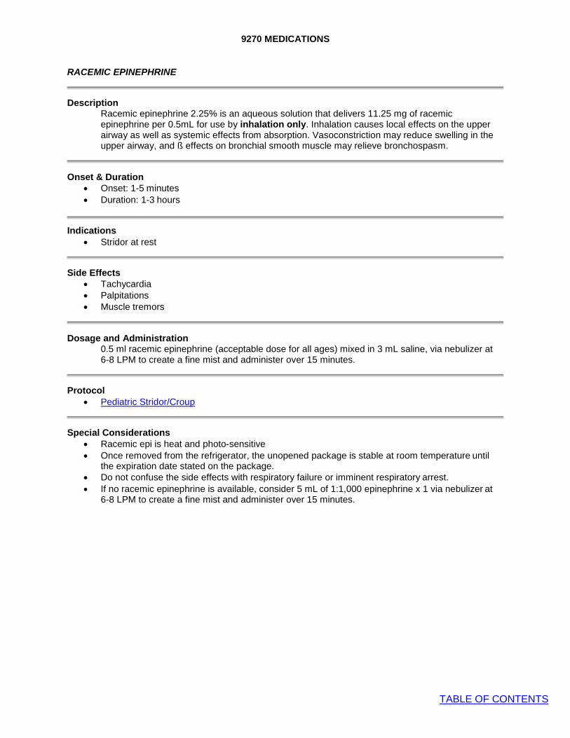
9270 MEDICATIONS
TABLE OF CONTENTS
RACEMIC EPINEPHRINE
Description Racemic epinephrine 2.25% is an aqueous solution that delivers 11.25 mg of racemic epinephrine per 0.5mL for use by inhalation only. Inhalation causes local effects on the upper airway as well as systemic effects from absorption. Vasoconstriction may reduce swelling in the upper airway, and ß effects on bronchial smooth muscle may relieve bronchospasm.
Onset & Duration • Onset: 1-5 minutes
• Duration: 1-3 hours
Indications • Stridor at rest
Side Effects • Tachycardia
• Palpitations
• Muscle tremors
Dosage and Administration 0.5 ml racemic epinephrine (acceptable dose for all ages) mixed in 3 mL saline, via nebulizer at 6-8 LPM to create a fine mist and administer over 15 minutes.
Protocol • Pediatric Stridor/Croup
Special Considerations • Racemic epi is heat and photo-sensitive
• Once removed from the refrigerator, the unopened package is stable at room temperature until the expiration date stated on the package.
• Do not confuse the side effects with respiratory failure or imminent respiratory arrest. • If no racemic epinephrine is available, consider 5 mL of 1:1,000 epinephrine x 1 via nebulizer at
6-8 LPM to create a fine mist and administer over 15 minutes.

9280 MEDICATIONS
TABLE OF CONTENTS
SODIUM BICARBONATE
Description Sodium bicarbonate is an alkalotic solution, which neutralizes acids found in the body. Acids are increased when body tissues become hypoxic due to cardiac or respiratory arrest.
Indications • Tricyclic overdose with arrhythmias, widened QRS complex or hypotension. • Suspected hyperkalemic pulseless arrest: consider in patients with known renal failure/dialysis.
Contraindications • Metabolic and respiratory alkalosis
• Hypocalcemia
• Hypokalemia
Adverse Reactions • Metabolic alkalosis
• Paradoxical cerebral intracellular acidosis
• Sodium bolus can lead to volume overload
Drug Interactions • May precipitate in calcium solutions. • Alkalization of urine may increase half-lives of certain drugs. • Vasopressors may be deactivated.
Dosage and Administration Adults and children (> 10 kg), 8.4% Tricyclic OD with hypotension or prolonged QRS > 0.10 sec or suspected hyperkalemia- related pulseless arrest:
• 1 mEq/kg slow IV push. Repeat if needed in 10 minutes.
Protocol • Universal Pulseless Arrest • Poisoning/Overdose
Special Considerations • Sodium bicarbonate administration increases CO2 which rapidly enters cells, causing a
paradoxical intracellular acidosis. • Sodium bicarb is no longer recommended for routine use in prolonged cardiac arrest. Its use in
pulseless arrest should be limited to known or suspected hyperkalemia (e.g. dialysis patient), or arrest following tricyclic overdose.

9290 MEDICATIONS
TABLE OF CONTENTS
TOPICAL OPHTHALMIC ANESTHETICS
Description Proparacaine and tetracaine are local anesthetics approved for ocular administration for relief of eye pain caused by corneal abrasion or chemical injury.
Indications
• Pain secondary to eye injuries and corneal abrasions. • Topical anesthetic to facilitate eye irrigation.
Contraindications • Known allergy to local anesthetics. • Globe lacerations or rupture.
Precautions • Transient burning/stinging when initially applied.
Dosage and Administration • Instill 2 drops into affected eye. Contact Base for repeat dosing.
Special Considerations • This is single patient use. Unused portions should be discarded and only new bottles may be
used. • Do not administer until patient consents to transport and transport has begun. • Topical ophthalmic anesthetics should never be given to a patient for self-administration.
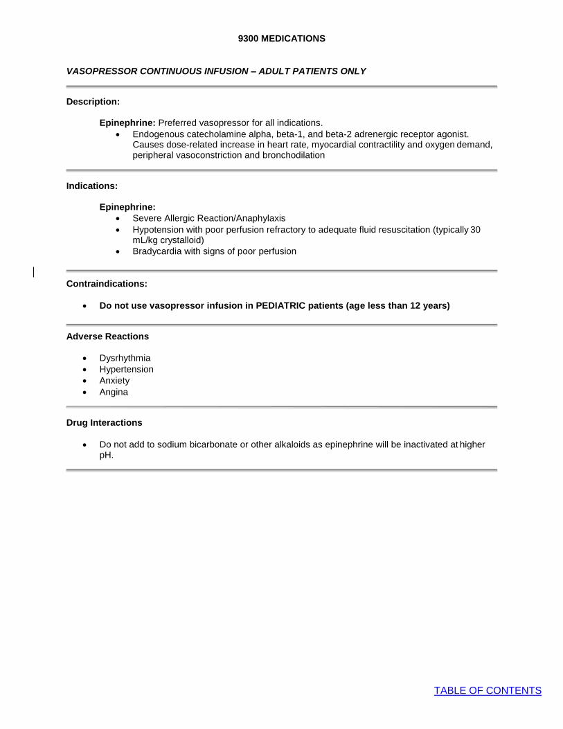
9300 MEDICATIONS
TABLE OF CONTENTS
VASOPRESSOR CONTINUOUS INFUSION – ADULT PATIENTS ONLY
Description:
Epinephrine: Preferred vasopressor for all indications. • Endogenous catecholamine alpha, beta-1, and beta-2 adrenergic receptor agonist.
Causes dose-related increase in heart rate, myocardial contractility and oxygen demand, peripheral vasoconstriction and bronchodilation
Indications:
Epinephrine: • Severe Allergic Reaction/Anaphylaxis • Hypotension with poor perfusion refractory to adequate fluid resuscitation (typically 30
mL/kg crystalloid) • Bradycardia with signs of poor perfusion
Contraindications:
• Do not use vasopressor infusion in PEDIATRIC patients (age less than 12 years)
Adverse Reactions
• Dysrhythmia
• Hypertension
• Anxiety
• Angina
Drug Interactions
• Do not add to sodium bicarbonate or other alkaloids as epinephrine will be inactivated at higher pH.

9300 MEDICATIONS
TABLE OF CONTENTS
Dosage and Administration:
Epinephrine: • Mix: inject 1 mg epinephrine into 1000 mL Normal Saline bag to achieve 1mcg/mL concentration
(This means 1 mL of 1:1000 or 10 mL of 1:10,000 – either way 1 mg of drug). Use macro drip set. • Adult IV/IO: Begin IV/IO infusion wide open to gravity to give small aliquots of fluid. Typical
volumes are less than 100 mL of total fluid, as typical doses are expected to be < 100 mcg. Titrate to desired hemodynamic effect with goal BP of > 90 mmHg systolic, improved respiratory status (bronchodilation), and improved perfusion/mentation.
Protocol • Post-Resuscitation Care with ROSC
• Bradycardia with Poor Perfusion
• Allergy and Anaphylaxis
• Medical Hypotension/Shock
• Overdose and Acute Poisoning
• Sepsis
Special Considerations • May increase myocardial oxygen demand and angina pectoris. Use with caution in patients with
known or suspected CAD

THREAT BASED CARE ADDENDUM
TABLE OF CONTENTS
COLD ZONE (AREA SECURED AND SAFE)
• Secondary triage of casualties arriving at CCP • Assess any previous interventions and address if not adequate • Refer to General Trauma Care protocol and complete full physical exam • Treatment Priorities:
1. Hemorrhage control (tourniquet and hemostatic gauze) 2. Airway management (NPA or OPA, ALS airway if indicated, Oxygen) 3. Assess/treat for tension pneumothorax 4. IV/IO access. Ref Traumatic Shock protocol for IV fluid management 5. Keep patient warm to prevent hypothermia 6. Spinal immobilization if indicated 7. Pain Control per protocol
• Transport casualties to appropriate trauma centers per protocol
WARM ZONE (AREA SECURED BUT NOT SAFE)
• Primary triage using SALT triage method if multiple casualties
• Assess any previous interventions and address if not adequate • Treatment Priorities:
1. Hemorrhage control (tourniquet and hemostatic gauze) 2. Basic airway management (NPA or OPA, place in recovery position, no ALS airway) 3. If penetrating chest wounds, apply vented chest seal. 4. Assess/treat for tension pneumothorax (auscultation of lung sounds not practical
usually in WARM ZONE. Assess and treat if respiratory distress, penetrating chest trauma, and shock (absent peripheral pulses, pale, cool, and diaphoretic)
5. If delay in extraction, consider hypothermia prevention and IV/IO if situation allows.
• Pain control and spinal immobilization not indicated in this phase of care. • Extraction teams should evacuate casualties to CCP
• The purpose of this addendum is to provide guidance and best practice principles for care of patients in a tactical environment. These concepts are based on Tactical Combat Casualty Care (TCCC) principles developed by the US military.
• This addendum applies to ASHER incidents as defined by departmental policy. This addendum is meant to augment the Aurora EMS Protocols, and will reference the applicable protocol. Any exceptions to an indication for a procedure in this addendum will be noted.
HOT ZONE (AREA NOT SECURED OR SAFE)
• Treatment priority is to have the patient move or move the patient to an area of cover and relative safety. Crews should utilize rescue techniques that minimize their exposure to potential threats.
• Treatment Priorities: 1. Hemorrhage control (tourniquet only. If patient is conscious, crews can facilitate self-
application of a tourniquet. Apply tourniquet “High and Tight”, which means as
proximal as possible, unless injury site is visible and apparent. • Airway management does not occur in this phase of care • Ideally, only SWAT medics operate in this zone. However, any WARM ZONE can become
HOT and AFR crews should always be aware of this.