ANTIDIABETIC ACTIVITY OF ALCOHOLIC FRUIT EXTRACT OF
MALLOTUS PHILIPPENSIS MUELL.ARG. IN STREPTOZOTOCIN
INDUCED DIBETIC RATS
Dissertation Submitted to
THE TAMILNADU Dr. M.G.R. MEDICAL UNIVERSITY,
CHENNAI- 32.
In partial fulfilment for the requirements for the award of the degree of
MASTER OF PHARMACY
IN
BRANCH – IV- PHARMACOLOGY
Submitted by
MUHAMMED SHABEER.A
REGISTER NO: 261425506
Under the guidance of
Mrs. G.SUMITHIRA, M. Pharm.,
Assistant Professor, Dept. of Pharmacology
THE ERODE COLLEGE OF PHARMACY AND RESEARCH INSTITUTE,
ERODE- 638112.
OCTOBER - 2016
EVALUATION CERTIFICATE
This is to certify that the dissertation work entitled “Antidiabetic activity of
alcoholic fruit extract of Mallotus Philippensis Muell.Arg. in Streptozotocin
induced diabetic rats” submitted by Register No: 261425506 to The Tamil Nadu
Dr. M.G.R Medical University, Chennai, in partial fulfilment for the degree of Master
of Pharmacy in Pharmacology is the bonafide work carried out under guidance and
direct supervision of Mrs. G.SUMITHIRA, M. Pharm., Assistant Professor at the
Department of Pharmacology, The Erode College of Pharmacy and Research
Institute, Erode-638112 and was evaluated by us during the academic year 2015-
2016.
1. INTERNAL EXAMINERS 2.EXTERNAL EXAMINERS
3. CONVENER OF EXAMINATION
Examination Centre: The Erode College of Pharmacy and Research Institute.
Date:
The Erode College of Pharmacy and Research Institute
Dr. V. Ganesan, M.Pharm., Ph.D.,
Principal,
Professor and Head, Department of Pharmaceutics,
The Erode College of Pharmacy and Research Institute,
Erode - 638112.
CERTIFICATE
This is to certify that the dissertation work entitled “Antidiabetic activity of
alcoholic fruit extract of Mallotus Philippensis Muell.Arg. in Streptozotocin
induced diabetic rats” submitted by Register No: 261425506 to The Tamil Nadu
Dr. M.G.R Medical University, Chennai, in partial fulfilment for the degree of Master
of Pharmacy in Pharmacology is the bonafide work carried out under the guidance
and direct supervision of Mrs. G.SUMITHIRA, M. Pharm., Assistant Professor at
the Department of Pharmacology, The Erode College of Pharmacy and Research
Institute, Erode- 638112, during the academic year 2015-2016.
Dr. V. Ganesan, M.Pharm., Ph.D.,
Principal
Place : Erode
Date :
The Erode College of Pharmacy and Research Institute
Prof. Dr. M. Periasamy, M.Pharm., Ph.D.,
Professor and Head,
Department of Pharmacology,
The Erode College of Pharmacy and Research Institute,
Erode - 638112.
CERTIFICATE
This is to certify that the dissertation work entitled “Antidiabetic activity of
alcoholic fruit extract of Mallotus Philippensis Muell.Arg. in Streptozotocin
induced diabetic rats” submitted by Register No: 261425506 to The Tamil Nadu
Dr. M.G.R Medical University, Chennai, in partial fulfilment for the degree of Master
of Pharmacy in Pharmacology is the bonafide work carried out under the guidance
and direct supervision of Mrs. G. Sumithira, M.Pharm., Assistant Professor at the
Department of Pharmacology, The Erode College of Pharmacy and Research
Institute, Erode- 638112, during the academic year 2015-2016.
Prof. Dr. M. Periasamy, M.Pharm., Ph.D.,
Prof. & HOD
Place : Erode
Date :
The Erode College of Pharmacy and Research Institute
Mrs. G.SUMITHIRA, M. Pharm.,
Assistant Professor,
Department of Pharmacology,
The Erode College of Pharmacy and Research Institute,
Erode - 638112.
CERTIFICATE
This is to certify that the dissertation work entitled “Antidiabetic activity of
alcoholic fruit extract of Mallotus Philippensis Muell.Arg. in Streptozotocin
induced diabetic rats” submitted by Register No: 261425506 to The Tamil Nadu
Dr. M.G.R Medical University, Chennai, in partial fulfilment for the degree of Master
of Pharmacy in Pharmacology is the bonafide work carried out under my guidance
and direct supervision at the Department of Pharmacology, The Erode College of
Pharmacy and Research Institute, Erode-638112, during the academic year 2015-
2016.
Mrs. G.Sumithira, M.Pharm.,
Assistant professor
Place: Erode
Date:
DECLARATION
I do hereby declare that the dissertation work entitled “Antidiabetic activity of alcoholic fruit extract of Mallotus Philippensis Muell.Arg. in Streptozotocin
induced diabetic rats” submitted to The Tamil Nadu Dr. M.G.R Medical University,
Chennai, in the partial fulfilment for the Degree of Master of Pharmacy in
Pharmacology, was carried out by myself under the guidance and direct supervision
of Mrs. G.SUMITHIRA, M. Pharm., Assistant Professor, at the Department of
Pharmacology, The Erode College of Pharmacy and Research Institute, Erode-
638112, during the academic year 2015-2016.
This work is original and has not been submitted in part or full for the award of
any other Degree or Diploma of this or any other University.
Place: Erode Register No: 261425506
Date:
ACKNOWLEDGEMENTS
The secret of success is undaunted ardor, motivation, dedication, confidence
on self and above all the blessing of god. I bow in reverence to the almighty for
bestowing upon me all his kindness that has helped me throughout the journey of my
life. Success is an outcome of collaborated efforts aimed that achieving different
goals. I hereby take this opportunity to acknowledge all those who have helped me in
the completion of this dissertation work.
It gives me an immense pleasure to express my deepest than heartfelt,
indebtedness and regards to my respected guide Mrs.G.Sumithira, M.Pharm.,
Asst. Professor, Department of Pharmacology for her inspiring nature, constant
encouragement, valuable guidance and support to me throughout the course of this
work.
I express my sincere thank and respectful regards to the President Dr .K.R.
Paramasivam M.sc., Ph.D., and the Secretary & Correspondant Mr. A.
Natarajan, B.A., H.D.C., for providing necessary facilities to carry out this
dissertation work successfully.I express my deep sense of gratitude to honourable
Principal & Prof. Dr. V. Ganesan, M.Pharm., Ph.D., and HOD, Dept of
Pharmaceutics, The Erode college of Pharmacy and Research Institute, for
providing necessary facilities to carry out this dissertation work successfully.
I now take this opportunity to express my sincere thanks to Prof.Dr. M.
Periasamy M.Pharm.,Ph.D., HOD, Dept of Pharmacology for giving his valuable
guidance and constant encouragement throughout the project work.
I express my heartful thank to Vice- Principal & Prof. Dr. V.S. Saravanan,
M.Pharm., Ph.D., and HOD, Dept of Pharmaceutical Analysis, for providing
necessary facilities to carry out this dissertation work successfully.
I express my sincere thanks to Mr. P. Royal Frank M.Pharm.,
Mrs. Rajamathanky M.Pharm., and Mrs.Rajeswari M.Pharm., Dept of
Pharmacology, for their support and encouragement throughout the study.
I express my sincere thanks to Prof. Dr. P Muralidharan, M.Pharm., Ph.D.,
Dept of Pharmacology, C.L.BAID METHA COLLEGE OF PHARMACY for
providing necessary facilities to carry out this dissertation work successfully.
I express my great thanks to Mrs. Uma Maheswari, M.Com, Lab attender,
(Department of Pharmacology), for her sincere help and technical support during the
extraction process.
I express my heartful thanks to Mrs. Chithra, D.pharm, (Store keeper),
Mr. Velmurugan, D.Pharm, Mr. Kannan, D.Pharm and Mrs. Kanimozhi for their
help during plant extraction process and phytochemical analysis.
I express my sincere thanks to Mr. Varatharajan Librarian who helped me to
take reference for carryout my project work.
I also thank to my friends Mr. Danish T.K, Mr.Tamilarasan K, Mr.
Muhammed Anas K.P, Ms. Meera Nadhini, Mr. Akhilan, Ms. Ashma,
Ms. Kavya, Mr. Subash, Mr. Ragupathi, Mr. Azharudheen T.P, Mr. Haneesh V,
Mr. Mohamed Fajir K, Mr. Mohamed Anees V.T, Mr. Shuaib M.V, Mr. Ansar T.P,
Mr. Muhammed Shafeeq, Mr. Muhammed Nashad K, Mr. Savya Sai K.P, Mr.
Safruq J.B and all others from the Department of Pharmacology for spending their
valuable time during various stages of my project work.
Last but not least I express my warmest and warm and most important
acknowledgement to my grand father Mr.Ayamu my parents Mr.Muhammedali A,
Mrs. Nafeesa and My small father Mr.Yousaf small mother Mrs.Sajitha my loving
sister Ms.Kamarunneesa and brothers Mr.Muhammed Kabeer, Mr.Muhammed
Muneer, Mr. Shihabudheen, Mr. Ramees and my friends Mr. Muhammed Rafi K.V
with deep appreciation and moral support encouragement and everlasting love that
served as a source of my inspiration, strength, determination and enthusiasm at
each and every front of my life, to transfer my dreams in to reality.
With Thanks
Reg.No: 261425506
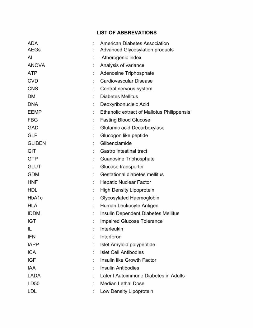
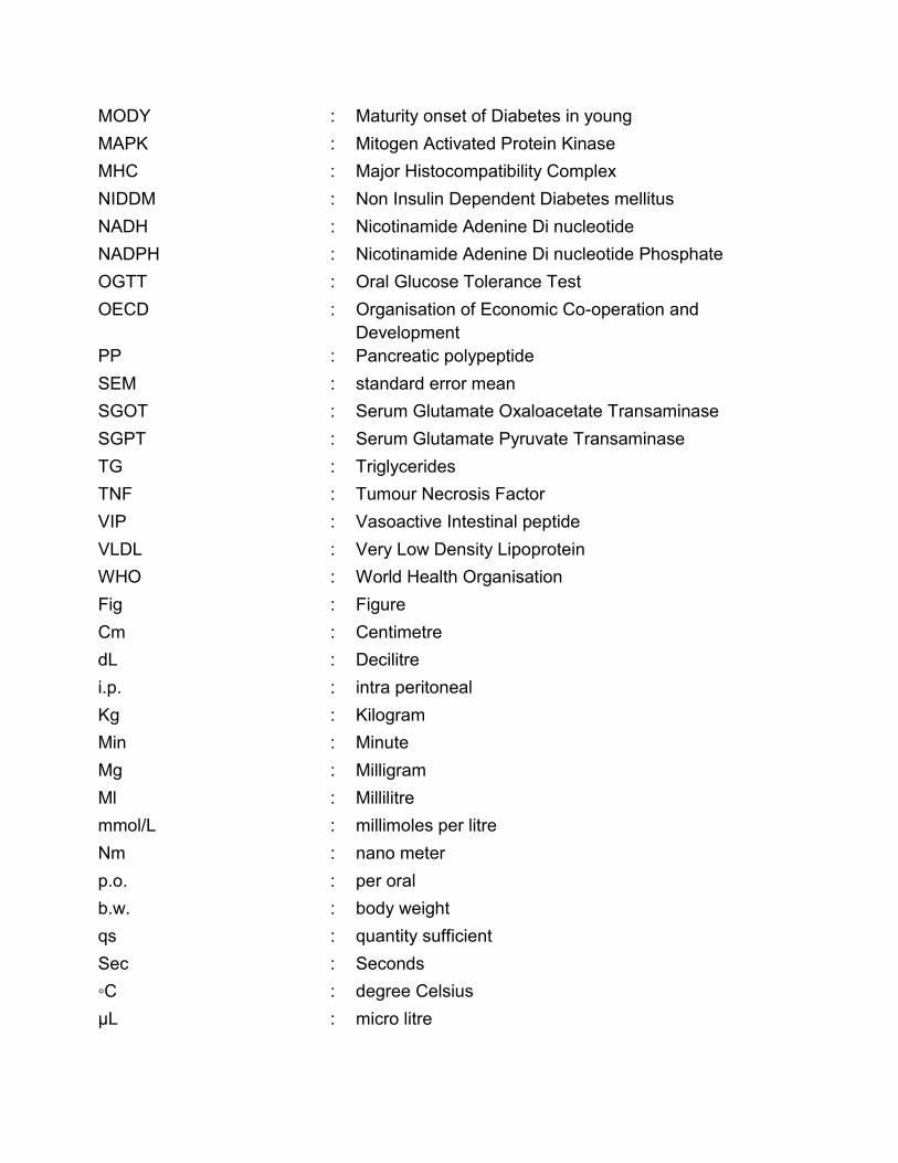
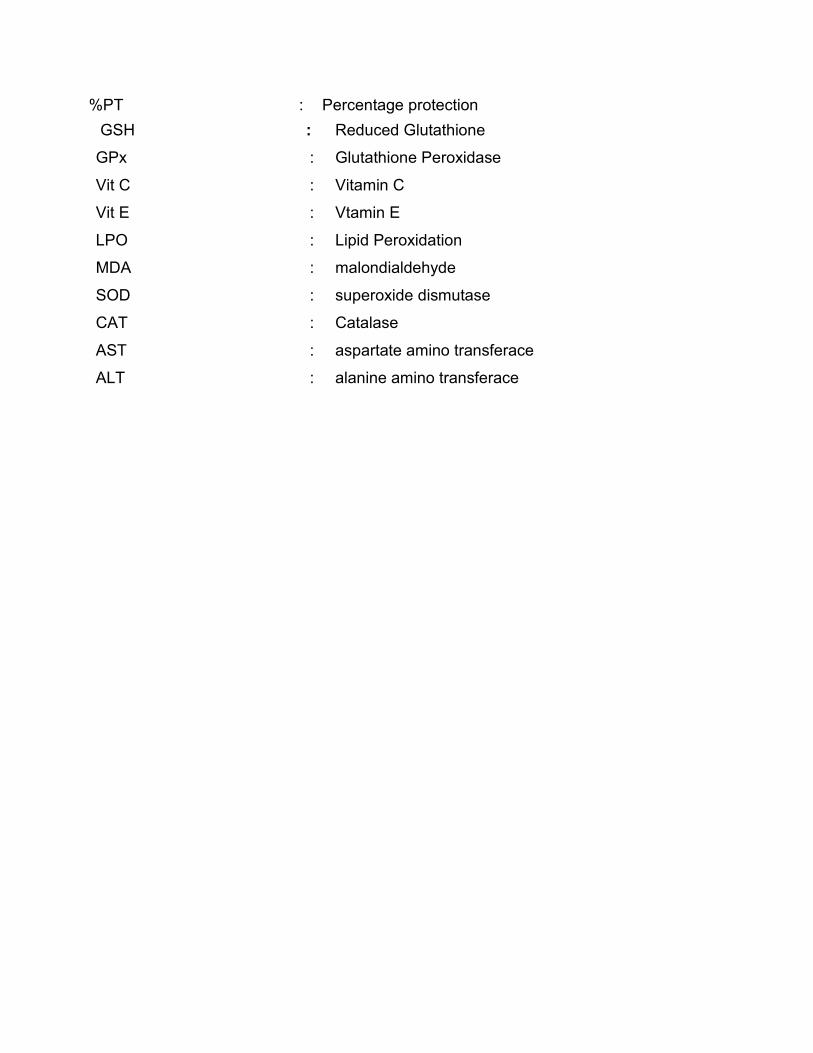
LIST OF ABBREVATIONS
ADA : American Diabetes Association
AEGs : Advanced Glycosylation products
AI : Atherogenic index
ANOVA : Analysis of variance
ATP : Adenosine Triphosphate
CVD : Cardiovascular Disease
CNS : Central nervous system
DM : Diabetes Mellitus
DNA : Deoxyribonucleic Acid
EEMP : Ethanolic extract of Mallotus Philippensis
FBG : Fasting Blood Glucose
GAD : Glutamic acid Decarboxylase
GLP : Glucogon like peptide
GLIBEN : Glibenclamide
GIT : Gastro intestinal tract
GTP : Guanosine Triphosphate
GLUT : Glucose transporter
GDM : Gestational diabetes mellitus
HNF : Hepatic Nuclear Factor
HDL : High Density Lipoprotein
HbA1c : Glycosylated Haemoglobin
HLA : Human Leukocyte Antigen
IDDM : Insulin Dependent Diabetes Mellitus
IGT : Impaired Glucose Tolerance
IL : Interleukin
IFN : Interferon
IAPP : Islet Amyloid polypeptide
ICA : Islet Cell Antibodies
IGF : Insulin like Growth Factor
IAA : Insulin Antibodies
LADA : Latent Autoimmune Diabetes in Adults
LD50 : Median Lethal Dose
LDL : Low Density Lipoprotein
MODY : Maturity onset of Diabetes in young
MAPK : Mitogen Activated Protein Kinase
MHC : Major Histocompatibility Complex
NIDDM : Non Insulin Dependent Diabetes mellitus
NADH : Nicotinamide Adenine Di nucleotide
NADPH : Nicotinamide Adenine Di nucleotide Phosphate
OGTT : Oral Glucose Tolerance Test
OECD : Organisation of Economic Co-operation and
Development
PP : Pancreatic polypeptide
SEM : standard error mean
SGOT : Serum Glutamate Oxaloacetate Transaminase
SGPT : Serum Glutamate Pyruvate Transaminase
TG : Triglycerides
TNF : Tumour Necrosis Factor
VIP : Vasoactive Intestinal peptide
VLDL : Very Low Density Lipoprotein
WHO : World Health Organisation
Fig : Figure
Cm : Centimetre
dL : Decilitre
i.p. : intra peritoneal
Kg : Kilogram
Min : Minute
Mg : Milligram
Ml : Millilitre
mmol/L : millimoles per litre
Nm : nano meter
p.o. : per oral
b.w. : body weight
qs : quantity sufficient
Sec : Seconds
◦C : degree Celsius
µL : micro litre
%PT : Percentage protection
GSH : Reduced Glutathione
GPx : Glutathione Peroxidase
Vit C : Vitamin C
Vit E : Vtamin E
LPO : Lipid Peroxidation
MDA : malondialdehyde
SOD : superoxide dismutase
CAT : Catalase
AST : aspartate amino transferace
ALT : alanine amino transferace
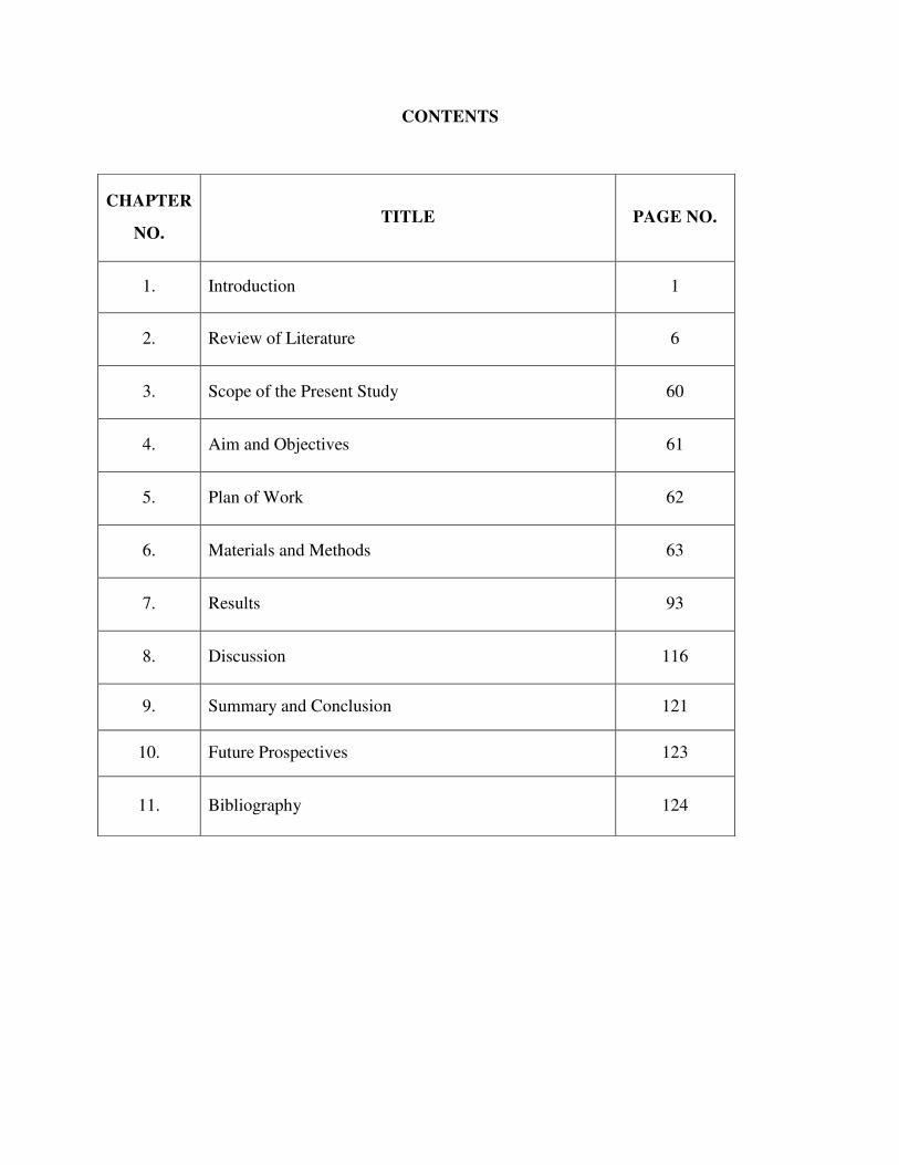
CONTENTS
CHAPTER
NO.
TITLE PAGE NO.
1. Introduction 1
2. Review of Literature 6
3. Scope of the Present Study 60
4. Aim and Objectives 61
5. Plan of Work 62
6. Materials and Methods 63
7. Results 93
8. Discussion 116
9. Summary and Conclusion 121
10. Future Prospectives 123
11. Bibliography 124
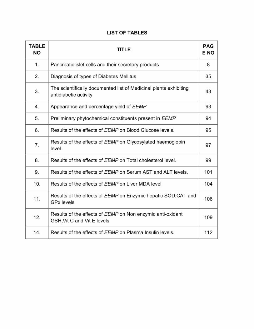
LIST OF TABLES
TABLE
NO TITLE
PAG
E NO
1. Pancreatic islet cells and their secretory products 8
2. Diagnosis of types of Diabetes Mellitus 35
3. The scientifically documented list of Medicinal plants exhibiting
antidiabetic activity 43
4. Appearance and percentage yield of EEMP 93
5. Preliminary phytochemical constituents present in EEMP 94
6. Results of the effects of EEMP on Blood Glucose levels. 95
7. Results of the effects of EEMP on Glycosylated haemoglobin
level. 97
8. Results of the effects of EEMP on Total cholesterol level. 99
9. Results of the effects of EEMP on Serum AST and ALT levels. 101
10. Results of the effects of EEMP on Liver MDA level 104
11. Results of the effects of EEMP on Enzymic hepatic SOD,CAT and
GPx levels 106
12. Results of the effects of EEMP on Non enzymic anti-oxidant
GSH,Vit C and Vit E levels 109
14. Results of the effects of EEMP on Plasma Insulin levels. 112
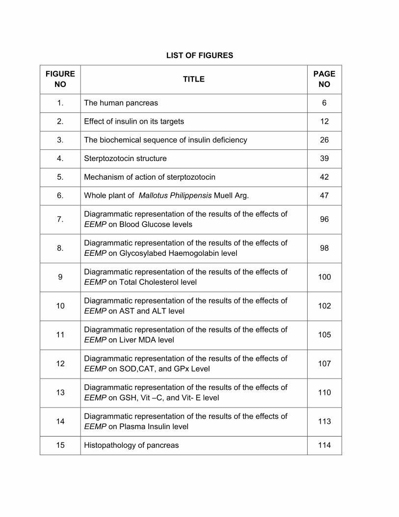
LIST OF FIGURES
FIGURE
NO TITLE
PAGE
NO
1. The human pancreas 6
2. Effect of insulin on its targets 12
3. The biochemical sequence of insulin deficiency 26
4. Sterptozotocin structure 39
5. Mechanism of action of sterptozotocin 42
6. Whole plant of Mallotus Philippensis Muell Arg. 47
7. Diagrammatic representation of the results of the effects of
EEMP on Blood Glucose levels 96
8. Diagrammatic representation of the results of the effects of
EEMP on Glycosylabed Haemogolabin level 98
9 Diagrammatic representation of the results of the effects of
EEMP on Total Cholesterol level 100
10 Diagrammatic representation of the results of the effects of
EEMP on AST and ALT level 102
11 Diagrammatic representation of the results of the effects of
EEMP on Liver MDA level 105
12 Diagrammatic representation of the results of the effects of
EEMP on SOD,CAT, and GPx Level 107
13 Diagrammatic representation of the results of the effects of
EEMP on GSH, Vit –C, and Vit- E level 110
14 Diagrammatic representation of the results of the effects of
EEMP on Plasma Insulin level 113
15 Histopathology of pancreas 114
INTRODUCTION
DEPARTMENT OF PHARMACOLOGY, THE ERODE COLLEGE OF PHARMACY, ERODE Page 1
1. INTRODUCTION
Diabetes mellitus commonly referred as a Diabetes, it is a group of metabolic
diseases in which there is high blood sugar levels over a prolonged period.1 Symptoms
of high blood sugar include frequent urination, increased thrist, and increased hunger. If
left untreated, diabetes can cause many complications.2 Acute complications can
include diabetic ketoacidosis, non ketotic hyperosmolar coma, or death.3 Serious long
term complications include heart disease, stroke, chronic kidney failure, foot ulcers, and
damage to eyes.2
Diabetes is due to either the pancreas not producing enough insulin or the cells
of the body not responding properly to the insulin produced.4 There are four types of
diabetes mellitus:
Type 1 diabetes results from the pancreas‘s failure to produce enough insulin.
This form referred as “insulin dependent diabetes mellitus” (IDDM) or “juvenile
diabetes”. The cause is unknown.2
Type 2 diabetes begins with insulin resistance, a condition in which cells fail to
respond to insulin properly. As the disease progress a lack of insulin may also
develop. This form referred as “non insulin dependent diabetes mellitus”
(NIDDM) or “adult onset diabetes”. The primary cause is excessive body weight
and not enough exercise.2
Gestational diabetes is the third main form and occurs when pregnant women
without a previous history of diabetes develop high blood sugar levels. They
prone to diabetes in future.5
MODY Maturity Onset Diabetes of the Young is the fourth type of diabetes.
Specific monogenetic defects of the beta-cells have been identified and usually
give rise to maturity onset diabetes of the young (MODY). MODY is defined as a
genetic defect in beta-cell function.6
Prevention and treatment involve maintaining a healthy diet, regular physical
exercise, a normal body weight, and avoiding use of tobacco. Control of blood pressure
and maintaining proper foot care are important for people with the disease. Type 1 DM
INTRODUCTION
DEPARTMENT OF PHARMACOLOGY, THE ERODE COLLEGE OF PHARMACY, ERODE Page 2
must be managed with insulin injections. Type 2 DM may be treated with medications
with or without insulin.7 Insulin and some oral medications can cause low blood sugar.8
Weight loss surgery in those with obesity is sometimes an effective measure in those
with type 2 DM.9 gestational diabetes usually resolves after the birth of the baby. Mody
type diabetes managed by sufonylureas treatment.6
As of 2015, an estimated 415 million people had diabetes worldwide, with type 2
DM making up about 90% of the cases. This represents 8.3% of the adult population,
with equal rates in women and men. As of 2014, trends suggested the rate would
continue to rise. Diabetes at least doubles a person’s risk of early death. From 2012 to
2015, approximately 1.5 to 5.0 million deaths each year resulted from diabetes.10
ROLE OF PHYTOMEDICINE IN THE TREATMENT OF DIABETES
The Ayurvedic concept appeared and developed between 200 and 500 B.C. in
India. The literal meaning of ayurveda is “science of life”, because ancient Indian
system of health care focused views of man and his illness. It is pointed out that the
positive health means metabolically well- balanced human beings. Ayurveda remains an
important system of medicine and drug therapy in India.10
In Ayurveda, diabetes falls under the term madhumeha. Various types of herbal
preparations such as decoctions (boiled extracts), swaras (expressed juices) Asav-
Arishta (fermented juices) and powders have been mentioned for the treatment of
madhumeha. These indigenous medicines may not have adverse effects in therapeutic
doses. It is mentioned in ancient texts such as the Charkas Samhinta that a single herb
exerts different actions on many diseases and that each herb may have one dominating
effect and other comparatively subsidiary effects. It is also mentioned that an herbal
drug can also have synergistic and antagonistic effects in combination with other
herbs.11
Out of an estimated 250000 higher plants, less than 1% have been screened
pharmacologically and very few in regard to diabetes mellitus.13 In India, indigenous
remedies have been used in the treatment of DM since the time of Charaka and
Sushruta (6th century BC). Plants have always been an exemplary source of drugs and
INTRODUCTION
DEPARTMENT OF PHARMACOLOGY, THE ERODE COLLEGE OF PHARMACY, ERODE Page 3
a many of the currently available drugs have been delivery directly or indirectly from
them. Ethnopharmacological surveys indicate that more than 1200 plants are used in
traditional medicine for their alleged hypoglycaemic activity. Medicinal plants, since
times immemorial, have been used in virtually all cultures as a source of medicine.14
The natural detoxification process of the body is effectively enhanced by herbal
medicines and also very good in boosting the immune system and also herbal
medicines in the treatment of a disease take into account pathogens, whole body
balance, body chemistry with scientific proof of them in the treatment of a disease.
Because of all these reasons, herbal medicines are preferred. Wide array of plant
derived active principles have demonstrated their anti-diabetic activity. The main active
constituents of these plants include guanidine, steroids, carbohydrates, glycopeptides,
terpenoids, glycosides, flavanoids, alkaloids, amino acids and inorganic ions. These
affect various metabolic cascades, which directly or indirectly affect the level of glucose
in the human body.15
PHYTOCONSTITUENTS HAVING ANTI DIABETIC ACTIVITY 16,17
The constituents that come under the category of polysaccharides, peptides,
alkaloids, glycopeptides, triterpenoids, amino acids, steroids, xanthenes, flavanoids
lipids, phenoics, coumarins, irirods, alkyl disulfides, inorganic ions and guanidine have
been reported to have anti-diabetic activity. Specifically the following constituents are
reported to have anti-diabetic activity, amino acid like hypoglycine A and hypoglycine B,
alkaloids like catharanthine, leurosine, lochnerine, arecoline and vindoline, pinitol,
epicatichin, bengalenoside, anemarans (A, B, C,D), atarctans (A,B,C), dioscorin
(A,B,C,D,E,F), ephedrans (A,B,C,D,E), glycoprotein (moran A), mucilage, nimbidin,
peptides (P insulin), S- methyl cysteine sulphoxide, S- allyl cysteine sulphoxide,
andrographollide, allicin (thio-2- propene-1- sulfinic acid S-allyl ester), shamimin, beta
vulgarosides I-IV, glycoside of leucopelargonidin and leucodelphindin, magniferin,
marsupsin, pterosupin, pterostilbene, pinoline, naringin, salacinol, hesperidin, berberine,
chlorgenic acid, charantin, swerchirin, epigallocatechin gallate, trigonelline, harmane,
norharmane, lactucaside, beta-sitosterol, lactucain C, kalopanax saponin A, gymnemic
acid IV, hederagenin, furfuran lignin, oleanoloc acid, elatosides (E,G,H,I), cryptolepine,
INTRODUCTION
DEPARTMENT OF PHARMACOLOGY, THE ERODE COLLEGE OF PHARMACY, ERODE Page 4
caffeoyl glucoside, momordin Ic, bellidifolin, kolaviron, scoparianosides A, B and C,
kaempferol glucosides, bakuchiol, trihydroxyoctadecadienoic acids, escins (Ia,Ia,IIa,IIb
and IIIa), thysanolactone, kotalanol, fagomine, 3-O-beta-D- glucopyranosylfagomine, 4-
O_beta- d glucopyranosylfagomine, 3-epifagomine.
Myrciacitrins I and II, myrciaphenones A and B, momordin, prunin, tormentic
acid, 8- debenzylpaenoniflorin, coutareagin, senticoside, lithosperman, senegin II, Z-
senegasaponins a and b and E and Z-senegasapponins, E and Z- senegins ( II, III and
IV), boussingoside, paenoflorin, pachymaran, saciharan, coixan, oleanolic acid
glycosides, ginsenoside, laminaran, masoprocol, senticoside A, abelmosan, ursolic
acid, trichosan also exhibit anti-diabetic activity.
Flavones C- glycoside, icarin, neomyrtillin, kakonein, acarbose, voglibose, ferulic
acid, brazilin, hyperin, sappanchalone, anisodamine, multiflorine, 3-deoxy sappanone,
protosappanin A also have anti-diabetic activity.
Oral hypoglycaemic agents like sulphonylureas and biguanides are still the major
players in the management of the disease, but there is growing interest in herbal
remedies due to the side effects associated with the oral hypoglycemic agents. Herbal
medicines have been highly esteemed source of medicine throughout the human
history. They are widely used today indicating that herbs are a growing part of modern
high-tech medicine. In recent times, there has been a revived interest within the plant
remedies. In this review article an attempt has been made to focus on hypoglyceamic
plants and may be useful to the health professionals, scientists and scholars working in
the field of pharmacology & therapeutics to develop evidence based alternative
medicine to cure different kinds of diabetes in man and animals.17
In addition, a major effort was directed towards discovery of novel anti-diabetic
agents. The interest in herbal drug research continues with an expectation that
someday rather the other day, we would be able to bring a safer and more effective
compound with all the desired parameters of a drug that could replace the synthetic
medicines, which resulted in the discovery of several patented compounds,
cryptolepine, maprouneacin, 3β, 30-dihydroxylupen-20 (29)-en-2-one, harunganin,
INTRODUCTION
DEPARTMENT OF PHARMACOLOGY, THE ERODE COLLEGE OF PHARMACY, ERODE Page 5
vismin, and quinines SP 18905. The most interesting discovery was nor
dihydroguaiaretic acid which besides being active orally in db/db diabetic mice, also
lowered cholesterol levels. This is considered as the unique quality of herbs, which was
not observed in any synthetic medicine.
REVIEW OF LITERATURE
DEPARTMENT OF PHARMACOLOGY, THE ERODE COLLEGE OF PHARMACY, ERODE Page 6
2. REVIEW OF LITERATURE
2.1 HUMAN PANCREAS
2.1.1 ANATOMY AND PHYSIOLOGY
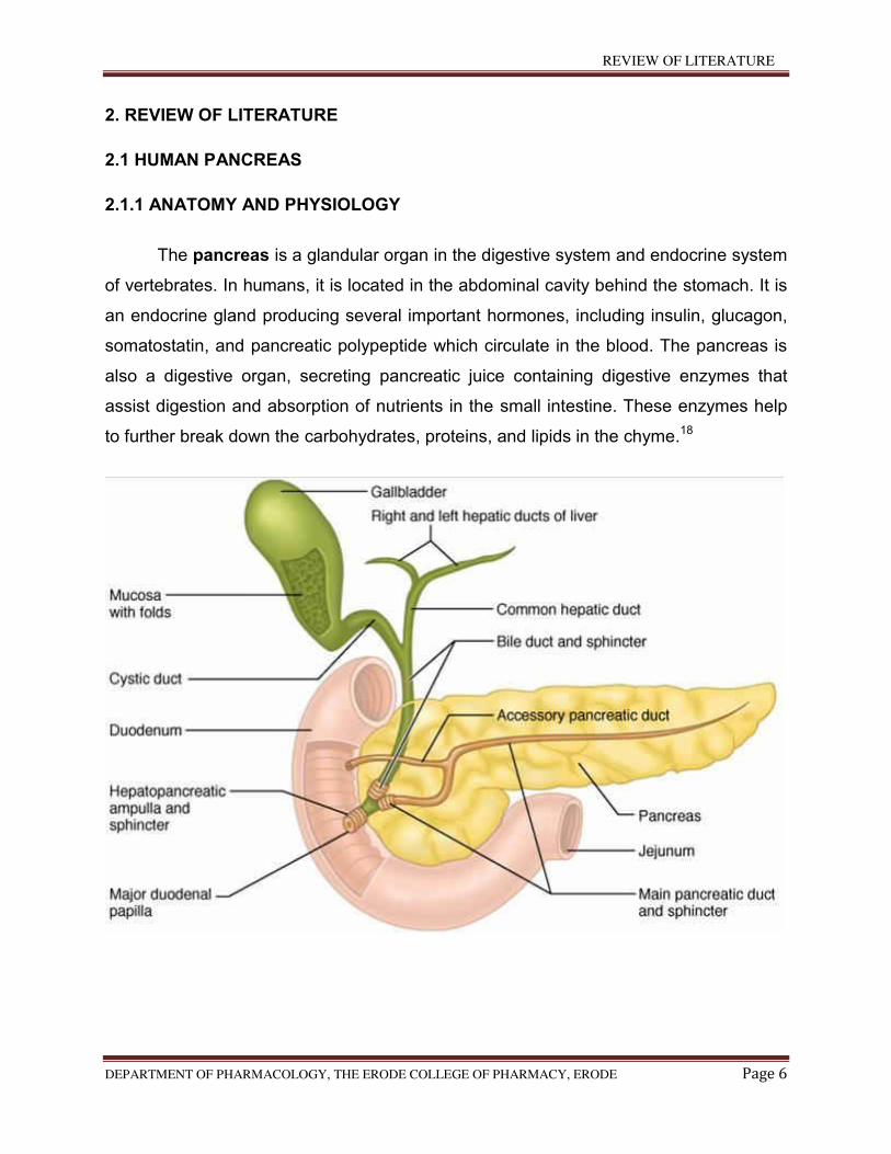
The pancreas is a glandular organ in the digestive system and endocrine system
of vertebrates. In humans, it is located in the abdominal cavity behind the stomach. It is
an endocrine gland producing several important hormones, including insulin, glucagon,
somatostatin, and pancreatic polypeptide which circulate in the blood. The pancreas is
also a digestive organ, secreting pancreatic juice containing digestive enzymes that
assist digestion and absorption of nutrients in the small intestine. These enzymes help
to further break down the carbohydrates, proteins, and lipids in the chyme.18
REVIEW OF LITERATURE
DEPARTMENT OF PHARMACOLOGY, THE ERODE COLLEGE OF PHARMACY, ERODE Page 7
The pancreas is an endocrine organ that lies in the upper left part of the
abdomen. It is found behind the stomach, with the head of the pancreas surrounded by
the duodenum. The pancreas is about 15 cm (6 in) long.19
Anatomically, the pancreas is divided into a head, which rests within the
concavity of the duodenum, a body lying behind the base of the stomach, and a tail,
which ends abutting the spleen. The neck of the pancreas lies between the body and
head, and lies anterior to the superior mesenteric artery and vein. The head of the
pancreas surrounds these two vessels, and a small uncinate process emerges from the
posterior part of the head, and extends posterior to the superior mesenteric vein, and
terminates at the superior mesenteric artery.20
The pancreas is a secretory structure with an internal hormonal role (endocrine)
and an external digestive role (exocrine). It has two main ducts, the main pancreatic
duct, and the accessory pancreatic duct. These drain enzymes through the ampulla of
Vater into the duodenum.21.
2.1.2 FUNCTION OF PANCREAS
2.1.2.1 THE EXOCRINE PANCREAS
The exocrine pancreas consist of acini, which resemble bunches of grapes. Each
acinus consists of a single layer of 40-50 pyramidal epithelial cells surrounding a lumen.
The epithelial cells produce the secretion (pancreatic juice) containing enzymes, ions
and water. The cells become wider during active section. The base of the acinar cells
are strongly basophylic owing to the presence of endoplasmic reticulum, where there is
a high concentration of RNA. This part of the cells therefore stains darker with
haemotoxylin and eosin. The apex of teh cells is abundant with secretory granules
containing the zymogen precursors of the pancreatic enzymes. The number of secretory
granules increases after fasting, and decreases after a meal.22
The lumen of the acini into intercalated duct. Intercalated ducts converg to make
larger interlobular ducts, which in turn converge to make interlobular ducts. Interlobular
ducts are found in the connective tissue septa between lobules. Interlobular ducts join to
REVIEW OF LITERATURE
DEPARTMENT OF PHARMACOLOGY, THE ERODE COLLEGE OF PHARMACY, ERODE Page 8
form either the pancreatic or the accessory duct, these ducts drain in to the duodenum.
In some cases, the pancreatic duct unites with the bile duct, the bile and pancreatic
juice enter the duodenum together.18
2.1.2.2 THE ENDOCRINE PANCREAS
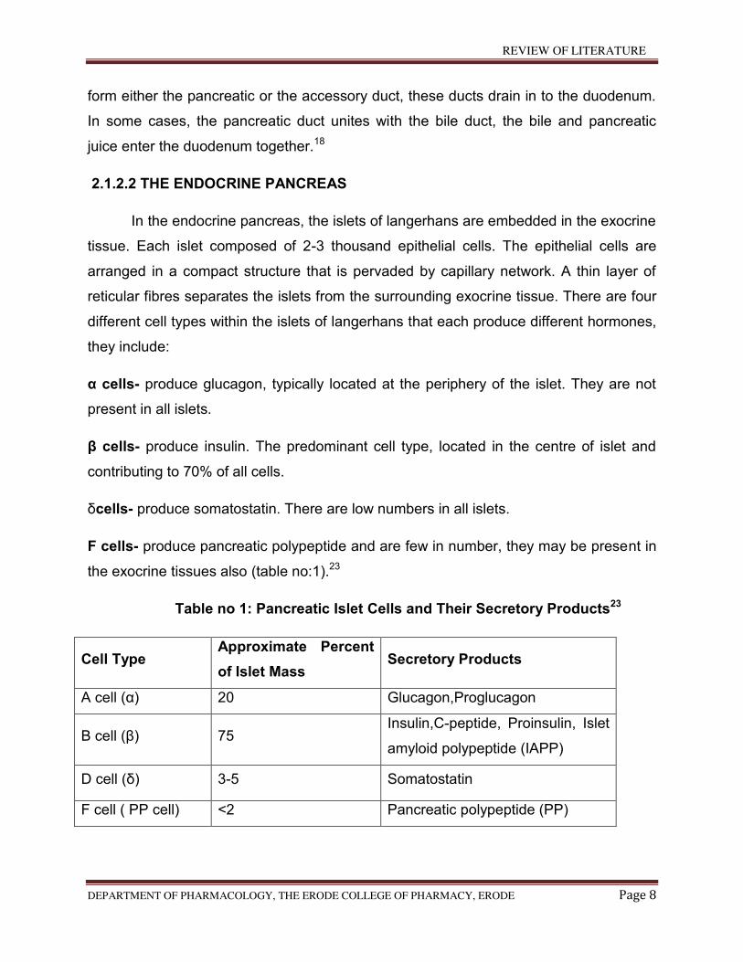
In the endocrine pancreas, the islets of langerhans are embedded in the exocrine
tissue. Each islet composed of 2-3 thousand epithelial cells. The epithelial cells are
arranged in a compact structure that is pervaded by capillary network. A thin layer of
reticular fibres separates the islets from the surrounding exocrine tissue. There are four
different cell types within the islets of langerhans that each produce different hormones,
they include:
α cells- produce glucagon, typically located at the periphery of the islet. They are not
present in all islets.
β cells- produce insulin. The predominant cell type, located in the centre of islet and
contributing to 70% of all cells.
δcells- produce somatostatin. There are low numbers in all islets.
F cells- produce pancreatic polypeptide and are few in number, they may be present in
the exocrine tissues also (table no:1).23
Table no 1: Pancreatic Islet Cells and Their Secretory Products23
Cell Type Approximate Percent
of Islet Mass Secretory Products
A cell (α) 20 Glucagon,Proglucagon
B cell (β) 75 Insulin,C-peptide, Proinsulin, Islet
amyloid polypeptide (IAPP)
D cell (δ) 3-5 Somatostatin
F cell ( PP cell) <2 Pancreatic polypeptide (PP)
REVIEW OF LITERATURE
DEPARTMENT OF PHARMACOLOGY, THE ERODE COLLEGE OF PHARMACY, ERODE Page 9
2.2 INSULIN
Insulin is a peptide hormone produced by beta cells of the pancreatic islets, and
by the Brockmann body in some teleost fish.24 It has important effects on the
metabolism of carbohydrates, fats and protein by promoting the absorption of,
especially, glucose from the blood into fat, liver and skeletal muscle cells. In these
tissues the absorbed glucose is converted into either glycogen or fats (triglycerides), or,
in the case of the liver, into both.25 Glucose production (and excretion into the blood) by
the liver is strongly inhibited by high concentrations of insulin in the blood. Circulating
insulin also affects the synthesis of proteins in a wide variety of tissues. In high
concentrations in the blood it is therefore an anabolic hormone, promoting the
conversion of small molecules in the blood into large molecules inside the cells. Low
insulin levels in the blood have the opposite effect by promoting widespread
catabolism.26
The pancreatic beta cells (β cells) are known to be sensitive to the glucose
concentration in the blood. When the blood glucose levels are high they secrete insulin
into the blood; when the levels are low they cease their secretion of this hormone into
the general circulation. Their neighboring alpha cells, probably by taking their cues from
the beta cells, secrete glucagon into the blood in the opposite manner: high secretion
rates when the blood glucose concentrations are low, and low secretion rates when the
glucose levels are high. High glucagon concentrations in the blood plasma powerfully
stimulate the liver to release glucose into the blood by glycogenolysis and
gluconeogenesis, thus having the opposite effect on the blood glucose level to that
produced by high insulin concentrations. The secretion of insulin and glucagon into the
blood in response to the blood glucose concentration is the primary mechanism
responsible for keeping the glucose levels in the extracellular fluids within very narrow
limits at rest, after meals, and during exercise and starvation.25,27
When the pancreatic beta cells are destroyed by an autoimmune process, insulin
can no longer be synthesized or be secreted into the blood. This results in type 1
diabetes mellitus, which is characterized by very high blood sugar levels, and
REVIEW OF LITERATURE
DEPARTMENT OF PHARMACOLOGY, THE ERODE COLLEGE OF PHARMACY, ERODE Page 10
generalized body wasting, which is fatal if not treated. This can only be corrected by
injecting the hormone, either directly into the blood if the patient is very ill and confused
or comatosed, or subcutaneously for routine maintenance therapy, which must be
continued for the rest of the person’s life. The exact details of how much insulin needs
to be injected, and when during the day, has to be adjusted according to the patient’s
daily routine of meals and exercise, in order to mimic the physiological secretion of
insulin as closely as is practically possible.28
2.2.1 NORMAL INSULIN PHYSIOLOGY
The insulin gene is expressed in the β- cells of the islets of langerhans, where
insulin is synthesized and stored in the form granules before secretion. Release from
beta cells occurs as a biphasic process involving two pools of insulin.30
Like other peptide hormones, insulin is synthesized as a precursor
(Preproinsulin) in the rough endoplasmic reticulum. Preproinsulin is transported to the
Golgi apparatus, of uncertain function called C-peptide. Insulin and C-peptide are stored
as granules in beta-cells, and are normally co-secreted by exocytosis in equimolar
amounts together with smaller and variable amounts of proinsulin.29
A rise in the blood glucose level calls forth an immediate release of insulin that is
stored in the β-cells granules. If the secretory stimulus persists, a delayed and
protracted response follows, which involves active synthesis of insulin.30
The main factor controlling the synthesis and secretion of insulin is the blood
glucose concentration. β-cells respond to both absolute glucose concentration and to
the rate of change of blood glucose. Other stimuli to insulin release include amino acids
(particularly arginine and leucine), fatty acids and the parasympathetic nervous system,
peptide hormones of the gut and drugs that act on sulfonylurea receptors.29
Insulin is the major anabolic hormone. It is necessary for 30
(1) Trans membrane transport of glucose and amino acids
(2) Glycogen formation in the liver and skeletal muscles
REVIEW OF LITERATURE
DEPARTMENT OF PHARMACOLOGY, THE ERODE COLLEGE OF PHARMACY, ERODE Page 11
(3) Nucleic acid synthesis
(4) Protein synthesis.
(5) Conversion of glucose into triglycerides
The principle metabolic function of insulin is to increase the rate of glucose transport
into certain cells of the body which are striated muscle cells, including myocardial cells;
fibroblasts and the fat cells, representing collectively about two third of the entire body
weight.30
Insulin interacts with its target cells by first binding to the insulin receptor; the
number and function of these receptors are important in regulating the action of insulin.
The insulin receptor is a tyrosine kinase that triggers a number of intracellular
responses that effect metabolic pathways. One of the important early responses to
insulin involves translocation of glucose transport units (GLUTs, of which there are
many tissue- specific types) from the Golgi apparatus to the plasma membrane, which
facilitates cellular uptake of glucose. Hence, removal of glucose from the circulation is a
primary outcome of insulin action.30
2.2.2 BIPHASIC INSULIN RESPONSE TO A CONSTANT GLUCOSE STIMULUS31
When the β- cells are stimulated, there will be a rapid first phase insulin response 1-
3 minutes after the glucose level is increased; this returns towards baseline in 6-10
minutes later. Thereafter, a gradual second phase insulin response that persists for the
duration of the stimulus. Type 2 diabetes mellitus is characterised by loss of the first
phase insulin response and a diminished second phase response.
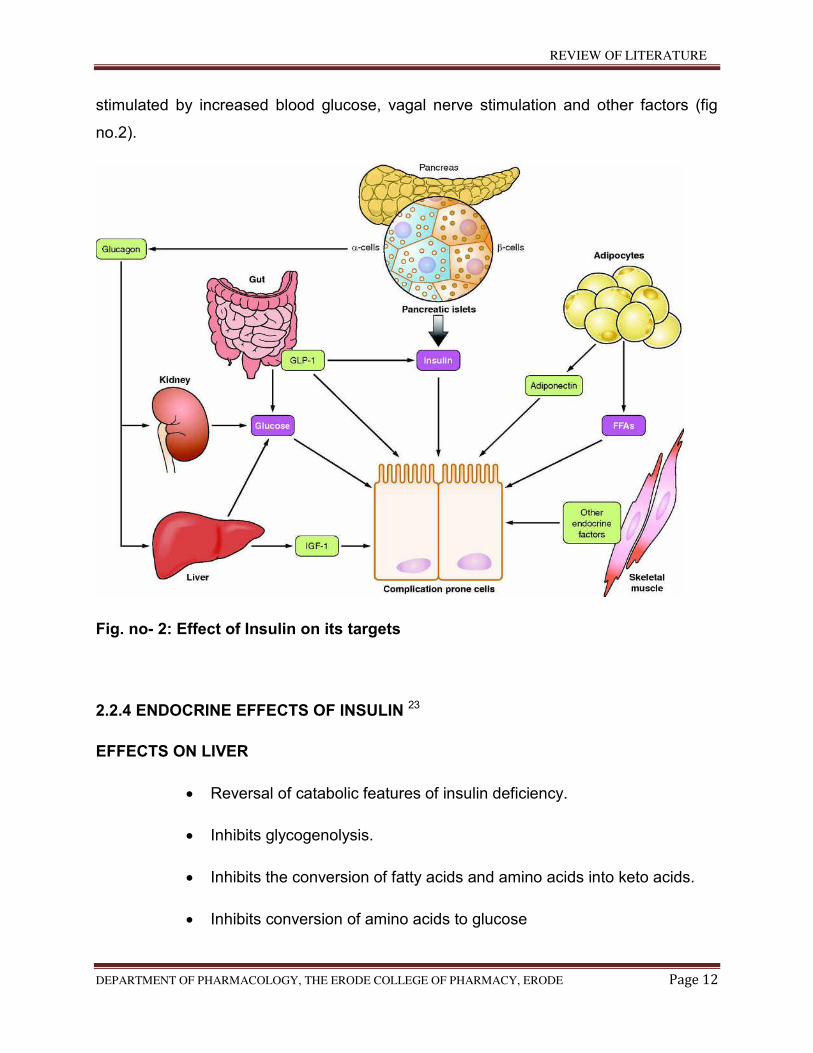
2.2.3 EFFECT OF INSULIN ON ITS TARGETS23
Insulin promotes the storage of fat as well as glucose (both are sources of
energy) within specialised target cells. (Fig no.2) Insulin promotes synthesis (from
circulating nutrients) and storage of glycogen, triglycerides and protein in its major
target tissues: liver, fat and muscle. The release of insulin from the pancreas is
REVIEW OF LITERATURE
DEPARTMENT OF PHARMACOLOGY, THE ERODE COLLEGE OF PHARMACY, ERODE Page 12
stimulated by increased blood glucose, vagal nerve stimulation and other factors (fig
no.2).
Fig. no- 2: Effect of Insulin on its targets
2.2.4 ENDOCRINE EFFECTS OF INSULIN 23
EFFECTS ON LIVER
Reversal of catabolic features of insulin deficiency.
Inhibits glycogenolysis.
Inhibits the conversion of fatty acids and amino acids into keto acids.
Inhibits conversion of amino acids to glucose
REVIEW OF LITERATURE
DEPARTMENT OF PHARMACOLOGY, THE ERODE COLLEGE OF PHARMACY, ERODE Page 13
Produces anabolic action
Promotes glucose storage as glycogen (induces glucokinase and
glycogen synthase and inhibits phosphorylase).
Increases triglycerides synthesis and very low density lipoprotein
formation.
EFFECT ON MUSCLE
Increases protein synthesis
Increases amino acid transport.
Increases ribosomal protein synthesis.
Increases glycogen synthesis.
Increases glucose transport.
Induces glycogen synthesis and inhibits phosphorylase.
EFFECT ON ADIPOSE TISSUE
Increases triglyceride storage.
Lipoprotein lipase is induced and activated by insulin to hydrolyse
triglycerides from lipoproteins.
Glucose transport into cells provides glycerol phosphate to permit
esterification of fatty acids supplied by lipoprotein transport.
Intracellular lipase is inhibited by insulin.
2.2.5 INSULIN AND CARBOHYDRATE METABOLISM32
After eating, various foods are broken into sugars in the stomach. The main
sugar is glucose which passes through the blood stream. But to remain healthy, the
blood glucose levels should not go too high or too low.
REVIEW OF LITERATURE
DEPARTMENT OF PHARMACOLOGY, THE ERODE COLLEGE OF PHARMACY, ERODE Page 14
When the blood glucose level starts to rise (after having food), at the same time
the level of the hormone called insulin also rises.
Insulin is the hormone that works on the body cells and helps them to take the
glucose from the blood stream for producing energy and some is converted into stores
of energy (glycogen or fat).
When the blood glucose level begins to fall (between meals), at the same time
the level of insulin also falls. When this happens the glycogen or fat is converted back
into glucose which is released from the cells into the blood stream.
2.2.6 INSULIN AND PROTEIN METABOLISM33
The present status of protein synthesis within cells has been outlined. Protein is formed
in the absence of insulin; the net formation of protein is accelerated by insulin. The
effects of insulin on protein metabolism take place independently of the transport of
glucose or amino acids into the cell; of glycogen synthesis; and of the stimulation of
high energy phosphate formation. In the case of protein metabolism, as in certain
studies on the pathways of glucose and fat metabolism, these observations reveal
striking intracellular effects of insulin in many tissues. Within most tissues the effect of
insulin appears to find expression predominantly at the microsomal level. Incidentally,
other hormones which affect protein metabolism such as growth or sex hormones
appear to act at the microsomes. The fact that insulin exerts effects on protein
metabolism at other intracellular sites as well as the above independent effects leads
one to agree that its action consists of a stimulation of multiple, seemingly unrelated,
metabolic events.
The fact that an immediate effect of insulin on protein synthesis is independent of the
immediate need for extracellular glucose or amino acids does not mean that the
sustained functioning of cells is likewise independent. The biochemist is fully aware of
metabolic defects in diabetes which are not altered by insulin in vitro, but which demand
varying periods of pre-treatment of the animal. It is also known that in diabetes some
REVIEW OF LITERATURE
DEPARTMENT OF PHARMACOLOGY, THE ERODE COLLEGE OF PHARMACY, ERODE Page 15
proteins (enzymes) may be deficient while others may be prodced in excess in the
absence of insulin.
2.2.7 INSULIN AND FAT METABOLISM34
The triacylglyceroles that comprise the bulk of lipids in the diet are hydrolyzed to free
fatty acids, monoacylglyceroles and glycerol in the intestinal tract. During absorption
through the intestinal tract mucosa, triacylglyceroles are re-synthesized from free fatty
acids, and glycerol-3-phosphate is formed in the intestinal mucose. These globules,
called chylomicrons, pass through the liver and adipose tissue, they are reduced in size
by an enzyme, lipoprotein lipase (LPL). In the postabsorptive period, free fatty acids and
glycerol are released from adipocytes by neural and hormonal stimulation. The free fatty
acids can be burned by almost all tissues of the body except the brain. They are burned
in the mitochondria by a process of b-oxidation to acetyl-CoA, which can then enter the
citrate acid cycle for conversion to CO2, adenosine triphosphate, and water. When
excessive quantities of glucose are ingested, the glucose can be converted to a storage
form, triacylglycerol. Fatty acids are synthesized by a series of reactions in which acetyl-
CoA and malonylo-CoA residues sequentially condense until the fatty acid chain is
completed. The fatty acids are then combined with glycerol-3-phosphate, generated in
the liver, to form the neutral triacylglyceroles. The insulin has effects on both the
synthetic (estrification) and breakdown (lipolysis) pathways. The promotion of
triacylglycerol storage in fat is one of the most important of the actions of insulin
DISORDERS OF PANCREAS35
Pancreatitis
Diabetes mellitus
Exocrine pancreatic insufficiency
Cystic fibrosis
Pseudocysts
Cysts
REVIEW OF LITERATURE
DEPARTMENT OF PHARMACOLOGY, THE ERODE COLLEGE OF PHARMACY, ERODE Page 16
2.3 DIABETES MELLITUS
Diabetes mellitus is simply referred to as diabetes which is either because the
body does not produce enough insulin or because the cells do not respond to insulin
that is produced. The classical symptoms of the diabetes mellitus are polyuria (frequent
urination), polydipsia (increased thirst) and polyphagia (increased hunger).
2.3.1 CLASSIFICATION OF DIABETES31
The American diabetes association has proposed four types of diabetes based on their
aetiology. They are;
Type 1 diabetes mellitus or insulin dependent diabetes mellitus (IDDM)
Type 2 diabetes mellitus or non insulin dependent diabetes mellitus (NIDDM)
Maturity onset diabetes of the young (MODY)
Gestational diabetes
2.3.2 TYPE 1 DIABETES MELLITUS31
Type 1diabetes (previously insulin dependent diabetes mellitus (IDDM)) is
characterised by beta –cell destruction, usually leading to absolute insulin deficiency
and associated with usually juvenile onset, a tendency to ketosis and diabetic
ketoacidosis and an absolute need for insulin treatment. Most patients have type1A
diabetes, which is caused by a cellular mediated autoimmune destruction of the beta-
cells of the pancreas.
2.3.3 PATHOGENESIS OF TYPE 1 DIABETES MELLITUS31
Type 1 diabetes mellitus (DM) is a disease of multi factorial autoimmune
causation. Worldwide, there is a marked geographic variation in prevalence. The overall
lifetime risk in Caucasian subjects is approximately 0-4%. Type 1 DM is caused by an
interaction between environmental factors and an inherited genetic predeposition.
REVIEW OF LITERATURE
DEPARTMENT OF PHARMACOLOGY, THE ERODE COLLEGE OF PHARMACY, ERODE Page 17
In twin studies, a significant genetic contribution is suggested by a
concordance value for type 1 DM of 30-50%. The risk to a first degree relative is
approximately 5%. The high discordance rate supports the notion that type 1 DM is
multifactorial in aetiology. Environmental triggers may account for up to two thirds of the
disease susceptibility. About 20 different regions of the human genome have been
found to have some degree of linkage with type 1 DM. To date, the strongest linkage
has been with genes encoded in the human leukocyte antigen (HLA) region located
within the major histocompatability (MHC), the contribution of which to disease risk is
now designated IDDM 1. This appears to be the most powerful determinant of genetic
susceptibility to the disease, accounting for approximately 40% of familial inheritance.
More than 90% of patients who develop type 1 DM have either DR3, DQ2 or DR4, DQ
haplotypes, whereas fewer than 40% normal controls who have these haplotypes. DR3-
DR4 heterozygosis is highest in children who develop diabetes before the age of 5
years (50%) and lowest in adults presenting with type 1 diabetes (20-30%) compared
with an overall US population prevalence of 2.4%. specific polymorphism of the DQBI
gene encoding the beta- chain of class II DQ molecules predispose to diabetes in
Caucasians but not in Japanese. In contrast, the HLS-DQ6 molecule protects against
the disease. HLA antigens (class I and II) are cell- surface glycoprotein that play crucial
role in presenting auto-antigen peptide fragments to T lymphocytes and thus initiating
an immune response. Polymorphism in the genes encoding specific peptide chains of
the HLA molecules may therefore modulate the ability of beta cells derived antigens to
trigger an autoimmune response against the beta- cells.
Only one non- HLA gene has been identified with certainty and that is the
insulin gene (INS) region on chromosomes 11p5.5, now designated as IDDm2.
Population studies of Caucasian type 1 subjects and non- diabetic controls initially
showed a positive association between alleles within the INS region and disease
susceptibility. However, recent genome screens have provided conflicting data
regarding the role of the INS gene region (IDDM2). It is thought that INS and HLA act
independently in the causation of type-1 diabetes and that the INS gene region (IDDM2)
accounts for 10% of familial clustering.
REVIEW OF LITERATURE
DEPARTMENT OF PHARMACOLOGY, THE ERODE COLLEGE OF PHARMACY, ERODE Page 18
The most likely environmental factor implicated in the causation of type-1 DM is
viral infection. Numerous viruses attack the pancreatic beta – cell either directly through
a cytolytic effect or by triggering an autoimmune attack against the beta cell. Evidence
for a viral factor in aetiology has come from animal models and in humans, from
observation of seasonal and geographic variations in the onset of disease. In addition,
patients newly presenting with type 1 DM may exhibit serologic evidence of viral
infection. Viruses that have been linked to human type 1 DM include mumps, Coxsackie
B, retroviruses, rubella, cytomegalovirus and Epstein- Barr virus. Bovine serum albumin,
a major constituent of cow’s milk has been implicated as a cause of type 1 DM in
children exposed at an early age, but definite proof is lacking and this remains
controversial. Nitrosamines (found in smoked and cured meats) may be diabetogenic as
many chemicals known to be toxic to pancreatic beta- cells, including alloxan,
sterptozotocin and the rat poison vector. Recent reports suggested that early ingestion
of cereal or gluten increases the risk of type 1 diabetes and remain to be confirmed.
Type 1 DM is associated with autoimmune destruction of the bets cells of the
endocrine pancreas. Examination of islet tissue obtained from pancreatic biopsy or that
obtained from post-mortem patients with recent onset type 1 DM confirms a mono
nuclear cell infiltrate (termed insulitis) with the presence of CD4 and CD8 T lymphocytes,
B lymphocytes and macrophages suggesting that these cells have a role in the
destruction of beta- cells. Although the precise mechanism of such as insulin has not
been elucidated, it seems to be that an environmental factor, such as viral infection, in a
subject with an inherited predisposition to the disease, triggers the damaging immune
response. This results in aberrant expression of class II MHC antigen by pancreatic
beta –cells. T lymphocytes recognise antigen presenting cells and are activated,
producing cytokines such as interleukin (IL)-2, interferon (INF) and tumour necrosis
factor (TNF). This generates a clone of T lymphocytes that carry receptors specific to
the presented antigen. Such T-helper cells assist B lymphocytes to produce antibodies
directed against the beta- cells. Such antibodies include islet cell antibodies (ICA)
directed against cytoplasmic components of the islet cells. ICA presence may precede
the development of type 1DM. Some subjects may develop ICA temporarily and not go
on to develop the disease, but persistence of ICA leads to progressive beta- cell
REVIEW OF LITERATURE
DEPARTMENT OF PHARMACOLOGY, THE ERODE COLLEGE OF PHARMACY, ERODE Page 19
destruction associated with the chronic inflammatory cell infiltrate termed ‘insulitis’. Type
1 DM ensures. Other antibodies associated with type 1 DM are islet cell- surface
antibodies (present in 30-60% of newly diagnosed type 1 DM patients), insulin
antibodies (IAA0 and antibodies to an isoform of glutamic acid decarboxylase (GAD).
2.3.4 TYPE 2 DIABETES MELLITUS31
Type 2 diabetes (previously non insulin dependent diabetes mellitus
(NIDDM)) is associated with obesity and an onset later in life. Patients, at least initially
and often throughout their lives, do not have a need for insulin therapy. The disorder
manifests as a result of insulin resistance and relative insulin deficiency. A precise
cause (or causes) has not been found. This type of diabetes frequently remains
undiagnosed for many years despite affected individuals being at risk of developing
serious macro vascular or micro vascular complications of the disease. Some patients
may masquerade as type 2 diabetic patients, but ultimately are recognised as having a
late onset slowly progressing immune mediated type 1 diabetes, so called latent
autoimmune diabetes in adults (LADA).
2.3.5 PATHOGENESIS OF TYPE 2 DIABETES MELLITUS31
Subjects with type-2 DM exhibit abnormalities in glucose homeostasis
owing to impaired insulin secretion, insulin resistance in muscle, liver, adipocytes and
abnormalities of splanchnic glucose uptake.
2.3.6 INSULIN SECRETION IN TYPE 2 DIABETES MELLITUS31
Impaired insulin secretion is a universal finding in patients with type 2
diabetes. In the early stages of type 2 diabetes mellitus, insulin resistance can be
compensated for by an increase in insulin secretion leading to normal glucose
tolerance. With increasing insulin resistance, the fasting plasma glucose will rise,
accompanied by an increase in fasting plasma insulin levels, until a fasting plasma
glucose level is reached when the beta- cell is unable to maintain its elevated rate of
insulin secretion at which point the fasting plasma insulin declines sharply. Hepatic
glucose production will begin to rise. When fasting plasma glucose reaches high levels,
REVIEW OF LITERATURE
DEPARTMENT OF PHARMACOLOGY, THE ERODE COLLEGE OF PHARMACY, ERODE Page 20
the plasma insulin response to a glucose challenge is markedly blunted. Although
fasting insulin levels remain elevated, postprandial insulin and C-peptide secretory rates
are decreased. This natural history of type 2 diabetes starting from normal glucose
tolerance followed by insulin resistance, compensatory hyperinsulinemia and then by
progression to impaired glucose tolerance and over diabetes has been documented in a
variety of populations.
Type 2 diabetes mellitus is characterised by loss of the first phase insulin
response to an intravenous glucose load, although this abnormality may be acquired
secondary to glucotoxicity. Loss of the first phase insulin response is important as this
early quick insulin secretion primes insulin target tissues, especially the liver. There may
be multiple possible causes of the impaired insulin secretion in type 2 diabetes mellitus
with several abnormalities having been shown to disturb the delicate balance between
islet neogenesis and apoptosis. Studies in first degree relatives of patients with type 2
DM and in twins have provided strong evidence for the genetic basis of abnormal beta-
cell function. Acquired defects in type 2 diabetes may lead to impairment of insulin
secretion. Clinical studies in man and animal studies, have supported the concept of
glucotoxicity, where by an elevation in plasma glucose levels, in the presence of a
reduced beta- cell mass, can lead to major impairment in insulin secretion.
Lipotoxicity has also been implicated as an acquired cause of impaired beta cell
function. Patients with type 2 DM exhibit a reduced response of the incretin glucagon
like peptide (GLP)-1 in response to oral glucose, while GLP-1 administration enhances
the postprandial insulin secretory response and may restore near normal glycaemia.
Amyloid deposits(islet amyloid polypeptide (IAPP) or amylin in the pancreas are
frequently observed in patients with type 2 diabetes and have been implicated as a
cause of progressive beta-cell failure. However, definitive evidence that amylin
contributes to beta-cell dysfunction in humans is lacking.
2.3.7 INSULIN RESISTANCE IN TYPE 2 DIABETES MELLITUS31
Insulin resistance is a characteristic feature of both lean obese individuals with
type 2 diabetes. In the fasting state, plasma insulin levels are increased in patients with
REVIEW OF LITERATURE
DEPARTMENT OF PHARMACOLOGY, THE ERODE COLLEGE OF PHARMACY, ERODE Page 21
type 2 diabetes. Since hyperinsulinemia is a potent inhibitor of hepatic glucose
production, an excessive rate of hepatic glucose production is the major abnormality
responsible for the elevated fasting plasma glucose in type 2 diabetes. It follows that
there must be hepatic resistance to the action of insulin. The liver is also resistant to the
inhibitory effect of hyperglycemia on hepatic glucose output. Most of the increase in
hepatic glucose production can be accounted by an increase in hepatic
gluconeogenesis. Muscle is the major site of insulin-stimulated glucose disposal in
humans. Muscle represents the primary site of insulin resistance in type 2 diabetic
subjects leading to a marketed blunting of glucose uptake into peripheral muscle. In
contrast, splanchnic tissue like the brain is relatively insensitive to insulin with respect to
stimulation of glucose uptake. Following glucose ingestion, both impaired suppression
of hepatic glucose production and decreased muscle glucose uptake are responsible for
the observed glucose intolerance leading to hyperglycemia.
There is a dynamic relationship between insulin resistance and impaired
insulin secretions. Insulin resistance is an early and characteristic feature of type 2
diabetes in high risk populations. More over diabetes develops only when the beta –
cells are unable to increase sufficiently their insulin output compensate for the defect in
insulin action (insulin resistance). Insulin resistance in type 2 diabetes is primarily due to
post binding defects in insulin action. Diminished insulin binding is modest and
secondary to down-regulation of the insulin receptor by chronic hyperglycemia. Post-
binding defects that have been recognised include reduced insulin receptor tyrosine
kinase activity, insulin signal transduction abnormalities, decreased glucose transport,
diminished glucose phosphorylation and impaired glycogen synthatase activity.
Quantitatively, impaired glycogen synthesis represents the major abnormality
responsible for insulin resistance in type 2 diabetic patients.
2.3.8 MATURITY ONSET DIABETS OF THE YOUNG (MODY)31
Specific monogenetic defects of the beta-cells has been identified and
usually gives rise to maturity onset diabetes of the young (MODY). MODY is defined as
a genetic defect in beta-cell function.
REVIEW OF LITERATURE
DEPARTMENT OF PHARMACOLOGY, THE ERODE COLLEGE OF PHARMACY, ERODE Page 22
2.3.9 PATHOGENESIS OF MODY31
Maturity onset diabetes of the young (MODY) is inherited as an autosomal
dominant and, upto date, abnormalities at six genetic locations on different
chromosomes have been identified. The most common form of MODY is associated
with mutations on chromosome 12 in hepatic nuclear factors (HNF)-1α and hence this is
referred to as transcription factor MODY. Other mutations affect such transcription
factor as HNF-1β, HNF-4α, insulin promoter factor-1 and NEUROD-1. Transcription
factor mutations alter insulin secretion in the mature β-cells as well as altering β-cell
development, proliferation and cell death. Cell dysfunction ensues until the emergence
of frank diabetes. Patients with transcription- factor mutations tend to be lean and
insulin sensitive rather than obese and insulin-resistant. Microvascular complications
are frequent. The first gene implicated in MODY was the glucokinse gene. Mutations on
the glucokinse gene on chromosome 7 result in a defective glucokinase molecule.
Glucokinse converts glucose to glucose-6-phosphate, the metabolism of which
stimulates insulin secretion by the β-cells tehreby glucokinase serves as a ‘glucose
sensor’. With defects in the glucokinase gene, increased plasma levels of glucose are
necessary to elicit normal levels of insulin secretion. Over 100 glucokinase gene
mutations have been found in families from several different countries. Fasting
hyperglycemia is present from both and worsens very slowly with age. Subjects are
usually detected by screening, e.g. in pregnancy or during coincidental illness or by
family studies. The mild hyperglycemia of this type of MODY rarely needs any treatment
other than diet and microvascular complications are rare.
Other specific genetic defects leading to diabetes include point mutations in
mitochondrial DNA, genetic abnormalities which leads to the inability to convert
proinsulin to insulin and the production of mutant insulin molecules and also the
mutations of the insulin receptor. Diabetes may also result from over disease of
exocrine pancreas, secondary to endocrinopathies and also due to some drugs and
chemicals. Viruses like cytomegalovirus, coxsackievirus B, adeno virus, mumps and
congenital rubella are also associated in the destruction of β-cells. Genetic syndromes
REVIEW OF LITERATURE
DEPARTMENT OF PHARMACOLOGY, THE ERODE COLLEGE OF PHARMACY, ERODE Page 23
like Down’s syndrome, Turner’s syndrome, Wolfram’s syndrome, Klinefelter’s syndrome
are accompanied by an increased incidence of diabetes mellitus.
2.3.10 GESTATIONAL DIABETES MELLITUS31
Gestational diabetes mellitus (GDM) was first defined as decreased
carbohydrate tolerance that develops or may be identified during pregnancy and this
definition was changed in 2010 as that GDM is a carbohydrate intolerance that develops
during pregnancy or has been discovered for the first time during pregnancy which is
not diabetes. Therefore, the GDM definition was not included in overt diabetes in
pregnancy.
PATHOGENESIS31
Gestational diabetes mellitus (GDM) is a carbohydrate intolerance that
develops first time during pregnancy. Type 1 and type2 diabetes also presents in
pregnancy occasionally. For GDM there is lack of agreed diagnostic criteria, but this
should not detract from the detrimental impact of maternal hyperglycemia on the
pregnancy and the future health of the mother and the child. The American Diabetes
Association recommends immediate recommendation for those women who are to be at
high risk of GDM with marked obesity, previous history of GDM, glycosuria or strong
family history of diabetes.
The fasting plasma glucose ≥126 mg/dl (7mmol/1) or a random plasma glucose
≥200 mg/dl (11mmol/1) meets the threshold for diagnosis of GDM and should conform
on a subsequent day.
In high-risk and average-risk women of GDM, it will not be found in the initial
screening and they should be screened between 24 and 26 weeks of gestation by either
a one-step approach using a 100g oral glucose tolerance test (OGTT) or a two-step
approach which involves in measuring the plasma glucose level 1 hour after a 50g oral
glucose load and performing a 100 g OGTT on those women who exceed the glucose
threshold 1 hour after the 50g oral glucose load.
REVIEW OF LITERATURE
DEPARTMENT OF PHARMACOLOGY, THE ERODE COLLEGE OF PHARMACY, ERODE Page 24
As if the glucose threshold value is ≥140mg/dl (7.8 mg/dl), then around 80% the
woman is with GDM. The diagnostic criteria for the 100 g OGTT are as follows;
≥95mg/dl (5.3mmol/1) fasting and ≥180 mg/dl (10mmol/1) at 1 hour, ≥155 mg/dl
(8.6mmol/1) at 2 hours and ≥140 mg/dl (7.8mmol/1) at 3 hours. To diagnose the GDM,
two or more of the plasma glucose values must meet or exceed. In many countries
these testing methods are not used and therefore a 75g IGTT was recommended by the
WHO.
Gestational diabetes mellitus is mainly seen in women with obesity, increased
maternal age and groups with a high background incidence of type 2 diabetes mellitus.
GDM usually occurs after the middle of the second trimester and can be detected by
suitable screening tests especially in the persons who are at high risk.
Prenatal morbidity in gestational diabetes mellitus increases with an increase in
maternal hyperglycemia. Most of the pregnancy related morbidity of gestational
diabetes mellitus is associated with delivering a large–fore gestational- age infant.
Caesarean rates have been increased due to increase in gestational diabetes patients
but this type of caesarean delivery can be reduced by intensive management of
maternal hyperglycemia. In the majority of the mothers with gestational diabetes, it can
be managed by diet alone by a dietician. Along with the mother, sequential tests will be
done from the foetus to estimate the foetal growth and abdominal circumferences and
also to identify features of inappropriate foetal growth and inform the mother the need of
maintaining blood glucose level intensively.
The American Diabetes Association recommends that if the dietary management
does not maintain fasting plasma glucose level below 5.8 mg/dl (105mmol/1) and the 2
hour post prandial glucose level below 6.7 mg/dl (120mmol/1), then insulin therapy
should be considered.
2.3.11 OTHER TYPES OF DIABETES MELLITUS31
Diabetes can also result from another process that adversely affects the
pancreas and such acquired processes pancreatitis, trauma, pancreatectomy,
pancreatic cancer. Hemochromatosis, fibrocalculous pancreatopathy and cystic fibrosis
REVIEW OF LITERATURE
DEPARTMENT OF PHARMACOLOGY, THE ERODE COLLEGE OF PHARMACY, ERODE Page 25
may also cause diabetes. Diabetes may also caused by the other endocrine diseases
especially when there is over-secretion of hormones that antagonize the normal effects
of insulin including Cushing’s syndrome, acromegaly and pheochromocytoma.
Diabetes may also result from certain rare diseases associated with abnormalities
of insulin or the insulin receptor which causes extreme insulin resistance and
sometimes found in association with acanthosis nigricans. These disorders are
categorized as insulin resistance syndromes. Diabetes may also occur due to a wide
array of genetic syndromes like Down’s syndrome, Klinefelter’s syndrome, Turner’s
syndrome. Diabetes may also result from the drugs like glucocorticoids, diazoxide,
thiazides which have similar effect may also cause diabetes.
REVIEW OF LITERATURE
DEPARTMENT OF PHARMACOLOGY, THE ERODE COLLEGE OF PHARMACY, ERODE Page 26
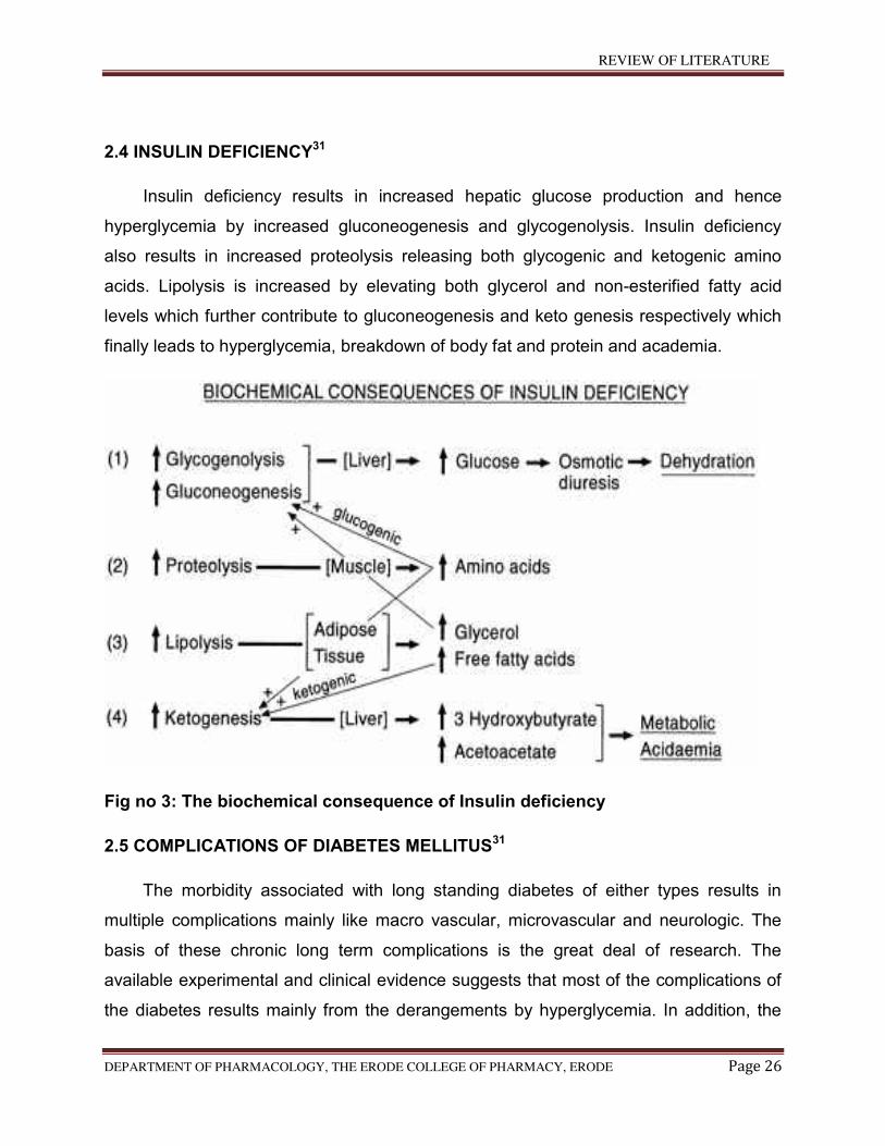
2.4 INSULIN DEFICIENCY31
Insulin deficiency results in increased hepatic glucose production and hence
hyperglycemia by increased gluconeogenesis and glycogenolysis. Insulin deficiency
also results in increased proteolysis releasing both glycogenic and ketogenic amino
acids. Lipolysis is increased by elevating both glycerol and non-esterified fatty acid
levels which further contribute to gluconeogenesis and keto genesis respectively which
finally leads to hyperglycemia, breakdown of body fat and protein and academia.
Fig no 3: The biochemical consequence of Insulin deficiency
2.5 COMPLICATIONS OF DIABETES MELLITUS31
The morbidity associated with long standing diabetes of either types results in
multiple complications mainly like macro vascular, microvascular and neurologic. The
basis of these chronic long term complications is the great deal of research. The
available experimental and clinical evidence suggests that most of the complications of
the diabetes results mainly from the derangements by hyperglycemia. In addition, the
REVIEW OF LITERATURE
DEPARTMENT OF PHARMACOLOGY, THE ERODE COLLEGE OF PHARMACY, ERODE Page 27
existence of hypertension is common in diabetes which leads to atherosclerosis. It is
evident that when a kidney is transplanted to diabetic patient from non diabetic donors
develops lesions due to diabetic nephropathy which is due to metabolic abnormalities in
diabetic patients. Conversely, the kidneys with lesions due to diabetic nephropathy
causes the reversal of the lesions when transplanted into the normal recipients
2.5.1 MACROVASCULAR COMPLICATIONS36
Macro vascular refers to the large blood vessels of the heart, brain, and legs. The
commonest manifestation of the macro vascular disease is in the coronary arteries and
the legs. Atherosclerosis of the coronary arteries is common in most of the people with
diabetes which is the most common cause for the death in people with diabetes which
may occur at a much younger age than in the general population and even females are
not immune in getting the disease. The mechanism for the development of macro
vascular disease in people with diabetes is similar to that of people without diabetes
which varies in the speed of development between diabetic and non-diabetic patients.
The risk of coronary artery disease is enhanced at all levels by the risk factors like
cholesterol, smoking, sedentary lifestyle, obesity, hypertension, etc
2.5.2 MICROVASCULAR COMPLICATIONS36
A Microvascular disease affects capillaries all over the body and so the
manifestations of the disease can be diffused. The eyes and kidneys are the most
affected organs. In the eyes, retinopathy which causes blindness, cataracts and
glaucoma are the complications. In kidneys, nephropathy accounts for half of the people
who go for dialysis and receive kidney transplantation.
2.5.3 PATHOGENESIS OF THE COMPLICATIONS OF DIABETES
3 main mechanisms linking hyperglycemia to the complications of long-standing
diabetes have been explored. Currently two such mechanisms are considered
important.
REVIEW OF LITERATURE
DEPARTMENT OF PHARMACOLOGY, THE ERODE COLLEGE OF PHARMACY, ERODE Page 28
First mechanism30
Glucose chemically attaches to free amino acid groups of proteins without the
aid of enzymes by the process called nonenzymatic glycosylation. The degree of this
nonenzymatic glycosylation is directly related to the level of blood glucose. In the
management of diabetes mellitus, the measurement of glycosylated haemoglobin
(HbA1c) levels in blood is useful, because it provides an index of the average blood
glucose levels over the 120 day life span of the erythrocytes. The early glycosylation
products of collagen and other long lived proteins undergo a slow series of chemical
rearrangements in the interstitial tissues and blood vessel walls to form irreversible
advanced glycosylation end products (AGEs) and these products accumulate on the
vessel wall over the life time.
AGE have a number of chemical and biological properties which are potentially
pathogenic.
The formation of glycosylated end products (AGEs) on the proteins such as
collagen causes cross-linkages between polypeptides, which may trap nonglycosylated
plasma and interstitial proteins. The circulating low density lipoprotein (LDL) is trapped
and retards its efflux from the vessel wall and promotes the deposition of cholesterol
and thus causes atherogenesis. AGEs also affects the structure and function of
capillaries including those on the glomeruli where the basement membranes become
thickened and becomes leaky.
AGEs also bind to receptors of many cell types like monocytes, macrophages,
endothelium and mesengial cells. The binding of AGEs on these receptors induces a
variety of biological activities, which includes monocytes emigration, release of
cytokines and growth factors from macrophages, increased endothelial permeability and
enhanced proliferation of fibroblasts and smooth muscle cells and synthesis of
extracellular matrix. All these effects can be potentially contributed to diabetic
complications.
REVIEW OF LITERATURE
DEPARTMENT OF PHARMACOLOGY, THE ERODE COLLEGE OF PHARMACY, ERODE Page 29
Second mechanism30
The second major mechanism proposed for the complications of intracellular
hyperglycemia is the disturbances in the polyol pathways. Some tissues like nerves,
lens, kidneys, and blood vessels do not require insulin for the transport of glucose, so
hyperglycemia leads to an increase in intracellular glucose, which is then metabolised
by aldose reductase into an polyol and eventually to fructose. Thus, the accumulated
sorbitol and fructose leads to increase intracellular osmolarity and influx of water and
then causes osmotic cell injury. In the lens, osmotically accumulated water causes
swelling and opacity. The accumulation of sorbitol also impairs ion pumps and promotes
the injury of Schwann cells and pericytes of retinal capillaries, with resultant peripheral
neuropathy and retinal micro aneurysms. Inhibition of aldose reductase is capable of
ameliorating the development of cataracts and neuropathy.
2.5.4 DIABETIC DYSLIPIDEMIA31
Dyslipidemia is the major macro vascular risk factor for the macro vascular
complications which leads to the cardiovascular diseases (CVD) in type 2 diabetes
mellitus. Along with this, endothelial dysfunction, platelet hyperactivity, impaired
fibrinolytic balance and abnormal blood flow which causes atherosclerosis and
increases the risk of thrombotic vascular events. In type 2 diabetes mellitus, the most
common lipoprotein abnormality is the elevation of triglycerides and very low density
lipoprotein (VLDL) which is caused by the overproduction of VLDL triglycerides.
The alteration in the distribution of lipids increases the risk of atherosclerosis in
diabetic patients. So, the condition with insulin deficiency and insulin resistance was
identified as phenotype of dyslipidemia in diabetes mellitus which is characterised with
high plasma triglyceride level, low HDL cholesterol level and increased level of small
dense LDL cholesterol. In addition to this, in diabetic patients, there will be an increment
of free fatty- acid release which is due to insulin resistance. So, due presence of
sufficient glycogen stores in the liver will promote triglyceride production which
stimulates the secretion of apolipoprotein B (Apo B) and VLDL cholesterol. This
REVIEW OF LITERATURE
DEPARTMENT OF PHARMACOLOGY, THE ERODE COLLEGE OF PHARMACY, ERODE Page 30
production of VLDL cholesterol by liver is enhanced due to the disability of insulin to
inhibit the release of free fatty-acids. There are many associations between
dyslipidemia and increased risk of cardiovascular disease in type2 diabetes mellitus
patients due to increased triglyceride levels and low HDL cholesterol
The management of dyslipidemia in diabetes mellitus includes changes in the
lifestyle of the patients such as increased physical activity and dietary modifications.
Besides this, antihyperlipidemic agents have been utilised for the management of
dyslipidemia. For the prevention of primary and secondary cardiovascular disease in
type 2 diabetes mellitus, anti platelet agents were recommended in contrast.
Dyslipidemia is categorised as one of the cardiovascular risk factors beside to the family
history of hypertension, CHD, smoking. Patients with type 2 diabetes mellitus having
dyslipidemia are eligible for the prevention of cardiovascular disease with anti-platelet
agents.
2.6 SIGNS AND SYMPTOMS OF DIABETES MELLITUS37
Polyuria (frequent urination)
The insulin is ineffective, kidneys cannot filter the glucose back into the blood
and the kidneys will take water from the blood to dilute the glucose which in turn fills up
the bladder and causes frequent urination in diabetic patients.
Polydipsia
High thirst due to osmosis of water from cells into the blood in an attempt to
dilute the high blood glucose concentration.
Intense hunger
Tendency to take food frequently more hunger and causes weight gaining.
Unusual weight loss
Weight loss is more common in patients with diabetes. As the body is not making
enough insulin then the body will seek out for the other energy source where the cells
REVIEW OF LITERATURE
DEPARTMENT OF PHARMACOLOGY, THE ERODE COLLEGE OF PHARMACY, ERODE Page 31
are not getting glucose. Muscle tissues and fat will be broken down for the energy to the
cells. So, when these muscle tissues and fat is broken down automatically there will be
loss in body weight.
Blurred vision
This can be caused by tissue being pulled from the eye lenses and effects the
eye’s ability to focus and can be treated. If it is severe prolonged vision problems and
blindness can occur.
Other symptoms
Cuts, bruises, skin or yeast infections do not heal properly or quickly because if
there is more sugar in the body, its ability to recover from infections is affected.
Specially, it is difficult to cure bladder and vaginal infections in women with
diabetes.
Red and swollen gums
If the gums are red, tender and swollen this could be the sign of diabetes. The
gums will pull away the teeth and the teeth will become loose. Numbness / tingling in
feet and palm
2.6.1 Long-term complications of Diabetes:38,39
Microangiopathy: Ischemic heart disease (IHD), stroke, peripheral vascular
disease.
Microangiopathy: retinopathy, nephropathy.
Neuropathy: peripheral neuropathy, autonomic neuropathy
Cataract
Diabetic foot
Diabetic heart
REVIEW OF LITERATURE
DEPARTMENT OF PHARMACOLOGY, THE ERODE COLLEGE OF PHARMACY, ERODE Page 32
2.6.2 RISK FACTORS OF TYPE 1 DIABETES40
Family history
Genetics
Age
Exposure to certain virus, such as the Epstein-Barr virus, Coxsackie virus,
mumps virus and cytomegalovirus
Early exposure to cow’s milk
Low vitamin D levels
Drinking water that contains nitrates
Early (before 4 months) or late ( after 7 months) introduction of cereal and
gluten into a baby’s diet
Having a mother who had preeclampsia during pregnancy
Being born with jaundice.
2.6.3 RISK FACTORS FOR TYPE 2 DIABETES
Obesity
Age
Family history of diabetes
History of gestational diabetes (diabetes during pregnancy)
High blood pressure(˃130/80mm/Hg)
Impaired glucose metabolism
Physical inactivity
REVIEW OF LITERATURE
DEPARTMENT OF PHARMACOLOGY, THE ERODE COLLEGE OF PHARMACY, ERODE Page 33
Race/ethnicity- African Americans, Hispanic/ Latino Americans, American
Indians and some Asian Americans and Native Hawaiians/Pacific Islanders
are particularly at high risk.
2.7 DIAGNOSIS OF DIABETES MELLITUS38,39
The diagnosis of the diabetes is an asymptomatic subject should never be made
on the basis of a single abnormal blood glucose value. For the asymptomatic person, at
least one additional plasma blood glucose tolerance test result with a value in the
diabetic range is essential, either fasting, from a random sample, or from the oral
glucose tolerance test (OGTT). If such sample fails to confirm the diagnosis of diabetes
mellitus, it will be usually be advisable to maintain surveillance with periodic retesting
until the diagnostic situation becomes clear. In these circumstances, the clinician should
take into consideration such additional factors as ethnicity, family history, age, adiposity,
and concomitant disorders, before deciding on a diagnostic or therapeutic course of
action. An alternative to blood glucose estimation, the OGTT has long been sought to
simplify the diagnosis of diabetes. Glycated haemoglobin, reflecting average glycaemia
over a period of weeks, was thought to provide such a test. Although in certain cases it
gives equal or almost equal sensitivity and specificity to glucose measurement, it is not
available in many parts of the world and is not well enough standardised for its use to
be recommended at this time.
2.7 DIAGNOSTIC CRITERIA38,39
The clinical diagnosis of diabetes is often prompted by symptoms such as
increased thirst and urine volume, recurrent infections, unexplained weight loss and in
severe cases drowsiness and coma; high levels of glycosuria are usually present. A
single blood glucose estimation is excess of the diagnostic values indicated it and also
defines levels of blood glucose below which a diagnosis of diabetes is unlikely in non
pregnant individuals. For clinical purposes, an OGTT (Oral glucose Tolerance Test) only
be considered to establish diagnostic status if casual blood glucose values lie in the
uncertain range between the levels that establish or exclude diabetes and fasting blood
glucose levels are below those which establish the diagnosis of the diabetes. If an
REVIEW OF LITERATURE
DEPARTMENT OF PHARMACOLOGY, THE ERODE COLLEGE OF PHARMACY, ERODE Page 34
OGTT is performed, it is sufficient to measure the blood glucose values while fasting
and at 2 hours after a 75g oral glucose load. For children, the oral glucose load is
related to body weight: 1.75 g per kg. The diagnostic criteria in children are same as for
the adults.
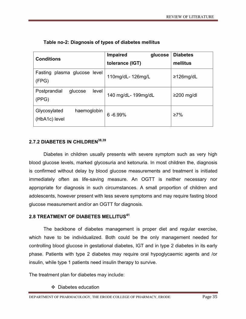
REVIEW OF LITERATURE
DEPARTMENT OF PHARMACOLOGY, THE ERODE COLLEGE OF PHARMACY, ERODE Page 35
Table no-2: Diagnosis of types of diabetes mellitus
Conditions Impaired glucose
tolerance (IGT)
Diabetes
mellitus
Fasting plasma glucose level
(FPG) 110mg/dL- 126mg/L ≥126mg/dL
Postprandial glucose level
(PPG) 140 mg/dL- 199mg/dL ≥200 mg/dl
Glycosylated haemoglobin
(HbA1c) level 6 -6.99% ≥7%
2.7.2 DIABETES IN CHILDREN38,39
Diabetes in children usually presents with severe symptom such as very high
blood glucose levels, marked glycosuria and ketonuria. In most children the, diagnosis
is confirmed without delay by blood glucose measurements and treatment is initiated
immediately often as life-saving measure. An OGTT is neither necessary nor
appropriate for diagnosis in such circumstances. A small proportion of children and
adolescents, however present with less severe symptoms and may require fasting blood
glucose measurement and/or an OGTT for diagnosis.
2.8 TREATMENT OF DIABETES MELLITUS41
The backbone of diabetes management is proper diet and regular exercise,
which have to be individualized. Both could be the only management needed for
controlling blood glucose in gestational diabetes, IGT and in type 2 diabetes in its early
phase. Patients with type 2 diabetes may require oral hypoglycaemic agents and /or
insulin, while type 1 patients need insulin therapy to survive.
The treatment plan for diabetes may include:
Diabetes education
REVIEW OF LITERATURE
DEPARTMENT OF PHARMACOLOGY, THE ERODE COLLEGE OF PHARMACY, ERODE Page 36
Meal planning and nutritional requirements
Exercise
Anti-diabetic agents
Insulin therapy
Management of associated conditions and complications.
2.8.1 PHARMACOLOGICAL THERAPY16,23,29,42
When the lifestyle modifications fails, therapeutic methods should be used that
consists of the following categories:-
Sulphonyl urea (Secretagogues)
Acetohexamide
Chlorpropamide
Tolbutamide
Glibenclamide
Glipizide
Gliclazide
Glimipiride
Biguanides
Metformin
Buformin
Thiazolidinediones
Pioglitazone
REVIEW OF LITERATURE
DEPARTMENT OF PHARMACOLOGY, THE ERODE COLLEGE OF PHARMACY, ERODE Page 37
Rosiglitazone
Dual agonist (PPAR)
Aleglitazar
Muraglitazar
Tesaglitazar
Alpha glycosidase inhibitors
Acarbose
Voglibose
Miglitol
Meglitinides
Repaglinide
Nateglinide
Mitiglinide
Lixisenatide
Taspoglutide
Dipeptidyl peptidase 4 inhibitors
Saxagliptin
Sitagliptin
Vidagliptin
SGLT2 Inhibitors
Asoartame
REVIEW OF LITERATURE
DEPARTMENT OF PHARMACOLOGY, THE ERODE COLLEGE OF PHARMACY, ERODE Page 38
Benfluorex
Chronimum picolinate
Epalrestat
Glucomannan
Tolrestat
Oral agents may counteract insulin resistance, improve β- cell glucose sensing
and insulin secretion or control the rate of intestinal glucose absorption. Combinations
of oral agents, in particular sulfonylurea plus metformin or thiazolidinediones plus
metformin, have improved the care of diabetic patients and may be used when
monotherapy is in effective.
2.9 PRECLINICAL IN VIVO ANIMAL MODEL OF DIABETES MELLITUS FOR
SCREENING OF POTENTIAL ANTI-DIABETIC ACTIVITY
2.9.1 PHARMACOLOGICAL INDUCTION OF DIABETES
Alloxan structure and Sterptozotocin43
The cytotoxic glucose analogues alloxan and sterptozotocin are the most
prominent diabetogenic chemical agents in experimental diabetes research. While the
mechanism of the selectivity of pancreatic beta cell toxicity is identical, the mechanism
of the cytotoxic action of the two compounds are different. Both are selectively toxic to
beta cells because they preferentially accumulate in beta cells as glucose analogues
through uptake via the GLUT2 glucose transporter. Both compounds are cytotoxic
glucose analogues. While the mechanism of cytotoxic action of the two compounds is
different, the mechanism of the selectivity of the beta cell action is identical.
2.9.2 Alloxan induced diabetes44
Alloxan (2, 4, 5, 6- tetra oxy pyrimidine; 2, 4, 5, 6-pyrimidinetetrone) is an
oxygenated pyrimidine derivative which is present as alloxan hydrate in aqueous
solution. Brugnatelli originally isolated alloxan in 1818 and the name is given by Wohler
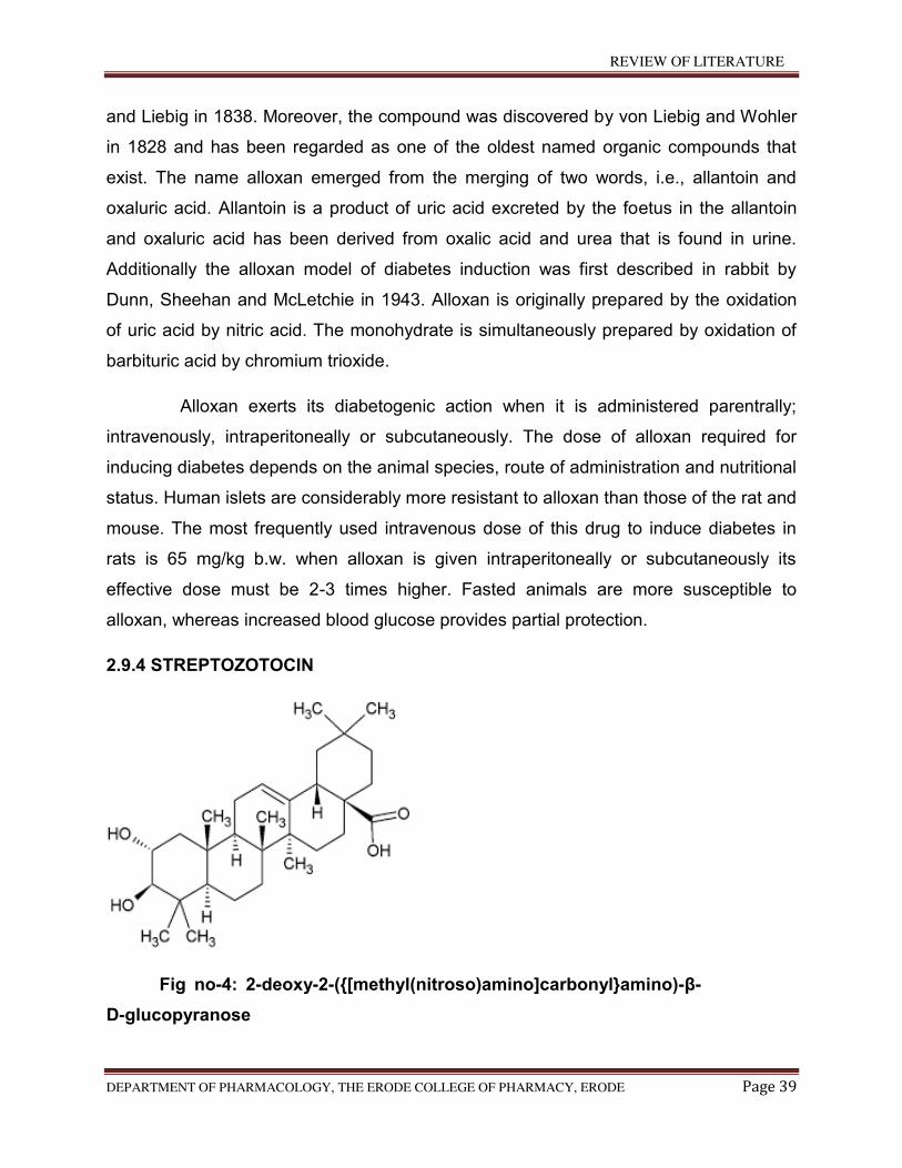
REVIEW OF LITERATURE
DEPARTMENT OF PHARMACOLOGY, THE ERODE COLLEGE OF PHARMACY, ERODE Page 39
and Liebig in 1838. Moreover, the compound was discovered by von Liebig and Wohler
in 1828 and has been regarded as one of the oldest named organic compounds that
exist. The name alloxan emerged from the merging of two words, i.e., allantoin and
oxaluric acid. Allantoin is a product of uric acid excreted by the foetus in the allantoin
and oxaluric acid has been derived from oxalic acid and urea that is found in urine.
Additionally the alloxan model of diabetes induction was first described in rabbit by
Dunn, Sheehan and McLetchie in 1943. Alloxan is originally prepared by the oxidation
of uric acid by nitric acid. The monohydrate is simultaneously prepared by oxidation of
barbituric acid by chromium trioxide.
Alloxan exerts its diabetogenic action when it is administered parentrally;
intravenously, intraperitoneally or subcutaneously. The dose of alloxan required for
inducing diabetes depends on the animal species, route of administration and nutritional
status. Human islets are considerably more resistant to alloxan than those of the rat and
mouse. The most frequently used intravenous dose of this drug to induce diabetes in
rats is 65 mg/kg b.w. when alloxan is given intraperitoneally or subcutaneously its
effective dose must be 2-3 times higher. Fasted animals are more susceptible to
alloxan, whereas increased blood glucose provides partial protection.
2.9.4 STREPTOZOTOCIN
Fig no-4: 2-deoxy-2-({[methyl(nitroso)amino]carbonyl}amino)-β-
D-glucopyranose
REVIEW OF LITERATURE
DEPARTMENT OF PHARMACOLOGY, THE ERODE COLLEGE OF PHARMACY, ERODE Page 40
Sterptozotocin (STZ) (2-deoxy-2-({[methyl(nitroso)amino]carbonyl}amino)-
β-D-glucopyranose) is synthesized by Streptomycetes achromogenes and is used to
induce both insulin-dependent and non-insulin-dependent diabetes mellitus (IDDM and
NIDDM respectively).The range of the STZ dose is not as narrow as in the case of
alloxan. It is a naturally occurring chemical that is particularly toxic to the insulin-
producing beta cells of the pancreas in mammals. The frequently used single
intravenous dose in adult rats to induce IDDM is between 40 and 60 mg/kg b.w. But
higher doses are also used22. STZ is also efficacious after intraperitoneal administration
of a similar or higher dose, but single dose below 40 mg/kg b.w. may be ineffective. For
instance, when 50 mg/kg b.w. STZ are injected intravenously to fed rats, blood glucose
(determined 2 weeks after treatment) can reach about 15mM. ). STZ may also be given
in multiple low doses. Such treatment is used predominantly in the mouse and the
induction of IDDM is mediated by the activation of immune mechanisms. However, that
the non- specific activation of the immune system via complete Freund's adjuvant prior
to STZ injections allows to reduce its diabetogenic dose even in the rat. NIDDM can
easily be induced in rats by intravenous or intraperitoneal treatment with 100 mg/kg
b.w.45
2.9.5 Usage of STZ for diabetes mellitus
Sterptozotocin is approved by the U.S food and drug administration (FDA) for
treating metastatic cancer of the pancreatic islet cells. Since it carries a substantial risk
of toxicity and rarely cures the cancer, its use is generally limited to patients whose
cancer cannot be removed by surgery. In these patients, sterptozotocin can reduce the
tumour size and reduce symptoms (especially hypoglycaemia due to excessive insulin
secretion by insulinomas). A typical dose is 500 mg/m²/day by intravenous injection, for
5 days, repeated every 4-6 weeks.
Due to its high toxicity to beta cells, in scientific research, sterptozotocin has also
been long used for inducing insulitis and diabetes on experimental animals.
REVIEW OF LITERATURE
DEPARTMENT OF PHARMACOLOGY, THE ERODE COLLEGE OF PHARMACY, ERODE Page 41
2.9.6 MECHANISM OF STREPTOZOTOCIN46,47 , 48, 49
Sterptozotocin is a nitrosamine glucosamine- nitrosourea. As with other alkylating
agents in the nitrosourea class, it is toxic to cells by causing damage to the DNA,
though other mechanisms may also contribute. DNA damage induces activation of poly
ADP- ribosylation, which is likely more important for diabetes induction than DNA
damage itself. Sterptozotocin is similar enough to glucose to be transported into the cell
by the glucose transport protein GLUT2, but is not recognised by the other glucose
transporters. This explains its relative toxicity to beta cells, since these cells have
relatively high levels of GLUT 2.
STZ is taken up by pancreatic B cells via glucose transporter GLUT2. A reduced
expression of GLUT2 has been found to prevent the diabetogenic action of STZ
observed that STZ itself restricts GLUT2 expression in vivo and in vitro when
administered in multiple doses. Intracellular action of STZ results in changes of DNA in
pancreatic B cells comprising its fragmentation. Recent experiments have proved that
the main reason for the STZ-induced B cell death is alkylation of DNA. The alkylating
activity of STZ is related to its nitrosourea moiety, especially at the O6 position of
guanine. After STZ injection to rats, different methylated purines were found in tissues
of these animals.
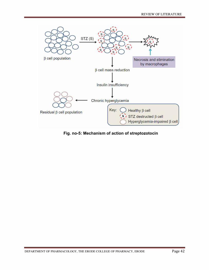
REVIEW OF LITERATURE
DEPARTMENT OF PHARMACOLOGY, THE ERODE COLLEGE OF PHARMACY, ERODE Page 42
Fig. no-5: Mechanism of action of streptozotocin
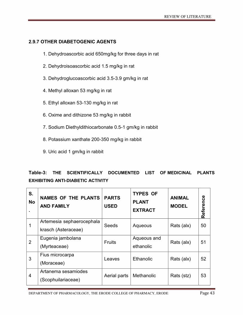
REVIEW OF LITERATURE
DEPARTMENT OF PHARMACOLOGY, THE ERODE COLLEGE OF PHARMACY, ERODE Page 43
2.9.7 OTHER DIABETOGENIC AGENTS
1. Dehydroascorbic acid 650mg/kg for three days in rat
2. Dehydroisoascorbic acid 1.5 mg/kg in rat
3. Dehydroglucoascorbic acid 3.5-3.9 gm/kg in rat
4. Methyl alloxan 53 mg/kg in rat
5. Ethyl alloxan 53-130 mg/kg in rat
6. Oxime and dithizone 53 mg/kg in rabbit
7. Sodium Diethyldithiocarbonate 0.5-1 gm/kg in rabbit
8. Potassium xanthate 200-350 mg/kg in rabbit
9. Uric acid 1 gm/kg in rabbit
Table-3: THE SCIENTIFICALLY DOCUMENTED LIST OF MEDICINAL PLANTS
EXHIBITING ANTI-DIABETIC ACTIVITY
S.
No
.
NAMES OF THE PLANTS
AND FAMILY
PARTS
USED
TYPES OF
PLANT
EXTRACT
ANIMAL
MODEL R
efe
ren
ce
1 Artemesia sephaerocephala
krasch (Asteraceae) Seeds Aqueous Rats (alx) 50
2 Eugenia jambolana
(Myrteaceae) Fruits
Aqueous and
ethanolic Rats (alx) 51
3 Fius microcarpa
(Moraceae) Leaves Ethanolic Rats (alx) 52
4 Artanema sesamiodes
(Scophuilariaceae) Aerial parts Methanolic Rats (stz) 53
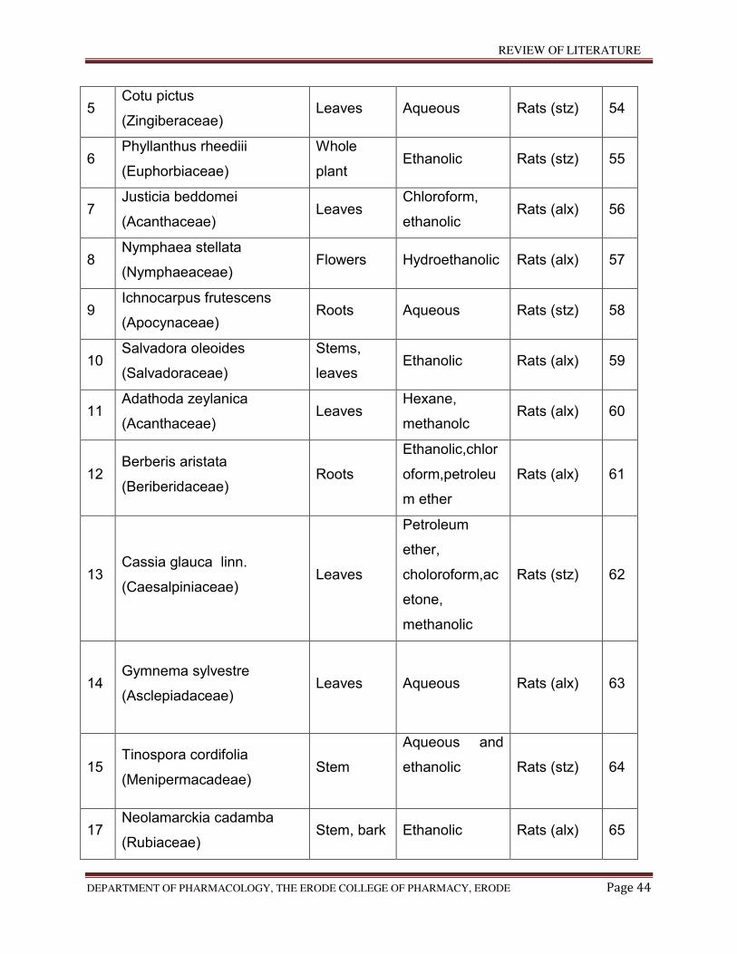
REVIEW OF LITERATURE
DEPARTMENT OF PHARMACOLOGY, THE ERODE COLLEGE OF PHARMACY, ERODE Page 44
5 Cotu pictus
(Zingiberaceae) Leaves Aqueous Rats (stz) 54
6 Phyllanthus rheediii
(Euphorbiaceae)
Whole
plant Ethanolic Rats (stz) 55
7 Justicia beddomei
(Acanthaceae) Leaves
Chloroform,
ethanolic Rats (alx) 56
8 Nymphaea stellata
(Nymphaeaceae) Flowers Hydroethanolic Rats (alx) 57
9 Ichnocarpus frutescens
(Apocynaceae) Roots Aqueous Rats (stz) 58
10 Salvadora oleoides
(Salvadoraceae)
Stems,
leaves Ethanolic Rats (alx) 59
11 Adathoda zeylanica
(Acanthaceae) Leaves
Hexane,
methanolc Rats (alx) 60
12 Berberis aristata
(Beriberidaceae) Roots
Ethanolic,chlor
oform,petroleu
m ether
Rats (alx) 61
13 Cassia glauca linn.
(Caesalpiniaceae) Leaves
Petroleum
ether,
choloroform,ac
etone,
methanolic
Rats (stz) 62
14 Gymnema sylvestre
(Asclepiadaceae) Leaves Aqueous Rats (alx) 63
15 Tinospora cordifolia
(Menipermacadeae) Stem
Aqueous and
ethanolic
Rats (stz) 64
17 Neolamarckia cadamba
(Rubiaceae) Stem, bark Ethanolic Rats (alx) 65
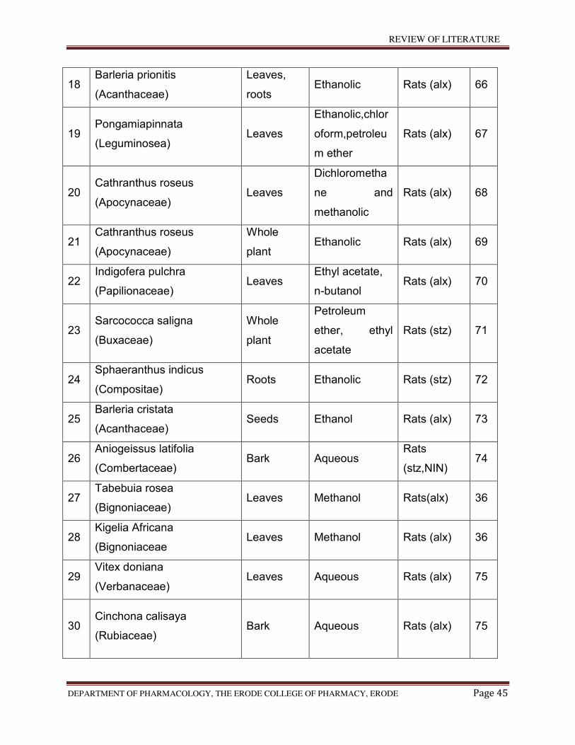
REVIEW OF LITERATURE
DEPARTMENT OF PHARMACOLOGY, THE ERODE COLLEGE OF PHARMACY, ERODE Page 45
18 Barleria prionitis
(Acanthaceae)
Leaves,
roots Ethanolic Rats (alx) 66
19 Pongamiapinnata
(Leguminosea) Leaves
Ethanolic,chlor
oform,petroleu
m ether
Rats (alx) 67
20 Cathranthus roseus
(Apocynaceae) Leaves
Dichlorometha
ne and
methanolic
Rats (alx) 68
21 Cathranthus roseus
(Apocynaceae)
Whole
plant Ethanolic Rats (alx) 69
22 Indigofera pulchra
(Papilionaceae) Leaves
Ethyl acetate,
n-butanol Rats (alx) 70
23 Sarcococca saligna
(Buxaceae)
Whole
plant
Petroleum
ether, ethyl
acetate
Rats (stz)
71
24 Sphaeranthus indicus
(Compositae) Roots Ethanolic Rats (stz) 72
25 Barleria cristata
(Acanthaceae) Seeds Ethanol Rats (alx) 73
26 Aniogeissus latifolia
(Combertaceae) Bark Aqueous
Rats
(stz,NIN) 74
27 Tabebuia rosea
(Bignoniaceae) Leaves Methanol Rats(alx) 36
28 Kigelia Africana
(Bignoniaceae Leaves Methanol Rats (alx) 36
29 Vitex doniana
(Verbanaceae) Leaves Aqueous Rats (alx) 75
30 Cinchona calisaya
(Rubiaceae) Bark Aqueous Rats (alx)
75
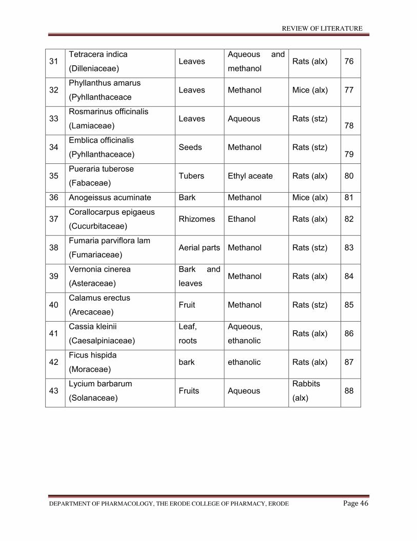
REVIEW OF LITERATURE
DEPARTMENT OF PHARMACOLOGY, THE ERODE COLLEGE OF PHARMACY, ERODE Page 46
31 Tetracera indica
(Dilleniaceae) Leaves
Aqueous and
methanol Rats (alx) 76
32 Phyllanthus amarus
(Pyhllanthaceace Leaves Methanol Mice (alx) 77
33 Rosmarinus officinalis
(Lamiaceae) Leaves Aqueous Rats (stz)
78
34 Emblica officinalis
(Pyhllanthaceace) Seeds Methanol Rats (stz)
79
35 Pueraria tuberose
(Fabaceae) Tubers Ethyl aceate Rats (alx) 80
36 Anogeissus acuminate Bark Methanol Mice (alx) 81
37 Corallocarpus epigaeus
(Cucurbitaceae) Rhizomes Ethanol Rats (alx) 82
38 Fumaria parviflora lam
(Fumariaceae) Aerial parts Methanol Rats (stz) 83
39 Vernonia cinerea
(Asteraceae)
Bark and
leaves Methanol Rats (alx) 84
40 Calamus erectus
(Arecaceae) Fruit Methanol Rats (stz) 85
41 Cassia kleinii
(Caesalpiniaceae)
Leaf,
roots
Aqueous,
ethanolic Rats (alx) 86
42 Ficus hispida
(Moraceae) bark ethanolic Rats (alx) 87
43 Lycium barbarum
(Solanaceae) Fruits Aqueous
Rabbits
(alx) 88
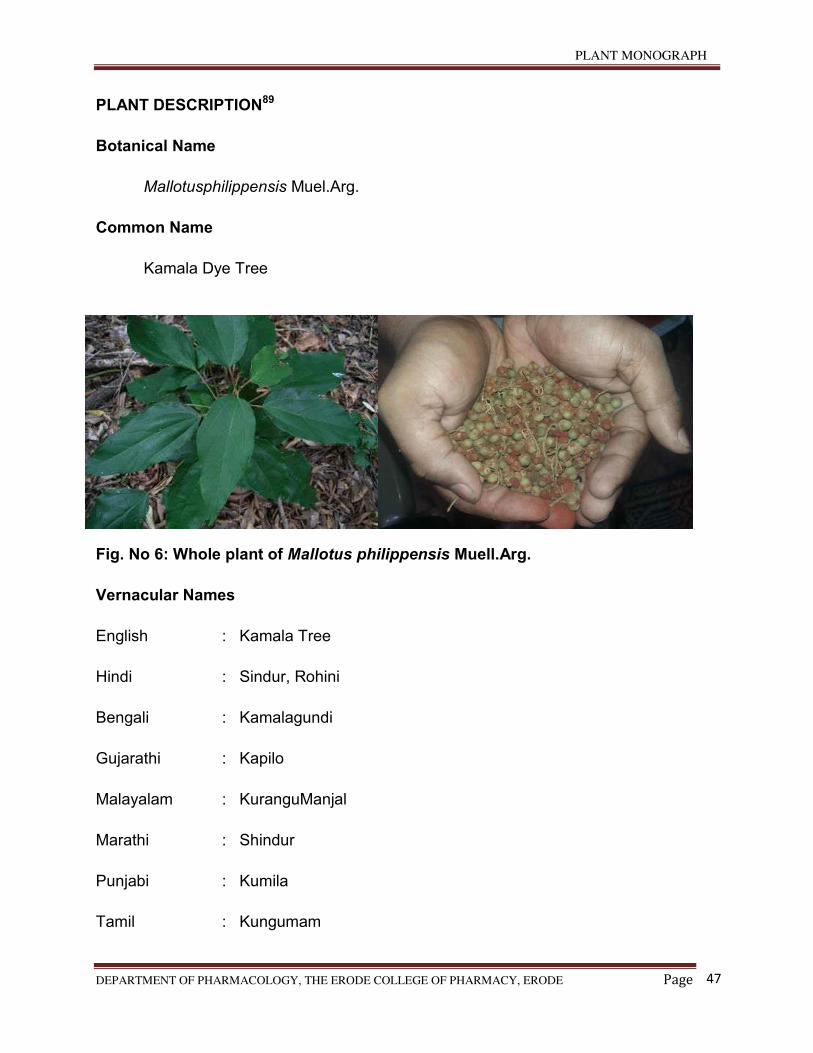
PLANT MONOGRAPH
DEPARTMENT OF PHARMACOLOGY, THE ERODE COLLEGE OF PHARMACY, ERODE Page
47
PLANT DESCRIPTION89
Botanical Name
Mallotusphilippensis Muel.Arg.
Common Name
Kamala Dye Tree
Fig. No 6: Whole plant of Mallotus philippensis Muell.Arg.
Vernacular Names
English : Kamala Tree
Hindi : Sindur, Rohini
Bengali : Kamalagundi
Gujarathi : Kapilo
Malayalam : KuranguManjal
Marathi : Shindur
Punjabi : Kumila
Tamil : Kungumam
PLANT MONOGRAPH
DEPARTMENT OF PHARMACOLOGY, THE ERODE COLLEGE OF PHARMACY, ERODE Page
48
Telugu : Kunkuma
Assami : Gangai
Oriya : Bosonto-gundi
TAXONAMY
Kingdom : Plantae
Subkingdom : Tracheobionta
Superdivision
Division
:
:
Spermatophyta
Magnoliopsida
Class : Rosidae
Order : Euphobiales
Family : Euphorbiaceae
Genus : Mallotus
Species : Philippensis
BOTANY
Leaves are alternate and simple, more or less leathery, ovate to lanceolate, cuneate to
rounded with two glands at base. Leaves are mostly acute or acuminate at apex,
conspicuously 3-nerved, hairy and reddish glandular beneath, petiole size 1–4 cm long,
puberulous and reddish-brown in colour. Male flowers in terminal and axillary position,
2–10 cm long, solitary or fascicled paniculates spikes, each flowers are with numerous
stamens, small; female flowers have spikes or slender racemes, each flower with a
stellate hairy, 3 celled ovary with 3 papillose stigmas. Fruit is a depressed-globose; 3-
lobed capsule; 5, 7 mm, and 10 mm; stellate; puberulous; with abundant orange or
reddish glandular granules; 3-seeded . Seeds are subglobose and black in colour and
4 mm across.
PLANT MONOGRAPH
DEPARTMENT OF PHARMACOLOGY, THE ERODE COLLEGE OF PHARMACY, ERODE Page
49
Distribution
Mallotus philippensis Muell.Arg. (Family: Euphorbiaceae) is a branched herb found in
hills of southern districts of Tamilnadu and kerala. It is also common in America and
occurs in Eastern Africa and East India.
Parts Used
Fruits
Chemical Constituents
Major phytochemicals present in this genus contain different natural compounds,
mainly phenols, diterpenoids, steroids, flavonoids, cardenolides, triterpenoids,
coumarin, isocoumarins, and many more to discover. Present knowledge about this
endangered species of medicinal plant is still limited with respect to its phytochemistry
and biological activity.One of the major chemical constituent, that is, rottlerin of M.
philippinensis, is listed below with its chemical structure and its major biological
activities along with other phytochemicals
Cardenolides
(Corotoxigenin,L-rhamnoside, corogl-aucigenin and L-rhamnoside.)
Steroids
(β-Sitosterol.)
Phenolic Compounds
(Bergenin, Kamalachalcone A.,Kamalachalcone B.,Mallotophilippen
A.,Mallotophilippen B.,Mallotophilippen C.,Mallotophilippen D. and Mallotophilippen E)
phloroglucinol derivatives
(Rottlerin, Isoallorottlerin and Isorottlerin.)
PLANT MONOGRAPH
DEPARTMENT OF PHARMACOLOGY, THE ERODE COLLEGE OF PHARMACY, ERODE Page
50
Triterpenoids
(Betulin-3-acetate,Friedelin,Acetylaleuritolic acid’γb-Acetoxy-22b-hydroxyolean-
18-ene and α-Amyrin.)
Properties and Uses
According to Ayurveda, leaves are bitter, cooling, and appetizer. All parts of plant like
glands and hairs from the capsules or fruits are used as heating, purgative,
anthelmintic, vulnerary, detergent, maturant, carminative, Anti-diabetic,90 and alexiteric
and are useful in treatment of bronchitis, abdominal diseases, and spleen enlargement,
and if taken with milk or curd (yoghurt), it can be quite useful for expelling tapeworms .
Kamala or Kampillakah is also used as an oral contraceptive. The powder and a few
other parts of Kamala are also used in external applications to promote the healing of
ulcers and wounds. They are used to treat parasitic affections of the skin like scabies,
ringworm, and herpes.
Earlier Work Done On Mallotus philippensis Muell.Arg.
Cuong NX et al. have studied ‘’A new lignan dimer from Mallotus philippensis’’. A
new lignan dimer, bilariciresinol (1), was isolated from the leaves of Mallotus
philippensis, along with platanoside (2), isovitexin (3), dihydromyricetin (4),
bergenin (5), 4-O-galloylbergenin (6), and pachysandiol A (7). Their structures
were elucidated by spectroscopic experiments including 1D and 2D NMR and
FTICR-MS91.
Chan TK et al. have studied ‘’Anti-allergic actions of rottlerin
from Mallotus philippensis in experimental mast cell-mediated anaphylactic
models’’. Allergy is an acquired hypersensitivity reaction of the immune system
mediated by cross-linking of the allergen-specific IgE-bound high-affinity IgE
receptors, leading to immediate mast cell degranulation. Rottlerin is an active
molecule isolated from Mallotus philippensis, a medicinalplant used in Ayurvedic
Medicine System for anti-allergic and anti-helminthic treatments. The present
PLANT MONOGRAPH
DEPARTMENT OF PHARMACOLOGY, THE ERODE COLLEGE OF PHARMACY, ERODE Page
51
study investigated potential anti-allergic effects of rottlerin in animal models of
IgE-dependent anaphylaxis and the anti-allergic mechanisms of action of rottlerin
in mast cells. Anti-allergic actions of rottlerin were evaluated in passive
cutaneous anaphylaxis and passive systemic anaphylaxis mouse models, and in
anaphylactic contraction of bronchial rings isolated from sensitized guinea pigs.
Direct mast cell-stabilizing effect of rottlerin was examined in RBL-2H3 mast cell
line. Anti-allergic signaling mechanisms of action of rottlerin in mast cells were
also examined. Rottlerin prevented IgE-mediated cutaneous vascular
extravasation, hypothermia, elevation in plasma histamine level and tracheal
tissue mast cell degranulation in mice in a dose-dependent manner. In addition,
rottlerin suppressed ovalbumin-induced guinea pig bronchial smooth muscle
contraction. Furthermore, rottlerin concentration-dependently blocked IgE-
mediated immediate release of -hexosaminidase from RBL-2H3 mast cells.
Rottlerin was found to inhibit IgE-induced PLC 1 and Akt phosphorylation
production of IP3 and rise in cytosolic Ca+2 level in mast cells. We report here for
the first time that rottlerin possesses anti-allergic activity by blocking IgE-induced
mast cell degranulation, providing a foundation for developing rottlerin for the
treatment of allergic asthma and other mast cell-mediated allergic disorders.92
Hong Q et al. have studied ‘’Anti-tuberculosis compounds
from Mallotus philippensis’’. Bioassay-directed fractionation of the organic extract
of Mallotus philippensis gave five compounds (1-5), the most active of which
against Mycobacterium tuberculosis was a new compound, 8-cinnamoyl-5,7-
dihydroxy-2,2-dimethyl-6-geranylchromene (1) for which the name
mallotophilippen F is suggested. Compound (2), 8-cinnamoyl-2,2-dimethyl-7-
hydroxy-5-methoxychromene, was isolated from a natural source for the first
time, while the remaining three compounds, rottlerin (3),
isoallorottlerin=isorottlerin (4) and the so-called "red compound," 8-cinnamoyl-
5,7-dihydroxy-2,2,6-trimethylchromene (5), had been isolated previously from
this plant. All compounds were identified by analysis of their spectra including
2D-NMR, which was used to correct the literature NMR spectral assignments of
compounds 2-4. The C-13 NMR of 5 is reported for the first time.93
PLANT MONOGRAPH
DEPARTMENT OF PHARMACOLOGY, THE ERODE COLLEGE OF PHARMACY, ERODE Page
52
Kulkarni RR et al. have studied ‘’Antifungal dimericchalcone derivative
kamalachalcone E from Mallotus philippensis’’. From the red coloured extract
(Kamala) prepared through acetone extraction of the fresh whole uncrushed
fruits of Mallotus philippensis, one new dimericchalcone (1) along with three
known compounds 1-(5,7-dihydroxy-2,2,6-trimethyl-2H-1-benzopyran-8-yl)-3-
phenyl-2-propen-1-one (2), rottlerin (3) and 4'-hydroxyrottlerin (4) were isolated.
The structure of compound 1 was elucidated by 1D and 2D NMR analyses that
included HSQC, HMBC, COSY and ROESY experiments along with the literature
comparison. Compounds 1-4 were evaluated for antifungal activity against
different human pathogenic yeasts and filamentous fungi. The antiproliferative
activity of the compounds was evaluated against Thp-1 cell lines. Compounds 1
and 2 both exhibited IC50 of 8, 4 and 16 μg/mL against Cryptococcus
neoformans PRL518, C. neoformans ATCC32045 and Aspergillusfumigatus,
respectively. Compound 4, at 100 μg/mL, showed 54% growth inhibition of Thp-1
cell lines.94
Khan H et al. have studied ‘’Antioxidant and Antiplasmodial Activities of Bergenin
and 11-O-Galloylbergenin Isolated from Mallotus philippensis’’. Two important
biologically active compounds were isolated from Mallotus philippensis. The
isolated compounds were characterized using spectroanalytical techniques and
found to be bergenin (1) and 11-O-galloylbergenin (2). The in vitro antioxidant
and antiplasmodial activities of the isolated compounds were determined. For the
antioxidant potential, three standard analytical protocols, namely, DPPH radical
scavenging activity (RSA), reducing power assay (RPA), and total antioxidant
capacity (TAC) assay, were adopted. The results showed that compound 2 was
found to be more potent antioxidant as compared to 1. Fascinatingly, compound
β displayed better EC50 results as compared to α-tocopherol while being
comparable with ascorbic acid. The antiplasmodial assay data showed that both
the compound exhibited good activity against chloroquine sensitive strain of
Plasmodium falciparum (D10) and IC50 values were found to be less than 8 μM.
The in silico molecular docking analyses were also performed for the
determination of binding affinity of the isolated compounds using P. falciparum
PLANT MONOGRAPH
DEPARTMENT OF PHARMACOLOGY, THE ERODE COLLEGE OF PHARMACY, ERODE Page
53
proteins PfLDH and Pfg27. The results showed that compound 2 has high
docking score and binding affinity to both protein receptors as compared to
compound 1. The demonstrated biological potentials declared that compound 2
could be the better natural antioxidant and antiplasmodial candidate.95
Gangwar M et al. have studied ‘’Antioxidant capacity and radical scavenging
effect of polyphenol rich Mallotus philippensis fruit extract on human
erythrocytes: an in vitro study’’. Mallotus philippensis is an important source of
molecules with strong antioxidant activity widely used medicinal plant. Previous
studies have highlighted their anticestodal, antibacterial, wound healing activities,
and so forth. So, present investigation was designed to evaluate the total
antioxidant activity and radical scavenging effect of 50% ethanol fruit glandular
hair extract (MPE) and its role on Human Erythrocytes. MPE was tested for
phytochemical test followed by its HPLC analysis. Standard antioxidant assays
like DPPH, ABTS, hydroxyl, superoxide radical, nitric oxide, and lipid
peroxidation assay were determined along with total phenolic and flavonoids
content. Results showed that MPE contains the presence of various
phytochemicals, with high total phenolic and flavonoid content. HPLC analysis
showed the presence of rottlerin, a polyphenolic compound in a very rich
quantity. MPE exhibits significant strong scavenging activity on DPPH and ABTS
assay. Reducing power showed dose dependent increase in concentration
absorption compared to standard, Quercetin. Superoxide, hydroxyl radical, lipid
peroxidation, nitric oxide assay showed a comparable scavenging activity
compared to its standard. Our finding further provides evidence that Mallotus fruit
extract is a potential natural source of antioxidants which have a protective role
on human Erythrocytes exhibiting minimum hemolytic activity and this justified its
uses in folklore medicines.96
Khan M et al. have studied ‘’Hexane soluble extract of Mallotus philippensis
Muell.Arg. root possesses anti-leukaemic activity’’. Root extract of
M. philippensis was initially extracted in organic solvents, hexane, ethyl acetate,
and n-butanol. The hexane extract showed highest toxicity against p53-deficient
HL-60 cells (IC50 1.5 mg dry roots equivalent/ml medium) after 72 h and
PLANT MONOGRAPH
DEPARTMENT OF PHARMACOLOGY, THE ERODE COLLEGE OF PHARMACY, ERODE Page
54
interestingly, inhibition of cell proliferation was preceded by the upregulation of
the proto-oncogenes Cdc25A and cyclin D1 within 24 h. The hexane extract
induced 18% apoptosis after 48 h of treatment. Chemical composition of the
hexane extract was analyzed by GC-MS and the 90% fragments were matched
with polyphenolic compounds. The present study confirms that the hexane
fraction of M. philippensis root extract possesses anti-leukemic activity in HL-60
cells. The polyphenols were the main compounds of the hexane extract that
inhibited proliferation and induced apoptosis.97
Gangwar M et al. have studied ‘’In-vitro scolicidal activity of Mallotus philippensis
Muell.Arg. fruit glandular hair extract against hydatid cyst
Echinococcusgranulosus’’. To investigate new scolicidal agent from natural
resources to cope with the side effects associated with synthetic drugs in
Echinococcosis. The scolicidal potential of methanolic fruit powder extract (10
and 20 mg/mL) of Mallotus philippensis was investigated. Viability of
protoscoleces was confirmed by trypan blue exclusion method, where mortality
was observed at concentration of 10 and 20 mg/mL in 60 min treatment against
Echinococcusgranulosus (E. granulosus), under in-vitro conditions with reference
to the known standard drug Praziquantel. At concentration 10 and 20 mg/mL, the
mortality rate was observed 97% and 99% respectively for 60 min treatment;
while up to 93% mortality was observed with 20 mg/mL for only 10 min treatment.
The concentration above 20 mg/mL for above 2 h showed 100% mortality,
irrespective of further incubation. As compared with the standard anti-parasitic
drug Praziquantel our extract has significant scolicidal activity with almost no
associated side effects.98
Goel RK et al. have studied ‘’Mallotus philippensis Muell.Arg. (Euphorbiaceae):
ethnopharmacology and phytochemistry review. Mallotus philippensis Muel.Arg”.
(Euphorbiaceae) are widely distributed perennial shrub or small tree in tropical
and subtropical region in outer Himalayas regions with an altitude below 1,000 m
and are reported to have wide range of pharmacological
activities. Mallotus philippensis species are known to contain different natural
compounds, mainly phenols, diterpenoids, steroids, flavonoids, cardenolides,
PLANT MONOGRAPH
DEPARTMENT OF PHARMACOLOGY, THE ERODE COLLEGE OF PHARMACY, ERODE Page
55
triterpenoids, coumarins, isocoumarins, and many more especially phenols; that
is, bergenin, Malloto philippensis, rottlerin, and isorottlerin have been isolated,
identified, and reported interesting biological activities such as antimicrobial,
antioxidant, antiviral, cytotoxicity, antioxidant, anti-inflammatory,
immunoregulatory activity protein inhibition against cancer cell. We have selected
all the pharmacological aspects and toxicological and all its biological related
studies. The present review reveals that Mallotus philippensis is a valuable
source of medicinally important natural molecules and provides convincing
support for its future use in modern medicine. However, the existing knowledge is
very limited about Mallotus philippensis and its different parts like steam, leaf,
and fruit. Further, more detailed safety data pertaining to the acute and subacute
toxicity and cardio- and immunotoxicity also needs to be generated for crude
extracts or its pure isolated compounds. This review underlines the interest to
continue the study of this genus of the Euphorbiaceae.99
Furumoto T et al. have studied ‘’Mallotus philippensis bark extracts promote
preferential migration of mesenchymal stem cells and improve wound healing in
mice’’. In the present study, we report the effects of the ethanol extract
from Mallotus philippensis bark (EMPB) on mesenchymal stem cell (MSC)
proliferation, migration, and wound healing in vitro and in a mouse model.
Chemotaxis assays demonstrated that EMPB acted an MSC chemoattractant
and that the main chemotactic activity of EMPB may be due to the effects of
cinnamtannin B-1. Flow cytometric analysis of peripheral blood mononuclear
cells in EMPB-injected mice indicated that EMPB enhanced the mobilization of
endogenous MSCs into blood circulation. Bioluminescent whole-animal imaging
of luciferase-expressing MSCs revealed that EMPB augmented the homing of
MSCs to wounds. In addition, the efficacy of EMPB on migration of MSCs was
higher than that of other skin cell types, and EMPB treatment improved of wound
healing in a diabetic mouse model. The histopathological characteristics
demonstrated that the effects of EMPB treatment resembled MSC-induced tissue
repair. Taken together, these results suggested that EMPB activated the
PLANT MONOGRAPH
DEPARTMENT OF PHARMACOLOGY, THE ERODE COLLEGE OF PHARMACY, ERODE Page
56
mobilization and homing of MSCs to wounds and that enhancement of MSC
migration may improve wound healing.100
Gautam MK et al. have studied ‘’Mallotus philippensis Muell.Arg. fruit glandular
hairs extract promotes wound healing on different wound model in rats’’. The
study includes acute toxicity and wound healing potential of 50% ethanol extract
of MP fruit glandular hair (MPE). MPE (200 mg/kg) was administered orally, once
daily for 10 days (incision and dead space wound) and 22 days (excision wound).
MPE was found safe when given to ratsupto 10 times of optimal effective dose.
Wound breaking strength (WBS) in Incision wound and rate of contraction, period
of epithelization and scar area in Excision wound were evaluated. Granulation
tissue free radicals (nitric oxide and lipid peroxidation), antioxidants (catalase,
superoxide dismutase, and reduced glutathione), acute inflammatory marker
(myeloperoxidase), connective tissue markers (hydroxyproline, hexosamine, and
hexuronic acid), and deep connective tissue histology were studied in Dead
space wound. MPE significantly increased WBS and enhanced wound
contraction, and decreased both epithelization period and scar area compared
with control group. MPE was found to decrease free radicals (50.8 to 55.2%,
P<0.001) and myeloperoxidase (44.0%, P<0.001) but enhanced antioxidants
(41.1 to 54.5%, P<0.05 to P<0.001) and connective tissue markers (39.5 to
67.3%, P<0.05 to P<0.01). Histopathological evaluation revealed more density of
collagen formation with minimal inflammatory cells in deeper tissues. Thus, the
study revealed Mallotus philippensis fruit hair extract, safe and effective in wound
healing and the healing effects seemed to be due to decrease in free radical
generated tissue damage, promoting effects on antioxidant status and faster
collagen deposition as evidenced biochemically and histology.101
Oyedemi BO et al. have studied ‘’Novel R-plasmid conjugal transfer inhibitory
and antibacterial activities of phenolic compounds from Mallotus
philippensis Mull.Arg’’. Antimicrobial resistance severely limits the therapeutic
options for many clinically important bacteria. In Gram-negative bacteria,
multidrug resistance is commonly facilitated by plasmids that have the ability to
accumulate and transfer refractory genes amongst bacterial populations. The aim
PLANT MONOGRAPH
DEPARTMENT OF PHARMACOLOGY, THE ERODE COLLEGE OF PHARMACY, ERODE Page
57
of this study was to isolate and identify bioactive compounds from the
medicinal plant Mallotus philippensis Mull.Arg. with both direct antibacterial
properties and the capacity to inhibit plasmid conjugal transfer. A chloroform-
soluble extract of M. philippensis was subjected to bioassay-guided fractionation
using chromatographic and spectrometric techniques that led to the isolation of
the known compounds rottlerin [5,7-dihydroxy-2,2-dimethyl-6-(2,4,6-trihydroxy-3-
methyl-5-acetylbenzyl)-8-cinnamoyl-1,2-chromene] and the red compound (8-
cinnamoyl-5,7-dihydroxy-2,2,6-trimethylchromene). Both compounds were
characterised and elucidated using one-dimensional and two-dimensional
nuclear magnetic resonance (NMR). Rottlerin and the red compound showed
potent activities against a panel of clinically relevant Gram-positive bacteria,
including meticillin-resistant Staphylococcus aureus (MRSA). No significant direct
activities were observed against Gram-negative bacteria. However, both rottlerin
and the red compound strongly inhibited conjugal transfer of the plasmids
pKM101, TP114, pUB307 and R6K amongst Escherichia coli at a subinhibitory
concentration of 100mg/L. Interestingly, despite the planar nature of the
compounds, binding to plasmid DNA could not be demonstrated by a DNA
electrophoretic mobility shift assay. These results show that rottlerin and the red
compound are potential candidates for antibacterial drug lead development.
Further studies are needed to elucidate the mode of inhibition of the conjugal
transfer of plasmids.102
Chhiber N et al. have studied ‘’Rottlerin, a polyphenolic compound from the fruits
of Mallotus phillipensis Mull.Arg.’’ impedes oxalate/calcium oxalate induced
pathways of oxidative stress in male wistar rats. Oxalate and/or calcium oxalate,
is known to induce free radical production, subsequently leading to renal
epithelial injury. Oxidative stress and mitochondrial dysfunction have emerged as
new targets for managing oxalate induced renal injury. Hyperoxaluria was
induced by administering 0.4% ethylene glycol and 1% ammonium chloride in
drinking water to male wistar rats for 9 days. Rottlerin was administered
intraperitoneally at 1mg/kg/day along with the hyperoxaluric agent. Prophylactic
efficacy of rottlerin to diminish hyperoxaluria induced renal dysfunctionality and
PLANT MONOGRAPH
DEPARTMENT OF PHARMACOLOGY, THE ERODE COLLEGE OF PHARMACY, ERODE Page
58
crystal load was examined along with its effect on free radicals generating
pathways in hyperoxaluric rats. 0.4% ethylene glycol and 1% ammonium chloride
led to induction of hyperoxaluria, oxiadtive stress and mitochondrial damage in
rats. Rottlerin treatment reduced NADPH oxidase activity, prevented
mitochondrial dysfunction and maintained antioxidant environment. It also
refurbished renal functioning, tissue integrity and diminished urinary crystal load
in hyperoxaluric rats treated with rottlerin. Thus, the present investigation
suggests that rottlerin evidently reduced hyperoxaluric consequences and the
probable mechanism of action of this drug could be attributed to its ability to
quench free radicals by itself and interrupting signaling pathways involved in
pathogenesis of stone formation.103
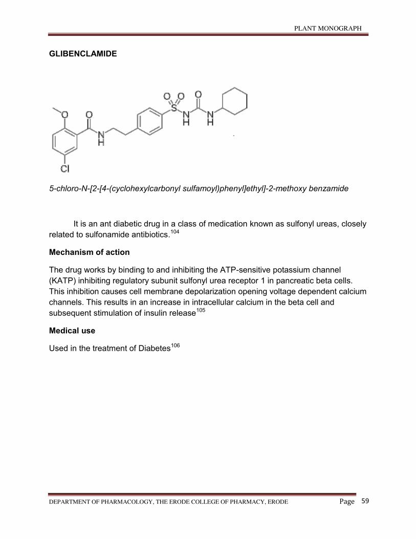
PLANT MONOGRAPH
DEPARTMENT OF PHARMACOLOGY, THE ERODE COLLEGE OF PHARMACY, ERODE Page
59
GLIBENCLAMIDE
5-chloro-N-[2-[4-(cyclohexylcarbonyl sulfamoyl)phenyl]ethyl]-2-methoxy benzamide
It is an ant diabetic drug in a class of medication known as sulfonyl ureas, closely
related to sulfonamide antibiotics.104
Mechanism of action
The drug works by binding to and inhibiting the ATP-sensitive potassium channel
(KATP) inhibiting regulatory subunit sulfonyl urea receptor 1 in pancreatic beta cells.
This inhibition causes cell membrane depolarization opening voltage dependent calcium
channels. This results in an increase in intracellular calcium in the beta cell and
subsequent stimulation of insulin release105
Medical use
Used in the treatment of Diabetes106
SCOPE OF THE PRESENT STUDY
DEPARTMENT OF PHARMACOLOGY, THE ERODE COLLEGE OF PHARMACY, ERODE Page 60
SCOPE OF THE PRESENT STUDY
In the present situation, diabetes is possibly the world’s largest growing
metabolic disorders and as the knowledge on the heterogenicity of this disorder is
advanced, the need for more appropriate therapy increases. A number of allopathic
drugs are used for the anti-diabetic effect like Tolbutamide, metformin, phenformin
and acarbose which have danger of drug interaction, adverse effects etc. Traditional
plant medicines are used throughout the world for a range of diabetic presentations.
There are many medicinal plants known to be used in the treatment of diabetes and
a number of plants had been screened positively for their anti-diabetic effect. Most of
these plants were found to belong to the chemical group glycosides, alkaloids and
flavanoids.
Diabetes mellitus has to become one of the world’s biggest health problems
owing to the projected increase in new cases. In India, the prevalence rate of
diabetes is estimated to be 1-5%. Complications are the major cause of morbidity
and mortality in diabetes mellitus.
MallotusPhilippensis Muell.Arg. Fruit is having the anti-diabetic activity. As per
the literature review, still no anti-diabetic activity has been reported on this plant.
Hence, this study has been taken to explore the anti-diabetic activity of Mallotus
Philippensis Muell.Arg. in sterptozotocin induced diabetes in Wistar albino rats.
AIM AND OBJECTIVES
DEPARTMENT OF PHARMACOLOGY, THE ERODE COLLEGE OF PHARMACY, ERODE Page 61
AIM AND OBJECTIVES
Aim
The present investigation was aimed to screen the anti-diabetic activity of
Mallotus Philippensis Muell.Arg. on sterptozotocin induced diabetic Wistar albino
rats.
Objectives
The objective of the present study to:
a. Find out the phytochemical constituents present in the ethanolic extract of the
whole plant of Mallotus Philippensis Muell.Arg.
b. Acute toxicity studies
c. Evaluation of anti-diabetic activity
Blood glucose level
Glycosylated haemoglobin
d. Estimation of Serum parameters
Total cholesterol level
Serum aspartate amino transferace level (AST)
Serum alanine amino transferace level (ALT)
e. Estimation of Oxidative stress parameters
Liver malondialdehyde level (MDA)
f. Estimation of enzymic hepatic antioxidant parameters
Liver superoxide dismutase level (SOD)
Liver catalase level (CAT)
Liver glutathione peroxide level (GPx)
g. Estimation of non-enzymic anti-oxidant activity
Reduced glutathione (GSH) level
Ascorbic acid (Vitamin C) level
α –tocopherol (Vitamin E) level
h. Estimation of plasma insulin level
i. Histopathology of pancreas.
j. Statistical analysis
PLAN OF WORK
DEPARTMENT OF PHARMACOLOGY, THE ERODE COLLEGE OF PHARMACY, ERODE Page 62
PLAN OF WORK
1. Collection of plant.
2. Authentication of plant, shade drying of the Fruits.
3. Extraction of plant materials with solvents.
4. Preliminary phytochemical screening of ethanolic extract of Fruits Mallotus
Philippensis Muell.Arg.
5. Evaluation of acute toxicity studies to determine LD50 value
6. Evaluation of anti-diabetic activity.
7. Estimation of biochemical parameters.
8. Study of histopathology of pancreas.
9. Statistical analysis
MATERIALS AND METHODS
DEPARTMENT OF PHARMACOLOGY, THE ERODE COLLEGE OF PHARMACY, ERODE Page 63
6. MATERIALS AND METHODS
6.1 PLANT MATERIAL
6.1.1 Collection and authentication, shade drying and granulation of plant
material
1. The Fruits of Mallotus Philippensis Muell.Arg. were collected in the month of
December from, Cherpulassery, Palakkad (Dist), Kerala South India. The plant
material was taxonomically identified and authenticated by
Dr.A.Balasubramanian, Director, ABS Botanical conservation, Research &
Training center, Kaaripatti, Salem.
6.1.2 Preparation of Extract
The Fruits of Mallotus Philippensis Muell.Arg. were dried under shade and
then powdered with a mechanical grinder. The powder was passed through sieve No 40
and stored in an airtight container for further use.
Purification of Solvent
Ethanol
Rectified spirit was soaked in slaked lime overnight and distilled.
6.1.3 Extraction procedure
The coarse Fruit powder was extracted with ethanol by continuous hot percolation
using soxhlet apparatus. After completion of extraction, extract was filtered and the
solvent was removed by under reduced pressure. The dried extract was stored in
desiccators.
6.1.4 METHOD OF EXTRACTION
Soxhlet extraction
Soxhlet extraction is the process of continuous extraction in which the same
solvent can be circulated through the extractor for several times. This process involves
in the extraction followed by evaporation of the solvent. The vapours of the solvent are
MATERIALS AND METHODS
DEPARTMENT OF PHARMACOLOGY, THE ERODE COLLEGE OF PHARMACY, ERODE Page 64
taken to a condenser and the condensed liquid is returned to the drug for continuous
extraction. Soxhlet apparatus designed for such continuous extraction consists of a
body of extractor attached with a side tube and siphon tube. The lower of the extractor
is attached to distillation flask and the mouth of extractor is fixed to a condenser by the
standard joints. The powdered crude drug is packed in the soxhlet apparatus directly or
in a thimble of filter paper or fine muslin cloth. The diameter of the thimble corresponds
to the internal diameter of the soxhlet extractor.
6.2 PRELIMINARY PHYTOCHEMICAL SCREENING
Qualitative Phytochemical Analysis107
The ethanolic extract of dried Fruits of crude Mallotus Philippensis was analyzed
for the presence of various phytoconstituents.
A) CARBOHYDRATE TESTS
1. Molisch’s test
To the test solution, few drops of α-naphthol was added, then few drops of
concentrated sulphuric acid was added through the sides of test tube, purple to
violet colour ring appeared at the junction, indicated the presence of
carbohydrates.
2. Fehling’s test
To the test solution, equal quantity of Fehling’s A and B were added and heated
on water bath, brick red precipitate was formed, indicated the presence of
carbohydrates.
3. Benedict’s test
To the test solution, 5ml of Benedict’s reagent was added and heated on water
bath, red precipitate was formed, indicated the presence of carbohydrates.
MATERIALS AND METHODS
DEPARTMENT OF PHARMACOLOGY, THE ERODE COLLEGE OF PHARMACY, ERODE Page 65
4. Barfoed’s test
To 1 ml of the test solution, add 1ml of Barfoed’s reagent was added and heated
on a water bath, red cupric oxide was formed, presence of monosaccharide.
5. Test for pentoses
To the test solution, equal volume of hydrochloric acid and phloroglucinol was
added and heated, no red colour was produced, indicated the absence of
pentoses.
6. Selivanoff’s test (test for ketones)
To the test solution, crystals of resorcinol and equal volume of concentrated
hydrochloric acid were added and heated on a water bath, rose colour was
produced, indicated the presence of ketones.
B) ALKALOIDS
1. Dragendroff’s test
To 1ml of the extract, Dragendroff’s reagent (potassium bismuth iodine solution)
was added, no reddish brown precipitate was formed, indicated the absence of
alkaloids.
2. Wagner’s test
To 1ml of the extract, Wagner’s reagent (iodine potassium iodide solution) was
added, no reddish brown precipitate was formed, indicated the absence of
alkaloids.
3. Mayer’s test
To 1 ml of the extract, Mayer’s reagent (potassium mercuric iodine solution) was
added, no cream colour precipitate was formed, indicated the absence of alkaloids.
MATERIALS AND METHODS
DEPARTMENT OF PHARMACOLOGY, THE ERODE COLLEGE OF PHARMACY, ERODE Page 66
4. Hager’s test
To 1 ml of the extract, Hager’s reagent (saturated aqueous solution of picric acid)
was added, no yellow coloured precipitate was formed, indicated the absence of
alkaloids.
5. Tannic acid test
To the extract tannic acid solution was added, no buff colour precipitate was
produced, indicated the absence of alkaloids.
C) GLYCOSIDE TEST
1. Legal’s test
The extract in pyridine and sodium nitroprusside solution was added to make it
alkaline, formation of pink to red colour showed the presence of glycosides.
2. Baljet’s test
To 1 ml of the extract, 1 ml of sodium picrate solution was added and the change
of yellow to orange colour reveals the presence of glycosides.
3. Borntrager’s test
Few ml of dilute sulphuric acid was added to 1 ml of the extract solution. Boiled
and filtered and then the filtrate was extracted with chloroform. The chloroform
layer was treated with 1 ml of ammonia. The formation of red colour of the
ammonical layer showed the presence of anthraquinone glycosides.
4. Keller killani test
1. 1gm of powdered drug was extracted with 10ml of 70% alcohol for 2 minutes,
filtered, the filtrate was added to 10 ml of water and 0.5 ml of strong solution of
lead acetate and filter, and the filtrate was shaken with 5ml of chloroform.
MATERIALS AND METHODS
DEPARTMENT OF PHARMACOLOGY, THE ERODE COLLEGE OF PHARMACY, ERODE Page 67
2. The chloroform layer was separated in a porcelain dish and the solvent was
removed by gentle evaporation. The cooled residue was dissolved in 3 ml of
glacial acetic acid containing 2 drops of 5% ferric chloride solution.
The solution was carefully transferred to the surface of 2ml of
concentrated sulphuric acid. A reddish brown layer formed at the junction of the
two liquids and the upper layer slowly became bluish green, darkening with
standing.
D) TEST FOR SAPONINS
Foam test
Small quantity of alcoholic extract was taken and 20ml of distilled water
was added and shaked in a graduated cylinder for 15 minutes length wise. No
Layer of foam, indicated the absence of saponins.
E) TEST FOR FLAVANOIDS
1. Shinoda’s test
To the test solution, few magnesium turnings were added and
concentrated hydrochloric acid was added drop wise, pink scarlet, crimson red or
occasionally green to blue colour appeared after few minutes indicating the
presence of flavanoids.
2. Alkaline reagent test
To the test solution, few drops of sodium hydroxide solution was added,
intense yellow colour was formed which turned colourless on addition of few
drops of dilute acid indicating presence of flavanoids.
MATERIALS AND METHODS
DEPARTMENT OF PHARMACOLOGY, THE ERODE COLLEGE OF PHARMACY, ERODE Page 68
3. Zinc hydrochloride test
To the test solution, a mixture of zinc dust and concentrated hydrochloric
acid were added. It gave red colour after few minutes showing the
presence of flavanoids.
F) TEST FOR TANNINS
1. Ferric chloride test
To the test solution, ferric chloride solution was added, green colour
appeared showing the presence of condensed tannins.
2. Phenazone test
To the test solution, 0.5 grams of sodium phosphate was added, warmed
and filtered. To the filtrate 2% phenazone solution was added, bulky precipitate
was formed which was often coloured, indicating the presence of tannins.
3. Gelatin test
To the test solution, 1% gelatine solution containing 10% sodium chloride
was added. Precipitate was formed, indicating the presence of tannins.
4. Test for Catechin
Match stick was dipped in the test solution, it was dried and lastly
moistened with concentrated hydrochloric acid. Then the stick was warmed
near to flame. The colour of the wood changed to pink due to phloroglucinol.
(Phloroglucinol was formed when catechins were treated with acids), indicating
the presence of tannins.
MATERIALS AND METHODS
DEPARTMENT OF PHARMACOLOGY, THE ERODE COLLEGE OF PHARMACY, ERODE Page 69
TEST FOR PROTEINS
1. Warming test
The test solution was heated in a boiling water bath, coagulation was
observed, indicating the presence of proteins.
2. Test with Trichloroacetic acid
To the test solution trichloroacetic acid was added, precipitate was
formed, indicating the presence of proteins
3. Biuret test
To the test solution (2ml), Biuret (2ml) was added, violet colour was
produced, indicating the presence of proteins.
4. Xanthoproteic test
5ml of the test solution, 1ml of concentrated nitric acid was added and
boiled, yellow precipitate was formed. After cooling it, 40% sodium hydroxide
solution was added orange colour was formed, indicating the presences of
proteins.
G) TEST FOR AMINO ACIDS
1. Million’s test
To the test solution, about 2ml of Million’s reagent was added, white
precipitate was obtained indicating the presence of amino acids.
2. Ninhydrin test
To the test solution, ninhydrin solution was added, boiled, violet colour
was produced indicating the presence of amino acid.
MATERIALS AND METHODS
DEPARTMENT OF PHARMACOLOGY, THE ERODE COLLEGE OF PHARMACY, ERODE Page 70
H) TEST FOR STEROIDS AND TRITERPENOIDS
1. Libermann-Buchard test
The extract was treated with few drops of acetic anhydride, boiled and
cooled. Concentrated sulphuric acid was added from the side of the test tube,
brown ring was formed at the junction of two layers and upper layer turned
green which showed the presence of steroids and formation of deep red colour
indicating the presence of triterpenoids.
2. Salkowski Test
The extract was treated with few drops of concentrated sulphuric acid, red
colour formed at lower layer indicated the presence of steroids and formation
of yellow coloured lower layer indicating the presence of triterpenoids.
3. Hesses reaction
The residue dissolved in chloroform and an equal quantity of concentrated
sulphuric acid was then added along the side of the tube and observed for the
formation of pink coloured ring, which on shaking diffused in both the layers.
4. Hersch’s Sohn’s reaction
To the residue 2-3 ml of trichloroacetic acid was added, heated and
observed for the formation of red to violet colour.
Test for Gums and Mucilage
Small quantity of the extract was added separately to 25ml of absolute alcohol
with constant stirring and filtered. No precipitate was formed, it indicates that
absence of gums and mucilage.
MATERIALS AND METHODS
DEPARTMENT OF PHARMACOLOGY, THE ERODE COLLEGE OF PHARMACY, ERODE Page 71
6.3 ACUTE TOXICITY STUDY108
Whenever an investigator administers a chemical substance to a biological
system, different types of interactions can occur and a series of dose-related responses
result. In most cases these responses are desired and useful, but there are a number of
other effects which are not advantageous. These may or may not be harmful to the
patients. The types of toxicity tests which are routinely performed by pharmaceutical
manufacturers in the investigation of new drug involve acute, sub-acute and chronic
toxicity. Acute toxicity is involved in estimation of LD50 (the dose has proved to be lethal
(causing death) to 50% of the tested group of animals).
Determination of oral toxicity is usually an initial screening step in the
assessment and the evaluation of the toxic characteristics of all compounds. This article
reviews the methods of so far utilized for the determination of median lethal dose
(LD50) and the new changes which would be made. This has to go through the entire
process of validation with different categories of substances before its final acceptance
by regulatory bodies.
Organisation for Economic co-operation and Development (OECD) regulates
guidelines for oral acute toxicity study. It is an international organisation which works
with the aim of reducing both the number of animals and the level of pain associated
with acute toxicity testing. To determine the acute oral toxicity OECD frames the
following guideline methods.
OECD 401 – Acute Oral Toxicity
OECD 420 – Acute Oral Toxicity: Fixed Dose procedure
OECD 423 –Acute Oral Toxicity: Acute Toxic Classic method
OECD 425 – Acute Oral Toxicity: Up and own Procedure
In the present study the acute oral toxicity of Mallotus Philippensis Muell.Arg.
was carried out according to OECD 423 guideline (Acute Oral Toxicity: acute Toxic
Classic Method).
MATERIALS AND METHODS
DEPARTMENT OF PHARMACOLOGY, THE ERODE COLLEGE OF PHARMACY, ERODE Page 72
ACUTE ORAL TOXICITY
Acute oral toxicity refers to those adverse effects that occur following oral
administration of a single dose of a substance or multiple doses given within 24 hours.
LD50 (median lethal oral dose)
LD50 (median lethal oral dose) is a statistically derived single dose of a substance
that can be expected to cause death in 50 per cent of animals when administered by the
oral route. The LD50 value is expressed in terms of weight of test substance per unit
weight of test animal (mg/kg).
PRINCIPLE
It is based on a stepwise procedure with the use of a minimum number of
animals per step; sufficient information is obtained on the acute toxicity of the test
substance to enable its classification. The substance is administered orally to a group of
experimental animals at one of the defined doses. The substance is tested using a step
wise procedure, each step using three animals of a single sex (normally females).
Absence or presence of compound- related mortality of the animals dosed at one step
will determine the next step, i.e.
No further testing is needed
Dosing of three additional animals, with the same dose
Dosing of three additional animals at the next higher or the next lower
dose level.
SELECTION OF ANIMAL SPECIES
The preferred rodent species was the rat. Normally females were used.
Females were generally slightly more sensitive. Healthy young adult animals of
commonly used laboratory strains were employed. Females were nulliparous and non-
pregnant. Each animal, at the commencement of it’s dosing, were between 8 to 12
weeks old.
ADMINISTRATION OF DOSES
The test substance was administered in a single dose by gavages using a
oral feeding needle. Animals were fasted prior to dosing (e.g. with the rat, food but not
water should be withheld over-night, with the mouse, food but not water was withheld
for 3-4 hours). Following the period of fasting, the animals were weighed and the test
MATERIALS AND METHODS
DEPARTMENT OF PHARMACOLOGY, THE ERODE COLLEGE OF PHARMACY, ERODE Page 73
substance administered. After the substance has been administered, food was withheld
for a further 3-4 hours in rats.
OBSERVATION
Animals were observed individually after dosing at least once during the
first 30 minutes, periodically during the first 24 hours, with special attention given during
the first 4 hours, and daily thereafter, for a total of 21 days, except where they need to
be removed from the study and humanely killed for animal welfare reasons or are found
dead. However, the duration of observation was not fixed rigidly. It was determined by
the toxic reactions, time of onset and length of recovery period and extended when
considered necessary. The times at which signs of toxicity appeared and disappeared
were important, when toxic signs were to be delayed. All observations were
systematically recorded with individual records being maintained for each animal.
6.4 PHARMACOLOGICAL SCREENING
ANIMALS
Adult Wistar albino rats of either sex weighing 150-160 gm were procured from
the animal house of Kings institute, guindy, Chennai, Tamilnadu, India. used. The
animals were maintained on the suitable nutritional and environmental condition
throughout the experiment.
The animals were housed in polypropylene cages with paddy house bedding
under standard laboratory conditions for an acclimatization periods of 7 days prior to
performing the experiment. The animals had access to laboratory chow ad libitum
(H.G.Vogel, 2002).
The Institutional Animal Ethics Committee approved the experimental protocol
and the conditions in the animal house approved by Committee for Supervision on
Experiments on Animals. The study was conducted in accordance with IAEC guidelines
(IAEC approval No: IAEC/XLVIII/04/CLBMCP/2016 dated on 04/05/2016).
MATERIALS AND METHODS
DEPARTMENT OF PHARMACOLOGY, THE ERODE COLLEGE OF PHARMACY, ERODE Page 74
6.4.1 STERPTOZOTOCIN INDUCED DIABETES59
Experimental induction of diabetes
A freshly prepared solution of STZ (45 mg/kg in 0.1 M citrate buffer, pH
4.5) was injected intraperitoneally to overnight-fasted rats The rats exhibited
hyperglycaemia within 48 h of STZ administration. The rats having fasting blood glucose
(FBG) values of 250 mg/dl or above were considered for the study.
6.4.2 EXPERIMENTAL DESIGN
The anti diabetic activity was tested on a total of 30 rats (24 diabetic rats and 6 normal
rats) and they were divided into five groups and each group consists of 6 animals as
follows,
Group I- Served as control, received 0.5% CMC (1ml/kg; p.o) for 21 days.
Group II- Diabetic control received single streptozocin injection (45mg/kg; b.wt; i.p)
freshly prepared in citrate buffer on Day 1 and received citrate buffer for 21 days.
Group III- STZ+ plant extract low dose (200mg/kg, b.wt; p.o) suspended in 0.5% CMC
for 21days
Group IV- STZ+ plant extract high dose (400mg/kg, b.wt; p.o) suspended in 0.5% CMC
for 21days
Group V- STZ+ Standard Glibenclamide (600 µg/kg, b.wt; p.o) dissolved in 5% CMC for
21days
6.4.3 DIABETIC STUDY
The study involved repeated administration of EEMP for 21 days at a prefixed
times and blood glucose levels were estimated in samples withdrawn after 1st day, 7, 14
and 21st day. The animals had free access to food and water during this period.
Blood samples from the experimental rats were collected from the tail by using
pricking lancet. The collected blood samples were analyzed for blood glucose levels by
the glucometer using strip technique and blood glucose levels were expressed in mg/dl.
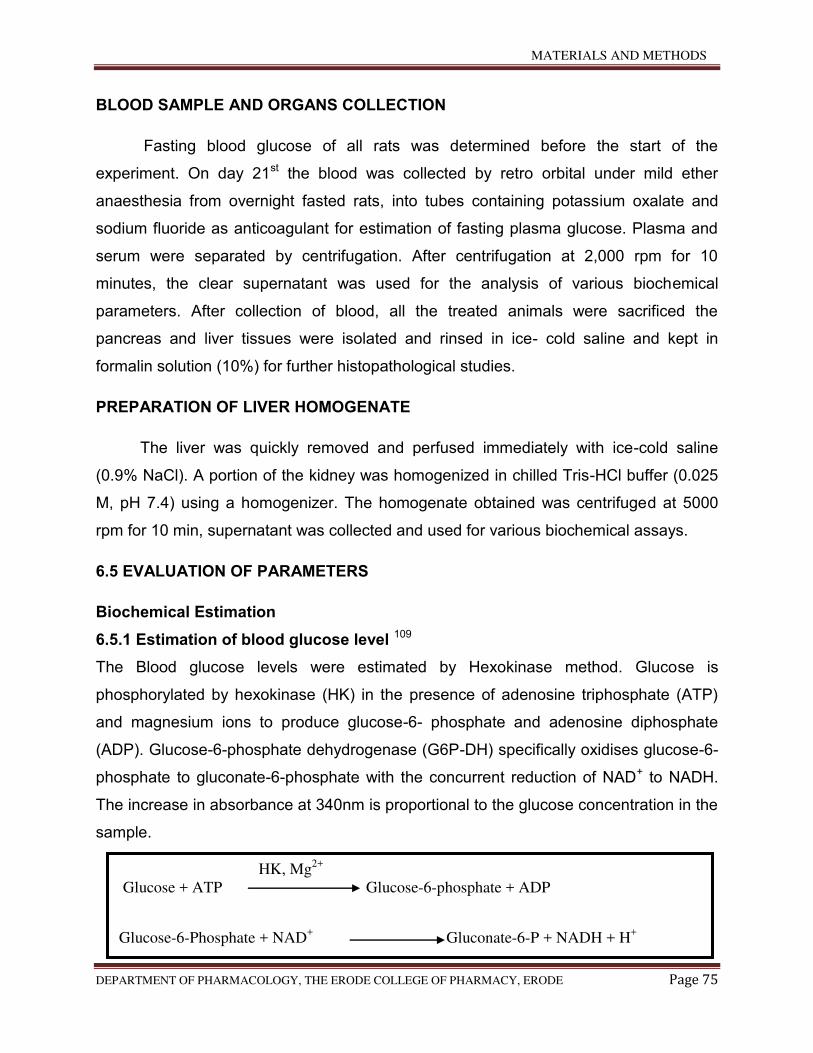
MATERIALS AND METHODS
DEPARTMENT OF PHARMACOLOGY, THE ERODE COLLEGE OF PHARMACY, ERODE Page 75
BLOOD SAMPLE AND ORGANS COLLECTION
Fasting blood glucose of all rats was determined before the start of the
experiment. On day 21st the blood was collected by retro orbital under mild ether
anaesthesia from overnight fasted rats, into tubes containing potassium oxalate and
sodium fluoride as anticoagulant for estimation of fasting plasma glucose. Plasma and
serum were separated by centrifugation. After centrifugation at 2,000 rpm for 10
minutes, the clear supernatant was used for the analysis of various biochemical
parameters. After collection of blood, all the treated animals were sacrificed the
pancreas and liver tissues were isolated and rinsed in ice- cold saline and kept in
formalin solution (10%) for further histopathological studies.
PREPARATION OF LIVER HOMOGENATE
The liver was quickly removed and perfused immediately with ice-cold saline
(0.9% NaCl). A portion of the kidney was homogenized in chilled Tris-HCl buffer (0.025
M, pH 7.4) using a homogenizer. The homogenate obtained was centrifuged at 5000
rpm for 10 min, supernatant was collected and used for various biochemical assays.
6.5 EVALUATION OF PARAMETERS
Biochemical Estimation
6.5.1 Estimation of blood glucose level 109
The Blood glucose levels were estimated by Hexokinase method. Glucose is
phosphorylated by hexokinase (HK) in the presence of adenosine triphosphate (ATP)
and magnesium ions to produce glucose-6- phosphate and adenosine diphosphate
(ADP). Glucose-6-phosphate dehydrogenase (G6P-DH) specifically oxidises glucose-6-
phosphate to gluconate-6-phosphate with the concurrent reduction of NAD+ to NADH.
The increase in absorbance at 340nm is proportional to the glucose concentration in the
sample.
HK, Mg2+
Glucose + ATP Glucose-6-phosphate + ADP
Glucose-6-Phosphate + NAD+
Gluconate-6-P + NADH + H+
MATERIALS AND METHODS
DEPARTMENT OF PHARMACOLOGY, THE ERODE COLLEGE OF PHARMACY, ERODE Page 76
Reagents
Reagent 1
0.05 M Tris HCl buffer, pH 8.0 with 13.3 mM MgCl2
0.67 M Glucose in Tris-MgCl2 buffer
16.5 mM ATP in Tris-MgCl2 buffer
6.8 mM NAD in Tris-MgCl2 buffer
Reagent 1A
300 IU/ml Glucose-6-phosphate dehydrogenase in Tris-MgCl2 buffer
300 IU/ml Hexokinase in Tris-MgCl2 buffer
Procedure
150 μl of reagent 1 was added with 30 μl of reagent 1A and to this 20 μl of suitable
diluents was added and the contents were mixed thoroughly. To this mixture, 2 μl of
serum sample was added. Then the contents were mixed and incubated at 37°C for 10
seconds. After zeroing the instrument with blank the absorbance of standard followed
by the test sample was measured at 340 nm. The values were expressed as mg/dl.
MATERIALS AND METHODS
DEPARTMENT OF PHARMACOLOGY, THE ERODE COLLEGE OF PHARMACY, ERODE Page 77
6.5.1.1 Estimation of Glycosylated Haemoglobin (HbA1C)110
Method: “Tina-quant” (turbidimetric inhibition immunoassay”
The concentration A1c (HbA1c) is measured as a percentage of a total
hemoglobin in human whole blood (%HbA1c). Consequently, the hemoglobin
contained in red blood cells is released by hemolysis of the sample. This method
uses the Hemolyzing Reagent containing a detergent (tetradecyltrimethylammonium
bromide - TTAB) to specifically lysate red blood cells. HbA1c and Hemoglobin
levels in the sample are determined from the obtained hemolysate by two
independent reactions. HbA1c During the first stage of the reaction: the HbA1c in
the sample reacts with the anti-HbA1c specific antibody (Reagent A1) to form
soluble antigen-antibody complexes. Then the polyhapten is added (Reagent A2). The
polyhapten reacts with the specific antibody excess from the first reaction,
producing insoluble immune complexes which can be measured turbidimetrically at
340 nm.
Reagents
Reagent A1: Monospecific antibodies anti-HbA1c in pH 6.2 buffer.
Reagent A2: Polyhapten-HbA1c in pH 6.2 buffer.
Calculation
%HbA1c = 91.5 x HbA1c (g/dl) / Hb( gm/dl) + 2.15
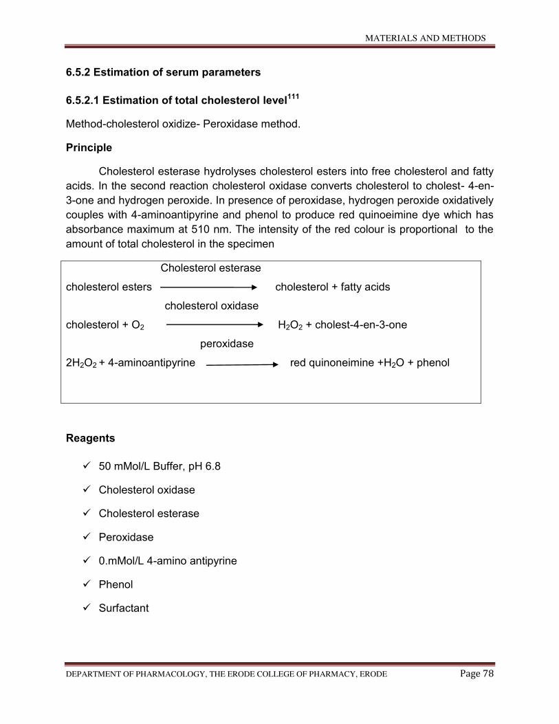
MATERIALS AND METHODS
DEPARTMENT OF PHARMACOLOGY, THE ERODE COLLEGE OF PHARMACY, ERODE Page 78
6.5.2 Estimation of serum parameters
6.5.2.1 Estimation of total cholesterol level111
Method-cholesterol oxidize- Peroxidase method.
Principle
Cholesterol esterase hydrolyses cholesterol esters into free cholesterol and fatty
acids. In the second reaction cholesterol oxidase converts cholesterol to cholest- 4-en-
3-one and hydrogen peroxide. In presence of peroxidase, hydrogen peroxide oxidatively
couples with 4-aminoantipyrine and phenol to produce red quinoeimine dye which has
absorbance maximum at 510 nm. The intensity of the red colour is proportional to the
amount of total cholesterol in the specimen
Cholesterol esterase
cholesterol esters cholesterol + fatty acids
cholesterol oxidase
cholesterol + O2 H2O2 + cholest-4-en-3-one
peroxidase
2H2O2 + 4-aminoantipyrine red quinoneimine +H2O + phenol
Reagents
50 mMol/L Buffer, pH 6.8
Cholesterol oxidase
Cholesterol esterase
Peroxidase
0.mMol/L 4-amino antipyrine
Phenol
Surfactant
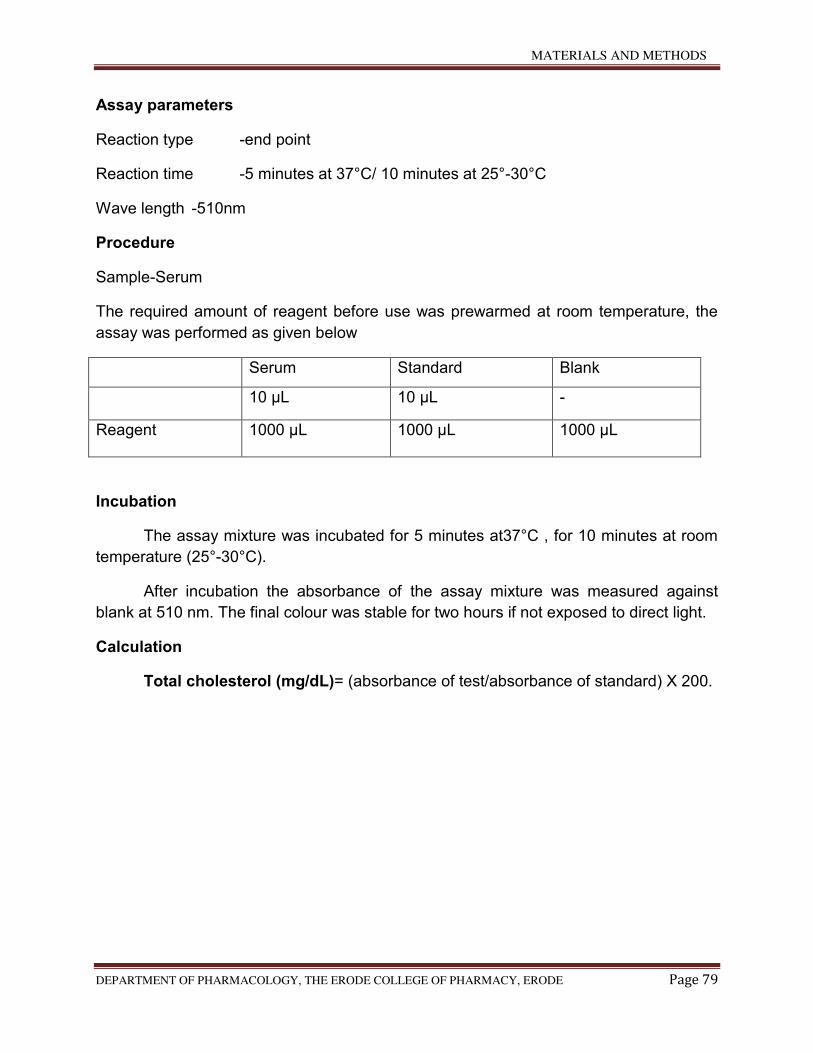
MATERIALS AND METHODS
DEPARTMENT OF PHARMACOLOGY, THE ERODE COLLEGE OF PHARMACY, ERODE Page 79
Assay parameters
Reaction type -end point
Reaction time -5 minutes at 37°C/ 10 minutes at 25°-30°C
Wave length -510nm
Procedure
Sample-Serum
The required amount of reagent before use was prewarmed at room temperature, the
assay was performed as given below
Serum Standard Blank
10 µL 10 µL -
Reagent 1000 µL 1000 µL 1000 µL
Incubation
The assay mixture was incubated for 5 minutes at37°C , for 10 minutes at room
temperature (25°-30°C).
After incubation the absorbance of the assay mixture was measured against
blank at 510 nm. The final colour was stable for two hours if not exposed to direct light.
Calculation
Total cholesterol (mg/dL)= (absorbance of test/absorbance of standard) X 200.
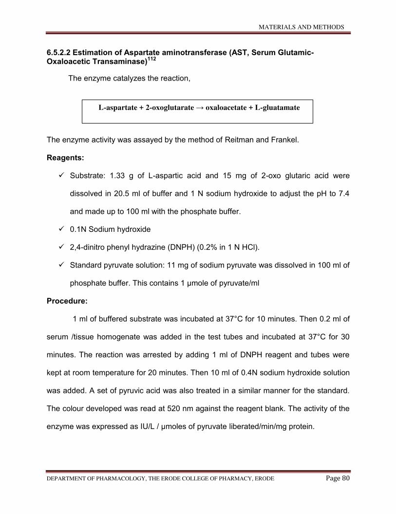
MATERIALS AND METHODS
DEPARTMENT OF PHARMACOLOGY, THE ERODE COLLEGE OF PHARMACY, ERODE Page 80
6.5.2.2 Estimation of Aspartate aminotransferase (AST, Serum Glutamic-Oxaloacetic Transaminase)112
The enzyme catalyzes the reaction,
The enzyme activity was assayed by the method of Reitman and Frankel.
Reagents:
Substrate: 1.33 g of L-aspartic acid and 15 mg of 2-oxo glutaric acid were
dissolved in 20.5 ml of buffer and 1 N sodium hydroxide to adjust the pH to 7.4
and made up to 100 ml with the phosphate buffer.
0.1N Sodium hydroxide
2,4-dinitro phenyl hydrazine (DNPH) (0.2% in 1 N HCl).
Standard pyruvate solution: 11 mg of sodium pyruvate was dissolved in 100 ml of
phosphate buffer. This contains 1 μmole of pyruvate/ml
Procedure:
1 ml of buffered substrate was incubated at 37°C for 10 minutes. Then 0.2 ml of
serum /tissue homogenate was added in the test tubes and incubated at 37°C for 30
minutes. The reaction was arrested by adding 1 ml of DNPH reagent and tubes were
kept at room temperature for 20 minutes. Then 10 ml of 0.4N sodium hydroxide solution
was added. A set of pyruvic acid was also treated in a similar manner for the standard.
The colour developed was read at 520 nm against the reagent blank. The activity of the
enzyme was expressed as IU/L / μmoles of pyruvate liberated/min/mg protein.
L-aspartate + 2-oxoglutarate → oxaloacetate + L-gluatamate
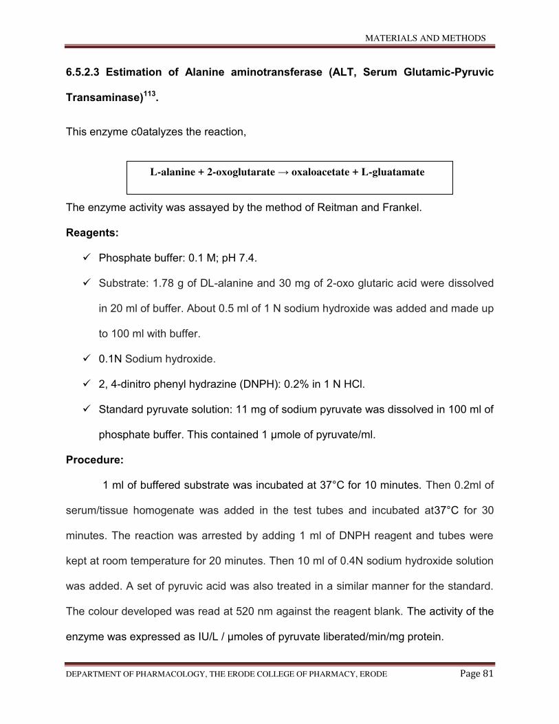
MATERIALS AND METHODS
DEPARTMENT OF PHARMACOLOGY, THE ERODE COLLEGE OF PHARMACY, ERODE Page 81
6.5.2.3 Estimation of Alanine aminotransferase (ALT, Serum Glutamic-Pyruvic
Transaminase)113.
This enzyme c0atalyzes the reaction,
The enzyme activity was assayed by the method of Reitman and Frankel.
Reagents:
Phosphate buffer: 0.1 M; pH 7.4.
Substrate: 1.78 g of DL-alanine and 30 mg of 2-oxo glutaric acid were dissolved
in 20 ml of buffer. About 0.5 ml of 1 N sodium hydroxide was added and made up
to 100 ml with buffer.
0.1N Sodium hydroxide.
2, 4-dinitro phenyl hydrazine (DNPH): 0.2% in 1 N HCl.
Standard pyruvate solution: 11 mg of sodium pyruvate was dissolved in 100 ml of
phosphate buffer. This contained 1 μmole of pyruvate/ml.
Procedure:
1 ml of buffered substrate was incubated at 37°C for 10 minutes. Then 0.2ml of
serum/tissue homogenate was added in the test tubes and incubated at37°C for 30
minutes. The reaction was arrested by adding 1 ml of DNPH reagent and tubes were
kept at room temperature for 20 minutes. Then 10 ml of 0.4N sodium hydroxide solution
was added. A set of pyruvic acid was also treated in a similar manner for the standard.
The colour developed was read at 520 nm against the reagent blank. The activity of the
enzyme was expressed as IU/L / μmoles of pyruvate liberated/min/mg protein.
L-alanine + 2-oxoglutarate → oxaloacetate + L-gluatamate
MATERIALS AND METHODS
DEPARTMENT OF PHARMACOLOGY, THE ERODE COLLEGE OF PHARMACY, ERODE Page 82
6.5.3 Estimation of oxidative stress parameters
6.5.3.1 Estimation of Lipid Peroxidation (LPO)114
Lipid peroxidation (LPO) was assayed by the method of Ohkawa et al, in which
the malondialdehyde (MDA) released served as the index of LPO. The extent of LPO in
the hepatic tissue was assayed by measuring one of the end products of this process,
the thiobarbituric acid-reactive substances (TBARS). As 99% TBARS is
malondialdehyde (MDA), thus this assay is based on the reaction of 1 molecule of MDA
with 2 molecules of TBARS at low pH (2- 3) and at a temperature of 95°C for 60 min.
The resultant pink chromogen can be detected spectrophonometrically at 532 nm.
Reagents
Standard: 1, 1, 3, 3-tetra ethoxypropane (TEP).
8.1% Sodium dodecyl sulphate (SDS)
20%Acetic acid
0.8%Thiobarbituric acid (TBA)
15:1 v/v n-butanol: pyridine mixture
Procedure
To 0.2 ml of tissue homogenate, 0.2 ml of 8.1% SDS, 1.5 ml of 20% acetic acid
(pH 3.5) and 1.5 ml of 0.8% TBA were added. The mixture was made up to 4 ml with
water and then heated in a water bath at 95.8ºC for 60 min using glass ball as a
condenser. After cooling, 1 ml of water and 5 ml of n-butanol: pyridine (15:1 v/v) mixture
were added and shaken vigorously. After centrifugation at 4000 rpm for 10 min, the
organic layer was taken and its absorbance was measured at 532 nm. The level of lipid
peroxides was expressed as nmoles of MDA formed/mg of protein.
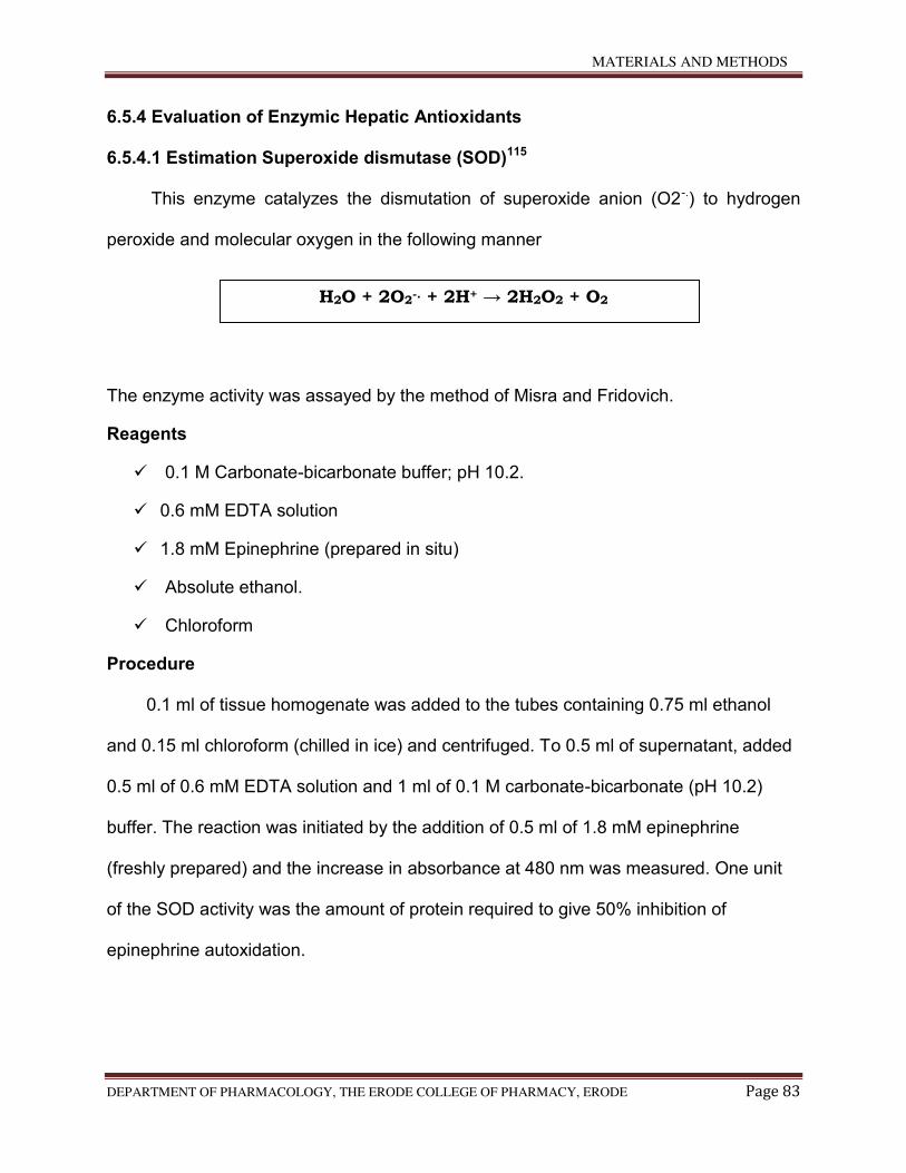
MATERIALS AND METHODS
DEPARTMENT OF PHARMACOLOGY, THE ERODE COLLEGE OF PHARMACY, ERODE Page 83
6.5.4 Evaluation of Enzymic Hepatic Antioxidants
6.5.4.1 Estimation Superoxide dismutase (SOD)115
This enzyme catalyzes the dismutation of superoxide anion (O2-.) to hydrogen
peroxide and molecular oxygen in the following manner
The enzyme activity was assayed by the method of Misra and Fridovich.
Reagents
0.1 M Carbonate-bicarbonate buffer; pH 10.2.
0.6 mM EDTA solution
1.8 mM Epinephrine (prepared in situ)
Absolute ethanol.
Chloroform
Procedure
0.1 ml of tissue homogenate was added to the tubes containing 0.75 ml ethanol
and 0.15 ml chloroform (chilled in ice) and centrifuged. To 0.5 ml of supernatant, added
0.5 ml of 0.6 mM EDTA solution and 1 ml of 0.1 M carbonate-bicarbonate (pH 10.2)
buffer. The reaction was initiated by the addition of 0.5 ml of 1.8 mM epinephrine
(freshly prepared) and the increase in absorbance at 480 nm was measured. One unit
of the SOD activity was the amount of protein required to give 50% inhibition of
epinephrine autoxidation.
H2O + 2O2-. + 2H+ → 2H2O2 + O2
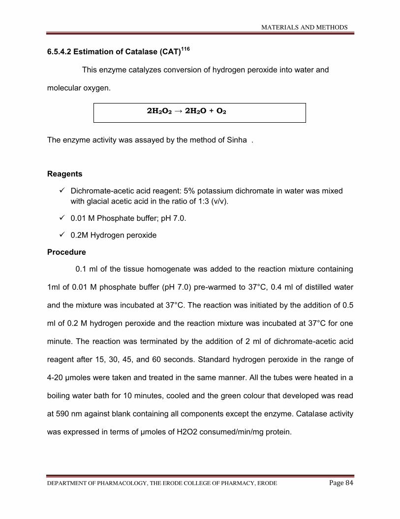
MATERIALS AND METHODS
DEPARTMENT OF PHARMACOLOGY, THE ERODE COLLEGE OF PHARMACY, ERODE Page 84
6.5.4.2 Estimation of Catalase (CAT)116
This enzyme catalyzes conversion of hydrogen peroxide into water and
molecular oxygen.
The enzyme activity was assayed by the method of Sinha .
Reagents
Dichromate-acetic acid reagent: 5% potassium dichromate in water was mixed
with glacial acetic acid in the ratio of 1:3 (v/v).
0.01 M Phosphate buffer; pH 7.0.
0.2M Hydrogen peroxide
Procedure
0.1 ml of the tissue homogenate was added to the reaction mixture containing
1ml of 0.01 M phosphate buffer (pH 7.0) pre-warmed to 37°C, 0.4 ml of distilled water
and the mixture was incubated at 37°C. The reaction was initiated by the addition of 0.5
ml of 0.2 M hydrogen peroxide and the reaction mixture was incubated at 37°C for one
minute. The reaction was terminated by the addition of 2 ml of dichromate-acetic acid
reagent after 15, 30, 45, and 60 seconds. Standard hydrogen peroxide in the range of
4-20 μmoles were taken and treated in the same manner. All the tubes were heated in a
boiling water bath for 10 minutes, cooled and the green colour that developed was read
at 590 nm against blank containing all components except the enzyme. Catalase activity
was expressed in terms of μmoles of H2O2 consumed/min/mg protein.
2H2O2 → 2H2O + O2
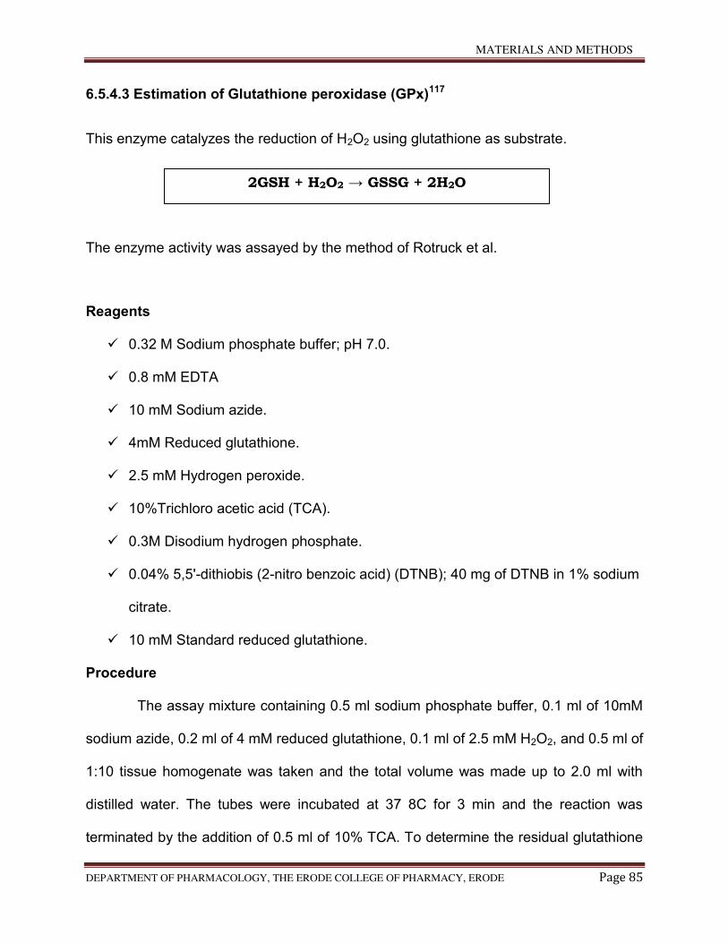
MATERIALS AND METHODS
DEPARTMENT OF PHARMACOLOGY, THE ERODE COLLEGE OF PHARMACY, ERODE Page 85
6.5.4.3 Estimation of Glutathione peroxidase (GPx)117
This enzyme catalyzes the reduction of H2O2 using glutathione as substrate.
The enzyme activity was assayed by the method of Rotruck et al.
Reagents
0.32 M Sodium phosphate buffer; pH 7.0.
0.8 mM EDTA
10 mM Sodium azide.
4mM Reduced glutathione.
2.5 mM Hydrogen peroxide.
10%Trichloro acetic acid (TCA).
0.3M Disodium hydrogen phosphate.
0.04% 5,5'-dithiobis (2-nitro benzoic acid) (DTNB); 40 mg of DTNB in 1% sodium
citrate.
10 mM Standard reduced glutathione.
Procedure
The assay mixture containing 0.5 ml sodium phosphate buffer, 0.1 ml of 10mM
sodium azide, 0.2 ml of 4 mM reduced glutathione, 0.1 ml of 2.5 mM H2O2, and 0.5 ml of
1:10 tissue homogenate was taken and the total volume was made up to 2.0 ml with
distilled water. The tubes were incubated at 37 8C for 3 min and the reaction was
terminated by the addition of 0.5 ml of 10% TCA. To determine the residual glutathione
2GSH + H2O2 → GSSG + 2H2O
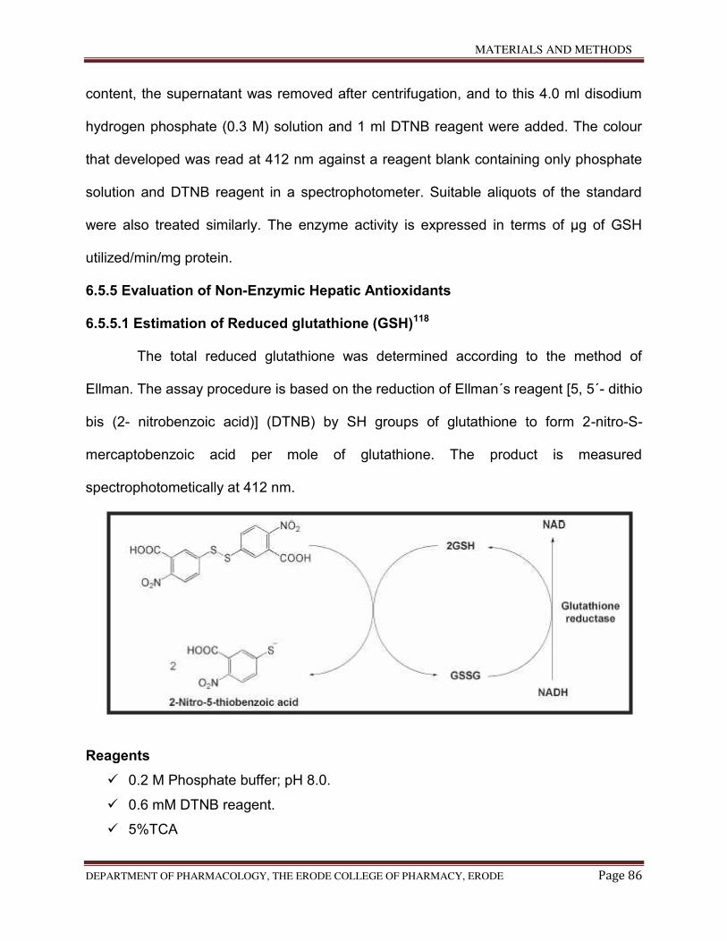
MATERIALS AND METHODS
DEPARTMENT OF PHARMACOLOGY, THE ERODE COLLEGE OF PHARMACY, ERODE Page 86
content, the supernatant was removed after centrifugation, and to this 4.0 ml disodium
hydrogen phosphate (0.3 M) solution and 1 ml DTNB reagent were added. The colour
that developed was read at 412 nm against a reagent blank containing only phosphate
solution and DTNB reagent in a spectrophotometer. Suitable aliquots of the standard
were also treated similarly. The enzyme activity is expressed in terms of μg of GSH
utilized/min/mg protein.
6.5.5 Evaluation of Non-Enzymic Hepatic Antioxidants
6.5.5.1 Estimation of Reduced glutathione (GSH)118
The total reduced glutathione was determined according to the method of
Ellman. The assay procedure is based on the reduction of Ellman´s reagent [5, 5΄- dithio
bis (2- nitrobenzoic acid)] (DTNB) by SH groups of glutathione to form 2-nitro-S-
mercaptobenzoic acid per mole of glutathione. The product is measured
spectrophotometically at 412 nm.
Reagents
0.2 M Phosphate buffer; pH 8.0.
0.6 mM DTNB reagent.
5%TCA
MATERIALS AND METHODS
DEPARTMENT OF PHARMACOLOGY, THE ERODE COLLEGE OF PHARMACY, ERODE Page 87
Procedure
0.1 ml of tissue homogenate was precipitated with 5% TCA. The contents were
mixed well for complete precipitation of proteins and centrifuged. To 0.1 ml of
supernatant, 2 ml of 0.6 mM DTNB reagent and 0.2 M phosphate buffer (pH 8.0) were
added to make up to a final volume of 4 ml. The absorbance was read at 412 nm
against a blank containing TCA instead of sample. A series of standards treated in a
similar way also run to determine the glutathione content. The amount of glutathione
was expressed as nmoles/g tissue.
6.5.5.2 Estimation of Vitamin C (Ascorbic acid)119
The level of vitamin C was estimated by the method of Omaye et al. Ascorbic
acid is oxidised by copper to form dehydroascorbic acid. The product was treated with
2,4 dinitrophenyl hydrazine to form tris 2,4 dinitrophenyl hydrazone which undergoes
rearrangement to form a product with the absorption maximum at 520 nm in
spectrophotometer.
Reagents
5% TCA
DTC reagent (3 gm of 2,4 dinitrophenyl hydrazine, 0.4 gm thiourea and 0.05 gm
of copper sulphate were dissolved in 100 ml of 9 N H2SO4).
65 % H2SO4 (ice cold)
Standard ascorbic acid
MATERIALS AND METHODS
DEPARTMENT OF PHARMACOLOGY, THE ERODE COLLEGE OF PHARMACY, ERODE Page 88
Procedure
To 0.5 ml of tissue homogenate, 0.5 ml of water and 1 ml of TCA were added,
mixed thoroughly and centrifuged. To 1 ml of the supernatant, 0.2 ml of DTC reagent
was added and incubated at 37°C for 3 hours. Then 1.5 ml of H2SO4 was added, mixed
well and the solution was allowed to stand for 30 minutes at room temperature. The
colour developed was read at 520 nm in spectrophotometer. The level of vitamin C was
expressed as nmole/ g of wet tissue.
6.5.5.3 Estimation of Vitamin E (α- Tocopherol)120
Vitamin E content was estimated by the method of Palan et al. This method
involves the conversion of ferric ions to ferrous ions by a-tocopherol and the formation
of red colored complex with 2, 2 dipyridyl. Absorbance of chromophore was measured
at 520 nm in the spectrophotometer.
Reagents
2% 2,2 dipyridyl solution
5 % FeCl3 solution
Standard : 100 mg of α-tocopherol in 0.1% ethanol
n- Butanol
Procedure
To 0.5 ml of tissue homogenate, 1.5 ml of ethanol was added, mixed and
centrifuged. The supernatant was dried at 80°C for 3 hours. To this 0.2 ml of 2, 2
dipyridyl solution and 0.2 ml of FeCl3 solution were added, mixed well and 4 ml of
butanol was added. The colour developed was read at 520 nm in the
spectrophotometer. The level of vitamin C was expressed as nmole/ g of wet tissue.
MATERIALS AND METHODS
DEPARTMENT OF PHARMACOLOGY, THE ERODE COLLEGE OF PHARMACY, ERODE Page 89
6.5.6 ESTIMATION OF PLASMA INSULIN LEVEL121
Insulin was assayed in plasma using a commercial kit by enzyme linked
immunosorbant assay (ELISA) technique.
Reagents used for the estimation of plasma insulin level:
1. Monoclonal anti-insulin antibody
2. Enzyme conjugate: Anti-insulin antibodies conjugated to horseradish
peroxidise
3. Standard: Human insulin
4. Solution A: Buffer solution containing hydrogen peroxide
5. Solution B: Tetramethylbenzidine
6. Concentrated wash buffer
7. Stock solution: 2 N HCL
Procedure:
25 µl of the plasma taken into micro wells coated with anti-insulin antibody. To
this, 100µl of the enzyme conjugate was added to each well, mixed for 5 sec and
incubated at 25°C for 30 min. The wells were rinsed for five times with washing buffer.
Then, 100µl of solution A and then 100µl of solution B were added to each well. This
was incubated for 15 min at room temperature. The reaction was stopped by the
addition of 50µl of 2 N HCL to each well and read at 450 nm.
MATERIALS AND METHODS
DEPARTMENT OF PHARMACOLOGY, THE ERODE COLLEGE OF PHARMACY, ERODE Page 90
6.7 HISTOPATHOLOGICAL STUDIES113
Hematoxylin, a basic bye is oxidized to hematein with a mordant, a metallic ion
such as the salts of aluminium. The positively charged aluminium-hematein complex
combines with the negatively charged phosphate groups of the nucleic acids (DNA and
RNA) forming blue/purple colour, which is characteristic of hematoxylin stains. Eosin is
an acidic dye, which is considered to have a selective affinity for the basic parts of the
cell, i.e., the cytoplasm. Thus, the hematoxylin and eosin (H & E) stain is used to
demonstrate different structures of the tissue.
The various steps involved in the preparation of pancreatic tissues for
histological studies are as follows:
Fixation
In order to avoid tissue by the lysosomal enzymes and to preserve its
physical and chemical structure, a bit of tissue from each organ was cut and fixed in
bouin’s fluid immediately after removal from the animal body. The tissues were fixed in
bouin’s fluid for about 24 hours. The tissues were then taken and washed in glass
distilled water for a day to remove excess of picric acid.
Dehydration
The tissues were kept in the following solutions for an hour each; 30%,
50%, 70% and 100% alcohol. Inadequately dehydrated tissues cannot be satisfactorily
infiltered with paraffin. At the same time over dehydration results in making the tissues
brittle, which would be difficult for sectioning. So, careful precautions were followed
while performing the dehydration process
Clearing
Dealcoholization or replacement of alcohol from the tissues with a
clearing agent is called as clearing. Xylene was used as the clearing agent for one or
two hours, two or three times. Since, the clearing agent is miscible with both
dehydration and embedding agents, it permits paraffin to infilterate the tissues. So, the
clearing was carried out as the next step after dehydration to permit tissue spaces to be
filled with paraffin. The tissues were kept in the clearing agent till they become
transparent and impregnated with xylene.
MATERIALS AND METHODS
DEPARTMENT OF PHARMACOLOGY, THE ERODE COLLEGE OF PHARMACY, ERODE Page 91
Impregnation
In this process the clearing agent xylene was placed by paraffin wax.
The tissues were taken out of xylene and were kept in molten paraffin embedding bath,
which consists of metal pots filled with molten wax maintained at about 50oC. The
tissues were given three changes in the molten wax at half an hour intervals.
Embedding
The paraffin wax used for embedding was fresh and heated upto the
optimum melting point at about 56–58oC. A clear glass plate was smeared with
glycerine. L-shaped mould was placed on it to from a rectangular cavity. The molten
paraffin wax was poured and air bubbles were removed by using a hot needle. The
tissue was placed in the paraffin and oriented with the surface to be sectioned. Then the
tissue was pressed gently towards the glass plate to make settle uniformly with a metal
pressing rod and allowed the wax to settle and solidity room temperature. The paraffin
block was kept in cold water for cooling.
Section Cutting
Section cutting was done with a rotatory microtome. The excess of
paraffin around the tissue was removed by trimming, leaving ½ cm around the tissue.
Then the block was attached to the gently heated holder. Additional support was given
by some extra wax, which was applied along the sides of the block. Before sectioning,
all set screws holding the object holder and knife were hand tightened to avoid vibration.
To produce uniform sections, the microtome knife was adjusted to the proper angle in
the knife holder with only the cutting edge coming in contact with the paraffin block. The
tissue was cut in the thickness range of about 7µm.
Flattening and Mounting of Sections
The procedure was carried out in tissue flotation warm water bath. The
sections were spread on a warm water bath after they were detached from the knife
with the help of hair brush. Dust free clean slides were coated with egg albumin over the
whole surface. Required sections were spread on clean slide and kept at room
temperature
MATERIALS AND METHODS
DEPARTMENT OF PHARMACOLOGY, THE ERODE COLLEGE OF PHARMACY, ERODE Page 92
Staining of Tissue Sections
The sections were stained as follows; deparaffinization with xylene two
times
each for five minutes
Dehydration through descending grades of ethyl alcohol
100% alcohol (absolute) - 2 minute
90% alcohol - 1minute
50% alcohol - 1 minute
Staining with Ehrlich’s Haemaoxylin was done for 15-20 minutes. Then the sectioned
tissues were thoroughly washed in tap water for 10 minutes. Rinsed with distilled water
and stained with Eosin. Dehydration again with ascending grades of alcohol.
70% alcohol - 2 minute
90% alcohol - 2 minute
100%alcohol - 1 minute
Finally the tissues were cleared with xylene two times, each for about 3 minutes
interval.
Mounting
On the stained slide, DPX mountant was applied uniformly and micro
glass cover slides were spread. The slides were observed in Nikon microscope and
microphotographs were taken.
STATISTICAL ANALYSIS
The data of all the results were represented as Mean ± S.E.M. on statistically
analysed by one-way ANOVA followed by Tukey’s multiple comparison test was used
for statistical analysis p<0.05 was considered significant.
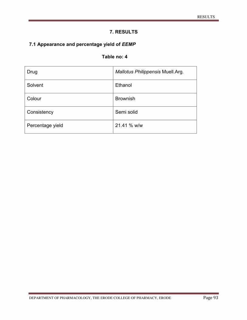
RESULTS
DEPARTMENT OF PHARMACOLOGY, THE ERODE COLLEGE OF PHARMACY, ERODE Page 93
7. RESULTS
7.1 Appearance and percentage yield of EEMP
Table no: 4
Drug Mallotus Philippensis Muell.Arg.
Solvent Ethanol
Colour Brownish
Consistency Semi solid
Percentage yield 21.41 % w/w
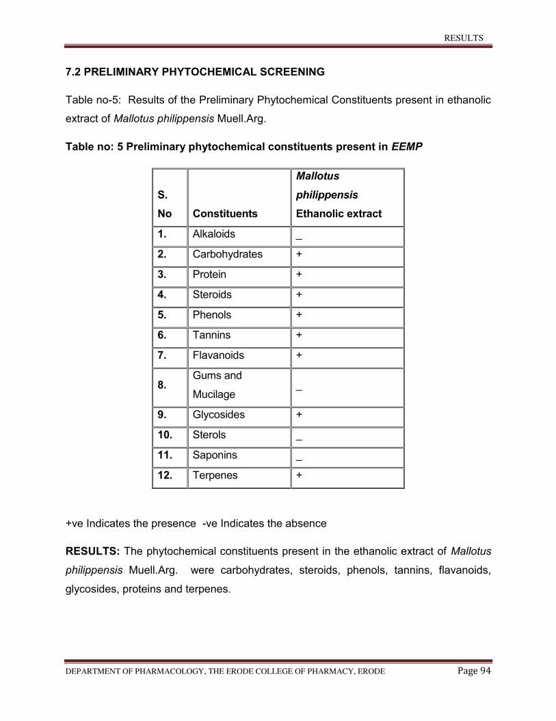
RESULTS
DEPARTMENT OF PHARMACOLOGY, THE ERODE COLLEGE OF PHARMACY, ERODE Page 94
7.2 PRELIMINARY PHYTOCHEMICAL SCREENING
Table no-5: Results of the Preliminary Phytochemical Constituents present in ethanolic
extract of Mallotus philippensis Muell.Arg.
Table no: 5 Preliminary phytochemical constituents present in EEMP
S.
No Constituents
Mallotus
philippensis
Ethanolic extract
1. Alkaloids _
2. Carbohydrates +
3. Protein +
4. Steroids +
5. Phenols +
6. Tannins +
7. Flavanoids +
8. Gums and
Mucilage _
9. Glycosides +
10. Sterols _
11. Saponins _
12. Terpenes +
+ve Indicates the presence -ve Indicates the absence
RESULTS: The phytochemical constituents present in the ethanolic extract of Mallotus
philippensis Muell.Arg. were carbohydrates, steroids, phenols, tannins, flavanoids,
glycosides, proteins and terpenes.
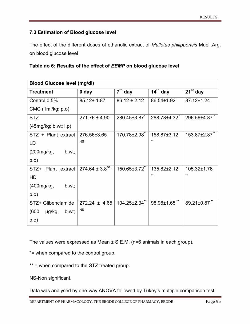
RESULTS
DEPARTMENT OF PHARMACOLOGY, THE ERODE COLLEGE OF PHARMACY, ERODE Page 95
7.3 Estimation of Blood glucose level
The effect of the different doses of ethanolic extract of Mallotus philippensis Muell.Arg.
on blood glucose level
Table no 6: Results of the effect of EEMP on blood glucose level
The values were expressed as Mean ± S.E.M. (n=6 animals in each group).
*= when compared to the control group.
** = when compared to the STZ treated group.
NS-Non significant.
Data was analysed by one-way ANOVA followed by Tukey’s multiple comparison test.
Blood Glucose level (mg/dl)
Treatment 0 day 7th day 14th day 21st day
Control 0.5%
CMC (1ml/kg; p.o)
85.12± 1.87 86.12 ± 2.12 86.54±1.92 87.12±1.24
STZ
(45mg/kg; b.wt; i.p)
271.76 ± 4.90 280.45±3.87* 288.78±4.32 * 296.56±4.87 *
STZ + Plant extract
LD
(200mg/kg, b.wt;
p.o)
276.56±3.65
NS
170.78±2.98** 158.87±3.12
**
153.87±2.87**
STZ+ Plant extract
HD
(400mg/kg, b.wt;
p.o)
274.64 ± 3.8NS 150.65±3.72** 135.82±2.12
**
105.32±1.76
**
STZ+ Glibenclamide
(600 µg/kg, b.wt;
p.o)
272.24 ± 4.65
NS
104.25±2.34** 98.98±1.65 ** 89.21±0.87 **
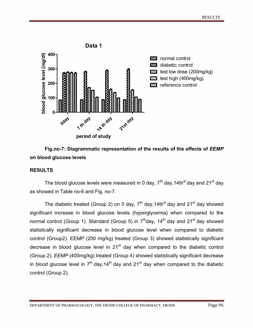
RESULTS
DEPARTMENT OF PHARMACOLOGY, THE ERODE COLLEGE OF PHARMACY, ERODE Page 96
Data 1
0day
7 th
day
14 th
day
21st
day
0
100
200
300
400normal control
diabetic control
test low dose (200mg/kg)
test high (400mg/kg)
reference control
period of study
blo
od
glu
co
se level (m
g/d
l)
Fig.no-7: Diagrammatic representation of the results of the effects of EEMP
on blood glucose levels
RESULTS
The blood glucose levels were measured in 0 day, 7th day,14thst day and 21st day
as showed in Table no-6 and Fig. no-7.
The diabetic treated (Group 2) on 0 day, 7th day,14thst day and 21st day showed
significant increase in blood glucose levels (hyperglycemia) when compared to the
normal control (Group 1). Standard (Group 5) in 7thday, 14th day and 21st day showed
statistically significant decrease in blood glucose level when compared to diabetic
control (Group2). EEMP (200 mg/kg) treated (Group 3) showed statistically significant
decrease in blood glucose level in 21st day when compared to the diabetic control
(Group 2). EEMP (400mg/kg) treated (Group 4) showed statistically significant decrease
in blood glucose level in 7th day,14th day and 21st day when compared to the diabetic
control (Group 2).
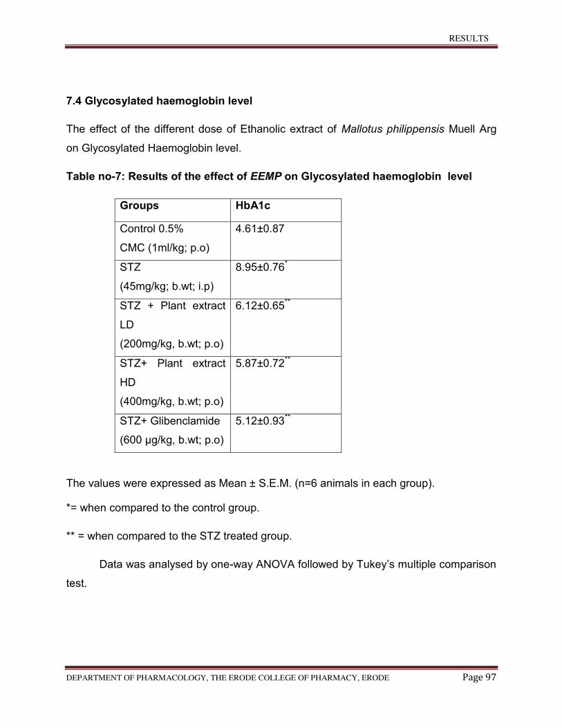
RESULTS
DEPARTMENT OF PHARMACOLOGY, THE ERODE COLLEGE OF PHARMACY, ERODE Page 97
7.4 Glycosylated haemoglobin level
The effect of the different dose of Ethanolic extract of Mallotus philippensis Muell Arg
on Glycosylated Haemoglobin level.
Table no-7: Results of the effect of EEMP on Glycosylated haemoglobin level
The values were expressed as Mean ± S.E.M. (n=6 animals in each group).
*= when compared to the control group.
** = when compared to the STZ treated group.
Data was analysed by one-way ANOVA followed by Tukey’s multiple comparison
test.
Groups HbA1c
Control 0.5%
CMC (1ml/kg; p.o)
4.61±0.87
STZ
(45mg/kg; b.wt; i.p)
8.95±0.76*
STZ + Plant extract
LD
(200mg/kg, b.wt; p.o)
6.12±0.65**
STZ+ Plant extract
HD
(400mg/kg, b.wt; p.o)
5.87±0.72**
STZ+ Glibenclamide
(600 µg/kg, b.wt; p.o)
5.12±0.93**
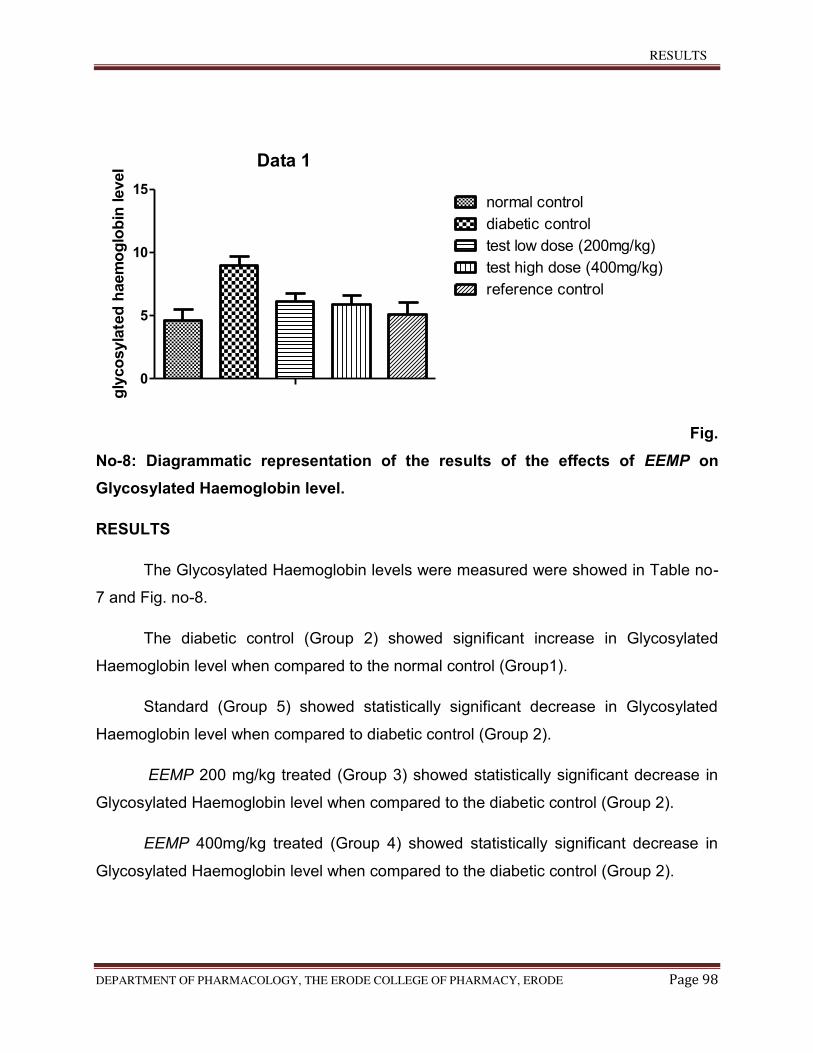
RESULTS
DEPARTMENT OF PHARMACOLOGY, THE ERODE COLLEGE OF PHARMACY, ERODE Page 98
Data 1
0
5
10
15normal control
diabetic control
test low dose (200mg/kg)
test high dose (400mg/kg)
reference control
gly
co
syla
ted
haem
og
lob
in level
Fig.
No-8: Diagrammatic representation of the results of the effects of EEMP on
Glycosylated Haemoglobin level.
RESULTS
The Glycosylated Haemoglobin levels were measured were showed in Table no-
7 and Fig. no-8.
The diabetic control (Group 2) showed significant increase in Glycosylated
Haemoglobin level when compared to the normal control (Group1).
Standard (Group 5) showed statistically significant decrease in Glycosylated
Haemoglobin level when compared to diabetic control (Group 2).
EEMP 200 mg/kg treated (Group 3) showed statistically significant decrease in
Glycosylated Haemoglobin level when compared to the diabetic control (Group 2).
EEMP 400mg/kg treated (Group 4) showed statistically significant decrease in
Glycosylated Haemoglobin level when compared to the diabetic control (Group 2).
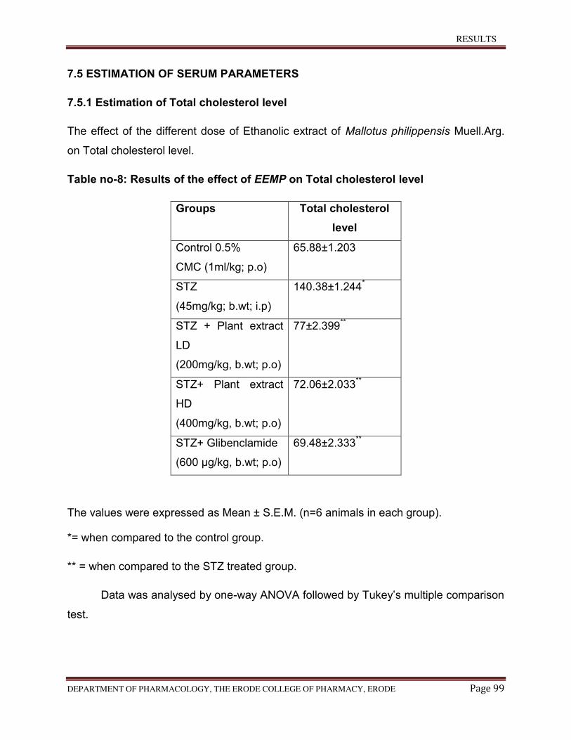
RESULTS
DEPARTMENT OF PHARMACOLOGY, THE ERODE COLLEGE OF PHARMACY, ERODE Page 99
7.5 ESTIMATION OF SERUM PARAMETERS
7.5.1 Estimation of Total cholesterol level
The effect of the different dose of Ethanolic extract of Mallotus philippensis Muell.Arg.
on Total cholesterol level.
Table no-8: Results of the effect of EEMP on Total cholesterol level
Groups Total cholesterol
level
Control 0.5%
CMC (1ml/kg; p.o)
65.88±1.203
STZ
(45mg/kg; b.wt; i.p)
140.38±1.244*
STZ + Plant extract
LD
(200mg/kg, b.wt; p.o)
77±2.399**
STZ+ Plant extract
HD
(400mg/kg, b.wt; p.o)
72.06±2.033**
STZ+ Glibenclamide
(600 µg/kg, b.wt; p.o)
69.48±2.333**
The values were expressed as Mean ± S.E.M. (n=6 animals in each group).
*= when compared to the control group.
** = when compared to the STZ treated group.
Data was analysed by one-way ANOVA followed by Tukey’s multiple comparison
test.
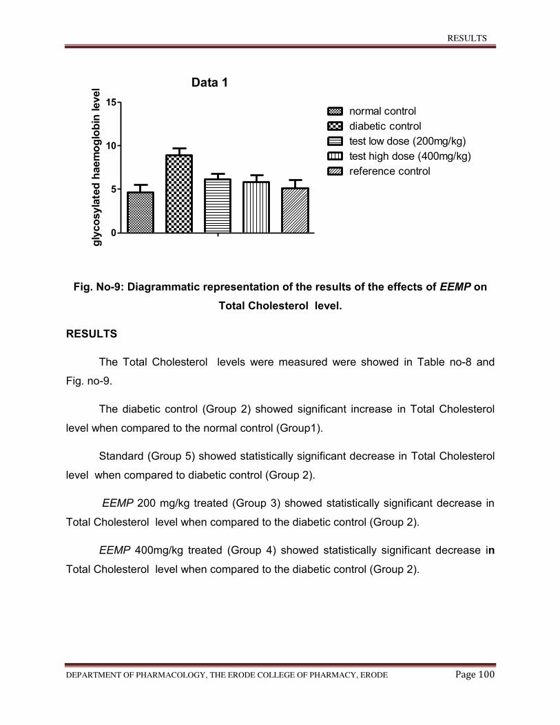
RESULTS
DEPARTMENT OF PHARMACOLOGY, THE ERODE COLLEGE OF PHARMACY, ERODE Page 100
Data 1
0
5
10
15normal control
diabetic control
test low dose (200mg/kg)
test high dose (400mg/kg)
reference control
gly
co
syla
ted
haem
og
lob
in level
Fig. No-9: Diagrammatic representation of the results of the effects of EEMP on
Total Cholesterol level.
RESULTS
The Total Cholesterol levels were measured were showed in Table no-8 and
Fig. no-9.
The diabetic control (Group 2) showed significant increase in Total Cholesterol
level when compared to the normal control (Group1).
Standard (Group 5) showed statistically significant decrease in Total Cholesterol
level when compared to diabetic control (Group 2).
EEMP 200 mg/kg treated (Group 3) showed statistically significant decrease in
Total Cholesterol level when compared to the diabetic control (Group 2).
EEMP 400mg/kg treated (Group 4) showed statistically significant decrease in
Total Cholesterol level when compared to the diabetic control (Group 2).
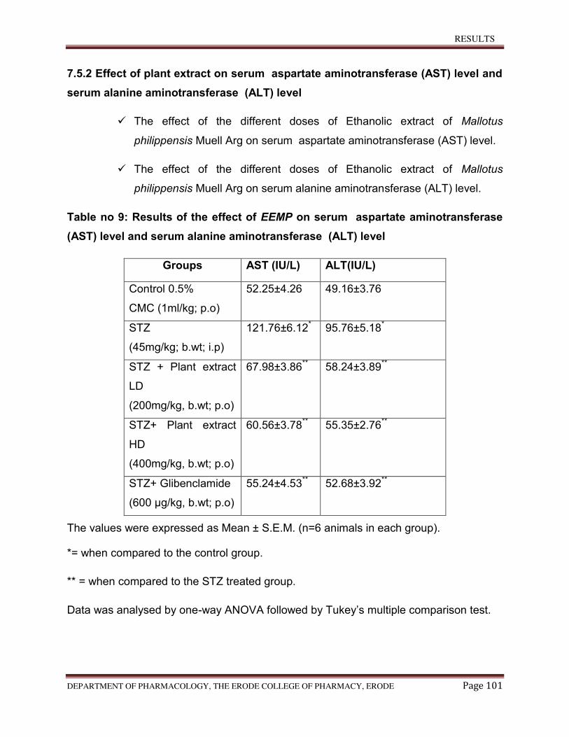
RESULTS
DEPARTMENT OF PHARMACOLOGY, THE ERODE COLLEGE OF PHARMACY, ERODE Page 101
7.5.2 Effect of plant extract on serum aspartate aminotransferase (AST) level and
serum alanine aminotransferase (ALT) level
The effect of the different doses of Ethanolic extract of Mallotus
philippensis Muell Arg on serum aspartate aminotransferase (AST) level.
The effect of the different doses of Ethanolic extract of Mallotus
philippensis Muell Arg on serum alanine aminotransferase (ALT) level.
Table no 9: Results of the effect of EEMP on serum aspartate aminotransferase
(AST) level and serum alanine aminotransferase (ALT) level
Groups AST (IU/L) ALT(IU/L)
Control 0.5%
CMC (1ml/kg; p.o)
52.25±4.26 49.16±3.76
STZ
(45mg/kg; b.wt; i.p)
121.76±6.12* 95.76±5.18*
STZ + Plant extract
LD
(200mg/kg, b.wt; p.o)
67.98±3.86** 58.24±3.89**
STZ+ Plant extract
HD
(400mg/kg, b.wt; p.o)
60.56±3.78** 55.35±2.76**
STZ+ Glibenclamide
(600 µg/kg, b.wt; p.o)
55.24±4.53** 52.68±3.92**
The values were expressed as Mean ± S.E.M. (n=6 animals in each group).
*= when compared to the control group.
** = when compared to the STZ treated group.
Data was analysed by one-way ANOVA followed by Tukey’s multiple comparison test.
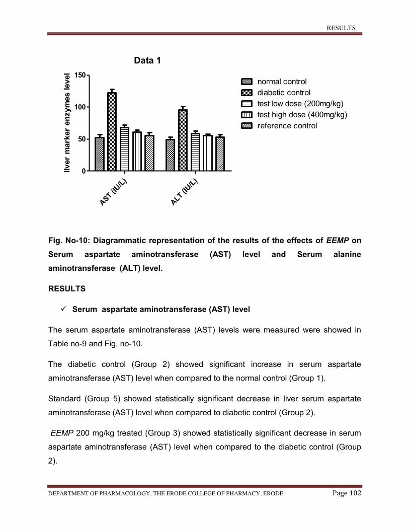
RESULTS
DEPARTMENT OF PHARMACOLOGY, THE ERODE COLLEGE OF PHARMACY, ERODE Page 102
Data 1
AST (I
U/L
)
ALT (I
U/L
)
0
50
100
150normal control
diabetic control
test low dose (200mg/kg)
test high dose (400mg/kg)
reference control
liver
mark
er
en
zym
es level
Fig. No-10: Diagrammatic representation of the results of the effects of EEMP on
Serum aspartate aminotransferase (AST) level and Serum alanine
aminotransferase (ALT) level.
RESULTS
Serum aspartate aminotransferase (AST) level
The serum aspartate aminotransferase (AST) levels were measured were showed in
Table no-9 and Fig. no-10.
The diabetic control (Group 2) showed significant increase in serum aspartate
aminotransferase (AST) level when compared to the normal control (Group 1).
Standard (Group 5) showed statistically significant decrease in liver serum aspartate
aminotransferase (AST) level when compared to diabetic control (Group 2).
EEMP 200 mg/kg treated (Group 3) showed statistically significant decrease in serum
aspartate aminotransferase (AST) level when compared to the diabetic control (Group
2).
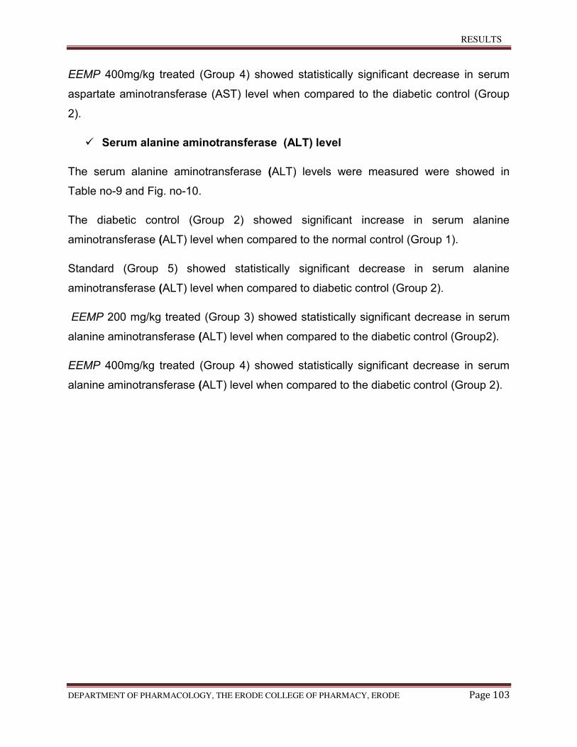
RESULTS
DEPARTMENT OF PHARMACOLOGY, THE ERODE COLLEGE OF PHARMACY, ERODE Page 103
EEMP 400mg/kg treated (Group 4) showed statistically significant decrease in serum
aspartate aminotransferase (AST) level when compared to the diabetic control (Group
2).
Serum alanine aminotransferase (ALT) level
The serum alanine aminotransferase (ALT) levels were measured were showed in
Table no-9 and Fig. no-10.
The diabetic control (Group 2) showed significant increase in serum alanine
aminotransferase (ALT) level when compared to the normal control (Group 1).
Standard (Group 5) showed statistically significant decrease in serum alanine
aminotransferase (ALT) level when compared to diabetic control (Group 2).
EEMP 200 mg/kg treated (Group 3) showed statistically significant decrease in serum
alanine aminotransferase (ALT) level when compared to the diabetic control (Group2).
EEMP 400mg/kg treated (Group 4) showed statistically significant decrease in serum
alanine aminotransferase (ALT) level when compared to the diabetic control (Group 2).
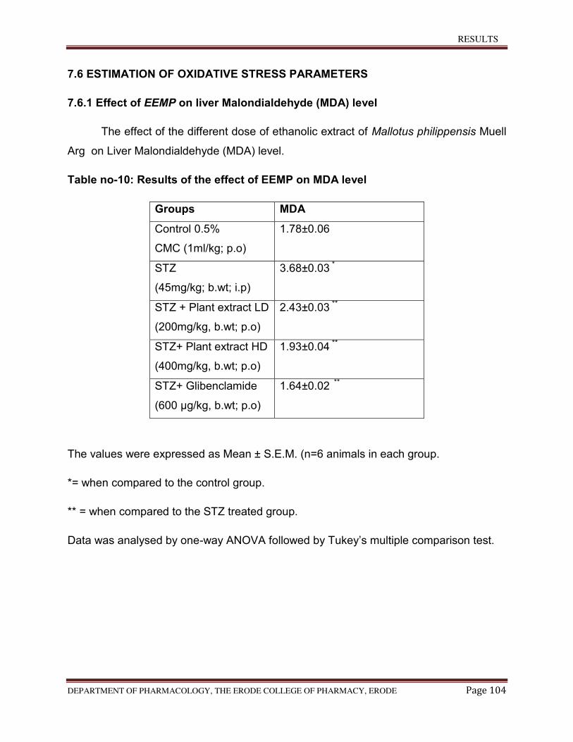
RESULTS
DEPARTMENT OF PHARMACOLOGY, THE ERODE COLLEGE OF PHARMACY, ERODE Page 104
7.6 ESTIMATION OF OXIDATIVE STRESS PARAMETERS
7.6.1 Effect of EEMP on liver Malondialdehyde (MDA) level
The effect of the different dose of ethanolic extract of Mallotus philippensis Muell
Arg on Liver Malondialdehyde (MDA) level.
Table no-10: Results of the effect of EEMP on MDA level
Groups MDA
Control 0.5%
CMC (1ml/kg; p.o)
1.78±0.06
STZ
(45mg/kg; b.wt; i.p)
3.68±0.03 *
STZ + Plant extract LD
(200mg/kg, b.wt; p.o)
2.43±0.03 **
STZ+ Plant extract HD
(400mg/kg, b.wt; p.o)
1.93±0.04 **
STZ+ Glibenclamide
(600 µg/kg, b.wt; p.o)
1.64±0.02 **
The values were expressed as Mean ± S.E.M. (n=6 animals in each group.
*= when compared to the control group.
** = when compared to the STZ treated group.
Data was analysed by one-way ANOVA followed by Tukey’s multiple comparison test.
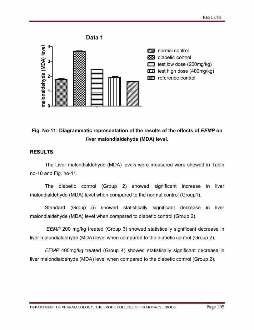
RESULTS
DEPARTMENT OF PHARMACOLOGY, THE ERODE COLLEGE OF PHARMACY, ERODE Page 105
Data 1
0
1
2
3
4normal control
diabetic control
test low dose (200mg/kg)
test high dose (400mg/kg)
reference control
malo
nald
eh
yd
e (
MD
A)
level
Fig. No-11: Diagrammatic representation of the results of the effects of EEMP on
liver malondialdehyde (MDA) level.
RESULTS
The Liver malondialdehyde (MDA) levels were measured were showed in Table
no-10 and Fig. no-11.
The diabetic control (Group 2) showed significant increase in liver
malondialdehyde (MDA) level when compared to the normal control (Group1).
Standard (Group 5) showed statistically significant decrease in liver
malondialdehyde (MDA) level when compared to diabetic control (Group 2).
EEMP 200 mg/kg treated (Group 3) showed statistically significant decrease in
liver malondialdehyde (MDA) level when compared to the diabetic control (Group 2).
EEMP 400mg/kg treated (Group 4) showed statistically significant decrease in
liver malondialdehyde (MDA) level when compared to the diabetic control (Group 2).
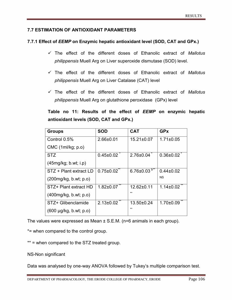
RESULTS
DEPARTMENT OF PHARMACOLOGY, THE ERODE COLLEGE OF PHARMACY, ERODE Page 106
7.7 ESTIMATION OF ANTIOXIDANT PARAMETERS
7.7.1 Effect of EEMP on Enzymic hepatic antioxidant level (SOD, CAT and GPx.)
The effect of the different doses of Ethanolic extract of Mallotus
philippensis Muell Arg on Liver superoxide dismutase (SOD) level.
The effect of the different doses of Ethanolic extract of Mallotus
philippensis Muell Arg on Liver Catalase (CAT) level
The effect of the different doses of Ethanolic extract of Mallotus
philippensis Muell Arg on glutathione peroxidase (GPx) level
Table no 11: Results of the effect of EEMP on enzymic hepatic
antioxidant levels (SOD, CAT and GPx.)
Groups SOD CAT GPx
Control 0.5%
CMC (1ml/kg; p.o)
2.66±0.01 15.21±0.07 1.71±0.05
STZ
(45mg/kg; b.wt; i.p)
0.45±0.02 * 2.76±0.04 * 0.36±0.02 *
STZ + Plant extract LD
(200mg/kg, b.wt; p.o)
0.75±0.02** 6.76±0.03 b** 0.44±0.02
NS
STZ+ Plant extract HD
(400mg/kg, b.wt; p.o)
1.82±0.07 ** 12.62±0.11
**
1.14±0.02 **
STZ+ Glibenclamide
(600 µg/kg, b.wt; p.o)
2.13±0.02 ** 13.50±0.24
**
1.70±0.09 **
The values were expressed as Mean ± S.E.M. (n=6 animals in each group).
*= when compared to the control group.
** = when compared to the STZ treated group.
NS-Non significant
Data was analysed by one-way ANOVA followed by Tukey’s multiple comparison test.
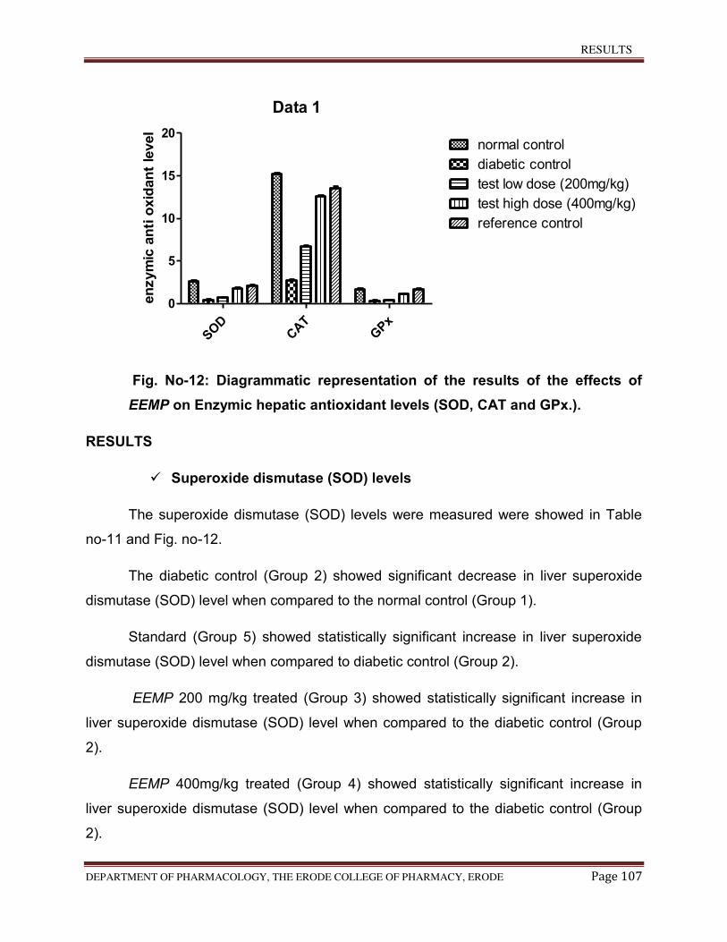
RESULTS
DEPARTMENT OF PHARMACOLOGY, THE ERODE COLLEGE OF PHARMACY, ERODE Page 107
Data 1
SOD
CAT
GPx
0
5
10
15
20normal control
diabetic control
test low dose (200mg/kg)
test high dose (400mg/kg)
reference control
en
zym
ic a
nti
oxid
an
t le
vel
Fig. No-12: Diagrammatic representation of the results of the effects of
EEMP on Enzymic hepatic antioxidant levels (SOD, CAT and GPx.).
RESULTS
Superoxide dismutase (SOD) levels
The superoxide dismutase (SOD) levels were measured were showed in Table
no-11 and Fig. no-12.
The diabetic control (Group 2) showed significant decrease in liver superoxide
dismutase (SOD) level when compared to the normal control (Group 1).
Standard (Group 5) showed statistically significant increase in liver superoxide
dismutase (SOD) level when compared to diabetic control (Group 2).
EEMP 200 mg/kg treated (Group 3) showed statistically significant increase in
liver superoxide dismutase (SOD) level when compared to the diabetic control (Group
2).
EEMP 400mg/kg treated (Group 4) showed statistically significant increase in
liver superoxide dismutase (SOD) level when compared to the diabetic control (Group
2).
RESULTS
DEPARTMENT OF PHARMACOLOGY, THE ERODE COLLEGE OF PHARMACY, ERODE Page 108
Catalase (CAT) level
The CAT levels were measured were showed in Table no-11 and Fig. no-12.
The diabetic control (Group 2) showed significant decrease in liver catalase
(CAT) level when compared to the normal control (Group 1).
Standard (Group 5) showed statistically significant increase in liver catalase
(CAT) level when compared to diabetic control (Group 2).
EEMP 200 mg/kg treated (Group 3) showed statistically significant increase in
liver catalase (CAT) level when compared to the diabetic control (Group 2).
EEMP 400mg/kg treated (Group 4) showed statistically significant increase in
liver catalase (CAT) level when compared to the diabetic group.
Glutathione peroxidise (GPx) level
The glutathione peroxidase (GPx) levels were measured were showed in Table
no-11 and Fig. no-12.
The diabetic control (Group 2) showed significant decrease in glutathione
peroxidase (GPx) level when compared to the normal control (Group 1).
Standard (Group 5) showed statistically significant increase in glutathione
peroxidase (GPx) level when compared to diabetic control (Group 2).
EEMP 200 mg/kg treated (Group 3) showed statistically significant increase in
glutathione peroxidise (GPx) level when compared to the diabetic control (Group 2).
EEMP 400mg/kg treated (Group 4) showed statistically significant increase in
glutathione peroxidise (GPx) level when compared to the diabetic group.
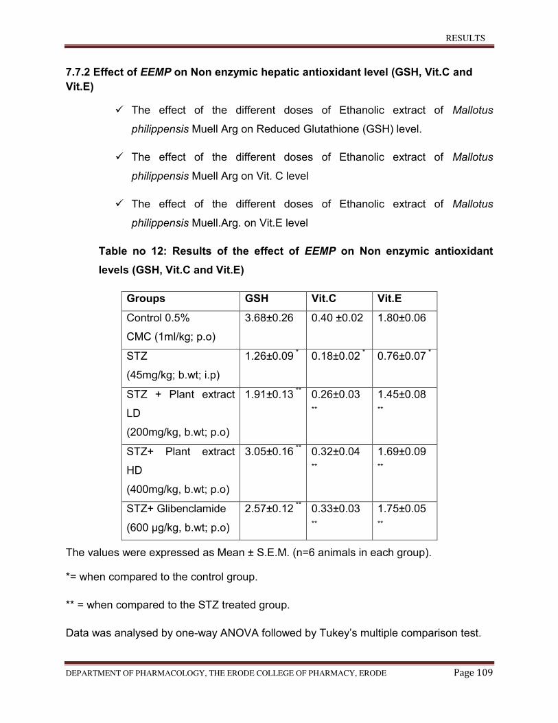
RESULTS
DEPARTMENT OF PHARMACOLOGY, THE ERODE COLLEGE OF PHARMACY, ERODE Page 109
7.7.2 Effect of EEMP on Non enzymic hepatic antioxidant level (GSH, Vit.C and
Vit.E)
The effect of the different doses of Ethanolic extract of Mallotus
philippensis Muell Arg on Reduced Glutathione (GSH) level.
The effect of the different doses of Ethanolic extract of Mallotus
philippensis Muell Arg on Vit. C level
The effect of the different doses of Ethanolic extract of Mallotus
philippensis Muell.Arg. on Vit.E level
Table no 12: Results of the effect of EEMP on Non enzymic antioxidant
levels (GSH, Vit.C and Vit.E)
Groups GSH Vit.C Vit.E
Control 0.5%
CMC (1ml/kg; p.o)
3.68±0.26 0.40 ±0.02 1.80±0.06
STZ
(45mg/kg; b.wt; i.p)
1.26±0.09 * 0.18±0.02 * 0.76±0.07 *
STZ + Plant extract
LD
(200mg/kg, b.wt; p.o)
1.91±0.13 ** 0.26±0.03
**
1.45±0.08
**
STZ+ Plant extract
HD
(400mg/kg, b.wt; p.o)
3.05±0.16 ** 0.32±0.04
**
1.69±0.09
**
STZ+ Glibenclamide
(600 µg/kg, b.wt; p.o)
2.57±0.12 ** 0.33±0.03
**
1.75±0.05
**
The values were expressed as Mean ± S.E.M. (n=6 animals in each group).
*= when compared to the control group.
** = when compared to the STZ treated group.
Data was analysed by one-way ANOVA followed by Tukey’s multiple comparison test.
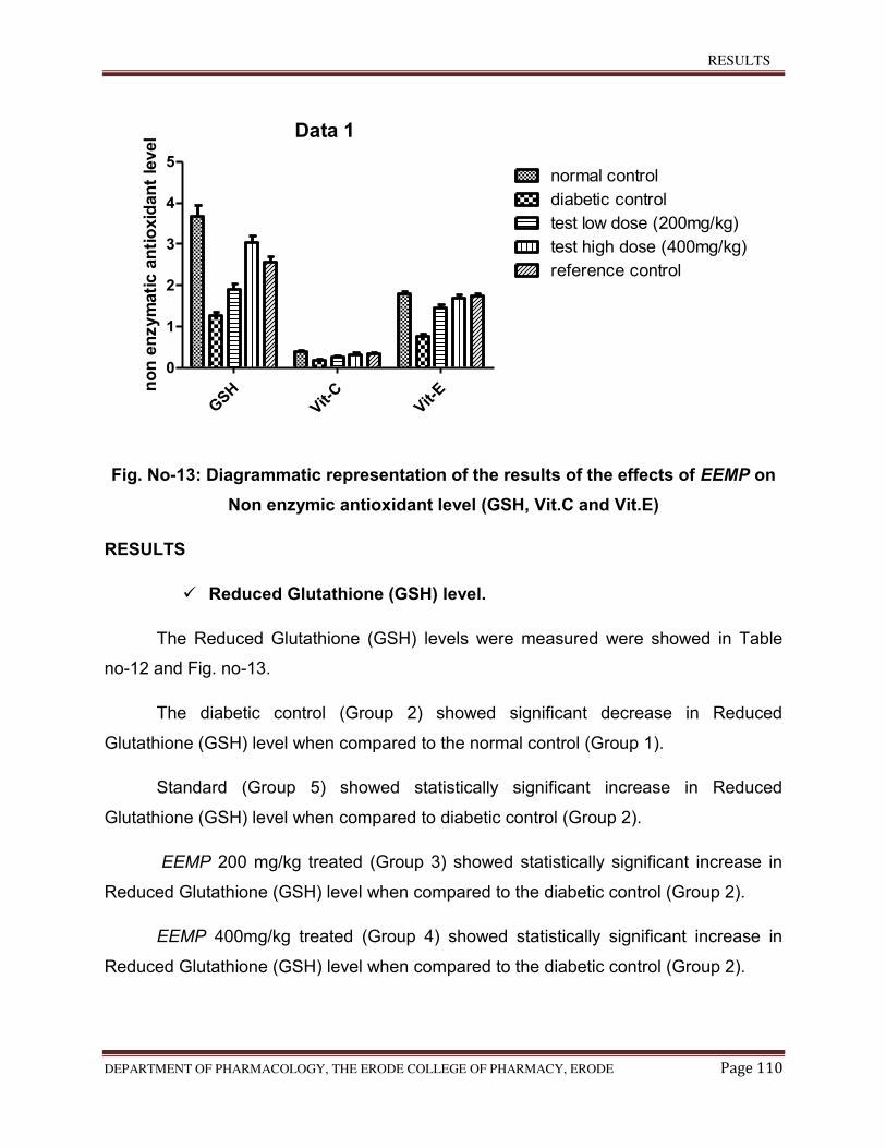
RESULTS
DEPARTMENT OF PHARMACOLOGY, THE ERODE COLLEGE OF PHARMACY, ERODE Page 110
Data 1
GSH
Vit-
CVit-
E
0
1
2
3
4
5normal control
diabetic control
test low dose (200mg/kg)
test high dose (400mg/kg)
reference control
no
n e
nzym
ati
c a
nti
oxid
an
t le
vel
Fig. No-13: Diagrammatic representation of the results of the effects of EEMP on
Non enzymic antioxidant level (GSH, Vit.C and Vit.E)
RESULTS
Reduced Glutathione (GSH) level.
The Reduced Glutathione (GSH) levels were measured were showed in Table
no-12 and Fig. no-13.
The diabetic control (Group 2) showed significant decrease in Reduced
Glutathione (GSH) level when compared to the normal control (Group 1).
Standard (Group 5) showed statistically significant increase in Reduced
Glutathione (GSH) level when compared to diabetic control (Group 2).
EEMP 200 mg/kg treated (Group 3) showed statistically significant increase in
Reduced Glutathione (GSH) level when compared to the diabetic control (Group 2).
EEMP 400mg/kg treated (Group 4) showed statistically significant increase in
Reduced Glutathione (GSH) level when compared to the diabetic control (Group 2).
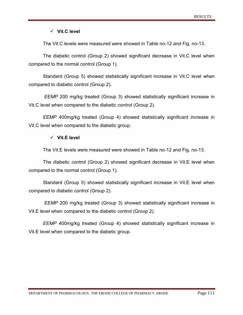
RESULTS
DEPARTMENT OF PHARMACOLOGY, THE ERODE COLLEGE OF PHARMACY, ERODE Page 111
Vit.C level
The Vit.C levels were measured were showed in Table no-12 and Fig. no-13.
The diabetic control (Group 2) showed significant decrease in Vit.C level when
compared to the normal control (Group 1).
Standard (Group 5) showed statistically significant increase in Vit.C level when
compared to diabetic control (Group 2).
EEMP 200 mg/kg treated (Group 3) showed statistically significant increase in
Vit.C level when compared to the diabetic control (Group 2).
EEMP 400mg/kg treated (Group 4) showed statistically significant increase in
Vit.C level when compared to the diabetic group.
Vit.E level
The Vit.E levels were measured were showed in Table no-12 and Fig. no-13.
The diabetic control (Group 2) showed significant decrease in Vit.E level when
compared to the normal control (Group 1).
Standard (Group 5) showed statistically significant increase in Vit.E level when
compared to diabetic control (Group 2).
EEMP 200 mg/kg treated (Group 3) showed statistically significant increase in
Vit.E level when compared to the diabetic control (Group 2).
EEMP 400mg/kg treated (Group 4) showed statistically significant increase in
Vit.E level when compared to the diabetic group.
RESULTS
DEPARTMENT OF PHARMACOLOGY, THE ERODE COLLEGE OF PHARMACY, ERODE Page 112
7.8 Effect of EEMP on plasma Insulin level
The effect of the different doses of Ethanolic extract of Mallotus Philippensis
Muell Arg on in plasma insulin level.
Table no 13: Results of the effect of EEMP on Plasma insulin level
Treatment and Dose Insulin levels
(μIU/ml)
Control 0.5%
CMC (1ml/kg; p.o)
7.74 ± 0.41
STZ
(60mg/kg; b.wt; i.p)
3.50 ± 0.73*
STZ + Plant extract LD
(200mg/kg, b.wt; p.o)
5.01± 0.36**
STZ+ Plant extract HD
(400mg/kg, b.wt; p.o)
6.75±0.98**
STZ+ Glibinclamide
(600 µg/kg, b.wt; p.o)
7.58 ± 0.72**
The values were expressed as Mean ± S.E.M. (n=6 animals in each group).
*= when compared to the control group.
** = when compared to the STZ treated group.
Data was analysed by one-way ANOVA followed by Tukey’s multiple comparison test.
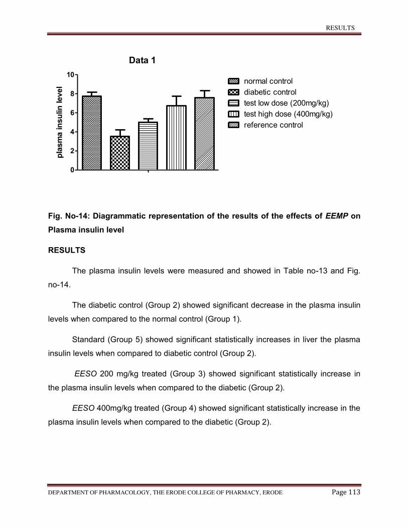
RESULTS
DEPARTMENT OF PHARMACOLOGY, THE ERODE COLLEGE OF PHARMACY, ERODE Page 113
Data 1
0
2
4
6
8
10normal control
diabetic control
test low dose (200mg/kg)
test high dose (400mg/kg)
reference control
pla
sm
a in
su
lin
level
Fig. No-14: Diagrammatic representation of the results of the effects of EEMP on
Plasma insulin level
RESULTS
The plasma insulin levels were measured and showed in Table no-13 and Fig.
no-14.
The diabetic control (Group 2) showed significant decrease in the plasma insulin
levels when compared to the normal control (Group 1).
Standard (Group 5) showed significant statistically increases in liver the plasma
insulin levels when compared to diabetic control (Group 2).
EESO 200 mg/kg treated (Group 3) showed significant statistically increase in
the plasma insulin levels when compared to the diabetic (Group 2).
EESO 400mg/kg treated (Group 4) showed significant statistically increase in the
plasma insulin levels when compared to the diabetic (Group 2).
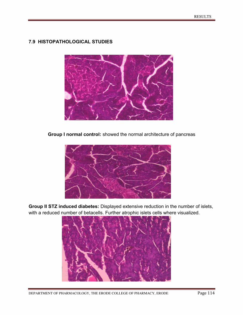
RESULTS
DEPARTMENT OF PHARMACOLOGY, THE ERODE COLLEGE OF PHARMACY, ERODE Page 114
7.9 HISTOPATHOLOGICAL STUDIES
Group I normal control: showed the normal architecture of pancreas
Group II STZ induced diabetes: Displayed extensive reduction in the number of islets,
with a reduced number of betacells. Further atrophic islets cells where visualized.
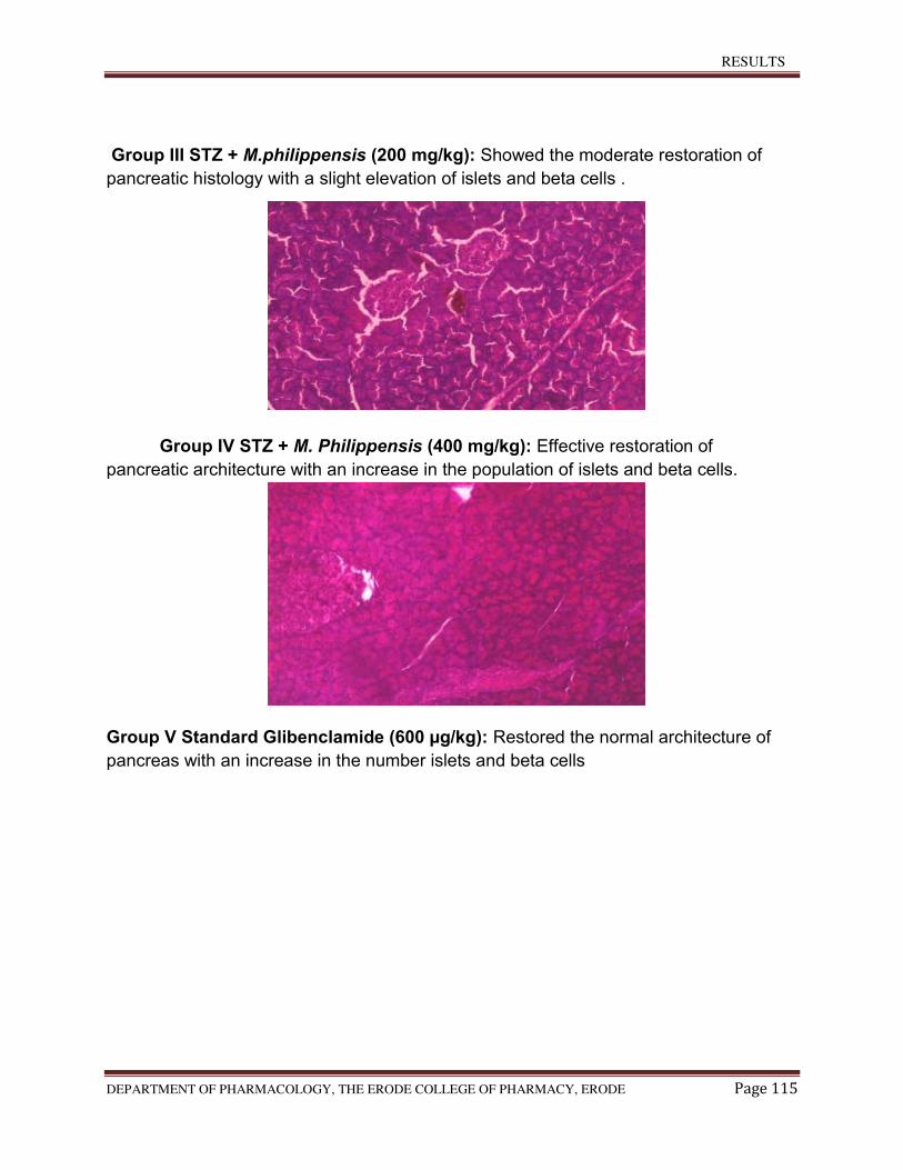
RESULTS
DEPARTMENT OF PHARMACOLOGY, THE ERODE COLLEGE OF PHARMACY, ERODE Page 115
Group III STZ + M.philippensis (200 mg/kg): Showed the moderate restoration of
pancreatic histology with a slight elevation of islets and beta cells .
Group IV STZ + M. Philippensis (400 mg/kg): Effective restoration of
pancreatic architecture with an increase in the population of islets and beta cells.
Group V Standard Glibenclamide (600 µg/kg): Restored the normal architecture of
pancreas with an increase in the number islets and beta cells
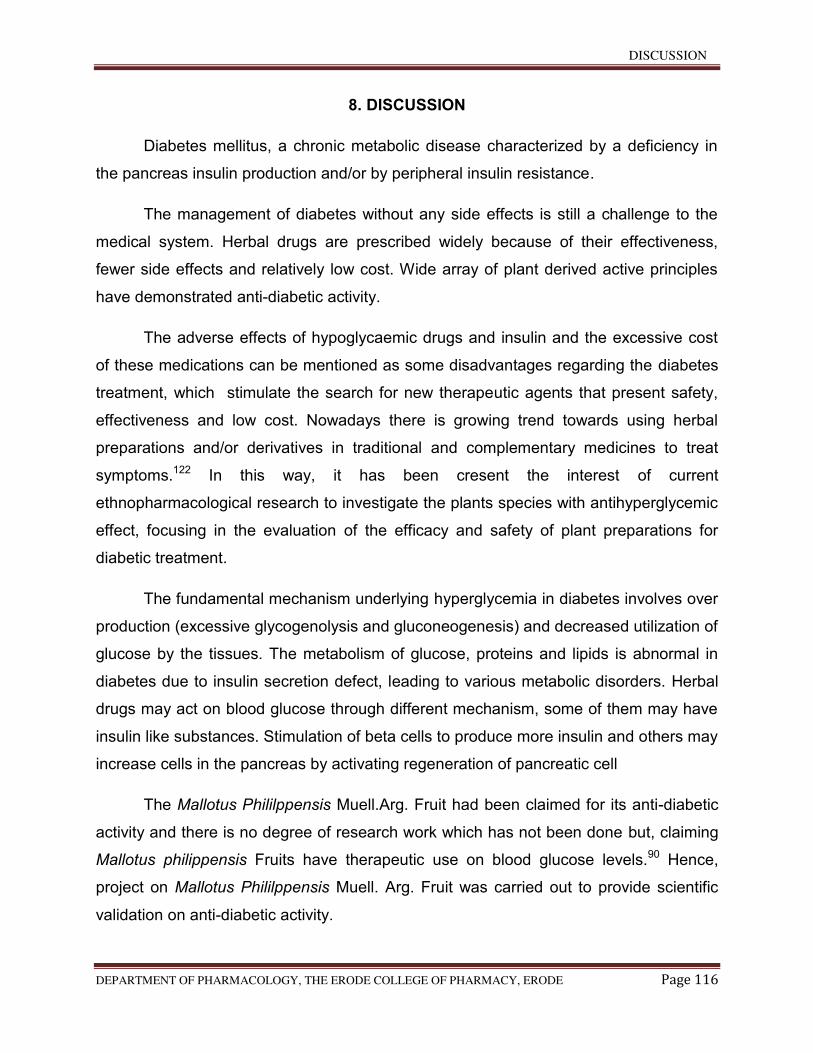
DISCUSSION
DEPARTMENT OF PHARMACOLOGY, THE ERODE COLLEGE OF PHARMACY, ERODE Page 116
8. DISCUSSION
Diabetes mellitus, a chronic metabolic disease characterized by a deficiency in
the pancreas insulin production and/or by peripheral insulin resistance.
The management of diabetes without any side effects is still a challenge to the
medical system. Herbal drugs are prescribed widely because of their effectiveness,
fewer side effects and relatively low cost. Wide array of plant derived active principles
have demonstrated anti-diabetic activity.
The adverse effects of hypoglycaemic drugs and insulin and the excessive cost
of these medications can be mentioned as some disadvantages regarding the diabetes
treatment, which stimulate the search for new therapeutic agents that present safety,
effectiveness and low cost. Nowadays there is growing trend towards using herbal
preparations and/or derivatives in traditional and complementary medicines to treat
symptoms.122 In this way, it has been cresent the interest of current
ethnopharmacological research to investigate the plants species with antihyperglycemic
effect, focusing in the evaluation of the efficacy and safety of plant preparations for
diabetic treatment.
The fundamental mechanism underlying hyperglycemia in diabetes involves over
production (excessive glycogenolysis and gluconeogenesis) and decreased utilization of
glucose by the tissues. The metabolism of glucose, proteins and lipids is abnormal in
diabetes due to insulin secretion defect, leading to various metabolic disorders. Herbal
drugs may act on blood glucose through different mechanism, some of them may have
insulin like substances. Stimulation of beta cells to produce more insulin and others may
increase cells in the pancreas by activating regeneration of pancreatic cell
The Mallotus Phililppensis Muell.Arg. Fruit had been claimed for its anti-diabetic
activity and there is no degree of research work which has not been done but, claiming
Mallotus philippensis Fruits have therapeutic use on blood glucose levels.90 Hence,
project on Mallotus Phililppensis Muell. Arg. Fruit was carried out to provide scientific
validation on anti-diabetic activity.
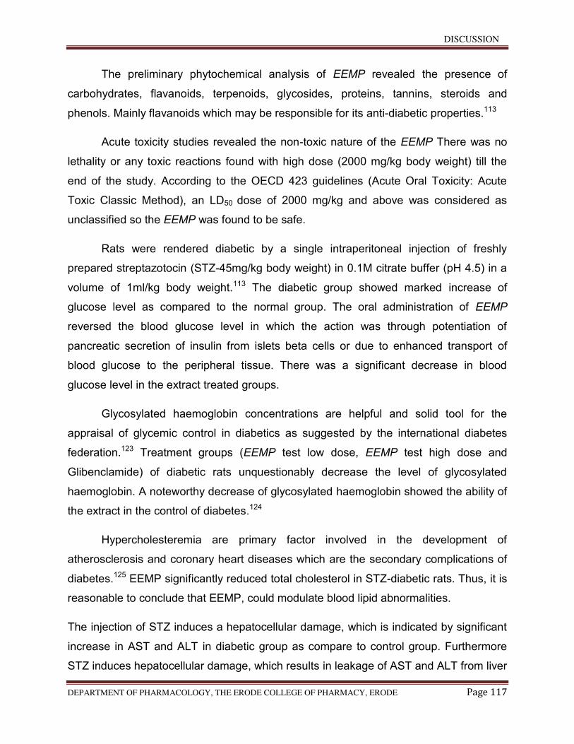
DISCUSSION
DEPARTMENT OF PHARMACOLOGY, THE ERODE COLLEGE OF PHARMACY, ERODE Page 117
The preliminary phytochemical analysis of EEMP revealed the presence of
carbohydrates, flavanoids, terpenoids, glycosides, proteins, tannins, steroids and
phenols. Mainly flavanoids which may be responsible for its anti-diabetic properties.113
Acute toxicity studies revealed the non-toxic nature of the EEMP There was no
lethality or any toxic reactions found with high dose (2000 mg/kg body weight) till the
end of the study. According to the OECD 423 guidelines (Acute Oral Toxicity: Acute
Toxic Classic Method), an LD50 dose of 2000 mg/kg and above was considered as
unclassified so the EEMP was found to be safe.
Rats were rendered diabetic by a single intraperitoneal injection of freshly
prepared streptazotocin (STZ-45mg/kg body weight) in 0.1M citrate buffer (pH 4.5) in a
volume of 1ml/kg body weight.113 The diabetic group showed marked increase of
glucose level as compared to the normal group. The oral administration of EEMP
reversed the blood glucose level in which the action was through potentiation of
pancreatic secretion of insulin from islets beta cells or due to enhanced transport of
blood glucose to the peripheral tissue. There was a significant decrease in blood
glucose level in the extract treated groups.
Glycosylated haemoglobin concentrations are helpful and solid tool for the
appraisal of glycemic control in diabetics as suggested by the international diabetes
federation.123 Treatment groups (EEMP test low dose, EEMP test high dose and
Glibenclamide) of diabetic rats unquestionably decrease the level of glycosylated
haemoglobin. A noteworthy decrease of glycosylated haemoglobin showed the ability of
the extract in the control of diabetes.124
Hypercholesteremia are primary factor involved in the development of
atherosclerosis and coronary heart diseases which are the secondary complications of
diabetes.125 EEMP significantly reduced total cholesterol in STZ-diabetic rats. Thus, it is
reasonable to conclude that EEMP, could modulate blood lipid abnormalities.
The injection of STZ induces a hepatocellular damage, which is indicated by significant
increase in AST and ALT in diabetic group as compare to control group. Furthermore
STZ induces hepatocellular damage, which results in leakage of AST and ALT from liver
DISCUSSION
DEPARTMENT OF PHARMACOLOGY, THE ERODE COLLEGE OF PHARMACY, ERODE Page 118
systole to the blood stream and/or may change the permeability of liver cell
membrane.126 in the present study, EEMP significantly decreased AST and ALT
enzyme activities in diabetic rats. The improvements in the levels of the enzyme are a
consequence of an improvement in the carbohydrate, fat and protein metabolism. The
restoration of AST and ALT after treatment also indicates a revival of insulin
secretion.127The results from present study indicates that EEMP may reduce the level of
serum cholesterol, SGOT and SGPT. It confirms that functions are on the protection of
vital tissues Pancreas, thereby reducing the causation of diabetis in experimental
animals.
Lipid peroxidation eventually leads to extensive membrane damage and
disfunction.128 Decreased lipid peroxidation and improved antioxidant status may be one
of the mechanism by which drug treatment could contribute to the prevention of diabetic
complications.129 In our study, EEMP significantly attenuated the increased lipid
peroxidation which could be due to the antioxidant effect of flavanoids, detected in the
preliminary phytochemical screening of the extract.
Oxidative stress is a condition of reduction in antioxidative enzymes like SOD,
CAT, GPx.130
Superoxide dismutase (SOD), is an important defence enzyme which catalyses the
dismutation of superoxide radicals, which scavenges the superoxide ions by catalyzing
its dismutation.
Catalase (CAT), a heme enzyme which removes hydrogen peroxide.131 The decreased
activities of CAT and SOD thereby result in the increased production of hydrogen
peroxide and oxygen by auto oxidation of glucose and non-enzymatic glycation,132
which is known to occur during diabetis.
Glutathione peroxidise (GPx) is an antioxidant enzyme, catalyses the scavenging and
inactivation of hydrogen and lipid peroxidise.133
The results showed that hepatic activity of catalase, superoxide dismutase and
glutathione peroxidase decreased significantly in STZ induced diabetic group (Group II).

DISCUSSION
DEPARTMENT OF PHARMACOLOGY, THE ERODE COLLEGE OF PHARMACY, ERODE Page 119
The normal control group maintained optimal value for activity of antioxidants. EEMP
treatment in diabetic rats significantly increased the antioxidant enzyme activities and
reversed them to their normal values. The same phenomenon was seen in the results of
glibenclamide treated groups.
An array of non-enzymatic antioxidant like GSH, Vitamin C and Vitamin E are
involved in scavenging free radicals in vivo.
Reduced glutathione (GSH), a tripeptide present in the all cells, is an important
antioxidant.134 It is essential to maintain structural and functional integrity of cells.
Hyperglycemia can increase oxidative stress and change the redox potential of
glutathione.135 Decreased levels of GSH in liver of diabetic rats may increase their
susceptibility to oxidative injury.
Vitamin C is an excellent hydrophilic, dietary antioxidant and it readily scavenges ROS
and peroxyl radicals.136 it also acts as co-antioxidant by generating Vitamin A, E and
GSH from radicals. A decrease in the level of Vitamin C was observed in liver of
diabetic rats. Such a fall in level of Vitamin C could be due to the increased utilization of
Vitamin C in the deactivation of increased level of ROS or due to decrease in GSH
level, since GSH is required in recycling of Vitamin C.137Another possibility is that
hyperglycemia inhibits ascorbic acid and its cellular transport.
Vitamin E is an antioxidant, a substance that helps prevent damage to the body’s
cells.138 Streptozotocin induced diabetic rats were found to have decreased GSH,
Vitamin C and Vitamin E levels in liver as compared to control rats. Treatment with
EEMP and the standard drug, glibenclamide produced significant increase in the levels
of these non-enzymatic antioxidants.
The serum insulin level decreased in diabetic rats, whereas EEMP extract,
brought about a marked increase in serum insulin in streptozotocin-induced diabetic
rats. This increase may be a consequence of the stimulation of insulin synthesis and
secretion.139
The histopathological investigation along with the biochemical evaluation
suggests the possibility of the islets regeneration and recovery of normal carbohydrate
metabolism in treated group EEMP. The regenerative effect of the pancreatic cells by
DISCUSSION
DEPARTMENT OF PHARMACOLOGY, THE ERODE COLLEGE OF PHARMACY, ERODE Page 120
Mallotus philippensis via exocrine cells of pancreas may enlighten the positive effects of
these agents on the production of insulin. Reports on histopathological analysis of
pancreas of the Mallotus philippensis alone treated rats showed results that were very
similar as that of the control group.
Based on the above results, it was concluded that Mallotus Phililppensis
Muell.Arg. exerted statistically significant anti-diabetic activity against STZ induced
diabetic rats.
SUMMARY AND CONCLUSION
DEPARTMENT OF PHARMACOLOGY, THE ERODE COLLEGE OF PHARMACY, ERODE Page 121
9. SUMMARY AND CONCLUSION
The present study was undertaken to scientifically elucidate the anti-diabetic
activity of alcoholic fruit extract of Mallotus Phililppensis Muell.Arg.
The phytochemical investigation revealed the presence of carbohydrates,
steroids, phenols, tannins, flavanoids, glycosides, and terpenes in the EEMP.
The STZ 45 mg/kg effectively induced diabetes, which was similar to diabetes in
human. Therefore it is an effective and an ideal model for diabetes research.
The Blood glucose level and Glycosylated haemoglobin level significantly
decreased with treatment of EEMP proves it having anti-diabetic activity.
Attenuating the increased serum parameters like total cholesterol, AST and ALT
with treatment of EEMP showed its anti-diabetic activity.
Reduced level of lipid peroxidation in EEMP treated rats, showed EEMP has
protective effect in oxidative stress induced diabetes.
The enzymic antioxidant parameters such as SOD, CAT and GPx and
Non-enzymic antioxidant parameters such as GSH, Vitamin C and Vitamin E are
increased significantly with treatment of EEMP, which proved as it having antioxidant
activity.
The plasma insulin level significantly increased with treatment of EEMP, showed
the EEMP in managing hyperglycemia and diabetic complications.
Mallotus Phililppensis Muell.Arg.fruit extract also have favourable effects to
inhibit the histopathological change of the pancreas in STZ induced diabetes.
In summary, alcoholic fruit extract of Mallotus Phililppensis Muell.Arg showed
statistically significant anti-diabetic activity.
In conclusion, the alcoholic fruit extract of Mallotus Phililppensis Muell.Arg.
showed and offered a promising therapeutic value in prevention of diabetes. These
effects was mainly attributed to its antioxidant properties by significant quenching
SUMMARY AND CONCLUSION
DEPARTMENT OF PHARMACOLOGY, THE ERODE COLLEGE OF PHARMACY, ERODE Page 122
impact of the extract on lipid peroxidation along with enhancement of enzymatic and
non-enzymatic antioxidant defense systems in liver and pancreatic tissue.
Further studies will be needed in future to determine the exact phytoconstituents
in the extract, which having anti-diabetic activity.
FUTURE PROSPECTIVES
DEPARTMENT OF PHARMACOLOGY, THE ERODE COLLEGE OF PHARMACY, ERODE Page 123
10. FUTURE PROSPECTIVES
Further study is required.,
1. To isolate and separate the active phytochemicals present in the ethanolic extract of
the Fruit of Mallotus Phililppensis Muell Arg.
2. Formulation of the isolated lead molecule can be designed.
3. Clinical trial of the formulated molecule in healthy human volunteers or diseased
persons.
4. The formulated lead molecule can be subjected to the clinical trials, patented and
marketed for the treatment of diabetes.
REFERENCE
DEPARTMENT OF PHARMACOLOGY, THE ERODE COLLEGE OF PHARMACY, ERODE Page 124
11. REFERENCE
1. "About diabetes". World Health Organization. Retrieved 4 April 2014.
2. "Diabetes Fact sheet N°312". WHO. October 2013. Archived from the original
on 26 Aug 2013. Retrieved 25 March 2014.
3. Kitabchi, AE; Umpierrez, GE; Miles, JM; Fisher, JN (Jul 2009).
"Hyperglycemic crises in adult patients with diabetes.". Diabetes Care. 32 (7):
1335–43. doi:10.2337/dc09-9032. PMC 2699725. PMID 19564476.
4. Shoback, edited by David G. Gardner, Dolores (2011). "Chapter 17".
Greenspan's basic & clinical endocrinology (9th ed.). New York: McGraw-Hill
Medical. ISBN 0-07-162243-8.
5. Maggard MA, Yermilov I, Li Z, et al; Pregnancy and fertility following bariatric
surgery: a systematic review. JAMA. 2008 Nov 19;300(19):2286-96
6. RSSDI textbook of diabetes mellitus. (Rev. 2nd ed.). New Delhi: Jaypee
Brothers Medical Publishers. 2012. p. 235. ISBN 9789350254899.
7. "The top 10 causes of death Fact sheet N°310". World Health Organization.
Oct 2013.
8. Rippe, edited by Richard S. Irwin, James M. (2010). Manual of intensive care
medicine (5th ed.). Philadelphia: Wolters Kluwer Health/Lippincott Williams &
Wilkins. p. 549. ISBN 9780781799928.
9. Picot, J; Jones, J; Colquitt, JL; Gospodarevskaya, E; Loveman, E; Baxter, L;
Clegg, AJ (September 2009). "The clinical effectiveness and cost-
effectiveness of bariatric (weight loss) surgery for obesity: a systematic review
and economic evaluation". Health Technology Assessment (Winchester,
England). 13 (41): 1–190, 215–357, iii–iv. doi:10.3310/hta13410.
PMID 19726018.
10. Williams textbook of endocrinology (12th ed.). Philadelphia:
Elsevier/Saunders. pp. 1371–1435. ISBN 978-1-4377-0324-5.
11. Abha Saxena and Naval Kishore Vikram. “Role of Selected Indian Plants in
Management of Type 2 Diabetes: A Review”. The journal of alternative and
Complementary medicine. 2004, 10 (2): 369-378.
12. Pranav K, Prabhakar and Mukesh Doble. “A target based therapeutic
Approach towards Diabetes Mellitus using Medicinal Plants”. Current
Diabetes reviews. 2008, 4:291-308.
REFERENCE
DEPARTMENT OF PHARMACOLOGY, THE ERODE COLLEGE OF PHARMACY, ERODE Page 125
13. KA wadkar, CS Magdum,SS Patil,NS Naikwade, anti-diabetic potential and
Indian medicinal plants, J herbal med toxicol, volume 2, 2008.
14. Gerard j. Tortora, bryan H. Derrickson. 2009. Principles of Anatomy and
physiology”. Wiley (Asia) private limited. 12 (2); 942-944.
15. H.P. Rang, M.M Dale, J.M. Ritter, R. J. Flower. 2007. Rang and Dale’s
Pharmacology”. Churchill Living stone Elsevier. (6):307-409.
16. Raju. S, Hemamalini “In vivo animal model for screening of Anti-diabetic
activity” Asian Journal of pharmaceutical and Clinical Research Vol 5, Suppl
4, 2012 ISSN- 0974-2441.
17. Review ArticleMani Rupeshkumar,Kunchu Kavitha,et al, Role of herbal plants
in the diabetes mellitus therapy: an overreview. East Point College of
Pharmacy, Bidarahalli, Bangalore-49, Karnataka, India, 2Department of
Pharmaceutical Technology, Jadavpur University, Kolkata-32, West Bengal,
India.
18. Khan, Ali Nawaz. "Chronic Pancreatitis Imaging". Medscape. Retrieved 5
January 2014.
19. "Cancer of the Pancreas". NHS. Retrieved 5 November 2014.
20. Drake, Richard L.; Vogl, Wayne; Tibbitts, Adam W.M. Mitchell; illustrations by
Richard; Richardson, Paul (2005). Gray's anatomy for students. Philadelphia:
Elsevier/Churchill Livingstone. pp. 288–290, 297, 303. ISBN 978-0-8089-
2306-0.
21. Young, Barbara, ed. (2006). Wheater's functional histology : a text and colour
atlas (5th ed.). Churchill Livingstone/Elsevier. pp. 299–301. ISBN 978-0-443-
06850-8.
22. Hall, John (2011). Guyton and Hall textbook of medical physiology (12th ed.).
Philadelphia, Pa.: Saunders/Elsevier. p. 781. ISBN 978-1-4160-4574-8.
23. World health Organisation Laboratory Diagnosis and Mdonitoring of Diabetes
mellitus 2002.
24. Wright JR, Yang H, Hyrtsenko O, Xu BY, Yu W, Pohajdak B (2014). "A review
of piscine islet xenotransplantation using wild-type tilapia donors and the
production of transgenic tilapia expressing a "humanized" tilapia insulin".
Xenotransplantation. 21 (6): 485–95. doi:10.1111/xen.12115. PMC 4283710
. PMID 25040337.
REFERENCE
DEPARTMENT OF PHARMACOLOGY, THE ERODE COLLEGE OF PHARMACY, ERODE Page 126
25. Stryer, Lubert (1995). Biochemistry. (Fourth ed.). New York: W.H. Freeman
and Company. pp. 773–774. ISBN 0 7167 2009 4.
26. Sonksen P, Sonksen J (July 2000). "Insulin: understanding its action in health
and disease". British Journal of Anaesthesia. 85 (1): 69–79.
doi:10.1093/bja/85.1.69. PMID 10927996.
27. Sonksen P, Sonksen J (July 2000). "Insulin: understanding its action in health
and disease". British Journal of Anaesthesia. 85 (1): 69–79.
doi:10.1093/bja/85.1.69. PMID 10927996.
28. American Society of Health-System Pharmacists (2009-02-01). "Insulin
Injection". PubMed Health. National Center for Biotechnology Information,
U.S. National Library of Medicine. Retrieved 2012-10-12.
29. “Definition, Diagnosis and Classification of Diabetes mellitus and its
complications’ World health organisation department of Noncommunicable
Disease Survelillance Geneva WHO/NCD/ncS/99.2.
30. Duncan C. Moore, Caius D Christoe, Andrew Baum. “Diabetes: a 21st Century
Epidemic” Morgan Stanley Research Europe Page no: 1-93.
31. Ian N Scobie. 2007. “Atlas of Diabetes mellitus”. Informa healthcare, United
Kingdom. (3): 1-121.
32. www,ncbi.nlm.nih.gov/pubmed/23683103.
33. American journal of diabetics insulin and protein metabolism
34. Lipid metabolism. Role of insulin in lipid metabolism.Otto-Buczkowska
EJarosz-Chobot P.
35. Disorder of pancreas Wikipedia and encyclopedia
36. Goodman &Gilman’s 2008. “Manual of Pharmacology and Therapeutics”
McGraw Hill 1035-1050.
37. Sigurd lenzen. “Alloxan and Streptozotocin diabetes”. 120-138.
38. Szkudelski T. The mechanism of alloxan and sterptozotocin action in beta
cells of the pancreas. Physiol Res 2001; 50:536-46.
39. Ganda OP, Rossi AA, Like AA: Studies on streptozotocin diabetes, 1976 25:
595-603.
40. Ziegler M, Ziegler B, Hehmke B: Severe hyperglycaemia caused
autoimmunization to beta cells in rats. Diabetologia, 1984 27: 163-165.
41. Oussama MN Khatib. “Guidelines for the prevention, management and care of
diabetes mellitus”. World Health Organisation, 2006. 10-43.
REFERENCE
DEPARTMENT OF PHARMACOLOGY, THE ERODE COLLEGE OF PHARMACY, ERODE Page 127
42. Schnedl WJ, Ferber S, Johnson JH, Newgard CB: STZ transport and
cytotoxicity. Specific enhancement in GLUT2- expressing cells. Diabetes,
1994, 43: 1326-1333.
43. Wang Z, Gleichmann H: GLUT2 in pancreatic islets: crucial target molecule in
diabetes induced with multiple low doses of streptozotocin in mice. Diabetes,
1998 47: 50-56,
44. Delaney CA, Dunger A, Di Matteo M, Cunningham JM, Green MH, Green IC:
Comparison of inhibition of glucose-stimulated insulin secretion in rat islets of
Langerhans by streptozotocin and methyl and ethyl nitrosoureas and
methanesulphonates. Lack of correlation with nitric oxide-releasing or O 6 -
alkylating ability. Biochem Pharmaco, 1995l 50: 2015-2020.
45. Ji Zhanga, Yulong Huanga, Tiande Houa, Yunpu Wangb. “hyperglycemic
effect of Artemesia sephaerocephala Krasch seed polysaccharide in alloxan
– induced diabetic rats’. Swiss med Wkly.2006, 136; 529-532.
46. Suman Bala Sharma, Afreena Nasir, Krishna Madhava Prabhu, Potharagada
Surynaryana Murthy. “Antihyperglycemic effect of the fruit-pulp of Eugenia
jambolana in experimental diabetes mellitus”. Journal of Ethnopharmacology.
2006, 104: 367-373.
47. K. Ashok Kumar, M.Uma Maheswari,A.T. Sivashanmugam, V. Subhadradevi,
N.V. Prasant and T.K. Ravi. “Hypoglycemic effect of Ficus microcarpa leaves
on alloxan induced diabetic rats”. Journal of Biological sciences. 2007, 7(2).
48. Selvan VT, Mankandan l, Senthil Kumar GP, Suresh R, Kakoti BB, Gomathi
P, Kumar DA, Sha P, Gupta M, Mazumder UK. “ Anti-diabetic and antioxidant
effect of Artenema sesamoides in sterptozotocin induced diabetic rats”.
International Journal of applied research in natural products. 2008, 1(1): 25-
33.
49. M.A. Jayasri, S. Gunasekaran, A. Radha and T.L. Mathew. “Anti-diabetic
effect of Coptus pictus leaves in normal and sterptozotocin induced Diabetic
rats”. International Journal of Diabetes and Metabolism. 2008, 16: 117-122.
50. Vaiyapuri Sivajothi, alanka Dey, Balasundarm Jyakar and Balasubramanian
rajkapoor. “ Antihypreglycemic, antihyperlipidemic and antioxidant effect of
Phyllanthus rheediii on sterptozotocin induced diabetic rats”. Iranian journal of
pharmaceutical research. 2008,7(1):53-59.
REFERENCE
DEPARTMENT OF PHARMACOLOGY, THE ERODE COLLEGE OF PHARMACY, ERODE Page 128
51. U.Srinivasa, J. Venkateshwara rao, A.M. krupanidhi and K. Divakar.
“Antidaibetic activity of Justicia beddomei leaves in alloxan induced diabetic
rats”. Journal of research and education of Indian medicine. 2008:45-47
52. K. Rajagopal and K. Sasikala. “Anti-diabetic activity of hydroethanolic extracts
of Nymphaea stellata flowers in normal African Journal of Pharmacy and
Pharmacology 2008,2(8): 173-178.and alloxan induced diabetic rats”.
53. Rakesh Barik, Sanjay Jain, Deep Qwatra, Amit Joshi, Girraj Sharan Tripathi,
Ravi Goyal. “Anti-diabetic activity of aqueous root of Ichnocarpus frutescens
in sterptozotocin-nicotinamide induced type-II diabetes in rats”. Indian Journal
of Pharmacology. 2008, 40(1): 19-22.
54. A.N.Kalia, J.P.Yadav, Sushila Saini, A.S.Dangi. “Hypoglycaemic and
Hypolipidemic activity of ethanolic extract of Salvadora oleoides in normal
and alloxan induced diabetic rats”. Indain Journal of Pharmacology
2008,40(1): 23-27.
55. K. Illango, V.Chitra, P.Kanimozhi and G. Balaji. “Anti-diabetic antioxidant and
antibacterial activities of leaf extracts of Adathoda zeylanica. Medic
(Acanthaceae)”. Journal of Pharmaceutical Sciences and Research. 2009,
1(2): 57-63.
56. Yogesh Kumar, Bhupesh Chander Semwal, Jitendra Gupta, Sonia Singh,
Mahendra Giri. “Antihyperglycemic activity of roots of Berberis aristata D.C in
alloxan induced diabetic rats” International Journal of Green Pharmacy. 2009:
259-262.
57. Papiya Mitra Mazumder, Mamta Farswan, V. Parcha. “Effects of an isolated
active compound (cg-1) of Cassia glauca Linn. Leaf on blood glucose, lipid
profile, and atherogenic index in diabetic rats”. Indian journal of Pharmacology
2009, 41 (4): 182-186.
58. Pankaj Kishor Mishra, Grijesh Kumar Mall and Veeru Prakash. “Anti-diabetic
activity and hypolipidemic activity of Gymnema sylvestre in alloxan induced
diabetic rats”. Global Journal of Biotechnology & Biochemistry. 2009, 4(1): 37-
42.
59. Nagaraj Puranik, Kararashah Fakruddin Kammar, Sheela Devi. “Anti-diabetic
activity of Tinospora cordifolia (Willd.) in sterptozotocin diabetic rats; does it
act like sulfonylureas?”. Turk Journal of Medical Sciences. 2010, 40(2):265-
270.
REFERENCE
DEPARTMENT OF PHARMACOLOGY, THE ERODE COLLEGE OF PHARMACY, ERODE Page 129
60. Sravan Kumar Bussa, Jyothi Pinnapareddy. “Anti-diabetic activity of stem bark
of Neolamarckia cadamba in alloxan induced diabetic rats”. International
Journal of Pharmacy technology. 2010(2): 314-324.
61. Reema Dheer, Pradeep Bhatnagar. “A study of the Anti-diabetic activity of
Barleria prionitis Linn’. Indian Journal of Pharmacology. 2010, 42(2): 70-73.
62. Mekesh S Sikarwan, M B Patil. “Anti-diabetic activity of Pongamia pinnata leaf
extracts in alloxan induced diabetic rats”. International Journal of Ayurveda
Research 2010, 1(4):199-204.
63. V Sivakumar, M Jayanth, N Sowbala, G Rajalakshmi, U Kanagavalli. “Study of
antihyperglycemic effect of Cathranthus roseus in alloxan induced diabetic
rats”. International Journal of Pharmacy and Pharmaceutical Sciences. 2010,
2(4): 114-116.
64. Syed Mohammed Kazim, Mohammed Fazil, Syed Safiullah Ghori, Syeda
Sughra Mehjabeen, Shaik Rasheed Ahmed, Shaik Mehboob Ali, Mohammed
Ibrahim. “Anti-diabetic activity of Vinca roseus extract in alloxan induced
diabetic ats”. International Journal of Endocrionology 2010; 1-6.
65. Y. Tanko, M.A. Mabrouk, A.B. Adelaiye, M.Y. Fatihu and K.Y. Musa. “Anti-
diabetic and some haematological effects of ethylacetate and n-
butanolfractions of Indigofera pulchra extract on alloxan induced diabetic
Wistra rats”. Journal of Diabetes and Endocrinology, 2011,2(1):1-7.
66. T. Harikant,A. Zabeer, Pooja, K.C. Samanta. “Anti hyperglycemic activity of
petroleum ether extract ðyl acetate extract of Sarcococca saligna”.
International Journal of Current Pharmaceutical Research 2011, 3 (1): 26-29.
67. Rajeev Kumar Jha, Mangilal, Anil Bhandari, Rajesh Kumar Nema. “Anti-
diabetic activity of flower head petroleum ether extracts of Sphaeranthus
indicus Linn”. Asian Journal of Pharmaceutical and Clinical Research. 2011.
3(1): 16-19.
68. Ranjit Singh, P. H. Rajasree and C. Sankar, “Screening for Anti-diabetic
activity of the ethanolic extract of Barleria cristata seeds”. International
Journal of Pharmacy Life Sciences 3(10):Oct., 2012.
69. Subramaniam Ramachandran,Koikaramparambil Robert Naveen, Baskaran
Rajinikanth, Mohammad Akbar, Aiyalu Rajasekaran. “Anti-diabetic,
Antihyperlipidemic and in vivo Antioxidant potential of aqueous exyract of
REFERENCE
DEPARTMENT OF PHARMACOLOGY, THE ERODE COLLEGE OF PHARMACY, ERODE Page 130
Aniogeissus latifolia bark in type 2 diabetic rats”. Asian Pacific Journal of
Tropical Diseases 2012,S596-S602.
70. C.N. Ezekwesili, H.A. Ogbunugafor and J.O. Ezekwsili-Ofili. “Anti-diabetic
activity of aqueous extract of Vitex doniana leaves and chincina calisaya bark
in alloxan induced diabetic rats”. International Journal of Tropical Diseases
&Health, 2012 2(4):290-300.
71. Qamar Uddin Ahmed, Bashar B.S. Dogarai, Mohamed Zaffar Ali M.
Amiroydine, Muhammed Taher, Jalifah Latip, Abdulrashid Umar and Bala Y.
Muhammed. “Anti-diabetic activity of the leaves of tetracera indica merr.
(Dilleniaceae) in vivo and in vitro”. Journal of Medicinal Plants Research. 25
December, 2012. vol. 6(49), pp. 5912-5922.
72. Shetti, R. D. Sanakal and B. Kaliwal. “anti-diabetic effect of Ethanolic leaf
extract of Phyllanthus amarus in alloxan induced diabetic mice”. Asian journal
of plant Science and Research, 2012,2(1):11-15.
73. Olfat A. Khalil, Kholoud S. Ramadan, Enas N. Danial, Hanan S, Alnahdi and
NAjla O. Ayaz. “Anti-diabetic activity of Rosmarinus officinalis and its
relationship with antioxidant property”. African Journal of Pharmacy and
Pharmacology, 15 April 2012, vol. 6(14),pp. 1031-1036.
74. Parminder Nain, Vipin Saini, and Sunil Sharma. “Anti-diabetic activity of
Emblica officinalis seed extraction sterptozotocin induced type-2
diabetesmellitus in rat” International Journal of Natural Products Science,
2012; Spl Issue 1:135.
75. Rashmi Raghuwanshi and Bharati Jain. “Hypoglycaemic effect of Pueraria
tuberose tubers in healthy and alloxan diabetic rats “ Journal of Chemical,
Biological and Physical Sciences Nov.2011-Jan2012, vol.2.no.1, 270-272 E-
ISSN:2249-1929.
76. Moses Z Zaruwa, Jiredej Manosroi, Toshihiro Akihisa, Worapaka Manosroi,
Aranya Manosroi “Anti-diabetic acivity of Anogeissus acuminate a medicinal
plant selected from the Thai medicinal plant recepie database MANOSROI II”
Wudpecker Journal of Medicinal Plants, October 2012Vol.1(20, pp.O11-018.
77. Kattamnachi Gnanath. Kontham Ramakanth reddy, Gudur Pavan Kumar,
Bheemanapally Krishna, Karka Srinivasa Reddy, Avvuri Sanjeeva Kumarn
“Evaluation of Anti-diabetic activity in Corallocarpus epigaeus rhizomes”
International Current Pharamceutical Journal, February 2013, 2(3);53-56.
REFERENCE
DEPARTMENT OF PHARMACOLOGY, THE ERODE COLLEGE OF PHARMACY, ERODE Page 131
78. Fatemeh Fathiazad, Sanaz Hamedeyazdan, Mohamad Karim Khsropanah,
Arash Khaki “Hypoglycaemic activity of Fumaria parviflora LSam in
sterptozotocin induced diabetic rats,” Advanced Pharmaceutical Bulletin,
2013,3(1), 207-210.
79. Md. Ahsanul haque, Chowdhury Sayef Abdullah, Bilquis Romana, Md.
Bodrudoza rafiquel, Gazi Md. Zia-ul-Huda, Sarder Fahim Hossain, Bilkis
Begum “Evaluation of Anti diarrhoeal and Anti-diabetic activities of the stem,
barks and leaves of the plant Vernonia cinerea (Family: Asteraceae)” Journal
of Applied Pharmaceutical Science, October 2012 vol.3 (1),pp. 069-072.
80. Mitali Ghosal and Palash Mandal “Evaluation of Anti-diabetic activity of
Calamus erectus in sterptozotocin induced diabetic rats’ Asian Journal of
Plant Science and Research, 2013, 3 (1): 47-53.
81. R. Sundhararajan and V. Ravichandiran. ‘Antiinflammatory activity of
Spermacoe ocymoides Burm F”. Journal of Pharmacy Research 2012,5(1),
719-721.
82. C.K. Kokate. A.P.Purohit, S.B. Gokhale. 2008. “Pharmacology”. Nirali
Prakasan, Pune, India. A.1-1.6
83. Anonymous. WWW.OECD.com.
84. Mrs. P.H. Agarkar, Dr.J.S. Kulkarni, Dr. V.L. Maheshwari, Dr. R.A. Fursule.
2008. “Practical Biochemistry”. Nirali Prakashan, Pune, India.
85. Misra HP,Fridovich I,1972.The role of superoxide anion in the autooxidation of
epinephrine anion in the autooxidation of epinephrine and a simple assay of
superoxide dismutase.J Biol Chem 247,3170-3184.
86. Indian pharmacopoeia, Government of India, Ministry of Health & welfare,
controller of publications, New Delhi, 1996,Vol 2, A53-54.
87. Jayakumari S, Malarkodi Velraj, Vijayalakshmi A and Arthanarieswaran P.
Pharmacognostical studies on the leaves of Pisonia grandis R. Br. Research
Journal of Pharmaceutical, Biological and Chemical Sciences, 2(2),(2011).
88. Johnsen DA. Plant microtechnique; New Work, Mc Graw Hill co, 1940,PP-
523.
89. Mayank Gangwar, R. K. Goel, and Gopal Nath Mallotus philippinensis Muell.
Arg (Euphorbiaceae): Ethnopharmacology and Phytochemistry Review.
90. Indian medicinal plants an illustrated dictionary page no:76
91. Cuong NX et al. A new lignan dimer from Mallotusphilippensis.pubmed ncbi.
REFERENCE
DEPARTMENT OF PHARMACOLOGY, THE ERODE COLLEGE OF PHARMACY, ERODE Page 132
92. Chan TK et al. Anti-allergic actions of rottlerin from Mallotus philippinensis in
experimental mast cell-mediated anaphylactic models. pubmed ncbi.
93. Hong Q et al. Anti-tuberculosis compounds from Mallotus philippinensis.
pubmed ncbi.
94. Kulkarni RR et al. Antifungal dimericchalcone derivative kamalachalcone E
from Mallotus philippinensis. pubmed ncbi.
95. Khan H et al. Antioxidant and Antiplasmodial Activities of Bergenin and 11-O-
Galloylbergenin Isolated from Mallotusphilippensis. pubmed ncbi.
96. Gangwar M et al. Antioxidant capacity and radical scavenging effect of
polyphenol rich Mallotus philippenensis fruit extract on human erythrocytes.
pubmed ncbi.
97. Khan M et al. Hexane soluble extract of Mallotusphilippensis (Lam.) Muell.
Arg. root possesses anti-leukaemic activity. pubmed ncbi.
98. Gangwar M et al. in-vitro scolicidal activity of Mallotus philippinensis (Lam.)
Muell Arg. fruit glandular hair extract against hydatid cyst
Echinococcusgranulosus. pubmed ncbi.
99. Goel RK et al. Mallotus philippinensisMuell. Arg (Euphorbiaceae):
ethnopharmacology and phytochemistry review. pubmed ncbi.
100. Furumoto T et al. Mallotus philippinensis bark extracts promote
preferential migration of mesenchymal stem cells and improve wound healing
in mice. pubmed ncbi.
101. Gautam MK et al. Mallotus philippinensisMuell. Arg fruit glandular hairs
extract promotes wound healing on different wound model in rats. pubmed
ncbi.
102. Oyedemi BO et al. Novel R-plasmid conjugal transfer inhibitory and
antibacterial activities of phenolic compounds from Mallotus
philippensis (Lam.) Mull. Arg. pubmed ncbi.
103. Chhiber N et al. Rottlerin, a polyphenolic compound from the fruits
of Mallotus phillipensis (Lam.) Müll.Arg. pubmed ncbi.
104. Marble A (1971) ‘’Glibenclamide, a new sulfonylureas whither oral
hypoglycemic agents, drugs 1 (2) 109-25.
105. A b Serrano-martin x, prayins G, Mendoza Leona (December 2006)
REFERENCE
DEPARTMENT OF PHARMACOLOGY, THE ERODE COLLEGE OF PHARMACY, ERODE Page 133
106. Riddle mc (February 2003) ‘’Editorial sulfonylureas differ in effects on
ischemic pres conditioning- is it time to retire glyburde J cin endocrine, metab
88 (2): 528-30
107. Okhawa H,Ohigni N,Yagi K,1979.Assay of lipid peroxides in animal
tissues by thiobarbituric acid reaction.Anal Biochem95,351-359
108. Reitman, S., Frankel, S.A., 1957. Colorimetric method for the
determination of serum glutamic oxaloacetic and glutamic pyruvic
transaminases. Am. J. Clin. Pathol. 28, 56–63.
109. Mrs. P.H. Agarkar, Dr.J.S. Kulkarni, Dr. V.L. Maheswari, Dr. R.A.
Fursule. 2008. ‘’Practical Biochemistry’’. Nirali Prakashan, Pune, India.
110. B.Jayaprasad, P.S. Sharavanan et al, Anti-diabetic effect of
Chloroxylon swietenia bark extracts on streptozotocin induced diabetic rats,
Dept of Botany Annamalai university.
111. Rajaram K et al, Antioxidant and anti-diabetic activity of tectona grandis
linnn.in alloxan induced albino rats, Dept of Biotechnology Bharathidasan
Institute of Technology Anna university.
112. Reitman, S., Frankel, S.A., 1957. Colorimetric method for
determination of serum glutamic oxaloacetic and glutamic pyruvic
transaminases. Am. J. Clin. Pathol.28, 56-63.
113. Anupama V, Narmadha R, et al, Enzymatic alteration in the vital organs
of streptozotocin diabetic rats treated with aqueous extract of erythrina
variegate bark, Dept of Biochemistry Karpagam university Coimbatore.
114. Okhawa H, Ohigni N, Yagi K, 1979. Assay of lipid peroxides in animal
tissues by thiobarbituric acid reaction. Anal Biochem 95, 351-359.
115. Misra HP, Fridovich I, 1972. The role of superoxide anion in the auto
oxidation of epinephrine and simple assay of superoxide dismutase.J Biol
Chem 247,3170-3184
116. Sinha AK, Colorimetric assay of catalase, Analytical Biochemistry,
1972, 47:2:389-394.
117. Halim Eshrat M et al, Effect of ocimum sanctum (Tulasi) and vitamin E
in biochemical parameters and retinopathy in streptozotocin induced diabetic
rats, Dept of laboratory medicine, All India Institute of Medical Science New
Delhi.
REFERENCE
DEPARTMENT OF PHARMACOLOGY, THE ERODE COLLEGE OF PHARMACY, ERODE Page 134
118. Bhesh Raj Sharma, et al, Nelumbo nucifera leaf extract attenuated
pancreatic β-cell toxicity induced by interleukin-1β and interferon- ý, and
increased insulin secretion of pancreatic β-cell in streptozotocin-induced
diabetic rats.
119. Stephen O. Owa PhD, Adeolu A. Taiwo, BSc, et al, The biochemical
effect of lime concentrate Aporo and mucuana puriens seed extract on
alloxan- induced diabetic rats, Biochemistry university Dept of Biological
Science Landmark University Omu-Aran,Nigeria.
120. Lachin T, Reza H. Anti-diabetic effect of cheries in alloxan induced
dibetes rats. Resent patent on endocrine. Metab Immune Drug Discov 2012;
6: 67-72.
121. Ellaman GL. Tissue sulfhydryl groups. Arch Biochem Biophy 1959;
179(4073): 70-77.
122. Yeh GY et al., ‘’systemic review of herbs and dietary supplements for
glycemic control in diabetes’’ diabetes care 26:1277-1294.
123. Consensus committee. Consensus statement on the world wide
standardization of the hemoglobin measurement:the American diabetes
association, European association for the study of diabetes, international
federation of clinical chemistry and laboratory medicine , and the international
diabetes federation diab care 2007;30:2399-400.
124. Shirwaikar A et al., ‘’Anti diabetic activity of alcoholic stem extract of
coscinium fenestaratum in STZ induced type 2 diabetic rats ethnopharmacol
2005;97:369-74
125. Ananthan R et al., ‘’Effect of gymnema montanum leaves on serum
and tissue lipids in alloxan induced diabetes exp diabetesres 2003:4:183-189.
126. Carlos Roberto porto dechandt et al, ‘’Combretum lanceolatum flowers
extract shows anti-diabetic activity through activation of AMPK by quercetin’’
23 (2). 291-300 Apr,2013.
127. Karuppusamy Arunachalam et al, ‘’Anti-diabetic activity of aqueous root
extract of merremia tridentate in streptozocin induced diabetics rats’’ (2012)
175-179.
128. P. Daisy et al, ‘’ A novel steroid from elephantopus scaber an
etnomedicinal plant with anti-diabetic activity 16 (2009) 252-257.
REFERENCE
DEPARTMENT OF PHARMACOLOGY, THE ERODE COLLEGE OF PHARMACY, ERODE Page 135
129. Snehal S et al., ‘’Antihyperglycemic, antihyperlipidemic and antioxidant
effects on dihar, a polyherbal formulation, in STZ induced diabetes Indian
journal of experimental biology 2009;47:41:1-15.
130. Lee SE et al., ‘’Free radical scavenging and antioxidant enzyme
fortifying activities of extract from smilax china roots’’ expt.mol.med
2001;34:263-268.
131. Ali SS et al., “Indian medicinal herbs as sources of antioxidant.” Food
research international 2008; 41: 1-15.
132. Lubec B et al., ”Brain lipid peroxidation and the hydroxyl radical attack
following the IV infusion of hydrogen peroxide in an infant.” Free rad biol.Med
1996; 21:219-233.
133. Lu SC. “Regulation hepatic glutathione synthesis: current concept and
controversies”. Faseb J 1999;6:69-83.
134. Reshmi CR et al., “Antioxidant activity of albizzia lebbeck benth inn
alloxan diabetic rats”. Indian J physiol pharmacol 2006;50:297-302.
135. Irshad M et al., “Oxidant-antioxidant system: roll and significant in
human body”. Indian J exp biol 2002;40:1233-1239.
136. Madhu CG et al., ”Antioxidant status of STZ induced diabetuic rats”.
Indian J biol 1996;34:264-266.
137. Ragavan B et al., ” Effect of terminalia arjuna stem bark on antioxidant
status in liver and kidney of alloxan diabetic rats”. Indian J physiol pharmacol
2006;50:133-142.
138. Chatergy IB et al., “Ascorbic acid- a scavenger of oxy radicals”. Indian
J physiol pharmacol 1991;28:233-236.
139. Venkateswaran S et al., “Effect of coccinia indica leaves on antioxidant
status in streptozotocin induced diabetic rats”. J ethnopharmacol
2003;84:163-168.


![Duniyapost.com Pharmacist Paper 1 · The dried alcoholic extract of the unripe capsules d. concentrated alcoholic extract of the poppy straw [ a ] 22. Atropine is a 1:1 mixture of](https://static.cupdf.com/doc/110x72/5e4d3ba7057a59277f62ecc5/pharmacist-paper-1-the-dried-alcoholic-extract-of-the-unripe-capsules-d-concentrated.jpg)