
ANATYOMY OF The thigh

Ι) Skin of the thigh1- Lateral
cutaneous nerve of
the thigh
7 -Posterior cutaneous nerve of the thigh
4- Medial cutaneous nerve
of the thigh
6- Branches from the obturator
nerve
5- Intermediate cutaneous nerve of the thigh
from the Sacral plexus
1, 2 and 3 are From the lumber plexus
4 and 5 are branches from the femoral nerve
3- Ilioinguinal nerve
2- Femoral branch of the genitofemoral
nerve
Anterior view

The Lateral
cutaneous nerve of
the thigh
Intermediate cutaneous
nerve of the thigh
Branches from the obturator
nerve
Posterior cutaneous nerve of the thigh

ΙΙ) Fascia
1- Cutaneous nerves all nerves that have been mentioned above.
2- Superficial arteries (branches from the femoral artery)that emerge through the Saphenous opining
A-The superficial fascia of the thigh
A- Superficial fascia of the thigh B- Deep fascia of the thigh
(fascia lata)

3- Superficial inguinal lymph nods
Lies below the inguinal ligament
Divided into two groups;
horizontal and vertical.
A-The horizontal group lies
below and parallel to the
inguinal ligament.
It divides into medial and
lateral groups
B-The vertical group lies
along the terminal part of
Saphenous vein.
An
teri
or v
iew
of
the
thig
h
Sh
owin
g th
e ly
mp
hat
ic d
rain
age
of t
he
Rig
ht
Low
er li
mb
Note:
Lymph nodes cannot bee palpated or
seen unless they are enlarged

The medial members of the horizo
ntal group receive superficial lymph vessels
from:
1-The anterior abdominal wall below the level of the umbilicus
2-The perineum
3-The urethra
4-The external genitalia of both sexes (EXCEPT the
testes)?!!!!!
5-The lower half of the anal canal
6- The lower third of the vagina
Remember that if the patient presented to you with an
enlarged superficial inguinal lymph nods you should ask about and check the
above mentioned areas
The vertical group receives most of the
superficial lymph vessels of the lower limbs
The lateral members of the horizontal group receive
superficial lymph vessels from the back below the level of
the iliac crests
The efferent lymph vessels from the superficial inguinal
nodes pass through the saphenous opening in the deep
fascia and
join the deep inguinal nodes.
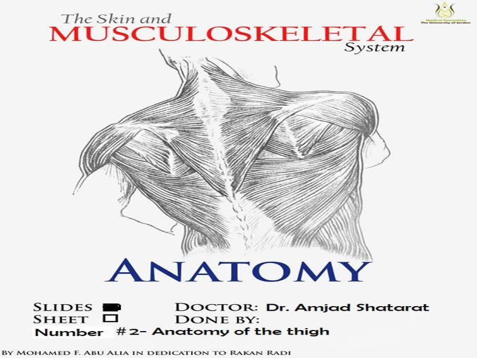
4- Superficial veins
G r e a t S a p h e n o u s v e i n .The great Saphenous vein
drains the medial end of the dorsal venous arch. passes directly in front of the medial malleolus of the tibia.
ascends in a company with the Saphenous nerve. in the superficial fascia over the medial side of the leg.
passes behind the knee and then curves around the medial side of the thigh.
pierces the Saphenous opining and then joins
the femoral vein about 4cm below and lateral to the pubic tubercle.
Great Saphenous vein
cutdown at the ankle?
When we need this
procedure
The most important superficial vein is the
impo
rtan
t

Forms on the anterio-medial side of the thigh the
Saphenous opening (fossa ovalis).
Saphenous opening (fossa ovalis) is a gap in the fascia
lata which is covered by loose connective tissue called
cribriform fascia.
The cribriform fascia is pierced by:
1- Great Saphenous vein
2- superficial branches of the femoral artery
3- Lymphatics.
Fascia lata is connected to the linea aspera by
three intermuscular septa;1- Medial intermuscular septum2- Lateral intermuscular septum
3- Posterior intermuscular septum
Thus the deep fascia and septa divide the thigh into three compartment; Anterior,
Posterior and Medial.
B- Deep fascia of the thigh (fascia lata)

FASCIAL COMPARTMENTS OF THE THIGH

Fascia lata is connected to the
linea aspera by
three intermuscular septa;1- Medial intermuscular septum2- Lateral intermuscular septum
3- Posterior intermuscular septum
Fascial Compartments of the Thigh
Thus the deep fascia and septa divide the thigh into three
compartment; AnteriorPosterior Medial.
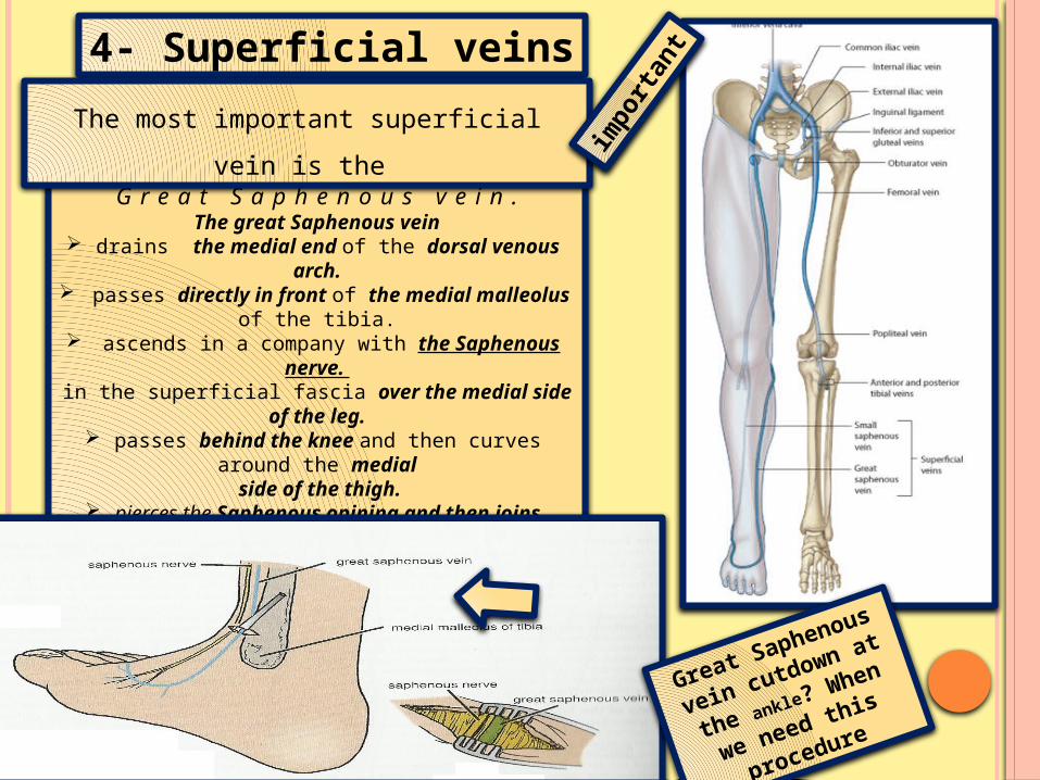
Contents of the Anterior Fascial Compartment of the Thigh
1-Muscles: Sartorius, iliacus, psoas, pectineus, and quadriceps femoris
2-Blood supply: Femoral artery
3-Nerve supply: Femoral nerve
Note: that not all the contents of the anterior compartment have the Same function. For example psoas is the m a i n f l e x o r of the thigh at the hip
joint while quadriceps femoris is the m a i n e x t e n s o r of the leg at the knee joint.

S a r t o r i u sOrigin: Anterior superior iliac spine
Insertion: Upper medial surface of shaft of tibia
Nerve supply: Femoral nerve
Actions: Flexes, abducts, laterally rotates thigh at hip joint
Flexes and medially rotates leg at knee joint
P e c t i n e u s
Origin: Superior ramus of pubis
Insertion: Upper end of linea aspera of shaft of femur
Nerve supply: Femoral nerve?
Actions: Flexes and adducts thigh at hip joint

P s o a s
Origin: Transverse processes, bodies, and intervertebral discs of the 12th thoracic and
five lumbar vertebrae
Insertion: With iliacus into
lesser trochanter of femur
Nerve supply: Lumbar plexus
Actions: Flexes thigh on trunk;
if thigh is fixed, it flexes the
trunk on thigh as in sitting up
from lying down.
I l i a c u s
Origin: Iliac fossa of hip bone
Insertion: With psoas into lesser trochanter of femur
Nerve supply: Femoral nerve
Actions: Flexes thigh on trunk; if thigh is fixed, it flexes the trunk on the thigh as in sitting up from lying down(the same as psoas).

Consisting of:
1- The rectus femoris
2- The vastus intermedius
3- The vastus lateralis
4- The vastus medialis
Originates by two heads
The quadriceps femoris muscle
R e c t u s f e m o r i s
Straight head from anterior inferior iliac spine
Reflected head from ilium above acetabulum
V a s t u s l a t e r a l i s
Origin : Upper end and shaft of femur (linear origin)
V a s t u s m e d i a l i s
Origin : Upper end and shaft of femur (linear origin)