Toxicology in Vitro 19 (2005) 11–20
www.elsevier.com/locate/toxinvit
Analysis of oxidative stress in SK-N-MC neuronsexposed to styrene-7,8-oxide
M.V. Vettori a,*, A. Caglieri b, M. Goldoni a,b, A.F. Castoldi c,E. Dar�e d, R. Alinovi b, S. Ceccatelli d, A. Mutti b
a ISPESL Research Center at the University of Parma, via Gramsci 14, Parma 43100, Italyb Laboratory of Industrial Toxicology, Department of Clinical Medicine, Nephrology and Health Sciences, University of Parma, Parma, Italy
c Toxicology Division, Research Center, Salvatore Maugeri Foundation IRCCS, Institute of Pavia, Italyd Institute of Environmental Medicine, Division of Toxicology and Neurotoxicology, Karolinska Institutet, Box 210, Stockholm S-171 77, Sweden
Received 15 December 2003; accepted 28 April 2004
Abstract
Styrene-7,8-oxide (SO) is the main metabolite of styrene, a neurotoxic volatile organic compound used industrially. Here we
report the novel observation that several markers of oxidative stress were affected in SK-N-MC cells exposed for 16 h to con-
centrations of SO that induce apoptotic cell death. The production of Thiobarbituric Acid Reactive Substances (TBARS), rose from
69.1± 15.7 nmol/g protein (control) to 119.3± 39.2 and 102.0± 17.3 nmol/g protein after exposure to 0.3 and 1 mM SO, respectively.
Carbonyl group levels were significantly enhanced by SO at both concentrations. The lower dose also decreased sulphydryl groups.
SO caused a marked oxidative DNA damage, as shown by a fivefold increase in 8-hydroxy-20-deoxyguanosine (8-OHdG). In
addition, SO exposure resulted in alterations of scavenging abilities, given the reduction of both glutathione (GSH) and glutathione-
S-transferase (GST) activity. Induction of expression of the oxidative stress response gene heme-oxygenase-1 (HO-1) and an in-
creased HO-1 activity were also observed. These data provide compelling evidence that oxidative stress significantly contributes to
SO toxicity in neuronal cells.
� 2004 Elsevier Ltd. All rights reserved.
Keywords: Oxidative stress; Styrene oxide; Neurotoxicity; SK-N-MC neurons
1. Introduction
Styrene-7,8-oxide (SO) is an industrial chemical used
in the manufacture of epoxy resins, as an intermediate in
the preparation of various agricultural chemicals, cos-
metics, and plastics and in the processing of textiles andfibers. SO is also the main electrophilic intermediate
metabolite of styrene, a chemical used in several indus-
trial applications, e.g. manufacture of glass fiber rein-
Abbreviations: 1-Chloro-2,4-dinitrobenzene (CDNB); Dinitro-
phenylhydrazine (DNPH); Fetal bovine serum (FBS); Glutathione
(GSH); Glutathione-S-transferase (GST); Heme-oxygenase-1 (HO-1);
8-Hydroxy-20-deoxyguanosine (8-OHdG); Reactive oxygen species
(ROS); Sodium dodecyl sulfate (SDS); Styrene-7,8-oxide (SO); Thio-
barbituric acid (TBA)*Corresponding author. Tel.: +39-0521-033092; fax: +39-0521-
033076.
E-mail address: [email protected] (M.V. Vettori).
0887-2333/$ - see front matter � 2004 Elsevier Ltd. All rights reserved.
doi:10.1016/j.tiv.2004.04.015
forced plastics and production of styrene monomer,
polyester resins and synthetic rubber. As an epoxide, SO
is able to undergo reactions with various nucleophilic
groups in tissue components, causing effects such as
growth inhibition, cytotoxicity and mutagenicity. Fur-
thermore, the most probable risk of SO is related to itsgenotoxicity. In particular, SO has been shown to
induce DNA adducts, chromosomal aberrations, sister
chromatide exchanges (SCE) micronuclei and DNA
damage in several in vitro systems (Vodicka et al., 2002;
Laffon et al., 2001, 2002). SO is a proven animal car-
cinogen and is classified as a possible human carcinogen
(group 2B) by IARC (1994). The role of SO in the
neurotoxic effects reported in workers exposed to sty-rene is still controversial. Alternative explanations to
styrene neurotoxicity have been put forward by our
group years ago. Brain dopamine depletion has been
reported upon exposure to styrene or styrene metabo-
lites in experimental studies (Mutti et al., 1984b, 1988),

12 M.V. Vettori et al. / Toxicology in Vitro 19 (2005) 11–20
and the dopaminergic system has been suggested as a
specific target for styrene neurotoxicity both in humans
(Mutti et al., 1984a; Arfini et al., 1987; Mutti and
Franchini, 1987) and laboratory animals (Mutti et al.,1984b, 1988). SO at high doses has been shown to be
neurotoxic in animals and using in vitro models (Katoh
et al., 1989; Trenga et al., 1991; Kohn et al., 1995; Bei-
swanger et al., 1993; Chakrabarti, 1999) and the mech-
anisms by which it acts are still unclear. SO exposure
induces depletion of cells in rat brain and is cytotoxic to
neurons in vitro (Kohn et al., 1995; Dypbukt et al.,
1992). In a recent work (Dar�e et al., 2002) we haveshown that exposure of primary cerebellar granule
neurons and human neuroblastoma SK-N-MC cells to
SO (0.3–1 mM) induces apoptosis. Cell shrinkage,
chromatin condensation and DNA cleavage into high
molecular weight fragments of regular size were
accompanied by the activation of class II caspases. In
addition, the presence of the 150-kDa cleavage product
of alpha-fodrin suggested a possible activation of cal-pains. Apoptosis can be triggered by oxidative stress
(Fleury et al., 2002). The ability to increase the synthesis
of reactive oxygen species (ROS) is a common factor in
the toxicity of several neurotoxic compounds including
metals (Naarala et al., 1995; Flora and Seth, 2000;
Olivieri et al., 2002; Belletti et al., 2002; Migheli et al.,
1999) and solvents (Nordmann, 1987; Uysal et al.,
1989). Several lines of evidence suggest that styrene andits metabolite SO can induce oxidative stress. Depletion
of GSH, the primary cellular defense against oxidative
stress, was observed in different brain regions of rats
exposed to SO (Trenga et al., 1991). Lower levels of
GSH were found to be associated with increased lipid
peroxidation in the liver of rodents exposed subchroni-
cally either to styrene or to SO (Katoh et al., 1989). In
addition, increased levels of DNA oxidation productswere detected in blood cells exposed to styrene oxide
in vitro (Marczynski et al., 1997).
In the present study, we have further explored the
mechanisms of SO toxicity and tested, evaluating the
hypothesis that SO increases the levels of ROS in neu-
rons. To clarify intracellular events involved in neuronal
cells death, we have used the human neuroblastoma SK-
N-MC cells. This cell line is a well established experi-mental model to study mechanisms of neurotoxicity in
vitro (Hyun et al., 2002; Dar�e et al., 2002; Sherer et al.,2003). Since apoptotic chromatin rearrangements and
increased caspase activity were observed at 0.3 and 1
mM SO in SK-N-MC cells (Dar�e et al., 2002), theseconcentrations were chosen here to examine different
markers of oxidative stress. Cellular responses have been
studied at the 16 h time point, based on initial timecourse experiments (Dar�e et al., in press). The followingmarkers have been analyzed: (1) Thiobarbituric Acid
Reactive Substances (TBARS) as indicators of oxidative
damage to cell membranes; (2) protein sulphydryl
groups and carbonyl groups as markers of protein oxi-
dation; (3) 8-hydroxy-20-deoxyguanosine (8-OHdG) as
an index of DNA oxidation; (4) expression and activity
of heme-oxigenase-1 (HO-1) as a stress response tooxidative damage mediated by gene regulation. Experi-
ments were also performed to assess the scavenging
potential of cells (GSH intracellular levels and gluta-
thione-S-transferase (GST) activity) as an indirect
mechanism possibly accounting for oxidative stress
associated with SO exposure.
2. Materials and methods
2.1. Chemicals
1,1,3,3-Tetraethoxypropane (Malondialdehyde) was
obtained from Fluka Chemie (Buchs, CH-9471), while
Bioxytech GSH-400 Assay Kit from Oxis International,
Inc. (Portland, OR, USA). DNA isolation Kit was
bought from Gentra System (Minneapolis, MN, USA).Styrene-7,8-oxide (97%) was purchased from Sigma–
Aldrich (Stockholm, Sweden). Dinitrophenylhydrazine
(DNPH) and n-butanol were obtained from Aldrich
(Steinheim, Germany and Milwakee, USA, respec-
tively). Trichloroacetic acid and ethanol were bought
from Analar-BDH (England) and from Carlo Erba
(Milano, Italy), respectively. Ethyl acetate was bought
from Lab-Scan (Dublin, Ireland). Sodium dodecyl sul-fate (SDS) was obtained from Gibco (Paisley, Scotland).
The polyclonal anti-HO-1 antibody was obtained from
Stressgen (Victoria, BC, Canada), the goat antirabbit
peroxidase conjugated antibody was from Pierce
(Rockford, USA), and 30,30-diaminobenzidine substrate
was from Boehringer Mannheim Biochimica (Milan,
Italy).
All the other chemicals were obtained from SigmaChemical Co. (St Louis, MO, USA).
2.2. Cell cultures
The human neuroblastoma SK-N-MC clonal cell line
was purchased from the American Type Culture Col-
lection. Cells were routinely seeded at the density of
40,000 cells/cm2 in DMEM containing 10% fetal bovine
serum (FBS) and 50 units/ml of penicillin and 50 lg/mlof streptomycin. Cells were maintained at 37�C in a 5%CO2 humidified incubator and sub-cultured twice a
week. Treatments with SO were carried out using dilu-tions in medium without serum of a stock solution
prepared in DMSO (final DMSO concentration in the
cell exposure medium¼ 0.05%) and were started 24 hafter seeding, the density of seeding being always 4 · 104cells/cm2. For harvesting, the floating cells, collected
from the medium by centrifugation, were pooled
together with the cells detached by scraping or with

M.V. Vettori et al. / Toxicology in Vitro 19 (2005) 11–20 13
trypsin. In all experiments, the protein concentration
was determined by the BCA method (Pierce, Rockford,
USA), using bovine serum albumin as a standard. Mean
values of SO within the incubation period were extrap-olated from a time-dependent curve, similarly to the
method described in Dar�e et al. (2002) but using theexperimental conditions described above. Briefly, after
0, 8, 16 and 24 h of incubation, 1 ml of supernatant was
placed in 4 ml SPME vials to measure SO concentration
in medium by SPME-GC/MS (Poli et al., 2004). For
each exposure time, samples were collected in triplicate.
Styrene-d5 was added to samples as Internal Standard (2ll of the stock solution, final concentration 2 · 10�6 M).Samples were stored at )80 �C until analysis.
2.3. Detection of TBARS
Cellular TBARS were measured according to the
method of Jentzsch et al. (1996), adapted for cells.Briefly, after three cycles of freezing and thawing ()80�C to +37 �C), control and treated cells were centrifugedat 3000g for 5 min. Two hundred ll of supernatant werediluted with 200 ll of 0.2 M orthophosphoric acid. After
vortexing, 25 ll of a 0.11 M thiobarbituric acid (TBA)
solution prepared in 0.1 M NaOH were added and the
vortexed solution was incubated at 95 �C for 45 min.TBARS were extracted adding 500 ll of n-butanol and50 ll of a saturated solution of NaCl. After a vigorousmixing, the reaction mixture was centrifuged at 3000g
for 10 min and the upper solution was collected. TBARS
concentrations were measured using the difference in
absorption between 530 and 650 nm (the zero sig-
nal). Malondialdehyde was used as a standard for
the calibration curve (range 0–10 lM). TBARSconcentrations were normalized for the total proteincontent.
2.4. Quantification of protein carbonyl groups
Cellular carbonyl groups of proteins were measured
according to the method of Reznick and Packer (1994)adapted for cells. Briefly, after three cycles of freezing
and thawing ()80 �C to +37 �C), control and treatedcells were centrifuged at 3000g for 5 min. DNA was
precipitated from the supernatant adding an amount of
a 10% (w/v) solution of streptomycin sulphate prepared
in PBS, equal to 1/10 of the volume. After 10 min at
room temperature, the supernatant was centrifuged at
3000g for 5 min and the pellet was removed. Twohundred and fifty ll of the supernatant were added to600 ll of a solution of 15 mM DNPH prepared in 2.5 M
HCl. The mixture was left at room temperature for 60
min in the dark, and vortexed every 15 min. Six hundred
ll of a 20% (w/v) trichloroacetic acid solution were
added to precipitate proteins. After 10 min, samples
were centrifuged at 5000g for 5 min and the supernatant
was discarded. This procedure was repeated twice.
Finally, the pellet was washed three times with 1 ml of
an ethanol–ethyl acetate solution (1:1, v/v) to remove
the residue of free DNPH and lipid contaminants. Theprecipitates were dissolved in 1 ml of an 8 M guanidine
hydrochloride solution and were left at 37 � C for 10min. No insoluble material was observed thereafter.
Carbonyl content was calculated by the peak absor-
bance at 365 nm, subtracting the zero signal, using an
extinction coefficient of 22,000 M�1 cm�1. The concen-
tration of carbonyls was normalized for the total protein
content.
2.5. Measurement of protein sulphydryl groups
Cellular protein sulphydryl groups were measured
according to the method of Inayama et al. (1996) with
some modifications. Briefly, after three cycles of freezing
and thawing ()80 �C to +37 �C), control and treatedcells were centrifuged at 3000g for 5 min. Two hundred
ll of supernatant were diluted with 600 ll of water andultra-filtered with a 5-kDa cut-off filter at 4000g for 15
min. The procedure was repeated three times to elimi-
nate free sulphydryl groups. After centrifugation, no
protein aggregation was observed. The upper solution in
the filter, corrected with water to restore the initial
volume of 200 ll, was mixed with 475 ll of a solutioncontaining 0.12 M Tris–HCl, 15 mM EDTA, 3% SDS
and 1 mM DTNB and incubated for 15 min at room
temperature. Sulphydryl content was calculated by the
peak absorbance at 412 nm, subtracting the zero signal.
LL-cysteine was used as a standard for the calibration
curve (range 0–200 lM). The concentration of sul-phydryl groups was normalized for the total non-filtered
protein content.
2.6. Quantification of 8-OHdG in DNA samples
DNA was extracted from cells exposed to SO and
controls using a commercial kit (Gentra System). The
DNA samples were digested with nuclease P1 for 30 minat 37 �C, and then incubated with alkaline phosphatasefor 60 min. To measure hydroxyl adducts to DNA, the
amount of 8-OHdG present in the DNA was measured
by HPLC with electrochemical detector as previously
described by Kim and Lee (1997).
2.7. Evaluation of HO-1 activity
HO-1 activity was measured by bilirubin generation.
The cells were exposed to SO for 16 h, then collected
from the flasks and centrifuged at 1000g at 4 �C for 10min. The pellet was resuspended in 500 ll of phosphatebuffer (100 mM KH2 PO4 and 2 mM MgCl2, pH 7.4)
and frozen–thawed ()80 to +37 �C) three times. After

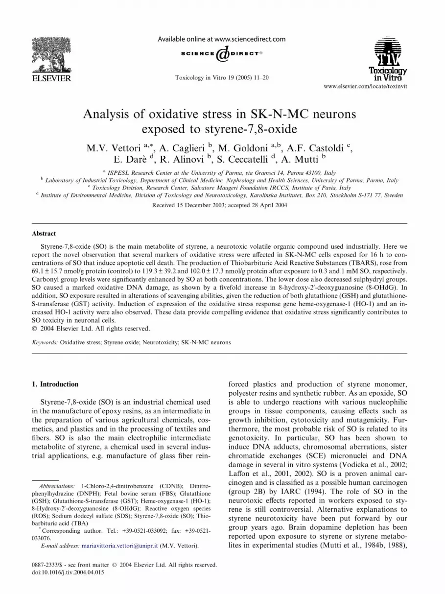
Fig. 1. Time-course of SO concentration in the medium during
exposure of SK-N-MC cells. Cells were treated with the initial con-
centration of 0.3 mM SO, and SO was measured in aliquots of the
medium at different time points (8, 16 and 24 h). Values are the
means±S.D. of three determinations. (j¼ experimental data points;continuous line¼fitting curve; dotted line¼fitting curve reported byDar�e et al., 2002).
14 M.V. Vettori et al. / Toxicology in Vitro 19 (2005) 11–20
sonication on ice and centrifugation at 18,000g for 10
min at 4 �C, the reaction was started by adding thesupernatant to the NADPH-generating system con-
taining 0.8 mM NADPH, 2 mM glucose-6-phosphate,0.2 U glucose-6-phosphate dehydrogenase, 3 mg protein
of rat liver cytosol (prepared from 105,000g supernatant
fraction as source of biliverdin reductase), potassium
phosphate buffer (100 mM, pH 7.4), and hemin (20 lM)in a final volume of 1 ml. The reaction was incubated for
1 h at 37 �C in the dark and was terminated by additionof 1 ml chloroform. The solution was vortexed for 30 s,
centrifuged for 5 min at 4000g and the lower phase wascollected. The extracted bilirubin was calculated by the
difference in OD units recorded at 464 and 530 nm using
a quartz cuvette (extinction coefficient, 40 mM�1 cm�1
for bilirubin). HO-1 activity was expressed as picomol of
bilirubin/mg of protein/h.
2.8. Detection of HO-1 by immunocytochemistry
SK-N-MC cells grown on coverslips were exposed to
SO, then fixed with 80% methanol at 20 �C for 10 minand with acetone at 20 �C for 10 s. Since the 1 mM dose
induced cell detachment from the glass slide, only sam-
ples exposed to 0.3 mM SO and controls were analyzed
by immunocytochemistry. After three washes with PBS,
inhibition of endogenous peroxidases (1% H2O2 for 10min) and blocking with FBS (30 min at room tempera-
ture), the coverslips were incubated with a rabbit anti-
HO-1 polyclonal antibody (1:500) at 37 �C for 45 min.The cells were then rinsed with PBS and incubated for
1 h at room temperature with a goat peroxidase conju-
gated secondary antibody (1:1000). After washing with
PBS, the coverslips were incubated with 30,30-diam-
inobenzidine substrate for 15 min at room temperature,rinsed in water and then counterstained with methyl-
green (6 min at room temperature). The cells were then
examined at the light microscope (Olympus) and images
were collected with a digital camera.
2.9. Detection of intracellular glutathione-S-transferase
(GST) activity
For GST activity assays the cells were collected by
centrifugation (1100 rpm for 10 min at 4 �C). The pelletswere sonicated in cold phosphate buffered saline and
then centrifuged at 10,000g for 15 min at 4 �C. Thesupernatants were transferred into new tubes and stored
on ice. GST activity was determined in 100 mM potas-sium phosphate buffer pH 6.5 by measuring the conju-
gation of 1-chloro-2,4-dinitrobenzene (CDNB) with
reduced glutathione, which was accompanied by an
increase in absorbance at 340 nm (Habig et al., 1974).
The rate of product formation was monitored by mea-
suring the increase in absorbance at 340 nm in a Beck-
man DU 640 UV/visible spectrophotometer for 10 min.
GST activity was expressed as mU/mg protein. The di-
rect effect of SO on pure GST enzyme from human
placenta (Sigma–Aldrich, St Louis, MO, USA) was also
investigated. The reaction mixture was prepared byadding 30 lg of pure GST enzyme to different solutionsof SO (0.3 and 1 mM) in a 100 mM phosphate buffer pH
6.5, and incubated at 30 �C for different periods of time(1, 2, 4, 8 and 16 h). At the end of the incubation an
aliquot of the mixture was analyzed to determine the
GST activity, using the above-described method.
2.10. Measurement of intracellular GSH
Intracellular levels of GSH were determined in freshsamples using a commercial colorimetric kit (Oxis
International, Portland, USA). Briefly, at the end of
the exposure period SK-N-MC cells were pelleted by
centrifugation at 1200 rpm for 5 min at 4 �C, and thenwashed in 5% cold metaphosphoric acid; the samples
were centrifuged at 4500g for 10 min at 4 �C and
the supernatants were immediately used to measure the
GSH content according to the protocol indicated in thekit. The sample GSH concentration was extrapolated
from a freshly prepared standard curve (0–60 lM) andnormalized to the protein content.
3. Results
3.1. Measurement of the SO concentration in the
medium
Fig. 1 (continuous line) shows the time-decay of 0.3
mM SO in the medium containing SK-N-MC cells. The

M.V. Vettori et al. / Toxicology in Vitro 19 (2005) 11–20 15
1 mM dose showed a similar trend (data not shown).
Using the same algorithm for the decay of SO given by
Dar�e et al. (2002) (Fig. 1, dotted line), the nominalconcentrations of 0.3 and 1 mM SO were shown tocorrespond to average concentrations of 0.15 and 0.51
mM, respectively (during the 16 h of incubation). To
simplify the presentation of the results, the concentra-
tions of SO given throughout this paper refer to the
initial levels in the medium.
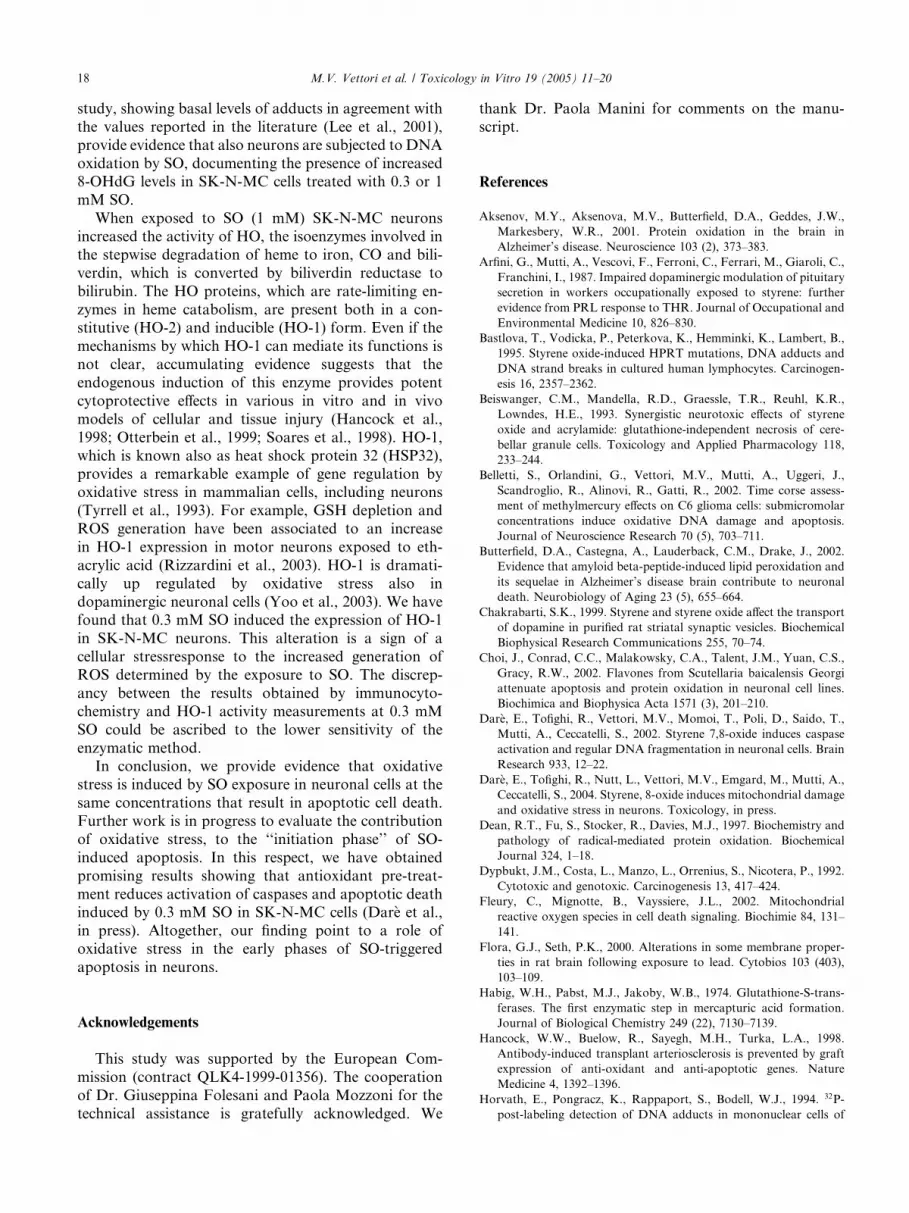
3.2. Lipid peroxidation in SO-exposed neurons
To evaluate the effect of SO on lipid peroxidation the
concentration of TBARS was measured in lysates of
SK-N-MC cells exposed to 0.3 or 1 mM SO for 16 h.
Both doses caused a statistically significant enhancementof MDA levels relatively to control values (Fig. 2A).
The production of TBARS increased from 69.08± 15.7
nmol/g protein in control samples to 119.32 ± 39.25 and
102.05 ± 17.27 in samples exposed to 0.3 and 1 mM SO,
respectively.
3.3. Protein modifications induced by SO
As a measure of protein oxidation, the levels of both
carbonyl and sulphydryl groups were measured in SK-
Fig. 2. Effects of SO on TBARS (A), 8-OHdG levels (B), carbonyl group
means±S.D. of at least four determinations. The concentrations of SO given
medium and correspond to an extrapolated average of SO concentration o
performed by one-way ANOVA, followed by the post-hoc Dunnett’s test. � S
different from untreated control (p < 0:01); ��� Significantly different from u
N-MC cell lysates following 16 h exposure to SO. Car-
bonyl groups were significantly increased in cells ex-
posed to either 0.3 or 1 mM SO, as compared with
control levels (Fig. 2C) (4.71 ± 0.46 nmol/mg protein at0.3 mM and 4.73± 0.48 nmol/mg protein at 1 mM, vs.
4.17 ± 0.12 nmol/mg protein of controls). With respect
to protein sulphydryl groups, the latter were decreased
by about 26% in cell lysates treated with 0.3 mM
SO (Fig. 2D). In contrast, no significant changes in the
level of sulphydryl groups were observed in proteins
extracted from cells exposed to 1 mM SO for 16 h
(Fig. 2D).
3.4. Effects of SO on DNA oxidation
Measurements of hydroxyl adducts to DNA (8-
OHdG) were performed to evaluate SO-induced DNA
oxidation. Both 0.3 and 1 mM SO caused a marked
oxidative DNA damage after 16 h, as indicated by the
more than fivefold increase in 8-OHdG levels in SO-exposed cell lysates as compared to controls (Fig. 2B).
In basal conditions samples contained 0.6 8-OHdG/105
dG. Upon treatment with 0.3 and 1 mM SO, the levels
of 8-OHdG/105 dG were enhanced to 3.5 ± 0.5 and
3.75± 0.8, respectively, as compared to untreated con-
trols (0.6 ± 0.4).
s (C), and sulphydryl groups (D) in SK-N-MC cells. Values are the
in all the figures refer to the initial levels of SO (0.3 and 1 mM) in the
f 0.15 and 0.51 mM within 16 h, respectively. Statistical analysis was
ignificantly different from untreated control (p < 0:05); �� Significantly
ntreated control ðp < 0:001Þ.

Fig. 3. HO-1 activity (A) and expression (B and C) in SK-N-MC neurons exposed to SO for 16 h. Values are the means±S.D. of at least four
determinations. The concentrations of SO given in all the figures refer to the initial levels of SO (0.3 and 1 mM) in the medium and correspond to an
extrapolated average of SO concentration of 0.15 and 0.51 mM within 16 h, respectively. Statistical analysis was performed by one-way ANOVA,
followed by the post-hoc Dunnett’s test. (��� Significantly different from untreated control, p < 0:001). The expression of HO-1 was evaluated in
neurons exposed to 0.3 mM for 16 h (B) and in unexposed control cells (C) using immunocytochemistry (bar is 10 lm).
16 M.V. Vettori et al. / Toxicology in Vitro 19 (2005) 11–20
3.5. SO-induced changes in HO-1 activity and
expression
HO-1 activity was measured in SK-N-MC cells ly-
sates by bilirubin generation. A significant increase ðp <0:001Þ in HO-1 activity was seen in cells exposed to 1mM SO (Fig. 3A). HO-1 activity showed an increasing
trend also in cells treated with 0.3 mM SO, although
such changes did not reach statistical significance. Be-
sides, this lower dose of SO increased the expression
HO-1 in comparison with control cultures, as shown byimmunocytochemical staining (Fig. 3B and C). Since the
1 mM dose induced cell detachment from the glass slide,
these samples were not analyzed for this end-point.
3.6. Alterations of GST activity and GSH levels
in SO-exposed cells
A dose-dependent decrease in the activity of GST was
detected in SK-N-MC cell lysates exposed to 0.3 and 1
mM SO for 16 h. In particular, the extent to which the
enzyme activity was inhibited by SO increased from
about 54% upon exposure to 0.3 mM to approximately
70% upon treatment with 1 mM (Table 1). To assesswhether the inhibition of GST activity by SO exposure
occurred through a direct mechanism, we studied the
effects of SO on GST enzymes purified from human
Table 1
GST activity and GSH levels in SK-N-MC cells exposed to either 0.3
or 1 mM initial concentrations of SO for 16 h
Control 0.3 mM SO 1 mM SO
GST (mU/mg prot)a 83.88± 13.71 38.38± 14� 25.46± 8.58�
GSH (nmoles/mg
prot)a16.11± 1.28 18.47± 5.21 7.66± 1.39�
The concentrations of SO given refer to the initial levels of SO (0.3 and
1 mM) in the medium and correspond to an extrapolated average of
SO concentration of 0.15 and 0.51 mM within 16 h, respectively.* Significantly different from untreated control ðp < 0:02Þ.a Values are the means of six determinations± S.D. Statistical
analysis was performed by one-way ANOVA.
placenta. The results of these measurements showed the
inability of SO to affect directly the GST enzyme activity
(data not shown).Additional experiments were performed to assess the
GSH level in cell lysates. Statistically significant de-
creases in GSH were observed in neurons exposed to
1 mM SO for 16 h as compared to controls (Table 1).
No changes were detected with the 0.3 mM dose after
16 h (Table 1). The same trend in GSH level was also
observed at earlier time points (after 8 h, data not
shown).
4. Discussion
Although oxidative stress has been shown to con-
tribute significantly to the noxious effects of many neu-
rotoxic agents, so far little attention has been paid tothe role of ROS in the mechanism of SO neurotoxicity
in vitro. In this study, we report the novel observation
that several markers of oxidative stress are affected by
SO exposure in neuronal cells. Since at 0.3 and 1 mM
apoptotic chromatin rearrangements and increased
caspase activity were observed (Dar�e et al., 2002), thesedoses were chosen to perform our study. Notably, here
we refer to initial concentrations prepared in the med-ium, although mean values of SO within the incubation
period of 16 h were 0.15 and 0.51 mM corresponding to
0.3 and 1 mM, respectively. Significant increases in lipid
peroxidation (TBARS content), protein and DNA oxi-
dation were observed in SK-N-MC cells exposed to ei-
ther 0.3 or 1 mM SO for 16 h. Both SO concentrations
markedly influenced the detoxifying capabilities of cells,
as they strongly decreased GST activity. The higher dosewas also effective in reducing the cellular GSH content.
In addition, SO induced HO-1 expression and increased
HO-1 activity. Taken together, these data suggest that
an excess of oxidative reactions is an important mech-
anism contributing to SO neurotoxicity in vitro and
point to a role of oxidative stress in the early phase of
SO-triggered apoptosis in neurons.

M.V. Vettori et al. / Toxicology in Vitro 19 (2005) 11–20 17
In the present study, a higher level of lipid peroxi-
dation was detected in neurons exposed to SO in vitro,
as compared to controls. The values measured in SK-N-
MC cells were in the same order of magnitude as thosereported in other studies on neurons (Naito et al., 1995).
The occurrence of lipid peroxidation has been demon-
strated in the liver but not in the brain of rats sub-
chronically exposed to styrene (300–500 mg/kg i.p., 3
times/week, 7 weeks) and styrene oxide (200–400 mg/kg)
(Katoh et al., 1989). These authors suggested that the
enhancement of hepatic lipid peroxidation resulted from
GSH depletion to certain critical levels and delayedrecovery of lipid peroxides (Katoh et al., 1989). Al-
though the same investigators also reported a decrease
in cerebral GSH levels in SO-exposed rats, they
hypothesized that such decrement was not critical en-
ough, in terms of degree and duration, to produce an
increase in brain lipid peroxidation (Katoh et al., 1989).
In our cell model, both 0.3 and 1 mM SO caused the
enhancement of lipid peroxidation at 16 h, but the lowerSO concentration did not affect GSH levels at the same
time point. The latter result may be ascribed to the lack
of SO effect on this target at the 0.3 mM dose. Although
the basal levels of GSH measured in our study are in
accordance with the ones reported in the literature
(Verity and Sarafian, 1991) no differences in GSH levels
were observed at 0.3 mM SO after 2, 4 and 8 h (data not
shown). The discrepancies with the in vivo data could bedue both to the different method of exposure and to SO
concentrations to which neuronal cells have been ex-
posed. The decrease in GST activity reflects a compro-
mised ability of the cell to scavenge electrophilic species.
GST is the enzyme that conjugates electrophilic species,
like SO, to GSH, and it is one of the essential cellular
antioxidant enzymes that can counteract the toxicity of
free radicals. Indeed, Xie et al. (2001) have demon-strated that over-expression of GST confers resistance
to oxidative stress in SY5Y neuroblastoma cells and that
GST activity decreases after exposure to 4-hydroxy-
nonenal, a compound formed during lipid peroxidation,
in a dose-dependent manner. Here we show that after
exposure to 1 mM SO for 16 h both GST activity and
GSH content were decreased. These concomitant alter-
ations were consistent with the presence of diffuse celldamage. In a previous study we have documented that 1
mM SO induced secondary necrosis in 57% of SK-N-
MC cells after 16 h (Dar�e et al., 2002). In agreementwith our findings, GSH depletion by 1 mM SO has been
previously reported in PC12 cells (Dypbukt et al., 1992).
A different pattern of changes has been observed in SK-
N-MC cells treated with 0.3 mM SO for 16 h, where
only 24% of the cells were necrotic (Dar�e et al., 2002). Inthis case GST activity was 50% lower than that of
controls in the absence of significant GSH changes. To
explain the reduced GST activity, we hypothesized a
direct action of SO on the catalytic site of the enzyme.
However, no changes were seen in the activity of GST
purified from human placenta in the presence of SO,
ruling out a direct interaction between the enzyme and
the toxic compound (data not shown). SO may insteadexert an indirect effect, increasing generation of ROS
that can cause enzyme modification. Moreover it is
possible a direct interaction of SO with GST genes and a
subsequent inactivation, SO being capable to form ad-
ducts with guanine and adenine (Vodicka et al., 2002).
The determination of carbonyl groups level has been
widely employed as a parameter of protein oxidation to
characterize tissue alterations occurring in neurodegen-erative diseases (Yatin et al., 1999; Aksenov et al., 2001;
Butterfield et al., 2002) and to unravel the mechanisms
of neurotoxicity in vitro (Keller et al., 2000; Kanski
et al., 2001; Choi et al., 2002). An increased level of
carbonyl groups has been related to a decrease in pro-
teasome activity in PC12 cells (Keller et al., 2000),
damage of mitochondrial proteins (Kim et al., 2001) and
to a general impairment of cellular functions (Deanet al., 1997). Under our experimental conditions, the
levels of carbonyl groups measured in SK-N-MC neu-
rons exposed to either 0.3 or 1 mM SO for 16 h are in
accordance with the values reported in the literature
(Lee et al., 2001) and were significantly increased as
compared to controls, providing evidence that SO
damages cellular proteins by oxidative reactions. In
SK-N-MC cells treated with 0.3 mM SO, a significantdecrease in protein sulphydryl groups was observed,
suggesting a higher number of oxidized cysteine and
methionine residues in the polypeptidic chains of SO
exposed neurons. Recent evidence indicates that the
oxidation of protein sulphydryl groups may result in
mitochondrial dysfunction, e.g. mitochondrial complex
I is highly vulnerable to inactivation through oxidation
(Sriram et al., 1998; Kenchappa et al., 2002). We foundthat SO at higher concentrations, namely 1 mM, was
devoid of significant effects toward the level of protein
sulphydryl groups in SK-N-MC cells. This difference in
protein oxidation observed at the two doses of SO after
16 h may relate to the fact that neurons have reached
different stages in the process of cell death, e.g. more
cells in secondary necrosis are detected at the higher SO
concentration (Dar�e et al., 2002). Disruption of disul-phide bridges and exposure of cysteine and methionine
residues, which are the measured in our assay, vary in
relation to the degree of protein denaturation and pro-
teolysis occurring in the cells.
The genotoxic potential of SO is well known and its
ability to induce DNA adducts and DNA strand breaks
both in vitro and in vivo has been reported (Bastlova
et al., 1995; Dypbukt et al., 1992; Horvath et al., 1994;Vodicka et al., 1993; Walles et al., 1993). Oxidative DNA
damage has also been implicated as a causative factor
in the DNA strand breaks detected in blood samples
exposed to SO in vitro (Marczynski et al., 1997). Our
18 M.V. Vettori et al. / Toxicology in Vitro 19 (2005) 11–20
study, showing basal levels of adducts in agreement with
the values reported in the literature (Lee et al., 2001),
provide evidence that also neurons are subjected to DNA
oxidation by SO, documenting the presence of increased8-OHdG levels in SK-N-MC cells treated with 0.3 or 1
mM SO.
When exposed to SO (1 mM) SK-N-MC neurons
increased the activity of HO, the isoenzymes involved in
the stepwise degradation of heme to iron, CO and bili-
verdin, which is converted by biliverdin reductase to
bilirubin. The HO proteins, which are rate-limiting en-
zymes in heme catabolism, are present both in a con-stitutive (HO-2) and inducible (HO-1) form. Even if the
mechanisms by which HO-1 can mediate its functions is
not clear, accumulating evidence suggests that the
endogenous induction of this enzyme provides potent
cytoprotective effects in various in vitro and in vivo
models of cellular and tissue injury (Hancock et al.,
1998; Otterbein et al., 1999; Soares et al., 1998). HO-1,
which is known also as heat shock protein 32 (HSP32),provides a remarkable example of gene regulation by
oxidative stress in mammalian cells, including neurons
(Tyrrell et al., 1993). For example, GSH depletion and
ROS generation have been associated to an increase
in HO-1 expression in motor neurons exposed to eth-
acrylic acid (Rizzardini et al., 2003). HO-1 is dramati-
cally up regulated by oxidative stress also in
dopaminergic neuronal cells (Yoo et al., 2003). We havefound that 0.3 mM SO induced the expression of HO-1
in SK-N-MC neurons. This alteration is a sign of a
cellular stressresponse to the increased generation of
ROS determined by the exposure to SO. The discrep-
ancy between the results obtained by immunocyto-
chemistry and HO-1 activity measurements at 0.3 mM
SO could be ascribed to the lower sensitivity of the
enzymatic method.In conclusion, we provide evidence that oxidative
stress is induced by SO exposure in neuronal cells at the
same concentrations that result in apoptotic cell death.
Further work is in progress to evaluate the contribution
of oxidative stress, to the ‘‘initiation phase’’ of SO-
induced apoptosis. In this respect, we have obtained
promising results showing that antioxidant pre-treat-
ment reduces activation of caspases and apoptotic deathinduced by 0.3 mM SO in SK-N-MC cells (Dar�e et al.,in press). Altogether, our finding point to a role of
oxidative stress in the early phases of SO-triggered
apoptosis in neurons.
Acknowledgements
This study was supported by the European Com-
mission (contract QLK4-1999-01356). The cooperation
of Dr. Giuseppina Folesani and Paola Mozzoni for the
technical assistance is gratefully acknowledged. We
thank Dr. Paola Manini for comments on the manu-
script.
References
Aksenov, M.Y., Aksenova, M.V., Butterfield, D.A., Geddes, J.W.,
Markesbery, W.R., 2001. Protein oxidation in the brain in
Alzheimer’s disease. Neuroscience 103 (2), 373–383.
Arfini, G., Mutti, A., Vescovi, F., Ferroni, C., Ferrari, M., Giaroli, C.,
Franchini, I., 1987. Impaired dopaminergic modulation of pituitary
secretion in workers occupationally exposed to styrene: further
evidence from PRL response to THR. Journal of Occupational and
Environmental Medicine 10, 826–830.
Bastlova, T., Vodicka, P., Peterkova, K., Hemminki, K., Lambert, B.,
1995. Styrene oxide-induced HPRT mutations, DNA adducts and
DNA strand breaks in cultured human lymphocytes. Carcinogen-
esis 16, 2357–2362.
Beiswanger, C.M., Mandella, R.D., Graessle, T.R., Reuhl, K.R.,
Lowndes, H.E., 1993. Synergistic neurotoxic effects of styrene
oxide and acrylamide: glutathione-independent necrosis of cere-
bellar granule cells. Toxicology and Applied Pharmacology 118,
233–244.
Belletti, S., Orlandini, G., Vettori, M.V., Mutti, A., Uggeri, J.,
Scandroglio, R., Alinovi, R., Gatti, R., 2002. Time corse assess-
ment of methylmercury effects on C6 glioma cells: submicromolar
concentrations induce oxidative DNA damage and apoptosis.
Journal of Neuroscience Research 70 (5), 703–711.
Butterfield, D.A., Castegna, A., Lauderback, C.M., Drake, J., 2002.
Evidence that amyloid beta-peptide-induced lipid peroxidation and
its sequelae in Alzheimer’s disease brain contribute to neuronal
death. Neurobiology of Aging 23 (5), 655–664.
Chakrabarti, S.K., 1999. Styrene and styrene oxide affect the transport
of dopamine in purified rat striatal synaptic vesicles. Biochemical
Biophysical Research Communications 255, 70–74.
Choi, J., Conrad, C.C., Malakowsky, C.A., Talent, J.M., Yuan, C.S.,
Gracy, R.W., 2002. Flavones from Scutellaria baicalensis Georgi
attenuate apoptosis and protein oxidation in neuronal cell lines.
Biochimica and Biophysica Acta 1571 (3), 201–210.
Dar�e, E., Tofighi, R., Vettori, M.V., Momoi, T., Poli, D., Saido, T.,Mutti, A., Ceccatelli, S., 2002. Styrene 7,8-oxide induces caspase
activation and regular DNA fragmentation in neuronal cells. Brain
Research 933, 12–22.
Dar�e, E., Tofighi, R., Nutt, L., Vettori, M.V., Emgard, M., Mutti, A.,Ceccatelli, S., 2004. Styrene, 8-oxide induces mitochondrial damage
and oxidative stress in neurons. Toxicology, in press.
Dean, R.T., Fu, S., Stocker, R., Davies, M.J., 1997. Biochemistry and
pathology of radical-mediated protein oxidation. Biochemical
Journal 324, 1–18.
Dypbukt, J.M., Costa, L., Manzo, L., Orrenius, S., Nicotera, P., 1992.
Cytotoxic and genotoxic. Carcinogenesis 13, 417–424.
Fleury, C., Mignotte, B., Vayssiere, J.L., 2002. Mitochondrial
reactive oxygen species in cell death signaling. Biochimie 84, 131–
141.
Flora, G.J., Seth, P.K., 2000. Alterations in some membrane proper-
ties in rat brain following exposure to lead. Cytobios 103 (403),
103–109.
Habig, W.H., Pabst, M.J., Jakoby, W.B., 1974. Glutathione-S-trans-
ferases. The first enzymatic step in mercapturic acid formation.
Journal of Biological Chemistry 249 (22), 7130–7139.
Hancock, W.W., Buelow, R., Sayegh, M.H., Turka, L.A., 1998.
Antibody-induced transplant arteriosclerosis is prevented by graft
expression of anti-oxidant and anti-apoptotic genes. Nature
Medicine 4, 1392–1396.
Horvath, E., Pongracz, K., Rappaport, S., Bodell, W.J., 1994. 32P-
post-labeling detection of DNA adducts in mononuclear cells of
M.V. Vettori et al. / Toxicology in Vitro 19 (2005) 11–20 19
workers occupationally exposed to styrene. Carcinogenesis 15,
1309–1315.
Hyun, D.H., Lee, M.H., Halliwell, B., Jenner, P., 2002. Proteasomal
dysfunction induced by 4-hydroxy-2,3-trans-nonenal, an end-
product of lipid peroxidation: a mechanism contributing to
neurodegeneration? Journal of Neurochemistry 83 (2), 360–
370.
IARC, 1994. Styrene-7,8-oxide. IARC Monographs on the Evaluation
of Carcinogenic Risks to Humans 60, 321–346.
Inayama, T., Kumagai, Y., Sakane, M., Saito, M., Matsuda, M., 1996.
Plasma protein-bound sulfhydryl group oxidation in humans
following a full marathon race. Life Sciences 59 (7), 573–
578.
Jentzsch, A.M., Bachmann, H., Furst, P., Biesalski, H.K., 1996.
Improved analysis of malondialdehyde in human blood fluids. Free
Radical Biology and Medicine 20 (2), 251–256.
Kanski, J., Drake, J., Aksenova, M., Engman, L., Butterfield, D.A.,
2001. Antioxidant activity of the organotellurium compound 3-[4-
(N,N-dimethylamino) benzenetellurenyl] propanesulfonic acid
against oxidative stress in synaptosomal membrane systems and
neuronal cultures. Brain Research 911 (1), 12–21.
Katoh, T., Higashi, K., Inoue, N., 1989. Sub-chronic effects of styrene
and styrene oxide on lipid peroxidation and the metabolism of
gluthathione in rat liver and brain. Journal of Toxicological
Sciences 14, 1–9.
Keller, J.N., Huang, F.F., Dimayuga, E.R., Maragos, W.F., 2000.
Dopamine induces proteasome inhibition in neural PC12 cell line.
Free Radical Biology and Medicine 29 (10), 1037–1042.
Kenchappa, R.S., Diwakar, L., Boyd, M.R., Ravindranath, V., 2002.
Thioltransferase (glutaredoxin) mediates recovery of motor neu-
rons from excitotoxic mitochondrial injury. Journal of Neurosci-
ence 22, 8402–8410.
Kim, K.B., Lee, B.M., 1997. Oxidative stress to DNA, protein, and
antioxidant enzymes (superoxide dismutase and catalase) in rats
treated with benzo(a)pyrene. Cancer Letters 113, 205–212.
Kim, D.H., Jang, Y.Y., Han, E.S., Lee, C.S., 2001. Protective effect of
harmaline and harmalol against dopamine- and 6-hydroxydop-
amine-induced oxidative damage of brain mitochondria and
synaptosomes, and viability loss of PC12 cells. European Journal
of Neuroscience 13 (10), 1861–1872.
Kohn, J., Minotti, S., Durham, H., 1995. Assessment of the neuro-
toxicity of styrene, styrene oxide, and styrene glycol in primary
cultures of motor and sensory neurons. Toxicology Letters 75, 29–
37.
Laffon, B., P�asaro, E., M�endez, J., 2001. Genotoxic effects of styrene-
7,8-oxide in human white blood cells: comet assay in relation to the
induction of sister-chromatid exchanges and micronuclei. Mutation
Research 491, 163–172.
Laffon, B., P�asaro, E., M�endez, J., 2002. DNA damage and repair in
human leukocytes exposed to styrene-7,8-oxide measured by the
comet assay. Toxicology Letters 126, 61–68.
Lee, M., Hyun, D.H., Marshall, K.A., Ellerby, L.M., Bredesen, D.E.,
Jenner, P., Halliwell, B., 2001. Effect of overexpression of BCL-2
on cellular oxidative damage, nitric oxide production, antioxidant
defenses, and the proteasome. Free Radical Biology and Medicine
31 (12), 1550–1559.
Marczynski, B., Peel, M., Baur, X., 1997. Changes in high molecular
weight DNA fragmentation following human blood exposure to
styrene-7,8-oxide. Toxicology 120, 111–117.
Migheli, R., Godani, C., Sciola, L., Delogu, M.R., Serra, P.A.,
Zangani, D., De Natale, G., Miele, E., Desole, M.S., 1999.
Enhancing effect of manganese on L-DOPA-induced apoptosis in
PC12 cells: role of oxidative stress. Journal of Neurochemistry 73
(3), 1155–1163.
Mutti, A., Mazzucchi, A., Rustichelli, P., Frigeri, G., Arfini, G.,
Franchini, I., 1984a. Exposure-effect and exposure-response rela-
tionships between occupational exposure to styrene and neuropsy-
chological functions. American Journal of Industrial Medicine 5,
275–286.
Mutti, A., Falzoi, M., Romanelli, A., Franchini, I., 1984b. Regional
alterations of brain catecholamines by styrene exposure in rabbits.
Archives of Toxicology 55, 173–177.
Mutti, A., Franchini, I., 1987. Toxicity of metabolites to dopaminergic
systems and the behavioural effects of organic solvents. British
Journal of Industrial Medicine 44, 721–723.
Mutti, A., Falzoi, M., Romanelli, A., Bocchi, M.C., Ferroni, C.,
Franchini, I., 1988. Brain dopamine as a target for solvent toxicity:
effects of some monocyclic aromatic hydrocarbons. Toxicology 49,
77–82.
Naarala, J.T., Loikkanen, J.J., Ruotsalainen, M.H., Savolainen, K.M.,
1995. Lead amplifies glutamate-induced oxidative stress. Free
Radical Biology and Medicine 19 (5), 689–693.
Naito, M., Umegaki, H., Iguchi, A., 1995. Protective effects of
probucol against glutamate-induced cytotoxicity in neuronal cell
line PC12. Neuroscience Letters 186 (2-3), 211–213.
Nordmann, R., 1987. Oxidative stress from alcohol in the brain.
Alcoholism Alcohol Suppl. 1, 75–82.
Olivieri, G., Novakovic, M., Savaskan, E., Meier, F., Baysang, G.,
Brockhaus, M., Muller-Spahn, F., 2002. The effects of beta-
estradiol on SHSY5Y neuroblastoma cells during heavy metal
induced oxidative stress, neurotoxicity and beta-amyloid secretion.
Neuroscience 113 (4), 849–855.
Otterbein, L.E., Mantell, L.L., Choi, A.M., 1999. Carbon monoxide
provides protection against hyperoxic lung injury. American
Journal of Physiology: Lung Cellular and Molecular Physiology
276, L688–L649.
Poli, D., Vettori, M.V., Andreoli, R., Manini, P., Alinovi, R.,
Ceccatelli, S., Mutti, A., 2004. A novel approach based on solid
phase micro extraction gas chromatography and mass spectrom-
etry to the determination of highly reactive organic compounds in
cell cultures: styrene-7,8-oxide. Chemical and Research in Toxicol-
ogy 17 (1), 104–109.
Reznick, A.Z., Packer, L., 1994. Oxidative damage to proteins:
spectrophotometric method for carbonyl assay. Methods in Enzy-
mology 233, 357–363.
Rizzardini, M., Lupi, M., Bernasconi, S., Mangolini, A., Cantoni, L.,
2003. Mitochondrial dysfunction and death in motor neurons
exposed to the glutathione-depleting agent ethacrylic acid. Journal
of the Neurological Sciences 8798, 1–8.
Sherer, T.B., Betarbet, R., Testa, C.M., Seo, B.B., Richardson, J.R.,
Kim, J.H., Miller, G.W., Yagi, T., Matsuno-Yagi, A., Greenamyre,
J.T., 2003. Mechanism of toxicity in rotenone models of Parkin-
son’s disease. Journal of Neuroscience 23 (34), 10756–10764.
Soares, M.P., Lin, Y., Anrather, J., Csizmadia, E., Takigami, K., Sato,
K., Grey, S.T., Colvin, R.B., Choi, A.M., Poss, K.D., Bach, F.H.,
1998. Expression of heme oxygenase-1 can determine cardiac
xenograft survival. Nature Medicine 4, 1073–1077.
Sriram, K., Shankar, S.K., Boyd, M.R., Ravindranath, V., 1998. Thiol
oxidation and loss of mitochondrial complex I precede excitatory
amino acid-mediated neurodegeneration. Journal of Neuroscience
18, 10287–10296.
Trenga, C.A., Kunkel, D.D., Eaton, D.L., Costa, L.G., 1991. Effect of
styrene oxide on rat brain glutathione. Neurotoxicology 12, 165–
178.
Tyrrell, R.M., Applegate, L.A., Tromvoukis, Y., 1993. The proximal
promoter region of the human heme oxygenase gene contains
elements involved in stimulation of transcriptional activity by a
variety of agents including oxidants. Carcinogenesis 14, 761–
765.
Uysal, M., Kutalp, G., Odzemirler, G., Ayka, G., 1989. Ethanol-
induced changes in lipid peroxidation and glutathione content in
rat brain. Drug and Alcohol Dependence 23, 227–230.
Verity, M.A., Sarafian, T., 1991. Role of oxidative damage in the
pathogenesis of methylmercury neurotoxicity. In: Suzuki, T.,

20 M.V. Vettori et al. / Toxicology in Vitro 19 (2005) 11–20
Imura, N., Clarkson, T.W. (Eds.), Advances in Mercury Toxicol-
ogy. Plenum Press, New York, pp. 209–222.
Vodicka, P., Koskinen, M., Arand, M., Oesch, F., Hemminki, K.,
2002. Spectrum of styrene-induced DNA adducts: the relationship
to other biomarkers and prospects in human biomonitoring.
Mutation Research 511 (3), 239–254.
Vodicka, P., Vodickova, L., Hemminki, K., 1993. 32P-postlabeling of
DNA adducts of styrene-exposed lamination workers. Carcino-
genesis 14, 2059–2061.
Walles, S.A., Edling, C., Anundi, H., Johanson, G., 1993. Exposure
dependent increase in DNA single strand breaks in leucocytes from
workers exposed to low concentrations of styrene. British Journal
of Industrial Medicine 50, 570–574.
Xie, C., Lovell, M.A., Xiong, S., Kindy, M.S., Guo, J., Xie, J.,
Amaranth, V., Montine, T.J., Markesbery, W.R., 2001. Expression
of glutathione-S-transferase isozyme in the SY5Y neuroblastoma
cell line increases resistance to oxidative stress. Free Radical
Biology and Medicine 31 (1), 73–81.
Yatin, S.M., Varadarajan, S., Link, C.D., Butterfield, D.A., 1999. In
vitro and in vivo oxidative stress associated with Alzheimer’s
amyloid beta-peptide (1–42). Neurobiology of Aging 20 (3), 325–
330.
Yoo, M.S., Chun, H.S., Son, J.J., Giorgio, L.A., Kim, D.J., Peng, C.,
Son, J.H., 2003. Oxidative stress regulated genes in nigral dopami-
nergic neuronal cells: correlation with the known pathology in
Parkinson’s Disease. Molecular Brain Research 110 (1), 76–84.


![Palladium(II) Catalyzed Cyclization-Carbonylation-Cyclization ......oxidative 1,4-addition of nucleophiles to conjugated dienes [7,8]. p-Benzoquinone is the most common stoichiometric](https://static.cupdf.com/doc/110x72/60c64e82c52a2c59774f33be/palladiumii-catalyzed-cyclization-carbonylation-cyclization-oxidative.jpg)


![Weak Temperature Dependence of Structure in Hydrophobic … · 2016. 7. 20. · styrene and sodium styrene sulfonate [poly-(sodium styrene sulfonate) f-(styrene) 1 f] (PSSNa) whose](https://static.cupdf.com/doc/110x72/6121e88d85512935481dfaad/weak-temperature-dependence-of-structure-in-hydrophobic-2016-7-20-styrene-and.jpg)