http://www.elsevier.com/locate/bba
Biochimica et Biophysica Ac
Aluminum exposure affects transferrin-dependent and -independent iron
uptake by K562 cells
Gladys Pereza,*, Nicolas Pregia, Daniela Vittoria, Cecilia Di Risiob,
Graciela Garbossaa, Alcira Nessea
aDepartamento de Quımica Biologica, Facultad de Ciencias Exactas y Naturales, Universidad de Buenos Aires, Pabellon II, Piso 4,
Ciudad Universitaria, Ciudad de Buenos Aires (C1428EHA), ArgentinabCiclo Basico Comun, Universidad de Buenos Aires, Argentina
Received 15 October 2004; received in revised form 1 December 2004; accepted 20 December 2004
Available online 5 January 2005
Abstract
Aluminum (Al) and iron (Fe) share several physicochemical characteristics and they both bind to transferrin (Tf), entering the cell via Tf
receptors (TfR). Previously, we found similar values of affinity constant for the binding of TfR to Tf carrying either Al or Fe. The competitive
interaction between both metals prevented normal Fe incorporation into K562 cells and triggered the upregulation of Fe transport. In the
present work we demonstrated that Al modified Fe uptake without affecting the expression of Tf receptors. Both TfR and TfR2 mRNA
levels, evaluated by RT-PCR, and TfR antigenic sites, analyzed by flow cytometry, were found unchanged after Al exposure. In turn, Al did
induce upregulation of non-Tf bound Fe (NTBI) uptake. This modulation was not due to intracellular Fe decrease since NTBI transport
proved not to be regulated by Fe depletion. Unlike its behavior in the presence of Tf, Al was unable to compete with NTBI uptake, suggesting
that both metals do not share the same alternative transport pathway. We propose that Al interference with TfR-mediated Fe incorporation
might trigger the upregulation of NTBI uptake, an adaptation aimed at incorporating the essential metal required for cellular metabolism
without allowing the simultaneous access of a potentially toxic metal.
D 2004 Elsevier B.V. All rights reserved.
Keywords: Aluminum; Iron metabolism; Transferrin receptor; Transferrin-mediated iron uptake; Non-transferrin bound iron transport; K562 cell line
1. Introduction
Aluminum (Al) is an element which has no known
biological role. However, the widespread use of products
made of/or containing Al makes it unlikely that this metal is
absent from any tissue in the body. Even though much
controversy surrounds its role in human diseases, Al
accumulation has been considered as an etiopathogenic
factor affecting the erythropoietic [1] and nervous [2,3]
systems.
Al and the essential metal iron (Fe) share several
physicochemical characteristics (ionic radius, charge den-
sity, chelation by particular compounds) [4], and they both
0167-4889/$ - see front matter D 2004 Elsevier B.V. All rights reserved.
doi:10.1016/j.bbamcr.2004.12.002
* Corresponding author. Tel./fax: +54 011 4576 3342.
E-mail address: [email protected] (G. Perez).
bind to transferrin (Tf), entering the cellular environment via
specific Tf receptors (TfR) [5].
The same properties that make Fe an essential metal for
basic biological processes also make it toxic. Fe is able to
promote oxidative damage to vital biological structures, and
thus its homeostasis should be tightly regulated balancing
metal uptake with intracellular storage and utilization [6]. To
guarantee this equilibrium, TfR expression is post-transcrip-
tionally regulated by iron regulatory proteins (IRPs) that are
sensitive to intracellular Fe concentration and interact with
iron responsive elements (IREs) located in the untranslated
region of TfR mRNA [7]. On the other hand, a recent study
reported that the newly identified receptor TfR2 is involved
in another Tf-dependent Fe uptake pathway [8].
In addition to the well-described Tf-dependent pathways,
many studies have demonstrated the existence of an uptake
system involving non-transferrin bound iron (NTBI) [9–12].
ta 1745 (2005) 124–130

G. Perez et al. / Biochimica et Biophysica Acta 1745 (2005) 124–130 125
Even though the function and regulation of this transport
have not been completely elucidated yet, at least two
mechanisms have been reported. Ferric ions are transported
into cells via the h3-integrin–mobilferrin pathway [12],
whereas the uptake of ferrous ions is mediated by the
divalent metal transporter 1 (DMT1, Nramp2, DCT1) [13].
Unlike essential metals, no physiological role has been
attributed to Al. Thus, its toxicity represents a major concern
due to the growing bioavailability originated in its natural
abundance and the expanding future of Al chemistry [14]. In
previous studies on the relationship between Al and the
development of anemia [1,15,16] we demonstrated the
existence of significant Fe deposits in the bone marrow of
animals chronically overloaded with Al, concurrently with
erythroid progenitor cell growth depression and impairment
of hemoglobin synthesis [16]. In this context, the presence of
Al decreased Fe incorporation to the prosthetic group heme
and inhibited erythroid differentiation of K562 cells [5,17].
These data agreed with impaired Fe utilization which could
be a consequence of lower Fe uptake because of Al
interference. In order to characterize the interaction of Al–
Tf at the membrane TfR level, we performed kinetic studies.
The affinity constant for Al–Tf binding, which was similar in
magnitude to that of Fe–Tf, allowed us to report the first
evidence on the behavior of Al–Tf as a non-physiological
Fe–Tf competitor [17]. Therefore, the simultaneous presence
of Al, Fe and Tf hindered Fe from being taken and
incorporated to the hem group by K562 cells. On the other
hand, the Al removal favored a rapid increase in cell Fe
uptake by non-induced and hemin-stimulated cells [5],
suggesting that the competitive interaction between Al–Tf
and Fe–Tf at the cell surface would entail intracellular Fe
depletion, which in turn might represent a stimulus to
modulate Fe transport mechanisms. K562 cells induced to
differentiate by sodium butyrate showed a quite different
behavior after being exposed to Al. Even though adequate Fe
uptake was achieved after Al withdrawal, the Fe incorpo-
ration to hem continued being inhibited. Therefore, Al might
cause reversible or permanent effects on Fe metabolism
depending on the cell’s physiological condition [5].
Taking into consideration our results as a whole, it was
interesting to know whether the disruption of the different
cellular metabolic pathways attributed to Al could be
explained by its interference with Fe homeostasis. There-
fore, the purpose of this work was to elucidate the
contribution of Fe incorporation mechanisms to the cell
adaptation in response to Al exposure under different
conditions of erythroid maturation.
2. Materials and methods
2.1. Cell cultures
Human erythroleukemic K562 cells (American Type
Culture Collection, Manassas, VA) were grown in HEPES-
buffered RPMI 1640 medium, pH 7.0F0.3 (Sigma-Aldrich,
St Louis, MO), supplemented with 10% heat-inactivated
fetal bovine serum (FBS, Bioser, Argentina) and 100 U/ml
penicillin–100 Ag/ml streptomycin (PAA Laboratories
GmbH, Austria).
2.2. Induction of erythroid differentiation
In order to have models of K562 cells under different
conditions of erythroid maturation, cell differentiation was
induced by freshly prepared solutions of either sodium
butyrate or hemin (Sigma-Aldrich), at final concentrations
of 1.5 mM and 25 AM, respectively [5].
Cell viability, which was evaluated by the Trypan Blue
(ICN Biomedicals) exclusion test, varied between 81% and
90% in the different assays, proving to be unaffected by
either the erythroid differentiation induction or the Al
treatment. In contrast, cell growth was 15% and 40% lower
under the effect of hemin and butyrate than in non-induced
cultures, respectively.
2.3. Al solutions and measurement of cellular Al content
On the day of the assay, Al citrate was freshly prepared in
0.1 M Tris–HCl buffer (pH 7.3) by mixing Al chloride and
sodium citrate solutions (1:1.5 molar ratio). Al–Tf was
prepared by adding Al citrate to human apoTf (Sigma-
Aldrich) to yield a molar ratio Al:Tf of 4.5:1 as previously
described [17].
Al content was determined in the cell lysates by atomic
absorption spectrometry as previously reported [5].
2.4. Fe uptake assays
On completing culture periods, cells were washed,
suspended in serum-free medium (RPMI-BSA) in which
FBS was replaced by 1% bovine serum albumin (Sigma-
Aldrich) and later subjected to either of the following
protocols:
a) Tf-independent Fe uptake: Cells (5�106 cells/ml) were
incubated during 2 h, at 37 8C, in RPMI-BSA medium
containing 0.5 AM 59Fe citrate (Perkin Elmer Life
Sciences Inc.).
b) Tf-mediated Fe uptake: a 4-h incubation of 2�106
cells/ml was performed at 37 8C in RPMI-BSA
medium with the addition of 1.0 AM 59Fe–Tf. This
radiolabeled compound was prepared from 59Fe citrate
and apoTf, as previously reported [5]. During the 59Fe
pulse, Al–Tf was either absent or present at 1.0 AMfinal concentration.
In order to remove non-bound radioiron, cells were
carefully washed (100 mM Tris buffer, pH 7.4, 25 mM
NaHCO3, 40 mM NaCl). The efficiency of this step was
controlled by measuring the 59Fe activity in supernatants.
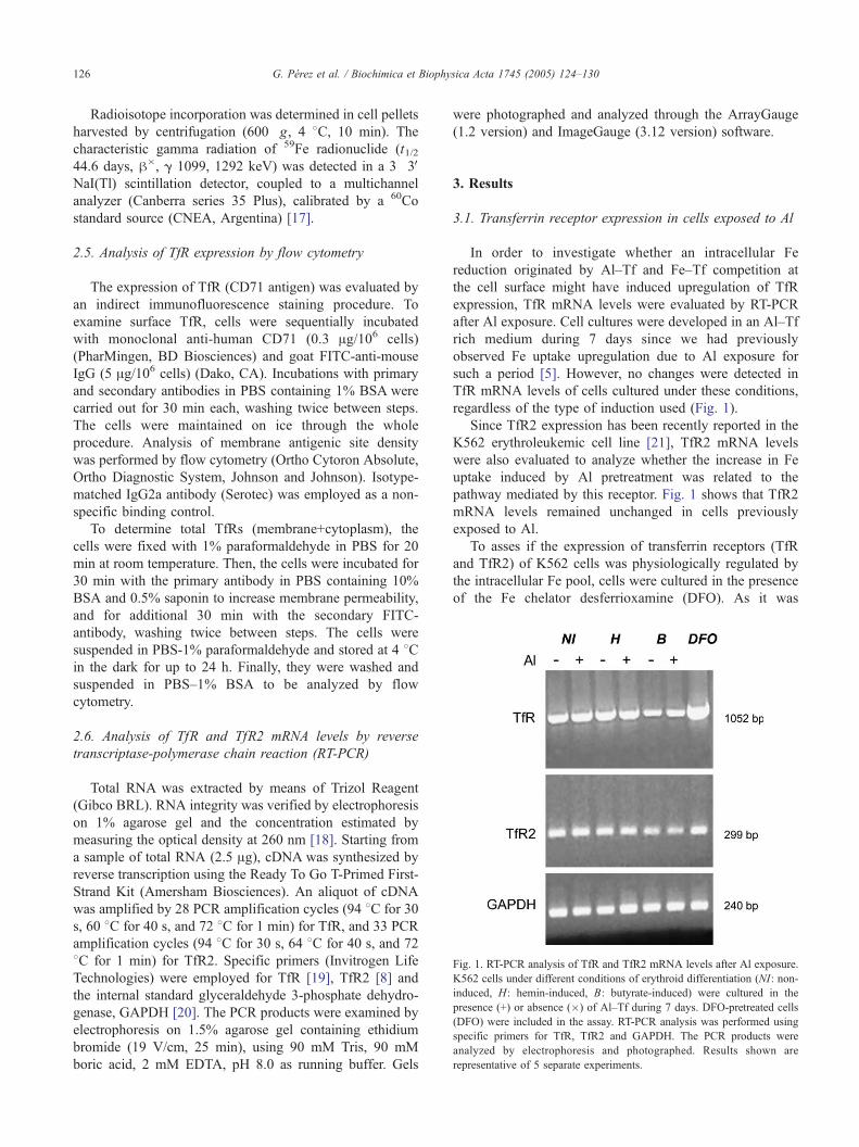
Fig. 1. RT-PCR analysis of TfR and TfR2 mRNA levels after Al exposure.
K562 cells under different conditions of erythroid differentiation (NI: non-
induced, H: hemin-induced, B: butyrate-induced) were cultured in the
presence (+) or absence (�) of Al–Tf during 7 days. DFO-pretreated cells
(DFO) were included in the assay. RT-PCR analysis was performed using
specific primers for TfR, TfR2 and GAPDH. The PCR products were
analyzed by electrophoresis and photographed. Results shown are
representative of 5 separate experiments.
G. Perez et al. / Biochimica et Biophysica Acta 1745 (2005) 124–130126
Radioisotope incorporation was determined in cell pellets
harvested by centrifugation (600�g, 4 8C, 10 min). The
characteristic gamma radiation of 59Fe radionuclide (t1/244.6 days, h�, g 1099, 1292 keV) was detected in a 3�3VNaI(Tl) scintillation detector, coupled to a multichannel
analyzer (Canberra series 35 Plus), calibrated by a 60Co
standard source (CNEA, Argentina) [17].
2.5. Analysis of TfR expression by flow cytometry
The expression of TfR (CD71 antigen) was evaluated by
an indirect immunofluorescence staining procedure. To
examine surface TfR, cells were sequentially incubated
with monoclonal anti-human CD71 (0.3 Ag/106 cells)
(PharMingen, BD Biosciences) and goat FITC-anti-mouse
IgG (5 Ag/106 cells) (Dako, CA). Incubations with primary
and secondary antibodies in PBS containing 1% BSA were
carried out for 30 min each, washing twice between steps.
The cells were maintained on ice through the whole
procedure. Analysis of membrane antigenic site density
was performed by flow cytometry (Ortho Cytoron Absolute,
Ortho Diagnostic System, Johnson and Johnson). Isotype-
matched IgG2a antibody (Serotec) was employed as a non-
specific binding control.
To determine total TfRs (membrane+cytoplasm), the
cells were fixed with 1% paraformaldehyde in PBS for 20
min at room temperature. Then, the cells were incubated for
30 min with the primary antibody in PBS containing 10%
BSA and 0.5% saponin to increase membrane permeability,
and for additional 30 min with the secondary FITC-
antibody, washing twice between steps. The cells were
suspended in PBS-1% paraformaldehyde and stored at 4 8Cin the dark for up to 24 h. Finally, they were washed and
suspended in PBS–1% BSA to be analyzed by flow
cytometry.
2.6. Analysis of TfR and TfR2 mRNA levels by reverse
transcriptase-polymerase chain reaction (RT-PCR)
Total RNA was extracted by means of Trizol Reagent
(Gibco BRL). RNA integrity was verified by electrophoresis
on 1% agarose gel and the concentration estimated by
measuring the optical density at 260 nm [18]. Starting from
a sample of total RNA (2.5 Ag), cDNA was synthesized by
reverse transcription using the Ready To Go T-Primed First-
Strand Kit (Amersham Biosciences). An aliquot of cDNA
was amplified by 28 PCR amplification cycles (94 8C for 30
s, 60 8C for 40 s, and 72 8C for 1 min) for TfR, and 33 PCR
amplification cycles (94 8C for 30 s, 64 8C for 40 s, and 72
8C for 1 min) for TfR2. Specific primers (Invitrogen Life
Technologies) were employed for TfR [19], TfR2 [8] and
the internal standard glyceraldehyde 3-phosphate dehydro-
genase, GAPDH [20]. The PCR products were examined by
electrophoresis on 1.5% agarose gel containing ethidium
bromide (19 V/cm, 25 min), using 90 mM Tris, 90 mM
boric acid, 2 mM EDTA, pH 8.0 as running buffer. Gels
were photographed and analyzed through the ArrayGauge
(1.2 version) and ImageGauge (3.12 version) software.
3. Results
3.1. Transferrin receptor expression in cells exposed to Al
In order to investigate whether an intracellular Fe
reduction originated by Al–Tf and Fe–Tf competition at
the cell surface might have induced upregulation of TfR
expression, TfR mRNA levels were evaluated by RT-PCR
after Al exposure. Cell cultures were developed in an Al–Tf
rich medium during 7 days since we had previously
observed Fe uptake upregulation due to Al exposure for
such a period [5]. However, no changes were detected in
TfR mRNA levels of cells cultured under these conditions,
regardless of the type of induction used (Fig. 1).
Since TfR2 expression has been recently reported in the
K562 erythroleukemic cell line [21], TfR2 mRNA levels
were also evaluated to analyze whether the increase in Fe
uptake induced by Al pretreatment was related to the
pathway mediated by this receptor. Fig. 1 shows that TfR2
mRNA levels remained unchanged in cells previously
exposed to Al.
To asses if the expression of transferrin receptors (TfR
and TfR2) of K562 cells was physiologically regulated by
the intracellular Fe pool, cells were cultured in the presence
of the Fe chelator desferrioxamine (DFO). As it was
Fig. 2. Flow cytometry analysis of surface TfR expression after Al
exposure. Non-induced (NI), hemin- (H) and butyrate-induced (B) cells
were cultured in the presence or absence of Al–Tf for 7 days (n=6). Flow
cytometry analysis was performed after consecutive incubations (0 8C, 30min, each) with monoclonal anti-human CD71 and FITC-anti-mouse IgG
antibodies. The amount of TfR surface antigenic sites is expressed as
arbitrary units of fluorescence. *Statistically significant differences between
B-induced cells and those under other treatments, irrespective of the Al
exposure (Pb0.01, Kruskal–Wallis test).
Fig. 3. Effect of Al exposure on Fe uptake. K562 cells non-induced or
induced to differentiate by hemin or sodium butyrate were cultured during 3
(Al 3d) or 7 days (Al 7d) in RPMI-FBS containing Al–Tf. Then, cells were
washed and suspended in RPMI-BSA. In order to measure Fe incorpo-
ration, a subsequent 59Fe pulse was carried out with (Panel B) or without
(Panel A) Tf, and radioiron activity was measured in the packaged pellets.
Results (meanFS.E.) are expressed as percentage of the 59Fe incorporated
to Al-pretreated cells (NI+Al, H+Al, B+Al) with respect to the activity
measured in the corresponding control grown in the absence of Al during
the whole procedure (NI, H, B) considered as 100%. Statistically significant
increase with respect to the control value, Wilcoxon signed rank test,
*Pb0.01, n=6; **Pb0.05, n=5.
Table 1
Effect of Al exposure upon the rate of Fe uptake
Rate of 59Fe uptake (ng/107 cells/h)
Basal Increment after Al exposure
NI Tf free 0.76F0.16 0.30F0.07 (z39%)
Tf present 1.31F0.17 0.47F0.11 (z36%)
H Tf free 0.59F0.09 0.21F0.04 (z36%)
Tf present 1.00F0.19 0.30F0.10 (z30%)
Data of 59Fe activity measured in cell pellets, corresponding to experiences
detailed in Fig. 3 (Panels A and B), were expressed as the mass of 59Fe
incorporated to 107 cells per hour. Cells induced to differentiate by hemin
(H) and non-induced cells (NI) were exposed to Al for 3 and 7 days,
respectively. Then, Fe incorporation was determined by incubation with
either 59Fe citrate (Tf free) or 59Fe–Tf (Tf present). The differences of Fe
uptake rate values between Al-pretreated cells and Al-unexposed cells
(Basal) are displayed (Increment after Al exposure).
G. Perez et al. / Biochimica et Biophysica Acta 1745 (2005) 124–130 127
expected, the upregulation of TfR mRNA under Fe
depletion condition confirmed that the physiological regu-
latory mechanism was intact. In turn, TfR2 mRNA levels
showed no response to DFO treatment (Fig. 1).
To further investigate whether Al effect was exerted at
the post-translational level, TfR expression was analyzed by
flow cytometry. Data described in Fig. 2 show no changes in
TfR (CD71) surface antigenic sites due to Al exposure,
despite the finding of significant differences related to the
differentiation induction. Besides, total TfRs (membrane
plus cytoplasm) were evaluated to investigate whether Al
could induce rearrangements in TfR cellular distribution. As
expected total TfR quantity was higher than membrane TfR
number. However, no changes were observed due to Al cell
exposure (data not shown).
3.2. Effect of Al exposure on NTBI uptake
The preceding experiments demonstrated that, under the
conditions assayed, the Al effect on Fe uptake cannot be
ascribed to a modified expression of Tf receptors. Therefore,
the study was focused on the NTBI transport system. Cells
induced to differentiation by hemin or sodium butyrate, as
well as non-induced cells, were cultured in RPMI-FBS in
the presence of Al–Tf. Incubations were carried out for
different periods (3 or 7 days), in which different behavior
Table 2
Rate of NTBI uptake—effect of Al or Fe excess
Rate of NTBI uptake (ng/107 cells/h)
Basal 0.43F0.03
Al excess 0.41F0.03
Fe excess 1.14F0.26*
K562 cells suspended in RPMI-BSA medium were incubated with 0.5 AM59Fe citrate for 2 h at 37 8C (Basal). During the radioisotope pulse either 20
AM Al citrate (Al excess) or 20 AM FeCl3 (Fe excess) was present. 59Fe
activity associated to cell pellets was measured and the rate of Fe uptake
was expressed as the mass of 59Fe incorporated to 107 cells per hour
(meanFS.E.).
* Statistically significant differences compared with Basal and Al
excess (Pb0.05, n=5, Kruskal–Wallis test).

G. Perez et al. / Biochimica et Biophysica Acta 1745 (2005) 124–130128
had been observed between non-induced and induced cells
[5]. Then, 59Fe uptake was measured in a fresh medium free
of Al and Tf (RPMI-BSA). Hemin-induced and non-
stimulated cells showed significantly higher 59Fe incorpo-
ration than did Al-unexposed cells after 3 and 7 days of Al
exposure, respectively. On the contrary, Al failed to increase
NTBI uptake in butyrate-induced cells (Fig. 3A).
These data were compared with the results obtained in
parallel assays in which the 59Fe uptake was measured in the
presence of Tf (Fig. 3B). With this purpose, 59Fe activity
corresponding to experiences detailed in Fig. 3 (Panels A
and B) was expressed in terms of mass of 59Fe incorporated
to 107 cells per hour (Table 1). Through this comparison, the
increase in 59Fe uptake rates induced by Al was evident both
in the presence or absence of Tf. As can be calculated from
these data (increment of 59Fe uptake rate in Tf free medium
relative to the increment in a Tf-containing medium), 64–
70% of the increase in Fe uptake rate observed in the
presence of Tf after Al exposure can be ascribed to positive
modulation of the alternative Fe transport pathway.
The next step was focused to elucidate whether Al might
be able to compete with Fe for the sites involved in NTBI
transport pathway. However, such effect was not observed
in assays carried out with the simultaneous presence of both
cations, even though Al concentration was 40 times higher
than that of the essential metal. Conversely, the excess of
unlabeled ferric ions during the 59Fe pulse significantly
increased the radioisotope uptake (Table 2). On the other
hand, the Fe depletion caused by DFO did not induce
significant modulation of the NTBI transport system. The59Fe incorporated into DFO-treated cells was 1780/1730–
2250 cpm vs. 2110/1610–2830 cpm into control cells
(median/range).
3.3. Al incorporation to cells
To analyze if Al is able to enter the cellular environment
by mechanisms not related to Tf, cells were cultured in a Tf-
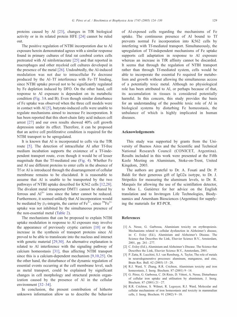
Fig. 4. Al incorporation to cells. K562 cells were cultured in a Tf-free
medium (RPMI-BSA) in the presence of Al citrate (Al). At the end of a 4-
day incubation period, the Al content was determined in cell lysates by
atomic absorption spectrometry. Two controls were simultaneously assayed:
cells grown without Al (C) and cells cultured in an Al–Tf containing
medium (Al–Tf). Results are expressed as meanFS.E. *Statistically
significant differences from both Al–Tf and C ( Pb0.05, n=4, Kruskal–
Wallis test).
free medium (RPMI-BSA) with the addition of Al citrate.
The amounts of Al detected in the lysates were significantly
higher than in control cells grown without Al (Fig. 4, Al vs.
C). Although Al entered the cell by a Tf independent
pathway, the contribution of this mechanism to total Al
incorporation was of lesser magnitude than that of the Tf-
mediated pathway (Fig. 4, Al vs. Al+Tf).
4. Discussion
This study provides information hitherto unknown on
the action of the non-essential metal Al upon mechanisms
of Fe homeostasis. Indeed, Al exposure proved to induce
the modulation of the non-Tf bound Fe uptake rather than
the classical Tf-dependent Fe transport.
Human K562 cells have been chosen for this inves-
tigation because they can mediate Tf-dependent [22] as well
as Tf-independent [11,12] Fe uptake. In order to employ
cellular models with different Fe requirements, cells were
either non-induced or induced to differentiation by hemin or
sodium butyrate, under which stimulus K562 cells showed
different kinetic of erythroid maturation [5].
We have previously demonstrated the interference of Tf-
bound Al with Fe uptake mediated by Tf receptors, which
could be explained by the similar affinity for Fe–Tf and Al–
Tf demonstrated for TfR in K562 cells [17]. This prevention
of normal Fe uptake caused by Al proved to be reversible.
Moreover, when Al was removed from the cell environment
an Fe uptake stimulation was revealed [5]. However, the
lack of changes in TfR mRNA levels despite previous Al
exposure (Fig. 1) simultaneously with no variations in CD-
71 antigenic sites (Fig. 2) strongly suggests that the
intracellular Fe deprivation caused by the competitive
interaction between Al–Tf and Fe–Tf was not enough to
trigger the upregulation of TfR expression through the IRE–
IRP interaction.
We then considered possible Al effects upon the
expression of the TfR2 receptor, also expressed in K562
cells [8,21]. However, no changes were detected in TfR2
mRNA levels (Fig. 1).
The most outstanding fact herein demonstrated is that
NTBI transport is positively modulated by Al exposure
(Fig. 3A). The similar response exhibited by Al-treated cells
irrespective of Tf availability (Fig. 3A and B) strongly
suggests that, in the presence of Tf, Fe should have been
simultaneously incorporated both through Tf-dependent and
Tf-independent pathways. Whereas 59Fe–Tf is transported
into cells via the TfR mediated route, low molecular weight59Fe complexes (e.g. ferric citrate) would be incorporated
through alternative pathways. Therefore, NTBI uptake
would be responsible for the increase in Fe acquisition. In
terms of Fe uptake rate, 64–70% of the increment observed
in the presence of Tf could be ascribed to the increase in Fe
acquisition through Tf-independent routes (Table 1). Based
on previous reports showing structural alterations of certain
G. Perez et al. / Biochimica et Biophysica Acta 1745 (2005) 124–130 129
proteins caused by Al [23], changes in TfR biological
activity or in its related protein HFE [24] cannot be ruled
out.
The positive regulation of NTBI incorporation due to Al
exposure herein demonstrated agrees with a similar response
found in primary cultures of fetal rat cerebral cortex cells
pretreated with Al nitrilotriacetate [25] and that reported in
macrophages and other myeloid cell cultures developed in
the presence of the metal [26]. Undoubtedly, the Al-induced
modulation was not due to intracellular Fe decrease
produced by the Al–Tf interference with Fe–Tf binding,
since NTBI uptake proved not to be significantly regulated
by Fe depletion induced by DFO. On the other hand, cell
response to Al exposure is dependent on its metabolic
condition (Fig. 3A and B). Even though similar disturbance
of Fe uptake was observed when the three cell models were
in contact with Al [5], butyrate-induced cells were unable to
regulate mechanisms aimed to increase Fe incorporation. It
has been reported that this short-chain fatty acid induces cell
arrest [27] and our own results showed 40% cell growth
depression under its effect. Therefore, it can be proposed
that an active cell proliferative condition is required for the
NTBI transport to be upregulated.
It is known that Al is incorporated to cells via the TfR
route [5]. The detection of intracellular Al after Tf-free
medium incubation supports the existence of a Tf-inde-
pendent transport route, even though it would be of lesser
magnitude than the Tf-mediated one (Fig. 4). Whether Fe
and Al use different proteins to enter cells in the absence of
Tf or Al is introduced through the disarrangement of cellular
membrane remains to be elucidated. It is reasonable to
assume that Al is unable to be transported by the two
pathways of NTBI uptake described for K562 cells [12,28].
The divalent metal transporter DMT1 cannot be shared by
ferrous and Al3+ ions since the latter cannot be reduced.
Furthermore, it seemed unlikely that Al incorporation would
be mediated by h3-integrin, the carrier of Fe3+, since 59Fe3+
uptake was not inhibited by the simultaneous presence of
the non-essential metal (Table 2).
The mechanisms that can be proposed to explain NTBI
uptake modulation in response to Al exposure may involve
the appearance of previously cryptic carriers [10] or the
increase in the synthesis of transport proteins since Al
proved to be able to translocate into the nucleus and interact
with genetic material [29,30]. An alternative explanation is
related to Al interference with the signaling pathway of
calcium homeostasis [31], thus affecting NTBI transport
since this is a calcium-dependent mechanism [9,10,25]. On
the other hand, the disturbance of the dynamic regulation of
essential events occurring at the cell membrane level, such
as metal transport, could be explained by significant
changes in cell morphology and structural protein organ-
ization caused by the presence of Al in the cellular
environment [32–34].
In conclusion, the present contribution of hitherto
unknown information allow us to describe the behavior
of Al-exposed cells regarding the mechanisms of Fe
uptake. The continuous presence of Al bound to Tf
prevents normal Fe incorporation into K562 cells by
interfering with Tf-mediated transport. Simultaneously, the
upregulation of Tf-independent mechanisms of Fe uptake
supports cell adaptation in response to Al exposure
whereas an increase in TfR affinity cannot be discarded.
It seems that through the regulation of NTBI transport
rather than through Tf-mediated system, cells would be
able to incorporate the essential Fe required for metabo-
lism and growth without allowing the simultaneous access
of a potentially toxic metal. Although no physiological
role has been attributed to Al, or perhaps because of that,
its accumulation in tissues is considered potentially
harmful. In this concern, this study provides the basis
for an understanding of the possible toxic role of Al in
biological systems by disturbing Fe homeostasis, the
unbalance of which is highly implicated in human
diseases.
Acknowledgements
This study was supported by grants from the Uni-
versity of Buenos Aires and the Scientific and Technical
National Research Council (CONICET, Argentina).
Results included in this work were presented at the Fifth
Keele Meeting on Aluminium, Stoke-on-Trent, United
Kingdom, 2003.
The authors are grateful to Dr. A. Fosati and Dr. P.
Baldi for their generous gift of IgG2a isotype, to Dr. J.
Stripeikis for measuring the aluminum levels, to Dr. R.
Marques for allowing the use of the scintillation detector,
to Miss L. Gutierrez for her advice on the English
translation and to Invitrogen Life Technologies, Biody-
namics and Amersham Biosciences (Argentina) for supply-
ing the materials for RT-PCR.
References
[1] A. Nesse, G. Garbossa, Aluminium toxicity on erythropoiesis.
Mechanisms related to cellular dysfunction in Alzheimer’s disease,
in: C. Exley (Ed.), Aluminium and Alzheimer’s Disease. The
Science that Describes the Link, Elsevier Science B.V., Amsterdam,
2001, pp. 261–277.
[2] C. Exley (Ed.), Aluminium and Alzheimer’s Disease. The Science that
Describes the Link, Elsevier Science B.V., Amsterdam, 2001.
[3] P. Zatta, R. Lucchini, S.J. van Rensburg, A. Taylor, The role of metals
in neurodegenerative processes: aluminum, manganese, and zinc,
Brain Res. Bull. 62 (2003) 15–28.
[4] R.J. Ward, Y. Zhang, R.R. Crichton, Aluminium toxicity and iron
homeostasis, J. Inorg. Biochem. 87 (2001) 9–14.
[5] G. Perez, G. Garbossa, C. Di Risio, D. Vittori, A. Nesse, Disturbance
of cellular iron uptake and utilisation by aluminium, J. Inorg.
Biochem. 87 (2001) 21–27.
[6] R.R. Crichton, S. Wilmet, R. Legssyer, R.J. Ward, Molecular and
cellular mechanisms of iron homeostasis and toxicity in mammalian
cells, J. Inorg. Biochem. 91 (2002) 9–18.

G. Perez et al. / Biochimica et Biophysica Acta 1745 (2005) 124–130130
[7] P. Aisen, M. Wessling-Resnick, E.A. Leibold, Iron metabolism, Curr.
Opin. Chem. Biol. 3 (1999) 200–206.
[8] H. Kawabata, R. Yang, T. Hirama, P.T. Vuong, S. Kawano, A.F.
Gombart, H.P. Koeffler, Molecular cloning of transferrin receptor 2, J.
Biol. Chem. 274 (1999) 20826–20832.
[9] A. Sturrock, J. Alexander, J. Lamb, C.M. Craven, J. Kaplan,
Characterization of a transferrin-independent uptake system for iron
in HeLa cells, J. Biol. Chem. 265 (1990) 3139–3145.
[10] J. Kaplan, I. Jordan, A. Sturrock, Regulation of the transferrin-
independent iron transport system in cultured cells, J. Biol. Chem. 266
(1991) 2997–3004.
[11] R.S. Inman, M. Wessling-Resnick, Characterization of transferrin-
independent iron transport in K562 cells, J. Biol. Chem. 268 (1993)
8521–8528.
[12] M.E. Conrad, J.N. Umbreit, E.G.Moore, C. Uzel,M.R. Berry, Alternate
iron transport pathway, J. Biol. Chem. 269 (1994) 7169–7173.
[13] F. Canonne-Hergaux, A.-S. Zhang, P. Ponka, P. Gros, Character-
ization of the iron transporter DMT1 (NRAMP2/DCT1) in red
blood cells of normal and anemic mk/mk mice, Blood 98 (2001)
3823–3830.
[14] D.A. Atwood, B.C. Yearwood, The future of aluminum chemistry, J.
Organomet. Chem. 600 (2000) 186–197.
[15] G. Garbossa, A. Gutnisky, A. Nesse, Depressed erythroid progenitor
cell activity in aluminum-overloaded mice, Miner. Electrolyte Metab.
22 (1996) 214–218.
[16] D. Vittori, A. Nesse, G. Perez, G. Garbossa, Morphologic and
functional alterations of erythroid cells induced by long term ingestion
of aluminium, J. Inorg. Biochem. 76 (1999) 113–120.
[17] G. Perez, G. Garbossa, B. Sassetti, C. Di Risio, A. Nesse, Interference
of aluminium on iron metabolism in erythroleukemia K562 cells, J.
Inorg. Biochem. 76 (1999) 105–112.
[18] J. Sambrook, D. Russell, Molecular Cloning: A Laboratory Manual,
Cold Spring Harbor Laboratory Press, New York, 2001.
[19] Z. Tsuchihashi, S.L. Hansen, L. Quintana, G.S. Kronmal, F.A. Mapa,
J.N. Feder, R.K. Wolff, Transferrin receptor mutation analysis in
hereditary hemochromatosis patients, Blood Cells Mol. Diseases 24
(1998) 317–321.
[20] M. McKinney, M. Robbins, Chronic atropine administration up-
regulates rat cortical muscarinic m1 receptor mRNA molecules:
assessment with the RT/PCR, Brain Res. Mol. Brain Res. 12 (1992)
39–45.
[21] H. Kawabata, T. Nakamaki, P. Ikonomi, R.D. Smith, R.S. Germain,
H.P. Koeffler, Expression of transferrin receptor 2 in normal and
neoplastic hematopoietic cells, Blood 98 (2001) 2714–2719.
[22] R.D. Klausner, J. Van Renswoude, G. Ashwell, C. Kempf, A.N.
Schechter, A. Dean, K.R. Bridges, Receptor-mediated endocytosis of
transferrin in K562 cells, J. Biol. Chem. 258 (1983) 4715–4724.
[23] T. Kiss, M. Hollosi, The interaction of aluminum with peptides and
proteins, in: C. Exley (Ed.), Aluminium and Alzheimer’s Disease. The
Science that Describes the Link, Elsevier Science B.V., Amsterdam,
2001, pp. 361–392.
[24] C.N. Roy, D.M. Penny, J.N. Feder, C.A. Enns, The hereditary
hemochromatosis protein, HFE, specifically regulates transferrin-
mediated iron uptake, J. Biol. Chem. 274 (1999) 9022–9028.
[25] S. Oshiro, M. Kawahara, S. Mika, K. Muramoto, K. Kobayashi, R.
Ishige, K. Nozawa, M. Hori, C. Yung, S. Kitajima, Y. Kuroda,
Aluminum taken up by transferrin-independent iron uptake affects the
iron metabolism in rat cortical cells, J. Biochem. 123 (1998) 42–46.
[26] O. Olakanmi, J.B. Stokes, S. Pathan, B.E. Britigan, Polyvalent
cationic metals induce the rate of transferrin-independent iron
acquisition by HL-60 cells, J. Biol. Chem. 272 (1997) 2599–2606.
[27] B. Chenais, I. Molle, C. Trentesaux, P. Ieannesson, Time-course of
butyric acid-induced differentiation in human K562 leukemic cell line:
rapid increase in g-globin, porphobilinogen deaminase and NF-E2
mRNA levels, Leukemia 11 (1997) 1575–1579.
[28] M.E. Conrad, J.N. Umbreit, E.G. Moore, L.N. Hainsworth, M.
Porubcin, M.J. Simovich, M.T. Nakada, K. Dolan, M.D. Garrick,
Separate pathways for cellular uptake of ferric and ferrous iron, Am. J.
Physiol.: Gastrointest. Liver Physiol. 279 (2000) G767–G774.
[29] P.R. Walker, J. LeBlanc, M. Sikorska, Effects of aluminum and other
cations on the structure of brain and liver chromatin, Biochemistry 28
(1989) 3911–3915.
[30] R.Y. Zhang, Y. Liu, D.W. Pang, R.X. Cai, Y.P. Qi, Spectroscopic and
voltammetric study on the binding of aluminium(III) to DNA, Anal.
Sci. 18 (2002) 761–766.
[31] W.R. Mundy, T.J. Shafer, Aluminum-induced alteration of phosphoi-
nositide and calcium signaling, in: C. Exley (Ed.), Aluminium and
Alzheimer’s Disease. The Science that Describes the Link, Elsevier
Science B.V., Amsterdam, 2001, pp. 345–360.
[32] D. Vittori, G. Garbossa, C. Lafourcade, G. Perez, A. Nesse, Human
erythroid cells are affected by aluminium. Alteration of membrane
band 3 protein, Biochim. Biophys. Acta 1558 (2002) 142–150.
[33] M. Suwalsky, B. Ungerer, F. Villena, B. Norris, H. Cardenas, P. Zatta,
Effects of AlCl3 on toad skin, human erythrocyte, and model cell
membranes, Brain Res. Bull. 55 (2001) 205–210.
[34] S.J. Van Rensburg, M.E. Carstens, F.C. Potocnik, A.K. Aucamp, J.J.
Taljaard, K.R. Koch, Membrane fluidity of platelets and erythrocytes
in patients with Alzheimer’s disease and the effect of small amounts of
aluminium on platelets and erythrocyte membranes, Neurochem. Res.
17 (1992) 825–829.