8. Abdominal injuries
Abdominal injuries often co-exist with chest and pelvic injuries.
More children in Yorkshire & Humber suffer blunt force trauma through motor vehicle collisions,
falls and assaults, than penetrating trauma. The management guidance differs between blunt and
penetrating mechanisms of injury, so these will be considered separately.
Blunt Injury
Clinical assessment
The patient will be assessed by the trauma team in line with Trauma Management principles.
Abdominal examination should be included within “C” as a potential site of bleeding. Patients
in shock and suspected to have intra-abdominal injury (including at time of pre-alert) need
immediate transfer to the Paediatric MTC. This should be ED to ED and does not need
discussion with specialities within the MTC as automatic acceptance is Network standard.
Any patients not meeting criteria for immediate transfer should be discussed early with:
o MTC: the on call Paediatric Surgical Consultant or Middle Grade. Alert the interventional
radiologist on-call where appropriate.
o TU: the on call General Surgical Consultant.
Ensure O Negative blood will be available and warn that the Massive Haemorrhage Protocol
may be activated (Section 5).
Inspection: Abdominal wall bruising is highly indicative of intra-abdominal injury. This is
infrequently associated with abdominal distension. Swallowed air is the most common cause of
distension - insert a gastric tube. New and progressive abdominal distension in a shocked
patient suggests exsanguinating intra-abdominal haemorrhage.
Palpation: Tenderness on examination should prompt further investigation but examination in a
distressed child is challenging and may be compromised by other distracting injuries or reduced
level of consciousness. Absence of clinical signs does not exclude injury.
Percussion and auscultation: Add little to the examination. The presence or absence of bowel
sounds has no diagnostic value.
Repeated clinical assessment is valuable
Investigation
Bloods: FBC, U&E, clotting, venous gas and cross-match (with activation of Massive
Haemorrhage Protocol if appropriate) should be taken for all significantly injured patients.
Consider a pregnancy test, if relevant.
Ultrasound: In the acute paediatric trauma setting there is no role for ultrasound outside of
assisting in interventional procedures.
CT scan: Contrast-enhanced CT is the modality of choice for the assessment of acute traumatic
intra-abdominal injury. Where there is concern for significant intra-abdominal injury, all
patients should undergo a CT scan using appropriate paediatric imaging protocols (Section 17)
unless there is rapid haemodynamic deterioration that requires immediate transfer to theatre.
CT is best performed at the Major Trauma Centre (MTC), however for some less severe injuries
the CT may be performed at the Trauma Unit. The findings will need to be discussed with the
Paediatric Surgical Consultant at the MTC.
Management (see Appendix 4a)
The guidance below covers expected management at the Major Trauma Centre. At a Trauma Unit
management may be limited by the available resources. When the treatment necessary exceeds
the TUs capabilities the patient will require transfer to the MTC. The MTC can be contacted for
advice at any time.
The management of patients with unresponsive or transiently responding shock/hypotension is
challenging. Early consideration must be given to blood transfusion in line with the Massive
Haemorrhage Protocol. Any patient considered to have significant on going intra-abdominal
bleeding requires rapid transfer to theatre for resuscitation and potential damage control
surgery - laparotomy, pelvic stabilization, thoracotomy etc.
Patients whose shock is not rapidly deteriorating should have a trauma or targeted CT scan in
line with the Y&H guidance on imaging in paediatric trauma.
Patients with radiological evidence of ongoing bleeding from solid organs (spleen, kidney, liver)
must be discussed with the Consultant Paediatric Surgeon, Consultant Paediatric Radiologist/

Interventional Radiologist, Consultant Paediatric Intensivist and Consultant Paediatric
Anaesthetist to decide the optimal method and location of haemorrhage control.
Patients with radiological evidence of pseudoaneurysm rather than free, active bleeding from
the spleen, liver or kidney must be discussed with the Consultant Paediatric Surgeon and
Consultant Paediatric Radiologist/ Interventional Radiologist with a view to angio-embolisation.
This may require Vascular Intervention in Leeds.
Patients with solid organ (spleen, kidney, liver) injury but no evidence of ongoing bleeding or
pseudoaneurysm must be discussed with the Consultant Paediatric Surgeon. Non-operative
management is superior in such cases. This should only be undertaken in a specialist paediatric
high dependency setting, with appropriate staff and equipment should there be deterioration. It
is appropriate to transfer these patients early to the MTC, rather than transfer on deterioration.
The patient must be adequately resuscitated to correct hypoperfusion. In a minority of patients
due to the increase in perfusion pressure, bleeding may recur.
During non-operative treatment regular clinical examinations and hemoglobin measurements
must be undertaken. If re-bleeding is suspected (progressive shock and / or falling hemoglobin)
transfer to theatre or further CT angiography is required. If confirmed, then angio-embolisation
or operative control of bleeding is required. Increasing abdominal pain, tenderness,
inflammatory markers or deranged liver function tests may be the result of a missed hollow
viscus injury, pancreatic injury or a local complication of solid organ injury e.g. biliary peritonitis.
Mesenteric bleeding can lead to slowly developing local intestinal ischaemia and delayed
intestinal perforation as well as the risk of ongoing haemorrhage. Further CT imaging is
indicated to attempt to identify the underlying problem.
Patients with Grade IV or more splenic or hepatic injuries undergoing non-operative
management should be considered for angiography as a proportion will reveal significant
vascular injury which if treated should reduce the risk of re-bleeding. This may require Vascular
Intervention in Leeds. For more detail on the solid organ injury grading system see
https://www.wymtn.com/uploads/5/1/8/9/51899421/abdominal_trauma_-_paediatrics.pdf (Appx 1-3).
Patients with evidence of hollow viscus injury, mesenteric injury or diaphragmatic injury on the
initial CT will almost certainly require laparotomy and should be discussed with the Consultant
Paediatric Surgeon.

The Embrace conferencing system allows TU and MTC to talk directly to each other and can
facilitate discussion between multiple clinicians. Embrace www.embrace.sch.nhs.uk can also
give advice on transfers if needed. For immediate transfer procedure see here.
Penetrating Injury
Background
Paediatric penetrating injuries are very uncommon. Within the trauma network, gunshot
wounds are very rare but stabbing and impalements do occur. The mechanism of wounding
needs to be established as it strongly influences management decisions. Adult patients
suffering stab injury are less likely to require laparotomy (25-33%) than those suffering gunshot
injury (80-95%). Note, 55-60% of patients with any stab wound that has entered the peritoneum
have hypovolemic shock, peritonitis or bowel / omental evisceration and require a laparotomy.
In the remainder, 50% will eventually require operation if observed. Most patients with
abdominal gunshot wounds have significant intraperitoneal injury and therefore justify
laparotomy.
Clinicians have a responsibility to inform the police if a patient attends the Emergency
Department with a knife or gunshot wound after an assault but demographic information
should, in the first instance, only be shared with the patient’s consent. Reporting is the
responsibility of the ED consultant in charge. Further information can be found at
https://www.gmc-uk.org/ethical-guidance/ethical-guidance-for-doctors/confidentiality---
reporting-gunshot-and-knife-wounds
Clinical assessment
The patient must be assessed by the trauma team in line with Trauma Management Principles.
Abdominal examination should be included within “C” as a potential site of bleeding. As with
blunt injury, patients in shock with penetrating chest and / or abdominal injury need immediate
transfer to the Paediatric MTC. This should be ED to ED and does not need discussion with
specialities within the MTC as automatic acceptance is Network standard.
Any patients not meeting criteria for immediate transfer should be discussed early with:
o MTC: the on call Paediatric Surgical Consultant or Middle Grade. Alert the interventional
radiologist on-call where appropriate.
o TU: the on call General Surgical Consultant.

Ensure O Negative blood will be available and warn that the Massive Haemorrhage Protocol
may be activated.
Inspection: Do not exclude significant injury on the basis of perceived depth or direction injury
from the entry point of the wound; few patients are in the anatomical position at the time of
injury. Unless the patient requires an emergency department thoracotomy, the patient must be
log rolled to identify all injuries. Particular care should be taken to inspect the axillae and
perineum as wounds in these sites can be missed. Skin wounds should be marked with radio
opaque markers e.g. closed paper clip taped to anterior wounds and opened paper clip to
posterior wounds. Never remove protruding weapon or foreign body. Abdominal distension
may be a sign of significant intra-abdominal bleeding, but a significant volume of blood can
collect without undue distension.
Palpation: Tenderness around the wound is to be expected but progressive pain and tenderness
remote from the initial wound suggests intra peritoneal hollow viscus injury. As with blunt
injury, the reliability of clinical examination will be reduced when there are remote but
distracting injuries or reduced consciousness (head injury, intoxication, sedating medication,
spinal cord injury).
Percussion and auscultation: Add little to the examination. The presence or absence of bowel
sounds has no diagnostic value.
Investigation
Bloods: FBC, U&E, clotting, venous gas and cross-match (with activation of Massive
Haemorrhage Protocol if appropriate) should be taken for all significantly injured patients.
Consider a pregnancy test, if relevant.
Ultrasound: FAST scan has no role in the exclusion of hollow viscus injury.
CT scan: discussed in the management section below.
Management of penetrating injuries.
The guidance below covers expected management at the Major Trauma Centre. At a Trauma Unit
management may be limited by the available resources. When the treatment necessary exceeds

the TUs capabilities the patient will require transfer to the MTC. The MTC can be contacted for
advice at any time.
Management of stab wounds (see Appendix 4b)
For patients with penetrating injury, balanced resuscitation should be utilized unless
contraindicated (traumatic brain injury).
The management of patients with unresponsive or transiently responding shock/hypotension is
challenging. Early consideration must be given to blood transfusion in the Massive
Haemorrhage Protocol. Any patient considered to have significant ongoing intra-abdominal
bleeding requires rapid transfer to theatre for resuscitation and potential damage control
surgery - laparotomy, pelvic stabilization, thoracotomy etc.
Other causes of shock need to be considered e.g. bleeding (chest, limbs, bleeding from
wounds), tension pneumothorax and cardiac tamponade. Clearly, patients with multiple
wounds can have life threatening pathology in more than one body cavity.
Patients with foreign bodies (eg. knives) protruding from the abdomen require these to be
removed in the operating theatre with the abdomen open if there is any concern that they may
have entered the peritoneum. Preoperative CT scan is likely to be degraded by artefact but may
be considered if findings would influence surgical approach.
Patients without overt shock but with clinical signs of peritonitis or bowel / omental
evisceration require a laparotomy (bowel evisceration is associated with a 75% risk of bowel
perforation). A preoperative CT scan may be undertaken but the trauma scan is poor at
detecting fresh hollow organ injury.
Patients without overt shock but with an unreliable examination e.g. brain injury, spinal cord
injury, intoxication or sedating medication, should have further investigation with a CT scan or
undergo exploratory laparotomy / laparoscopy.
Patients who are conscious, cooperative and can concentrate and with no signs of peritonitis or
diffuse abdominal tenderness (away from the wounding site) may be initially managed non-
operatively. A CT scan should be performed to help quantify the depth of injury. Repeated /
serial examination preferably by the same experienced surgeon should be undertaken. At hand
over, ideally both surgeons should examine the patient together and agree on the clinical
findings. Any injury is likely to reveal itself within 24 hours or so after this time.

Stab wounds can be classified as anterior (between the anterior axillary lines), flank (between
anterior and posterior axillary lines) and posterior (posterior to posterior axillary line). In
general, one third of anterior wounds do not penetrate the peritoneum. One third penetrate
the peritoneum but no not require intervention, and the remaining third penetrate the
peritoneum and require surgical repair. Anterior abdominal wounds may be explored under
local anaesthetic within the emergency department if the child is older and compliant or under
a general anaesthetic in theatre. If the wound extends deep to the anterior fascia then the
chance of intraperitoneal hollow viscus perforation is increased although not definite. Patients
with posterior fascial penetration proceed to theatre to laparotomy / laparoscopy. Hollow
viscus injury can be difficult to detect even at laparotomy. Exclusion of visceral injury by
laparoscopy should only be performed by those with significant experience in such cases.
Exploration of flank and posterior wounds is rarely indicated. In the absence of a need for
immediate laparotomy (shock or generalized peritonitis), a CT scan helps to determine depth of
injury.
Thoraco-abdominal injuries can present a diagnostic dilemma as penetrating wounds between
the nipples and costal margin may damage structures within the chest cavity, within the
peritoneal cavity and make a hole in the intervening diaphragm.
o Patients with unresponsive or transiently responding shock and considered to have
ongoing abdominal or thoracic bleeding require rapid chest drain insertion and transfer
to theatre for laparotomy and any other surgery required to control bleeding.
o For patients without overt shock, a CT scan will give some indication of the trajectory of
the wound although may not detect incised wounds of the diaphragm. If concern
regarding diaphragmatic injury persists, then a laparoscopy/laparotomy should be
performed. If an injury is detected, then the defect should be repaired, and visceral
injury excluded. Both diaphragmatic repair and exclusion of visceral injury are possible
laparoscopically but only by those with appropriate skills and experience.
Management of low velocity (hand gun / shotgun) gunshot wounds (see Appendix 4c)
These are extremely rare in the paediatric age range and management should follow adult
guidelines.

Patients with abdominal gunshot wounds have a very high chance of intraperitoneal injury and
must undergo laparotomy to exclude injury rather than define it. Importantly projectiles may
move in non-linear planes and ricochet. Few patients are shot in the anatomical position.
Patients with unresponsive or transiently responding shock require immediate laparotomy.
Those without overt shock may undergo a CT scan to guide surgical planning and identify those
few patients with tangential injuries. Close range shot gun injuries are locally destructive and
likely to penetrate the peritoneum mandating laparotomy. For those delivered at distance, CT
scanning may demonstrate pellet penetration deep to peritoneum although scatter may limit
image quality.
Management of high velocity and ballistic injuries
The experiences from Manchester and London highlight the need for consideration of
management of high velocity and ballistic injuries. There is very little civilian experience in such
management and expert advice is best sought on the management of such patients. Key
learning points from the Manchester are
1. The importance of CT scanning to identify shrapnel injuries
2. The importance of considering the need for prophylaxis for possible blood borne
infection (see latest Public Health England and NHS England guidance)
3. In the event of a Mass Casualty Incident, different rules may apply, and staff in all
hospitals receiving paediatric major trauma patients should be familiar with their own
Major Incident Policy.
Venous Thromboembolic (VTE) prophylaxis in patients with abdominal injury
Mechanical prophylaxis eg. TED stockings can be used for all patients where an appropriate size
exists, unless precluded by lower limb injury.
Pharmacological prophylaxis with LMWH should be commenced when the risk of further
bleeding becomes less than the risk of VTE - usually at 18:00 following the day of surgery and if
there is no coagulopathy (normal INR and APTT).

Appendix 4a - Management algorithm for blunt abdominal injury
Appendix 4 a - KEY
A. Abdominal examination should be included within assessment of “C” as a potential source of
bleeding
B. Senior decision makers (Consultant Paediatric Surgeon or equivalent in TU) / Consultant in
Emergency Medicine/Consultant Paediatric/Interventional Radiologist) to assess and decide if
patient’s hemodynamic status is deteriorating too fast to proceed to CT.
C. Unresponsive or transiently responsive shock is usually due to bleeding. Potential sites (chest,
abdomen, pelvis, limbs and external loss) of bleeding should be evaluated. Obstructive /
mechanical causes of shock (tension pneumothorax and cardiac tamponade) should also be
considered. Rarer causes of shock include myocardial contusion, neurogenic shock, myocardial
infarction and air embolus. Non-abdominal sources of shock will need intervention in parallel
with intra-abdominal assessment and intervention e.g. chest drain, pelvic binder, wound
compression etc.
D. If bleeding or “blush” reported on CT scan a discussion between paediatric surgical team and
radiological team is required to clarify precise nature of abnormality detected. Evidence of
bleeding in to peritoneal cavity will almost certainly require intervention. Contained blush
within a solid organ may not. If evidence of active bleeding and hemodynamic deterioration,
requires discussion between Consultant Paediatric Surgeon (or equivalent in TU) and
Paediatric/Interventional Radiologist to determine suitability for embolisation or laparotomy.
Factors to consider include rate of hemodynamic deterioration, constellation of injuries and
physiological reserve. If embolization felt to be appropriate this may necessitate transfer to
Leeds.
E. Patients undergoing a trial of non-operative management require regular clinical assessment
and hemoglobin measurements ideally initially within a critical care environment. Evidence of
hemodynamic deterioration, falling hemoglobin, coagulopathy, increasing abdominal pain or
tenderness or rising inflammatory markers requires discussion with the Consultant Paediatric
Surgeon. Depending on the rate of deterioration and clinical suspicion, the patient should
undergo CT imaging or more rarely emergency transfer to theatre. The CT scan may reveal re-
bleeding, missed hollow viscus injury, pancreatic injury or complication of known solid organ
injury. Further bleeding may be treated with embolization or surgery determined by
hemodynamic deterioration, constellation of injuries and physiological reserve. Missed injuries
or complications may require a combination of radiological or surgical intervention depending
on the exact diagnosis.

Appendix 4b - Management algorithm for penetrating stab injury
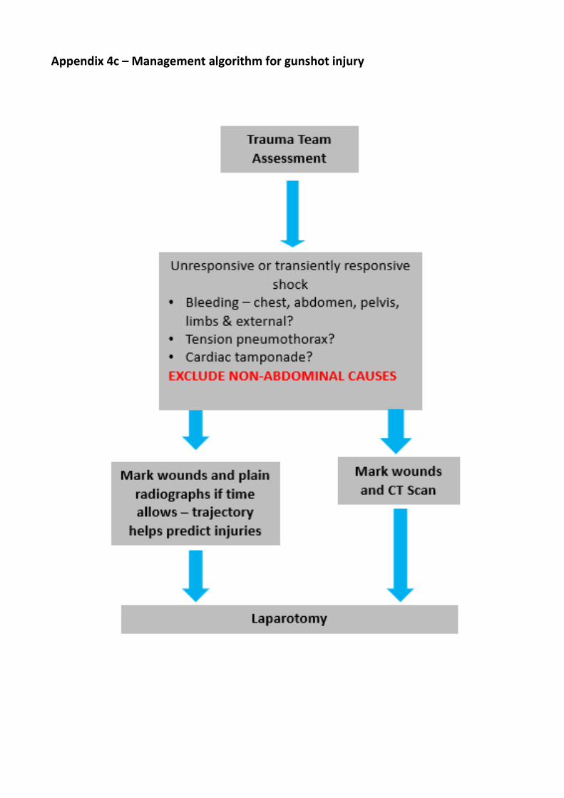
Appendix 4c – Management algorithm for gunshot injury


![Laparoscopy in Abdominal Trauma - Springer · dard of care in blunt abdominal solid organ injuries [37, 38], its implementation in penetrating abdominal trauma has been considerably](https://static.cupdf.com/doc/110x72/5ca43df888c99355658be93f/laparoscopy-in-abdominal-trauma-springer-dard-of-care-in-blunt-abdominal-solid.jpg)