79 YEAR OLD WOMAN
PRESENTS WITH BILATERAL
HAND AND ARM NUMBNESS /
DECREASED COORDINATION
Emily Arciero, MS4
Michael Baldwin, MD

Clinical History
• 79 year old female with history significant for L1-L5 spinal fusion years prior, recently diagnosed with L5-S1 spondylolisthesis– Other PMHx- HTN, HLD, T2DM, PAD, depression
• Patient had been admitted to hospital for syncopal episode (May 2020)– Head CT without contrast showed severe central spinal
stenosis/cord compression at atlantoaxial joint due to possible soft tissue mass
– CT of cervical spine without contrast performed, corroborated initial head CT findings
• Subsequently referred to outpatient orthopaedic spine clinic, had first visit in June 2020

Presenting Symptoms
• Initial visit to orthopaedic spine clinic:– Patient endorsed a 2 month history of bilateral
hand numbness with loss of coordination • Numbness in hands radiating to mid-forearm
– Loss of balance, felt as though she was “drunk” when trying to walk
– No change in bowel/bladder function
– Symptoms progressively worsening
– Patient reported being in car accident about 2 months ago, started noticing symptoms after the event

Extra Information
• Physical exam– First visit (June 2020)
• Significant ataxic gait
• 3 beats of clonus bilaterally
• Hoffmann negative bilaterally
• Strength 5/5, reflexes 2+ bilaterally in upper and lower extremities
– Second visit (July 2020)• Hyperreflexia of bilateral upper extremities
• Inversion of brachioradialis reflex
• Labs – Rheumatoid factor negative in 2018
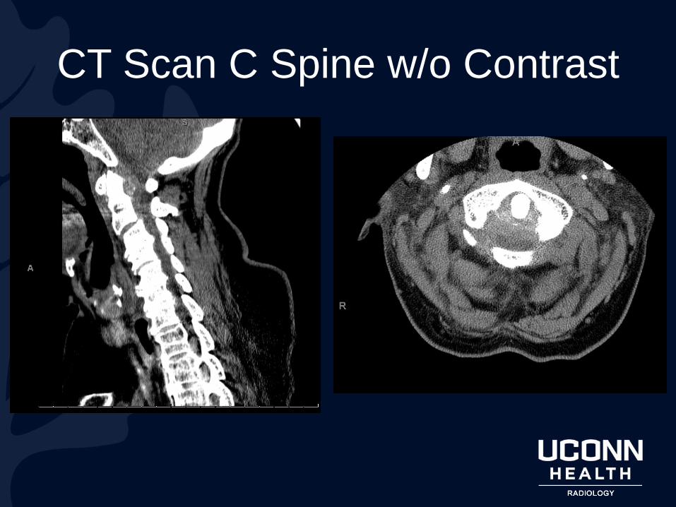
CT Scan C Spine w/o Contrast

SagittalAxial
Punctate
hyperdensities
within mass
Border of
heterogeneous
mass of soft
tissue density
Spinal cord compression
Retro-odontoid mass

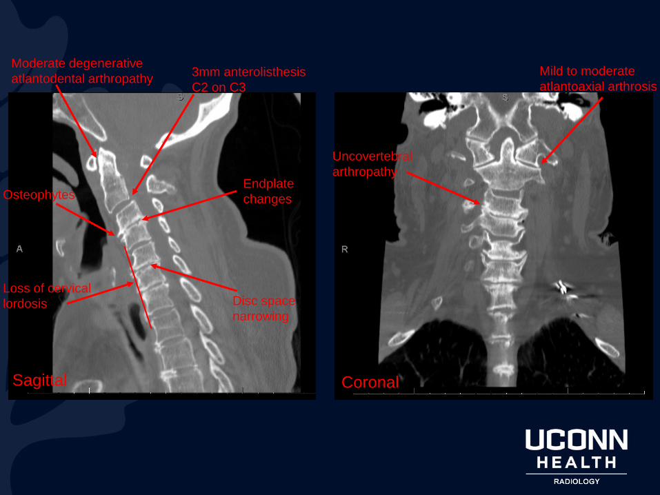
Sagittal Coronal
Moderate degenerative
atlantodental arthropathy3mm anterolisthesis
C2 on C3
Endplate
changes
Disc space
narrowing
Loss of cervical
lordosis
Osteophytes
Uncovertebral
arthropathy
Mild to moderate
atlantoaxial arthrosis

MRI C Spine w/o Contrast
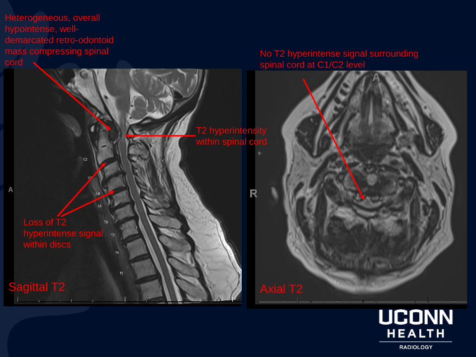
Heterogeneous, overall
hypointense, well-
demarcated retro-odontoid
mass compressing spinal
cord
T2 hyperintensity
within spinal cord
Loss of T2
hyperintense signal
within discs
Sagittal T2 Axial T2
No T2 hyperintense signal surrounding
spinal cord at C1/C2 level

MRI C Spine w/ and w/o Contrast

Sagittal T1 pre-contrast Sagittal T1 post-contrast
Retro-odontoid hypointense
mass non enhancing

Diagnosis?

Dx = Retro-Odontoid Pseudotumor
• Retro-odontoid pseudotumor (ROP)– Defined as soft tissue proliferative changes at the
atlantoaxial junction surrounding the region of the transverse ligament
• Clinical presentation – Often asymptomatic
– Acute inflammation in these masses can manifest as neck pain/headache
– As a chronic process, mass effect on cervical spine can manifest as myelopathy, including sensory and motor deficits

DDx Retro-Odontoid Pseudotumor
The following entities may cause retro-odontoid pseudotumor:• Chondrocalcinosis
– Calcific deposits in transverse ligament- tend to be linear and diffuse
– Associated pyrophosphate arthropathy → narrowing of intervertebral discs, bony sclerosis, osteophyte formation, often with bony erosions and subchondral cysts
• Hemodialysis-associated amyloidosis– Bony erosion and cystic change
• Pigmented villonodular synovitis– Heterogeneously low signal intensity
– Rarely forms osteolytic bone lesions
– Blooming on GRE sequences secondary to hemosiderin content
• Chronic odontoid fracture– Poor healing → altered/increased movement → formation of fibrous soft tissue mass around
fracture site
• Gout– Tophi may be faintly calcified
– MRI appearance of tophus can be indistinguishable from calcium hydroxyapatite crystal deposit
• Pannus (RA)- can be hypervascular, hypovascular, or fibrous
• Osteoarthritis
*although ROP has been shown to be related to mechanical instability, prior studies have not shown a single specific cause for soft tissue proliferation

DDx Retro-Odontoid PseudotumorOther retro-odontoid entities:
• Retro-odontoid synovial cyst– Simple fluid signal intensity cystic structure (although
occasionally can have a complex appearance due to neovascularization and hemorrhage)
• Epidural lipomatosis– High T1 signal, fat suppression on T1 FS, low T2 signal
• Epidural hematoma– MRI appearance of hemorrhage varies based on acuity
– High T1 signal, no fat suppression on T1 FS, low T2 signal
– >75% found dorsally within spinal canal
• Ossification of posterior longitudinal ligament– Isointense to cortex and usually extends caudal to craniocervical
junction• Unlike in pseudotumor- varied signal intensity, centered at posterior
aspect of dens

Surgical Intervention
• C1-C3 posterior decompression and C1-C4 instrumented posterior spinal fusion
• Resulted in significant interval decrease in prominence of soft tissue pannus along the posterior aspect of the dens, along with no cord compression at the C1 level

Post-Op XR C SpineBilateral lateral mass screws at C1
Bilateral pedicle
screws C2-C4
Bilateral parallel rods

Post-Op MRI C Spine w/o Contrast
(6 mo f/u)
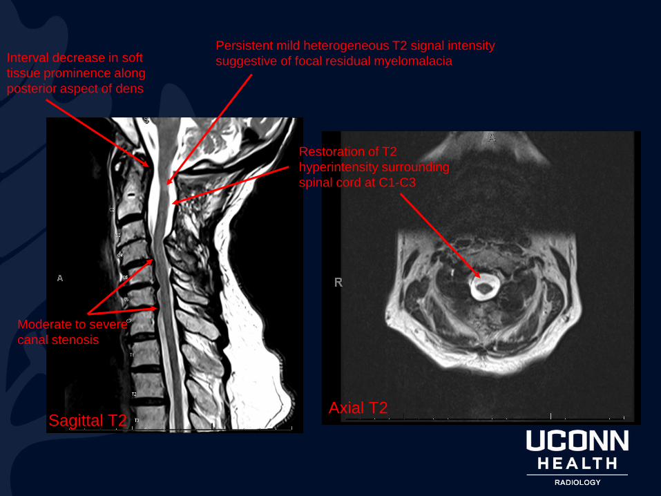
Sagittal T2Axial T2
Restoration of T2
hyperintensity surrounding
spinal cord at C1-C3
Persistent mild heterogeneous T2 signal intensity
suggestive of focal residual myelomalaciaInterval decrease in soft
tissue prominence along
posterior aspect of dens
Moderate to severe
canal stenosis


Anatomy
• Normal soft-tissue anatomy of retro-odontoid region:
– Cruciate ligament (transverse ligament and longitudinal band)
– Posterior longitudinal ligament
– Thecal sac
• Given the synovial articulation b/w the transverse ligament and odontoid, there is a potential space that can be affected by inflammatory and reparative processes resulting in proliferative soft tissue

Pathophysiology• Repetitive cycle of
ligamentous injury and repair likely drive development, initiated by transverse ligament damage from inflammation, minor trauma, degenerative changes, or altered biomechanics
• *Regardless of the etiology, spontaneous regression of ROP is commonly observed following surgical fusion (as in this case)– This further supports the
theory that XS motion at the craniocervical junction may be a major contributing factor in the development of abnormal soft tissue

Radiographic Features
Soft tissue thickening at the atlantoaxial junction around the expected location of the transverse atlantal ligament, posterior to the dens
• CT– Mineralization within the pseudotumor
– Bony erosion may be present in RA, CPPD, gout, pigmented villonodular synovitis
• MRI- signal changes vary by etiology → no reliable imaging features that distinguish RA-related pannus from non-RA retro-odontoid pseudotumor– T1- usually hypointense
• T1 precontrast- identification of hemorrhage, fibrous tissue, mineralization
• T2 post contrast- to characterize vascularity of pseudotumor
– T2- variable, often heterogeneous• This sequence can be used to identify edema within odontoid,
pseudotumor, or C spine

Discussion/Conclusion
• Retro-odontoid pseudotumor is an uncommon entity with varied and overlapping imaging appearances described on MRI in patients with and without RA– ROP is a diverse entity commonly mislabeled as pannus
• In the setting of RA, retro-odontoid soft tissue proliferation is most commonly referred to as pannus, although histological reports are rare and do not always confirm inflammation
• Pannus-like lesions have been reported in patients with severe degenerative disease in the cervical spine– This patient had extensive multilevel degenerative changes
– No prior diagnosis of RA
• Imaging findings in this case (low T1 signal (nonenhancing) and low T2 signal) consistent with soft tissue proliferation seen in osteoarthritis (dense fibrous tissue with immature bone formation), or a fibrous pannus (nonvascular fibrous connective tissue with few cells)
• Regardless of etiology, ROP tend to regress after stabilization of the C spine, which was observed in this patient
References
• Shi J, Ermann J, Weissman BN, Smith SE, Mandell JC. Thinking beyond pannus: a review of retro-odontoid pseudotumor due to rheumatoid and non-rheumatoid etiologies. Skeletal Radiol. 2019 Oct;48(10):1511-1523. doi: 10.1007/s00256-019-03187-z. Epub 2019 Mar 13. PMID: 30868232.
• Kushchayev SV, Glushko T, Jarraya M, Schuleri KH, Preul MC, Brooks ML, TeytelboymOM. ABCs of the degenerative spine. Insights Imaging. 2018 Apr;9(2):253-274. doi: 10.1007/s13244-017-0584-z. Epub 2018 Mar 22. PMID: 29569215; PMCID: PMC5893484.
• Joyce AA, Williams JN, Shi J, Mandell JC, Isaac Z, Ermann J. Atlanto-axial Pannus in Patients with and without Rheumatoid Arthritis. J Rheumatol. 2019 Nov;46(11):1431-1437. doi: 10.3899/jrheum.181429. Epub 2019 Apr 1. PMID: 30936276.
• Park JH, Lee E, Lee JW, Kang Y, Ahn JM, Yeom JS, Kang HS. Postoperative Regression of Retro-odontoid Pseudotumor After Atlantoaxial Posterior Fixation: 11 Years of Experience in Patients With Atlantoaxial Instability. Spine (Phila Pa 1976). 2017 Dec 1;42(23):1763-1771. doi: 10.1097/BRS.0000000000002222. PMID: 28459776.
• Pierce JL, Donahue JH, Nacey NC, Quirk CR, Perry MT, Faulconer N, Falkowski GA, Maldonado MD, Shaeffer CA, Shen FH. Spinal Hematomas: What a Radiologist Needs to Know. Radiographics. 2018 Sep-Oct;38(5):1516-1535. doi: 10.1148/rg.2018180099. PMID: 30207937.
• https://radiopaedia.org/articles/retro-odontoid-pseudotumour?lang=us


![[XLS] · Web view79 0 79 79000 79 79332 79 79085 79 79005 79 10051 79 79328 79 79148 79 10061 79 79476 79 79971 79 79045 79 79772 79 79301 79 79333 79 79154 79 10018 79 79101 79 79335](https://static.cupdf.com/doc/110x72/5adf13517f8b9a6e5c8bad58/xls-view79-0-79-79000-79-79332-79-79085-79-79005-79-10051-79-79328-79-79148-79.jpg)


![OAV dysplasia handout.ppt [Compatibiliteitsmodus] · maximum multipoint lod score of 3.00. familial simplex bilateral involvement 71% 46% hearing loss 26% 87% aural atresia 41% 79%](https://static.cupdf.com/doc/110x72/5c68865809d3f206678ba166/oav-dysplasia-compatibiliteitsmodus-maximum-multipoint-lod-score-of-300.jpg)

![HIGHLIGHTS OF PRESCRIBING INFORMATION Caution should …total, irreversible, bilateral congenital deafness when administered to a pregnant woman [Warnings and Precautions (5.5)]. Although](https://static.cupdf.com/doc/110x72/5f6c061c593a6e5572386379/highlights-of-prescribing-information-caution-should-total-irreversible-bilateral.jpg)