Analysis of individual differences in radiosensitivity using genome editing
3rd International Symposium on the System of Radiological Protection
ICRP2015
Shinya Matsuura
Department of Genetics and Cell Biology, Research Institute for
Radiation Biology and Medicine, Hiroshima University,
Hiroshima 734-8553, Japan
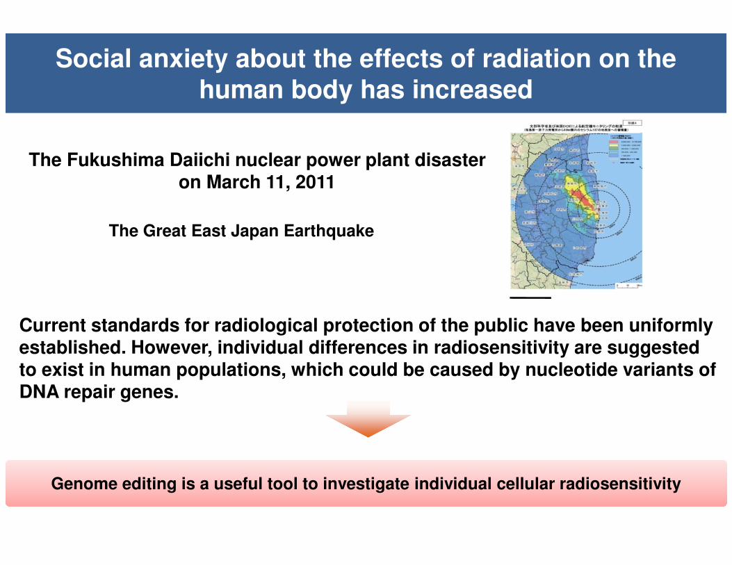
Current standards for radiological protection of the public have been uniformly established. However, individual differences in radiosensitivity are suggested to exist in human populations, which could be caused by nucleotide variants of DNA repair genes.
The Fukushima Daiichi nuclear power plant disaster on March 11, 2011
Genome editing is a useful tool to investigate individual cellular radiosensitivityGenome editing is a useful tool to investigate individual cellular radiosensitivity
Social anxiety about the effects of radiation on the human body has increased
The Great East Japan Earthquake
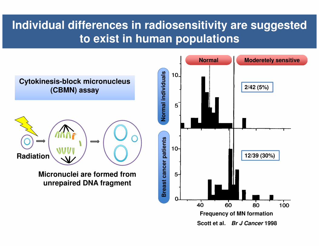
Cytokinesis-block micronucleus (CBMN) assay
Cytokinesis-block micronucleus (CBMN) assay
Individual differences in radiosensitivity are suggested to exist in human populations
Radiation
Micronuclei are formed from unrepaired DNA fragment
Scott et al. Br J Cancer 1998
Frequency of MN formation
No
rmal in
div
idu
als
Moderetely sensitiveModeretely sensitiveNormalNormal
Bre
ast
can
cer
pati
en
ts
2/42 (5%)
12/39 (30%)
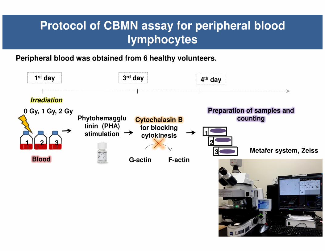
3rd day 4th day1st day
Blood
Cytochalasin B for blocking cytokinesis
Phytohemagglutinin (PHA) stimulation
Preparation of samples and counting
G-actin F-actin
Irradiation
1 2 3
1
2
3 Metafer system, Zeiss
Protocol of CBMN assay for peripheral blood lymphocytes
Peripheral blood was obtained from 6 healthy volunteers.
0 Gy, 1 Gy, 2 Gy

CBMN assay detects individual differences in radiosensitivity among normal individuals
Two individuals showed difference in radiation sensitivity
Volunteer 1 was a 53-year-old man and volunteer 2 was a 46-year-old woman

0
10
20
30
40
50
60
70
80
90
0Gy 1Gy 2Gy
MN
/BN
ra
tio
(%
)
Radiation dose
#87 (AT)
#88 (Carrier)
#89 (Carrier)
#90 (Carrier)
#91 (Normal)
#92 (Normal)
A-T patient
CBMN assay of Ataxia-telangiectasia (A-T) family members
8266A>G1141ins4
#88 #89
#87 #90 #92 #91
Skin fibroblasts were obtained from A-T family members, and were analyzed by CBMN assay.
0
0.5
1
1.5
#87 #88 #89 #90 #91 #92
ATM/b-actin Average
qRT-PCR analysis of ATM mRNA

0
2
4
6
8
10
12
14
0Gy 1Gy 2Gy
MN
/BN
rati
o (
%)
Radiation dose
#88 (Carrier)
#89 (Carrier)
#90 (Carrier)
#91 (Normal)
#92 (Normal)
Heterozygouscarriers(ATM+/-)
Normalindividuals(ATM+/+)
A-T heterozygous carries showed increased frequency of MN formation as compared to normal individuals
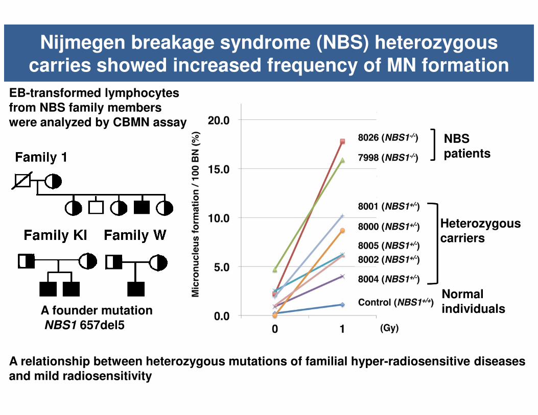
Nijmegen breakage syndrome (NBS) heterozygous carries showed increased frequency of MN formation
EB-transformed lymphocytes from NBS family members were analyzed by CBMN assay
Heterozygouscarriers
Normalindividuals
NBS patients
A relationship between heterozygous mutations of familial hyper-radiosensitive diseases and mild radiosensitivity
A founder mutationNBS1 657del5

Gene Amino acid change Change of base Phenotype
XRCC1 Q399R c.1196A>G Acute/Late radiation reaction
XRCC1 R194W c.580C>T Acute/Late radiation reaction
XRCC1 R280H c.839G>A Cancer risk, late radiation reaction
XRCC3 Y241M c.722C>T Late radiation reaction
LIG4 A3V c.8C>T Lung cancer risk
LIG4 T9I c.26C>T Lung cancer risk
ATM c.8850+60A>G Late radiation reaction
ATM c.5674+1518T>A Breast cancer risk
XPD/ERCC2 D711D c.2133C>T Late radiation reaction
MDC1 A1657A c.4971C>G Acute/Late radiation reaction
CHEK1 c.1233+35G>A Pancreatic cancer risk
XRCC6/Ku70 G593G c.1779G>T Breast cancer risk
XRCC5/Ku80 c..2110-2408G>A Breast cancer risk
RAD51C c.-98G>C Head/neck cancer risk
MRE11 c.*2501A>G Bladder cancer risk
NBS1 I171V c.511A>G Breast cancer risk
RAD50 c.3390-1922T>G Non-Hodgkin lymphoma risk
Individual radiosensitivity may be attributed to SNPs in DNA repair genes
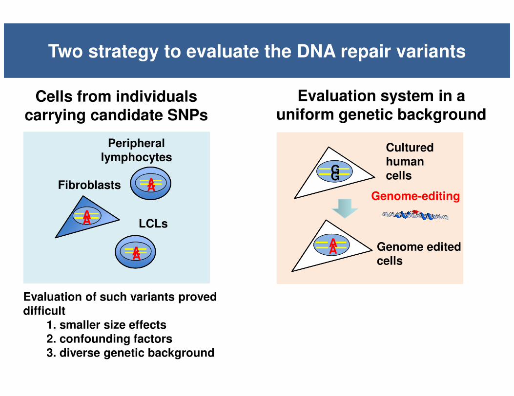
Two strategy to evaluate the DNA repair variants
GG
AA
Cultured human cells
Genome-editing
Genome edited cells
Evaluation system in a uniform genetic background
LCLs
Peripheral lymphocytes
AA
AA
AA
Fibroblasts
Cells from individuals carrying candidate SNPs
Evaluation of such variants proved difficult
1. smaller size effects2. confounding factors3. diverse genetic background

Artificial nucleases and genome editing
Zinc finger nuclease (ZFN)Transcription Activator-like Effector Nuclease (TALEN)
Clustered Regulatory Interspaced Short Palindromic Repeat /Cas9 based RNA-guided DNA endonuclease (CRISPR/CAS9)
Homologous recombination (HR)
Gene knock-in
Targeting vector
DSB

Genome editing identification of an intergenic mutation as causative of genetic disorder
One-year-old boy with a severe diseaseWilms tumor, seizures, and nonverbal.His parents expected to have a third healthy child.However, prenatal DNA diagnosis was difficult because no coding mutation in BUBR1 was found, suggesting that causative mutation is a non-coding one.
Premature chromatid separation (PCS) syndromeAutosomal recessive disorderLoss-of–function mutations in a gene encoding BUBR1, a spindle assembly checkpoint protein
BUBR1
G>A
A single base substitution (G>A) in an intergenic region 44-kb upstream of BUBR1 was identified as potentially causative
Is this the causal mutation or merely correlates with the syndrome ?
To answer this question, we used genome editingTo answer this question, we used genome editing
Premature chromatid separation (PCS)

Two-step single-base-pair editing strategy
CMV tk neor
CMV tk neor
G
G
A
A
Integration of a selection cassette
Removal of the selection cassetteand introduction of the substitution
Wild type allele
Targeted allele
Genome edited allele
Targeting vector
G
Targeting vector
+ neomycin
+ ganciclovir
CMV tk neor
G
Targeted allele
1st step
2nd step
TALEN target site

Patient typeWild type
The nucleotide substitution identified was the causal mutation of the syndrome
Ochiai et al., PNAS 2014
The parents performed amniocentesis during the third pregnancy.It was found to be heterozygous. A healthy baby boy was born.
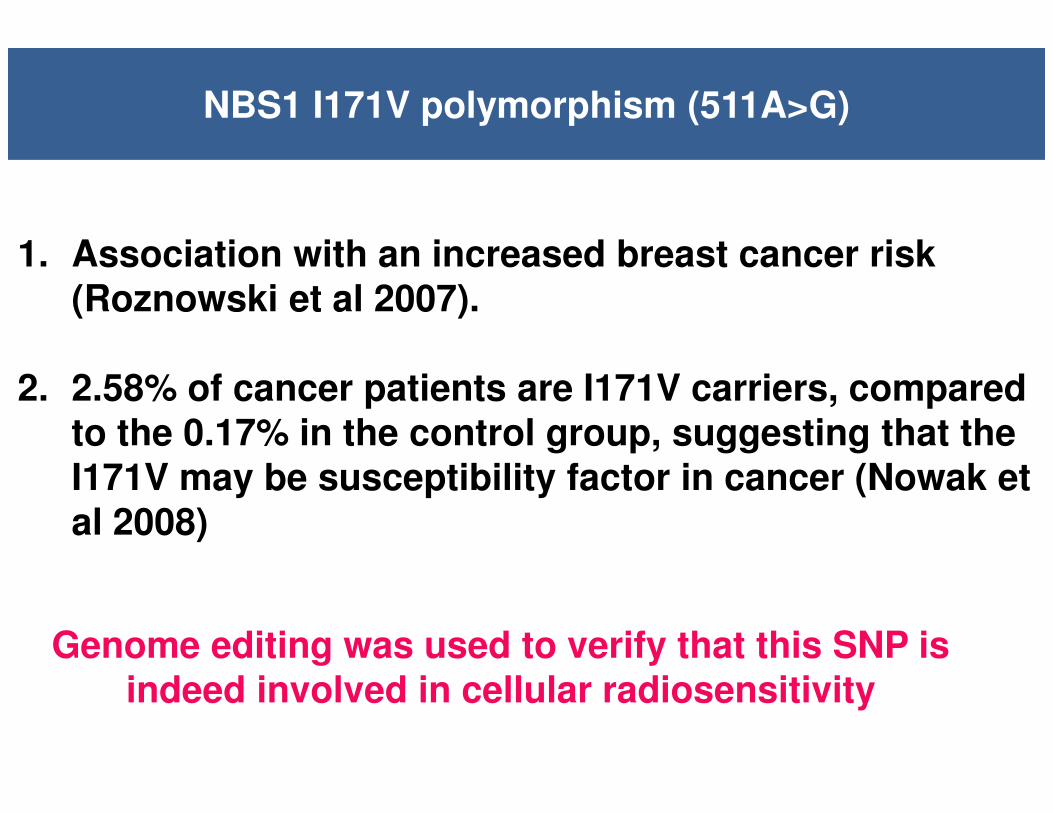
NBS1 I171V polymorphism (511A>G)
1. Association with an increased breast cancer risk (Roznowski et al 2007).
2. 2.58% of cancer patients are I171V carriers, compared to the 0.17% in the control group, suggesting that the I171V may be susceptibility factor in cancer (Nowak et al 2008)
Genome editing was used to verify that this SNP is indeed involved in cellular radiosensitivity

A
(ssODN, 150 mer)
sgRNAsgRNA
CRISPR/Cas9
G
G
G
PAM seq (-NGG)
A
One-step genome editing strategy
One-step
Wild type allele
Genome edited allele
Targeting vector

ScaI: - + - +
WT I171V
number of
clones analysed
ScaI-digested
clones
96 3 (3.15%)
↑ ↑ ↑ ↑↑
NBS1 I171V homozygous cloneNBS1 wild type clone
Restriction enzyme and sequence analysis of genome edited cells
↑
A/A G/G
Targeting
vector5’-AGGTCAACAcTTCGGcCTcATgAAAATGA-(150mer)-3’
Sca IT→C

0.001
0.01
0.1
10 2 4 6
Su
rviv
al
fra
cti
on
Radiation dose (Gy)
NBS1-/-
cells
NBS1 I171V cells
Wild type cells
Genome edited cells showed increased frequency of MN formation
0
2
4
6
8
10
12
14
16
18
20
0 1 2 3
MN
/BN
(%
)
Radiation dose (Gy)
NBS1-/-
cells
NBS1 I171V cells
Wild type cells
1. Individual differences in radiosensitivity exist in human
populations.
2. We designed TALEN-mediated two-step single-base-pair editing,
which we used to introduce a nucleotide variant associated
with a chromosomal instability syndrome into human cultured
cells to demonstrate that it is the causative mutation.
3. We designed CRISPR/CAS9-based one-step genome editing
and applied it to the evaluation of NBS1 I171V polymorphism
for cellular radiosensitivity.
4. Genome editing is now widely used and become a valuable tool
to investigate individual radiosensitivity.
Conclusion

Ekaterina Royba, Silvia Natsuko Akutsu, Hiromi Yanagihara, Tatsuo MiyamotoDepartment of Genetics and Cell Biology, Research Institute for Radiation Biology and Medicine, Hiroshima University
Takashi Yamamoto, Hiroshi OchiaiDepartment of Mathematical and Life Sciences, Graduate School of Science, Hiroshima University
Yoshiki KudoDepartment of Obstetrics and Gynecology, Graduate School of Biomedical Sciences, Hiroshima University
Satoshi TashiroDepartment of Cellular Biology, Research Institute for Radiation Biology and Medicine, Hiroshima University
Acknowledgements