1 Zoonoses Pavel Chalupa Dep. of Infectious and Tropical Diseases First Faculty of Medicine Charles University in Prague 2018/2019 ZOONOSES Infection diseases transmitted from animals to man (opposite transmission is also possible) Causal organism: • viruses • bacteria • parasites • fungi • prions Most frequent zoonoses in CR campylobacteriosis, salmonelosis, yersiniosis, toxoplasmosis, tularemia, EHEC,leptospirosis,listeriosis,ornithosis, toxocarosis, taeniasis, erysipeloid, cat scratch disease, Lyme disease, ehrlichiosis, tick-borne encephalitis, Q fever, hepatitis E (genotype 3) • virus Nipah (1999, Malaysia) –severe febrile encephalitis with high mortality in pig keepers • New variant of Creutzfeldt-Jakob disease (vCJD) in human – relation to bovine spongiforme encephalopathy (BSE) in cattle (still is studied) The 1st case of BSE was in 1986 and the 1st case of vCJD in 1996 • Acute HE HEV genotypes 3 a 4 (transmission from pigs, boars, deer on man in non-endemic areas) • TIBOLA (1996, Hungary) – new rickettsiosis, TIck-BOrne LymfAdenopathy, Rickettsia slovaca • Enteroaggregative hemorrhagic E. coli (EAHEC) O104:H4 strain (2011, large outbreak in Germany) • Novel coronavirus MERS-CoV (Middle East Respiratory Syndrome-Coronavirus) (2012, Saudi Arabia - in a patient who died of pneumonia and renal failure) transmission from camels • Human infections with Highly Pathogenic Avian Influenza A (H7N9) Virus (since 2013 in China) NEW ZOONOSES Tick-borne lymphadenopathy (TIBOLA). A 67-year old woman with an inoculation eschar with central necrosis, edematous margins and erythematous halo at the former site of the tick bite (Dermacentor marginatus). BMC Infectious Diseases 2011;vol.11,art.no.167 ZOONOSES – bioterrorism and biological weapons • Anthrax (Bacillus anthracis) • Plague (Yersinia pestis) • Q fever (Coxiella burnetti) • Tularemia (Francisella tularensis) • Brucelosis (Brucella abortus, B. melitensis, B. suis) • Glanders (Burgholderia mallei) • Melioidosis (Burgholderia pseudomallei) • Viral hemorrhagic fevers

Welcome message from author
This document is posted to help you gain knowledge. Please leave a comment to let me know what you think about it! Share it to your friends and learn new things together.
Transcript

1
Zoonoses
Pavel Chalupa
Dep. of Infectious and Tropical Diseases
First Faculty of Medicine
Charles University in Prague
2018/2019
ZOONOSESInfection diseases transmitted from
animals to man (opposite transmission is
also possible)
Causal organism:
• viruses
• bacteria
• parasites
• fungi
• prions
Most frequent zoonoses in CR
campylobacteriosis, salmonelosis,
yersiniosis, toxoplasmosis, tularemia,
EHEC,leptospirosis,listeriosis,ornithosis,
toxocarosis, taeniasis, erysipeloid,
cat scratch disease, Lyme disease,
ehrlichiosis, tick-borne encephalitis,
Q fever, hepatitis E (genotype 3)
• virus Nipah (1999, Malaysia) –severe febrile encephalitis with high mortality in pig keepers
• New variant of Creutzfeldt-Jakob disease (vCJD) in human – relation to bovine spongiforme
encephalopathy (BSE) in cattle (still is studied)
The 1st case of BSE was in 1986 and the 1st case of vCJD in 1996
• Acute HE
HEV genotypes 3 a 4 (transmission from pigs, boars, deer on man in non-endemic areas)
• TIBOLA (1996, Hungary) – new rickettsiosis, TIck-BOrne LymfAdenopathy, Rickettsia slovaca
• Enteroaggregative hemorrhagic E. coli (EAHEC) O104:H4 strain(2011, large outbreak in Germany)
• Novel coronavirus MERS-CoV (Middle East Respiratory Syndrome-Coronavirus)(2012, Saudi Arabia - in a patient who died of pneumonia and renal failure)
transmission from camels
• Human infections with Highly Pathogenic Avian Influenza A (H7N9) Virus(since 2013 in China)
NEW ZOONOSES
Tick-borne lymphadenopathy (TIBOLA). A 67-year old woman with an inoculation eschar
with central necrosis, edematous margins and erythematous halo at the former site of the
tick bite (Dermacentor marginatus). BMC Infectious Diseases 2011;vol.11,art.no.167ZOONOSES – bioterrorism and
biological weapons
• Anthrax (Bacillus anthracis)
• Plague (Yersinia pestis)
• Q fever (Coxiella burnetti)
• Tularemia (Francisella tularensis)
• Brucelosis (Brucella abortus, B. melitensis,
B. suis)
• Glanders (Burgholderia mallei)
• Melioidosis (Burgholderia pseudomallei)
• Viral hemorrhagic fevers

2
Lyme disease (LD)
• Seasonal zoonosis
• Causal organism:
Borrelia burgdorferi sensu lato
• Most often is affected:
skin (65%)
musculoskeletal system (17%)
nervous system (12%)
less other organs – heart (Lyme carditis)
eye, or any other tissue in the body
B. burgdorferi sensu lato
Three main species:
• B. burgdorferi sensu stricto (mainly in
America, in Europe has other antigens) –
causes mainly affections of joints
• B. garinii (in Europe and Asia) – causes
mainly affections of nervous system
• B. afzelii (in Europe and Asia) – causes
mainly affections of the skin
Other species:
B. bavariensis – neurotropic as B. garinii,
B. spielmanii, B. valaisiana, B. lusitanie
History of LD I.
• 1883 Buchwald – acrodermatitis chronica
atrophicans
• 1909 Artvid Afzelius – erythema migrans
• 1922 Garin and Bujadoux „paralyse par les
tiques“
• 1941 Bannwarth – syndrome of
meningopolyradiculoneuritis
• 1943 Bäferstedt – lymphadenosis benigna
cutis
History of LD II.
• 1947 Lehnhof – microscopic detection of spirochetes from skin biopsy of periphery of erythema migrans
• 1951 Hellström – PNC administration was effective on above-mentioned affections
• 1975 Steere – endemic arthrities in children in the area of the city Old Lyme (Connecticut, US)
• 1982 Burgdorfer – isolation of spirochaete from the gut of the tick Ixodex dammini
• 1984 Borrelia burgdorferi was marked as etiological agent of Lyme disease
Clinical characteristics of LD
Stage Incub. per. Clinical manifestations
Early infection:
1. Localized
Infection
2. Disseminated
Infection
3-30 days
(max. 8 weeks)
Weeks-months
Erythema migrans
Erythema migrans multiplex
Lymphadenosis benigna cutis
Bannwarth syndrome
(meningopolyradiculoneuritis)
Lyme carditis (A-V blocades, myocarditis,
pericarditis)
Arthritis of small joints, tendovaginitis,
myositis, bursitis
Late infection:
3. Persistent
Infection
Months-years Acrodermatitis chronica atrophicans
Neurological involvement
(chron. encephalitis, chron. polyneuritis)
Arthritis of large joint (e.g. gonitis)

4
Epidemiology of LD I.
• Reservoir of B. burgdorferi: small terrestrial mammals
• Transmission on human is by the ticks:
in Europe Ixodes ricinus
in America I. scapularis (I. dammini) and I.pacificus
• Risk of transmission increases with duration of sucking – 48 hours is enough time for transmission of infection from the infected tick
Epidemiology of LD II.
• Seroconversion (appearance of antibodies)
after infected tick is only in 5-10% of
persons. Not all of them have some clinical
manifestations of LD
• Positive LD antibodies are detected in CR
population cca in 10% (in forest workers
more than 20%)
• Transmission from mother to foetus is
possible
• Blood transfusion can transfer infection
in case of borreliaemia in blood donor
LD and coinfection
• Contemporary occurrence of other
disseases transmitted by ticks was
registered in 10% of cases in the CR
• First of all human granulocytic
anaplasmosis (ehrlichiosis) and other
rickettsioses can be transferred
simultaneouslyMedicína po promoci 2005;6(8):20-27
Infections with possible transmission by the
ticks in Europe
Etiological agent Disease
TBE virus
B. burgdorferi sensu lato
Rickettsia conori
Coxiella burneti
Rickettsia slovaca
Anaplasma
phagocytophilum
Bartonella henselae
Francisella tularensis
Babesia microti
Crimean-Congo
hemorrhagic fever virus
Tick-borne encephalitis
Lyme disease
Marseilles fever
Q fever
TIBOLA (tick-borne lymfadenopathy)
human granulocytic anaplasmosis
(ehrlichiosis)
Cat scratch disease
Tularemia
Babesiosis
Crimean-Congo hemorrhagic fever
(cases in the last 10 years: Albania,
Kosovo, Turkey, Greece, Ukraine,
SW Russia, Bulgaria)

5
Diagnosis of LD I.
• ELISA and Western blot (WB) – detection of antibodies against antigen components of B. burgdorferi
• Production of IgM antibodies starts in the 3rd or 4th week after the primoinfection
• At the time of erythema migrans IgM antibodies are not usually produced
• IgM persists sometimes very long time and their detection has not to be a marker of the activity of infection
• It is always important to correlate laboratory and clinical findings!!
• Antibodies could be also negative and clinical finding is clear!!
Diagnostic of LD II.
• PCR from CSF, urine, synovial liquid,
sample from placenta, or from other
medium
• Intrathecal production of antibodies
from CSF
• Electronmicroscopy detection of
B. burgdorferi mostly from CSF
(not in routine practice in the CR )
• DNA microarrays = method of the future
B. burgdorferi from CSF in EM
Zapůjčila RNDr. D. Hulínská, CSc., NRL pro lymeskou borreliózu, SZÚ
Cyst (L-form) of B. burgdorferi
from CSF in EM
Zapůjčila RNDr. D. Hulínská, CSc., NRL pro lymeskou borreliózu, SZÚ
Therapy of LD
Therapy is indicated only when clear
clinical symptomatology is present and
we are glad, if also laboratory findings
are possitive
• Causal therapy = ATB
• Symptomatic treatment = analgetics,
antiedematous therapy, vitamins,
anxiolytics, antidepresives
Therapy of LD
(Czech Guidelines 2018)
Antibioticum Diagnosis Duration of
treatment (days)
Adults Children
Administration per os
Doxycycline1 Skin LD, NB, LA, LC 10-14 2x 100 mg nebo
1x 200 mg/d
4 mg/kg/d1)
Amoxicillin2 Skin LD, LA, LC 10-14 3x 500-1000 mg/d 50 mg/kg/d
Fenoxymethylpenicillin2 EM, EMM, LBC 10-14 3x 1-1,5 mil. IU/d 80-100 000 IU/kg/d
Cefuroxim-axetil EM,EMM,LBC,LA,LC 10-14 2x 500 mg/d 30 mg/kg/d
Azithromycin3 EM, EMM, LBC 5 1. day: 2x 500 mg
2.-5. day: 1x 500 mg
1. day: 20 mg/kg
2.-5.day: 10 mg/kg
Clarithromycin3 EM, EMM, LBC 10-14 2x 500 mg/d 15 mg/kg/d
Administration i.v.
Ceftriaxon NB, LA, ACA, LC 14-28 1x 2 g/d 50-75 mg/kg/d
Cefotaxim NB, LA, ACA, LC 14-28 3x 1 g/d 150-200 mg/kg/d
Benzylpenicillin
(penicillin G)
NB 14-28 4-5 mil. IU à 4-6 hod. 200-400 000 IU/kg/d
LD = Lyme disease, EM = erythema migrans, EMM = erythema migrans multiplex, LBC = lymphadenosis benigna cutis,
LA = lyme arthritis, LC = lyme carditis, NB = neuroborreliosis1 doxycycline not in children younger than 8 years, not in pregnancy2 penicillin and amoxicillin not during nursing3 azithromycin and clarithromycin –prolongation of QT interval is possible and ventricular arrhythmia and ventricular tachycardia (type
of torsade de pointes); azithromycin not in pregnancy

6
Repetition of the therapy
1. Reinfection
2. Relaps
3. Insufficient answer to the therapy
Repeat the therapy during 3 months is
not indicated
Not more than 3x ATB treatment in one
patient with LD!!!
Tularemia
• 1910 etiological agent was identified by Mc Coy as the agent of disease in sisels in the surroundings of the town Tulare in California
• 1914 Whery and Lamb – isolation this microbe from hares and possibility of transmission to human was pronounced
• 1921 E. Francis in USA - isolation the same microbe from blood and pus from the patients with „deer-fly-fever“ and the disease was named tularemia
• Etiological agent was named Francisella tularensis
F.tularensis is G- cocobacillus,
Francis agar is used for cultivationOccurrence of tularemia in animals
in CR – year 2009
New occurrence - red
7
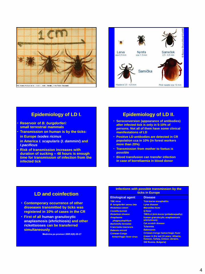
A21 Tularémie
222 225
10394
110
6051
83 87
54
113
6553
0
100
200
300
1998 1999 2000 2001 2002 2003 2004 2005 2006 2007 2008 2009 2010
Po
čet
on
em
ocn
ěn
í
EPIDAT, SZÚ, Praha
Clinical manifestation of
tularemia
• External manifestation
- ulceroglandular
- glandular
- oroglandular (pharyngeal)
- oculoglandular
• Internal manifestation
- pulmonal
- abdominal
• Sepsis (generalization of infection)
• Combinations of external or internal manifestations
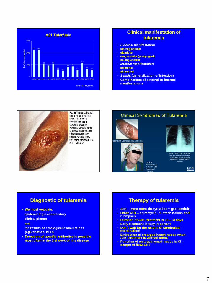
Clinical Syndromes of Tularemia
Cervical
lymphadenitis
in patient with
pharyngeal
tularemia
Patient with ulceroglandular tularemia
Chest radiograph of patient
with pulmonary tularemia(Radiograph shows bilateral
pneumonitis and left pleural
effusion)
Diagnostic of tularemia
• We must evaluate:
epidemiologic case-history
clinical picture
and
the results of serological examinations
(aglutination, KFR)
• Detection of specific antibodies is possible
most often in the 3rd week of this disease
Therapy of tularemia
• ATB – most often doxycyclin + gentamicin• Other ATB – spiramycin, fluofochinolons and
rifampicin
• Duration of ATB treatment is 10 - 14 days
• Early treatment is very important
• Don´t wait for the results of serological examination!
• Extirpation of enlarged lymph nodes when ATB treatment is without effect
• Punction of enlarged lymph nodes is KI –danger of fistulas!!!

8
Ulceroglandular form of tularemia
(extirpation of enlarged lymph nodes was done) Toxoplasmosis
• Etiological agent Toxoplasma gondii
• Incubation period 5-20 days
• Definitiv host is cat and some other felidae.
• Sexual stage of the development of this parazite is in the the cat
• Human is inficated by the oocysts, that are in the excrements of cats
• Duration of excretion of oocysts is 2-3 weeks and is present only when the cat is inficated with T. gondii for the first time.
Toxoplasmosis
3 life forms of T. gondii:
• Tachyzoit – vegetative form
• Bradyzoit – this form ramains in the
body for the whole life as tissue cysts
• Oocysts – they are very resistant and
they are in the stool of the cat
Toxoplasmosis – transmission of
infection• Alimentary – consumation of raw or not good
cooked meat, contaminated foodstuff or water
• Also is possible – geophagia, contact with contaminated soil or with animals
• Very rare – through the mucous of respiratory tract, urogenital tract, conjunctiva or damaged skin.
• Other modes: with blood transfusion, transplanted tissue, or in laboratory
• Transplacentar transmission leads to endouterine (congenital) infection. Danger of involvement the foetus is only when the primoinfection is in pregnancy or not longer than 6 months before the pregnancy
Toxoplasmosis
clinical manifestations
• About 90% of infections are inaparent
• Implications of primoinfection in pregnancy: abortus, premature birth, birth of lifeless foetus. Typical changes in congenital toxoplasmosis = Sabin trias (hydrocephalus, calcifications in brain and chorioretinitis). Sabin tetrade = convulsions are present also
Also is possible: microcephalia, microophthalmus, icterus, hepatosplenomegaly, atrophia n. optici, cataracta, strabismus, myocarditis, purpura, psychomotoric retardation, disorders with hearing or vision
• Most danger is primoinfection in pregnancy, not reactivation of chronic toxoplasmosis

9
Toxoplasmosis – clinical
manifestations• Only 10-20% infections are symptomatic and
the most often manifestation is enlargement of lymph nodes. Toxoplasmosis is also the most often cause of the enlargement of lymph nodes in the CR. We do not treat it in immunocompetent patients.
• Involvement of eyes – it is acute chorioretinitis, most often only on one side
• Involvement of other organs (rarely) - hepatitis, myocarditis, pneumonitis
• Septic form with the involvement of different organs including CNS in the patients with immunosuppression or immunodeficiency
Toxoplasmosis - diagnostics
• Serological detection of antibodies using at least two methods: KFR and ELISA (IgG, IgM, IgA and IgE)
• Avidity of IgG antibodies
(low values – primoinfection)
• Detection of DNA of Toxoplasma gondii from amniotic fluid using PCR method
• Western blot

10
Toxoplasmosis - therapy
We treat:
• Pregnant women with acute infection
• Children with congenital infection
• Patients with organ involvement (eyes or other)
• Acute toxoplasmosis in immunocompromised persons
• Children until 5 years of the age
We don´t treat asymptomatic infection in immunocompetent adults
Therapy of toxoplasmosis
• Duration of the therapy: 3-4 weeks
• We use:
pyrimethamin + sulfadiazin
or
pyrimethamin + clindamycin
• Patients with involvement of eyes:
moreover corticosteroids
• KI of pyrimethamin: to 16.-18. week of gravidity
• We combine pyrimethamin with acidum folinicum (Leucovorin) to reduce myelotoxicity of pyrimethamin
Therapy of toxoplasmosis
in pregnancy
• We use:
spiramycin
or
pyrimethamin + sulfadiazin
• Therapeutic schema:
rotation of usage 3 weeks spiramycin and next 3 weeks pyrimethamin + sulfadiazin
• KI of pyrimethamin: the 1st trimestr ofgravidity
• KI of sulfadiazin: the 1st trimestr of gravidity and the last 4-6 weeks before the birth
Cerebral form of toxoplasmosis
• Great oportunic infection in HIV+
• It is reactivation of latent infection
• Therapy: pyrimetamin + sulfadiazin
or pyrimethamin + clindamycine
• Duration of the therapy 6 (4-8) weeks
• Antiedematic therapy:
5 days dexamethazon i.v.
Prevention of cerebral form of
toxoplasmosis
• Primary prophylaxis when is decrease of CD4+ below 100-150/μl – cotrimoxazol 3x960 mg/week or 1x480 mg/day
• Secundary prophylaxis after the cerebral form of toxoplasmosis:
pyrimethamin
or
pyrimethamin + sulphadiazin
or
pyrimethamin + clindamycin

11
Cerebral form of toxoplasmosis
• No or bad treated – always is fatal
• Serological tests are not very useful
(helpful) in HIV+ patients:
- we do not detect strong changes in
the level of antibodies,
- IgG is +, but not pronounced,
- IgM is mostly negative
Related Documents