Zoledronic Acid Decreased Osteolysis But Not Bone Metastasis in a Nude Mouse Model of Canine Prostate Cancer With Mixed Bone Lesions Nanda K. Thudi 1 , Chelsea K. Martin 1 , Murali V.P. Nadella 1 , Soledad A. Fernandez 2 , Jillian L. Werbeck 1 , Joseph J. Pinzone 3 , and Thomas J. Rosol 1,* 1 Department of Veterinary Biosciences, The Ohio State University, Columbus, Ohio 2 Center for Biostatistics, The Ohio State University, Columbus, Ohio 3 Department of Internal Medicine, The Ohio State University, Columbus, Ohio Abstract BACKGROUND—Bone metastasis is the most common cause of morbidity and mortality in patients with advanced prostate cancer and is manifested primarily as mixed osteoblastic and osteolytic lesions. However, the mechanisms responsible for bone metastases in prostate cancer are not clearly understood, in part due to the lack of relevant in vivo models that mimic the clinical presentation of the disease in humans. We previously established a nude mouse model with mixed bone metastases using intracardiac injection of canine prostate cancer cells (Ace-1). In this study, we hypothesized that tumor-induced osteolysis promoted the incidence of bone metastases and osteoblastic activity. METHODS—We studied the effect of inhibition of osteolysis with zoledronic acid (ZA) on the prevention and progression of Ace-1 bone metastases in nude mice using prophylactic and delayed treatment protocols. Bioluminescent imaging, radiography, and histopathological evaluation were performed to monitor the effect of ZA on the incidence, progression and nature of bone metastases. RESULTS—Unexpectedly, there was no significant difference in tumor burden and the incidence of metastasis between control and treatment groups as detected by bioluminescent imaging and bone histomorphometry. However, radiographic and histopathological analysis showed a significant treatment-related decrease in osteolysis, but no effect on tumor-induced trabecular bone thickness in both treatment groups compared to controls. CONCLUSION—Our results demonstrated that the incidence of prostate cancer bone metastases in vivo was not reduced by zoledronic acid even though zoledronic acid inhibited bone resorption and bone loss associated with the mixed osteoblastic/osteolytic bone metastases in the Ace-1 model. Keywords prostate cancer; bioluminescent imaging; bone metastases; zoledronic acid; osteoblastic metastases; osteolysis © 2008 Wiley-Liss, Inc. *Correspondence to: Dr. Thomas J. Rosol, Department of Veterinary Biosciences, College of Veterinary Medicine, The Ohio State University, 1925 Coffey Road, Columbus, OH 43210. [email protected]. NIH Public Access Author Manuscript Prostate. Author manuscript; available in PMC 2010 March 5. Published in final edited form as: Prostate. 2008 July 1; 68(10): 1116–1125. doi:10.1002/pros.20776. NIH-PA Author Manuscript NIH-PA Author Manuscript NIH-PA Author Manuscript

Welcome message from author
This document is posted to help you gain knowledge. Please leave a comment to let me know what you think about it! Share it to your friends and learn new things together.
Transcript

Zoledronic Acid Decreased Osteolysis But Not Bone Metastasisin a Nude Mouse Model of Canine Prostate Cancer With MixedBone Lesions
Nanda K. Thudi1, Chelsea K. Martin1, Murali V.P. Nadella1, Soledad A. Fernandez2, Jillian L.Werbeck1, Joseph J. Pinzone3, and Thomas J. Rosol1,*1Department of Veterinary Biosciences, The Ohio State University, Columbus, Ohio2Center for Biostatistics, The Ohio State University, Columbus, Ohio3Department of Internal Medicine, The Ohio State University, Columbus, Ohio
AbstractBACKGROUND—Bone metastasis is the most common cause of morbidity and mortality inpatients with advanced prostate cancer and is manifested primarily as mixed osteoblastic andosteolytic lesions. However, the mechanisms responsible for bone metastases in prostate cancer arenot clearly understood, in part due to the lack of relevant in vivo models that mimic the clinicalpresentation of the disease in humans. We previously established a nude mouse model with mixedbone metastases using intracardiac injection of canine prostate cancer cells (Ace-1). In this study,we hypothesized that tumor-induced osteolysis promoted the incidence of bone metastases andosteoblastic activity.
METHODS—We studied the effect of inhibition of osteolysis with zoledronic acid (ZA) on theprevention and progression of Ace-1 bone metastases in nude mice using prophylactic and delayedtreatment protocols. Bioluminescent imaging, radiography, and histopathological evaluation wereperformed to monitor the effect of ZA on the incidence, progression and nature of bone metastases.
RESULTS—Unexpectedly, there was no significant difference in tumor burden and the incidenceof metastasis between control and treatment groups as detected by bioluminescent imaging and bonehistomorphometry. However, radiographic and histopathological analysis showed a significanttreatment-related decrease in osteolysis, but no effect on tumor-induced trabecular bone thicknessin both treatment groups compared to controls.
CONCLUSION—Our results demonstrated that the incidence of prostate cancer bone metastasesin vivo was not reduced by zoledronic acid even though zoledronic acid inhibited bone resorptionand bone loss associated with the mixed osteoblastic/osteolytic bone metastases in the Ace-1 model.
Keywordsprostate cancer; bioluminescent imaging; bone metastases; zoledronic acid; osteoblastic metastases;osteolysis
© 2008 Wiley-Liss, Inc.*Correspondence to: Dr. Thomas J. Rosol, Department of Veterinary Biosciences, College of Veterinary Medicine, The Ohio StateUniversity, 1925 Coffey Road, Columbus, OH 43210. [email protected].
NIH Public AccessAuthor ManuscriptProstate. Author manuscript; available in PMC 2010 March 5.
Published in final edited form as:Prostate. 2008 July 1; 68(10): 1116–1125. doi:10.1002/pros.20776.
NIH
-PA Author Manuscript
NIH
-PA Author Manuscript
NIH
-PA Author Manuscript

INTRODUCTIONProstate cancer is the most frequently diagnosed cancer and second leading cause of cancer-related deaths in men [1]. In spite of themarked improvements in early diagnosis and efficientlocal and systemic therapeutic approaches, 65–75%of the patients with advanced prostatecancer develop skeletal metastasis due to the resistance of tumor cells to conventional therapies[2–4]. In prostate cancer, most patients die because of metastases to bone rather than theprimary tumor [5].
Prostate cancer bone metastasis is frequently osteoblastic in nature with increased woven boneformation often preceded by osteoclastic activity [2,6]. Currently, in prostate cancer, themechanisms responsible for osteoblastic bone metastases are not clear due to the lack ofprostate cancer cell lines that consistently metastasize to bone and develop mixed osteoblasticand osteolytic lesions in animal models. Therefore, prostate cancer cell lines that reliablydevelop mixed osteoblastic and osteolytic lesions in vivo can be used to help understand themechanisms underlying mixed bone metastases of prostate cancer as they occur in men.Recently, we have established a new canine prostate cancer cell line (Ace-1) from a prostateadenocarcinoma that consistently produces mixed osteoblastic and osteolytic bone metastasesafter intracardiac injection in nude mice [7,8]. Metastases of prostate cancer occur in the axialand appendicular bones in humans. Spontaneous prostate cancer in dogs has importantsimilarities to human prostate cancer in the clinical presentation of disease including tumorgrowth over a long period of time, individual and intratumor heterogeneity, extensive genomehomology, and metastasis to distant sites, such as bone. Metastasis of Ace-1 cells anddevelopment of mixed osteoblastic/osteoblastic lesions in nude mice recapitulates thecharacteristics of human and canine prostate cancer metastases in a mouse model [9,10].Therefore, the Ace-1 model is useful to study the pathogenesis of prostate cancer metastasisand investigate the complex interactions between tumor cells and the bone microenvironment.
Preferential metastasis and growth of prostate cancer cells in bone is associated with a complexinteractions between the cancer cells (seed), bone cells, and the bone marrowmicroenvironment (fertile soil) [3,11]. Metastatic prostate cancer cells in bone produce factorssuch as parathyroid hormone related protein (PTHrP) and receptor activator for nuclear factorκ B ligand (RANKL) that stimulate increased bone resorption [5]. This results in the furtherrelease of growth factors and proteins from the bone matrix that promote the growth of cancercells in bone and possibly increase the maturation and function of osteoblasts. Therefore, it hasbeen hypothesized that bone resorption contributes significantly to the development ofosteoblastic metastases. However, the role of osteolysis in prostate cancer bone metastasis,growth of metastases, and induction of osteoblasts is not well understood [5,12–15]. Therefore,targeting osteoclast activity in mixed osteoblastic and osteolytic bone metastases will improveour understanding of the mechanisms underlying prostate cancer bone metastasis. Insights intothe pathogenesis of prostate cancer bone metastasis will help identify specific targets foreffective therapeutic approaches to help treat this devastating malignancy.
Previous studies have shown that zoledronic acid (ZA) is a potent inhibitor ofosteoclastogenesis and osteoclast-mediated bone resorption in animal models of bonemetastasis associated with prostate cancer, breast cancer and myeloma [16–20]. In the presentstudy, we used ZA to inhibit osteoclast activity and investigate the role of osteoclast activityin prostate cancer bone metastasis.
Thudi et al. Page 2
Prostate. Author manuscript; available in PMC 2010 March 5.
NIH
-PA Author Manuscript
NIH
-PA Author Manuscript
NIH
-PA Author Manuscript

MATERIALSANDMETHODSCell Culture
Ace-1 is a spontaneously immortalized canine prostate cancer cell line derived from a prostateadenocarcinoma that was previously established in our laboratory [7]. Ace-1 cells weremaintained at 37°C in Dulbecco’s Modified Eagle’s Medium/Ham’s Nutrient Mixture F12(Invitrogen Corp., Carlsbad, CA) supplemented with 10% fetal bovine serum, 250 U/mlpenicillin, 250 µg/ml streptomycin, and 2 mM L-glutamine (Invitrogen Corp.) in a 5% CO2-humidified chamber.
Establishment of Ace-1 Cells Stably Expressing the YFP-Luc Reporter GeneAce-1 cells were transfected with 6 µg of pcDNA3.1(+)/YFP-LUC, a dual reporter geneconstruct under control of the CMV promoter (a generous gift from Dr. Christopher Contag,Stanford University, Stanford, CA), and 10 µl of Lipofectamine 2000 (Invitrogen Corp.).Stably integrated cells were selected using 400 µg/ml of G418 (Sigma–Aldrich Co., St. Louis,MO) for 17 days. Flow cytometry (BDFACSVantage SE; BD Biosciences, San Jose, CA) wasused to sort and clone YFP-positive cells.
Intracardiac Inoculation of Ace-1 Cells Into Nude MiceMale nu/nu mice, 4–6 weeks old (Charles River Laboratories, Wilmington, MA) were housedin microisolator cages, and were provided food pellets and water ad libitum. Animal careprocedures were approved by the Ohio State University Institutional Lab Animal Care and UseCommittee using criteria based on both the Animal Welfare Act and the Public Health Services“Guide for the Care and Use of Laboratory Animals.” Mice were anesthetized with ketamine(100 mg/kg) and xylazine (10 mg/kg) administered intraperitoneally (IP) and positioned ondorsal recumbency. 1 × 10⁵ Ace-1 cells were suspended in 100 µl of sterile Dulbecco’s PBS(Invitrogen Co.) and were injected into the left ventricle using a 27 gauge needle afterconfirmation of location of the tip of the needle in the left ventricle indicated by pulsatile bloodflow in the hub of the needle [21]. Successful Ace-1 intracardiac injections were confirmedusing bioluminescent imaging (BLI) at 10 min after injection and were characterized by adiffuse emission of light from the entire body. Mice were euthanized 28 days after Ace-1inoculation.
TreatmentNude mice were divided into three groups: (a) Control group(n = 11) received PBS from –1to 4weeks. (b) Prophylaxis group (n = 9) received ZA from –1 to 4 weeks. (c) Delayed treatment(n = 7) group received ZA from 2 to 4 weeks. Mice were administered ZA at 100 µg/kg BW(Novartis, Basel, Switzerland) or vehicle (PBS) twice a week subcutaneously (SQ) (Fig. 1).
Bioluminescent Imaging (BLI)Mice were injected intraperitoneally (IP) with 150 µl 40 mg/ml luciferin (Caliper Life Sciences,Hopkinton, MA) dissolved in PBS. Mice were anesthetized with 3% isoflurane–air mixtureand transferred to the light-tight 37°C imaging chamber of an In Vivo Imaging System (IVIS;Caliper Life Sciences). BLI was performed 10 min after IP administration of luciferin. BLIwas performed on mice with dorso-ventral positioning under anesthesia with 1.5% isoflurane–air mixture once per week for 4 weeks. The BLI signal intensity was analyzed usingLivingImage software version 2.50 (Caliper Life Sciences) and was quantified serially bymeasurement of peak photon flux at the individual metastasis foci by selecting a region ofinterest (ROI) around the BLI signal.
Thudi et al. Page 3
Prostate. Author manuscript; available in PMC 2010 March 5.
NIH
-PA Author Manuscript
NIH
-PA Author Manuscript
NIH
-PA Author Manuscript

Faxitron RadiographyRadiographic images of mice were obtained using a Faxitron cabinet X-ray system (Hewlett-Packard, McMinnville, OR) at 45 kVp for 3.5 min at day 28.
HistopathologyComplete necropsies were performed on the mice. Bones were fixed in 10% neutral-bufferedformalin at 4°C for 24 hr, decalcified in 10% EDTA (pH 7.4) for 2 weeks at 4°C, and embeddedin paraffin. The specimens were sectioned (5 µm) and were stained with hematoxylin and eosin(H&E) to evaluate by histopathology or stained for tartrate-resistant acid phosphatase (TRAP)to identify active osteoclasts. TRAP staining was performed on non-stained sections that weredeparaffinized by three 1 min washes with xylene (Hemo-De, Fisher Scientific, and Bay Shore,NY) and rehydrated in decreasing concentrations of ethanol (100%, 95% and finally 70%). Forthe effective staining of TRAP, antigen retrieval on the sections was performed using heattreatment at 60°C for 10 min in preheated antigen retrieval solution (Dako-Cytomation,Carpinteria, CA) and then stained for TRAP (Acid Phosphatase Kit 387-A; Sigma Diagnostics,St. Louis, MO) as directed by the manufacturer.
Bone HistomorphometryBone histomorphometry was performed using computer software designed forhistomorphometric analyses (Image Pro plus version 5.0; Media Cybernetics, Silver Spring,MD). The number of large active osteoclasts (TRAP-positive osteoclasts with three or morenuclei) per millimeter of trabecular bone were measured along the tumor-bone interface in fivedifferent fields at 200× magnification for each bone. Trabecular volume in the metaphyses oflong bones was measured in five different fields at 200× magnification for each bone. Totaltumor area in the medullary cavity of each bone was measured at 400× magnification. Theterminology used to describe bone histomorphometric parameters was recommended by theHistomorphometry Nomenclature Committee of the American Society for Bone and MineralResearch [22].
Serum Dickkopf-1 Enzyme-Linked Immunosorbent Assay (ELISA)Serum Dkk1 levels were measured using the DuoSet Human Dkk1 ELISA Kit, asrecommended by the manufacturer (R&D Systems, Minneapolis, MN). The lowest standardof the assay was 62.5 pg/ml.
Serum Osteocalcin AssaySerum mouse osteocalcin levels were measured using an immunoradiometric assay accordingto the manufacturer’s protocol (American Laboratory Products Company, Salem, NH). Thepolyclonal goat antibody used in this assay detects the mid-region and C-terminal portion ofosteocalcin. The sensitivity of the assay was 0.1 ng/ml.
StatisticsResults were displayed as means ± standard error of mean (SEM). Data were analyzed usingStudent’s t-test and multiple group comparisons were made by one-way ANOVA and Kruskal–Wallis test followed by Dunn’s post hoc test. Data with P values less than 0.05 were consideredstatistically significant. ANOVA models were used for the comparisons of serum Dkk1 levels(Fig. 6C) and serum osteocalcin (Fig. 6A), and Dunnett’s method was used to adjust formultiple comparisons between control or vehicle groups. One observation from the “delayed”group was removed because statistical diagnostics indicated that it was highly influential andresulted in bad fits. For the outcome variable serum osteocalcin, the Bonferroni correction
Thudi et al. Page 4
Prostate. Author manuscript; available in PMC 2010 March 5.
NIH
-PA Author Manuscript
NIH
-PA Author Manuscript
NIH
-PA Author Manuscript

method was used to adjust for multiplicity for the six pairwise comparisons of interest. Thestatistical software SAS v.9 (Institute Inc., Cary, NC) was used for all the analyses.
RESULTSSerial In Vivo Bioluminescent Imaging (BLI) of Ace-1 Tumor Growth and Metastasis Incidencein Nude Mice
To visualize and measure the effect of osteolysis on real-time tumor growth and incidence ofmetastasis in bone, Ace-1 prostate cancer cells stably expressing YFP-Luc reporter gene wereinjected into the left cardiac ventricle of nude mice. BLI of mice 10 min after intracardiacinjection of Ace-1 cells revealed a bioluminescent signal over their entire bodies, whichconfirmed successful intracardiac injection of cancer cells. It was apparent from the signalintensity that tumor cells initially accumulated in the lungs, kidney, and brain immediatelyafter intracardiac injection on day 0. However, metastases did not develop in these organs (Fig.2A). At day 7, the diffuse BLI signal over the entire body was gone. By day 14, bone metastasesin control, prophylactic and delayed treatment groups were identifiable in the vertebrae,humeri, tibias and femurs. There was no change in the number of metastatic foci during thecourse of the experiment, but there was increased intensity of bioluminescence signals at days21 and 28 demonstrating progressive tumor growth in the bones. All mice in control,prophylactic and delayed treatment groups developed bone metastases over a period of 4 weeksafter intracardiac injection of Ace-1 cells. Zoledronic acid (ZA) treatment had no significanteffect on the BLI of tumor growth and incidence rate of metastases in prophylactic and delayedtreatment groups compared to control mice (Fig. 2B,C). The BLI signals correlated well withthe metastases in bones as demonstrated by radiography and histopathology (see below).
Faxitron RadiographyTo characterize the Ace-1 bone metastases and confirm the metastatic sites observed by BLI,radiographs of all mice were taken on the day of sacrifice (day 28 after intracardiac inoculationof tumor cells) and representative radiographs are illustrated in Figure 3. In the tumor-bearingvehicle treatment group, the bone metastases had evidence of osteolysis characterized by lossof cortical and medullary bone in the metaphyses of long bones compared to intact corticalbone and radio-opaque medullary bone in the nontumor-bearing vehicle treatment group. Inthe delayed treatment group, intact cortical bone with mild osteolysis in the medullary regionof the metaphysis was observed demonstrating inhibition of osteolysis by ZA. In theprophylactic treatment group, intact cortical bone with increased radio-opacity in themetaphyseal regions of the long bones demonstrated inhibition of osteolysis and theosteoblastic nature of the Ace-1 tumors following inhibition of osteolysis by ZA.
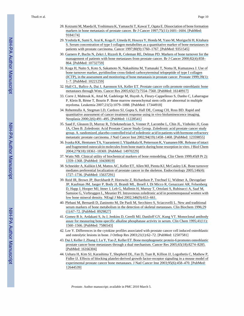
HistopathologyBased on BLI and radiography, we selected 13, 12, and 7 long bones from the control,prophylactic and delayed treatment groups, respectively, and stained sections from the boneswith H&E. In the control group, the prostate carcinoma cells induced woven bone formationin the metaphyses adjacent to the neoplastic cells demonstrating their osteoblastic phenotype(Fig. 4A). There were multiple areas of cortical and trabecular bone resorption caused by tumor-induced increased osteoclast activity along the cortical and trabecular endosteum. In some ofthe bones there was reactive new bone formation in the periosteum adjacent to the tumor. InZA-treated mice, Ace-1 metastases extended from the growth plate to the diaphysis and filledthe marrow cavities in between metaphyseal trabeculae (Fig. 4A,B). To determine the effectof ZA on tumor-induced bone formation, we measured metaphyseal trabecular thicknessadjacent to intramedullary metastases. Trabecular thickness adjacent to metastases wassignificantly (P < 0.001) increased in all tumor-bearing mice compared to contralateral bonesin non-tumor bearing mice. ZA had no effect on tumor-induced trabecular thickness in
Thudi et al. Page 5
Prostate. Author manuscript; available in PMC 2010 March 5.
NIH
-PA Author Manuscript
NIH
-PA Author Manuscript
NIH
-PA Author Manuscript

treatment groups compared to the control group (Fig. 4C). Histomorphometry demonstratedthat ZA had no significant effect on the tumor area in bone metastases (Fig. 4D).
TRAP staining was performed on the bone sections to identify the active osteoclasts. NumerousTRAP-positive osteoclasts (with three or more nuclei) were observed along the tumor-boneinterface in vehicle-treated mice compared to few active osteoclasts in the mice treated withZA (Fig. 5A). Bone histomorphometry was performed on the TRAP-stained bone sections toquantify the effect of ZA on the osteoclast activity at the tumor-bone interface. ZA decreasedTRAP-positive osteoclasts by five- to sixfold in tumor-bearing mice, as compared to vehicle-treated tumor-bearing mice (Fig. 5B).
Bone Remodeling IndicesOur results showed that ZA inhibited the prostate cancer-induced osteolysis, To determine andcompare the effect of ZA on bone remodeling in healthy mice and in tumor-bearing mice, wemeasured serum osteocalcin following treatment of mice for 4 weeks. ZA treatmentsignificantly (P < 0.0001) decreased serum osteocalcin concentrations in nontumor-bearingmice. In contrast, ZA treatment did not reduce serum osteocalcin in tumor-bearing mice (Fig.6A). Bone histomorphometric analysis showed that ZA treatment of healthy mice for 4 weeksincreased the trabecular volume by twofold (Fig. 6B) compared to the vehicle-treated mice.The decreased serum osteocalcin and increased trabecular bone volume in ZA-treated healthymice demonstrated the effect of ZA on the inhibition of normal physiological bone remodeling.
It has been shown that Dkk1 promotes osteolysis and inhibits osteoblast differentiation byantagonizing the Wnt signaling pathway in prostate cancer bone metastases [23,24]. Todetermine the role of Dkk1 in this study, we measured serum Dkk1 concentrations. Dkk1concentrations in tumor-bearing mice (avg = 4.2 ng/ml) were significantly increased comparedto nontumor-bearing mice (avg = 1.9 ng/ml). ZA significantly decreased Dkk1 concentrationsin the prophylactic treatment group but had no effect in delayed treatment mice as comparedto vehicle-treated mice (Fig. 6C). This could be due to the long duration of ZA treatment inthe prophylactic treatment compared to the delayed treatment. These data suggest that ZA-mediated inhibition of Dkk1 might contribute to the decreased osteolysis.
DISCUSSIONProstate cancer metastases to bone are typically characterized by a predominance of new wovenbone formation with a lesser amount of osteolysis (‘osteoblastic’ metastases) [23]. Thecontribution of osteolysis in the development of osteoblastic lesions in prostate cancer bonemetastases is not well understood partially due to a lack of preclinical models that consistentlydevelop mixed osteoblastic and osteolytic lesions [24–26]. This study showed that bonemetastasis and osteoblastic lesions are independent of osteolysis in a nude mouse model ofcanine prostate cancer-induced mixed bone metastases.
Studies have shown that normal bone homeostasis is maintained by the balanced coupling ofbone resorption and bone formation [27]. However, the importance of the bone remodelingsequence in the development of prostate cancer bone metastasis is unknown. Metastasis ofprostate cancer cells to bone disrupts the balanced coupling of bone remodeling. Althoughbone formation and bone resorption are both increased, bone formation is favored and theremodeling process becomes unbalanced, resulting in a net gain of bone. Many studies havereported that both osteoclast and osteoblast activity are important for prostate cancer bonemetastases [13]. Previously, the lack of preclinical models that recapitulate the sequentialcourse of mixed bone metastases has prevented investigations on the in vivo significance ofosteoclast activity in the development of osteoblastic lesions [23,28–31]. We have developeda canine prostate cancer cell line (Ace-1) that develops mixed bone metastases in nude mice
Thudi et al. Page 6
Prostate. Author manuscript; available in PMC 2010 March 5.
NIH
-PA Author Manuscript
NIH
-PA Author Manuscript
NIH
-PA Author Manuscript

[7,8]. This model allows studies on the sequential biological events involved in thedevelopment of prostate cancer bone metastases.
To investigate the role of osteoclastic bone resorption in the development of osteoblasticmetastases, we inhibited osteoclastic-mediated bone resorption in Ace-1 tumor-bearing miceusing zoledronic acid (ZA). As expected, radiographic and histopathological findings showedthat ZA markedly inhibited osteolysis of cortical and trabecular bone when compared to thecontrol group, regardless of the administration schedule. Decreased number of TRAP-stainedosteoclasts and decreased serum Dkk1 levels in the ZA-treated groups additionally supportedthis observation. Dkk1 was shown to decrease osteoblast maturation and mineralization in vitroand has the potential to switch the phenotype of bone metastasis from osteoblastic to osteolytic[32]. In the bone microenvironment, bone marrow mesenchymal stem cells (BMMSC) are thesource of Dkk1 [33]. The exact mechanism of ZA inhibition on Dkk1 is not clear. Based onthe findings in this study that ZA had no effect on the incidence of prostate cancer metastasisto bone or growth of bone metastases, but had decreased Dkk1 levels, we speculate that prostatecancer metastasis to bone stimulated Dkk1 expression in BMMSC. ZA inhibited the tumor-mediated stimulation of Dkk1 expression by BMMSC. Decreased Dkk1 levels might be oneof the potential factors contributing be the decrease in osteolysis and increased bone formationin this model. The ability of ZA to inhibit osteolysis in our study was consistent with previousreports [16–18]. ZA was less effective in the delayed treatment group compared to theprophylactic group, suggesting that preventive therapy may be more effective to treat patientswith osteolytic metastases in bone. On the other hand, the increased efficacy of prophylactictreatment in this study might be attributed to the longer duration of treatment (5 weeks)compared to shorter (2 weeks) administration in the delayed treatment group.
Recent studies have documented the ability of in vivo bioluminescent imaging (BLI) tomeasure tumor progression and response to therapy in animal models [34]. The BLI datarevealed that ZA treatment had no significant effect on the incidence of bone metastases or therate of metastatic tumor growth. Furthermore, analysis of tumor area using histomorphometryrevealed no significant difference between treatment and control groups despite the significantinhibition of osteolysis. Our data is in agreement with the report from Saad et al. [35] thatshowed ZA had no effect on tumor progression and survival rate of prostate cancer patients.Lee et al. [16] showed that ZA was effective in inhibiting bone resorption induced by theprostate cancer cell line (PC-3); however, there was no effect on osteoblastic lesions inducedby the LAPC-9 prostate cancer cell line. In contrast, Corey et al. [17] demonstrated that ZAinhibited tumor progression and osteoblastic lesions in an experimental mouse model withLuCaP 23.1 prostate cancer cells. These findings suggest the ability of ZA to inhibit tumorprogression and osteoblastic metastasis depends on the specific biology of the prostate cancercell line evaluated. Variation in the nature of different cell lines can be attributed to theheterogeneity of the primary prostate cancer from which cell lines are derived. Our findingsdemonstrate that the osteoclastic component of Ace-1- induced bone metastases were notnecessary for the survival and growth of tumor in bone or development of osteoblasticmetastases.
Osteocalcin, which is secreted by osteoblasts, is a bone turnover marker because the osteoblast-secreted osteocalcin is deposited in bone matrix and released during bone resorption [36,37].ZA significantly reduced serum osteocalcin in nude mice, indicating a reduction in boneturnover. This finding was consistent with previously published work showing that ZA reducedosteocalcin concentrations in patients with prostate cancer bone metastases [38,39]. There wasno significant difference in serum osteocalcin levels in Ace-1-bearing control mice comparedto control nontumor-bearing mice. Koizumi et al. previously showed that serum osteocalcinconcentrations were similar in human patients with and without prostate cancer bonemetastases [28,40]. This correlated with our findings and suggested that osteocalcin is not a
Thudi et al. Page 7
Prostate. Author manuscript; available in PMC 2010 March 5.
NIH
-PA Author Manuscript
NIH
-PA Author Manuscript
NIH
-PA Author Manuscript

useful marker to identify bone metastases. Serum bone specific alkaline phosphatase, secretedby osteoblasts, is a specific and reliable bone formation marker [41]. We were unable tomeasure the bone specific alkaline phosphatase concentrations in the mice due to the lack ofassays that detect this marker in mice.
Many studies have reported that osteoclast and osteoblast regulatory factors expressed byprostate cancer cells can alter bone homeostasis either directly or indirectly. Prostate (PC-3)and breast (MDA-231) cancer cell lines that induce osteolytic bone metastases expresscytokines that include RANKL, interleukin-1 (IL-1), tumor necrosis factor-a (TNF-α), PTHrPand cathepsin K, which are associated with increased osteoclastogenesis. In contrast, C4-2Band LAPC-9 cells (prostate cancer) and ZR-75-1 and MCF-7 cells (breast cancer), which induceosteoblastic metastases, express abundant osteoprotegerin (OPG), bone morphogenic protein-2(BMP-2), BMP-4, BMP-6, vascular endothelial growth factor (VEGF), endothelin-1 (ET-1),platelet derived growth factor-BB (PDGF-BB), insulin growth factor-1 (IGF-1) and fibroblastgrowth factor-2 (FGF-2), which are known to stimulate osteoblast activity [16,24,42–45]. Yinet al. [46] showed that ET-1 production in ZR-75-1 cells stimulated osteoblast activity resultingin increased new bone formation. PDGF-BB, VEGF and urokinase plasminogen activator(uPA) also contributed to the increased bone formation in prostate and breast cancer bonemetastases [24,47,48]. PCR analysis revealed that Ace-1 cells express a wide variety of factorsknown to stimulate both osteoclasts (RANKL, IL-6, cathepsin K, PTHrP) and osteoblastactivity (PDGF-BB, ET-1, VEGF, uPA, OPG, FGF-2 and IGF-1) (unpublished data). Thecytokine expression profile of Ace-1 cells suggests that multiple factors contribute to theosteoblastic and osteolytic phenotypes at metastatic sites. However, further investigations willbe required to understand the role various cytokines in the development of mixed bonemetastases in this model. In this regard, the Ace-1 model will be a very useful translationalmodel to study the pathogenesis and treatment of prostate cancer bone metastasis.
AcknowledgmentsWe would like to thank Fu-Sheng Chou for his assistance with the ELISA assay and Tim Vojt for illustrations. Thiswork was supported by the National Cancer Institute (CA100730 and CA77911) and the National Center for ResearchResources (RR00168).
Grant sponsor: National Cancer Institute; Grant numbers: CA100730, CA77911; Grant sponsor: National Center forResearch Resources; Grant number: RR00168.
REFERENCES1. Jemal A, Siegel R, Ward E, Murray T, Xu J, Smigal C, Thun MJ. Cancer statistics 2006. CA Cancer
J Clin 2006;56(2):106–130. [PubMed: 16514137]2. Adami S. Bisphosphonates in prostate carcinoma. Cancer 1997;80(8 Suppl):1674–1679. [PubMed:
9362435]3. Fidler IJ. The pathogenesis of cancer metastasis: The ’seed and soil’ hypothesis revisited. Nat Rev
Cancer 2003;3(6):453–458. [PubMed: 12778135]4. Crawford ED. Skeletal complications in men with prostate cancer: Effects on quality-of-life outcomes
throughout the continuum of care. Eur Urol Suppl 2004;3(5):10–15.5. Mundy GR. Metastasis to bone: Causes, consequences and therapeutic opportunities. Nat Rev Cancer
2002;2(8):584–593. [PubMed: 12154351]6. Charhon SA, Chapuy MC, Delvin EE, Valentin-Opran A, Edouard CM, Meunier PJ.
Histomorphometric analysis of sclerotic bone metastases from prostatic carcinoma special referenceto osteomalacia. Cancer 1983;51(5):918–924. [PubMed: 6681595]
7. Leroy BE, Thudi NK, Nadella MV, Toribio RE, Tannehill-Gregg SH, van Bokhoven A, Davis D, CornS, Rosol TJ. New bone formation and osteolysis by a metastatic, highly invasive canine prostatecarcinoma xenograft. Prostate 2006;66(11):1213–1222. [PubMed: 16683269]
Thudi et al. Page 8
Prostate. Author manuscript; available in PMC 2010 March 5.
NIH
-PA Author Manuscript
NIH
-PA Author Manuscript
NIH
-PA Author Manuscript

8. Halvorson KG, Kubota K, Sevcik MA, Lindsay TH, Sotillo JE, Ghilardi JR, Rosol TJ, Boustany L,Shelton DL, Mantyh PW. A blocking antibody to nerve growth factor attenuates skeletal pain inducedby prostate tumor cells growing in bone. Cancer Res 2005;65(20):9426–9435. [PubMed: 16230406]
9. Khanna C, Lindblad-Toh K, Vail D, London C, Bergman P, Barber L, Breen M, Kitchell B, McNeilE, Modiano JF, Niemi S, Comstock KE, Ostrander E, Westmoreland S, Withrow S. The dog as a cancermodel. Nat Biotechnol 2006;24(9):1065–1066. [PubMed: 16964204]
10. Waters DJ, Hayden DW, Bell FW, Klausner JS, Qian J, Bostwick DG. Prostatic intraepithelialneoplasia in dogs with spontaneous prostate cancer. Prostate 1997;30(2):92–97. [PubMed: 9051147]
11. Hart IR, Fidler IJ. Role of organ selectivity in the determination of metastatic patterns of B16melanoma. Cancer Res 1980;40(7):2281–2287. [PubMed: 7388794]
12. Clines GA, Guise TA. Hypercalcaemia of malignancy and basic research on mechanisms responsiblefor osteolytic and osteoblastic metastasis to bone. Endocr Relat Cancer 2005;12(3):549–583.[PubMed: 16172192]
13. Keller ET. Overview of metastasis and metastases. J Musculoskelet Neuronal Interact 2002;2(6):567–569. [PubMed: 15758397]
14. Yoneda T, Michigami T, Yi B, Williams PJ, Niewolna M, Hiraga T. Actions of bisphosphonate onbone metastasis in animal models of breast carcinoma. Cancer 2000;88(12 Suppl):2979–2988.[PubMed: 10898341]
15. Goltzman D. Mechanisms of the development of osteoblastic metastases. Cancer 1997;80(8 Suppl):1581–1587. [PubMed: 9362425]
16. Lee YP, Schwarz EM, Davies M, Jo M, Gates J, Zhang X, Wu J, Lieberman JR. Use of zoledronateto treat osteoblastic versus osteolytic lesions in a severe-combined-immunodeficient mouse model.Cancer Res 2002;62(19):5564–5570. [PubMed: 12359769]
17. Corey E, Brown LG, Quinn JE, Poot M, Roudier MP, Higano CS, Vessella RL. Zoledronic acidexhibits inhibitory effects on osteoblastic and osteolytic metastases of prostate cancer. Clin CancerRes 2003;9(1):295–306. [PubMed: 12538482]
18. Croucher PI, De Hendrik R, Perry MJ, Hijzen A, Shipman CM, Lippitt J, Green J, Van Marck E, VanCamp B, Vanderkerken K. Zoledronic acid treatment of 5T2MM-bearing mice inhibits thedevelopment of myeloma bone disease: Evidence for decreased osteolysis, tumor burden andangiogenesis, and increased survival. J Bone Miner Res 2003;18(3):482–492. [PubMed: 12619933]
19. Lipton A, Small E, Saad F, Gleason D, Gordon D, Smith M, Rosen L, Kowalski MO, Reitsma D,Seaman J. The new bisphosphonate, zometa (zoledronic acid), decreases skeletal complications inboth osteolytic and osteoblastic lesions: A comparison to pamidronate. Cancer Invest 2002;20:45–54. [PubMed: 12442349]
20. Saad F, Schulman CC. Role of bisphosphonates in prostate cancer. Urology 2004;45:26–34.21. Guise TA, Yin JJ, Taylor SD, Kumagai Y, Dallas M, Boyce BF, Yoneda T, Mundy GR. Evidence
for a causal role of parathyroid hormone-related protein in the pathogenesis of human breast cancer-mediated osteolysis. J Clin Invest 1996;98(7):1544–1549. [PubMed: 8833902]
22. Parfitt AM, Drezner MK, Glorieux FH, Kanis JA, Malluche H, Meunier PJ, Ott SM, Recker RR.Bone histomorphometry: Standardization of nomenclature, symbols, and units. Report of theASBMR histomorphometry nomenclature committee. J Bone Miner Res 1987;2(6):595–610.[PubMed: 3455637]
23. Percival RC, Urwin GH, Harris S, Yates AJ, Williams JL, Beneton M, Kanis JA. Biochemical andhistological evidence that carcinoma of the prostate is associated with increased bone resorption. EurJ Surg Oncol 1987;13(1):41–49. [PubMed: 3102281]
24. Kitagawa Y, Dai J, Zhang J, Keller JM, Nor J, Yao Z, Keller ET. Vascular endothelial growth factorcontributes to prostate cancer-mediated osteoblastic activity. Cancer Res 2005;65(23):10921–10929.[PubMed: 16322239]
25. Mundy GR. Endothelin-1 and osteoblastic metastasis. Proc Natl Acad Sci USA 2003;100(19):10588–10589. [PubMed: 12963808]
26. Roodman GD. Mechanisms of bone metastasis. N Engl J Med 2004;350(16):1655–1664. [PubMed:15084698]
27. Rodan GA. Bone homeostasis. Proc Natl Acad Sci USA 1998;95:13361–13362. [PubMed: 9811806]
Thudi et al. Page 9
Prostate. Author manuscript; available in PMC 2010 March 5.
NIH
-PA Author Manuscript
NIH
-PA Author Manuscript
NIH
-PA Author Manuscript
28. Koizumi M, Maeda H, Yoshimura K, Yamauchi T, Kawai T, Ogata E. Dissociation of bone formationmarkers in bone metastasis of prostate cancer. Br J Cancer 1997;75(11):1601–1604. [PubMed:9184174]
29. Yoshida K, Sumi S, Arai K, Koga F, Umeda H, Hosoya Y, Honda M, Yano M, Moriguchi H, KitaharaS. Serum concentration of type I collagen metabolites as a quantitative marker of bone metastases inpatients with prostate carcinoma. Cancer 1997;80(9):1760–1767. [PubMed: 9351545]
30. Garnero P, Buchs N, Zekri J, Rizzoli R, Coleman RE, Delmas PD. Markers of bone turnover for themanagement of patients with bone metastases from prostate cancer. Br J Cancer 2000;82(4):858–864. [PubMed: 10732759]
31. Koga H, Naito S, Koto S, Sakamoto N, Nakashima M, Yamasaki T, Noma H, Kumazawa J. Use ofbone turnover marker, pyridinoline cross-linked carboxyterminal telopeptide of type I collagen(ICTP), in the assessment and monitoring of bone metastasis in prostate cancer. Prostate 1999;39(1):1–7. [PubMed: 10221259]
32. Hall CL, Bafico A, Dai J, Aaronson SA, Keller ET. Prostate cancer cells promote osteoblastic bonemetastases through Wnts. Cancer Res 2005;65(17):7554–7560. [PubMed: 16140917]
33. Corre J, Mahtouk K, Attal M, Gadelorge M, Huynh A, Fleury-Cappellesso S, Danho C, LaharragueP, Klein B, Rème T, Bourin P. Bone marrow mesenchymal stem cells are abnormal in multiplemyeloma. Leukemia 2007;21(5):1079–1088. [PubMed: 17344918]
34. Rehemtulla A, Stegman LD, Cardozo SJ, Gupta S, Hall DE, Contag CH, Ross BD. Rapid andquantitative assessment of cancer treatment response using in vivo bioluminescence imaging.Neoplasia 2000;2(6):491–495. [PubMed: 11228541]
35. Saad F, Gleason D, Murray R, Tchekmedyian S, Venner P, Lacombe L, Chin JL, Vinholes JJ, GoasJA, Chen B. Zoledronic Acid Prostate Cancer Study Group. Zoledronic acid prostate cancer studygroup. A. randomized, placebo-controlled trial of zoledronic acid in patients with hormone-refractorymetastatic prostate carcinoma. J Natl Cancer Inst 2002;94(19):1458–1468. [PubMed: 12359855]
36. Ivaska KK, Hentunen TA, Vaaraniemi J, Ylipahkala H, Pettersson K, Vaananen HK. Release of intactand fragmented osteocalcin molecules from bone matrix during bone resorption in vitro. J Biol Chem2004;279(18):18361–18369. [PubMed: 14970229]
37. Watts NB. Clinical utility of biochemical markers of bone remodeling. Clin Chem 1999;45(8 Pt 2):1359–1368. [PubMed: 10430819]
38. Schneider A, Kalikin LM, Mattos AC, Keller ET, Allen MJ, Pienta KJ, McCauley LK. Bone turnovermediates preferential localization of prostate cancer in the skeleton. Endocrinology 2005;146(4):1727–1736. [PubMed: 15637291]
39. Reid IR, Brown JP, Burckhardt P, Horowitz Z, Richardson P, Trechsel U, Widmer A, DevogelaerJP, Kaufman JM, Jaeger P, Body JJ, Brandi ML, Broell J, Di Micco R, Genazzani AR, FelsenbergD, Happ J, Hooper MJ, Ittner J, Leb G, Mallmin H, Murray T, Ortolani S, Rubinacci A, Saaf M,Samsioe G, Verbruggen L, Meunier PJ. Intravenous zoledronic acid in postmenopausal women withlow bone mineral density. NEngl J Med 2002;346(9):653–661.
40. Plebani M, Bernardi D, Zaninotto M, De Paoli M, Secchiero S, Sciacovelli L. New and traditionalserum markers of bone metabolism in the detection of skeletal metastases. Clin Biochem 1996;29(1):67–72. [PubMed: 8929827]
41. Gomez B Jr, Ardakani S, Ju J, Jenkins D, Cerelli MJ, Daniloff GY, Kung VT. Monoclonal antibodyassay for measuring bone-specific alkaline phosphatase activity in serum. Clin Chem 1995;41(11):1560–1566. [PubMed: 7586543]
42. Lee Y. Differences in the cytokine profiles associated with prostate cancer cell induced osteoblasticand osteolytic lesions in bone. J Orthop Res 2003;21(1):62–72. [PubMed: 12507581]
43. Dai J, Keller J, Zhang J, Lu Y, Yao Z, Keller ET. Bone morphogenetic protein-6 promotes osteoblasticprostate cancer bone metastases through a dual mechanism. Cancer Res 2005;65(18):8274–8285.[PubMed: 16166304]
44. Uehara H, Kim SJ, Karashima T, Shepherd DL, Fan D, Tsan R, Killion JJ, Logothetis C, Mathew P,Fidler IJ. Effects of blocking platelet-derived growth factor-receptor signaling in a mouse model ofexperimental prostate cancer bone metastases. J Natl Cancer Inst 2003;95(6):458–470. [PubMed:12644539]
Thudi et al. Page 10
Prostate. Author manuscript; available in PMC 2010 March 5.
NIH
-PA Author Manuscript
NIH
-PA Author Manuscript
NIH
-PA Author Manuscript

45. Guise TA, Yin JJ, Mohammad KS. Role of endothelin-1 in osteoblastic bone metastases. Cancer2003;97(3 Suppl):779–784. [PubMed: 12548575]
46. Yin JJ, Mohammad KS, Kakonen SM, Harris S, Wu-Wong JR, Wessale JL, Padley RJ, Garrett IR,Chirgwin JM, Guise TA. A causal role for endothelin-1 in the pathogenesis of osteoblastic bonemetastases. Proc Natl Acad Sci USA 2003;100(19):10954–10959. [PubMed: 12941866]
47. Yi B, Williams PJ, Niewolna M, Wang Y, Yoneda T. Tumor-derived platelet-derived growth factor-BB plays a critical role in osteosclerotic bone metastasis in an animal model of human breast cancer.Cancer Res 2002;62(3):917–923. [PubMed: 11830552]
48. Achbarou A, Kaiser S, Tremblay G, Ste-Marie LG, Brodt P, Goltzman D, Rabbani SA. Urokinaseoverproduction results in increased skeletal metastasis by prostate cancer cells in vivo. Cancer Res1994;54(9):2372–2377. [PubMed: 8162583]
Thudi et al. Page 11
Prostate. Author manuscript; available in PMC 2010 March 5.
NIH
-PA Author Manuscript
NIH
-PA Author Manuscript
NIH
-PA Author Manuscript

Fig. 1.Treatment protocol. Nude mice were divided into three groups: the control group received PBSfrom –1 to 4 weeks (top line). The prophylaxis group received ZA from –1 to 4 weeks (middleline).The delayed treatment group received ZA from 2 to 4 weeks (bottom line). Mice wereadministered 100 µg/kg ZA BW, twice a week subcutaneously (SQ) to their respective groups.Ace-1YFP-LUC cells were injected on week 0.
Thudi et al. Page 12
Prostate. Author manuscript; available in PMC 2010 March 5.
NIH
-PA Author Manuscript
NIH
-PA Author Manuscript
NIH
-PA Author Manuscript

Fig. 2.Effect of ZA on the Ace-1 tumor growth and metastasis incidence in nude mice was monitoredusing bioluminescence imaging (BLI). A: Representative images of serial BLI of Ace-1 tumorprogression from control, delayed treatment and prophylaxis groups taken at 0,7,14,21, and 28days, after intracardiac injection of cancer cells. On day 0, immediately after intracardiacinjections, ACE-1 cells were present throughout the entire body and accumulated in the kidney,liver and brain. On day 7, BLI signals were gone. BLI signals were detected on day 14 at varioussites of bone metastasis. At day 21 and 28, the intensity of BLI signals increased, whichdemonstrated progressive growth of the metastases. BLI on days 21 and 28 did not reveal anynew metastatic sites compared to day14. Panel B graph represents the average intensity of theBLI signal measured at each metastatic region of interest (ROI) per group at the specific timepoints. Intensity of BLI was measured using Living Image software Version 2.50. Panel Cgraph shows the average number of metastases per group. Each region of interest was countedas one metastatic site.
Thudi et al. Page 13
Prostate. Author manuscript; available in PMC 2010 March 5.
NIH
-PA Author Manuscript
NIH
-PA Author Manuscript
NIH
-PA Author Manuscript

Fig. 3.Radiographic evaluation of ZA on Ace-1 metastases to long bones. Radiographs of mice weretaken 28 days after injection of Ace-1 tumor cells into the left cardiac ventricle. Cortical andtrabecular bone lysis (thin arrows) was observed in the metaphyseal regions of long bones ofmice that received Ace-1 cancer cells and vehicle (C) compared to mice that received vehiclealone (A). ZA-treated nontumor-bearing mice (B) had a mild increase in radioopacity in theproximal metaphysis of long bones compared to vehicle-treated nontumor-bearing mice (A).Mild trabecular bone loss and intact cortices were present in the delayed treatment group (D),whereas in the prophylactic group (E), intact cortices and increased radio-opacity of bone (thickarrow) was present in the metaphyseal region.
Thudi et al. Page 14
Prostate. Author manuscript; available in PMC 2010 March 5.
NIH
-PA Author Manuscript
NIH
-PA Author Manuscript
NIH
-PA Author Manuscript
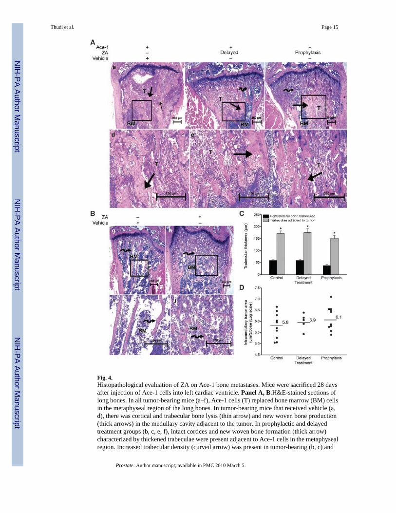
Fig. 4.Histopathological evaluation of ZA on Ace-1 bone metastases. Mice were sacrificed 28 daysafter injection of Ace-1 cells into left cardiac ventricle. Panel A, B:H&E-stained sections oflong bones. In all tumor-bearing mice (a–f), Ace-1 cells (T) replaced bone marrow (BM) cellsin the metaphyseal region of the long bones. In tumor-bearing mice that received vehicle (a,d), there was cortical and trabecular bone lysis (thin arrow) and new woven bone production(thick arrows) in the medullary cavity adjacent to the tumor. In prophylactic and delayedtreatment groups (b, c, e, f), intact cortices and new woven bone formation (thick arrow)characterized by thickened trabeculae were present adjacent to Ace-1 cells in the metaphysealregion. Increased trabecular density (curved arrow) was present in tumor-bearing (b, c) and
Thudi et al. Page 15
Prostate. Author manuscript; available in PMC 2010 March 5.
NIH
-PA Author Manuscript
NIH
-PA Author Manuscript
NIH
-PA Author Manuscript

nontumor-bearing (h, j) mice that received ZA compared to the mice that received vehiclealone (g, i). Panel C: histomorphometric analysis showed that trabecular thickness in longbones adjacent to metastases was significantly greater than contralateral bones withoutmetastases. *P < 0.001 (t-test) and data represent the mean ± SEM. Panel D shows a verticaldot plot of the individual values of tumor area from each long bone that was quantified.
Thudi et al. Page 16
Prostate. Author manuscript; available in PMC 2010 March 5.
NIH
-PA Author Manuscript
NIH
-PA Author Manuscript
NIH
-PA Author Manuscript
Fig. 5.Effect of ZA on tartrate-resistant acid phosphatase (TRAP) activity of osteoclasts in Ace-1tumor-bearing mice. TRAP-stained sections of bones demonstrated numerous red TRAP-positive osteoclasts (thick arrow) along the tumor (T)–bone (B) interface in Ace-1-bearingmice compared to few osteoclasts in ZA-treated mice (A, top panels).Higher magnificationof TRAP-positive osteoclasts demonstrated multiple nuclei (thin head arrow) (A, lowerpanels). Histomorphometric analysis showed a significant decrease in TRAP-positiveosteoclasts with three or more nuclei along the tumor-bone interface between control and ZA-treated groups (Panel B). *P < 0.01 (ANOVA and Dunn’s test for posthoc analysis). Datarepresent the mean ± standard error of mean (SEM).
Thudi et al. Page 17
Prostate. Author manuscript; available in PMC 2010 March 5.
NIH
-PA Author Manuscript
NIH
-PA Author Manuscript
NIH
-PA Author Manuscript
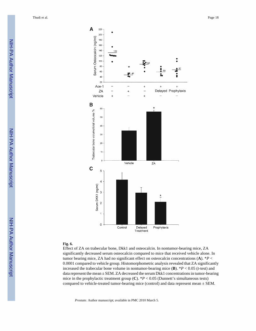
Fig. 6.Effect of ZA on trabecular bone, Dkk1 and osteocalcin. In nontumor-bearing mice, ZAsignificantly decreased serum osteocalcin compared to mice that received vehicle alone. Intumor bearing mice, ZA had no significant effect on osteocalcin concentrations (A). *P <0.0001 compared to vehicle group. Histomorphometric analysis revealed that ZA significantlyincreased the trabecular bone volume in nontumor-bearing mice (B). *P < 0.05 (t-test) anddata represent the mean ± SEM. ZA decreased the serum Dkk1 concentrations in tumor-bearingmice in the prophylactic treatment group (C). *P < 0.05 (Dunnett’s simultaneous tests)compared to vehicle-treated tumor-bearing mice (control) and data represent mean ± SEM.
Thudi et al. Page 18
Prostate. Author manuscript; available in PMC 2010 March 5.
NIH
-PA Author Manuscript
NIH
-PA Author Manuscript
NIH
-PA Author Manuscript
Related Documents