Applied Catalysis B: Environmental 144 (2014) 386–393 Contents lists available at ScienceDirect Applied Catalysis B: Environmental jo ur nal home p ag e: www.elsevier.com/locate/apcatb ZnSnO 3 hollow nanospheres/reduced graphene oxide nanocomposites as high-performance photocatalysts for degradation of metronidazole Shuying Dong a , Jingyu Sun b,1 , Yukun Li a , Chongfei Yu a , Yihui Li c , Jianhui Sun a,∗ a School of Environment, Henan Normal University, Key Laboratory for Yellow River and Huai River Water Environmental and Pollution Control Ministry of Education, Henan Key Laboratory for Environmental Pollution Control, Xinxiang, Henan 453007, PR China b Department of Materials, University of Oxford, Parks Road, Oxford OX1 3PH, United Kingdom c School of Chemistry and Chemical Engineering, Henan Normal University, Xinxiang, Henan 453007, PR China a r t i c l e i n f o Article history: Received 3 April 2013 Received in revised form 7 July 2013 Accepted 15 July 2013 Available online 24 July 2013 Keywords: ZnSnO3 Hollow nanosphere Graphene oxide Metronidazole Photocatalytic a b s t r a c t The fabrication of novel ZnSnO 3 hollow nanospheres/reduced graphene oxide (RGO) hybrid nanocom- posite is reported for the first time. The nanocomposites were synthesized via a facile route, and were well characterized with the aid of XRD, FTIR, SEM, TEM, BET, UV–vis, and PL techniques. Moreover, the synthesized nanocomposites were used as photocatalysts in the application of the degradation of pharmaceutical wastewater. In this study, ZnSnO 3 hollow nanospheres showed high efficiency in pho- tocatalytic degradation of metronidazole under ultraviolet (UV) light irradiation. More interestingly, the photocatalytic activities of these nanospheres could be enhanced by coupling with RGO, where a large improvement (approx. 30.4% increase compared with pure ZnSnO 3 ) in photodegradation of metronida- zole was observed on the prepared ZnSnO 3 /RGO hybrid nanocomposites under visible light irradiation. This improvement might be attributed to the advanced adsorption efficiency of molecules and enhanced visible light absorption within the hybrid nanocomposites by the introduction of RGO. Such study might pave the way toward designing novel photocatalyst systems for efficient degradation of pharmaceutical wastewater. © 2013 Elsevier B.V. All rights reserved. 1. Introduction As a common nitroimidazole antibiotic derivative, metro- nidazole (2-methyl-5-nitroimidazole-1-ethanol) was abused for treating infections caused by a wide range of anaerobic bacte- ria, bacteroides and protozoa, including trichomoniasis, amoebiasis and gingivitis [1–3]. The maximum concentration of metronida- zole at 9400 and 127 ng L −1 have been found in the effluents of hospital and sewage treatment plant, respectively [4,5]. Being as a pharmaceutical substance as well as a potential carcinogen, met- ronidazole was prone to accumulating in aquatic environments, leading to the pollution of surface water and groundwater [6]. How- ever, the removal efficiency of metronidazole remains to be quite low by employing conventional sewage treatment methodologies [7]. Hence it is imperative to effectively remove metronidazole from the wastewater with the aid of relevant treatment techniques. Recent development of wastewater treatment technologies has enabled high-efficiency removal of pharmaceutical substances ∗ Corresponding author. Tel.: +86 373 3325971; fax: +86 373 3326336. E-mail address: [email protected] (J. Sun). 1 Current address: Center for Nanochemistry (CNC), College of Chemistry and Molecular Engineering, Peking University, Beijing 100871, PR China. in the aqueous environment, amongst which the photocatalytic degradation technique has been specifically ulitized. The advantage of the photocatalytic degradation technique lies mainly in the fact that it employes low-cost, environment-friendly semiconductor materials (e.g. ZnO) for complete mineralization of complex- structured organics (including pharmaceutical substances) [8,9]. Interest in ZnO for pollution treatment has been stimulated by its great photocatalytic capacity in the degradation of organic pol- lutants [10,11], and its photocatalytic efficiency can be further improved via various approaches such as introducing dopants, in order to suppress the fast-rated recombination of photogenerated electron/hole pairs within the material [12–14]. One of our recent studies has demonstrated that Sn-modified ZnO nanomaterials were characterized with increased sunlight photocatalytic activ- ities (compared to the single-phase ZnO), where Sn 4+ functioned as a good n-type dopant with extensive solid solubility in ZnO and similar value of the atomic radius with Zn 2+ (Sn 4+ : 0.069 nm, Zn 2+ : 0.074 nm) [15]. It is well realized that the photocatalytic performances of photo- catalysts strongly depends on the sizes and shapes of employed materials, especially at the nanoscale. Recent studies have shown that hollow-structured ZnO-based nanomaterials could act as promising photocatalysts, owing to their high specific surface area, good permeability, and high interfacial charge-transfer efficiency 0926-3373/$ – see front matter © 2013 Elsevier B.V. All rights reserved. http://dx.doi.org/10.1016/j.apcatb.2013.07.043

Welcome message from author
This document is posted to help you gain knowledge. Please leave a comment to let me know what you think about it! Share it to your friends and learn new things together.
Transcript
Za
Sa
Eb
c
a
ARRAA
KZHGMP
1
ntrazhprlel[t
h
M
0h
Applied Catalysis B: Environmental 144 (2014) 386– 393
Contents lists available at ScienceDirect
Applied Catalysis B: Environmental
jo ur nal home p ag e: www.elsev ier .com/ locate /apcatb
nSnO3 hollow nanospheres/reduced graphene oxide nanocompositess high-performance photocatalysts for degradation of metronidazole
huying Donga, Jingyu Sunb,1, Yukun Lia, Chongfei Yua, Yihui Li c, Jianhui Suna,∗
School of Environment, Henan Normal University, Key Laboratory for Yellow River and Huai River Water Environmental and Pollution Control Ministry ofducation, Henan Key Laboratory for Environmental Pollution Control, Xinxiang, Henan 453007, PR ChinaDepartment of Materials, University of Oxford, Parks Road, Oxford OX1 3PH, United KingdomSchool of Chemistry and Chemical Engineering, Henan Normal University, Xinxiang, Henan 453007, PR China
r t i c l e i n f o
rticle history:eceived 3 April 2013eceived in revised form 7 July 2013ccepted 15 July 2013vailable online 24 July 2013
eywords:nSnO3
a b s t r a c t
The fabrication of novel ZnSnO3 hollow nanospheres/reduced graphene oxide (RGO) hybrid nanocom-posite is reported for the first time. The nanocomposites were synthesized via a facile route, and werewell characterized with the aid of XRD, FTIR, SEM, TEM, BET, UV–vis, and PL techniques. Moreover,the synthesized nanocomposites were used as photocatalysts in the application of the degradation ofpharmaceutical wastewater. In this study, ZnSnO3 hollow nanospheres showed high efficiency in pho-tocatalytic degradation of metronidazole under ultraviolet (UV) light irradiation. More interestingly, thephotocatalytic activities of these nanospheres could be enhanced by coupling with RGO, where a large
ollow nanosphereraphene oxideetronidazole
hotocatalytic
improvement (approx. 30.4% increase compared with pure ZnSnO3) in photodegradation of metronida-zole was observed on the prepared ZnSnO3/RGO hybrid nanocomposites under visible light irradiation.This improvement might be attributed to the advanced adsorption efficiency of molecules and enhancedvisible light absorption within the hybrid nanocomposites by the introduction of RGO. Such study mightpave the way toward designing novel photocatalyst systems for efficient degradation of pharmaceuticalwastewater.
. Introduction
As a common nitroimidazole antibiotic derivative, metro-idazole (2-methyl-5-nitroimidazole-1-ethanol) was abused forreating infections caused by a wide range of anaerobic bacte-ia, bacteroides and protozoa, including trichomoniasis, amoebiasisnd gingivitis [1–3]. The maximum concentration of metronida-ole at 9400 and 127 ng L−1 have been found in the effluents ofospital and sewage treatment plant, respectively [4,5]. Being as aharmaceutical substance as well as a potential carcinogen, met-onidazole was prone to accumulating in aquatic environments,eading to the pollution of surface water and groundwater [6]. How-ver, the removal efficiency of metronidazole remains to be quiteow by employing conventional sewage treatment methodologies7]. Hence it is imperative to effectively remove metronidazole from
he wastewater with the aid of relevant treatment techniques.Recent development of wastewater treatment technologiesas enabled high-efficiency removal of pharmaceutical substances
∗ Corresponding author. Tel.: +86 373 3325971; fax: +86 373 3326336.E-mail address: [email protected] (J. Sun).
1 Current address: Center for Nanochemistry (CNC), College of Chemistry andolecular Engineering, Peking University, Beijing 100871, PR China.
926-3373/$ – see front matter © 2013 Elsevier B.V. All rights reserved.ttp://dx.doi.org/10.1016/j.apcatb.2013.07.043
© 2013 Elsevier B.V. All rights reserved.
in the aqueous environment, amongst which the photocatalyticdegradation technique has been specifically ulitized. The advantageof the photocatalytic degradation technique lies mainly in the factthat it employes low-cost, environment-friendly semiconductormaterials (e.g. ZnO) for complete mineralization of complex-structured organics (including pharmaceutical substances) [8,9].Interest in ZnO for pollution treatment has been stimulated by itsgreat photocatalytic capacity in the degradation of organic pol-lutants [10,11], and its photocatalytic efficiency can be furtherimproved via various approaches such as introducing dopants, inorder to suppress the fast-rated recombination of photogeneratedelectron/hole pairs within the material [12–14]. One of our recentstudies has demonstrated that Sn-modified ZnO nanomaterialswere characterized with increased sunlight photocatalytic activ-ities (compared to the single-phase ZnO), where Sn4+ functionedas a good n-type dopant with extensive solid solubility in ZnO andsimilar value of the atomic radius with Zn2+ (Sn4+: 0.069 nm, Zn2+:0.074 nm) [15].
It is well realized that the photocatalytic performances of photo-catalysts strongly depends on the sizes and shapes of employed
materials, especially at the nanoscale. Recent studies have shownthat hollow-structured ZnO-based nanomaterials could act aspromising photocatalysts, owing to their high specific surface area,good permeability, and high interfacial charge-transfer efficiency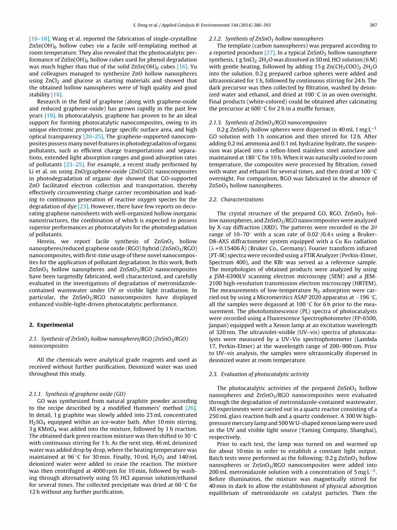
: Envi
[Zrfwauts
aysuopptoLiZeidrnso
nniZhecpe
2
2n
rt
2
tIH3Twwmdwif1
S. Dong et al. / Applied Catalysis B
16–18]. Wang et al. reported the fabrication of single-crystallinenSn(OH)6 hollow cubes via a facile self-templating method atoom temperature. They also revealed that the photocatalytic per-ormance of ZnSn(OH)6 hollow cubes used for phenol degradationas much higher than that of the solid ZnSn(OH)6 cubes [16]. Yu
nd colleagues managed to synthesize ZnO hollow nanospheressing ZnCl2 and glucose as starting materials and showed thathe obtained hollow nanospheres were of high quality and goodtability [18].
Research in the field of graphene (along with graphene-oxidend reduced graphene-oxide) has grown rapidly in the past fewears [19]. In photocatalysis, graphene has proven to be an idealupport for forming photocatalytic nanocomposites, owing to itsnique electronic properties, large specific surface area, and highptical transparency [20–25]. The graphene-supported nanocom-osites possess many novel features in photodegradation of organicollutants, such as efficient charge transportations and separa-ions, extended light absorption ranges and good adsorption ratesf pollutants [23–25]. For example, a recent study performed byi et al. on using ZnO/graphene-oxide (ZnO/GO) nanocompositesn photodegradation of organic dye showed that GO-supportednO facilitated electron collection and transportation, therebyffectively circumventing charge carrier recombination and lead-ng to continuous generation of reactive oxygen species for theegradation of dye [23]. However, there have few reports on deco-ating graphene nanosheets with well-organized hollow inorganicanostructures, the combination of which is expected to possessuperior performances as photocatalysts for the photodegradationf pollutants.
Herein, we report facile synthesis of ZnSnO3 hollowanospheres/reduced graphene oxide (RGO) hybrid (ZnSnO3/RGO)anocomposites, with first-time usage of these novel nanocompos-
tes for the application of pollutant degradation. In this work, BothnSnO3 hollow nanospheres and ZnSnO3/RGO nanocompositesave been targetedly fabricated, well characterized, and carefullyvaluated in the investigations of degradation of metronidazole-ontained wastewater under UV or visible light irradiation. Inarticular, the ZnSnO3/RGO nanocomposites have displayednhanced visible-light-driven photocatalytic performance.
. Experimental
.1. Synthesis of ZnSnO3 hollow nanospheres/RGO (ZnSnO3/RGO)anocomposites
All the chemicals were analytical grade reagents and used aseceived without further purification. Deionized water was usedhroughout this study.
.1.1. Synthesis of graphene oxide (GO)GO was synthesized from natural graphite powder according
o the recipe described by a modified Hummers’ method [26].n detail, 1 g graphite was slowly added into 23 mL concentrated
2SO4 equipped within an ice-water bath. After 10 min stirring, g KMnO4 was added into the mixture, followed by 1 h reaction.he obtained dark green reaction mixture was then shifted to 30 ◦Cith continuous stirring for 1 h. As the next step, 46 mL deionizedater was added drop by drop, where the heating temperature wasaintained at 96 ◦C for 30 min. Finally, 10 mL H2O2 and 140 mL
eionized water were added to cease the reaction. The mixture
as then centrifuged at 4000 rpm for 10 min, followed by wash-ng through alternatively using 5% HCl aqueous solution/ethanolor several times. The collected precipitate was dried at 60 ◦C for2 h without any further purification.
ronmental 144 (2014) 386– 393 387
2.1.2. Synthesis of ZnSnO3 hollow nanospheresThe template (carbon nanospheres) was prepared according to
a reported procedure [27]. In a typical ZnSnO3 hollow nanospheresynthesis, 1 g SnCl2·2H2O was dissolved in 50 mL HCl solution (6 M)with gentle heating, followed by adding 15 g Zn(CH3COO)2·2H2Ointo the solution. 0.2 g prepared carbon spheres were added andultrasonicated for 1 h, followed by continuous stirring for 24 h. Thedark precursor was then collected by filtration, washed by deion-ized water and ethanol, and dried at 100 ◦C in an oven overnight.Final products (white-colored) could be obtained after calcinatingthe precursor at 600 ◦C for 2 h in a muffle furnace.
2.1.3. Synthesis of ZnSnO3/RGO nanocomposites0.2 g ZnSnO3 hollow spheres were dispersed in 40 mL 1 mg L−1
GO solution with 1 h sonication and then stirred for 12 h. Afteradding 0.2 mL ammonia and 0.1 mL hydrazine hydrate, the suspen-sion was placed into a teflon-lined stainless steel autoclave andmaintained at 180 ◦C for 10 h. When it was naturally cooled to roomtemperature, the composites were processed by filtration, rinsedwith water and ethanol for several times, and then dried at 100 ◦Covernight. For comparison, RGO was fabricated in the absence ofZnSnO3 hollow nanospheres.
2.2. Characterizations
The crystal structure of the prepared GO, RGO, ZnSnO3 hol-low nanospheres, and ZnSnO3/RGO nanocomposites were analyzedby X-ray diffraction (XRD). The patterns were recorded in the 2�range of 10–70◦ with a scan rate of 0.02◦/0.4 s using a Bruker-D8-AXS diffractometer system equipped with a Cu K� radiation(� = 0.15406 A) (Bruker Co., Germany). Fourier transform infrared(FT-IR) spectra were recorded using a FTIR Analyzer (Perkin-Elmer,Spectrum 400), and the KBr was served as a reference sample.The morphologies of obtained products were analyzed by usinga JSM-6390LV scanning electron microscopy (SEM) and a JEM-2100 high-resolution transmission electron microscopy (HRTEM).The measurements of low-temperature N2 adsorption were car-ried out by using a Micromeritics ASAP 2020 apparatus at −196 ◦C,all the samples were degassed at 100 ◦C for 6 h prior to the mea-surement. The photoluminescence (PL) spectra of photocatalystswere recorded using a Fluorescence Spectrophotometer (FP-6500,Janpan) equipped with a Xenon lamp at an excitation wavelengthof 320 nm. The ultraviolet-visible (UV–vis) spectra of photocata-lysts were measured by a UV–Vis spectrophotometer (Lambda17, Perkin-Elmer) at the wavelength range of 200–900 nm. Priorto UV–vis analysis, the samples were ultrasonically dispersed indeionized water at room temperature.
2.3. Evaluation of photocatalytic activity
The photocatalytic activities of the prepared ZnSnO3 hollownanospheres and ZnSnO3/RGO nanocomposites were evaluatedthrough the degradation of metronidazole-contained wastewater.All experiments were carried out in a quartz reactor consisting of a250 mL glass reaction bulb and a quartz condenser. A 300 W high-pressure mercury lamp and 500 W U-shaped xenon lamp were usedas the UV and visible light source (Yaming Company, Shanghai),respectively.
Prior to each test, the lamp was turned on and warmed upfor about 10 min in order to establish a constant light output.Batch tests were performed as the following: 0.2 g ZnSnO3 hollownanospheres or ZnSnO3/RGO nanocomposites were added into
200 mL metronidazole solution with a concentration of 5 mg L−1.Before illumination, the mixture was magnetically stirred for40 min in dark to allow the establishment of physical adsorptionequilibrium of metronidazole on catalyst particles. Then the
388 S. Dong et al. / Applied Catalysis B: Environmental 144 (2014) 386– 393
10 20 30 40 50 60 70
28-1486 > ZnSnO3 - Zinc Tin Oxide
ZnSnO3
RGO
GO
Inte
nsit
y (
a.u
.)
2-Theta
ZnSnO3-RGO
Fn
smpbwat
sw1aem
d
wat
3
3n
Riar(daZwDnpniict
4000 3500 3000 2500 2000 1500 1000 500
20
40
60
80
100
120
140
ZnSnO3/RGO
GO
Tra
nsm
itta
nce (
a.u
.)
Wavenumber (cm-1)
ZnSnO3
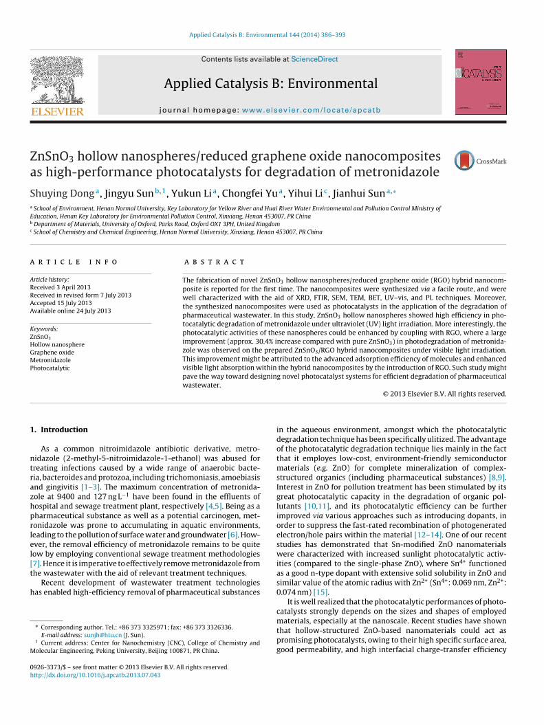
ig. 1. XRD patterns of GO, RGO, ZnSnO3 hollow nanosphere and ZnSnO3/RGOanocomposites.
olution was exposed to UV or visible light irradiation underagnetic stirring at 500 rpm, which marked the beginning of the
hotocatalytic degradation tests. An air diffuser was placed at theottom of the reactor to uniformly disperse air into the solutionith a flow rate of 0.2 m3 h−1. 5 mL of the suspension was sampled
t each 10 or 30 min intervals, which was immediately centrifugedo remove particles for further analysis.
The concentration of metronidazole was determined by mea-uring the absorption intensity at its maximum absorbanceavelength of � = 319 nm using a UV–Vis spectrophotometer (UV-
700, SHIMADU) with a 1 cm path length spectrometric quartz cell,nd was calculated from the calibration curve. The degradationfficiency of the metronidazole-contained wastewater was deter-ined according to the following equation:
egradation efficiency (%) = C0 − Ct
C0× 100% (1)
here C0 was the concentration of metronidazole after 40 mindsorption and Ct was the concentration of metronidazole at cer-ain reaction time t (min).
. Results and discussion
.1. XRD, FTIR, SEM, TEM, BET, UV–vis, and PL of the synthesizedanocomposites
XRD was employed to characterize the crystal phases of GO,GO, ZnSnO3 hollow nanospheres, and ZnSnO3/RGO nanocompos-
tes, the patterns of which are shown in Fig. 1. The sharp peak atround 2� = 10.8◦ (black curve), which corresponds to the (0 0 1)eflection of GO [28], almost disappears in the pattern of RGOred curve). Moreover, the RGO nanosheets exhibit a broad (0 0 2)iffraction peak at around 2� = 26◦and a (1 0 0) diffraction peakt around 2� = 44◦ [29]. The observed peaks of the synthesizednSnO3 hollow nanospheres (green curve) are in good agreementith the standard XRD pattern of ZnSnO3 (International Center foriffraction Data, JCPDS 28-1486), which confirms the crystallineature of obtained samples. The blue curve displays the XRDattern of the synthesized ZnSnO3/RGO nanocomposites, whereo apparent change in peak shapes and positions are observed
n comparison with that of the ZnSnO3 hollow nanospheres,ndicating that the introduction of RGO has no effect upon therystal orientations of ZnSnO3. However, it is worth-noting that noypical diffraction peaks of GO or RGO are observed within the blue
Fig. 2. FTIR spectra of GO, ZnSnO3 hollow nanosphere and ZnSnO3/RGO nanocom-posites.
curve, the reason of which may be due to the small amount andrelatively low diffraction intensity of RGO in the composite [29].
FTIR spectra of GO, ZnSnO3 hollow nanospheres, andZnSnO3/RGO nanocomposites are shown in Fig. 2. The FTIRspectrum of GO (black curve) displays several characteristic bandsat 1065, 1219, 1404, 1625, and 1724 cm−1, which correspond tothe C O C stretching vibrations, the C OH stretching peak, theO H deformation of the C OH groups, the C C stretching modeand the C O stretching vibrations of the COOH group, respec-tively, indicating the presence of the oxygen-containing functionalgroups on the surface of GO. The broad band at 3412 cm−1 (blackcurve) are assigned to O H stretching vibrations of adsorbedwater molecules on GO [30]. Compared to the spectrum of GO,the bands featuring oxygen-containing functional groups almostvanish in the spectrum (green curve) of the ZnSnO3/RGO nanocom-posites, confirming the successful and effective reduction of GOnanosheets. Moreover, the bands at 728, 2368, and 3468 cm−1 forboth the samples of ZnSnO3 hollow nanospheres and ZnSnO3/RGOnanocomposites are ascribed to the stretching modes of Zn O andSn O [31].
The representative SEM images of the prepared ZnSnO3 hol-low nanospheres and ZnSnO3/RGO nanocomposites are shown inFig. 3. Fig. 3a and b presents the top-view SEM observations ofthe ZnSnO3 hollow nanospheres. It can be seen from the imagesthat the obtained products are of uniform size distributions. Thenanomaterial possesses a hollow sphere structure, as one is able torandomly observe the shell-opened spheres (marked by red circlesin Fig. 3b and d). Fig. 3c and d displays the top-view SEM obser-vation of the ZnSnO3/RGO nanocomposites, although stratiformRGO nanosheets are hardly observed due to the high and uniformcoverages of ZnSnO3 hollow nanospheres on them.
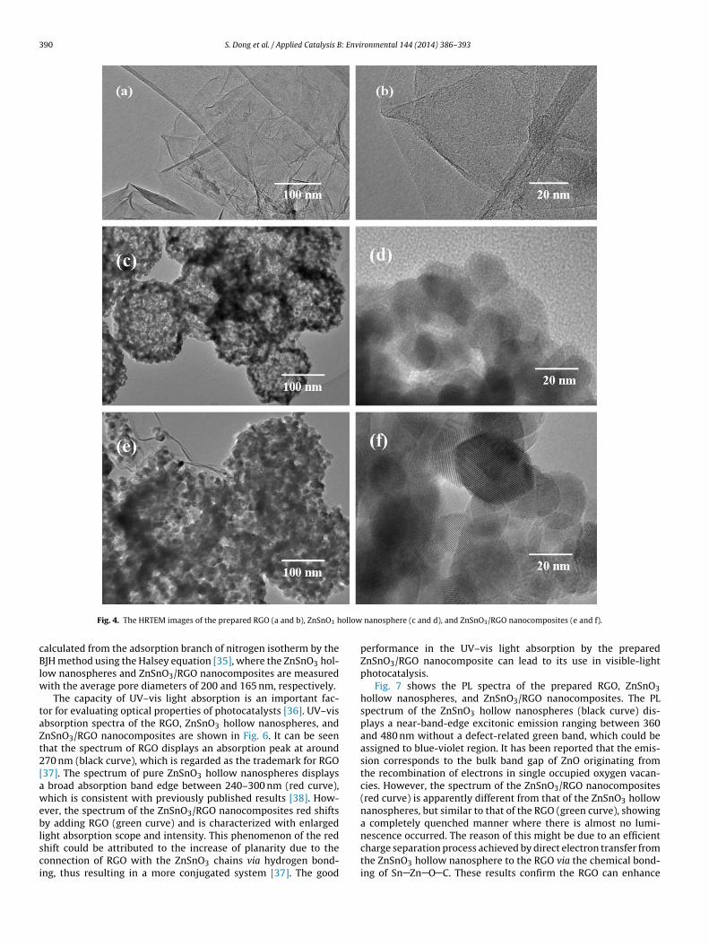
Fig. 4 displays the TEM characterization of the prepared RGO(Fig. 4a and b), ZnSnO3 hollow nanospheres (Fig. 4c and d), andZnSnO3/RGO nanocomposites (Fig. 4e and f), where detailed mor-phological information of synthesized materials can be obtained.Fig. 4a displays a general view of the produced RGO, with a highermagnified image shown in Fig. 4b. It can be seen from these imagesthat RGO flakes stack with each other and exhibit layered nano-structures. The TEM micrograph in Fig. 4c confirms the formationof hollow nanospheres, where the distinguished contrasts can be
found between the outer shells and the inner hollow cores. Also,the hollow nanospheres are of uniform size distributions, witha mean diameter at approx. 260 nm and a mean shell thicknessat approx. 7 nm. More close view of the hollow nanospheres is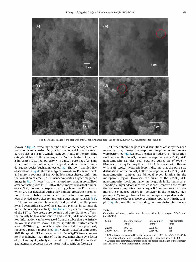
S. Dong et al. / Applied Catalysis B: Environmental 144 (2014) 386– 393 389
nosph
snpciwdoatiaowtR
iiotiharRioa
more, the enhanced adsorption behavior in the relatively highpressure (P/P0) range observed for both samples is a good indicationof the presence of large mesopores and macropores within the sam-ples. Fig. 5b shows the corresponding pore size distribution curves
Table 1Comparison of nitrogen adsorption characteristics of the samples ZnSnO3 andZnSnO3/RGO.
Sample BET surface areasa
(m2 g−1)Pore volumeb
(cm3 g−1)Pore diameterc
(nm)
ZnSnO3 10.2169 0.055351 200ZnSnO3/RGO 58.1951 0.256712 165
Fig. 3. The SEM images of the prepared ZnSnO3 hollow na
hown in Fig. 4d, revealing that the shells of the nanospheres areot smooth and consist of crystallized nanoparticles with a meanarticle size of 6–8 nm, which might contribute to the promisingatalytic abilities of these nanospheres. Another feature of the shells in regards to its high porosity with a mean pore size of 2–4 nm,
hich makes the hollow sphere a good candidate to accommo-ate guest species (such as molecules) [32]. The low-magnified TEMbservation in Fig. 4e shows the typical wrinkles of RGO nanosheetsnd uniform coatings of ZnSnO3 hollow nanospheres, confirminghe formation of ZnSnO3/RGO nanocomposites. Higher magnifiedmage in Fig. 4f shows that the nanospheres remain crystallizedfter contacting with RGO. Both of these images reveal that numer-us ZnSnO3 hollow nanospheres strongly bound to RGO sheets,hich are not detached during TEM sample preparation (sonica-
ion), this is probably due to the fact that the functional groups onGO provided active sites for anchoring guest nanomaterials [33].
The surface area of photocatalysts, depended upon the poros-ty and geometrical shape of the materials, plays an important rolen the photocatalytic activity [34]. Table 1 depicts the comparisonf the BET surface area, pore volume and pore diameter of bothhe ZnSnO3 hollow nanospheres and ZnSnO3/RGO nanocompos-tes. Information can be extracted from the table that the ZnSnO3ollow nanospheres shows a larger specific BET surface area atbout 10.2169 m2 g−1, which is higher than that of our previouslyeported ZnSnO3 nanoparticles [15]. Notably, that after composited
GO, the specific BET surface area of the ZnSnO3/RGO nanocompos-te is even higher than that of the hollow nanosphere by a factorf 5.8. This might partially attributed to the fact that RGO with 2Drrangements possesses large theoretical specific surface area.
ere (a and b) and ZnSnO3/RGO nanocomposites (c and d).
To further obtain the pore size distributions of the synthesizednanostructures, nitrogen adsorption-desorption measurementswere performed. Fig. 5a shows the nitrogen adsorption-desorptionisotherms of the ZnSnO3 hollow nanosphere and ZnSnO3/RGOnanocomposite samples. Both obtained curves are of type IV(Brunauer Deming Deming Teller (BDDT) classification) isothermswith a H3 typical hysteresis loop, indicating that the pore sizedistributions of the ZnSnO3 hollow nanosphere and ZnSnO3/RGOnanocomposite samples are bimodal types locating in themesoporous region. However, the curve of the ZnSnO3/RGOnanocomposites positions higher on the graph, indicating a corre-spondingly larger adsorbance, which is consistent with the resultsthat the nanocomposites have a larger BET surface area. Further-
a BET surface area calculated from the linear part of the BET plot (p/p0 = 0.18–0.30).b Total pore volume, taken from the volume of N2 adsorbed at p/p0 = 0.989.c Average pore diameter, estimated using the desorption branch of the isotherm
and the Barrett–Joyner–Halenda (BJH) formula.
390 S. Dong et al. / Applied Catalysis B: Environmental 144 (2014) 386– 393
ollow
cBlw
taZt2[aweblsci
Fig. 4. The HRTEM images of the prepared RGO (a and b), ZnSnO3 h
alculated from the adsorption branch of nitrogen isotherm by theJH method using the Halsey equation [35], where the ZnSnO3 hol-
ow nanospheres and ZnSnO3/RGO nanocomposites are measuredith the average pore diameters of 200 and 165 nm, respectively.
The capacity of UV–vis light absorption is an important fac-or for evaluating optical properties of photocatalysts [36]. UV–visbsorption spectra of the RGO, ZnSnO3 hollow nanospheres, andnSnO3/RGO nanocomposites are shown in Fig. 6. It can be seenhat the spectrum of RGO displays an absorption peak at around70 nm (black curve), which is regarded as the trademark for RGO37]. The spectrum of pure ZnSnO3 hollow nanospheres displays
broad absorption band edge between 240–300 nm (red curve),hich is consistent with previously published results [38]. How-
ver, the spectrum of the ZnSnO3/RGO nanocomposites red shiftsy adding RGO (green curve) and is characterized with enlarged
ight absorption scope and intensity. This phenomenon of the redhift could be attributed to the increase of planarity due to theonnection of RGO with the ZnSnO3 chains via hydrogen bond-ng, thus resulting in a more conjugated system [37]. The good
nanosphere (c and d), and ZnSnO3/RGO nanocomposites (e and f).
performance in the UV–vis light absorption by the preparedZnSnO3/RGO nanocomposite can lead to its use in visible-lightphotocatalysis.
Fig. 7 shows the PL spectra of the prepared RGO, ZnSnO3hollow nanospheres, and ZnSnO3/RGO nanocomposites. The PLspectrum of the ZnSnO3 hollow nanospheres (black curve) dis-plays a near-band-edge excitonic emission ranging between 360and 480 nm without a defect-related green band, which could beassigned to blue-violet region. It has been reported that the emis-sion corresponds to the bulk band gap of ZnO originating fromthe recombination of electrons in single occupied oxygen vacan-cies. However, the spectrum of the ZnSnO3/RGO nanocomposites(red curve) is apparently different from that of the ZnSnO3 hollownanospheres, but similar to that of the RGO (green curve), showinga completely quenched manner where there is almost no lumi-
nescence occurred. The reason of this might be due to an efficientcharge separation process achieved by direct electron transfer fromthe ZnSnO3 hollow nanosphere to the RGO via the chemical bond-ing of Sn Zn O C. These results confirm the RGO can enhance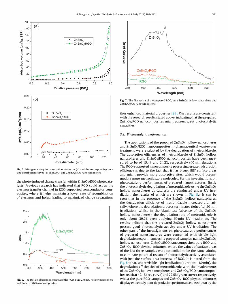
S. Dong et al. / Applied Catalysis B: Environmental 144 (2014) 386– 393 391
0.0 0.2 0.4 0.6 0.8 1.0
0
20
40
60
80
100
120
140
160
180
(a)
Ad
so
rbe
d v
olu
me
(c
m3/g
, S
TP
)
Rela tive press ure (P/P0)
ZnSnO3
ZnSnO3/RGO
0 20 40 60 80 100 120
0.00
0.05
0.10
0.15
0.20
0.25
(b)
dv/d
log
(D)(
cm
3/g
/nm
)
Pore diameter (nm)
SnZnO3
SnZnO3/RGO
Fs
tlepo
Fa
350 400 450 500 550 600
RGO
ZnSnO3/RGO
Inte
nsit
y (
a.u
)
Wavelength (nm)
ZnSnO3
ig. 5. Nitrogen adsorption desorption isotherms (a) and the corresponding poreize distribution curves (b) of ZnSnO3 and ZnSnO3/RGO nanocomposites.
he photo-induced charge transfer within ZnSnO3/RGO photocata-
ysts. Previous research has indicated that RGO could act as thelectron transfer channel in RGO-supported semiconductor com-osites, where it helps maintain a lower rate of recombinationf electrons and holes, leading to maximized charge separations200 300 400 500 600 700 800 900
0.0
0.5
1.0
1.5
2.0
2.5
ZnSnO3
RGO
Ab
s.
Wavelength (nm)
ZnSnO3/RGO
ig. 6. The UV–vis absorption spectra of the RGO, pure ZnSnO3 hollow nanospherend ZnSnO3/RGO nanocomposites.
Fig. 7. The PL spectra of the prepared RGO, pure ZnSnO3 hollow nanosphere andZnSnO3/RGO nanocomposites.
thus enhanced material properties [39]. Our results are consistentwith the research results stated above, indicating that the preparedZnSnO3/RGO nanocomposites might possess great photocatalyticcapacities.
3.2. Photocatalytic performances
The applications of the prepared ZnSnO3 hollow nanospheresand ZnSnO3/RGO nanocomposites in pharmaceutical wastewatertreatment were evaluated by the degradation of metronidazole.The adsorption efficiencies of metronidazole of ZnSnO3 hollownanospheres and ZnSnO3/RGO nanocomposites have been mea-sured to be of 15.4% and 24.2%, respectively (40 min duration).The RGO-supported nanocomposite possessing greater adsorptionefficiency is due to the fact that it has bigger BET surface areasand might provide more adsorptive sites, which would accom-modate more metronidazole molecules. For the investigations onphotocatalytic performances of prepared nanostructures, firstlythe photocatalytic degradation of metronidazole using the ZnSnO3hollow nanospheres as catalysts are conducted under UV irra-diation, the results of which are shown in Fig. 8a. It can beseen that in the presence of the ZnSnO3 hollow nanospheres,the degradation efficiency of metronidazole increases dramati-cally, where the degradation process terminates right after 20 minirradiation; whilst in the blank test (absence of the ZnSnO3hollow nanospheres), the degradation rate of metronidazole isonly about 39.7% even applying 60 min UV irradiation. Theresults indicate that the prepared ZnSnO3 hollow nanospherespossess good photocatalytic activity under UV irradiation. Theother part of the investigations on photocatalytic performancesof prepared nanostructures were concerned with visible lightdegradation experiments using prepared samples, namely, ZnSnO3hollow nanospheres, ZnSnO3/RGO nanocomposites, pure RGO, andZnSnO3–RGO physical mixtures, where the values of surface areasof the last three samples were controlled to be the same, aimingto eliminate potential reason of photocatalytic activity associatedwith just the surface area increase of RGO. It is noted from theFig. 8b that, under visible light irradiation (duration: 180 min), thedegradation efficiencies of metronidazole with the involvements
of the ZnSnO3 hollow nanospheres and ZnSnO3/RGO nanocompos-ites reach at 42.1% (red curve) and 72.5% (green curve), respectively,whilst the pure RGO samples and ZnSnO3–RGO physical mixturesdisplay extremely poor degradation performances, as shown by the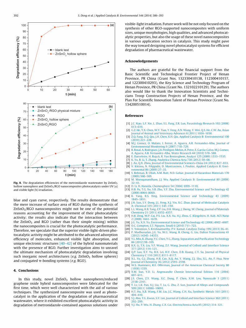
392 S. Dong et al. / Applied Catalysis B: Envi
0 10 20 30 40 50 60
0
10
20
30
40
50
60
70
80
90
100 (a)
Deg
rad
ati
on
eff
icie
ncy (
%)
t (min)
blank test
ZnSnO3 hollow sphere
0 20 40 60 80 100 120 140 160 180
0
10
20
30
40
50
60
70
80
(b)
De
gra
da
tio
n e
ffic
ien
cy
(%
)
t (min)
blank test
ZnSnO3-RGO physical mixture
RGO
ZnSnO3 hollow sphere
ZnSnO3/RGO
Fha
btZrattTteuwtsa
4
gfitcwd
[
[
[[
[
[
[
[
[[[[
[
[
[
[
[
[
[
[
ig. 8. The degradation efficiencies of the metronidazole wastewater by ZnSnO3
ollow nanosphere and ZnSnO3/RGO nanocomposites photocatalysts under UV (a)nd visible light (b) irradiation.
lue and cyan curve, respectively. The results demonstrate thathe mere increase of surface area of RGO during the synthesis ofnSnO3/RGO nanocomposites might not be one of the potentialeasons accounting for the improvement of their photocatalyticctivity; the results also indicate that the interaction betweenhe ZnSnO3 and RGO (rather than their simple mixing) withinhe nanocomposites is crucial for the photocatalytic performance.herefore, we speculate that the superior visible-light-driven pho-ocatalytic activity might be attributed to the advanced adsorptionfficiency of molecules, enhanced visible light absorption, andnique electronic structures [40–42] of the hybrid nanomaterialsith the presence of RGO. Further investigation aims to unravel
he ultimate mechanism of photocatalytic degradation involvinguch inorganic novel architectures (e.g. ZnSnO3 hollow spheres)nd conjugated �-bonding systems (e.g. RGO).
. Conclusions
In this study, novel ZnSnO3 hollow nanospheres/reducedraphene oxide hybrid nanocomposites were fabricated for therst time, which were well characterized with the aid of various
echniques. The synthesized nanocomposite was used as photo-atalyst in the application of the degradation of pharmaceuticalastewater, where it exhibited excellent photocatalytic activity inegradation of metronidazole-contained aqueous solutions under[
[
[
ronmental 144 (2014) 386– 393
visible-light irradiation. Future work will be not only focused on thesynthesis of other RGO-supported nanocomposites with uniformsizes, unique morphologies, high qualities, and advanced photocat-alytic properties, but also the usage of these novel nanocompositesin various application sectors in catalysis. This study might pavethe way toward designing novel photocatalyst systems for efficientdegradation of pharmaceutical wastewater.
Acknowledgements
The authors are grateful for the financial support from theBasic Scientific and Technological Frontier Project of HenanProvince, PR China (Grant Nos. 132330410138, 112300410157,and 122300410293), the Key Science and Technology Program ofHenan Province, PR China (Grant No. 132102210129). The authorsalso would like to thank the Innovation Scientists and Techni-cians Troop Construction Projects of Henan Province, and thePlan For Scientific Innovation Talent of Henan Province (Grant No.134200510014).
References
[1] J.C. Xiao, L.F. Xie, L. Zhao, S.L. Fang, Z.R. Lun, Parasitology Research 102 (2008)613–619.
[2] G.Z. He, Y.X. Chen, W.Y. Tian, Y. Feng, A.N. Wang, Y. Wei, Q.S. He, C.W. An, AsianJournal of Animal and Veterinary Advances 6 (2011) 1026–1030.
[3] Z.Q. Fang, X.Q. Qiu, J.H. Chen, X.H. Qiu, Applied Catalysis B: Environmental 100(2010) 221–228.
[4] M.J. Gomez, O. Malato, I. Ferrer, A. Aguera, A.R. Fernandez-Alba, Journal ofEnvironmental Monitoring 9 (2007) 718–729.
[5] R. Rosal, A. Rodriguez, J.A. Perdigon-Melon, A. Petre, E. Garcia-Calvo, M.J. Gomez,A. Aguera, A.R. Fernandez-Alba, Water Research 44 (2010) 578–588.
[6] E. Daeseleire, H. Ruyck, R. Van Renterghem, Analyst 125 (2000) 1533–1535.[7] K. Yu, B. Li, T. Zhang, Analytica Chimica Acta 738 (2012) 59–68.[8] J. An, Q.X. Zhou, Journal of Environmental Sciences-China 24 (2012) 827–833.[9] V. Kitsiou, N. Filippidis, D. Mantzavinos, I. Poulios, Applied Catalysis B: Envi-
ronmental 86 (2009) 27–35.10] S. Rehman, R. Ullah, A.M. Butt, N.D. Gohar, Journal of Hazardous Materials 170
(2009) 560–569.11] M. Muruganandham, J.J. Wu, Applied Catalysis B: Environmental 80 (2008)
32–41.12] D. Li, H. Haneda, Chemosphere 54 (2004) 1099–1110.13] H.B. Fu, T.G. Xu, S.B. Zhu, Y.F. Zhu, Environmental Science and Technology 42
(2008) 8064–8069.14] K. Yang, B.S. Xing, Environmental Science and Technology 43 (2009)
1845–1851.15] J.H. Sun, S.Y. Dong, J.L. Feng, X.J. Yin, X.C. Zhao, Journal of Molecular Catalysis
A: Chemical 335 (2011) 145–150.16] L.L. Wang, K.B. Tang, Z.P. Liu, D.K. Wang, J. Sheng, W. Cheng, Journal of Materials
Chemistry 21 (2011) 4352–4357.17] H.B. Zeng, W.P. Cai, P.S. Liu, X.X. Xu, H.J. Zhou, C. Klingshirn, H. Kalt, ACS Nano
2 (2008) 1661–1670.18] J.G. Yu, X.X. Yu, Environmental Science and Technology 42 (2008) 4902–4907.19] O.C. Compton, S.T. Nguyen, Small 6 (2010) 711–723.20] Y. Yokomizo, S. Krishnamurthy, P.V. Kamat, Catalysis Today 199 (2013) 36–41.21] P. Madhusudan, J.G. Yu, W.G. Wang, B. Cheng, G. Liu, Dalton Transactions 41
(2012) 14345–14353.22] Y.L. Min, K. Zhang, Y.C. Chen, Y.G. Zhang, Separation and Purification Technology
86 (2012) 98–105.23] B.X. Li, T.X. Liu, Y.F. Wang, Z.F. Wang, Journal of Colloid and Interface Science
377 (2012) 114–121.24] Q.P. Luo, X.Y. Yu, B.X. Lei, H.Y. Chen, D.B. Kuang, C.Y. Su, Journal of Physical
Chemistry C 116 (2012) 8111–8117.25] X.L. Yu, G.J. Zhang, H.B. Cao, X.Q. An, Y. Wang, Z.J. Shu, X.L. An, F. Hua, New
Journal of Chemistry 36 (2012) 2593–2598.26] W.S. Hummers, R.E. Offerman, Journal of the American Chemical Society 80
(1958) 1339.27] X.M. Sun, Y.D. Li, Angewandte Chemie International Edition 116 (2004)
607–611.28] J.S. Chen, Z.Y. Wang, X.C. Dong, P. Chen, X.W. Lou, Nanoscale 3 (2011)
2158–2161.29] T. Lv, L.K. Pan, X.J. Liu, T. Lu, G. Zhu, Z. Sun, Journal of Alloys and Compounds
509 (2011) 10086–10091.
30] H.T. Hu, X.B. Wang, F.M. Liu, J.C. Wang, C.H. Xu, Synthetic Metals 161 (2011)404–410.31] S.J. Ahn, T.G. Kwon, S.Y. Lee, Journal of Colloid and Interface Science 362 (2011)
292–299.32] Y.J. Hu, P. Wu, H. Zhang, C.X. Cai, Electrochimica Acta 85 (2012) 314–321.

: Envi
[
[
[
[
[
[
[
[
S. Dong et al. / Applied Catalysis B
33] Y. Fang, R.J. Wang, G.H. Jiang, H. Jin, Y. Wang, X.K. Sun, S. Wang, T. Wang, Bulletinof Materials Science 35 (2012) 495–499.
34] A.R. Liu, S.M. Wang, Y.R. Zhao, Z. Zheng, Materials Chemistry and Physics 99(2006) 131–134.
35] Y. Iizuka, T. Kubo, A. Nakahira, D. Onodera, N. Ozawa, T. Yao, Applied Catalysis
B: Environmental 76 (2007) 51–56.36] L.S. Wang, M.W. Xiao, X.J. Huang, Y.D. Wu, Journal of Hazardous Materials 161(2009) 49–54.
37] A. Kundu, R.K. Layek, A. Kuila, A.K. Nandi, ACS Applied Materials and Interfaces4 (2012) 5576–5582.
[
[
ronmental 144 (2014) 386– 393 393
38] C.H. Fang, B.Y. Geng, J. Liu, F.M. Zhan, Chemical Communications 17 (2009)2350–2352.
39] C.H. Wu, Y.Z. Zhang, S. Li, H.J. Zheng, H. Wang, J.B. Liu, K.W. Li, H. Yan, ChemicalEngineering Journal 178 (2011) 468–474.
40] J. Lin, R.L. Zong, M. Zhou, Y.F. Zhu, Applied Catalysis B: Environmental 89 (2009)
425–431.41] Y.J. Wang, R. Shi, J. Lin, Y.F. Zhu, Applied Catalysis B: Environmental 100 (2010)179–183.
42] T.G. Xu, L.W. Zhang, H.Y. Cheng, Y.F. Zhu, Applied Catalysis B: Environmental101 (2011) 382–387.
Related Documents